Article

A Trivalent Live Vaccine Elicits Cross-Species Protection Against Acute Otitis Media in a Murine Model

Department of Host-Microbe Interactions, St. Jude Children’s Research Hospital, Memphis, TN 38105, USA
*
Author to whom correspondence should be addressed.
These authors contributed equally to this work.
Vaccines 2024, 12(12), 1432; https://doi.org/10.3390/vaccines12121432
Submission received: 4 November 2024 / Revised: 9 December 2024 / Accepted: 12 December 2024 / Published: 19 December 2024
(This article belongs to the Section Attenuated/Inactivated/Live and Vectored Vaccines)

Abstract

:
Background: Acute otitis media (AOM) is a common pediatric infection worldwide and is the primary basis for pediatric primary care visits and antibiotic prescriptions in children. Current licensed vaccines have been incompletely ineffective at reducing the global burden of AOM, underscoring a major unmet medical need. The complex etiology of AOM presents additional challenges for vaccine development, as it can stem from multiple bacterial species including Streptococcus pneumoniae, Haemophilus influenzae, and Moraxella catarrhalis. As such, targeting multiple pathogens simultaneously may be required to significantly impact the overall disease burden. Methods: In this study, we aim to overcome this challenge by engineering a live-attenuated vaccine platform based on an attenuated mutant of S. pneumoniae that expresses H. influenzae and M. catarrhalis surface epitopes to induce protective immunity against all three pathogens. Results: The trivalent live-attenuated vaccine conferred significant protection against all three bacterial otopathogens as measured by seroconversion and the development of AOM, with the inclusion of the additional epitopes providing unexpected synergy and enhanced protection against S. pneumoniae. Conclusions: These data demonstrate a novel mechanism of introducing non-native immunogenic antigens into a live-attenuated vaccine platform to engender protection against AOM from multiple pathogenic species.

1. Introduction

Bacterial acute otitis media (AOM) is a frequent infection in children [1,2] causing over 700 million cases per year worldwide, with 75 percent of children experiencing at least one infection before the age of three. The leading bacterial species responsible for AOM are Streptococcus pneumoniae, Haemophilus influenzae, and Moraxella catarrhalis [3,4,5,6,7,8,9]. There has been notable success in developing vaccines targeting bacterial otopathogens, namely, with capsule-based conjugate vaccines greatly reducing the incidence of invasive disease by pneumococcus [10,11] and H. influenzae type B [12] in children and adults. While the pneumococcal conjugate vaccine has been very successful in preventing invasive disease, it has been less effective against otitis media [13] and does not decrease recurrent pneumococcal AOM [14]. The initial pneumococcal polysaccharide conjugate vaccine (PCV-7) effectively reduced the overall incidence of invasive disease [15,16] and partially reduced otitis media [17]. Expanding vaccine coverage from 7 to 13 serotypes (PCV-13) has further decreased pneumococcal AOM incidence, though significant disease burden persists [18]. To expand coverage beyond the pneumococcus, multiple vaccines have employed the conjugation of capsules from 10 or 11 pneumococcal serotypes to a surface-exposed lipoprotein of H. influenzae [19,20,21]. This strategy has somewhat decreased the incidence of AOM caused by both pneumococcus and H. influenzae [22,23,24,25]. Likewise, there has been considerable interest in investigating the potential utility of dual NTHi-Mcat vaccines that target unencapsulated H. influenzae (NTHi) and M. catarrhalis [26,27].
This phenomenon of reduced vaccine efficacy against AOM is not fully explained by serotype replacement with non-vaccine serotypes, as even vaccine serotypes continue to be isolated from pneumococcal AOM cases in vaccinated populations [19,28,29,30]. This issue may be due to poor mucosal antibody responses or the low expression of the capsular antigen during pneumococcal colonization [31]. The active removal of capsular polysaccharide in response to host antimicrobial peptides at the mucosal surface may also help S. pneumoniae evade anti-capsular antibodies conferred by vaccination [32]. Moreover, colonization with non-vaccine-serotype and non-typeable pneumococci and the predominance of NTHi have resulted in these pathogens continuing to be a significant medical burden for both community-acquired pneumonia [10,33] and AOM [34,35]. The recognition of diseases such as AOM with varying infectious etiologies underscores the value of exploring additional vaccine strategies that target several pathogens underlying the same disease to provide the broadest protective capacity.
The incidence of both mucosal and invasive pneumococcal disease decreases after early childhood, a phenomenon thought to be the result of accumulating protein antigen exposure leading to the development of broad protective antibody-mediated immunity [36]. There has been continued interest in the development of protein-based vaccines, which have demonstrated considerable promise both alone and in combination with capsule-based vaccines in conferring protective immunity against pneumococcal disease [37]. Another potential vaccine strategy that takes advantage of repeated exposure to bacterial antigens is the introduction of a live-attenuated vaccine in early childhood. Vaccination with live-attenuated strains acts as a means of exposure to multiple bacterial antigens, stimulating both humoral and cellular responses and, thereby, generating robust protection against both mucosal and invasive pneumococcal infection [38,39,40]. Many efforts have utilized highly attenuated vector strains to express important proteins and capsule-based pneumococcal antigens to induce protective immunity via either oral or intranasal administration. These have included approaches utilizing attenuated Salmonella vaccine platforms to express pneumococcal polysaccharides or protein antigens [41,42,43,44,45], commensal Lactobacillus and Lactococcus species to express pneumococcal antigens [46,47,48,49,50,51], vectors expressing either secreted or lipoprotein anchored pneumococcal antigens [52], and adenovirus vectors expressing multiple pneumococcal antigens [53]. These studies demonstrate that protective immunity against the pneumococcus can be elicited at the mucosal surface against subsequent disease development. In addition to the utilization of other bacterial species encoding pneumococcal antigens, there has been continued interest in modulating the pneumococcus to be avirulent whilst persisting at a sufficient density and duration to generate robust immunity and protection [38,39,40]. Initial studies using attenuated temperature-sensitive pneumococcal mutants demonstrated protection against challenge [54]. Subsequent investigations have utilized targeted deletions of the capsule locus coupled with mutations in iron or proline uptake systems, which induce robust antibody responses and confer serotype-independent immunity against invasive disease, though the deletion of the capsule appears to reduce heterologous protection during colonization [55,56]. Additional live-attenuated vaccines in which the capsule and DNA uptake machinery have been deleted induce both serum and mucosal antibody responses, with the additional benefit for the lack of reversion from recombination in a naturally competent pathogen [57]. Similar strategies have also proven successful via the modulation of virulence via the deletion of pep27 while simultaneously inactivating competence [58]. Other approaches include vaccination with strains lacking prolipoprotein diacylglyceryl transferase, which results in heterologous protection against subsequent pneumococcal challenge [38,59]. However, this deletion results in the impaired trafficking of multiple pneumococcal lipoproteins, many of which have been explored as candidates for protein-based vaccines [60]. These studies underscore that pneumococcal attenuation presents considerable potential for advancing live vaccines to confer serotype-independent immunity against both invasive and mucosal disease.
Antibody responses alone are not always indicative of protective responses, as live vaccines that induce antibody responses against conserved protein antigens may not confer protection against subsequent heterologous challenge [61]. As such, additional considerations for using the pneumococcus as a vaccine platform are the colonization duration of the vaccine strain, as colonization duration is a key aspect in generating protective immunity [62], and the retention of the major immunogenic surface proteins, many of which are virulence factors whose deletion may impact vaccine. The deletion of key virulence factors in certain serotypes have been shown to confer effective protective capacity while maintaining colonization [40]. Other strategies addressing these concerns include reducing virulence via synthetic changes in virulence factor codon-pair bias to reduce pathogenicity while retaining the native protein sequence [63]. In this study, we describe the advancement in a pneumococcal live vaccine platform based upon the deletion of ftsY, a key component of the Signal Recognition Particle Pathway that was previously demonstrated to be highly effective in conferring protective immunity against both invasive disease and AOM in a serotype-independent manner [39]. The vaccine strain was highly attenuated for invasive disease, while maintaining nasal carriage, and was found to be recalcitrant to genetic reversion, due to a pronounced defect in natural competence. Underscoring the importance of ftsY in pneumococcal fitness, horizontal transfer of the deletion mutation into other clinical strains was highly restricted, with many strains demonstrating markedly reduced or inability to uptake of the mutation. Prior colonization or administration of a conjugate vaccine did not reduce the vaccine efficacy. Moreover, as AOM is a disease caused by multiple bacterial pathogens, we engineered the pneumococcal ftsY mutant strain to express epitopes from both H. influenzae and M. catarrhalis on the bacterial surface, resulting in increased vaccine efficacy against the pneumococcus and protection against H. influenzae and M. catarrhalis. These data suggest that multiple bacterial pathogens of the mucosa can be targeted via the expression of the respective antigens in a live-attenuated pneumococcal vaccine platform to confer cross-species protection against AOM.

2. Materials and Methods

2.1. Bacterial Cultivation

Streptococcus pneumoniae strains were grown on solid TSA (Tryptic soy agar; Millipore Sigma, Burlington, MA, USA) supplemented with 3% sheep blood (Lampire Biologicals, Everett, PA, USA) and 20 μg/mL neomycin at 37 °C in a 5% CO2 atmosphere. Overnight growth was directly inoculated into semi-synthetic casein liquid media and 0.5% yeast extract (C + Y) [64] and grown in static conditions at 37 °C in a 5% CO2 atmosphere. Non-typeable Haemophilus influenzae 86-028NP was grown on solid chocolate agar media supplemented with 11,000 units/L bacitracin (Remel, San Diego, CA, USA) at 37 °C in a 5% CO2 atmosphere. Overnight growth was directly inoculated into brain–heart infusion broth (BD) supplemented with 0.2% yeast extract (BD) (BHI + Y), 10 μg/mL NAD+ (Sigma, St. Louis, MO, USA), and 10 μg/mL hemin (Sigma) with aeration. Moraxella catarrhalis strain O35E was grown in BHI + Y overnight at 37 °C with aeration. The strains used in this study are listed in Table S1. To determine growth kinetics, frozen stocks were diluted 1:100 in C + Y in a 96-well plate, and OD620 was measured every 30 min for 30 h using a Biotek Cytation 3 plate reader. The growth curves of the LAV strains were compared to those of BHN97 via two-way ANOVA, repeated measures using Prism 10 (GraphPad, San Diego, CA, USA).

2.2. Genetic Constructs

Capsule-swapped strains were generated as previously described [65]. Briefly, the TIGR4::19F capsule swap variant was generated through the transformation of an unencapsulated TIGR4SΔcps::NewSweetJanus with genomic DNA (gDNA) from BHN97 (19F). To obtain the capsule-swapped variants in the BHN97 background, an unencapsulated BHN97Δcps::SweetJanus strain [66] was transformed with either BHN97 (19F) or TIGR4 (4) gDNA.
For the generation of vaccine strains, all PCR products were amplified using exTaq polymerase (TAKARA) following the recommended guidelines; the primers are listed in Table S2. gDNA was extracted using the aqueous/organic extraction protocol as described previously [65]. For all transformations, S. pneumoniae strains were grown in C + Y until OD620 = 0.08 and were incubated with CSP1 and CSP2 [67] and the specific amplicon for three hours at 37 °C in a 5% CO2 atmosphere. The entire culture was plated on selection plates, and the correct transformants were confirmed through a lack of growth on counter-selection plates and through PCR. For transformations including the PhunSweetErm cassette, the selection plates contained 1 µg/mL erythromycin, and the counter-selection plates contained 15mM chlorinated-phenylalanine and 10% sucrose. For transformations replacing PhunSweetErm, the selection plates contained 15mM chlorinated-phenylalanine and 10% sucrose, and the counter-selection plates contained 1 µg/mL erythromycin as previously described [65,68].
To generate the live-attenuated S. pneumoniae strain (LAV), an internal deletion of ftsY (Sp1244) was generated, following a similar strategy employed previously [39]. The internal 867 nucleotides of ftsY were replaced with PhunSweetErm [68] through splicing-by-overlap-extension (SOE) PCR [69]. The upstream flanking region of ftsY and the first 213 bp of ftsY were amplified from BHN97 gDNA using the primer pair FtsY_Up_F/FtsY_Up_R. The downstream flanking region of ftsY and the last 210 bp of ftsY were amplified from BHN97 gDNA using the primer pair FtsY_Down_F/Fsy_Down_R. The ΔftsY::PhunSweetErm amplicon was generated through SOE PCR using the upstream, downstream, and PhunSweetErm fragments as the template and the primer pair FtsY_Up_F/FtsY_Down_R. BHN97 was transformed with the ΔftsY::PhunSweetErm amplicon to obtain the LAV strain.
To generate the LAV-D strain, BHN97 underwent three transformations steps. For the first transformation, an expression platform in the neutral CEP locus [70,71] was generated through the insertion of PhunSweetErm. The flanking regions in the CEP were amplified using BHN97 gDNA and the primer pair CEP_Up_F/CEP_Up_PS_R as well as CEP_Down_PS_F and CEP_Down_R. Of note, these regions are shared among several S. pneumoniae strains; however, in BHN97, an insertion at this site disrupts predicted ATP transporter proteins. The CEPΩPhunSweetErm amplicon was generated through SOE PCR using the CEP upstream, CEP downstream, and PhunSweetErm amplicons as the template and the primer pair CEP_Up_F/CEP_Down_R and was used to transform BHN97 to generate BHN97 CEPΩPhunSweetErm. For the second transformation step of generating the LAV-D strain, a codon-optimized gene expressing H. influenzae ProteinD (glpQ in non-typeable 86-028NP) [72] was inserted in the CEP locus, replacing PhunSweetErm, thereby making the strain markerless. Three strategies were employed to anchor ProteinD to the cell surface, i.e., with C-terminal LPXTG motif (ProteinD-LPXTG), with C-terminal choline-binding motif (ProteinD-CBD), and with N-terminal lipoprotein anchor domain (Lipo-ProteinD). For ProteinD-LPXTG and ProteinD-CBD, a Gram-positive signal sequence was incorporated into the N-terminus. All three proteinD genes were synthesized via Genscript (Piscataway) with codon optimization for S. pneumoniae and included a strong promoter (P3) and upstream and downstream terminators described previously [71] (see Figure S3 for protein sequences). The flanking regions in the CEP were amplified using BHN97 gDNA and the primer pairs CEP_Up_F/CEP_Up_D_R and CEP_Down_D_F/CEP_Down_R. The codon-optimized proteinD with the CBD and LPXTG motifs, along with the P3 promoter and terminators, were amplified from Genscript plasmids using the primer pair ProteinD_F/ProteinD_R. For the lipo-proteinD amplicon, Genscript was unable to synthesize the entire sequence and instead provided two fragments in two separate plasmids. To generate the entire sequence of lipo-proteinD, along with the P3 promoter and terminators, the two fragments were amplified from the plasmids using the primer pairs Lipo-ProteinD_1_F/Lipo-ProteinD_1_R and Lipo-ProteinD_2_F/Lipo-Protein_2_R. The two fragments were spliced together via SOE PCR using the primer pair Lipo-ProteinD_1_F/Lipo-ProteinD_2_R. The CEPΩproteinD-LPXTG, CEPΩproteinD-CBD, and CEPΩlipo-proteinD amplicons were generated through SOE PCR using the CEP upstream, CEP downstream, and proteinD-LPXTG/proteinD-CBD/lipo-proteinD amplicons as the template and the primer pair CEP_Up_F/CEP_Down_R. To generate BHN97 strains expressing differentially anchored ProteinD, BHN97 CEPΩPhunSweetErm was transformed using the CEPΩproteinD-LPXTG, CEPΩproteinD-CBD, or CEPΩlipo-proteinD amplicons. For the third transformation step, the internal 867 nucleotides of ftsY were replaced with PhunSweetErm. The ΔftsY::PhunSweetErm amplicon was amplified from BHN97ΔftsY::PhunSweetErm gDNA using the primer pair FtsY_Up_F/FtsY_Down_R. BHN97 CEPΩlipo-proteinD was transformed with the ΔftsY::PhunSweetErm amplicon to obtain the LAV-D strain.
To generate the LAV-D-M strain, the BHN97 CEPΩPhunSweetErm generated above underwent two transformation steps. For the first step, a gene expressing Lipo-ProteinD with an additional 23 amino acid UspA epitope (“NNINNIY”) [73] on the C-terminus was generated using SOE PCR. The upstream fragment was amplified from the gDNA of BHN97 CEPΩlipo-proteinD using the primers CEP_Up_F and Lipo-ProteinD_N_R such that the amplicon contained the upstream CEP flanking region and the lipo-proteinD gene. A 23 double-stranded ultramer fragment of the NNINNIY epitope was generated by annealing two ultramer oligos together, NNINNIY_Ultra_F and NNINNIY_Ultra_R. The downstream fragment was amplified from the gDNA of BHN97 CEPΩlipo-proteinD using the primers CEP_Down_N_F and CEP_Down_R such that the amplicon contained the terminators downstream of the lipo-proteinD gene and the downstream CEP flanking region. To enhance overlap during SOE PCR of the final product, the upstream and NNINNIY ultramer fragment were spliced together using the primers CEP_UP_F and NNINNIY_R, and the downstream and NNINNIY ultramer fragment were spliced together using the primers NNINNIY_F and CEP_Down_R. The final product, the CEPΩlipo-proteinD-M amplicon, was generated through SOE PCR using the CEP upstream-NNINNIY and the CEP downstream-NNINNIY amplicons as the template and the primer pair CEP_Up_F/CEP_Down_R. BHN97 CEPΩPhunSweetErm was transformed using the CEPΩ lipo-proteinD-M amplicon. For the next transformation step, the internal 867 nucleotides of ftsY were replaced with PhunSweetErm using the same method as that for the LAV-D strain. BHN97 CEPΩlipo-proteinD-M was transformed with the ΔftsY::PhunSweetErm amplicon to obtain the LAV-D-M strain.

2.3. Recombinant ProteinD Purification and Antibody Generation

To purify ProteinD for antibody generation, an E. coli codon-optimized proteinD was synthesized with an N-terminal His-tag in a pet28 expression plasmid by Genscript (see Table S3 for protein sequence). The pet28-his-proteinD plasmid was transformed into One Shot BL21 (DE3) chemically competent E. coli cells following manufacturer’s recommended protocol (Thermo Fisher, Waltham, MA, USA). Cells harboring pet28-his-proteinD were grown in 2TXY (2%Tryptone, 1% yeast extract, and 0.5% NaCl) media and 30 µg/mL kanamycin, and when the cells reached OD600 = 0.6, their protein expression was induced with 0.2 mM IPTG for 16 h at 18 °C. The culture was pelleted and lysed in lysis buffer (1x PBS, pH 7.4, and protease inhibitors) via a microfluidizer. The lysed pellet were incubated with nickel beads and washed, and His-ProteinD was eluted from beads using 200 mM imidazole in PBS. Rabbit polyclonal antibody against ProteinD was generated by Rockland Immunochemicals. Briefly, rabbits were immunized with 400 µg of recombinant His-ProteinD, followed by three boost vaccinations of 200 µg of recombinant His-ProteinD at two, three, and five weeks post initial vaccination. The rabbits were bled 12 weeks post final vaccination for the collection of sera.

2.4. Recombination Frequency

To determine the competence of the LAV strain, BHN97 and the LAV strain were transformed with 5 µg of gDNA from BHN97 Tn-seq, a nontargeted mutagenesis mechanism containing a spectinomycin resistance cassette [74]. Strains were grown in C + Y until OD620 = 0.08, and this was followed by transformation as described above. The transformations were serially diluted and plated on TSA blood plates and TSA blood plates supplemented with 200 µg/mL spectinomycin. To observe low-frequency recombination events, the transformation was also spread-plated on TSA blood plates supplemented with 200 µg/mL spectinomycin. The experiment was repeated eight times. To measure potential spread of the antibiotic resistance cassette originating from the LAV strain, a subset of S. pneumoniae strains representing different serotypes was transformed with either 3 µg of gDNA from the LAV strain or with 3 µg of gDNA from BHN97 CEPΩPhunSweetErm, which served as a neutral control for the antibiotic resistance cassette. The cells were incubated with either CSP1 (D39), CSP2 (TIGR4), or both (all other strains) and the gDNA for 3 h at 37 °C, 5% CO2, followed by serial dilution and plating on TSA blood plates and TSA blood plates supplemented with 1 µg/mL erythromycin. To observe low-frequency recombination events, the transformation was also spread-plated on TSA blood plates supplemented with 1 µg/mL erythromycin. The experiment was repeated six times. The plates were incubated overnight at 37 °C in a 5% CO2 atmosphere, and colonies were enumerated. Recombination frequency was calculated as the number of recombinants on the plates containing the selection antibiotic divided by the total number of cells on the plates without the selection antibiotic and was compared via non-parametric Mann–Whitney t-tests using Prism 10 (GraphPad).

2.5. Adhesion Assay

Pneumococcal adherence to A549 lung epithelial cells was determined from a modified adhesion assay [75]. A549 cells (ATCC) were seeded to ~95% confluency (2 × 10⁵ cells/mL) in 24-well tissue-culture-treated plates (Costar). The cells were treated for 2 h prior to infection with TNF-α (10 ng/mL) in F12K media (ATCC) with 10% FBS and 20 µg/mL gentamicin, followed by a 2x rinse with PBS. S. pneumoniae strains were grown in C + Y until OD620 = 0.4 and diluted 1:10 into F12K. The A549 cells were infected with the diluted bacterial culture at an MOI of ~50:1. The bacteria were allowed to adhere to the A549 cells for 30 or 90 min at 37 °C. Nonadherent bacteria were determined by serial dilution of the supernatant and subsequent plating on TSA blood agar plates. The A549 cells with adherent bacteria were washed with PBS and detached from the wells via incubation with 100 µL of 0.1% Trypsin in PBS at 37 °C for 5 min, followed by the addition of 900 µL of PBS. The adherent bacteria were determined by serial dilution of the cell solution in PBS and subsequent plating on TSA blood agar plates. Each strain was measured in five biological replicates per plate, in two independent experiments. Adherence was reported as the number of adherent bacteria divided by the total number of bacterial cells (nonadherent and adherent). The number of adherent colonies/total bacterial cells of the LAV strains was compared with that of wild-type BHN97 via non-parametric Mann–Whitney t-tests using Prism 10 (GraphPad).

2.6. Vaccination Regimens and Protection Efficacy

For all vaccinations, treatments, and challenges via intranasal (IN) installation, mice were anesthetized under inhaled 2.5% isoflurane. For the vaccination of mice with the LAV strains, bacterial strains were grown in C + Y until OD620 = 0.4. Bacterial cultures were pelleted and diluted in PBS to 105 CFUs/25 µL according to a previously determined standard curve. The bacteria were enumerated on TSA blood agar plates to confirm the inoculum. Eight-week-old female BALB/c mice (Jackson laboratory, Bar Harbor, ME, USA) were administered the vaccine via IN instillation of 105 CFUs in 25 µL of PBS of the LAV strains or 25 µL of PBS as a control [39]. Three weeks and six weeks after the initial vaccination, the mice were boosted with a second and third IN instillation of 105 CFUs in 25 µL of PBS of the LAV strains or 25 µL of PBS as above. For the co-vaccination of mice with Prevnar-13, 8-week-old female BALB/c mice were administered 100 µL of a 1:10 dilution of Prevnar-13 via intraperitoneal injection while conscious. Three, six, and nine weeks following Prevnar-13 vaccination, the mice were vaccinated with the LAV strains following the regimen above. As a control, a separate group of mice were administered 100 µL of a 1:10 dilution of Prevnar-13 via intraperitoneal injection followed by IN instillation of PBS at 3, 6, and 9 weeks. To determine the impact of previous colonization on vaccination efficacy, the bacterial strains were grown in C + Y until OD620 = 0.4. These strains included a homologous strain (BHN97) expressing its own capsule (Type 19F) as well as a variant capsule (Type 4) and a heterologous strain (TIGR4) expressing its own capsule (Type 4) as well as a variant capsule (Type 19F). Eight-week-old female BALB/c mice (Jackson laboratory) were administered the colonizing strains via IN instillation of 104 CFUs in 25 µL of PBS or 25 µL of PBS as a control. Three weeks following colonization, the mice were vaccinated with the LAV strains following the regimen above. The mice were monitored for indications of distress or disease over the course of the vaccination regimen, and no observable signs were detected. Prior to the initial vaccination and two weeks after the final vaccination, sera were collected via non-lethal retro-orbital bleeding to measure seroconversion. Three weeks after the final vaccination, the mice were challenged. For all challenge experiments, the mice were administered the challenge strain via IN instillation. The vaccinated mice were challenged with 106 CFUs/100 µL PBS of homologous S. pneumoniae strain (BHN97x) or heterologous S. pneumoniae strain (BHN54x) via IN instillation. For vaccination with LAV-D, an independent group of mice were challenged with H. influenzae, and for vaccination with LAV-D-M, independent groups of mice were challenged with either H. influenzae or M. catarrhalis. Similar to previous studies that leveraged influenza co-infection to facilitate NTHi translocation into the middle ear [76], we utilized an inflammatory stimulant poly (I:C) [77] to facilitate translocation to the middle ear in our challenge model as we did not observe appreciable middle ear translocation for either H. influenzae or M. catarrhalis without prior poly (I:C) sensitization (Figure S5). The mice were administered 50 μL of 1 mg/mL poly(I:C) HMW (Invivogen) IN daily for four days [68] followed by IN challenge with 107 CFUs/100 µL PBS of H. influenzae or M. catarrhalis grown in liquid media. Twenty-four hours after all the challenges, the mice were euthanized via CO2 asphyxiation followed by cervical dislocation. The lungs, nasal passages, and both ear bullae were immediately harvested, homogenized in PBS, serially diluted, and plated on TSA blood agar plates and 400 µg/mL kanamycin plates (S. pneumoniae), chocolate agar plates (H. influenzae), or BHI agar plates (M. catarrhalis). Plates were incubated overnight at 37 °C in a 5% CO2 atmosphere, and the bacterial titers were enumerated. The bacterial burden in each tissue (CFUs/mL) was compared using a non-parametric Mann–Whitney t-test using Prism 10 (GraphPad). The bacterial burden in each ear was determined and plotted individually [78].

2.7. Murine Challenge Experiments

To determine the virulence of the LAV strains in vivo, 8-week-old female BALB/c mice were infected via IN instillation of 105 CFUs/25 µL PBS of the wild-type BHN97 or LAV strains (N = 10). At 3 days post-challenge, the mice were euthanized via CO2 asphyxiation followed by cervical dislocation, and the lungs, nasal passages, and both ear bullae were immediately harvested and homogenized in PBS. The lungs and ears were serially diluted and plated on TSA blood agar plates. The nasal passages were serially diluted and plated, and the remainder was spread-plated on TSA blood agar plates. The plates were incubated overnight at 37 °C in a 5% CO2 atmosphere, and the bacterial titers were enumerated. The bacterial burden in each tissue (CFUs/mL) was compared using a non-parametric Mann–Whitney t-test using Prism 10 (GraphPad).
To investigate the potential reversion of the LAV strain in vivo, the bacterial outgrowth from the spread-plated nasal passages above was considered Passage 1 (P1) in an in vivo passaging experiment. The bacterial growth on the blood agar plates was collected in C + Y, mixed with glycerol at a final concentration of 20%, and frozen at −80 °C in several aliquots. A frozen aliquot of each P1 sample served as the inoculum to challenge the next group of mice for the subsequent passage. The six highest titered stocks of each group were used for infection of 8-week-old female BALB/c mice via IN instillation of 105 CFUs/25 µL PBS, N = 1 per P1 sample. At 3 days post-challenge, the mice were euthanized, and the lungs, nasal passages, and both ear bullae were harvested and processed as above. The bacterial burden was designated as Passage 2 (P2). Again, the bacterial outgrowth from the nasal passage was collected, frozen at −80 °C, and used to infect 8-week-old female BALB/c mice via IN instillation of 105 CFUs/25 µL PBS, N = 1 per P2 sample. At 3 days post-challenge, the mice were euthanized and the lungs, nasal passages, and both ear bullae were harvested and processed as above. The bacterial burden was designated as Passage 3 (P3). For each strain and tissue, the bacterial burden of the sequential passages was compared using Kruskal–Wallis one-way ANOVA using Prism 10 (GraphPad). For all collected passages, a frozen aliquot was used to inoculate C + Y media and grown to OD620 = 0.6. gDNA was extracted, and the deletion of ftsY was confirmed via PCR using primer pairs FtsY_Outer_F/FtsY_Outer_R and FtsY_Inner_F/FtsY_Inner_R.

2.8. Western Blots

Bacterial cultures were grown in C + Y until OD620 = 0.6. The culture was pelleted, and the pellet was lysed in 10% deoxycholate and 10% sodium dodecyl sulfate at 37 °C to disrupt the cellular membranes. The bacterial lysates were diluted in PBS, boiled with 4x LDS sample buffer (Thermo Fisher), and run on 4–12% NuPAGE Bis-Tris gels (Invitrogen, Waltham, MA, USA). Proteins were transferred to nitrocellulose membranes, which were subsequently blocked with 5% non-fat skim milk in PBS + 0.1% Tween. To determine ProteinD expression in S. pneumoniae, the membranes were probed with anti-ProteinD polyclonal antibody, purified through ProteinA columns (Pierce, Franklin, MA, USA), at a concentration of 3 µg/mL in blocking buffer. As a loading control, the samples were concurrently run in a separate gel, transferred, and the membrane was probed with anti-CbpA 3H11 monoclonal [79] in blocking buffer. The membranes were incubated with all primary antibodies overnight at 4 °C and then probed with anti-mouse (for monoclonal primary antibody) or anti-rabbit (for polyclonal primary antibody) HRP-conjugated secondary antibodies (Bio-Rad, Hercules, CA, USA), followed by detection using an HRP chemiluminescent substrate (Thermo Scientific, Waltham, MA, USA).

2.9. ELISA Antibody Measurements

To determine ProteinD expression in S. pneumoniae, strains were grown in liquid media until OD620 = 0.4. The cells were pelleted, resuspended in equal volumes of coating buffer (sodium carbonate–bicarbonate; Sigma), and bound to high-binding Nunc 96-well plates (Thermo Fisher) via centrifugation. The supernatant was removed, and the plates were allowed to dry at room temperature and were subjected to blocking buffer (10% heat-inactivated fetal bovine serum in PBS). After blocking, the cells were probed with anti-ProteinD polyclonal antibody (purified through Pierce IgG ProteinA columns). As a control for differential plate-binding in the strains, cells bound to other wells in the same plate were concurrently probed with polyclonal antibody against LytA, a cell surface antigen [32]. The samples in the wells were washed with Tris-buffered saline and 1% Tween-20 and then probed with AP-conjugated goat anti-rabbit IgG secondary antibody (Southern Biotech, Birmingham, AL, USA) in blocking buffer. The samples in wells were washed again and incubated with AP yellow substrate (Sigma), and this was followed by detection at OD405 with a Biotek Cytation 3 plate reader. The absorbance values of the cells probed with ProteinD were normalized to those of the cells probed with LytA. The immunoreactivity, as detected by absorbance at 405 nm, of ProteinD-expressing strains and LAV strains were compared to that of BHN97 via an unpaired t-test using Prism 10 (GraphPad).
The immunoreactivity of mouse sera post-vaccination was determined via whole-cell bacterial and Protein-D ELISA. Strains were prepared and bound to 96-well plates as above. Sera from the vaccinated mice were diluted 1:50 and incubated with the bound bacteria. The samples in the wells were washed, probed with AP-conjugated goat anti-mouse IgG secondary antibody (Southern Biotech) in blocking buffer, and incubated with AP yellow substrate (Sigma), followed by detection at OD405 with a Biotek Cytation 3 plate reader. The immunoreactivity of the LAV strains was compared with that of BHN97 with an unpaired t-test using Prism 10 (GraphPad).

3. Results

3.1. Genetic Stability of LAV

The live-attenuated ftsY mutant in S. pneumoniae 19F (LAV) induces protection against pneumococcal infection when challenged with both a homologous and heterologous serotype, demonstrated through both seroconversion and reduced bacterial burden in the vaccinated mice (Figure S1), recapitulating observations made previously [39]. A key potential shortcoming of a live-attenuated pneumococcal vaccine is the inherent capacity of the pneumococcus to uptake and combine foreign DNA [80]. This presents a potential risk for reversion of the LAV strain to wild-type virulence. To address this concern, we first monitored the fitness of the LAV strain during repeated murine colonization (Figure 1). The vaccine strain remained attenuated compared to wild-type 19F (Passage 1) in the lungs and ears between the first and last passage, while maintaining bacterial burden levels in the nasal passage (Figure 1a). The passaged LAV bacterial populations were confirmed to maintain the ftsY deletion through PCR. Interestingly, the wild-type 19F became attenuated in the lungs between the first and final passage, as observed previously whereby adaptation to murine nasal colonization confers fitness tradeoffs for lung infection [81]. These data suggest that the LAV strain remains attenuated for invasive disease and AOM despite repeated passages in vivo.
Next, we assessed the possibility of reversion of the LAV strain through a recombination event whereby the wild-type allele of a co-colonizing strain could replace the ftsY deletion. The level of competence in the LAV strain was determined by measuring the recombination frequency using a saturated Tn-seq library. The LAV strain demonstrated a severe defect in the recombination frequency of approximately four logs compared to the wild-type strain (Figure 1b). While the recombination of the saturated Tn-seq library in the LAV strain still occurred, genetic reversion due to recombination would not likely occur in the host considering the greatly reduced rate in ideal in vitro conditions, the growth defect imposed by the loss of ftsY (Figure S2a), and the lack of observable revertants when the LAV strain was passaged in the mice (Figure 1). Due to this pronounced reduction in competence, genetic manipulations for removing the erythromycin cassette were not successful, despite numerous attempts. This presents another potential risk of utilizing this strain as a live-attenuated vaccine in the unintentional spread of the resistance marker used in strain construction to non-vaccine strains during co-colonization. To determine the likelihood of resistance transfer, the frequency at which a panel of distinct serotypes recombined the antibiotic resistance marker from the gDNA of the LAV strain was measured (Figure 1c). For all serotypes, the frequency of recombining the resistance marker from the LAV strain was below levels of detection or severely impaired compared to obtaining the same resistance from a strain harboring the resistance marker in a neutral location. Of note, for three of the serotypes (2, 10 N, and 22 F), additional colonies were observed after 48 h of growth, demonstrating severe growth impairment upon obtaining the resistance from the gDNA of the LAV strain. These data underscore the genetic stability of the LAV strain and the inherently low risk of resistance mechanisms spreading to co-colonizing pneumococci.

3.2. Impact of Prior Colonization on LAV Efficacy

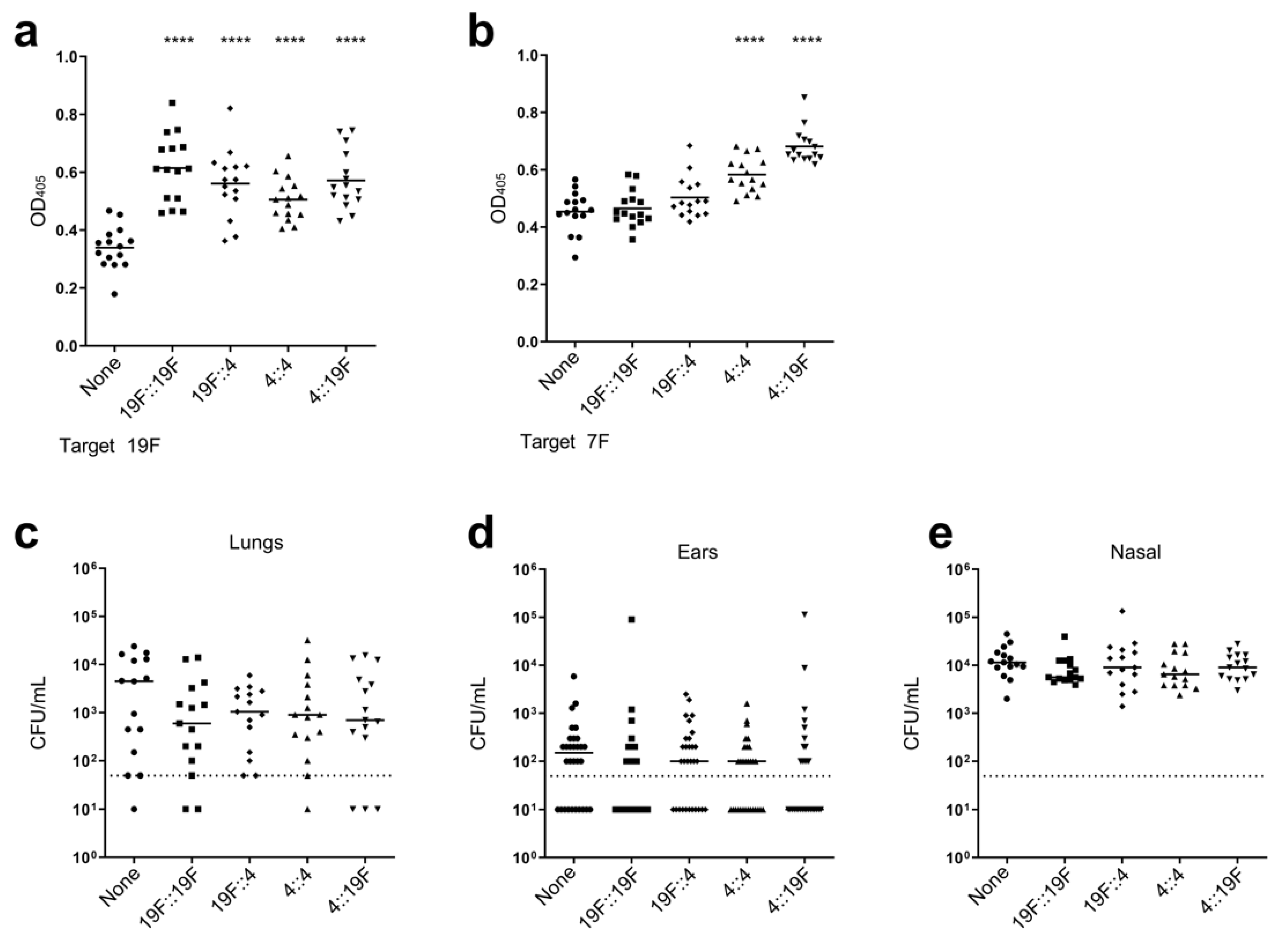
Colonization by S. pneumoniae occurs early in childhood [82,83]. Taking this into consideration, it is imperative that the impact of prior colonization on the efficacy of the LAV strain be determined. To model this, mice were pre-colonized with pneumococcal strains followed by vaccination with the LAV strain. The impact of several pre-colonization strains on vaccine efficacy was determined, including a homologous strain (serotype 19F) and a heterologous strain (serotype 4). As S. pneumoniae has the inherent ability to swap capsule loci and express a distinct serotype [84], we also determined the impact of pre-colonization with capsule-swapped variants on vaccine efficacy (Figure 2). Serum antibody titers following vaccination with the LAV strain demonstrated that the pre-colonized and vaccinated mice had either similar or enhanced IgG levels against homologous (Figure 2a) and heterologous serotypes (Figure 2b) compared to the non-colonized and vaccinated mice. Following challenge, we observed a slight enhancement in protection against bacterial burden in the lungs and ears of the pre-colonized, vaccinated mice compared to the non-colonized, vaccinated mice (Figure 2c–e). These data suggest that prior colonization is not detrimental to the protective efficacy of the LAV strain and may enhance the protective capacity.

3.3. Impact of Prior Conjugate Vaccine Exposure on LAV Efficacy

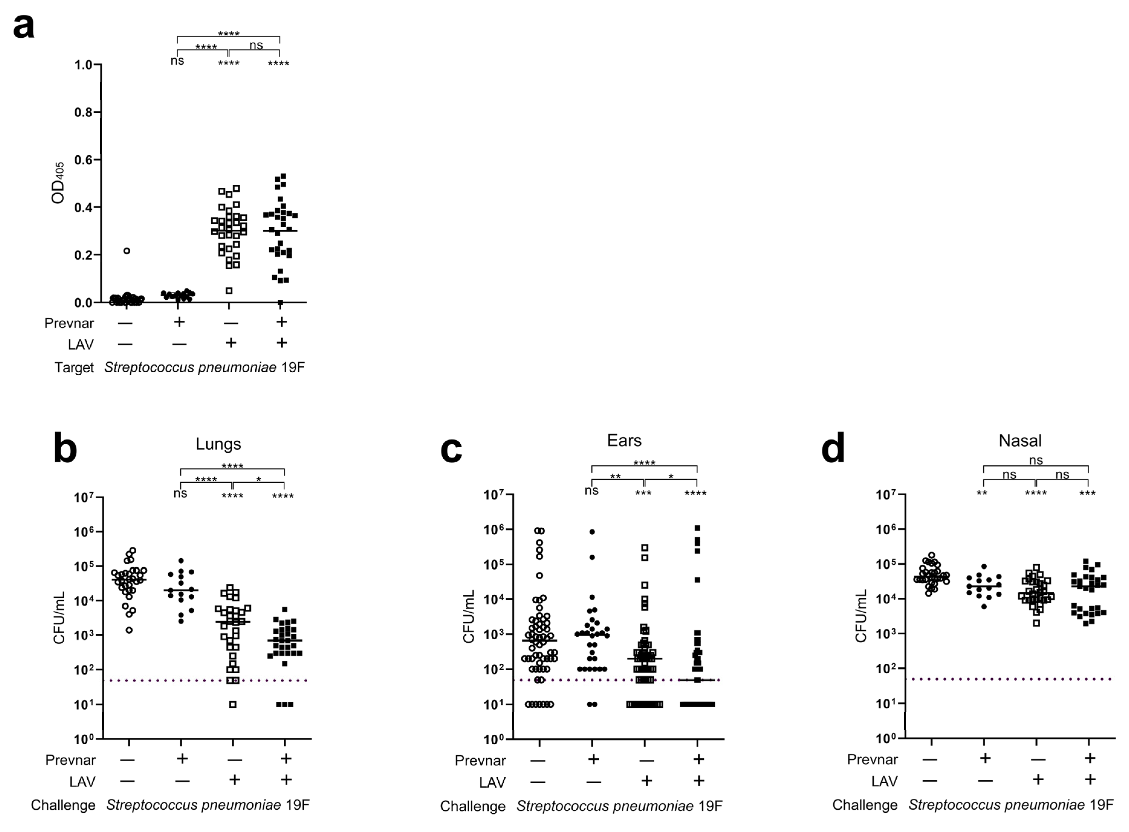
Various formulations of pneumococcal conjugate vaccines have been approved and are recommended as part of the standard series of childhood vaccinations [85,86]. Widespread introduction of these vaccines resulted in dramatic alterations in the population structure of pneumococci and serotype replacement [87]. This strong selective pressure might be expected to diminish the protective efficacy of the LAV strain, as colonization duration and/or immune response might be dampened by pre-existing vaccine-based immunity. This was evaluated in a model whereby mice were vaccinated with a single vaccination of Prevnar-13 prior to vaccination with the LAV strain. Serum antibody titers demonstrated no significant differences in IgG levels against homologous (19F) (Figure 3a) and heterologous serotypes (7F) (Figure S3a) between the mice that received Prevnar-13 prior to the LAV strain and those that received the LAV strain alone. In the mice challenged with either 19F or 7F strains, the efficacy of protection of the LAV strain was not diminished in the mice that received Prevnar-13 but rather demonstrated enhanced protective benefit in the lungs and ears, with no difference in the nasal passage burden (Figure 3b–d; Figure S3b–d). These data indicate that the efficacy of the LAV strain is not reduced with prior vaccination and may synergize with Prevnar-13 to confer enhanced protection against mucosal infections such as AOM.

3.4. Efficacy of Polyvalent Vaccines to Protect Against AOM from Multiple Pathogens

3.4.1. Engineering Polyvalent Vaccines

One challenge with utilizing vaccines to induce protection from AOM is the observation that multiple pathogens can cause AOM [88]. While S. pneumoniae is a leading cause of bacterial AOM, other bacterial pathogens, most notably H. influenzae and M. catarrhalis, are also highly prevalent causative agents of disease. This raises the distinct possibility that the deployment of a vaccine targeting a single pathogen may not result in an overall reduction in the clinical burden of AOM, despite promoting protection against one of the major pathogens. To address this pitfall, we sought to leverage the LAV strain as a platform to express non-native antigens on the cell surface to induce antibody responses and confer protection against multiple bacterial species.
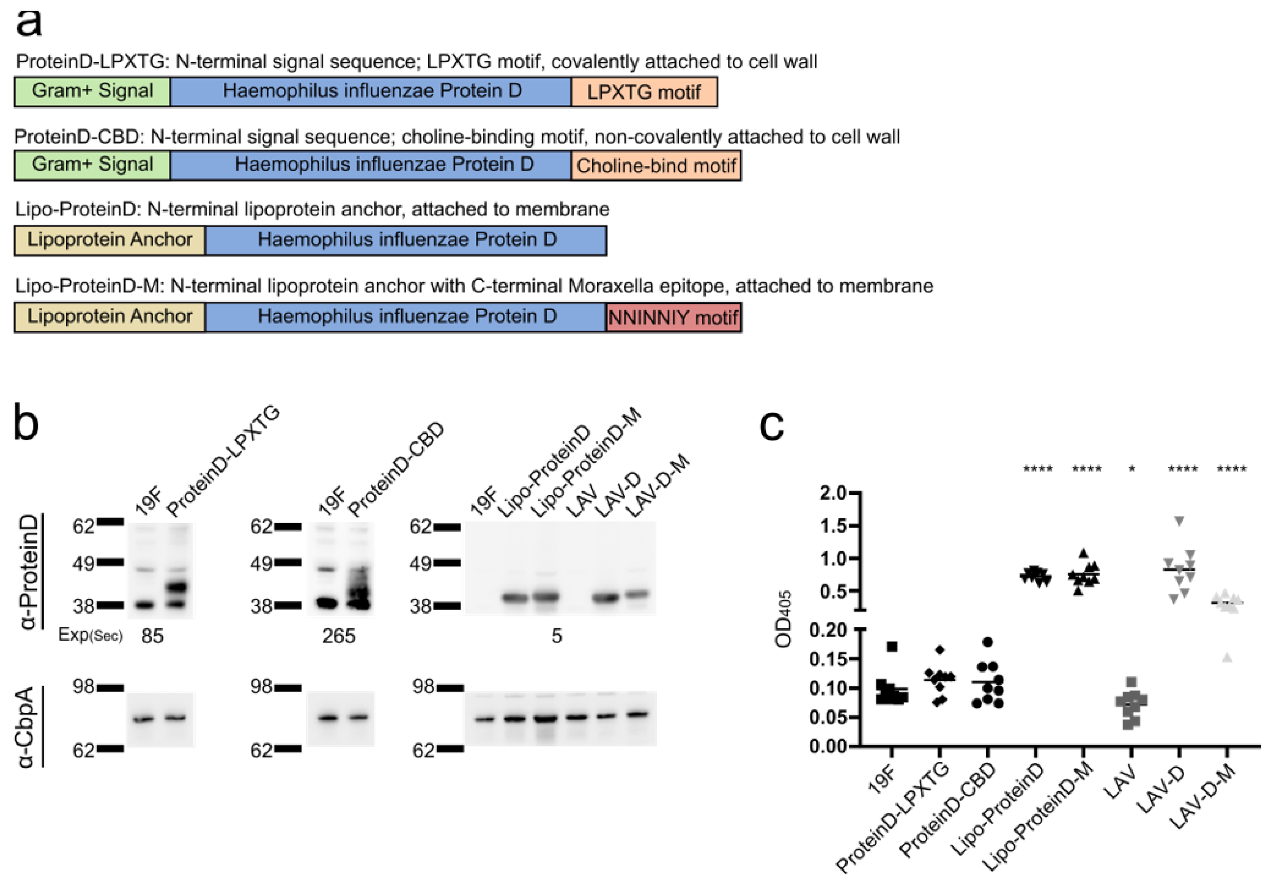
Streptococci utilize several strategies to anchor proteins to the cell surface, including lipoprotein anchoring, LPXTG-mediated covalent attachment to the peptidoglycan, and non-covalent attachment via choline-binding domains which bind to the phosphocholine moieties that decorate the pneumococcal peptidoglycan [89,90]. We generated S. pneumoniae strains that expressed a highly conserved and immunogenic H. influenzae antigen (ProteinD) [72] with distinct anchoring strategies (Figure 4a). All three anchoring strategies were successfully generated with S. pneumoniae expressing the ProteinD variants as determined via Western blot analysis (Figure 4b) and ELISA (Figure 4c). The S. pneumoniae strain expressing lipoprotein-anchored ProteinD consistently demonstrated the greatest level of expression and, hence, was selected for advancement in in vivo efficacy studies.
We next sought to determine if the strain expressing Lipo-ProteinD could be modified further to generate a single vaccine strain that would confer protection against all three major otopathogens: S. pneumoniae, H. influenzae, and M. catarrhalis. In this strategy, we fused the 23 amino acid protective epitope shared by UspA1 and UspA2 from M. catarrhalis (“NNINNIY”) [73,91,92] to the C-terminus of the Lipo-ProteinD construct (Figure 4a). S. pneumoniae expressed and anchored the Lipo-ProteinD-M to the cell surface as determined via Western blot analysis (Figure 4b) and ELISA (Figure 4c). To attenuate S. pneumoniae, the ∆ftsY mutation was introduced into the strain expressing the H. influenzae Lipo-ProteinD and the strain expressing both H. influenzae and M. catarrhalis antigens, which are herein referred to as LAV-D and LAV-D-M, respectively. Of note, the level of ProteinD expressed was reduced in the LAV-D-M strain compared to the LAV-D strain. Attenuation of the vaccine strains was confirmed by determining in vitro growth, adhesion to epithelial cells, and bacterial burden in the lungs, ears, and nasal passage upon challenging the mice with the vaccine strains (Figure S2). All three LAV strains exhibited slower growth in vitro, with the LAV-D-M strain demonstrating the most pronounced delay (Figure 2a) and reduced autolysis. While the vaccine strains had similar levels of adhesion to respiratory epithelial cells compared to the wild-type 19F (Figure S2b) and retained robust colonization capacity (Figure S2c), all three LAV strains were attenuated with dramatically reduced burden in the lungs and ears compared to the wild-type (Figure S2c).

3.4.2. Vaccination Efficacy

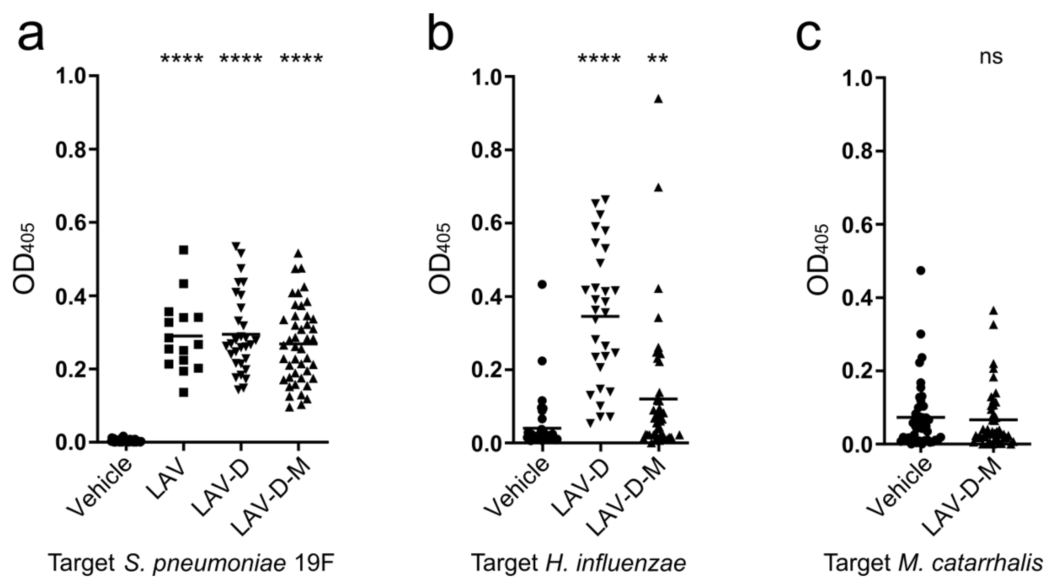
We next determined the efficacy of vaccination with the LAV-D and LAV-D-M strains to impart protection against the major otopathogens S. pneumoniae, non-typeable H. influenzae, and M. catarrhalis. Following intranasal vaccination, the levels of seroconversion was determined by ELISA (Figure 5). The mice vaccinated with LAV-D and LAV-D-M displayed significant IgG seroconversion against S. pneumoniae homologous 19F serotype and heterologous serotype 4 compared to the unvaccinated mice, at similar levels as the mice vaccinated with the LAV strain (Figure 5a, Supplemental Figure S4). Moreover, serum antibody titers against non-typeable H. influenzae were significantly increased compared to serum from the unvaccinated mice, with the level of protection in the mice vaccinated with LAV-D superseding that of LAV-D-M (Figure 5b). No notable differences were observed in serum titers against M. catarrhalis between the mice vaccinated with LAV-D-M and the unvaccinated mice (Figure 5c). However, the unvaccinated mice demonstrated some seroconversion to M. catarrhalis over the course of the vaccination regimen (baseline serum titers were ~0.028), suggesting the mice could have generated an antibody response to native flora similar to M. catarrhalis. These data indicate that the LAV-D and LAV-D-M strains, which add H. influenzae and M. catarrhalis antigens to the LAV strain, remained immunogenic against pneumococcus while conferring additional serum response against H. influenzae.
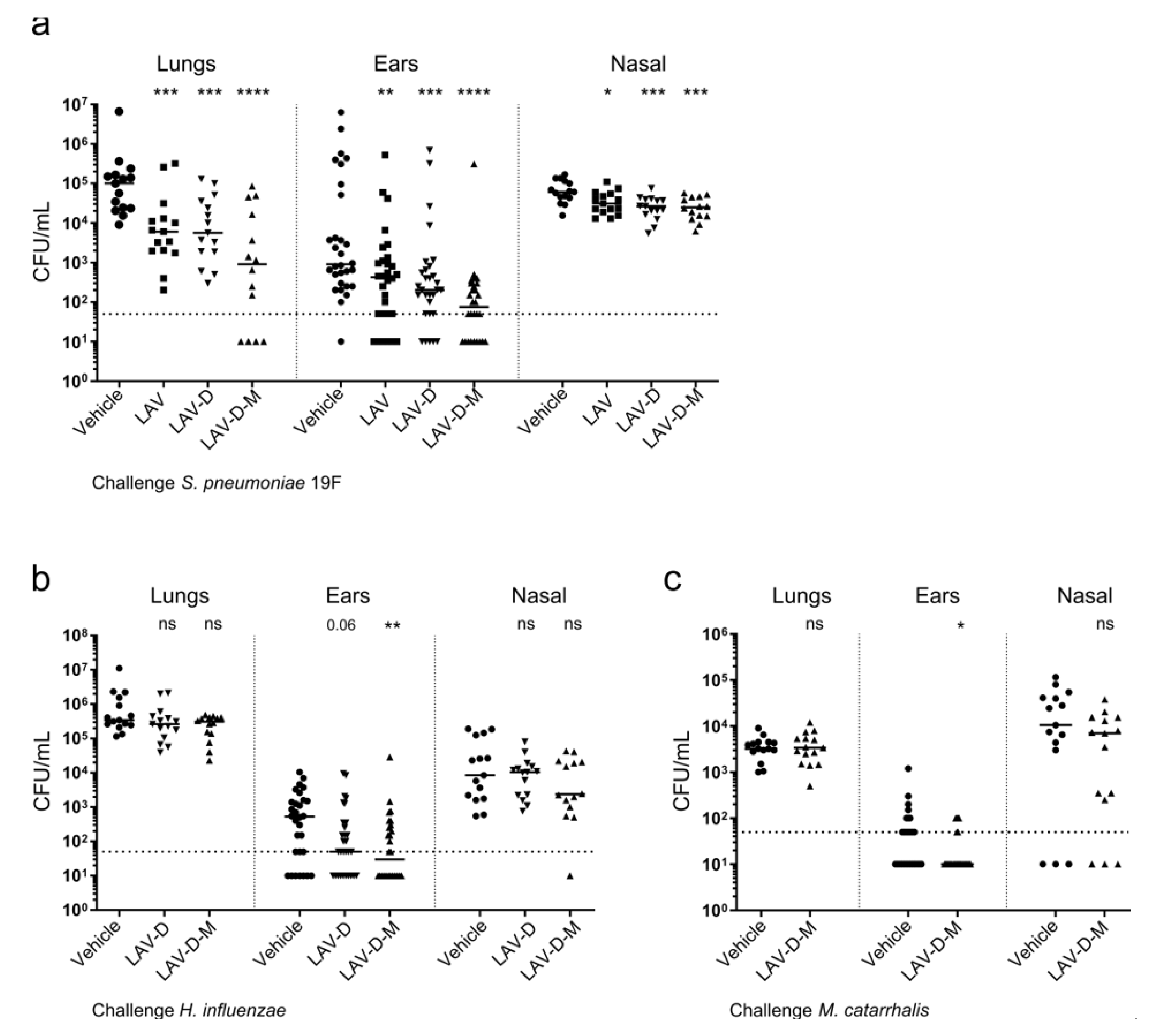
The vaccinated mice were subsequently challenged with S. pneumoniae, non-typeable H. influenzae, or M. catarrhalis, and protection against bacterial burden in the lungs, ears, and nasal passage was determined (Figure 6). All the mice challenged with H. influenzae or M. catarrhalis were administered inflammatory stimulant poly (I:C) [77] prior to challenge to facilitate translocation into the middle ear (Figure S5). Vaccine strains incorporating the H. influenzae and M. catarrhalis epitopes (LAV-D and LAV-D-M) demonstrated protection against bacterial burden in the lungs and ears when challenged with S. pneumoniae compared to the unvaccinated vehicle control (Figure 6a), similar to the original LAV strain and in line with the observed seroconversion against S. pneumoniae in the mice vaccinated with all three LAV strains (Figure 5a). Notably, vaccination with the LAV-D-M strain demonstrated significantly enhanced protection against pneumococcal burden in the ears compared with vaccination with the LAV strain (p < 0.05). In addition to protection against S. pneumoniae, the mice vaccinated with LAV-D and LAV-D-M gained AOM protection against H. influenzae challenge, exhibiting reduced bacterial burden in the ears (Figure 6b). Of note, the burden in the ears of the mice vaccinated with LAV-D-M was reduced similarly to those vaccinated with LAV-D despite a lower seroconversion against H. influenzae in LAV-D-M compared to LAV-D (Figure 5b). The mice vaccinated with LAV-D-M also gained protection against AOM caused by M. catarrhalis, demonstrating reduced bacterial burden in the ears compared to the unvaccinated mice (Figure 6c), despite no observable difference in IgG seroconversion (Figure 5c). These data present a potentially viable approach for targeting all three major bacterial pathogens responsible for pediatric AOM through engineering the LAV strain to express non-native bacterial epitopes on the cell surface.

4. Discussion

Streptococcus pneumoniae is a human pathogen and a major cause of morbidity and mortality particularly in young children and elderly populations [93]. The development of a pneumococcus vaccine began in the early 1910s, with the first license obtained for the pneumococcal polysaccharide vaccine (PPV) in 1977 followed by the pneumococcal conjugated vaccine (PCV) in 2000 [94]. The PPV was updated from the original 14 serotypes in 1983 to cover 23 serotypes, while the PCV was updated from 7 serotypes to 13 in 2010 [95] and then to 20 serotypes in 2021 [96]. The PPV is modestly effective at preventing invasive disease in adults, although most studies were conducted in populations with high carriage rates [97]. The PCV is currently recommended as part of the standard series of childhood vaccinations and provides effective protection against invasive disease, including bacteremia and meningitis [98], with an unexpected secondary protective effect on pneumonia and otitis media through herd immunity [99,100]. However, this protection is serotype-specific, does not include acute bacterial sinusitis, and is modest against AOM and pneumonia [101,102]. Thus, a multitude of vaccine strategies have been employed to develop serotype-independent pneumococcal vaccines that can prevent the entire spectrum of pneumococcal associated disease, including sinusitis, otitis media, pneumonia, bacteremia, and meningitis.
We and others have proposed that live-attenuated pneumococcal vaccines could represent a viable alternative to polysaccharide-based vaccines, since desirable, heterosubtypic responses to pneumococcal proteins are generated in the context of a natural, mucosal colonization [38,39,40]. In this study, we further investigated a pneumococcal live vaccine platform based upon the deletion of ftsY, which confers protective immunity against both invasive disease and AOM in a serotype-independent manner. Our data suggest that there would likely be minimal risk of genetic reversion of the LAV strain, as the deletion of ftsY conferred a significant impairment to uptake and recombine genomic DNA. The underlying mechanism for the reduced competence of the ftsY deletion is unclear but could be due to impaired assembly of the competence machinery into the pneumococcal cell membrane given the role of the SRP pathway for the appropriate targeting of integral membrane proteins [103]. The ftsY mutant also demonstrated reduced growth kinetics, suggesting that the deleterious fitness cost of obtaining such a mutation may also decrease its propensity for horizontal transfer. In many strain backgrounds, no recoverable ftsY mutants were recovered despite the transformed strain being competent. We postulate that this is due to strain-specific gene essentiality of the SRP pathway, restricting the transfer of this mutation to strain backgrounds permissive for deletion. This agrees with previous studies investigating gene essentiality across multiple strains of pneumococcus, whereby it was also observed that ftsY gene deletion was only tolerated in a subset of strains tested [104]. Further underscoring the genetic stability of the LAV strain, no reversion to the wild-type genotype was observed despite in vivo passaging.
Unlike traditional protein-based or polysaccharide vaccines, a potential limitation of LAVs is the interference by pre-existing immunity which could enhance clearance of the LAV and thus may adversely impact vaccine efficacy. For the pneumococcus, this could result from prior colonization by pneumococcal strains or immunity conferred by previous PCV vaccination. Prior colonization with strains harboring either a homologous or heterologous genotype/serotype did not significantly impact the protection imparted by vaccination with the LAV strain. Of note, it is unclear if the LAV strain colonized the nasal passage of the mice that were previously colonized as well as those that were non-colonized. It is unlikely that prior colonization would reduce LAV colonization to such an extent as to prevent an immune response against the vaccine strain. Previous studies have indicated that prior colonization can contribute to early clearance of secondary colonizing strains [105]. However, the secondary colonizing strain can initially colonize the nasal passage and persist up to 14 days [106,107,108]. The LAV strain itself is cleared after seven days [39], suggesting that the LAV strain would clear in the same time period regardless of prior colonization. Moreover, colonizing strains can induce antibody production in the murine model [107,109], and this may explain the enhanced seroconversion observed in the vaccinated mice in the context of prior colonization compared to a non-colonized control (Figure 2a). These data agree with prior observations that repeated exposure events confer heightened immune protection against pneumococcus [105,110]. As observed with prior colonization, the mice prevaccinated with Prevnar-13 demonstrated a similar or synergistic protective effect on the development of AOM. These data indicate that pre-existing immunity via either colonization or vaccination does not inhibit the protective capacity of the LAV strain and may enhance the protection conferred by the vaccine.
A main limitation of vaccines targeting any one etiological agent of AOM is the observation that multiple bacterial pathogens are responsible for the disease, making it unclear if a vaccine targeting any one organism would be successful in reducing overall clinical disease burden. To address this potential limitation, we engineered an LAV strain that expressed H. influenzae ProteinD via multiple anchoring mechanisms, indicating that novel epitopes could be introduced and expressed in the pneumococcus backbone. While all anchoring strategies were successful, the incorporation of a lipoprotein anchor, in a similar manner to how ProteinD is anchored in H. influenzae [111], yielded the highest level of protein expression and anchoring to the pneumococcal cell surface. This suggests that the exploration and validation of multiple routes of anchoring may be required to determine the optimal strategy for the surface expression of novel proteins.
We utilized the lipoprotein anchoring strategy to explore broadening the protective capacity of the LAV strain against multiple bacterial species. To further broaden pathogen coverage to include all three major AOM pathogens, we generated an LAV strain expressing a highly conserved epitope from M. catarrhalis UspA1/2 fused to Lipo-ProteinD. This epitope was chosen due to previous studies demonstrating the protective efficacy of monoclonal antibodies targeting this conserved antigen [91,92]. We were able to successfully integrate and express the Lipo-ProteinD and Moraxella epitope in the LAV strain background. These data indicate that a single LAV strain can be engineered to serve as an antigen presentation system for diverse bacterial antigens. All three vaccine strains conferred significant protection against pneumococcal pathogenesis, including pneumonia and AOM. Interestingly, vaccination with the LAV-D-M strain demonstrated significantly enhanced protection against pneumococcal burden in the ears compared to vaccination with the LAV strain. The mechanism behind this phenomenon is unclear and likely multifactorial. H. influenzae ProteinD, encoded by glpQ, is a glycerophosphodiester phosphodiesterase that catabolizes glycerophosphorylcholine (GCP) to free choline [112,113]. S. pneumoniae has one or two glpQ orthologs depending on the serotype, with the glpQ2 ortholog resembling that of H. influenzae. The deletion of glpQ2 in S. pneumoniae results in reduced choline on the cell surface, a lack of autolysis, reduced adhesion and cytotoxicity in cell lines, and reduced bacterial load in murine nasal passages and lungs [114]. While glpQ2 is present in S. pneumoniae 19A and Taiwan-19F strains, it is absent in the BHN97 19F strain used in this study. The expression of H. influenzae ProteinD in the BHN97 background may provide a means for BHN97 to obtain free choline from GCP. This, in turn, may increase phosphorylcholine in the cell wall and enhance binding to epithelial cells and colonization [115,116,117,118]. In addition to conferring protection against pneumococcal pathogenesis, the LAV strains expressing ProteinD conferred protection against H. influenzae challenge. Of note, the trivalent LAV-D-M strain was less effective than the LAV-D strain in seroconversion against H. influenzae. This was likely due to the reduced level of ProteinD expressed in the LAV-D-M strain compared to the LAV-D strain or perhaps the additional Moraxella epitope on the C-terminus end of ProteinD reduced its immunogenicity. Both LAV-D and LAV-D-M strains were protective against AOM caused by H. influenzae, while demonstrating no significant protective capacity in terms of reduction in bacterial load in the lungs, in line with previous ProteinD vaccine studies [119]. Similarly, vaccine strains expressing both H. influenzae and M. catarrhalis antigens protected against AOM caused by both otopathogens but had no significant impact on bacterial burden in the lungs.

5. Conclusions

Taken together, these data demonstrate a novel platform for engineering attenuated vaccine strains to express novel surface epitopes that are capable of inducing protection against multiple pathogens.

Supplementary Materials

The following supporting information can be downloaded at https://www.mdpi.com/article/10.3390/vaccines12121432/s1, File S1: The original Western blot figures; Table S1: Strains used in this study; Table S2: Primers used to generate LAV, LAV-D, and LAV-D-M; Table S3: ProteinD Protein Sequences; Figure S1: LAV strain confers protection against multiple pneumococcal serotypes; Figure S2: LAV strains are characterized by a slower growth rate and reduced virulence; Figure S3: The protective efficacy of the LAV strain is not diminished with Prevnar-13 vaccination; Figure S4: Vaccination with strains expressing non-native antigenic epitopes demonstrate seroconversion against heterologous S. pneumoniae serotype 4; Figure S5: Poly (I:C) treatment prior to challenge enhances localization to respiratory tissues. References [65,120,121,122,123] are cited in the Supplementary Materials.

Author Contributions

Conceptualization, H.E., A.I. and J.W.R.; methodology, H.E., A.I., A.M. and J.W.R.; software, H.E. and A.I.; validation, H.E. and A.I.; formal analysis, H.E. and A.I.; investigation, H.E., A.I and A.M.; resources, H.E., A.I., A.M. and J.W.R.; data curation, H.E., A.I., A.M. and J.W.R.; writing—original draft preparation, H.E., A.I. and J.W.R.; writing—review and editing, H.E., A.I., A.M. and J.W.R.; visualization, H.E., A.I. and J.W.R.; supervision, J.W.R.; project administration, J.W.R.; funding acquisition, J.W.R. All authors have read and agreed to the published version of the manuscript.

Funding

J.W.R. and the work described herein were supported in part by NIAID 1R21AI178085-01A1, NIAID 1R21AI178084-01A, NIAID U19AI158076, NIAID 1R01AI155614, NIAID 1R01AI171038, NIAID R01AI156898, and ALSAC.

Institutional Review Board Statement

All experiments involving animals were performed with the prior approval of and in accordance with the guidelines of the St. Jude Institutional Animal Care and Use Committee (Protocol 538, approved 10 June 2022). The St. Jude laboratory animal facilities are fully accredited by the American Association for Accreditation of Laboratory Animal Care. The laboratory animals are maintained in accordance with the applicable portions of the Animal Welfare Act and the guidelines prescribed in the DHHS publication Guide for the Care and Use of Laboratory Animals.

Informed Consent Statement

Not applicable.

Data Availability Statement

The raw data values underlying the graphed data and the reported means presented in both the main text and Supplementary Materials as well as any additional relevant information about the data are available upon request directly from the corresponding author.

Conflicts of Interest

The authors declare no conflicts of interest. The funders had no role in the design of the study; in the collection, analyses, or interpretation of data; in the writing of the manuscript; or in the decision to publish the results.

References

  1. Preventing and Treating Ear Infections. 2017. Available online: https://www.cdc.gov/ear-infection/media/pdfs/Ear-Infection-508.pdf (accessed on 11 December 2024).
  2. Short, K.R.; Reading, P.C.; Brown, L.E.; Pedersen, J.; Gilbertson, B.; Job, E.R.; Edenborough, K.M.; Habets, M.N.; Zomer, A.; Hermans, P.W.; et al. Influenza-induced inflammation drives pneumococcal otitis media. Infect. Immun. 2013, 81, 645–652. [Google Scholar] [CrossRef] [PubMed]
  3. Bergenfelz, C.; Hakansson, A.P. Streptococcus pneumoniae Otitis Media Pathogenesis and How It Informs Our Understanding of Vaccine Strategies. Curr. Otorhinolaryngol. Rep. 2017, 5, 115–124. [Google Scholar] [CrossRef] [PubMed]
  4. Ben-Shimol, S.; Givon-Lavi, N.; Leibovitz, E.; Raiz, S.; Greenberg, D.; Dagan, R. Impact of Widespread Introduction of Pneumococcal Conjugate Vaccines on Pneumococcal and Nonpneumococcal Otitis Media. Clin. Infect. Dis. 2016, 63, 611–618. [Google Scholar] [CrossRef] [PubMed]
  5. Block, S.L.; Hedrick, J.; Harrison, C.J.; Tyler, R.; Smith, A.; Findlay, R.; Keegan, E. Community-wide vaccination with the heptavalent pneumococcal conjugate significantly alters the microbiology of acute otitis media. Pediatr. Infect. Dis. J. 2004, 23, 829–833. [Google Scholar] [CrossRef]
  6. Casey, J.R.; Pichichero, M.E. Changes in frequency and pathogens causing acute otitis media in 1995–2003. Pediatr. Infect. Dis. J. 2004, 23, 824–828. [Google Scholar] [CrossRef]
  7. Jalalvand, F.; Riesbeck, K. Update on non-typeable Haemophilus influenzae-mediated disease and vaccine development. Expert. Rev. Vaccines 2018, 17, 503–512. [Google Scholar] [CrossRef]
  8. Marchisio, P.; Esposito, S.; Picca, M.; Baggi, E.; Terranova, L.; Orenti, A.; Biganzoli, E.; Principi, N.; Milan AOM Study Group. Prospective evaluation of the aetiology of acute otitis media with spontaneous tympanic membrane perforation. Clin. Microbiol. Infect. 2017, 23, 486.e1–486.e6. [Google Scholar] [CrossRef]
  9. Ngo, C.C.; Massa, H.M.; Thornton, R.B.; Cripps, A.W. Predominant Bacteria Detected from the Middle Ear Fluid of Children Experiencing Otitis Media: A Systematic Review. PLoS ONE 2016, 11, e0150949. [Google Scholar] [CrossRef]
  10. Ouldali, N.; Levy, C.; Minodier, P.; Morin, L.; Biscardi, S.; Aurel, M.; Dubos, F.; Dommergues, M.A.; Mezgueldi, E.; Levieux, K.; et al. Long-term Association of 13-Valent Pneumococcal Conjugate Vaccine Implementation with Rates of Community-Acquired Pneumonia in Children. JAMA Pediatr. 2019, 173, 362–370. [Google Scholar] [CrossRef]
  11. Berger, Y.; Adler, A.; Ariel, T.; Rokney, A.; Averbuch, D.; Grisaru-Soen, G. Paediatric community-acquired bacteraemia, pneumococcal invasive disease and antibiotic resistance fell after the pneumococcal conjugate vaccine was introduced. Acta Paediatr. 2018, 108, 1321–1328. [Google Scholar] [CrossRef]
  12. Wang, S.; Tafalla, M.; Hanssens, L.; Dolhain, J. A review of Haemophilus influenzae disease in Europe from 2000-2014: Challenges, successes and the contribution of hexavalent combination vaccines. Expert. Rev. Vaccines 2017, 16, 1095–1105. [Google Scholar] [CrossRef] [PubMed]
  13. Kawai, K.; Adil, E.A.; Barrett, D.; Manganella, J.; Kenna, M.A. Ambulatory Visits for Otitis Media before and after the Introduction of Pneumococcal Conjugate Vaccination. J. Pediatr. 2018, 207, 122–127. [Google Scholar] [CrossRef] [PubMed]
  14. Fortanier, A.C.; Venekamp, R.P.; Hoes, A.W.; Schilder, A.G.M. Does pneumococcal conjugate vaccination affect onset and risk of first acute otitis media and recurrences? A primary care-based cohort study. Vaccine 2019, 37, 1528–1532. [Google Scholar] [CrossRef] [PubMed]
  15. Keck, J.W.; Wenger, J.D.; Bruden, D.L.; Rudolph, K.M.; Hurlburt, D.A.; Hennessy, T.W.; Bruce, M.G. PCV7-induced changes in pneumococcal carriage and invasive disease burden in Alaskan children. Vaccine 2014, 32, 6478–6484. [Google Scholar] [CrossRef]
  16. Ramani, R.R.; Hall, W.N.; Boulton, M.; Johnson, D.R.; Zhu, B.P. Impact of PCV7 on invasive pneumococcal disease among children younger than 5 years: A population-based study. Am. J. Public. Health 2004, 94, 958–959. [Google Scholar] [CrossRef]
  17. Esposito, S.; Lizioli, A.; Lastrico, A.; Begliatti, E.; Rognoni, A.; Tagliabue, C.; Cesati, L.; Carreri, V.; Principi, N. Impact on respiratory tract infections of heptavalent pneumococcal conjugate vaccine administered at 3, 5 and 11 months of age. Respir. Res. 2007, 8, 12. [Google Scholar] [CrossRef]
  18. Ubukata, K.; Morozumi, M.; Sakuma, M.; Takata, M.; Mokuno, E.; Tajima, T.; Iwata, S. Etiology of Acute Otitis Media and Characterization of Pneumococcal Isolates After Introduction of 13-Valent Pneumococcal Conjugate Vaccine in Japanese Children. Pediatr. Infect. Dis. J. 2018, 37, 598–604. [Google Scholar] [CrossRef]
  19. Vesikari, T.; Forsten, A.; Seppa, I.; Kaijalainen, T.; Puumalainen, T.; Soininen, A.; Traskine, M.; Lommel, P.; Schoonbroodt, S.; Hezareh, M.; et al. Effectiveness of the 10-Valent Pneumococcal Nontypeable Haemophilus influenzae Protein D-Conjugated Vaccine (PHiD-CV) Against Carriage and Acute Otitis Media-A Double-Blind Randomized Clinical Trial in Finland. J. Pediatric Infect. Dis. Soc. 2016, 5, 237–248. [Google Scholar] [CrossRef]
  20. Nurkka, A.; Joensuu, J.; Henckaerts, I.; Peeters, P.; Poolman, J.; Kilpi, T.; Kayhty, H. Immunogenicity and safety of the eleven valent pneumococcal polysaccharide-protein D conjugate vaccine in infants. Pediatr. Infect. Dis. J. 2004, 23, 1008–1014. [Google Scholar] [CrossRef]
  21. Lee, H.; Choi, E.H.; Lee, H.J. Efficacy and effectiveness of extended-valency pneumococcal conjugate vaccines. Korean J. Pediatr. 2014, 57, 55–66. [Google Scholar] [CrossRef]
  22. Sigurdsson, S.; Eythorsson, E.; Hrafnkelsson, B.; Erlendsdottir, H.; Kristinsson, K.G.; Haraldsson, A. Reduction in All-Cause Acute Otitis Media in Children <3 Years of Age in Primary Care Following Vaccination with 10-Valent Pneumococcal Haemophilus influenzae Protein-D Conjugate Vaccine: A Whole-Population Study. Clin. Infect. Dis. 2018, 67, 1213–1219. [Google Scholar] [CrossRef] [PubMed]
  23. Berman-Rosa, M.; O’Donnell, S.; Barker, M.; Quach, C. Efficacy and Effectiveness of the PCV-10 and PCV-13 Vaccines Against Invasive Pneumococcal Disease. Pediatrics 2020, 145, e20190377. [Google Scholar] [CrossRef] [PubMed]
  24. Rosenblut, A.; Rosenblut, M.; Garcia, K.; Maul, X.; Santolaya, M.E. Frequency of Acute Otitis Media in Children Under 24 Months of Age Before and After the Introduction of the 10-valent Pneumococcal Conjugate Vaccine into the National Immunization Program in Chile. Pediatr. Infect. Dis. J. 2018, 37, 132–134. [Google Scholar] [CrossRef]
  25. Prymula, R.; Peeters, P.; Chrobok, V.; Kriz, P.; Novakova, E.; Kaliskova, E.; Kohl, I.; Lommel, P.; Poolman, J.; Prieels, J.P.; et al. Pneumococcal capsular polysaccharides conjugated to protein D for prevention of acute otitis media caused by both Streptococcus pneumoniae and non-typable Haemophilus influenzae: A randomised double-blind efficacy study. Lancet 2006, 367, 740–748. [Google Scholar] [CrossRef]
  26. Galgani, I.; Poder, A.; Jogi, R.; Anttila, V.J.; Milleri, S.; Borobia, A.M.; Launay, O.; Testa, M.; Casula, D.; Grassano, L.; et al. Immunogenicity and safety of the non-typable Haemophilus influenzae-Moraxella catarrhalis (NTHi-Mcat) vaccine administered following the recombinant zoster vaccine versus administration alone: Results from a randomized, phase 2a, non-inferiority trial. Hum. Vaccin. Immunother. 2023, 19, 2187194. [Google Scholar] [CrossRef]
  27. Van Damme, P.; Leroux-Roels, G.; Vandermeulen, C.; De Ryck, I.; Tasciotti, A.; Dozot, M.; Moraschini, L.; Testa, M.; Arora, A.K. Safety and immunogenicity of non-typeable Haemophilus influenzae-Moraxella catarrhalis vaccine. Vaccine 2019, 37, 3113–3122. [Google Scholar] [CrossRef]
  28. Fortanier, A.C.; Venekamp, R.P.; Boonacker, C.W.; Hak, E.; Schilder, A.G.; Sanders, E.A.; Damoiseaux, R.A. Pneumococcal conjugate vaccines for preventing otitis media. Cochrane Database Syst. Rev. 2014, 4, CD001480. [Google Scholar] [CrossRef]
  29. Ochoa-Gondar, O.; Figuerola-Massana, E.; Vila-Corcoles, A.; Aguirre, C.A.; de Diego, C.; Satue, E.; Gomez, F.; Raga, X. Epidemiology of Streptococcus pneumoniae causing acute otitis media among children in Southern Catalonia throughout 2007-2013: Incidence, serotype distribution and vaccine’s effectiveness. Int. J. Pediatr. Otorhinolaryngol. 2015, 79, 2104–2108. [Google Scholar] [CrossRef]
  30. Palmu, A.A.; Lahdenkari, M. Early Vaccine-type Pneumococcal Acute Otitis Media Does not Predispose to Subsequent Otitis When Compared with Early Acute Otitis Media Due to Other Bacterial Etiology. Pediatr. Infect. Dis. J. 2018, 37, 592–594. [Google Scholar] [CrossRef]
  31. Hall-Stoodley, L.; Nistico, L.; Sambanthamoorthy, K.; Dice, B.; Nguyen, D.; Mershon, W.J.; Johnson, C.; Hu, F.Z.; Stoodley, P.; Ehrlich, G.D.; et al. Characterization of biofilm matrix, degradation by DNase treatment and evidence of capsule downregulation in Streptococcus pneumoniae clinical isolates. BMC Microbiol. 2008, 8, 173. [Google Scholar] [CrossRef]
  32. Kietzman, C.C.; Gao, G.; Mann, B.; Myers, L.; Tuomanen, E.I. Dynamic capsule restructuring by the main pneumococcal autolysin LytA in response to the epithelium. Nat. Commun. 2016, 7, 10859. [Google Scholar] [CrossRef] [PubMed]
  33. De Schutter, I.; De Wachter, E.; Crokaert, F.; Verhaegen, J.; Soetens, O.; Pierard, D.; Malfroot, A. Microbiology of bronchoalveolar lavage fluid in children with acute nonresponding or recurrent community-acquired pneumonia: Identification of nontypeable Haemophilus influenzae as a major pathogen. Clin. Infect. Dis. 2011, 52, 1437–1444. [Google Scholar] [CrossRef]
  34. de Gier, C.; Granland, C.M.; Pickering, J.L.; Walls, T.; Bhuiyan, M.; Mills, N.; Richmond, P.C.; Best, E.J.; Thornton, R.B.; Kirkham, L.S. PCV7- and PCV10-Vaccinated Otitis-Prone Children in New Zealand Have Similar Pneumococcal and Haemophilus influenzae Densities in Their Nasopharynx and Middle Ear. Vaccines 2019, 7, 14. [Google Scholar] [CrossRef]
  35. Xu, Q.; Kaur, R.; Casey, J.R.; Sabharwal, V.; Pelton, S.; Pichichero, M.E. Nontypeable Streptococcus pneumoniae as an otopathogen. Diagn. Microbiol. Infect. Dis. 2011, 69, 200–204. [Google Scholar] [CrossRef]
  36. Ren, D.; Almudevar, A.L.; Pichichero, M.E. Synchrony in serum antibody response to conserved proteins of Streptococcus pneumoniae in young children. Hum. Vaccin. Immunother. 2015, 11, 489–497. [Google Scholar] [CrossRef]
  37. Rowe, H.M.; Mann, B.; Iverson, A.; Poole, A.; Tuomanen, E.; Rosch, J.W. A Cross-Reactive Protein Vaccine Combined with PCV-13 Prevents Streptococcus pneumoniae- and Haemophilus influenzae-Mediated Acute Otitis Media. Infect. Immun. 2019, 87, e00253-19. [Google Scholar] [CrossRef]
  38. Jang, A.Y.; Ahn, K.B.; Zhi, Y.; Ji, H.J.; Zhang, J.; Han, S.H.; Guo, H.; Lim, S.; Song, J.Y.; Lim, J.H.; et al. Serotype-Independent Protection Against Invasive Pneumococcal Infections Conferred by Live Vaccine with lgt Deletion. Front. Immunol. 2019, 10, 1212. [Google Scholar] [CrossRef]
  39. Rosch, J.W.; Iverson, A.R.; Humann, J.; Mann, B.; Gao, G.; Vogel, P.; Mina, M.; Murrah, K.A.; Perez, A.C.; Edward Swords, W.; et al. A live-attenuated pneumococcal vaccine elicits CD4+ T-cell dependent class switching and provides serotype independent protection against acute otitis media. EMBO Mol. Med. 2014, 6, 141–154. [Google Scholar] [CrossRef]
  40. Roche, A.M.; King, S.J.; Weiser, J.N. Live attenuated Streptococcus pneumoniae strains induce serotype-independent mucosal and systemic protection in mice. Infect. Immun. 2007, 75, 2469–2475. [Google Scholar] [CrossRef]
  41. Su, H.; Liu, Q.; Bian, X.; Wang, S.; Curtiss, R., 3rd; Kong, Q. Synthesis and delivery of Streptococcus pneumoniae capsular polysaccharides by recombinant attenuated Salmonella vaccines. Proc. Natl. Acad. Sci. USA 2021, 118, e2013350118. [Google Scholar] [CrossRef]
  42. Xin, W.; Li, Y.; Mo, H.; Roland, K.L.; Curtiss, R., 3rd. PspA family fusion proteins delivered by attenuated Salmonella enterica serovar Typhimurium extend and enhance protection against Streptococcus pneumoniae. Infect. Immun. 2009, 77, 4518–4528. [Google Scholar] [CrossRef] [PubMed]
  43. Wang, S.; Li, Y.; Shi, H.; Scarpellini, G.; Torres-Escobar, A.; Roland, K.L.; Curtiss, R., 3rd. Immune responses to recombinant pneumococcal PsaA antigen delivered by a live attenuated Salmonella vaccine. Infect. Immun. 2010, 78, 3258–3271. [Google Scholar] [CrossRef] [PubMed]
  44. Paton, J.C.; Morona, J.K.; Harrer, S.; Hansman, D.; Morona, R. Immunization of mice with Salmonella typhimurium C5 aroA expressing a genetically toxoided derivative of the pneumococcal toxin pneumolysin. Microb. Pathog. 1993, 14, 95–102. [Google Scholar] [CrossRef] [PubMed]
  45. Seo, S.U.; Kim, J.J.; Yang, H.; Kwon, H.J.; Yang, J.Y.; Curtiss Iii, R.; Kweon, M.N. Effective protection against secondary pneumococcal pneumonia by oral vaccination with attenuated Salmonella delivering PspA antigen in mice. Vaccine 2012, 30, 6816–6823. [Google Scholar] [CrossRef]
  46. Campos, I.B.; Darrieux, M.; Ferreira, D.M.; Miyaji, E.N.; Silva, D.A.; Areas, A.P.; Aires, K.A.; Leite, L.C.; Ho, P.L.; Oliveira, M.L. Nasal immunization of mice with Lactobacillus casei expressing the Pneumococcal Surface Protein A: Induction of antibodies, complement deposition and partial protection against Streptococcus pneumoniae challenge. Microbes Infect. 2008, 10, 481–488. [Google Scholar] [CrossRef]
  47. Vintini, E.O.; Medina, M.S. Host immunity in the protective response to nasal immunization with a pneumococcal antigen associated to live and heat-killed Lactobacillus casei. BMC Immunol. 2011, 12, 46. [Google Scholar] [CrossRef]
  48. Hanniffy, S.B.; Carter, A.T.; Hitchin, E.; Wells, J.M. Mucosal delivery of a pneumococcal vaccine using Lactococcus lactis affords protection against respiratory infection. J. Infect. Dis. 2007, 195, 185–193. [Google Scholar] [CrossRef]
  49. Hernani Mde, L.; Ferreira, P.C.; Ferreira, D.M.; Miyaji, E.N.; Ho, P.L.; Oliveira, M.L. Nasal immunization of mice with Lactobacillus casei expressing the pneumococcal surface protein C primes the immune system and decreases pneumococcal nasopharyngeal colonization in mice. FEMS Immunol. Med. Microbiol. 2011, 62, 263–272. [Google Scholar] [CrossRef]
  50. Medina, M.; Villena, J.; Vintini, E.; Hebert, E.M.; Raya, R.; Alvarez, S. Nasal immunization with Lactococcus lactis expressing the pneumococcal protective protein A induces protective immunity in mice. Infect. Immun. 2008, 76, 2696–2705. [Google Scholar] [CrossRef]
  51. Vintini, E.; Villena, J.; Alvarez, S.; Medina, M. Administration of a probiotic associated with nasal vaccination with inactivated Lactococcus lactis-PppA induces effective protection against pneumoccocal infection in young mice. Clin. Exp. Immunol. 2010, 159, 351–362. [Google Scholar] [CrossRef]
  52. Langermann, S.; Palaszynski, S.R.; Burlein, J.E.; Koenig, S.; Hanson, M.S.; Briles, D.E.; Stover, C.K. Protective humoral response against pneumococcal infection in mice elicited by recombinant bacille Calmette-Guerin vaccines expressing pneumococcal surface protein A. J. Exp. Med. 1994, 180, 2277–2286. [Google Scholar] [CrossRef] [PubMed]
  53. Arevalo, M.T.; Xu, Q.; Paton, J.C.; Hollingshead, S.K.; Pichichero, M.E.; Briles, D.E.; Girgis, N.; Zeng, M. Mucosal vaccination with a multicomponent adenovirus-vectored vaccine protects against Streptococcus pneumoniae infection in the lung. FEMS Immunol. Med. Microbiol. 2009, 55, 346–351. [Google Scholar] [CrossRef] [PubMed]
  54. Helms, C.M.; Grizzard, M.B.; Chanock, R.M. Temperature-sensitive mutants of type I Streptococcus pneumoniae: Preparation, characterization, and evidence for attenuation and immunogenicity. J. Infect. Dis. 1977, 136 (Suppl. S1), S208–S215. [Google Scholar] [CrossRef] [PubMed]
  55. Ramos-Sevillano, E.; Ercoli, G.; Felgner, P.; Ramiro de Assis, R.; Nakajima, R.; Goldblatt, D.; Heyderman, R.S.; Gordon, S.B.; Ferreira, D.M.; Brown, J.S. Preclinical Development of Virulence-attenuated Streptococcus pneumoniae Strains Able to Enhance Protective Immunity against Pneumococcal Infection. Am. J. Respir. Crit. Care Med. 2021, 203, 1037–1041. [Google Scholar] [CrossRef]
  56. Ramos-Sevillano, E.; Ercoli, G.; Guerra-Assuncao, J.A.; Felgner, P.; Ramiro de Assis, R.; Nakajima, R.; Goldblatt, D.; Tetteh, K.K.A.; Heyderman, R.S.; Gordon, S.B.; et al. Protective Effect of Nasal Colonisation with ∆cps/piaA and ∆cps/proABC Streptococcus pneumoniae Strains against Recolonisation and Invasive Infection. Vaccines 2021, 9, 261. [Google Scholar] [CrossRef]
  57. Amonov, M.; Simbak, N.; Wan Hassan, W.M.R.; Ismail, S.; NI, A.R.; Clarke, S.C.; Yeo, C.C. Disruption of the cpsE and endA Genes Attenuates Streptococcus pneumoniae Virulence: Towards the Development of a Live Attenuated Vaccine Candidate. Vaccines 2020, 8, 187. [Google Scholar] [CrossRef]
  58. Kim, S.J.; Seon, S.H.; Luong, T.T.; Ghosh, P.; Pyo, S.; Rhee, D.K. Immunization with attenuated non-transformable pneumococcal pep27 and comD mutant provides serotype-independent protection against pneumococcal infection. Vaccine 2019, 37, 90–98. [Google Scholar] [CrossRef]
  59. Chimalapati, S.; Cohen, J.M.; Camberlein, E.; MacDonald, N.; Durmort, C.; Vernet, T.; Hermans, P.W.; Mitchell, T.; Brown, J.S. Effects of deletion of the Streptococcus pneumoniae lipoprotein diacylglyceryl transferase gene lgt on ABC transporter function and on growth in vivo. PLoS ONE 2012, 7, e41393. [Google Scholar] [CrossRef]
  60. Pribyl, T.; Moche, M.; Dreisbach, A.; Bijlsma, J.J.; Saleh, M.; Abdullah, M.R.; Hecker, M.; van Dijl, J.M.; Becher, D.; Hammerschmidt, S. Influence of impaired lipoprotein biogenesis on surface and exoproteome of Streptococcus pneumoniae. J. Proteome Res. 2014, 13, 650–667. [Google Scholar] [CrossRef]
  61. Chimalapati, S.; Cohen, J.; Camberlein, E.; Durmort, C.; Baxendale, H.; de Vogel, C.; van Belkum, A.; Brown, J.S. Infection with conditionally virulent Streptococcus pneumoniae Deltapab strains induces antibody to conserved protein antigens but does not protect against systemic infection with heterologous strains. Infect. Immun. 2011, 79, 4965–4976. [Google Scholar] [CrossRef]
  62. Cohen, J.M.; Chimalapati, S.; de Vogel, C.; van Belkum, A.; Baxendale, H.E.; Brown, J.S. Contributions of capsule, lipoproteins and duration of colonisation towards the protective immunity of prior Streptococcus pneumoniae nasopharyngeal colonisation. Vaccine 2012, 30, 4453–4459. [Google Scholar] [CrossRef] [PubMed]
  63. Coleman, J.R.; Papamichail, D.; Yano, M.; Garcia-Suarez Mdel, M.; Pirofski, L.A. Designed reduction of Streptococcus pneumoniae pathogenicity via synthetic changes in virulence factor codon-pair bias. J. Infect. Dis. 2011, 203, 1264–1273. [Google Scholar] [CrossRef] [PubMed]
  64. Lacks, S.; Hotchkiss, R.D. A study of the genetic material determining an enzyme in Pneumococcus. Biochim. Biophys. Acta 1960, 39, 508–518. [Google Scholar] [CrossRef] [PubMed]
  65. Echlin, H.; Rosch, J.W. Advancing Genetic Tools in Streptococcus pneumoniae. Genes. 2020, 11, 965. [Google Scholar] [CrossRef]
  66. Rowe, H.M.; Livingston, B.; Margolis, E.; Davis, A.; Meliopoulos, V.A.; Echlin, H.; Schultz-Cherry, S.; Rosch, J.W. Respiratory Bacteria Stabilize and Promote Airborne Transmission of Influenza A Virus. mSystems 2020, 5, e00762-20. [Google Scholar] [CrossRef]
  67. Pozzi, G.; Masala, L.; Iannelli, F.; Manganelli, R.; Havarstein, L.S.; Piccoli, L.; Simon, D.; Morrison, D.A. Competence for genetic transformation in encapsulated strains of Streptococcus pneumoniae: Two allelic variants of the peptide pheromone. J. Bacteriol. 1996, 178, 6087–6090. [Google Scholar] [CrossRef]
  68. Echlin, H.; Iverson, A.; Sardo, U.; Rosch, J.W. Airway proteolytic control of pneumococcal competence. PLoS Pathog. 2023, 19, e1011421. [Google Scholar] [CrossRef]
  69. Horton, R.M.; Cai, Z.L.; Ho, S.N.; Pease, L.R. Gene splicing by overlap extension: Tailor-made genes using the polymerase chain reaction. Biotechniques 1990, 8, 528–535. [Google Scholar] [CrossRef]
  70. Guiral, S.; Henard, V.; Laaberki, M.H.; Granadel, C.; Prudhomme, M.; Martin, B.; Claverys, J.P. Construction and evaluation of a chromosomal expression platform (CEP) for ectopic, maltose-driven gene expression in Streptococcus pneumoniae. Microbiology 2006, 152, 343–349. [Google Scholar] [CrossRef]
  71. Sorg, R.A.; Kuipers, O.P.; Veening, J.W. Gene expression platform for synthetic biology in the human pathogen Streptococcus pneumoniae. ACS Synth. Biol. 2015, 4, 228–239. [Google Scholar] [CrossRef]
  72. Forsgren, A.; Riesbeck, K.; Janson, H. Protein D of Haemophilus influenzae: A protective nontypeable H. influenzae antigen and a carrier for pneumococcal conjugate vaccines. Clin. Infect. Dis. 2008, 46, 726–731. [Google Scholar] [CrossRef] [PubMed]
  73. Tan, T.T.; Nordstrom, T.; Forsgren, A.; Riesbeck, K. The respiratory pathogen Moraxella catarrhalis adheres to epithelial cells by interacting with fibronectin through ubiquitous surface proteins A1 and A2. J. Infect. Dis. 2005, 192, 1029–1038. [Google Scholar] [CrossRef] [PubMed]
  74. van Opijnen, T.; Lazinski, D.W.; Camilli, A. Genome-Wide Fitness and Genetic Interactions Determined by Tn-seq, a High-Throughput Massively Parallel Sequencing Method for Microorganisms. Curr. Protoc. Mol. Biol. 2014, 106, 7–16. [Google Scholar] [CrossRef] [PubMed]
  75. Orihuela, C.J.; Mahdavi, J.; Thornton, J.; Mann, B.; Wooldridge, K.G.; Abouseada, N.; Oldfield, N.J.; Self, T.; Ala’Aldeen, D.A.; Tuomanen, E.I. Laminin receptor initiates bacterial contact with the blood brain barrier in experimental meningitis models. J. Clin. Investig. 2009, 119, 1638–1646. [Google Scholar] [CrossRef]
  76. Michel, L.V.; Kaur, R.; Zavorin, M.; Pryharski, K.; Khan, M.N.; LaClair, C.; O’Neil, M.; Xu, Q.; Pichichero, M.E. Intranasal coinfection model allows for assessment of protein vaccines against nontypeable Haemophilus influenzae in mice. J. Med. Microbiol. 2018, 67, 1527–1532. [Google Scholar] [CrossRef]
  77. Stowell, N.C.; Seideman, J.; Raymond, H.A.; Smalley, K.A.; Lamb, R.J.; Egenolf, D.D.; Bugelski, P.J.; Murray, L.A.; Marsters, P.A.; Bunting, R.A.; et al. Long-term activation of TLR3 by poly(I:C) induces inflammation and impairs lung function in mice. Respir. Res. 2009, 10, 43. [Google Scholar] [CrossRef]
  78. Wren, J.T.; Blevins, L.K.; Pang, B.; King, L.B.; Perez, A.C.; Murrah, K.A.; Reimche, J.L.; Alexander-Miller, M.A.; Swords, W.E. Influenza A virus alters pneumococcal nasal colonization and middle ear infection independently of phase variation. Infect. Immun. 2014, 82, 4802–4812. [Google Scholar] [CrossRef]
  79. Mann, B.; Thornton, J.; Heath, R.; Wade, K.R.; Tweten, R.K.; Gao, G.; El Kasmi, K.; Jordan, J.B.; Mitrea, D.M.; Kriwacki, R.; et al. Broadly protective protein-based pneumococcal vaccine composed of pneumolysin toxoid-CbpA peptide recombinant fusion protein. J. Infect. Dis. 2014, 209, 1116–1125. [Google Scholar] [CrossRef]
  80. Claverys, J.P.; Havarstein, L.S. Extracellular-peptide control of competence for genetic transformation in Streptococcus pneumoniae. Front. Biosci. 2002, 7, d1798–d1814. [Google Scholar] [CrossRef]
  81. Cooper, V.S.; Honsa, E.; Rowe, H.; Deitrick, C.; Iverson, A.R.; Whittall, J.J.; Neville, S.L.; McDevitt, C.A.; Kietzman, C.; Rosch, J.W. Experimental Evolution In Vivo To Identify Selective Pressures during Pneumococcal Colonization. mSystems 2020, 5, e00352-20. [Google Scholar] [CrossRef]
  82. Syrjanen, R.K.; Kilpi, T.M.; Kaijalainen, T.H.; Herva, E.E.; Takala, A.K. Nasopharyngeal carriage of Streptococcus pneumoniae in Finnish children younger than 2 years old. J. Infect. Dis. 2001, 184, 451–459. [Google Scholar] [CrossRef] [PubMed]
  83. Tonkin-Hill, G.; Ling, C.; Chaguza, C.; Salter, S.J.; Hinfonthong, P.; Nikolaou, E.; Tate, N.; Pastusiak, A.; Turner, C.; Chewapreecha, C.; et al. Pneumococcal within-host diversity during colonization, transmission and treatment. Nat. Microbiol. 2022, 7, 1791–1804. [Google Scholar] [CrossRef] [PubMed]
  84. Croucher, N.J.; Kagedan, L.; Thompson, C.M.; Parkhill, J.; Bentley, S.D.; Finkelstein, J.A.; Lipsitch, M.; Hanage, W.P. Selective and genetic constraints on pneumococcal serotype switching. PLoS Genet. 2015, 11, e1005095. [Google Scholar] [CrossRef] [PubMed]
  85. Nuorti, J.P.; Whitney, C.G. Prevention of pneumococcal disease among infants and children—use of 13-valent pneumococcal conjugate vaccine Preventionand 23-valent pneumococcal polysaccharide vaccine—recommendations of the Advisory Committee on Immunization Practices (ACIP). MMWR Recomm. Rep. 2010, 59, 1–18. [Google Scholar]
  86. Centers for Disease Control and Prevention (CDC). Use of 13-valent pneumococcal conjugate vaccine and 23-valent pneumococcal polysaccharide vaccine among children aged 6-18 years with immunocompromising conditions: Recommendations of the Advisory Committee on Immunization Practices (ACIP). MMWR Morb. Mortal. Wkly. Rep. 2013, 62, 521–524. [Google Scholar]
  87. Johnson, C.N.; Wilde, S.; Tuomanen, E.; Rosch, J.W. Convergent impact of vaccination and antibiotic pressures on pneumococcal populations. Cell Chem. Biol. 2024, 31, 195–206. [Google Scholar] [CrossRef]
  88. Schilder, A.G.; Chonmaitree, T.; Cripps, A.W.; Rosenfeld, R.M.; Casselbrant, M.L.; Haggard, M.P.; Venekamp, R.P. Otitis media. Nat. Rev. Dis. Primers 2016, 2, 16063. [Google Scholar] [CrossRef]
  89. Fischetti, V.A. M Protein and Other Surface Proteins on Streptococcus pyogenes. In Streptococcus pyogenes: Basic Biology to Clinical Manifestations, 2nd ed.; Ferretti, J.J., Stevens, D.L., Fischetti, V.A., Eds.; University of Oklahoma Health Sciences Center: Oklahoma City, OK, USA, 2022. [Google Scholar]
  90. Bergmann, S.; Hammerschmidt, S. Versatility of pneumococcal surface proteins. Microbiology 2006, 152, 295–303. [Google Scholar] [CrossRef]
  91. Helminen, M.E.; Maciver, I.; Latimer, J.L.; Klesney-Tait, J.; Cope, L.D.; Paris, M.; McCracken, G.H., Jr.; Hansen, E.J. A large, antigenically conserved protein on the surface of Moraxella catarrhalis is a target for protective antibodies. J. Infect. Dis. 1994, 170, 867–872. [Google Scholar] [CrossRef]
  92. Aebi, C.; Maciver, I.; Latimer, J.L.; Cope, L.D.; Stevens, M.K.; Thomas, S.E.; McCracken, G.H., Jr.; Hansen, E.J. A protective epitope of Moraxella catarrhalis is encoded by two different genes. Infect. Immun. 1997, 65, 4367–4377. [Google Scholar] [CrossRef]
  93. Henriques-Normark, B.; Tuomanen, E.I. The pneumococcus: Epidemiology, microbiology, and pathogenesis. Cold Spring Harb. Perspect. Med. 2013, 3, a010215. [Google Scholar] [CrossRef] [PubMed]
  94. Kazanjian, P. Changing interest among physicians toward pneumococcal vaccination throughout the twentieth century. J. Hist. Med. Allied Sci. 2004, 59, 555–587. [Google Scholar] [CrossRef] [PubMed]
  95. Nunes, M.C.; Madhi, S.A. Review on the immunogenicity and safety of PCV-13 in infants and toddlers. Expert. Rev. Vaccines 2011, 10, 951–980. [Google Scholar] [CrossRef] [PubMed]
  96. Shirley, M. 20-Valent Pneumococcal Conjugate Vaccine: A Review of Its Use in Adults. Drugs 2022, 82, 989–999. [Google Scholar] [CrossRef]
  97. Mangtani, P.; Cutts, F.; Hall, A.J. Efficacy of polysaccharide pneumococcal vaccine in adults in more developed countries: The state of the evidence. Lancet Infect. Dis. 2003, 3, 71–78. [Google Scholar] [CrossRef]
  98. Black, S.; Shinefield, H.; Fireman, B.; Lewis, E.; Ray, P.; Hansen, J.R.; Elvin, L.; Ensor, K.M.; Hackell, J.; Siber, G.; et al. Efficacy, safety and immunogenicity of heptavalent pneumococcal conjugate vaccine in children. Pediatr. Infect. Dis. J. 2000, 19, 187–195. [Google Scholar] [CrossRef]
  99. Grijalva, C.G.; Poehling, K.A.; Nuorti, J.P.; Zhu, Y.; Martin, S.W.; Edwards, K.M.; Griffin, M.R. National impact of universal childhood immunization with pneumococcal conjugate vaccine on outpatient medical care visits in the United States. Pediatrics 2006, 118, 865–873. [Google Scholar] [CrossRef]
  100. Shea, K.M.; Weycker, D.; Stevenson, A.E.; Strutton, D.R.; Pelton, S.I. Modeling the decline in pneumococcal acute otitis media following the introduction of pneumococcal conjugate vaccines in the US. Vaccine 2011, 29, 8042–8048. [Google Scholar] [CrossRef]
  101. Shapiro, D.J.; Gonzales, R.; Cabana, M.D.; Hersh, A.L. National trends in visit rates and antibiotic prescribing for children with acute sinusitis. Pediatrics 2011, 127, 28–34. [Google Scholar] [CrossRef]
  102. Cutts, F.T.; Zaman, S.M.; Enwere, G.; Jaffar, S.; Levine, O.S.; Okoko, J.B.; Oluwalana, C.; Vaughan, A.; Obaro, S.K.; Leach, A.; et al. Efficacy of nine-valent pneumococcal conjugate vaccine against pneumonia and invasive pneumococcal disease in The Gambia: Randomised, double-blind, placebo-controlled trial. Lancet 2005, 365, 1139–1146. [Google Scholar] [CrossRef]
  103. Hwang Fu, Y.H.; Huang, W.Y.C.; Shen, K.; Groves, J.T.; Miller, T.; Shan, S.O. Two-step membrane binding by the bacterial SRP receptor enable efficient and accurate Co-translational protein targeting. eLife 2017, 6, e25885. [Google Scholar] [CrossRef] [PubMed]
  104. Rosconi, F.; Rudmann, E.; Li, J.; Surujon, D.; Anthony, J.; Frank, M.; Jones, D.S.; Rock, C.; Rosch, J.W.; Johnston, C.D.; et al. A bacterial pan-genome makes gene essentiality strain-dependent and evolvable. Nat. Microbiol. 2022, 7, 1580–1592. [Google Scholar] [CrossRef] [PubMed]
  105. Ramos-Sevillano, E.; Ercoli, G.; Brown, J.S. Mechanisms of Naturally Acquired Immunity to Streptococcus pneumoniae. Front. Immunol. 2019, 10, 358. [Google Scholar] [CrossRef] [PubMed]
  106. Zhang, Z.; Clarke, T.B.; Weiser, J.N. Cellular effectors mediating Th17-dependent clearance of pneumococcal colonization in mice. J. Clin. Investig. 2009, 119, 1899–1909. [Google Scholar] [CrossRef]
  107. Richards, L.; Ferreira, D.M.; Miyaji, E.N.; Andrew, P.W.; Kadioglu, A. The immunising effect of pneumococcal nasopharyngeal colonisation; protection against future colonisation and fatal invasive disease. Immunobiology 2010, 215, 251–263. [Google Scholar] [CrossRef]
  108. Malley, R.; Trzcinski, K.; Srivastava, A.; Thompson, C.M.; Anderson, P.W.; Lipsitch, M. CD4+ T cells mediate antibody-independent acquired immunity to pneumococcal colonization. Proc. Natl. Acad. Sci. USA 2005, 102, 4848–4853. [Google Scholar] [CrossRef]
  109. Wilson, R.; Cohen, J.M.; Jose, R.J.; de Vogel, C.; Baxendale, H.; Brown, J.S. Protection against Streptococcus pneumoniae lung infection after nasopharyngeal colonization requires both humoral and cellular immune responses. Mucosal Immunol. 2015, 8, 627–639. [Google Scholar] [CrossRef]
  110. Ferreira, D.M.; Neill, D.R.; Bangert, M.; Gritzfeld, J.F.; Green, N.; Wright, A.K.; Pennington, S.H.; Bricio-Moreno, L.; Moreno, A.T.; Miyaji, E.N.; et al. Controlled human infection and rechallenge with Streptococcus pneumoniae reveals the protective efficacy of carriage in healthy adults. Am. J. Respir. Crit. Care Med. 2013, 187, 855–864. [Google Scholar] [CrossRef]
  111. Janson, H.; Heden, L.O.; Forsgren, A. Protein D, the immunoglobulin D-binding protein of Haemophilus influenzae, is a lipoprotein. Infect. Immun. 1992, 60, 1336–1342. [Google Scholar] [CrossRef]
  112. Fan, X.; Goldfine, H.; Lysenko, E.; Weiser, J.N. The transfer of choline from the host to the bacterial cell surface requires glpQ in Haemophilus influenzae. Mol. Microbiol. 2001, 41, 1029–1036. [Google Scholar] [CrossRef]
  113. Munson, R.S., Jr.; Sasaki, K. Protein D, a putative immunoglobulin D-binding protein produced by Haemophilus influenzae, is glycerophosphodiester phosphodiesterase. J. Bacteriol. 1993, 175, 4569–4571. [Google Scholar] [CrossRef] [PubMed]
  114. Chuang, Y.P.; Peng, Z.R.; Tseng, S.F.; Lin, Y.C.; Sytwu, H.K.; Hsieh, Y.C. Impact of the glpQ2 gene on virulence in a Streptococcus pneumoniae serotype 19A sequence type 320 strain. Infect. Immun. 2015, 83, 682–692. [Google Scholar] [CrossRef] [PubMed]
  115. Cundell, D.R.; Gerard, N.P.; Gerard, C.; Idanpaan-Heikkila, I.; Tuomanen, E.I. Streptococcus pneumoniae anchor to activated human cells by the receptor for platelet-activating factor. Nature 1995, 377, 435–438. [Google Scholar] [CrossRef] [PubMed]
  116. Radin, J.N.; Orihuela, C.J.; Murti, G.; Guglielmo, C.; Murray, P.J.; Tuomanen, E.I. beta-Arrestin 1 participates in platelet-activating factor receptor-mediated endocytosis of Streptococcus pneumoniae. Infect. Immun. 2005, 73, 7827–7835. [Google Scholar] [CrossRef] [PubMed]
  117. Rosenow, C.; Ryan, P.; Weiser, J.N.; Johnson, S.; Fontan, P.; Ortqvist, A.; Masure, H.R. Contribution of novel choline-binding proteins to adherence, colonization and immunogenicity of Streptococcus pneumoniae. Mol. Microbiol. 1997, 25, 819–829. [Google Scholar] [CrossRef]
  118. Orihuela, C.J.; Gao, G.; Francis, K.P.; Yu, J.; Tuomanen, E.I. Tissue-specific contributions of pneumococcal virulence factors to pathogenesis. J. Infect. Dis. 2004, 190, 1661–1669. [Google Scholar] [CrossRef]
  119. Siggins, M.K.; Gill, S.K.; Langford, P.R.; Li, Y.; Ladhani, S.N.; Tregoning, J.S. PHiD-CV induces anti-Protein D antibodies but does not augment pulmonary clearance of nontypeable Haemophilus influenzae in mice. Vaccine 2015, 33, 4954–4961. [Google Scholar] [CrossRef]
  120. McCullers, J.A.; McAuley, J.L.; Browall, S.; Iverson, A.R.; Boyd, K.L.; Henriques Normark, B. Influenza enhances susceptibility to natural acquisition of and disease due to Streptococcus pneumoniae in ferrets. J. Infect. Dis. 2010, 202, 1287–1295. [Google Scholar] [CrossRef]
  121. McCullers, J.A.; Karlstrom, A.; Iverson, A.R.; Loeffler, J.M.; Fischetti, V.A. Novel strategy to prevent otitis media caused by colonizing Streptococcus pneumoniae. PLoS Pathog. 2007, 3, e28. [Google Scholar] [CrossRef]
  122. Iverson, A.; Meyer, C.J.; Vogel, P.; Waidyarachchi, S.; Das, N.; Bruhn, D.F.; Poole, A.; Butler, M.M.; Bowlin, T.L.; Lee, R.E.; et al. Efficacy of Aminomethyl Spectinomycins against Complex Upper Respiratory Tract Bacterial Infections. Antimicrob. Agents Chemother. 2019, 63, e02096. [Google Scholar] [CrossRef]
  123. Rowe, H.M.; Karlsson, E.; Echlin, H.; Chang, T.C.; Wang, L.; van Opijnen, T.; Pounds, S.B.; Schultz-Cherry, S.; Rosch, J.W. Bacterial Factors Required for Transmission of Streptococcus pneumoniae in Mammalian Hosts. Cell Host Microbe 2019, 25, 884–891.e6. [Google Scholar] [CrossRef]
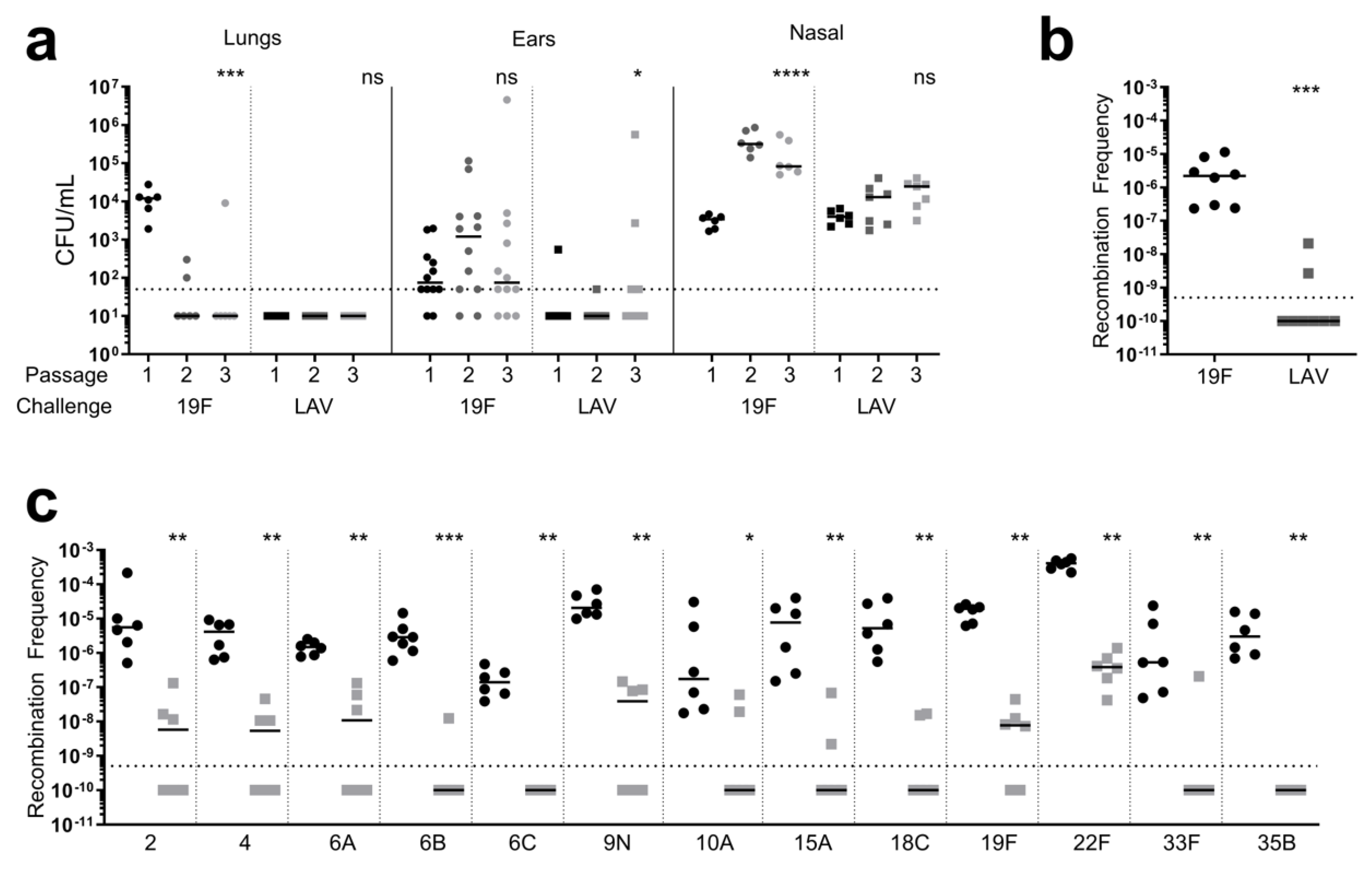
Figure 1. LAV is recalcitrant to reversion and is not readily obtained by other pneumococcal serotypes. (a) 19F (circles) and LAV (squares) strains were passaged in vivo via murine nasal colonization. Six mice were intranasally infected, and the lungs, ears, and nasal passages were harvested 3 days post-challenge. The bacterial burden was determined, and the bacterial growth on plates from the nasal passages were recovered (Passage 1). The recovered bacterial population from the nasal passage was used to challenge the mice for the next passage (2); N = 1 per each Passage 1 population. The bacterial population was passaged again using the same method (Passage 3). Each data point represents an individual mouse (lungs and nasal passage) or each ear from individual mice (ear), and the bars represent the median. The dashed line represents the limit of detection. For each strain and tissue, the bacterial burden of the sequential passages was compared via Kruskal–Wallis one-way ANOVA. (b) The competence of the LAV strain was determined by calculating the recombination frequency upon transformation with nontargeted Tn-seq gDNA. Each data point represents an individual biological replicate, and the bars represent the median. The dashed line represents the limit of detection. The recombination frequencies of the wild-type 19F and the LAV strain were compared via a non-parametric Mann–Whitney t-test. (c) The frequency of resistance spread from the LAV strain was determined by transforming strains of different serotypes with the LAV strain’s gDNA (squares, right). As a control, the same strains were transformed with gDNA from a strain harboring the resistance cassette at a neutral location (circles, left). Each data point represents an individual biological replicate, and the bars represent the median. The dashed line represents the limit of detection. For all strains, the recombination frequency of the strains transformed with the gDNA of the LAV strain was compared to that of the strains transformed with the control DNA via a non-parametric Mann–Whitney t-test. * p < 0.05, ** p < 0.01, *** p < 0.001, **** p < 0.0001, ns = non-significant.
Vaccines 12 01432 g001
Figure 2. Prior colonization in mice is not detrimental to the protective efficacy of the LAV strain. (ae) Mice (N = 15) were colonized with either a homologous strain (19F) expressing its own capsule (Type 19F; squares) or a variant capsule (Type 4; circles), or a heterologous strain (4) expressing its own capsule (Type 4; up triangles) or a variant capsule (Type 19F; down triangles). As a control, mice received a PBS vehicle and were not colonized (none; circles). Three weeks later, all the mice were vaccinated with the LAV strain. Following vaccination, all the mice were challenged with a 19F strain. (a,b) Sera were collected prior to challenge, and IgG seroconversion in the vaccinated mice was determined by ELISA against 19F (a) or 7F (b). Each data point represents an individual mouse, and the bars represent the mean. The immunoreactivity of sera from the mice colonized with each strain was compared to that of the non-colonized mice via an unpaired t-test. (ce) Post-challenge with 19F, the bacterial burden in the lungs (c), ears (d), and nasal passage (e) of the vaccinated mice was determined. Each data point represents an individual mouse (c,e) or each ear from individual mice (d), and the bars represent the median. The dashed line represents the limit of detection. The bacterial burden in each tissue from the mice colonized with each strain was compared to that of the non-colonized mice via a non-parametric Mann–Whitney t-test. **** p < 0.0001. No significant difference was observed for any comparison without a designated p value.
Vaccines 12 01432 g002
Figure 3. The protective efficacy of the LAV strain is not diminished with Prevnar-13 vaccination but rather demonstrates a significantly enhanced protective benefit. The mice were vaccinated with either a PBS vehicle control (N = 30; open circles), Prevnar-13 (N = 15; closed circles), LAV strain (N = 30; open squares), or Prevnar-13 followed by vaccination with the LAV strain (N = 30, closed squares). Following the final vaccination, the mice were challenged with a homologous serotype (19F). (a) Sera were collected prior to challenge, and IgG seroconversion was determined by ELISA against 19F. Each data point represents an individual mouse, and the bars represent the mean. The immunoreactivity of sera of the vaccinated mice were compared to that of the mice that received the PBS vehicle control or other vaccines via an unpaired t-test. (bd) Twenty-four hours post-challenge with 19F, the bacterial burden in the lungs (b), ears (c), and nasal passage (d) of the mice was determined. Each data point represents an individual mouse (b,d) or each ear from individual mice (c), and the bars represent the median. The dashed line represents the limit of detection. The bacterial burden in each tissue of the vaccinated mice was compared to the burden in the tissues of the mice that received the PBS vehicle control or other vaccines via a non-parametric Mann–Whitney t-test. * p < 0.05, ** p < 0.01, *** p < 0.001, **** p < 0.0001, ns = non-significant.
Vaccines 12 01432 g003
Figure 4. Novel platform for expressing non-native antigenic epitopes in S. pneumoniae. (a) Strategies for generating S. pneumoniae strains expressing antigenic epitopes of H. influenzae and M. catarrhalis. Three strategies were employed to anchor ProteinD to the cell surface, i.e., with C-terminal LPXTG motif (ProteinD-LPXTG), with C-terminal choline-binding motif (ProteinD-CBD), and with N-terminal lipoprotein anchor domain (Lipo-ProteinD). To generate a vaccine against all three otopathogens, both H. influenzae and M. catarrhalis epitopes were incorporated by expressing Lipo-ProteinD with an additional 23-amino-acid UspA epitope (“NNINNIY”) on the C-terminus. (b) The production of ProteinD in the cell lysates of S. pneumoniae expressing each ProteinD variant was measured via Western blot analysis using a polyclonal antibody against ProteinD. The strains included wild-type 19F, strains expressing each ProteinD variant, and the ftsY mutation in 19F (LAV), strain expressing Lipo-ProteinD (LAV-D), and strain expressing Lipo-ProteinD-M (LAV-D-M). The expression of ProteinD-LPXTG (51 kDa) and Protein-CBD (48 kDa) was detected upon longer exposure time than Lipo-ProteinD (42 kDa) and Lipo-ProteinD-M (45 kDa), and the exposure time (s) is listed for comparison. As a loading control, the samples were concurrently run and probed with an antibody against CbpA. (c) The anchoring of ProteinD on the S. pneumoniae cell surface was measured via whole-cell bacterial ELISA using a polyclonal antibody against ProteinD. As a control for differential binding to the plate, ProteinD immunoreactivity was normalized to immunoreactivity against LytA, a cell surface protein. Each data point represents a biological replicate, and the bars represent mean. The immunoreactivity of ProteinD-expressing strains and LAV strains was compared to that of 19F via an unpaired t-test. * p < 0.05, **** p < 0.0001.
Vaccines 12 01432 g004
Figure 5. Vaccination with strains expressing non-native antigenic epitopes demonstrate seroconversion against multiple respiratory pathogens. (ac) The mice were vaccinated with the LAV strain (squares), LAV-D strain (down triangles), LAV-D-M strain (up triangles), or PBS vehicle control (circles) and challenged with either S. pneumoniae, H. influenzae, or M. catarrhalis. Sera were collected prior to challenge, and IgG seroconversion in the vaccinated mice was determined by ELISA against 19F (a), H. influenzae (b), or M. catarrhalis (c) and included mice in all the challenge groups. Immunoreactivity against 19F was measured in the sera of the mice vaccinated with all the vaccine strains, against H. influenzae in the sera of the mice vaccinated with LAV-D or LAV-D-M, and against M. catarrhalis in the sera of the mice vaccinated with LAV-D-M; N = 15 for LAV, N = 30 for LAV-D, N = 45 for LAV-D-M and vehicle. Each data point represents an individual mouse, and the bars represent the mean. The immunoreactivity of the sera of the mice vaccinated with vaccine strains were compared to that of the mice that received the PBS vehicle control via an unpaired t-test. ** p < 0.01, **** p < 0.0001; ns = non-significant.
Vaccines 12 01432 g005
Figure 6. Vaccination with strains expressing non-native antigenic epitopes demonstrate protection against multiple respiratory pathogens. (ac) The mice were vaccinated with the LAV strain (squares), LAV-D strain (down triangles), LAV-D-M strain (up triangles), or PBS vehicle control (circles) and challenged with either S. pneumoniae, H. influenzae, or M. catarrhalis; N = 15 for each challenge. The bacterial burden in the lungs, ears, and nasal passage of the vaccinated mice was determined for challenge with 19F (a), H. influenzae (b), or M. catarrhalis (c). All the mice challenged with H. influenzae or M. catarrhalis were pre-sensitized with poly (I:C) to enhance translocation to the ear. Each data point represents an individual mouse (lungs and nasal passage) or each ear from individual mice (ear), and the bars represent the median. The dashed line represents the limit of detection. For each challenge, the bacterial burden in each tissue of the vaccinated mice was compared to the burden in the tissues of the mice that received the PBS vehicle control via a non-parametric Mann–Whitney t-test. * p < 0.05, ** p < 0.01, *** p < 0.001, **** p < 0.0001. ns = non-significant.
Vaccines 12 01432 g006
Disclaimer/Publisher’s Note: The statements, opinions and data contained in all publications are solely those of the individual author(s) and contributor(s) and not of MDPI and/or the editor(s). MDPI and/or the editor(s) disclaim responsibility for any injury to people or property resulting from any ideas, methods, instructions or products referred to in the content.