Abstract
Chikungunya virus (CHIKV) is an infectious agent spread by mosquitos, that has engendered endemic or epidemic outbreaks of Chikungunya fever (CHIKF) in Africa, South-East Asia, America, and a few European countries. Like most tropical infections, CHIKV is frequently misdiagnosed, underreported, and underestimated; it primarily affects areas with limited resources, like developing nations. Due to its high transmission rate and lack of a preventive vaccine or effective treatments, this virus poses a serious threat to humanity. After a 32-year hiatus, CHIKV reemerged as the most significant epidemic ever reported, in India in 2006. Since then, CHIKV-related research was begun in India, and up to now, more than 800 peer-reviewed research papers have been published by Indian researchers and medical practitioners. This review gives an overview of the outbreak history and CHIKV-related research in India, to favor novel high-quality research works intending to promote effective treatment and preventive strategies, including vaccine development, against CHIKV infection.
1. Introduction
Chikungunya fever (CHIKF) is a virus-borne illness transmitted by mosquitoes that is brought on by an alphavirus from the Togaviridae family. This virus is classified as an enveloped virus with positive-stranded RNA, and the primary vectors are Aedes aegypti and Aedes albopictus mosquitos. The term ‘chikungunya’ is derived from the root word ‘kungunyala’ of the Kimakonde language, and alludes to the bending posture of patients infected with the virus [1]. Like most tropical infections, the Chikungunya virus (CHIKV) is frequently misdiagnosed, underreported, and underestimated; it primarily affects areas with limited resources, like developing nations [2]. It has become a hazard to public health over the past 20 years, contributing to certain fatalities and substantial disease-associated morbidity [3]. Infection with CHIKV results in a feverish disease like the Dengue virus (DENV) and causes symptoms such as high body temperature, muscle and joint discomfort, flaccid limb weakness, headache, nausea, exhaustion, vomiting, conjunctivitis, rash, and is very rarely deadly in humans [4,5,6]. The key genotypes of CHIKV have been classified as Asian, East-Central-South-African (ECSA), and West African, based on their geographical distributions and all three variants can produce CHIKF in humans [7].
The very first report on CHIKV was from Tanzania, an eastern African country, in 1952 [8]. Later, it was reported from most parts of the African continent including Cameroon, Benin, Senegal, Angola, Burundi, the Democratic Republic of the Congo, Kenya, Madagascar, Gabon, Nigeria, Guinea, Sierra Leone, Liberia, Malawi, Uganda, southern Africa, and Sudan [3,9,10,11,12,13,14,15,16,17]. Subsequently, CHIKV outbreaks have been reported in many Asian countries including Pakistan, Bangladesh, Myanmar, Philippines, India, Sri Lanka, Bhutan, Saudi Arabia, Cambodia, Singapore, Maldives, China, Thailand, Indonesia, Taiwan, Laos, Vietnam, Malaysia, Timor and Yemen [18,19,20,21,22,23]. Since 2004, CHIKV outbreaks have increased in frequency and extent. Primarily as a result of a viral adaptation caused by a point mutation in the outer membrane E1 glycoprotein, resulting in an amino acid change at position 226, which makes it easier for A. albopictus to disseminate the virus [24]. Because, A. albopictus is an endemic mosquito that is found practically everywhere, unlike A. aegypti, which is only prevalent in the tropics and subtropics [25]. In 2005–2006, one-third of La Reunion Island residents from France, and in 2013, the Caribbean population were suffered from CHIKV outbreaks, which led to the peril to public health in Western Europe and Central America [26,27,28]. These two outbreaks have caused hundreds of casualties and more than a million people have fallen ill [29]. Argentina, Mexico, Brazil, Bolivia, Netherlands, Colombia, Panama, Ecuador, Papua New Guinea, Federated States of Micronesia, Italy, Peru, Jamaica, Venezuela, Paraguay, Latin, and North America also reported CHIKV outbreaks in European and western hemisphere [30,31,32,33,34,35,36,37].
Climate change and shifts in vector and host populations may have increased CHIKV’s geographic expansion in addition to potential alterations enhancing viral fitness and transmissibility. Economic growth can’t shield nations from vector-borne illnesses; rather modern living may exacerbate an outbreak through travel, the aging of the population, and the accumulation of solid waste that can serve as a mosquito breeding ground. The United States armed force has acknowledged that CHIKV may be a biological weapon due to its biosafety level 3 (BSL3) pathogenicity. The National Institute of Allergy and Infectious Disease (NIAID) in the United States has designated it as a priority pathogen in category B, which ranks as biological agents’ second-highest category [38]. The WHO ranks CHIKV in the Research and Development Blueprint for preparedness for priority diseases/pathogens because further spread and epidemics are probable [39].
Epidemiological evidence indicating that a single CHIKV infection gives lifetime immunity against all the current lineages raises the possibility that only one vaccination may be enough to guarantee lifetime immunity [40]. Therefore, vaccination is still the most effective method for containing widespread CHIKV epidemics. The first CHIKV vaccine was said to be under development more than 50 years ago. As of now, preclinical testing has been conducted on at least 30 vaccine candidates and of which nine have advanced to clinical trials including BBV87 from Bharat Biotech International Ltd. (BBIL). Despite this, there are currently no licensed vaccines or antiviral medications available to treat CHIKV.
Ergo, this review focuses on the current status of biomedical research on CHIKV in the most populous country, India, and its epidemiology, clinical features, laboratory findings, management, and prevention adopting varied treatment strategies, with reference to the researchers and their affiliated institutions.
2. Organization of the CHIKV Genome
The linear, positive-sense, single-stranded genome of the RNA alphavirus CHIKV is approximately 11.8 kb in size [41]. It encompasses two open reading frames, one of which codes for four non-structural proteins (nsP1, nsP2, nsP3, and nsP4), and the other codes for five structural proteins, including the capsid (C), the peptide 6K/TF, and three envelope proteins (E1, E2, and E3) (Figure 1) [42,43]. The nsP123 precursor and nsP4 work together to synthesize full-length negative-strand RNA intermediate, and then nsP123 is sequentially processed into each of its constituent proteins to produce positive-strand genomic RNA (49S) transcription and sub-genomic RNA [44]. nsP1 possesses RNA capping characteristics and participates in the production of the viral RNA’s negative strand. Whereas the shut-off of host cell transcription is aided by the actions of nsP2 that possess RNA helicase, RNA triphosphatase, and proteinase activities. The viral RNA polymerase is nsP4, and nsP3 is a component of the replicase unit [45]. The capsid and envelope glycoproteins (E1 and E2) that make up the viral particle are contained in the structural polyprotein, which is translated from 26S sub-genomic RNA [44]. A lipid bilayer host-derived envelope securely encases the viral RNA, which is encapsidated in a nucleocapsid of approximately 40 nm in size and displays the glycoproteins E1 and E2 of the viral envelope [46]. The glycoproteins are divided into 80 trimeric spikes, with three E2/E1 heterodimers in each spike [47]. These spikes are necessary for the budding and growth of new virus particles, recognition of host receptors, and entries of cells through pH-dependent endocytosis (via E1) [48]. The class II fusion protein E1 facilitates membrane fusion that is brought on by low pH during viral infection. The type I transmembrane glycoprotein E2 interacts with the receptor during the alphavirus life cycle [47]. During the translation of the structural polyprotein (C–pE2–6K–E1), the capsid protein C is autocatalytically separated from it, and encapsidation of cytoplasmic viral genomic RNA occurs. The remaining envelope polyproteins are processed in the endoplasmic reticulum (ER) of the host cell and are processed by the host signalases at the N- and C-terminal ends of the 6K peptide to produce E3-E2 (also known as precursor E2 or PE2), 6K and E1 [48]. E3-E2 and E1 will eventually unite to form heterotrimers in the early Golgi compartment, following the proteolytic cleavage and it is exported to the plasma membrane. Subsequently, the E3-E2 goes through furin-dependent cleavage and brings out E3 from E2, which makes the trimeric spike fusogenic [49]. Alphavirus 6K has been linked to the processing of envelope protein, virion assembly, membrane permeabilization, and virus budding, whereas alphavirus E3 has been linked to the maturation of envelope glycoprotein [50]. The sequence-specific interaction of both 6K and PE2 or E1 leads to the formation of an efficient virus budding [51]. 6K facilitates lipids from the membrane to flip from one side of the bilayer to the other, during virus budding. Most of the time, 6K is not integrated into new virions; nonetheless, changes in 6K result in an impaired capacity of virions to operate in fusion and core deformation [52]. CHIKV can also be transmitted through cell-to-cell transmission, which enables virions to prevent attacks from the host immune system effectively [53].
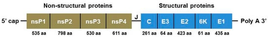
Figure 1.
Schematic representation of CHIKV genome: four non-structural genes (nsp1–4), five structural genes including (Capsid, E3, E2, 6K, and E1) and junction region (J). The genome has a 5’ cap and 3’ poly A tail.
3. CHIKV Research: Indian Scenario
3.1. Epidemiological Profile of CHIKV in India
The first CHIKV outbreak in India was recorded in the city of Kolkata in 1963, followed by Chennai, Puducherry, and Vellore in 1964 [54]. Retrospective serological tests, however, have revealed that chikungunya was present in India before 1963 [55]. After that, Visakhapatnam, Rajahmundry, and Kakinada in 1965, and the central part of India (Nagpur and Barsi) in 1973 were reported [56,57]. The virus reappeared in December 2005 after a 32-year gap, and it was actively spreading, infecting the Hyderabad district of Telangana and the Ananthapur district of Andhra Pradesh in South India, and ultimately affecting 1.4 million people in 13 states [58]. The worst-affected areas were Kerala state and Ahmedabad city in Gujarat [28]. By March 2006, 237 people had died and one-third of the Indian population was infected [29,59]. Until the 1970s, the source of CHICKV infection in India was due to the Asian genotype whereas the 2005–2006 outbreak was due to the ECSA genotype [60,61]. The illness spread to 15 states in 2009. By the years 2015, 2016, and 2019 it increased to 23, 28, and 30 states/Union territories respectively and the most laboratory-confirmed cases were reported in the years 2016, 2017, and 2019, in that order. The states of Maharashtra, Delhi, and Karnataka reported the most cases that were confirmed. A total of 81,914 clinically suspected CHIKV cases were reported in 2019, of which 12,205 (14.9%) were laboratory-confirmed cases in 21 Indian states and 3 Union territories. Karnataka reported the majority of CHIKV cases (3664), followed by Maharashtra (1646) and Telangana (1358) [62]. Till December 2019, Jammu and Kashmir, Mizoram, and Manipur reported 10 or fewer CHIKF cases [63,64]. Less spatial and temporal distribution of mosquitoes in the cold climate and lower urbanizations were major factors [65,66]. Based on the reports, there is a typical pattern of lower percentage positive values reported during the summer seasons, which rises when the rainy season begins, and stays high through the winter [67,68]. 2812 (15.2%) cases were laboratory confirmed as CHIKF until July 2020 out of a total of 18,533 clinically suspected CHIKV cases in 2020 [67]. CHIKF is a serious public health issue in our nation as it is still endemic in 22 Indian states and 4 union territories.
There have always been significant difficulties in the epidemiological surveillance of CHIKV infection, for instance, it can be challenging to distinguish CHIKF from other arbovirus-related ailments because of the similarities in their clinical symptoms or associated co-infections. Therefore, the severity of chikungunya is underestimated. Only precise serological and molecular assays measuring either the presence of anti-CHIKV antibodies in convalescent patients’ serum or the presence of CHIKV’s viral RNA can accurately diagnose CHIKF. To provide a more accurate assessment of incidence and prevalence, affected areas require more resources and surveillance. Nonetheless, there were only two virological testing laboratories in August 2006, the National Institute of Communicable Diseases (NICD) and the National Institute of Virology (NIV) for the entire country [69].
Over the past few decades, CHIKV outbreak studies have become popular in India’s research field to study how it spreads and affects the various populations living in different climatic, cultural, and environmental conditions [70]. A graphical representation of the year-wise publication on CHIKV research is presented in Figure 2. The major CHIKV research groups from India, based on their number of publications and the reputation of their affiliated institutions are summarized in Table 1.
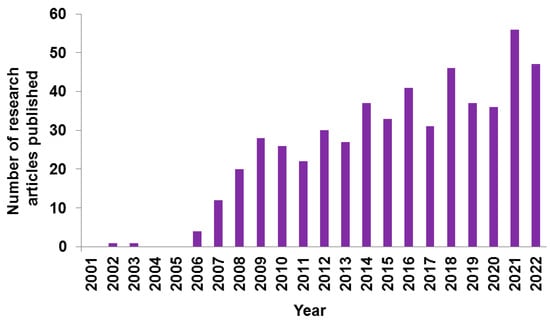
Figure 2.
Number of year-wise publications on Chikungunya from India, in PubMed Database.

Table 1.
Major Indian Biomedical Research Groups Working with the CHIKV.
3.2. Genomic and Proteomic Aspects of CHIKV
The genomic and proteomic data of CHIKV are vast. The comparison of various CHIKV strain genomes and the study of their genomic and proteomic relations give a reasoned knowledge of genetic diversity and phylogenetic relationships of CHIKV. Sreekumar et al., sequenced the 11.7 kb entire genome of the virus and genetic variants from six different viral isolates (2006 RGCB03, RGCB05; 2007 RGCB80, RGCB120; and 2008 RGCB355, RGCB356) from three consecutive Chikungunya epidemics in Kerala were examined [71]. Through full genome sequencing, Arankalle’s team investigated the relationship between the Indian isolates (IND-06) and Reunion Island isolates (RU) of CHIKV genomes, typical of various Indian states. The comparative genomic analysis of CHIKVs in India (1963–2006) with a reference to the 2005–2006 stormy epidemics has shown 99.9% nucleotide similarities. Three distinct substitutions were found in IND-06 isolates (2006): two for the Nsp1 region (T128K and T376M) and one for the capsid protein region (P23S) [72]. This team also verified that the CHIKV strain reported from India in 2000 shares 99% of its characteristics with a Ugandan strain from 1982. They concluded that the progenitor of the 2005–2007 CHIKV isolates originated from the neighborhood of Uganda [73].
Singh et al., reported that the ECSA genotype of CHIKV caused an epidemic between 2006–2010 and 2010–2014 in India [74,75]. Kumar and colleagues carried out a preliminary investigation to characterize genotyping of ECSA strain CHIKV involved in the outbreak of Kerala state and Puducherry. The Puducherry samples showed a mutation on K211E and Kerala samples showed a mutation on A226V in the CHIKV-E1 gene [76,77]. The ECSA genotype’s circulating CHIKV strains belonged to the Indian Ocean Lineage (IOL) category. But only a few different amino acids from the African strain S27 (AF369024) were found in the E1 polypeptide [78]. The crucial non-synonymous C/T mutation at position 10,670 in the E1 gene sequences from A. albopictus human isolates resulted in the alteration of the A226V amino acid, as mentioned vide-supra [79]. Santhosh from DRDE also disclosed E1: A226V shift in 2007 Indian CHIKV isolates [80]. Later, a team led by Dash from DRDE revealed new mutations namely E1:K211E and E2:V264A in CHIKV and it increased the replication and transmission effects of viruses in mosquitoes [81,82] and the same was corroborated in the clinical samples by Sunil’s group from ICGEB [83]. At the same time, the CHIKV-E2 K252H, another novel mutation, confirmed higher viral infectivity from a strain isolated from Kerala (2012–2013) [84]. Harsha et al., discovered yet another six non-synonymous mutations (D284E, V322A, K211E, M269V, V2201, and 1317V) in the CHIKV E1 gene between 2015 and 2016 [85]. By using double locus sequence typing, Muruganadam et al., identified the ECSA strain of the virus as being present in the Andaman and Nicobar Islands [86]. Whereas through E1 gene sequence analysis, Paramasivan et al., validated the involvement of Central/East African genotypes of CHIKV in the Lakshadweep Islands [87]. Dutta et al., also studied on ECSA strain and reported the variation of virus copy number from clinical symptoms among infected people [88]. They were also interested to look at toll-like receptor polymorphism associated with viral infection susceptibility and epitope prediction in-silico [89,90]. Chaaithanya’s team also carried out the gene cluster analysis of oligoadenylate synthetase and polymorphisms of the DC-SIGN (CD209) gene with patients’ symptoms due to CHIKV infections and also reported allele polymorphism of HLA class II in an outbreak from Andaman Island [91,92].
The amino acids from nsP2 (1–95) and nsP1 (170–288) are responsible for their straight interaction with ATPase enzymes demonstrated through mutational analysis [93]. The genetic analysis of nsP2 (378 bp), the E1 envelope protein (505 bp), and E2 (428 bp) by partial sequencing explained a mutation at L210Q (amino acids E2 200–220) in the coding region of E2.c [94]. The relationship between phylogenetic clade diversification and variations in in vitro infectivity among cosmopolitan CHIKV genotype strains was clarified by his team. They had novel mutations in the nsP1 (R171Q), nsP2 (I539S), nsP3 (N409T), and E2 (N72S) [84]. According to a study conducted by Jain and colleagues, the 2010 outbreak was the only one to have mutations in the nsP1 (G230R) and nsP3 (opal 524R) proteins, and these mutations were linked to the severity of the disease. Further, all of the sequenced 2016 samples included the nsP2 mutation H130Y [95].
Chaaithanya et al. investigated the role of chemokines and proinflammatory cytokines in CHIKV-induced chronic arthropathy and discovered that the main functions were played by the cytokines IFN-γ/β, IL-1, TNF-αl, MCP-1, IL-6, MIP-1β, and MIP-1α [96]. Few unusual reports of chronic arthropathy (CAR) and acute flaccid paralysis (AFP) following CHIKV infection were noticed in Andaman and Nicobar Islands, due to MIF gene polymorphism [97]. According to another study, conducted by Agarwal et al., mosquito saliva-induced cutaneous events promote the replication and progression of CHIKV, which is ensued by the induction of the anti-inflammatory genes IL-4, and IL-10 and the suppression of pro-inflammatory genes like IL-2, TLR-3, TNF-α, IFN-γ, and IFN-β is responsible for it [98]. According to Nair et al., with Ddx60, Usp18, Ifit1, Ifi44, Stat1, Rtp4, Mnda, Gbp3, Oas1g, Ly6a, Igtp, Gbp4, Gbp7, Oasl2, and Gbp10, the interferon-regulated gene (IRG) plays a significant role in CHIKV neurovirulence in the mice model [99,100].
Non-structural proteins nsP2 and nsP3 demonstrate RNA interference (RNAi) suppressor activity in the CHIKV proteins, according to research by Sunil’s team [101]. Through the use of next-generation sequencing techniques, this team identified 126 miRNAs from the A. aegypti cell line Aag2 that can control CHIKV replication, including miRNAs miR-100, miR-989, and miR-2b [102]. The same group also reported that the ubiquitin-related modifier is regulated by the microRNA miR-2b, which in turn regulates CHIKV replication and expansion in vitro [103,104]. Further, they investigated the transcriptome analysis and oxidative stress of vectors in response to DENV-2 and CHIKV mono- and co-infections [105,106]. In a coevolution study of CHIKV nonstructural proteins conducted by the same group, when each non-structural protein was taken into account separately, revealed 30 aa pairs coevolving in nsP1, 23 aa pairs coevolving in nsP2, 239 in nsP3, and 46 aa coevolving pairs in nsP4 [107]. Later, Parashar’s team studied altered microRNA expression in the CHIKV-infected fibroblast cells from the mammalian system and purported the possible use of let-7e, miR15, miR-16, miR-17, miR-23a, miR-99, and miR-125 as a biomarker in the viral infection [108].
A bioinformatics team led by Cherian from NIV examined the evolutionary rates and timeline comparability of CHIKV as inferred from the complete genome/E1 gene, paying particular attention to the early outbreaks (2005–2007) in the area bordering the Indian Ocean. The evolutionary timescale of CHIKV was calculated to be in the last 30 decades under a constant population relaxed clock model in bioinformatics [73]. This team also characterized the E1 and E2 genes of CHIKV collected from various parts of India during the 2015–2017 outbreaks [109]. The “Genome to Hits In Silico” strategy was proposed by Jayram’s team, who collaborated with a group of biologists from IIT Delhi, to illustrate novel CHIKV genome-based pathways. This strategy integrates several steps, including gene prediction, determination of the protein’s tertiary structure, identification of the active site, generation of hit molecules, docking of hits, and scoring of hits to arrive at lead compounds, adopting in-silico method [110,111,112].
Sreekumar and colleagues were involved in proteomic analysis and viral replication studies by CHIKV infection, adopting in vivo and in vitro systems [94,113]. Upon CHIKV infection in mammalian cells, their proteomic investigation of the nuclear and multifunctional chaperone nucleophosmin (NPM1)/B23 revealed non-permanent regulation and cytoplasmic aggregation, which limits viral multiplication in the host cells [114,115]. Proteomic analysis of CHKV-infected newborn mouse tissues was performed by Parida’s group and they discovered radical physiological reprogramming through differentially produced proteins from various classes, including those involved in stress, inflammation, apoptosis, urea cycle, cytoskeletal, lipid, and energy metabolism pathways of disease pathogenesis [116,117]. Through the proteome profiling of serum samples from virus-infected individuals, Pandey and his team provided information based on host reaction. During viral infection, the apolipoproteins, clusterin, and S100A family of proteins are active host response factors [118].
3.3. Diagnostics, Treatment, and Vaccines for CHIKV
3.3.1. Diagnostics
The lack of well-developed diagnostic techniques, licensed drugs, vaccines, and antiviral therapeutics led to the primary outbreaks of CHIKV in India. Usha’s group studied similarities between dengue and CHIKF and their co-infection [119] and they concluded that Rheumatologists should always consider Chikungunya infection as one of the causes of arthralgia, after finding seroprevalence of anti-chikungunya IgG antibodies among Rheumatoid Arthritis patients [120]. A research group led by Vijayachari revealed that CHIKV infection can change joint morphology in acute and chronic arthritis and is explained by the imaging technique [121]. Interestingly, four cases of CHIKV infection showed flaccid limb weakness with high body temperature, myalgia, bony erosion, arthralgia, and arthritis, and in a few cases maculopapular rashes in Andaman and Nicobar Islands. They concluded the infection of the virus by serological method (IgM ELISA method) [122,123].
Based on an examination of a variety of ocular symptoms in a cohort of CHIKV-infected individuals, conducted by Prajna, an ophthalmologist, the primary ocular symptom of the CHIKV infection included granulomatous and non-granulomatous anterior uveitis, retrobulbar neuritis, optic neuritis, and dendritic lesions [124]. Mahendradas, another ophthalmologist also reported various ocular manifestations associated with CHIKV infection [125,126,127,128]. The anterior uveitis, retinitis, and optic neuritis were the commonest manifestations, and a case of bilateral Fuchs’ heterochromic iridocyclitis was also reported [129,130]. Yet another ophthalmologist, Mittal also explained optic neuritis and uveitis associated with CHIKV infection in South India [131,132]. Further, Rose et al., noticed retrobulbar neuritis, bilateral papillitis, perineuritis, and neuro retinitis in CHIKV-infected individuals [133]. When Riyaz, a dermatologist reported cutaneous manifestations including purpuric macules with vesiculobullous lesions due to CHIKV infection [134,135,136], Bandyopadhyay observed hyperpigmentation, aphthous-like ulcers, excoriated papules, erythema, xerosis, vesiculobullous and lichenoid eruptions in the skin [137,138,139]. Kumar et al. concluded that anyone with painful oro-genital and intertriginous aphthous-like lesions linked to febrile polyarthralgia with a rash should be suspected of having the CHIKV virus [103]. In another study of febrile chikungunya patients, Murali-Krishna and colleagues described the antibody response patterns that had developed during the febrile phase and these differences could be used as biomarkers to predict future protection from or progression to chronic arthritis [140]. In yet another study of the clinical characteristics of CHIKV-infected patients at a tertiary care facility in Maharashtra, Dube identified some uncommon problems such as lymphadenopathy, mouth ulcers, and encephalitis [141]. Further, Lodha and colleagues discussed CHIKV infection in children and observed the same clinical features as elders [142,143]. However, Patel et al. were interested in studying the acute phase of CHIKV infection in infants and explained how the viral load causes clinical features in kids [144,145]. The assessment of CHIKV infection in Indian children conducted between 2009 and 2010 revealed that the southern region of India had a higher percentage of CHIKV infection than the northern region.
When Patil et al. assessed the level of CHIKV exposure by estimating the age-stratified prevalence of IgG antibodies [146,147], Ravi from NIMHANS studied the association of plasma viral loads and the presence of chikungunya IgM antibodies during acute CHIKV infection with cytokine/chemokine levels [148]. For CHIKV diagnosis, IgM ELISA, TaqMan Real-Time PCR, RT-LAMP assay, and reverse transcription PCR were employed [149]. Lakshmi et al. also discussed the clinical signs and symptoms of CHIKV infection as well as various molecular diagnostic methods, such as the RT-LAMP assay [150]. A single-step multiplex real-time RT-PCR technique for simultaneously detecting and quantifying the RNA of both the DENV and the CHIKV was developed by Cecilia’s dengue/chikungunya group from NIV and it worked well for CHIKV and DENV differential diagnosis [151]. IgM ELISA, nested RT-PCR, and real-time quantitative PCR (qPCR) were evaluated by Parashar’s lab as well, for their ability to diagnose CHIKV, with both assays being ideal for definitive diagnosis [152]. Besides, Tomar and colleagues from IITR recently developed tamarind chitinase (chi)-like lectin glycan-based virus capture assay as a point-of-care routine diagnostic and titration assay for CHIKV as well as for other re-emerging alphaviral infections [153]. Moreover, they recently reported that exploiting NLuc CHIK-VLPs as a detector probe offers the potential for large-scale screening of samples [154]. Further, Sunil’s group evaluated the immunochromatographic rapid diagnosis kit for the detection of CHIKV antigen in the country and performed serological, virological, and clinical analyses of people infected with the virus [64,155,156,157]. Ray’s group succeeded in preparing CHIKV-E2 recombinant protein in E.coli via cloning [158]. Adopting ELISA or Western blot, this isolated protein can accurately be utilized to identify CHIKV-specific antibodies in patients’ sera who had PCR or IgM-positive results [159,160,161]. When the truncated E2 protein was employed for antibody detection, they recently discovered a considerable overlap between CHIKV-infected participants and those with other febrile diseases [161]. Interestingly, when binding antibody titers were determined using fixed CHIKV particles as the test antigen, this overlap was more pronounced. Whereas, the in-vitro identification of the key molecules and pathways involved in CHIKV infection is made possible by the combined mRNA and miRNA signature by Saxena et al. [162].
A research team led by Parida from DRDE developed and analyzed the antigen capture ELISA for CHIKV early clinical diagnosis. Further, real-time RT-PCR assays for detection and quantification were also tested [163,164]. They also developed a quantitative competitive reverse transcription polymerase chain reaction (QC-RT–PCR) for the detection and quantitation of CHIKV [165]. Furthermore, the diagnostic ability of CHIKV viral antigens purified by recombinant envelope proteins and native cell culture was also evaluated [166]. In addition, they generated high-affinity E2-specific monoclonal antibodies against the recombinant protein E2 (rE2) of the CHIKV, and it was reported that these antibodies might be useful for early clinical diagnosis and cohort studies of the virus [167]. The recombinant GST-capsid protein was developed by prokaryotic expression, and ELISA and western blotting confirmed the immunoreactivity of the recombinant antigen. The clinical diagnostic examination of human patients demonstrated 100% sensitivity and 95% specificity in the identification of anti-CHIK IgM antibodies in the serum acute phase samples [168]. This team proved that the RT-LAMP test is a valuable technique for quick, real-time detection and CHIKV quantification in acute-phase serum samples [169]. Another team under the direction of Santhosh examined a one-step real-time RT-PCR assay based on SYBR Green I for the detection and quantification of CHIKV [170]. Besides, the nsP2 protease-based cell-free high-throughput screening assay was developed by Dash’s group to test inhibitors against emerging CHIKV [171].
3.3.2. Treatment
Chloroquine is a popular anti-malarial drug with the potential for both prophylaxis and treatment of CHIKV [172]. Parida’s group looked at the in vitro prophylactic and curative effects of chloroquine on Vero cell CHIKV replication. Chloroquine’s suppressive effects include reduced endosomal-mediated virus entry during the early stages of virus replication, mostly by preventing endocytosis and/or endosomal acidification [173]. Based on another study conducted by the same team, the four artificial microRNAs effectively inhibit Vero cell CHIKV replication [174]. Further, they proposed that the inhibition of CHIKV replication, and apoptosis brought on by viruses in cultured mammalian cells may be accomplished by inhibiting the activity of the cellular inosine monophosphate dehydrogenase (IMPDH) enzyme [175]. Moreover, their study of the effectiveness of combining RNAi-based therapy with traditional antivirals such as ribavirin, chloroquine, and mycophenolic acid provided insight into the decision to use RNAi-based combination therapy [174]. Chattopadhyay and colleagues studied the effect of Ibuprofen conjugated with sulfonamide and thiosemicarbazide and found that the conjugation imparted anti-CHIKV properties while maintaining the anti-inflammatory attributes of Ibuprofen [176].
According to Parashar et al., the administration of NS1 siRNAs and E2 can suppress CHIKV replication in-vitro and it can protect mice infected with the virus [177]. To create novel antiviral strategies, they also attempted to identify and describe the relationship between the host and the virus. Through the use of the viral overlay protein binding assay (VOPBA) and matrix-assisted laser desorption/ionization-time of flight analysis (MALDI TOF/TOF), HSP70 and actin were discovered to be virus-binding proteins in HEK-293T and Vero-E6 cells [178]. Chattopadhyay’s team compared the infection pattern of the Indian CHIKV outbreak (2006), the DRDE-06 strain, and the S-27 prototype strain in mammalian cells [179]. When these two viral strains are infected, the heat shock protein 90 (HSP90), which stabilizes nsP2, positively influences CHIKV proliferation [180]. 17-AAG, a putative HSP90 inhibitor, can significantly control viral infection, apoptosis, and the generation of pro-inflammatory cytokines and chemokines in the host macrophages [181].
Based on conformer and pharmacophore, Tomar and his team at the IITR discovered the peptidomimetic inhibitors of CHIKV-nsP2 protease. The molecular docking of the nsP3/4 junction peptide at the CHIKV nsP2 protease active site gave structural insight into the nsP3/4 peptide’s likely mode of binding and identified key molecular interactions for the substrate binding mode [182,183,184]. Besides, they showed that the peptidomimetic inhibitors Pep-I and Pep-II target virus-specific cysteine protease for CHIKV suppression [185]. Through a further study of CHIKV-nsP2 cysteine protease crystal structure, they have shown its active site with a putative flexible loop blocking (nsP2pro), and this might be beneficial for structure-based drug designing [186,187]. Tomar and her team also investigated the trans-proteolytic activity of the CHIKV capsid protease (CVCP) and discovered that it is crucial to create inhibitors against CHIKV enzymes to obstruct crucial steps in viral multiplication [188,189]. Furthermore, they reported that Piperazine and picolinic acid (PCA) suppresses viral multiplication in Vero cells by attaching to the hydrophobic pocket of the CHIKV capsid protein [190,191]. Moreover, the antiviral activity of NAG-specific chi-like lectin revealed the glycan-dependent chikungunya viral infection [192]. In terms of potential entry inhibitors for CHIKV, phenothiazine, bafilomycin, and 4-hydroxy-1-methyl-3-(3-morpholinopropanoyl)quinoline-2(1H)-one may be regarded as the lead drugs [193] and the same was revealed by structural modeling, molecular dynamic and in silico docking studies [194]. On top of that, Moralbanone, Kaempferol, and Rutin have been reported by Ray’s group to have better binding potential with CHIKV nsP2 and E1 proteins through in silico studies [195].
Dilip et al. compared different medical ideologies (such as ayurveda, allopathy, homeopathy, etc.) and investigated how conventional healthcare professionals treat CHIKV infections [196]. In southern India, people are using Dhanvantaram Gutika, Vilwadi Gulika, Sudarsanam Gulika, Vettumaran Gulika, Amruthotharam Kashayam, Amritarishta, Amruthadiguggulu, Rasnaeranda Kwatha Churna, Vanathulasi Patra and Chromolaena odorata as ayurvedic drugs to treat CHIKV infections. Homeopathic medicines like Eupatorium Perfoliatum Mother Tincture, Rhus tox, Influenzinum, Pyroginum, Arnica, Cedron, Belladonna, Ledumpal, Bryonia, and Nuxvomica are also used against CHKV [196]. The anti-CHIKV activities of Vitex negundo, Hyptis suaveolens, and Decalepis hamiltonii are reported by Swaminathan [197]. Brahmanandha Bairavam Mathirai and Vishnu Chakram, two polyherbal siddha formulations were tested and found to be effective against CHIKV infection by Sunil from ICGEB [198]. The ethanolic extract of Nilavembu kudineer, aqueous and ethanolic extracts of the Vathasura kudineer, and the traditional Indian remedy Amukkara Choornam (Withania somnifera-based) exhibit antiviral properties against CHIKV [199,200,201]. Besides, Patel’s lab from IIT Delhi also evaluated the anti-CHIKV activity of medicinal herbs. They identified that Cedrus deodara, Picrorhiza kurroa, Terminalia chebula, Ocimumtenui-florum, and Commiphora wightii plant extracts exhibited inhibition of CHIKV attachment via helicase and protease inhibition [202]. This team used an FDA-approved chemical library and an in-silico drug screening method to find inhibitors of the CHIKV-nsP2 protease, a crucial non-structural protein needed for the reproduction of the CHIKV virus. The antibiotic novobiocin and the antihypertensive medication telmisartan were among the top hits on the screen and were found to be significant inhibitors of viral replication [203,204].
3.3.3. Vaccine
Due to the pressing need for vaccinations to protect against various viral illnesses, the Indian government launched the National Immunization Program, which resulted in the establishment of numerous authorized vaccine manufacturing facilities around the nation. Vaccine institutes were established at the beginning of the 20th century, and they gradually evolved in the direction of privatization, like BBIL. Besides BBIL, a few Indian agencies are fervently working on the CHIKV vaccine, including the Defense Research Development Establishment, the National Institute of Virology, and the Serum Institute of India.
An ILS researcher Chattopadhyay intended to comprehend anti-CHIKV CD8+ T cell response and epitope advancement-based immunotherapy for CHIKV infection, in silico [205]. Polyclonal antibodies against synthetic peptides of CHIKV non-structural proteins were produced and characterized [206] and it was discovered that the monoclonal antibodies are very sensitive and specific for CHIKV-nsP2 and stop viral multiplication [207]. Arankalle’s group aimed to evaluate envelope-specific immune responses in individuals with CHIKV infection in 2006. The findings demonstrated that lower levels of IFN-γ may be associated with the severity of the disease in these individuals, and T cell epitopes recognized on the envelope region of CHIKV by all patients [208,209]. This team intended to create potential vaccines using either recombinant CHIKV-E2 protein or chemically inactivated whole CHIKV. They showed that the rE2p with alum adjuvant and the BPL/formalin-inactivated CHIKV are promising potential vaccines to prevent the infection [210]. Recently Rahatgi’s group from IITR provided a discernment of CHIKV E2-FL protein immunogenic epitopes, which can be used for the development of novel multiepitope-based anti-CHIKV vaccine strategies [211]. Kumar’s team conducted an in silico study for the development of a CHIKV epitope-based vaccine as well as diagnostic development [212]. Sreekumar and colleagues reported through another recent study the probable application of an attenuated CHIKV strain RGCB355/KL08-p75 as a probable candidate for vaccine development [213]. Using bioinformatics tools, Slathia’s team predicted the DNA vaccine design for CHIKV, based on the conserved T and B cell epitopes derived from structural protein [214]. The ECSA genotype of CHIKV provided the immunogenic potential of Vero-adapted formalin-inactivated vaccines, which were shown to have immunogenic potential to reduce the rate of virus infection by enhancing both humoral and cell-mediated immune response [215]. Further, using a novel yeast expression system (Pichia pastoris), chikungunya virus-like particles (CHIK-VLPs) were created by Parida’s team and tested as a potential vaccination candidate in Balb/c mice. Through the stimulation of TNF-α, IL-10, and significant quantities of IL-4, IL-2, and IFN-γ, a higher level of cellular immune response was seen, indicating a regulated response [216]. Furthermore, they investigated the immunogenic capacity of different adjuvants and recombinant CHIK-E1 and CHIK-E2 proteins produced in bacteria. Studies on neutralization conducted both in vitro and in vivo have verified the evaluation of the protective efficacy of the vaccine formulations [217].
Sumathy from BBIL looked at evolutionary dynamics and serotype analyses [218,219]. Based on the ECSA strain, they developed a pure, inactivated CHIKV immunogenic formulation (BBV87) that may be used as a vaccine to prevent viral infection [220,221]. Formalin, which is frequently used to produce inactivated viral vaccines and has also been tested in preclinical studies with CHIKV, is employed in the BBV87 vaccine as the technique of viral inactivation. The BBV87 vaccine was discovered to have the capacity to induce a sufficient immune response against CHIKV in an Indian phase I clinical research. BBIL submitted the study in 2017 to the Clinical Trial Registry-India and it has recently begun Phase II and III clinical trials in Costa Rica, according to a recent announcement from the International Vaccine Institute (IVI), a partner of BBIL in this endeavor. It was supported by the India Centric Epidemic Preparedness (Ind-CEPI) mission of the Department of Biotechnology, Government of India, and sponsored by the Coalition for Epidemic Preparedness Innovations (CEPI). Phase II/III clinical trials beginning in Costa Rica, and by others are key steps towards reaching the objective of quick vaccine development of Ind-CEPI.
3.4. Biotechnological Aspects of CHIKV
The function of male A. aegypti mosquitoes in the maintenance and transmission of CHIKV to female mosquitoes was demonstrated by Mishra’s team at NIV. They verified that CHIKV can spread to female mosquitoes during mating by infected male mosquitoes. Infant Swiss albino mice became sick after being exposed to the body fluids of venomous female mosquitoes, demonstrating the infectiousness of the venomous virus [222]. Another study from the same institution, by Mourya’s team, investigated how temperature stress affected juvenile stages and A. aegypti mosquitoes’ sensitivity to CHIKV. They established that mosquitoes perished at temperatures above 44.5 °C, which had an impact on the propagation of CHIKV [223]. By employing A. aegypti rosy eye mutants, they were able to determine the genetic basis of the virus’ vector competence for CHIKV [224]. The significance of the gregarine parasite Ascogregarina culicis in the maintenance of CHIKV in vector mosquitoes was also examined by this group. They observed that the oocyst of the parasite in the A. aegypti mosquitoes directly transmits the CHIKV virus vertically. It has been hypothesized that A. culicis may play a significant part in CHIKV maintenance throughout the inter-epidemic period [225]. Yadav et al. observed the potential CHIKV-specific receptor proteins on the midgut brush boundary membrane of A. aegypti, and carried out experimental CHIKV transmission via Anopheles stephensi mosquitoes [226,227]. They verified that A. aegypti infected with CHIKV showed post-inoculation alterations in the enzyme activity of acetylcholinesterase, glutathione-S-transferase, glucose-6-phosphate dehydrogenase, and non-specific esterases [228]. The team also attempted to study an in-depth perspective of the potential of Zika virus infectivity to already infected A. aegypti with DENV and CHIKV and the results proved that it multiplied effectively in the mosquitoes [229]. Ravi from NIMHANS revealed the ultrastructural morphological changes of host cells by the infection of CHIKV [230]. The pathogenesis is influenced by the CHIKV E1 protein’s molecular mimicry of host proteins [231]. Desai led a different team from the same institute that looked into the mechanisms underlying the three kinds of Aedes mosquitoes’ ability to serve as CHIKV vectors [232]. To enter the mosquito cell line, the CHIKV interacts with the protein heat shock cognate 70 (HSC 70), and C6/36 cells treated with quercetin and YM-01 showed dose-dependent inhibitory potential [233]. Sunil examined the effects of CHIKV replication on A. aegypti’s life table features and the pathways involved in egg-laying [234].
4. Research Priorities and Future Directions
Despite the earnest attempts and technological advancements, multiple concerns about CHIKV infection remain yet to be addressed. Point-of-care multiplex diagnostics to diagnose arboviruses are needed urgently, given the co-circulation of these viruses, including more specific serological assays for seroprevalence studies. More research and mitigation measures are required for the socioeconomic and environmental factors, that contribute to vector proliferation, particularly in low-income cities. Reasons behind the dramatic outbreaks in naive areas need to be unveiled. One of the economical and long-lasting approaches for reducing the disease is to combine therapies that are effective against several arboviral diseases. Lack of funding is a main crisis for the development of a successful CHIKV vaccine, although active immunization is the most economical preventative strategy. Therefore, there is an urgent need for global partnerships with industry, researchers, clinicians, public health officials, and other stakeholders in global health. These partnerships must include national and international, public, private, and government funding agencies to resolve this crisis. The quick deployment of SARS-CoV-2 vaccines in 2020 showed that the scientific community and financing organizations can overcome the customarily slow pace of vaccine development. Nonetheless, numerous potential vaccination candidates are currently undergoing clinical testing and will hopefully start the marketing authorization procedure soon. Yet, it is challenging to conduct vaccine efficacy studies due to the spontaneous emergence of CHIKV infections during outbreaks. An establishment of serological correlates of protection would be an appropriate strategy to make the marketing authorization simpler. The efficacy of the candidate vaccines is still unknown in the pediatric population because they are currently developed primarily for the adult population. Therefore, this part also needs to be considered.
5. Conclusions
In India, CHIKV-related research has boomed during the last decades. According to our current data, the CHIKV-related research has concentrated on its varied aspects such as epidemiology, vector entomology, virology, clinical microbiology, cell biology, immunology, molecular biology, vaccinology, genetics, geo-medical and disease environmental studies, and disease burden estimation. Remarkably, hundreds of articles of high quality have been published by Indian researchers in peer-reviewed journals such as The Lancet, JAMA, Clinical Infectious Diseases, Journal of Medical Virology, Travel Medicine and Infectious Disease, Journal of Infection and Public Health, Virus Research and Scientific Reports. Following the resurgence of viral infectious diseases like Zika, Nipah, and the COVID-19 pandemic, there have been significant advances in surveillance, disease diagnostics like the CRISPR system, convalescent plasma therapy, and vaccine development like the mRNA vaccine. Nonetheless, as a priority pathogen of great concern due to its propensity to produce incapacitating illness, CHIKV is still regarded as a neglected tropical illness. The lack of funding, the unpredictable nature of the disease’s epidemiology, and the erratic interest in this pathogen, which has risen and fallen with the frequency of CHIKV outbreaks, have all hindered the development of CHIKV research progress. The loss of lives during the latest COVID-19 epidemic and the 1918 Spanish flu pandemic emphasizes the usefulness of vaccines in containing epidemics. Albeit a wide range of potential preclinical and clinical vaccine candidates have been developed in recent years, and it can be considered a milestone in the fight against this debilitating disease, there are presently no approved medicines or vaccines to prevent CHIKV infections. Ergo, this review briefs the CHIKV research in India and put forward the priorities and suggestions to favor a positive outcome. Hitherto, it is alluring to conjecture that in par with the world’s hastening CHIKV research, and heightened necessity for research and development of efficient medication and/or vaccine for this neglected tropical illness, Indian scientists will be able to contribute adequately and the cure will be available sooner than we anticipate.
Author Contributions
M.M.V.: Conceived the idea, collected the data, designed the figures, and wrote the initial draft with input from all other authors; F.K., S.A. and T.-Y.W.: contributed to the design, edited, and proofread the MS. All authors have read and agreed to the published version of the manuscript.
Funding
This research received no external funding.
Institutional Review Board Statement
Not applicable.
Informed Consent Statement
Not applicable.
Data Availability Statement
Not applicable.
Acknowledgments
We acknowledge the contribution from Ace Bryan Sotelo Cabal, Department of Biosciences and Technology, College of Science, Chung Yuan Christian University, Taiwan, and Rajasekar V from the Department of Biotechnology, Bharathidasan University, Tiruchirappalli for their support.
Conflicts of Interest
The authors declare no conflict of interest.
References
- Schmidt, C.; Schnierle, B.S. Chikungunya Vaccine Candidates: Current Landscape and Future Prospects. Drug. Des. Dev. Ther. 2022, 16, 3663–3673. [Google Scholar] [CrossRef]
- Montalvo Zurbia-Flores, G.; Reyes-Sandoval, A.; Kim, Y.C. Chikungunya Virus: Priority Pathogen or Passing Trend? Vaccines 2023, 11, 568. [Google Scholar] [CrossRef]
- Sergon, K.; Njuguna, C.; Kalani, R.; Ofula, V.; Onyango, C.; Konongoi, L.S.; Bedno, S.; Burke, H.; Dumilla, A.M.; Konde, J. Seroprevalence of chikungunya virus (CHIKV) infection on Lamu Island, Kenya, October 2004. Am. J. Trop. Med. Hyg. 2008, 78, 333–337. [Google Scholar] [CrossRef]
- Amdekar, S.; Parashar, D.; Alagarasu, K. Chikungunya virus-induced arthritis: Role of host and viral factors in the pathogenesis. Viral Immunol. 2017, 30, 691–702. [Google Scholar] [CrossRef]
- Saxena, S.K.; Swamy, M.A.; Verma, S.K.; Saxena, R. The intolerable burden of Chikungunya: What’s new, what’s needed? Proc. Natl. Acad. Sci. India Sect. B Biol. Sci. 2012, 82, 153–165. [Google Scholar] [CrossRef]
- Saxena, S.K. Re-emergence of the knotty chikungunya virus: Facts, fear or fiction. Future Virol. 2007, 2, 121. [Google Scholar] [CrossRef]
- Teixeira, M.G.; Andrade, A.M.; Maria da Conceição, N.C.; Castro, J.S.; Oliveira, F.L.; Goes, C.S.; Maia, M.; Santana, E.B.; Nunes, B.T.; Vasconcelos, P.F. East/Central/South african genotype chikungunya virus, Brazil, 2014. Emerg. Infect. Dis. 2015, 21, 906. [Google Scholar] [CrossRef] [PubMed]
- Robinson, M.C. An epidemic of virus disease in Southern Province, Tanganyika territory, in 1952–1953. Trans. R. Soc. Trop. Med. Hyg. 1955, 49, 28–32. [Google Scholar] [CrossRef] [PubMed]
- Burt, F.J.; Chen, W.; Miner, J.J.; Lenschow, D.J.; Merits, A.; Schnettler, E.; Kohl, A.; Rudd, P.A.; Taylor, A.; Herrero, L.J. Chikungunya virus: An update on the biology and pathogenesis of this emerging pathogen. Lancet Infect. Dis. 2017, 17, e107–e117. [Google Scholar] [CrossRef] [PubMed]
- Eisenhut, M.; Schwarz, T.; Hegenscheid, B. Seroprevalence of dengue, chikungunya and Sindbis virus infections in German aid workers. Infection 1999, 27, 82. [Google Scholar] [CrossRef]
- Kuniholm, M.H.; Wolfe, N.D.; Huang, C.Y.-h.; Mpoudi-Ngole, E.; Tamoufe, U.; Burke, D.S.; Gubler, D.J. Seroprevalence and distribution of Flaviviridae, Togaviridae, and Bunyaviridae arboviral infections in rural Cameroonian adults. Am. J. Trop. Med. Hyg. 2006, 74, 1078–1083. [Google Scholar] [CrossRef]
- Peyrefitte, C.N.; Rousset, D.; Pastorino, B.A.; Pouillot, R.; Bessaud, M.; Tock, F.; Mansaray, H.; Merle, O.L.; Pascual, A.M.; Paupy, C. Chikungunya virus, Cameroon, 2006. Emerg. Infect. Dis. 2007, 13, 768. [Google Scholar] [CrossRef] [PubMed]
- Peyrefitte, C.N.; Bessaud, M.; Pastorino, B.A.; Gravier, P.; Plumet, S.; Merle, O.L.; Moltini, I.; Coppin, E.; Tock, F.; Daries, W. Circulation of Chikungunya virus in Gabon, 2006–2007. J. Med. Virol. 2008, 80, 430–433. [Google Scholar] [CrossRef] [PubMed]
- Jentes, E.S.; Robinson, J.; Johnson, B.W.; Conde, I.; Sakouvougui, Y.; Iverson, J.; Beecher, S.; Diakite, F.; Coulibaly, M.; Bausch, D.G. Acute arboviral infections in Guinea, west Africa, 2006. Am. J. Trop. Med. Hyg. 2010, 83, 388–394. [Google Scholar] [CrossRef] [PubMed]
- Pistone, T.; Ezzedine, K.; Schuffenecker, I.; Receveur, M.-C.; Malvy, D. An imported case of Chikungunya fever from Madagascar: Use of the sentinel traveller for detecting emerging arboviral infections in tropical and European countries. Travel. Med. Infect. Dis. 2009, 7, 52–54. [Google Scholar] [CrossRef]
- Moore, D.á.; Causey, O.; Carey, D.; Reddy, S.; Cooke, A.; Akinkugbe, F.; David-West, T.; Kemp, G. Arthropod-borne viral infections of man in Nigeria, 1964–1970. Ann. Trop. Med. Parasitol. 1975, 69, 49–64. [Google Scholar] [CrossRef]
- Pistone, T.; Ezzedine, K.; Boisvert, M.; Receveur, M.C.; Schuffenecker, I.; Zeller, H.; Lafon, M.E.; Fleury, H.; Malvy, D. Cluster of chikungunya virus infection in travelers returning from Senegal, 2006. J. Travel Med. 2009, 16, 286–288. [Google Scholar] [CrossRef]
- Lahariya, C.; Pradhan, S. Emergence of chikungunya virus in Indian subcontinent after 32 years: A review. J. Vector Borne Dis. 2006, 43, 151. [Google Scholar]
- Mackenzie, J.; Chua, K.; Daniels, P.; Eaton, B.; Field, H.; Hall, R.; Halpin, K.; Johansen, C.; Kirkland, P.; Lam, S. Emerging viral diseases of Southeast Asia and the Western Pacific. Emerg. Infect. Dis. 2001, 7, 497. [Google Scholar] [CrossRef]
- Pavri, K.; Banerjee, G.; Anderson, C.; Aikat, B. Virological and serological studies of cases of haemorrhagic fever in Calcutta: Material collected by the Institute of Post-graduate Medical Education and Research (IPGME), Calcutta. Indian J. Med. Res. 1964, 52, 692–697. [Google Scholar]
- Wangchuk, S.; Chinnawirotpisan, P.; Dorji, T.; Tobgay, T.; Dorji, T.; Yoon, I.-K.; Fernandez, S. Chikungunya fever outbreak, Bhutan, 2012. Emerg. Infect. Dis. 2013, 19, 1681. [Google Scholar] [CrossRef] [PubMed]
- Rezza, G.; El-Sawaf, G.; Faggioni, G.; Vescio, F.; Al Ameri, R.; De Santis, R.; Helaly, G.; Pomponi, A.; Metwally, D.; Fantini, M. Co-circulation of dengue and chikungunya viruses, Al Hudaydah, Yemen, 2012. Emerg. Infect. Dis. 2014, 20, 1351. [Google Scholar] [CrossRef]
- Hussain, R.; Alomar, I.; Memish, Z.A. Chikungunya virus: Emergence of an arthritic arbovirus in Jeddah, Saudi Arabia. East. Mediterr. Health J. 2013, 19, 506–508. [Google Scholar] [CrossRef]
- Tsetsarkin, K.A.; Vanlandingham, D.L.; McGee, C.E.; Higgs, S. A single mutation in chikungunya virus affects vector specificity and epidemic potential. PLoS Pathog. 2007, 3, e201. [Google Scholar] [CrossRef] [PubMed]
- Townson, H.; Nathan, M.B. Resurgence of chikungunya. Trans. R. Soc. Trop. Med. Hyg. 2008, 102, 308–309. [Google Scholar] [CrossRef]
- Parola, P.; De Lamballerie, X.; Jourdan, J.; Rovery, C.; Vaillant, V.; Minodier, P.; Brouqui, P.; Flahault, A.; Raoult, D.; Charrel, R.N. Novel chikungunya virus variant in travelers returning from Indian Ocean islands. Emerg. Infect. Dis. 2006, 12, 1493. [Google Scholar] [CrossRef]
- Weaver, S.C.; Forrester, N.L. Chikungunya: Evolutionary history and recent epidemic spread. Antivir. Res. 2015, 120, 32–39. [Google Scholar] [CrossRef] [PubMed]
- Sudeep, A.; Parashar, D. Chikungunya: An overview. J. Biosci. 2008, 33, 443–449. [Google Scholar] [CrossRef]
- Mavalankar, D.; Shastri, P.; Raman, P. Chikungunya epidemic in India: A major public-health disaster. Lancet Infect. Dis. 2007, 7, 306–307. [Google Scholar] [CrossRef]
- Nunes, M.R.T.; Faria, N.R.; de Vasconcelos, J.M.; Golding, N.; Kraemer, M.U.; de Oliveira, L.F.; da Silva Azevedo, R.d.S.; da Silva, D.E.A.; da Silva, E.V.P.; da Silva, S.P. Emergence and potential for spread of Chikungunya virus in Brazil. BMC Med. 2015, 13, 102. [Google Scholar] [CrossRef]
- Van Genderen, F.T.; Krishnadath, I.; Sno, R.; Grunberg, M.G.; Zijlmans, W.; Adhin, M.R. First chikungunya outbreak in Suriname; clinical and epidemiological features. PLoS Negl. Trop. Dis. 2016, 10, e0004625. [Google Scholar] [CrossRef]
- Madariaga, M.; Ticona, E.; Resurrecion, C. Chikungunya: Bending over the Americas and the rest of the world. Braz. J. Infect. Dis. 2016, 20, 91–98. [Google Scholar] [CrossRef]
- Carbajo, A.E.; Vezzani, D. Waiting for chikungunya fever in Argentina: Spatio-temporal risk maps. Memórias Do Inst. Oswaldo Cruz 2015, 110, 259–262. [Google Scholar] [CrossRef]
- Mattar, S.; Miranda, J.; Pinzon, H.; Tique, V.; Bolaños, A.; Aponte, J.; Arrieta, G.; Gonzalez, M.; Barrios, K.; Contreras, H. Outbreak of Chikungunya virus in the north Caribbean area of Colombia: Clinical presentation and phylogenetic analysis. J. Infect. Dev. Ctries. 2015, 9, 1126–1132. [Google Scholar] [CrossRef] [PubMed]
- Díaz, Y.; Carrera, J.-P.; Cerezo, L.; Arauz, D.; Guerra, I.; Cisneros, J.; Armién, B.; Botello, A.M.; Araúz, A.B.; Gonzalez, V. Chikungunya virus infection: First detection of imported and autochthonous cases in Panama. Am. J. Trop. Med. Hyg. 2015, 92, 482–485. [Google Scholar] [CrossRef]
- Nava-Frías, M.; Searcy-Pavía, R.E.; Juárez-Contreras, C.A.; Valencia-Bautista, A. Chikungunya fever: Current status in Mexico. Boletín Médico Hosp. Infant. México 2016, 73, 67–74. [Google Scholar]
- Wahid, B.; Ali, A.; Rafique, S.; Idrees, M. Global expansion of chikungunya virus: Mapping the 64-year history. Int. J. Infect. Dis. 2017, 58, 69–76. [Google Scholar] [CrossRef] [PubMed]
- National Institute of Allergy and Infectious Diseases. NIAID Emerging Infectious Diseases/Pathogens. Available online: https://www.niaid.nih.gov/research/emerging-infectious-diseases-pathogens (accessed on 17 April 2023).
- World Health Organization. Annual Review of Diseases Prioritized under the Research and Development Blueprint. Available online: https://www.who.int/docs/default-source/blue-print/first-annual-review-of-diseases-prioritized-under-r-and-d-blueprint.pdf?sfvrsn=1f6b5da0_4 (accessed on 27 April 2023).
- Silva, L.A.; Dermody, T.S. Chikungunya virus: Epidemiology, replication, disease mechanisms, and prospective intervention strategies. J. Clin. Investig. 2017, 127, 737–749. [Google Scholar] [CrossRef]
- Powers, A.M. Chikungunya. Clin. Lab. Med. 2010, 30, 209–219. [Google Scholar] [CrossRef]
- Konishi, E.; Hotta, S. Studies on Structural Proteins of Chikungunya Virus: I. Separation of Three Species of Proteins and Their Preliminary Characterization. Microbiol. Immunol. 1980, 24, 419–428. [Google Scholar] [CrossRef] [PubMed]
- Simizu, B.; Yamamoto, K.; Hashimoto, K.; Ogata, T. Structural proteins of Chikungunya virus. J. Virol. 1984, 51, 254–258. [Google Scholar] [CrossRef]
- Fros, J.J.; Liu, W.J.; Prow, N.A.; Geertsema, C.; Ligtenberg, M.; Vanlandingham, D.L.; Schnettler, E.; Vlak, J.M.; Suhrbier, A.; Khromykh, A.A. Chikungunya virus nonstructural protein 2 inhibits type I/II interferon-stimulated JAK-STAT signaling. J. Virol. 2010, 84, 10877–10887. [Google Scholar] [CrossRef]
- Strauss, J.H.; Strauss, E.G. The alphaviruses: Gene expression, replication, and evolution. Microbiol. Mol. Biol. Rev. 1994, 58, 491–562. [Google Scholar] [CrossRef] [PubMed]
- Metz, S.W.; Gardner, J.; Geertsema, C.; Le, T.T.; Goh, L.; Vlak, J.M.; Suhrbier, A.; Pijlman, G.P. Effective chikungunya virus-like particle vaccine produced in insect cells. PLoS Negl. Trop. Dis. 2013, 7, e2124. [Google Scholar] [CrossRef] [PubMed]
- Kielian, M.; Rey, F.A. Virus membrane-fusion proteins: More than one way to make a hairpin. Nat. Rev. Microbiol. 2006, 4, 67. [Google Scholar] [CrossRef] [PubMed]
- Kuhn, R. Togaviridae: The viruses and their replication. Fields Virol. 2007, 1, 1001–1022. [Google Scholar]
- Metz, S.W.; Geertsema, C.; Martina, B.E.; Andrade, P.; Heldens, J.G.; van Oers, M.M.; Goldbach, R.W.; Vlak, J.M.; Pijlman, G.P. Functional processing and secretion of Chikungunya virus E1 and E2 glycoproteins in insect cells. Virol. J. 2011, 8, 353. [Google Scholar] [CrossRef]
- Jose, J.; Snyder, J.E.; Kuhn, R.J. A structural and functional perspective of alphavirus replication and assembly. Future Microbiol. 2009, 4, 837–856. [Google Scholar] [CrossRef]
- Yao, J.S.; Strauss, E.G.; Strauss, J.H. Interactions between PE2, E1, and 6K required for assembly of alphaviruses studied with chimeric viruses. J. Virol. 1996, 70, 7910–7920. [Google Scholar] [CrossRef]
- Lusa, S.; Garoff, H.; Liueström, P. Fate of the 6K membrane protein of Semliki Forest virus during virus assembly. Virology 1991, 185, 843–846. [Google Scholar] [CrossRef]
- Hahon, N.; Zimmerman, W.D. Chikungunya virus infection of cell monolayers by cell-to-cell and extracellular transmission. Appl. Environ. Microbiol. 1970, 19, 389–391. [Google Scholar] [CrossRef] [PubMed]
- Myers, R.; Carey, D.; Reuben, R.; Jesudass, E.; De Ranitz, C.; Jadhav, M. The 1964 epidemic of dengue-like fever in South India: Isolation of chikungunya virus from human sera and from mosquitoes. Indian J. Med. Res. 1965, 53. [Google Scholar]
- Pavri, K. Disappearance of chikungunya virus from India and South East Asia. Trans. R. Soc. Trop. Med. Hyg. 1986, 80, 491. [Google Scholar] [CrossRef] [PubMed]
- Yergolkar, P.N.; Tandale, B.V.; Arankalle, V.A.; Sathe, P.S.; AB, S.; Gandhe, S.S.; Gokhle, M.D.; Jacob, G.P.; Hundekar, S.L.; Mishra, A.C. Chikungunya outbreaks caused by African genotype, India. Emerg. Infect. Dis. 2006, 12, 1580. [Google Scholar] [CrossRef]
- Padbidri, V.; Gnaneswar, T. Epidemiological investigations of chikungunya epidemic at Barsi, Maharashtra state, India. J. Hyg. Epidemiol. Microbiol. Immunol. 1979, 23, 445–451. [Google Scholar]
- Schuffenecker, I.; Iteman, I.; Michault, A.; Murri, S.; Frangeul, L.; Vaney, M.-C.; Lavenir, R.; Pardigon, N.; Reynes, J.-M.; Pettinelli, F. Genome microevolution of chikungunya viruses causing the Indian Ocean outbreak. PLoS Med. 2006, 3, e263. [Google Scholar] [CrossRef]
- Mourya, D.; Yadav, P. Vector biology of dengue & chikungunya viruses. Indian J. Med. Res. 2006, 124, 475. [Google Scholar]
- Naresh Kumar, C.; Sai Gopal, D. Reemergence of chikungunya virus in Indian subcontinent. Indian. J. Virol. 2010, 21, 8–17. [Google Scholar] [CrossRef]
- Kalantri, S.; Joshi, R.; Riley, L.W. Chikungunya epidemic: An Indian perspective. Natl. Med. J. India 2006, 19, 315. [Google Scholar]
- Translational Research Consortia (TRC) for Chikungunya Virus in India. Current Status of Chikungunya in India. Front. Microbiol. 2021, 12, 695173. [Google Scholar] [CrossRef]
- Muniaraj, M. Fading chikungunya fever from India: Beginning of the end of another episode? Indian J. Med. Res. 2014, 139, 468. [Google Scholar]
- Jain, J.; Kushwah, R.B.S.; Singh, S.S.; Sharma, A.; Adak, T.; Singh, O.P.; Bhatnagar, R.K.; Subbarao, S.K.; Sunil, S. Evidence for natural vertical transmission of chikungunya viruses in field populations of Aedes aegypti in Delhi and Haryana states in India—A preliminary report. Acta Trop. 2016, 162, 46–55. [Google Scholar] [CrossRef] [PubMed]
- Singh, P.K.; Dhiman, R.C. Climate change and human health: Indian context. J. Vector Borne Dis. 2012, 49, 55. [Google Scholar]
- Kakarla, S.G.; Mopuri, R.; Mutheneni, S.R.; Bhimala, K.R.; Kumaraswamy, S.; Kadiri, M.R.; Gouda, K.C.; Upadhyayula, S.M. Temperature dependent transmission potential model for chikungunya in India. Sci. Total Environ. 2019, 647, 66–74. [Google Scholar] [CrossRef]
- Department of Health and Family Welfare. Annual Report of Department of Health and Family Welfare 2017–2018, Chapter 5. Available online: https://main.mohfw.gov.in/sites/default/files/05Chapter.pdf (accessed on 20 May 2023).
- Ministry of Health and Family Welfare; Government of India. Integrated Disease Surveillance Programme (IDSP). Available online: https://idsp.mohfw.gov.in/ (accessed on 10 December 2010).
- National Center for Vector Borne Diseases Control. Chikungunya Cases in the Country since 2017. Available online: https://ncvbdc.mohfw.gov.in/index4.php?lang=1&level=0&linkid=486&lid=3765 (accessed on 1 June 2023).
- Kumar, M.S.; Kamaraj, P.; Khan, S.A.; Allam, R.R.; Barde, P.V.; Dwibedi, B.; Kanungo, S.; Mohan, U.; Mohanty, S.S.; Roy, S.; et al. Seroprevalence of chikungunya virus infection in India, 2017: A cross-sectional population-based serosurvey. Lancet Microbe 2021, 2, e41–e47. [Google Scholar] [CrossRef]
- Sreekumar, E.; Issac, A.; Nair, S.; Hariharan, R.; Janki, M.; Arathy, D.; Regu, R.; Mathew, T.; Anoop, M.; Niyas, K. Genetic characterization of 2006–2008 isolates of Chikungunya virus from Kerala, South India, by whole genome sequence analysis. Virus Genes 2010, 40, 14–27. [Google Scholar] [CrossRef]
- Arankalle, V.A.; Shrivastava, S.; Cherian, S.; Gunjikar, R.S.; Walimbe, A.M.; Jadhav, S.M.; Sudeep, A.; Mishra, A.C. Genetic divergence of Chikungunya viruses in India (1963–2006) with special reference to the 2005–2006 explosive epidemic. J. Gen. Virol. 2007, 88, 1967–1976. [Google Scholar] [CrossRef]
- Cherian, S.S.; Walimbe, A.M.; Jadhav, S.M.; Gandhe, S.S.; Hundekar, S.L.; Mishra, A.C.; Arankalle, V.A. Evolutionary rates and timescale comparison of Chikungunya viruses inferred from the whole genome/E1 gene with special reference to the 2005–07 outbreak in the Indian subcontinent. Infect. Genet. Evol. 2009, 9, 16–23. [Google Scholar] [CrossRef] [PubMed]
- Singh, P.; Mittal, V.; Rizvi, M.A.; Bhattacharya, D.; Chhabra, M.; Rawat, D.S.; Ichhpujani, R.L.; Chauhan, L.S.; Rai, A. Northward movement of East Central South African genotype of Chikungunya virus causing an epidemic between 2006–2010 in India. J. Infect. Dev. Ctries. 2012, 6, 563–571. [Google Scholar] [CrossRef] [PubMed]
- Singh, P.; Sharma, P.; Kumar, S.; Chhabra, M.; Rizvi, M.A.; Mittal, V.; Bhattacharya, D.; Venkatesh, S.; Rai, A. Continued persistence of ECSA genotype with replacement of K211E in E1 gene of Chikungunya virus in Delhi from 2010 to 2014. Asian Pac. J. Trop. Dis. 2016, 6, 564–566. [Google Scholar] [CrossRef]
- Kumar, N.P.; Mitha, M.M.; Krishnamoorthy, N.; Kamaraj, T.; Joseph, R.; Jambulingam, P. Genotyping of virus involved in the 2006 Chikungunya outbreak in South India (Kerala and Puducherry). Curr. Sci. 2007, 1412–1416. [Google Scholar]
- Kumar, N.P.; Joseph, R.; Kamaraj, T.; Jambulingam, P. A226V mutation in virus during the 2007 chikungunya outbreak in Kerala, India. J. Gen. Virol. 2008, 89, 1945–1948. [Google Scholar] [CrossRef] [PubMed]
- Raghavendhar, B.S.; Ray, P.; Ratagiri, V.H.; Sharma, B.; Kabra, S.K.; Lodha, R. Evaluation of chikungunya virus infection in children from India during 2009–2010: A cross sectional observational study. J. Med. Virol. 2016, 88, 923–930. [Google Scholar] [CrossRef]
- Kumar, N.P.; Sabesan, S.; Krishnamoorthy, K.; Jambulingam, P. Detection of Chikungunya virus in wild populations of Aedes albopictus in Kerala State, India. Vector-Borne Zoonotic Dis. 2012, 12, 907–911. [Google Scholar] [CrossRef]
- Santhosh, S.; Dash, P.; Parida, M.; Khan, M.; Tiwari, M.; Rao, P.L. Comparative full genome analysis revealed E1: A226V shift in 2007 Indian Chikungunya virus isolates. Virus Res. 2008, 135, 36–41. [Google Scholar] [CrossRef] [PubMed]
- Agarwal, A.; Sharma, A.K.; Sukumaran, D.; Parida, M.; Dash, P.K. Two novel epistatic mutations (E1: K211E and E2: V264A) in structural proteins of Chikungunya virus enhance fitness in Aedes aegypti. Virology 2016, 497, 59–68. [Google Scholar] [CrossRef] [PubMed]
- Agarwal, A.; Dash, P.K.; Singh, A.K.; Sharma, S.; Gopalan, N.; Rao, P.V.L.; Parida, M.M.; Reiter, P. Evidence of experimental vertical transmission of emerging novel ECSA genotype of chikungunya virus in Aedes aegypti. PLoS Negl. Trop. Dis. 2014, 8, e2990. [Google Scholar] [CrossRef]
- Shrinet, J.; Jain, S.; Sharma, A.; Singh, S.S.; Mathur, K.; Rana, V.; Bhatnagar, R.K.; Gupta, B.; Gaind, R.; Deb, M. Genetic characterization of Chikungunya virus from New Delhi reveal emergence of a new molecular signature in Indian isolates. Virol. J. 2012, 9, 100. [Google Scholar] [CrossRef]
- Abraham, R.; Manakkadan, A.; Mudaliar, P.; Joseph, I.; Sivakumar, K.C.; Nair, R.R.; Sreekumar, E. Correlation of phylogenetic clade diversification and in vitro infectivity differences among Cosmopolitan genotype strains of Chikungunya virus. Infect. Genet. Evol. 2016, 37, 174–184. [Google Scholar] [CrossRef]
- Harsha, P.K.; Reddy, V.; Rao, D.; Pattabiraman, C.; Mani, R.S. Continual circulation of ECSA genotype and identification of a novel mutation I317V in the E1 gene of Chikungunya viral strains in southern India during 2015–2016. J. Med. Virol. 2020, 92, 1007–1012. [Google Scholar] [CrossRef]
- Muruganandam, N.; Chaaithanya, I.; Senthil, G.; Shriram, A.; Bhattacharya, D.; JeevaBharathi, G.; Sudeep, A.; PradeepKumar, N.; Vijayachari, P. Isolation and molecular characterization of Chikungunya virus from the Andaman and Nicobar archipelago, India: Evidence of an East, Central, and South African genotype. Can. J. Microbiol. 2011, 57, 1073–1077. [Google Scholar] [CrossRef] [PubMed]
- Paramasivan, R.; Samuel, P.P.; Thenmozhi, V.; Rajendran, R.; Leo, S.V.J.; Dhananjeyan, K.J.; Krishnamoorthi, R.; Arunachalam, N.; Tyagi, B.K. Chikungunya virus isolated in Lakshadweep islands in the Indian Ocean: Evidence of the Central/East African genotype. Jpn. J. Infect. Dis. 2009, 62, 67–69. [Google Scholar] [CrossRef] [PubMed]
- Dutta, S.K.; Pal, T.; Saha, B.; Mandal, S.; Tripathi, A. Copy number variation of chikungunya ECSA virus with disease symptoms among Indian patients. J. Med. Virol. 2014, 86, 1386–1392. [Google Scholar] [CrossRef] [PubMed]
- Dutta, S.K.; Bhattacharya, T.; Tripathi, A. Chikungunya virus: Genomic microevolution in Eastern India and its in-silico epitope prediction. 3 Biotech. 2018, 8, 318. [Google Scholar] [CrossRef]
- Dutta, S.K.; Tripathi, A. Association of toll-like receptor polymorphisms with susceptibility to chikungunya virus infection. Virology 2017, 511, 207–213. [Google Scholar] [CrossRef]
- Chaaithanya, I.K.; Muruganandam, N.; Anwesh, M.; Rajesh, R.; Ghosal, S.R.; Kartick, C.; Prasad, K.N.; Muthumani, K.; Vijayachari, P. HLA class II allele polymorphism in an outbreak of chikungunya fever in M Iddle A Ndaman, I Ndia. Immunology 2013, 140, 202–210. [Google Scholar] [CrossRef]
- Chaaithanya, I.K.; Muruganandam, N.; Surya, P.; Anwesh, M.; Alagarasu, K.; Vijayachari, P. Association of Oligoadenylate Synthetase Gene Cluster and DC-SIGN (CD209) gene polymorphisms with clinical symptoms in Chikungunya virus infection. DNA Cell. Biol. 2016, 35, 44–50. [Google Scholar] [CrossRef]
- Kumar, S.; Kumar, A.; Mamidi, P.; Tiwari, A.; Kumar, S.; Mayavannan, A.; Mudulli, S.; Singh, A.K.; Subudhi, B.B.; Chattopadhyay, S. Chikungunya virus nsP1 interacts directly with nsP2 and modulates its ATPase activity. Sci. Rep. 2018, 8, 1045. [Google Scholar] [CrossRef]
- Niyas, K.P.; Abraham, R.; Unnikrishnan, R.N.; Mathew, T.; Nair, S.; Manakkadan, A.; Issac, A.; Sreekumar, E. Molecular characterization of Chikungunya virus isolates from clinical samples and adult Aedes albopictus mosquitoes emerged from larvae from Kerala, South India. Virol. J. 2010, 7, 189. [Google Scholar] [CrossRef]
- Jain, J.; Kaur, N.; Haller, S.L.; Kumar, A.; Rossi, S.L.; Narayanan, V.; Kumar, D.; Gaind, R.; Weaver, S.C.; Auguste, A.J. Chikungunya outbreaks in India: A prospective study comparing neutralization and sequelae during two outbreaks in 2010 and 2016. Am. J. Trop. Med. Hyg. 2020, 102, 857. [Google Scholar] [CrossRef]
- Chaaithanya, I.K.; Muruganandam, N.; Sundaram, S.G.; Kawalekar, O.; Sugunan, A.P.; Manimunda, S.P.; Ghosal, S.R.; Muthumani, K.; Vijayachari, P. Role of proinflammatory cytokines and chemokines in chronic arthropathy in CHIKV infection. Viral Immunol. 2011, 24, 265–271. [Google Scholar] [CrossRef] [PubMed]
- Fulsundar, S.R.; Roy, S.; Manimunda, S.P.; Singh, S.S.; Sugunan, A.; Vijayachari, P. Investigations on possible role of MIF gene polymorphism in progression of chikungunya infection into cases of acute flaccid paralysis and chronic arthropathy. J. Genet. 2009, 88, 123–125. [Google Scholar] [CrossRef]
- Agarwal, A.; Joshi, G.; Nagar, D.P.; Sharma, A.K.; Sukumaran, D.; Pant, S.C.; Parida, M.M.; Dash, P.K. Mosquito saliva induced cutaneous events augment Chikungunya virus replication and disease progression. Infect. Genet. Evol. 2016, 40, 126–135. [Google Scholar] [CrossRef] [PubMed]
- Nair, S.R.; Abraham, R.; Sundaram, S.; Sreekumar, E. Interferon regulated gene (IRG) expression-signature in a mouse model of chikungunya virus neurovirulence. J. Neurovirol. 2017, 23, 886–902. [Google Scholar] [CrossRef]
- Abraham, R.; Mudaliar, P.; Padmanabhan, A.; Sreekumar, E. Induction of cytopathogenicity in human glioblastoma cells by chikungunya virus. PLoS ONE 2013, 8, e75854. [Google Scholar] [CrossRef] [PubMed]
- Mathur, K.; Anand, A.; Dubey, S.K.; Sanan-Mishra, N.; Bhatnagar, R.K.; Sunil, S. Analysis of chikungunya virus proteins reveals that non-structural proteins nsP2 and nsP3 exhibit RNA interference (RNAi) suppressor activity. Sci. Rep. 2016, 6, 38065. [Google Scholar] [CrossRef]
- Kumar, R.; Srivastava, P.; Mathur, K.; Shrinet, J.; Dubey, S.K.; Chinnappan, M.; Kaur, I.; Nayak, D.; Chattopadhyay, S.; Bhatnagar, R.K.; et al. Chikungunya virus non-structural protein nsP3 interacts with Aedes aegypti DEAD-box helicase RM62F. Virusdisease 2021, 32, 657–665. [Google Scholar] [CrossRef]
- Dubey, S.K.; Shrinet, J.; Jain, J.; Ali, S.; Sunil, S. Aedes aegypti microRNA miR-2b regulates ubiquitin-related modifier to control chikungunya virus replication. Sci. Rep. 2017, 7, 17666. [Google Scholar] [CrossRef]
- Dubey, S.K.; Mehta, D.; Chaudhary, S.; Hasan, A.; Sunil, S. An E3 Ubiquitin Ligase Scaffolding Protein Is Proviral during Chikungunya Virus Infection in Aedes aegypti. Microbiol. Spectr. 2022, 10, e0059522. [Google Scholar] [CrossRef]
- Shrinet, J.; Srivastava, P.; Sunil, S. Transcriptome analysis of Aedes aegypti in response to mono-infections and co-infections of dengue virus-2 and chikungunya virus. Biochem. Biophys. Res. Commun. 2017, 492, 617–623. [Google Scholar] [CrossRef]
- Shrinet, J.; Bhavesh, N.S.; Sunil, S. Understanding oxidative stress in Aedes during chikungunya and dengue virus infections using integromics analysis. Viruses 2018, 10, 314. [Google Scholar] [CrossRef]
- Jain, J.; Mathur, K.; Shrinet, J.; Bhatnagar, R.K.; Sunil, S. Analysis of coevolution in nonstructural proteins of chikungunya virus. Virol. J. 2016, 13, 86. [Google Scholar] [CrossRef]
- Parashar, D.; Paingankar, M.S.; More, A.; Patil, P.; Amdekar, S. Altered microRNA expression signature in Chikungunya-infected mammalian fibroblast cells. Virus Genes 2018, 54, 502–513. [Google Scholar] [CrossRef] [PubMed]
- Patil, J.; More, A.; Patil, P.; Jadhav, S.; Newase, P.; Agarwal, M.; Amdekar, S.; Raut, C.; Parashar, D.; Cherian, S.S. Genetic characterization of chikungunya viruses isolated during the 2015-2017 outbreaks in different states of India, based on their E1 and E2 genes. Arch. Virol. 2018, 163, 3135–3140. [Google Scholar] [CrossRef]
- Soni, A.; Pandey, K.M.; Ray, P.; Jayaram, B. Genomes to hits in Silico-a country path today, a highway tomorrow: A case study of Chikungunya. Curr. Pharm. Des. 2013, 19, 4687–4700. [Google Scholar] [CrossRef]
- Soni, A.; Dhingra, P.; Mishra, A.; Singh, T.; Mukherjee, G.; Jayaram, B. 107 Genomes to hit molecules In Silico: A country path today, a highway tomorrow: A case study of Chikungunya. J. Biomol. Struct. Dyn. 2013, 31, 67–68. [Google Scholar] [CrossRef]
- Jain, N.; Bhat, R.; Jayaram, B. Development of Novel Antivirals for Chikungunya Virus. Stud. Indian. Place Names 2020, 40, 1196–1198. [Google Scholar]
- McPherson, R.L.; Abraham, R.; Sreekumar, E.; Ong, S.-E.; Cheng, S.-J.; Baxter, V.K.; Kistemaker, H.A.; Filippov, D.V.; Griffin, D.E.; Leung, A.K. ADP-ribosylhydrolase activity of Chikungunya virus macrodomain is critical for virus replication and virulence. Proc. Natl. Acad. Sci. USA 2017, 114, 1666–1671. [Google Scholar] [CrossRef]
- Abraham, R.; Mudaliar, P.; Jaleel, A.; Srikanth, J.; Sreekumar, E. High throughput proteomic analysis and a comparative review identify the nuclear chaperone, Nucleophosmin among the common set of proteins modulated in Chikungunya virus infection. J. Proteom. 2015, 120, 126–141. [Google Scholar] [CrossRef] [PubMed]
- Abraham, R.; Singh, S.; Nair, S.R.; Hulyalkar, N.V.; Surendran, A.; Jaleel, A.; Sreekumar, E. Nucleophosmin (NPM1)/B23 in the proteome of human astrocytic cells restricts chikungunya virus replication. J. Proteome Res. 2017, 16, 4144–4155. [Google Scholar] [CrossRef]
- Dhanwani, R.; Khan, M.; Alam, S.I.; Rao, P.V.L.; Parida, M. Differential proteome analysis of Chikungunya virus-infected new-born mice tissues reveal implication of stress, inflammatory and apoptotic pathways in disease pathogenesis. Proteomics 2011, 11, 1936–1951. [Google Scholar] [CrossRef] [PubMed]
- Dhanwani, R.; Khan, M.; Lomash, V.; Rao, P.V.L.; Ly, H.; Parida, M. Characterization of chikungunya virus induced host response in a mouse model of viral myositis. PLoS ONE 2014, 9, e92813. [Google Scholar] [CrossRef] [PubMed]
- Puttamallesh, V.N.; Sreenivasamurthy, S.K.; Singh, P.K.; Harsha, H.; Ganjiwale, A.; Broor, S.; Pandey, A.; Narayana, J.; Prasad, T.K. Proteomic profiling of serum samples from chikungunya-infected patients provides insights into host response. Clin. Proteom. 2013, 10, 14. [Google Scholar] [CrossRef] [PubMed]
- Kalawat, U.; Sharma, K.K.; Reddy, S.G. Prevalence of dengue and chickungunya fever and their co-infection. Indian. J. Pathol. Microbiol. 2011, 54, 844. [Google Scholar]
- Verma, A.; Mudhigeti, N.; Kommireddy, S.; Anagoni, S.; Sharma, K.; Nallapireddy, U.; Kalawat, U. Seroprevalence of Anti-Chikungunya IgG Antibodies among Rheumatoid Arthritis (RA) Patients. J. Clin. Diagn. Res. 2019, 13, 6–9. [Google Scholar] [CrossRef]
- Manimunda, S.P.; Vijayachari, P.; Uppoor, R.; Sugunan, A.P.; Singh, S.S.; Rai, S.K.; Sudeep, A.B.; Muruganandam, N.; Chaitanya, I.K.; Guruprasad, D.R. Clinical progression of chikungunya fever during acute and chronic arthritic stages and the changes in joint morphology as revealed by imaging. Trans. R. Soc. Trop. Med. Hyg. 2010, 104, 392–399. [Google Scholar] [CrossRef]
- Singh, S.; Manimunda, S.; Sugunan, A.; Vijayachari, P. Four cases of acute flaccid paralysis associated with chikungunya virus infection. Epidemiol. Infect. 2008, 136, 1277–1280. [Google Scholar] [CrossRef]
- Chaaithanya, I.K.; Muruganandam, N.; Raghuraj, U.; Sugunan, A.P.; Rajesh, R.; Anwesh, M.; Rai, S.K.; Vijayachari, P. Chronic inflammatory arthritis with persisting bony erosions in patients following chikungunya infection. Indian J. Med. Res. 2014, 140, 142. [Google Scholar]
- Lalitha, P.; Rathinam, S.; Banushree, K.; Maheshkumar, S.; Vijayakumar, R.; Sathe, P. Ocular involvement associated with an epidemic outbreak of chikungunya virus infection. Am. J. Ophthalmol. 2007, 144, 552–556. [Google Scholar] [CrossRef]
- Mahendradas, P.; Ranganna, S.K.; Shetty, R.; Balu, R.; Narayana, K.M.; Babu, R.B.; Shetty, B.K. Ocular manifestations associated with chikungunya. Ophthalmology 2008, 115, 287–291. [Google Scholar] [CrossRef]
- Mahendradas, P. Chikungunya and the Eye. In The Uveitis Atlas; Springer: New Delhi, India, 2020; pp. 363–367. [Google Scholar]
- Mahendradas, P. Chikungunya. In Emerging Infectious Uveitis; Springer: Berlin/Heidelberg, Germany, 2017; pp. 119–124. [Google Scholar]
- Khairallah, M.; Mahendradas, P.; Curi, A.; Khochtali, S.; Cunningham, E.T., Jr. Emerging viral infections causing anterior uveitis. Ocul. Immunol. Inflamm. 2019, 27, 219–228. [Google Scholar] [CrossRef]
- Mahendradas, P.; Avadhani, K.; Shetty, R. Chikungunya and the eye: A review. J. Ophthalmic Inflamm. Infect. 2013, 3, 35. [Google Scholar] [CrossRef] [PubMed]
- Mahendradas, P.; Shetty, R.; Malathi, J.; Madhavan, H. Chikungunya virus iridocyclitis in Fuchs’ heterochromic iridocyclitis. Indian. J. Ophthalmol. 2010, 58, 545. [Google Scholar] [CrossRef] [PubMed]
- Mittal, A.; Mittal, S.; Bharati, M.J.; Ramakrishnan, R.; Saravanan, S.; Sathe, P.S. Optic neuritis associated with chikungunya virus infection in South India. Arch. Ophthalmol. 2007, 125, 1381–1386. [Google Scholar] [CrossRef] [PubMed]
- Mittal, A.; Mittal, S.; Bharathi, J.M.; Ramakrishnan, R.; Sathe, P.S. Uveitis during outbreak of Chikungunya fever. Ophthalmology 2007, 114, 1798. [Google Scholar] [CrossRef] [PubMed]
- Rose, N.; Anoop, T.; John, A.P.; Jabbar, P.; George, K. Acute optic neuritis following infection with chikungunya virus in southern rural India. Int. J. Infect. Dis. 2011, 15, e147–e150. [Google Scholar] [CrossRef]
- Riyaz, N.; Riyaz, A.; Latheef, E.A.; Anitha, P.; Aravindan, K.; Nair, A.S.; Shameera, P. Cutaneous manifestations of chikungunya during a recent epidemic in Calicut, north Kerala, south India. Indian. J. Dermatol. Venereol. Leprol. 2010, 76, 671. [Google Scholar] [CrossRef]
- Pakran, J.; George, M.; Riyaz, N.; Arakkal, R.; George, S.; Rajan, U.; Khader, A.; Thomas, S.; Abdurahman, R.; Sasidharanpillai, S. Purpuric macules with vesiculobullous lesions: A novel manifestation of Chikungunya. Int. J. Dermatol. 2011, 50, 61–69. [Google Scholar] [CrossRef]
- Parish, L.C.; Virendra, N.; Sehgal, M.D.; Najeeba Riyaz, M.D. Commentary Chikungunya. SKINmed 2015, 13, 423–426. [Google Scholar]
- Bandyopadhyay, D.; Ghosh, S.K. Mucocutaneous manifestations of Chikungunya fever. Indian J. Dermatol. 2010, 55, 64. [Google Scholar] [CrossRef]
- Bandyopadhyay, D. Dengue and chikungunya fever: Resurgent viral infections with prominent mucocutaneous features. Indian J. Dermatol. 2010, 55, 53. [Google Scholar] [CrossRef]
- Bandyopadhyay, D.; Ghosh, S.K. Mucocutaneous features of Chikungunya fever: A study from an outbreak in West Bengal, India. Int. J. Dermatol. 2008, 47, 1148–1152. [Google Scholar] [CrossRef]
- Nayak, K.; Jain, V.; Kaur, M.; Khan, N.; Gottimukkala, K.; Aggarwal, C.; Sagar, R.; Gupta, S.; Rai, R.C.; Dixit, K.; et al. Antibody response patterns in chikungunya febrile phase predict protection versus progression to chronic arthritis. JCI Insight 2020, 5, e130509. [Google Scholar] [CrossRef]
- Suryawanshi, S.; Dube, A.; Khadse, R.; Jalgaonkar, S.; Sathe, P.; Zawar, S.; Holay, M. Clinical profile of chikungunya fever in patients in a tertiary care centre in Maharashtra, India. Indian J. Med. Res. 2009, 129, 438. [Google Scholar]
- Sebastian, M.R.; Lodha, R.; Kabra, S. Chikungunya infection in children. Indian J. Pediatr. 2009, 76, 185. [Google Scholar] [CrossRef]
- Dhochak, N.; Kabra, S.K.; Lodha, R. Dengue and Chikungunya infections in children. Indian J. Pediatr. 2019, 86, 287–295. [Google Scholar] [CrossRef] [PubMed]
- Patel, A.K.; Kabra, S.K.; Lodha, R.; Ratageri, V.H.; Ray, P. Virus load and clinical features during the acute phase of Chikungunya infection in children. PLoS ONE 2019, 14, e0211036. [Google Scholar]
- Ray, P.; Ratagiri, V.H.; Kabra, S.K.; Lodha, R.; Sharma, S.; Sharma, B.; Kalaivani, M.; Wig, N. Chikungunya infection in India: Results of a prospective hospital based multi-centric study. PLoS ONE 2012, 7, e30025. [Google Scholar] [CrossRef]
- Patil, H.P.; Rane, P.S.; Gosavi, M.; Mishra, A.C.; Arankalle, V.A. Standardization of ELISA for anti-chikungunya-IgG antibodies and age-stratified prevalence of anti-chikungunya-IgG antibodies in Pune, India. Eur. J. Clin. Microbiol. Infect. Dis. Off. Publ. Eur. Soc. Clin. Microbiol. 2020, 39, 1925–1932. [Google Scholar] [CrossRef] [PubMed]
- Gosavi, M.; Patil, H.P. Evaluation of monophosphoryl lipid A as an adjuvanted for inactivated chikungunya virus. Vaccine 2022, 40, 5060–5068. [Google Scholar] [CrossRef] [PubMed]
- Reddy, V.; Mani, R.S.; Desai, A.; Ravi, V. Correlation of plasma viral loads and presence of Chikungunya IgM antibodies with cytokine/chemokine levels during acute Chikungunya virus infection. J. Med. Virol. 2014, 86, 1393–1401. [Google Scholar] [CrossRef] [PubMed]
- Reddy, V.; Ravi, V.; Desai, A.; Parida, M.; Powers, A.M.; Johnson, B.W. Utility of IgM ELISA, TaqMan real-time PCR, reverse transcription PCR, and RT-LAMP assay for the diagnosis of Chikungunya fever. J. Med. Virol. 2012, 84, 1771–1778. [Google Scholar] [CrossRef] [PubMed]
- Lakshmi, V.; Neeraja, M.; Subbalaxmi, M.; Parida, M.; Dash, P.; Santhosh, S.; Rao, P. Clinical features and molecular diagnosis of Chikungunya fever from South India. Clin. Infect. Dis. 2008, 46, 1436–1442. [Google Scholar] [CrossRef] [PubMed]
- Cecilia, D.; Kakade, M.; Alagarasu, K.; Patil, J.; Salunke, A.; Parashar, D.; Shah, P. Development of a multiplex real-time RT-PCR assay for simultaneous detection of dengue and chikungunya viruses. Arch. Virol. 2015, 160, 323–327. [Google Scholar] [CrossRef]
- Parashar, D.; Paingankar, M.S.; Sudeep, A.; More, A.; Shinde, S.B.; Arankalle, V.A. Assessment of qPCR, nested RT-PCR and ELISA techniques in diagnosis of Chikungunya. Curr. Sci. 2014, 107, 2011–2013. [Google Scholar]
- Choudhary, S.; Neetu, N.; Singh, V.A.; Kumar, P.; Chaudhary, M.; Tomar, S. Chikungunya virus titration, detection and diagnosis using N-Acetylglucosamine (GlcNAc) specific lectin based virus capture assay. Virus Res. 2021, 302, 198493. [Google Scholar] [CrossRef]
- Singh, V.A.; Kumar, C.S.; Khare, B.; Kuhn, R.J.; Banerjee, M.; Tomar, S. Surface decorated reporter-tagged chikungunya virus-like particles for clinical diagnostics and identification of virus entry inhibitors. Virology 2023, 578, 92–102. [Google Scholar] [CrossRef]
- Jain, J.; Okabayashi, T.; Kaur, N.; Nakayama, E.; Shioda, T.; Gaind, R.; Kurosu, T.; Sunil, S. Evaluation of an immunochromatography rapid diagnosis kit for detection of chikungunya virus antigen in India, a dengue-endemic country. Virol. J. 2018, 15, 84. [Google Scholar] [CrossRef]
- Jain, J.; Nayak, K.; Tanwar, N.; Gaind, R.; Gupta, B.; Shastri, J.; Bhatnagar, R.K.; Kaja, M.K.; Chandele, A.; Sunil, S. Clinical, serological, and virological analysis of 572 chikungunya patients from 2010 to 2013 in India. Clin. Infect. Dis. 2017, 65, 133–140. [Google Scholar] [CrossRef]
- Londhey, V.; Agrawal, S.; Vaidya, N.; Kini, S.; Shastri, J.; Sunil, S. Dengue and chikungunya virus co-infections: The inside story. J. Assoc. Physicians India 2016, 64, 36–40. [Google Scholar]
- Verma, A.K.; Chandele, A.; Kaja, M.-K.; Arulandu, A.; Ray, P. Cloning, expression and purification of Chikungunya virus E2 recombinant protein in E. coli. BMC Infect. Dis. 2014, 14, P65. [Google Scholar] [CrossRef]
- Verma, A.; Chandele, A.; Nayak, K.; Kaja, M.K.; Arulandu, A.; Lodha, R.; Ray, P. High yield expression and purification of Chikungunya virus E2 recombinant protein and its evaluation for serodiagnosis. J. Virol. Methods 2016, 235, 73–79. [Google Scholar] [CrossRef]
- Raghavenhdar, S.; Kabra, S.; Ray, P. Evaluation of chikungunya virus infection and screening of antibodies. Int. J. Infect. Dis. 2016, 45, 240. [Google Scholar] [CrossRef]
- Verma, A.; Nayak, K.; Chandele, A.; Singla, M.; Ratageri, V.H.; Lodha, R.; Kabra, S.K.; Murali-Krishna, K.; Ray, P. Chikungunya-specific IgG and neutralizing antibody responses in natural infection of Chikungunya virus in children from India. Arch. Virol. 2021, 166, 1913–1920. [Google Scholar] [CrossRef]
- Saxena, T.; Tandon, B.; Sharma, S.; Chameettachal, S.; Ray, P.; Ray, A.R.; Kulshreshtha, R. Combined miRNA and mRNA signature identifies key molecular players and pathways involved in chikungunya virus infection in human cells. PLoS ONE 2013, 8, e79886. [Google Scholar] [CrossRef]
- Shukla, J.; Khan, M.; Tiwari, M.; Sannarangaiah, S.; Sharma, S.; Rao, P.V.L.; Parida, M. Development and evaluation of antigen capture ELISA for early clinical diagnosis of chikungunya. Diagn. Microbiol. Infect. Dis. 2009, 65, 142–149. [Google Scholar] [CrossRef] [PubMed]
- Parida, M.; Santhosh, S.; Dash, P.; Lakshmana Rao, P. Rapid and real-time assays for detection and quantification of chikungunya virus. Future Med. 2008, 3. [Google Scholar] [CrossRef]
- Sharma, S.; Dash, P.K.; Santhosh, S.; Shukla, J.; Parida, M.; Rao, P.L. Development of a Quantitative Competitive Reverse Transcription Polymerase Chain Reaction (QC-RT–PCR) for Detection and Quantitation of Chikungunya Virus. Mol. Biotechnol. 2010, 45, 49–55. [Google Scholar] [CrossRef]
- Khan, M.; Dhanwani, R.; Kumar, J.S.; Rao, P.L.; Parida, M. Comparative evaluation of the diagnostic potential of recombinant envelope proteins and native cell culture purified viral antigens of Chikungunya virus. J. Med. Virol. 2014, 86, 1169–1175. [Google Scholar] [CrossRef]
- Kumar, J.S.; Khan, M.; Gupta, G.; Bhoopati, M.; Lakshmana Rao, P.; Parida, M. Production, characterization, and application of monoclonal antibodies specific to recombinant (E2) structural protein in antigen-capture ELISA for clinical diagnosis of Chikungunya virus. Viral Immunol. 2012, 25, 153–160. [Google Scholar] [CrossRef]
- Priya, R.; Khan, M.; Rao, M.K.; Parida, M. Cloning, expression and evaluation of diagnostic potential of recombinant capsid protein based IgM ELISA for chikungunya virus. J. Virol. Methods 2014, 203, 15–22. [Google Scholar] [CrossRef] [PubMed]
- Parida, M.; Santhosh, S.; Dash, P.; Tripathi, N.; Lakshmi, V.; Mamidi, N.; Shrivastva, A.; Gupta, N.; Saxena, P.; Babu, J.P. Rapid and real-time detection of Chikungunya virus by reverse transcription loop-mediated isothermal amplification assay. J. Clin. Microbiol. 2007, 45, 351–357. [Google Scholar] [CrossRef] [PubMed]
- Santhosh, S.; Parida, M.; Dash, P.; Pateriya, A.; Pattnaik, B.; Pradhan, H.; Tripathi, N.; Ambuj, S.; Gupta, N.; Saxena, P. Development and evaluation of SYBR Green I-based one-step real-time RT-PCR assay for detection and quantification of Chikungunya virus. J. Clin. Virol. 2007, 39, 188–193. [Google Scholar] [CrossRef] [PubMed]
- Saha, A.; Acharya, B.N.; Priya, R.; Tripathi, N.K.; Shrivastava, A.; Rao, M.K.; Kesari, P.; Narwal, M.; Tomar, S.; Bhagyawant, S.S. Development of nsP2 protease based cell free high throughput screening assay for evaluation of inhibitors against emerging Chikungunya virus. Sci. Rep. 2018, 8, 10831. [Google Scholar] [CrossRef] [PubMed]
- Wellems, T.E.; Plowe, C.V. Chloroquine-resistant malaria. J. Infect. Dis. 2001, 184, 770–776. [Google Scholar] [CrossRef]
- Khan, M.; Santhosh, S.; Tiwari, M.; Lakshmana Rao, P.; Parida, M. Assessment of in vitro prophylactic and therapeutic efficacy of chloroquine against Chikungunya virus in vero cells. J. Med. Virol. 2010, 82, 817–824. [Google Scholar] [CrossRef]
- Saha, A.; Bhagyawant, S.S.; Parida, M.; Dash, P.K. Vector-delivered artificial miRNA effectively inhibited replication of Chikungunya virus. Antivir. Res. 2016, 134, 42–49. [Google Scholar] [CrossRef]
- Khan, M.; Dhanwani, R.; Patro, I.; Rao, P.; Parida, M. Cellular IMPDH enzyme activity is a potential target for the inhibition of Chikungunya virus replication and virus induced apoptosis in cultured mammalian cells. Antivir. Res. 2011, 89, 1–8. [Google Scholar] [CrossRef]
- Dash, R.N.; Prabhudutta, M.; De, S.; Swain, R.P.; Moharana, A.K.; Subudhi, B.B.; Chattopadhyay, S. Conjugates of ibuprofen inhibit CHIKV infection and inflammation. Mol. Divers. 2023. [Google Scholar] [CrossRef]
- Parashar, D.; Paingankar, M.S.; Kumar, S.; Gokhale, M.D.; Sudeep, A.; Shinde, S.B.; Arankalle, V. Administration of E2 and NS1 siRNAs inhibit chikungunya virus replication in vitro and protects mice infected with the virus. PLoS Negl. Trop. Dis. 2013, 7, e2405. [Google Scholar] [CrossRef]
- Paingankar, M.S.; Arankalle, V.A. Identification of chikungunya virus interacting proteins in mammalian cells. J. Biosci. 2014, 39, 389–399. [Google Scholar] [CrossRef] [PubMed]
- Kumar, A.; Mamidi, P.; Das, I.; Nayak, T.K.; Kumar, S.; Chhatai, J.; Chattopadhyay, S.; Suryawanshi, A.R.; Chattopadhyay, S. A novel 2006 Indian outbreak strain of Chikungunya virus exhibits different pattern of infection as compared to prototype strain. PLoS ONE 2014, 9, e85714. [Google Scholar] [CrossRef] [PubMed]
- Das, I.; Basantray, I.; Mamidi, P.; Nayak, T.K.; Pratheek, B.; Chattopadhyay, S.; Chattopadhyay, S. Heat shock protein 90 positively regulates Chikungunya virus replication by stabilizing viral non-structural protein nsP2 during infection. PLoS ONE 2014, 9, e100531. [Google Scholar] [CrossRef] [PubMed]
- Nayak, T.K.; Mamidi, P.; Kumar, A.; Singh, L.P.K.; Sahoo, S.S.; Chattopadhyay, S.; Chattopadhyay, S. Regulation of viral replication, apoptosis and pro-inflammatory responses by 17-AAG during Chikungunya virus infection in macrophages. Viruses 2017, 9, 3. [Google Scholar] [CrossRef]
- Dhindwal, S.; Kesari, P.; Singh, H.; Kumar, P.; Tomar, S. Conformer and pharmacophore based identification of peptidomimetic inhibitors of chikungunya virus nsP2 protease. J. Biomol. Struct. Dyn. 2017, 35, 3522–3539. [Google Scholar] [CrossRef]
- Bhatia, P.; Singh, V.A.; Rani, R.; Nath, M.; Tomar, S. Cellular uptake of metal oxide-based nanocomposites and targeting of chikungunya virus replication protein nsP3. J. Trace Elem. Med. Biol. 2023, 78, 127176. [Google Scholar] [CrossRef]
- Mudgal, R.; Mahajan, S.; Tomar, S. Inhibition of Chikungunya virus by an adenosine analog targeting the SAM-dependent nsP1 methyltransferase. FEBS Lett. 2020, 594, 678–694. [Google Scholar] [CrossRef]
- Singh, H.; Mudgal, R.; Narwal, M.; Kaur, R.; Singh, V.A.; Malik, A.; Chaudhary, M.; Tomar, S. Chikungunya virus inhibition by peptidomimetic inhibitors targeting virus-specific cysteine protease. Biochimie 2018, 149, 51–61. [Google Scholar] [CrossRef]
- Narwal, M.; Singh, H.; Pratap, S.; Malik, A.; Kuhn, R.J.; Kumar, P.; Tomar, S. Crystal structure of chikungunya virus nsP2 cysteine protease reveals a putative flexible loop blocking its active site. Int. J. Biol. Macromol. 2018, 116, 451–462. [Google Scholar] [CrossRef]
- Kumar, R.; Nehul, S.; Singh, A.; Tomar, S. Identification and evaluation of antiviral potential of thymoquinone, a natural compound targeting Chikungunya virus capsid protein. Virology 2021, 561, 36–46. [Google Scholar] [CrossRef]
- Aggarwal, M.; Sharma, R.; Kumar, P.; Parida, M.; Tomar, S. Kinetic characterization of trans-proteolytic activity of Chikungunya virus capsid protease and development of a FRET-based HTS assay. Sci. Rep. 2015, 5, 14753. [Google Scholar] [CrossRef] [PubMed]
- Fatma, B.; Kumar, R.; Singh, V.A.; Nehul, S.; Sharma, R.; Kesari, P.; Kuhn, R.J.; Tomar, S. Alphavirus capsid protease inhibitors as potential antiviral agents for Chikungunya infection. Antivir. Res. 2020, 179, 104808. [Google Scholar] [CrossRef] [PubMed]
- Sharma, R.; Fatma, B.; Saha, A.; Bajpai, S.; Sistla, S.; Dash, P.K.; Parida, M.; Kumar, P.; Tomar, S. Inhibition of chikungunya virus by picolinate that targets viral capsid protein. Virology 2016, 498, 265–276. [Google Scholar] [CrossRef] [PubMed]
- Aggarwal, M.; Kaur, R.; Saha, A.; Mudgal, R.; Yadav, R.; Dash, P.K.; Parida, M.; Kumar, P.; Tomar, S. Evaluation of antiviral activity of piperazine against Chikungunya virus targeting hydrophobic pocket of alphavirus capsid protein. Antivir. Res. 2017, 146, 102–111. [Google Scholar] [CrossRef]
- Kaur, R.; Mudgal, R.; Jose, J.; Kumar, P.; Tomar, S. Glycan-dependent chikungunya viral infection divulged by antiviral activity of NAG specific chi-like lectin. Virology 2019, 526, 91–98. [Google Scholar] [CrossRef]
- Islamuddin, M.; Afzal, O.; Khan, W.H.; Hisamuddin, M.; Altamimi, A.S.A.; Husain, I.; Kato, K.; Alamri, M.A.; Parveen, S. Inhibition of Chikungunya Virus Infection by 4-Hydroxy-1-Methyl-3-(3-morpholinopropanoyl)quinoline-2(1H)-one (QVIR) Targeting nsP2 and E2 Proteins. ACS Omega 2021, 6, 9791–9803. [Google Scholar] [CrossRef]
- Deeba, F.; Malik, M.Z.; Naqvi, I.H.; Haider, M.S.H.; Shafat, Z.; Sinha, P.; Ishrat, R.; Ahmed, A.; Parveen, S. Potential entry inhibitors of the envelope protein (E2) of Chikungunya virus: In silico structural modeling, docking and molecular dynamic studies. VirusDisease 2017, 28, 39–49. [Google Scholar] [CrossRef]
- Khan, N.; Bhat, R.; Patel, A.K.; Ray, P. Discovery of small molecule inhibitors of chikungunya virus proteins (nsP2 and E1) using in silico approaches. J. Biomol. Struct. Dyn. 2020, 39, 2607–2616. [Google Scholar] [CrossRef]
- Dilip, C.; Saraswathi, R.; Krishnan, P.; Azeem, A. Comparitive evaluation of different systems of medicines and the present scenario of chikungunya in Kerala. Asian Pac. J. Trop. Med. 2010, 3, 443–447. [Google Scholar] [CrossRef]
- Kothandan, S.; Swaminathan, R. Evaluation of in vitro antiviral activity of Vitex Negundo, L., Hyptis suaveolens (L.) poit., Decalepis hamiltonii Wight & Arn., to Chikungunya virus. Asian Pac. J. Trop. Dis. 2014, 4, S111–S115. [Google Scholar]
- Jain, J.; Pai, S.; Sunil, S. Standardization of in vitro assays to evaluate the activity of polyherbal siddha formulations against Chikungunya virus infection. Virusdisease 2018, 29, 32–39. [Google Scholar] [CrossRef] [PubMed]
- Jain, J.; Kumar, A.; Narayanan, V.; Ramaswamy, R.; Sathiyarajeswaran, P.; Devi, M.S.; Kannan, M.; Sunil, S. Antiviral activity of ethanolic extract of Nilavembu Kudineer against dengue and chikungunya virus through in vitro evaluation. J. Ayurveda Integr. Med. 2019, 11, 329–335. [Google Scholar] [CrossRef] [PubMed]
- Jain, J.; Narayanan, V.; Chaturvedi, S.; Pai, S.; Sunil, S. In Vivo Evaluation of Withania somnifera–Based Indian Traditional Formulation (Amukkara Choornam), Against Chikungunya Virus–Induced Morbidity and Arthralgia. J. Evid.-Based Integr. Med. 2018, 23, 1–7. [Google Scholar] [CrossRef] [PubMed]
- Hasan, A.; Devi Ms, S.; Sharma, G.; Narayanan, V.; Sathiyarajeswaran, P.; Vinayak, S.; Sunil, S. Vathasura Kudineer, an Andrographis based polyherbal formulation exhibits immunomodulation and inhibits chikungunya virus (CHIKV) under invitro conditions. J. Ethnopharmacol. 2023, 302, 115762. [Google Scholar] [CrossRef] [PubMed]
- Raghavendhar, S.; Tripati, P.K.; Ray, P.; Patel, A.K. Evaluation of medicinal herbs for Anti-CHIKV activity. Virology 2019, 533, 45–49. [Google Scholar] [CrossRef] [PubMed]
- Tripathi, P.K.; Soni, A.; Yadav, S.P.S.; Kumar, A.; Gaurav, N.; Siva, R.; Sharma, P.; Sunil, S.; Jayaram, B.; Patel, A.K. Evaluation of novobiocin and telmisartan for anti-CHIKV activity. Virology 2020, 548, 250–260. [Google Scholar] [CrossRef] [PubMed]
- Tripathi, P.K.; Singh, J.; Gaurav, N.; Garg, D.K.; Patel, A.K. In-silico and biophysical investigation of biomolecular interaction between naringin and nsP2 of the chikungunya virus. Int. J. Biol. Macromol. 2020, 160, 1061–1066. [Google Scholar] [CrossRef]
- Pratheek, B.; Suryawanshi, A.R.; Chattopadhyay, S.; Chattopadhyay, S. In silico analysis of MHC-I restricted epitopes of Chikungunya virus proteins: Implication in understanding anti-CHIKV CD8+ T cell response and advancement of epitope based immunotherapy for CHIKV infection. Infect. Genet. Evol. 2015, 31, 118–126. [Google Scholar] [CrossRef]
- Kumar, S.; Mamidi, P.; Kumar, A.; Basantray, I.; Bramha, U.; Dixit, A.; Maiti, P.K.; Singh, S.; Suryawanshi, A.R.; Chattopadhyay, S. Development of novel antibodies against non-structural proteins nsP1, nsP3 and nsP4 of chikungunya virus: Potential use in basic research. Arch. Virol. 2015, 160, 2749–2761. [Google Scholar] [CrossRef]
- Chattopadhyay, S.; Kumar, A.; Mamidi, P.; Nayak, T.K.; Das, I.; Chhatai, J.; Basantray, I.; Bramha, U.; Maiti, P.K.; Singh, S. Development and characterization of monoclonal antibody against non-structural protein-2 of Chikungunya virus and its application. J. Virol. Methods 2014, 199, 86–94. [Google Scholar] [CrossRef]
- Tripathy, A.S.; Tandale, B.V.; Balaji, S.S.; Hundekar, S.L.; Ramdasi, A.Y.; Arankalle, V.A. Envelope specific T cell responses & cytokine profiles in chikungunya patients hospitalized with different clinical presentations. Indian J. Med. Res. 2015, 141, 205. [Google Scholar] [PubMed]
- Patil, H.P.; Gosavi, M.; Mishra, A.C.; Arankalle, V.A. Age-Dependent Evaluation of Immunoglobulin G Response after Chikungunya Virus Infection. Am. J. Trop. Med. Hyg. 2021, 104, 1438–1443. [Google Scholar] [CrossRef] [PubMed]
- Kumar, M.; Sudeep, A.; Arankalle, V.A. Evaluation of recombinant E2 protein-based and whole-virus inactivated candidate vaccines against chikungunya virus. Vaccine 2012, 30, 6142–6149. [Google Scholar] [CrossRef]
- Shukla, M.; Chandley, P.; Tapryal, S.; Kumar, N.; Mukherjee, S.P.; Rohatgi, S. Expression, Purification, and Refolding of Chikungunya Virus Full-Length Envelope E2 Protein along with B-Cell and T-Cell Epitope Analyses Using Immuno-Informatics Approaches. ACS Omega 2022, 7, 3491–3513. [Google Scholar] [CrossRef]
- Sharma, P.; Sharma, P.; Ahmad, S.; Kumar, A. Chikungunya Virus Vaccine Development: Through Computational Proteome Exploration for Finding of HLA and cTAP Binding Novel Epitopes as Vaccine Candidates. Int. J. Pept. Res. Ther. 2022, 28, 50. [Google Scholar] [CrossRef]
- Nair, S.R.; Abraham, R.; Sreekumar, E. Generation of a Live-Attenuated Strain of Chikungunya Virus from an Indian Isolate for Vaccine Development. Vaccines 2022, 10, 1939. [Google Scholar] [CrossRef]
- Slathia, P.S. DNA Vaccine Design for Chikungunya Virus Based On the Conserved Epitopes Derived from Structural Protein. In Proceedings of the International Conference on Bioinformatics, Computational Biology and Biomedical Informatics, Northbrook, IL, USA, 7–10 August 2022; pp. 849–850. [Google Scholar]
- Tiwari, M.; Parida, M.; Santhosh, S.; Khan, M.; Dash, P.K.; Rao, P.L. Assessment of immunogenic potential of Vero adapted formalin inactivated vaccine derived from novel ECSA genotype of Chikungunya virus. Vaccine 2009, 27, 2513–2522. [Google Scholar] [CrossRef]
- Saraswat, S.; Athmaram, T.; Parida, M.; Agarwal, A.; Saha, A.; Dash, P.K. Expression and characterization of yeast derived chikungunya virus like particles (CHIK-VLPs) and its evaluation as a potential vaccine candidate. PLoS Negl. Trop. Dis. 2016, 10, e0004782. [Google Scholar] [CrossRef]
- Khan, M.; Dhanwani, R.; Rao, P.V.L.; Parida, M. Subunit vaccine formulations based on recombinant envelope proteins of Chikungunya virus elicit balanced Th1/Th2 response and virus-neutralizing antibodies in mice. Virus Res. 2012, 167, 236–246. [Google Scholar] [CrossRef]
- Sumathy, K.; Ella, K.M. Genetic diversity of chikungunya virus, India 2006–2010: Evolutionary dynamics and serotype analyses. J. Med. Virol. 2012, 84, 462–470. [Google Scholar] [CrossRef] [PubMed]
- Sheela, P.J.; Sumathy, K. Serological correlates of immune protection conferred by Chikungunya virus infection. Acta Virol. 2013, 57, 471–473. [Google Scholar] [CrossRef]
- Ella, M.K.; Sumathy, K.; Pydigummala, J.S.; Hegde, N.R. Vaccine for Chikungunya Virus Infection. U.S. Patent 8,865,184 B2, 21 October 2014. [Google Scholar]
- Caglioti, C.; Lalle, E.; Castilletti, C.; Carletti, F.; Capobianchi, M.R.; Bordi, L. Chikungunya virus infection: An overview. New Microbiol. 2013, 36, 211–227. [Google Scholar]
- Mavale, M.; Parashar, D.; Sudeep, A.; Gokhale, M.; Ghodke, Y.; Geevarghese, G.; Arankalle, V.; Mishra, A.C. Venereal transmission of chikungunya virus by Aedes aegypti mosquitoes (Diptera: Culicidae). Am. J. Trop. Med. Hyg. 2010, 83, 1242–1244. [Google Scholar] [CrossRef] [PubMed]
- Mourya, D.; Yadav, P.; Mishra, A. Effect of temperature stress on immature stages and susceptibility of Aedes aegypti mosquitoes to chikungunya virus. Am. J. Trop. Med. Hyg. 2004, 70, 346–350. [Google Scholar] [CrossRef] [PubMed]
- Mourya, D.; Gokhale, M.; Malunjkar, A.; Bhat, H.; Banerjee, K. Inheritance of oral susceptibility of Aedes aegypti to chikungunya virus. Am. J. Trop. Med. Hyg. 1994, 51, 295–300. [Google Scholar] [CrossRef] [PubMed]
- Mourya, D.; Singh, D.K.; Yadav, P.; Gokhale, M.; Barde, P.; Narayan, N.; Thakare, J.; Mishra, A.; Shouche, Y. Role of gregarine parasite Ascogregarina culicis (Apicomplexa: Lecudinidae) in the maintenance of Chikungunya virus in vector mosquito. J. Eukaryot. Microbiol. 2003, 50, 379–382. [Google Scholar] [CrossRef] [PubMed]
- Yadav, P.; Gokhale, M.; Barde, P.; Singh, D.K.; Mishra, A.; Mourya, D. Experimental transmission of Chikungunya virus by Anopheles stephensi mosquitoes. Acta Virol. 2003, 47, 45–47. [Google Scholar] [PubMed]
- Mourya, D.; Ranadive, S.; Gokhale, M.; Barde, P.; Padbidri, V.; Banerjee, K. Putative chikungunya virus-specific receptor proteins on the midgut brush border membrane of Aedes aegypti mosquito. Indian J. Med. Res. 1998, 107, 10–14. [Google Scholar]
- Mourya, D.; Hemingway, J.; Leake, C. Post-inoculation changes in enzyme activity of Aedes aegypti infected with Chikungunya virus. Acta Virol. 1995, 39, 31–35. [Google Scholar]
- Mourya, D.T.; Gokhale, M.D.; Majumdar, T.D.; Yadav, P.D.; Kumar, V.; Mavale, M.S. Experimental Zika virus infection in Aedes aegypti: Susceptibility, transmission & co-infection with dengue & chikungunya viruses. Indian J. Med. Res. 2018, 147, 88. [Google Scholar]
- Ghosh, A.; Alladi, P.; Narayanappa, G.; Vasanthapuram, R.; Desai, A. The time course analysis of morphological changes induced by Chikungunya virus replication in mammalian and mosquito cells. Acta Virol. 2018, 62, 360–373. [Google Scholar] [CrossRef] [PubMed]
- Reddy, V.; Desai, A.; Krishna, S.S.; Vasanthapuram, R. Molecular mimicry between chikungunya virus and host components: A possible mechanism for the arthritic manifestations. PLoS Negl. Trop. Dis. 2017, 11, e0005238. [Google Scholar] [CrossRef] [PubMed]
- Ghosh, A.; Mullapudi, T.; Bomanna, S.; Tyagi, B.; Ravi, V.; Desai, A. Understanding the mechanism of Chikungunya virus vector competence in three species of mosquitoes. Med. Vet. Entomol. 2019, 33, 375–387. [Google Scholar] [CrossRef] [PubMed]
- Ghosh, A.; Desai, A.; Ravi, V.; Narayanappa, G.; Tyagi, B.K. Chikungunya virus interacts with heat shock cognate 70 protein to facilitate its entry into mosquito cell line. Intervirology 2017, 60, 247–262. [Google Scholar] [CrossRef]
- Sirisena, P.; Kumar, A.; Sunil, S. Evaluation of Aedes aegypti (Diptera: Culicidae) life table attributes upon chikungunya virus replication reveals impact on egg-laying pathways. J. Med. Entomol. 2018, 55, 1580–1587. [Google Scholar] [CrossRef]
Disclaimer/Publisher’s Note: The statements, opinions and data contained in all publications are solely those of the individual author(s) and contributor(s) and not of MDPI and/or the editor(s). MDPI and/or the editor(s) disclaim responsibility for any injury to people or property resulting from any ideas, methods, instructions or products referred to in the content. |
© 2023 by the authors. Licensee MDPI, Basel, Switzerland. This article is an open access article distributed under the terms and conditions of the Creative Commons Attribution (CC BY) license (https://creativecommons.org/licenses/by/4.0/).