Article

Immune Activation Following Vaccination of Streptococcus iniae Bacterin in Asian Seabass (Lates calcarifer, Bloch 1790)

by
Pornpawit Tanpichai
1,
Surachart Chaweepack
2,
Saengchan Senapin
3,4,
Patharapol Piamsomboon
1 and
Janenuj Wongtavatchai
1,*
1
Department of Veterinary Medicine, Faculty of Veterinary Science, Chulalongkorn University, Bangkok 10330, Thailand
2
Chanthaburi Coastal Aquaculture Research and Development Center, Chanthaburi 22000, Thailand
3
Fish Health Platform, Center of Excellence for Shrimp Molecular Biology and Biotechnology (Centex Shrimp), Faculty of Science, Mahidol University, Bangkok 10400, Thailand
4
National Center for Genetic Engineering and Biotechnology (BIOTEC), National Science and Technology Development Agency (NSTDA), Pathum Thani 12120, Thailand
*
Author to whom correspondence should be addressed.
Vaccines 2023, 11(2), 351; https://doi.org/10.3390/vaccines11020351
Submission received: 29 December 2022 / Revised: 1 February 2023 / Accepted: 2 February 2023 / Published: 3 February 2023
(This article belongs to the Special Issue Fish Immunology, Vaccines and Novel Treatments)

Abstract

:
Juvenile Asian seabass (Lates calcarifer) (body weight 10 ± 0.7 g) were intraperitoneally injected with 1012 CFU fish−1 of formalin-killed Streptococcus iniae. The protective efficacy of the vaccine on survival and infection rate was assessed upon challenge at 4, 8, 12, 20, and 28 weeks post-vaccination. The results revealed that the challenged vaccinated fish showed no mortality at all time points, and the control fish presented 10–43.33% mortality. The infection rate at 2 weeks post-challenge was 0–13.33% in the vaccinated fish and 30–82.35% in the control group. At 8 weeks post-vaccination, the vaccinated fish showed comparable ELISA antibody levels with the control; however, the antibody levels of the vaccinated fish increased significantly after the challenge (p < 0.05), suggesting the presence of an adaptive response. Innate immune genes, including MHC I, MHC II, IL-1β, IL-4/13B, and IL-10, were significantly upregulated at 12 h post-challenge in the vaccinated fish but not in the control. In summary, vaccination with S. iniae bacterin provided substantial protection by stimulating the innate and specific immune responses of Asian seabass against S. iniae infection.

1. Introduction

Asian seabass or Barramundi (Lates calcarifer) is an important economic fish species cultured in Australia and southeast Asia, including Malaysia, Singapore, and Thailand. This euryhaline fish is suitable for culture in seawater, brackish water, and freshwater. In 2020, the amount of Asian seabass produced in Thailand was 45,415.25 tons, yielding a revenue of 450 million baht [1]. This increase in production requires high stocking density, leading to poor water quality and deteriorated fish health. Several diseases have emerged as a major constraint in Asian seabass culture, including streptococcosis caused by Streptococcus iniae [2]. This Gram-positive bacterium is associated with the large-scale mortality of Asian seabass culture in marine and freshwater. An infected fish shows clinical signs, such as a loss of equilibrium, erratic swimming, unilateral or bilateral exophthalmia, corneal opacity, darkening skin, and hemorrhaging at the fins [3]. The outbreak was first recorded in 1999 from marine-cultured seabass farms in Australia [4] and was subsequently reported in several countries, including Singapore [5], Vietnam [6], Iran [7], and Israel [8]. In Thailand, S. iniae was reported as a cause of mass mortality in marine and freshwater seabass culture [3,9,10].
The treatment of bacterial infection requires the use of antimicrobial drugs, which are usually accompanied by drug resistance and drug residue problems, especially in grown-out fish for human consumption. Vaccination is an alternative method to reduce the severity of disease and the imprudent use of antimicrobial drugs by stimulating fish immunity against the disease. In aquaculture, inactivated or killed vaccines are commonly utilized due to their stability in the field and lower production cost compared with other types of vaccine [11]. Although inactivated vaccines often show less efficacy against viral or intracellular bacterial infection [11], the bacterin provides sufficient protection against several bacterial infections, such as vibriosis [12,13], furunculosis [14], and streptococcosis [15]. Inactivated vaccines against S. iniae in fish have been widely studied in several species, such as Nile tilapia (Oreochromis niloticus) [15,16,17], grouper (Epinephelus coioides) [18], zebrafish (Danio rerio) [19], rainbow trout (Oncorhynchus mykiss) [20,21,22], and Asian seabass [23,24]. Studies in Asian seabass showed the efficacy of inactivated vaccines against experimental challenge at 60 and 79 days post-vaccination and the increasing IgM response to vaccination [23,24]. Considering that commercial S. iniae vaccine for Asian seabass is not available in Thailand, our study aimed to evaluate the efficacy of inactivated vaccines using several parameters, including mortality rate and infection rate after challenge, antibody levels, and expression levels of immune-related genes.

2. Materials and Methods

2.1. Experimental Fish and Husbandry

A total of 370 healthy juvenile Asian seabass with average body weight of 10 ± 0.7 g were stocked at the Chanthaburi Coastal Aquaculture Research and Development Center, Department of Fisheries, Ministry of Agriculture, and Cooperatives, Chanthaburi, Thailand. The fish were acclimatized in 500 L plastic tanks with 20 fish per tank supplied with filtered seawater (35 ppt salinity) and full aeration for 2 weeks prior to the experiment. The fish were fed with commercial diet (Profeed®, Thai Union Feedmill, Samut Sakhon, Thailand) containing 42% crude protein twice a day at 1% of bodyweight. Water temperature, pH, unionized ammonia, and nitrite were maintained at 26 °C–32 °C, 7.5–8.5, <1 mg L−1, and <0.01 mg L−1, respectively. Ten fish were randomly collected for prestudy health examination, including ectoparasite examination and bacterial culture from anterior kidney, to ensure that the fish were clinically healthy. This experiment was approved by the animal use ethics protocol of the Chulalongkorn University Animal Care and Use Committee (CU-ACUC: Approval No. 2231042).

2.2. Preparation of Formalin-Killed S. iniae Bacterin

For vaccine preparation, the protocol described by Jantrakajorn et al. [25] was adopted. Streptococcal isolate was obtained from the collection at the Department of Veterinary Medicine, Faculty of Veterinary Science, Chulalongkorn University. The isolate was recovered from −70 °C onto tryptic soy agar (TSA; Oxoid, Basingstoke, UK) and incubated at 33 °C for 24 h. The bacteria were resuspended in sterile normal saline solution (NSS) to yield a cell density of 0.5 McFarland (108 cells mL−1), inoculated in 100 mL of tryptic soy broth (Oxoid, Basingstoke, UK), and incubated at 33 °C for 12 h using a shaking incubator at 100 rpm (Sheldon MFG., Cornelius, OR, USA). A bacterial pellet was harvested by centrifugation at 6500× g for 20 min, treated with 3% formalin at 4 °C for 12 h, and centrifuged at 7500× g for 35 min. The cell pellet was washed twice with sterile NSS and adjusted to an optical density of 1.1 at 540 nm wavelength, equivalent to 1 × 10¹³ cells mL−1. The sterility of bacterins was determined as the lack of growth of any bacteria after being plated onto TSA and incubated at 33 °C for 48 h.

2.3. Vaccination and Challenge Protocol

A total of 150 fish were intraperitoneally injected with 0.1 mL of 1012 CFU fish−1 of S. iniae bacterin, and the other set of fish was assigned to control group and injected with sterile normal saline solution (NSS). Ten fish from each group were challenged with 1010 CFU mL−1 intraperitoneal injection of S. iniae (0.1 mL per 10 g fish) at 4, 8, 12, 20, and 28 weeks post-vaccination in triplicate. The fish were observed for morbidity, feeding behavior, clinical signs, and mortality at 14 days after challenge. At termination date, all fish were euthanized with overdose anesthetic solution (Aquanes®, Better Pharma, Bangkok, Thailand) and aseptically dissected approaching an anterior kidney for bacterial culture on TSA. The plates were incubated at 25 °C–27 °C overnight. Kidney and spleen tissues of each fish were preserved in 70% ethanol for further PCR assay, which was applied in fish with negative result from bacterial culture. Infection rate was calculated as the number of fish positive for S. iniae, indicated by bacterial culture and PCR assay.
For PCR assay, genomic DNA was extracted using Nucleospin® Tissue mini kit (Macherey-Nagel, Dueren, Germany) and replicated by PCR after mixing with DreamTaq PCR Master Mix (2X) (Thermo Fisher Scientific, Waltham, MA, USA) and PCR primers (SP1 5′-GAAAATAGGAAAGAGACGCAGTGTC-3′ and SP2 5′-CCTTATTTCCAGTCTTTCGACCTTC-3′) for a 377 bp fragment of the S. iniae 16S rRNA gene [26]. Thermal cycling conditions were as follows: initial denaturation at 95 °C for 3 min, followed by 35 cycles of 95 °C for 30 s, 60 °C for 30 s, and 72 °C for 60 s, and a final extension of 72 °C for 5 min. Finally, the amplified product was visualized in 1.5% agarose gel (Thermo Fisher Scientific, Waltham, MA, USA) using MUPID-exU® Electrophoresis System (Takara Bio Inc., Shiga, Japan).

2.4. Enzyme-Linked Immunosorbent Assay (ELISA)

Blood samples were collected from vaccinated and control fish at 8 weeks post-vaccination and 1-week post-challenge survivors to evaluate specific antibody (IgM) response. The ELISA protocol followed that in Thu-Lan et al. [24]. In brief, the collected blood samples were centrifuged at 2000× g 4 °C for 12 min in a universal centrifuge (SIGMA Laborzentrifugen GmbH, Osterode am Harz, Germany) to collect serum. Flat-bottom microplate wells (Costar®, Corning Inc., Corning, NY, USA) were coated with 100 μL well−1 of 108 CFU mL−1 S. iniae whole-cell antigen, incubated at 4 °C overnight, and washed three times with 1X PBS containing 0.05% Tween-20 (Amresco, Solon, OH, USA) (PBST). Serum samples were diluted with PBST containing 0.2% skimmed milk (PBSTM) at 1:1500 and incubated at 4 °C overnight. After being washed three times with PBST, anti-seabass IgM (Marine Leader, Bangkok, Thailand) [27] diluted with PBSTM (1:50) was overlayed and incubated for 2 h, followed by washing with PBST and adding commercial goat anti-mouse antibody horseradish peroxidase conjugate (Thermo Fisher Scientific, Waltham, MA, USA) diluted in PBSTM (1:3000) into each well for 1 h. After washing, 3, 3′, 5, 5′ -tetramethylbenzidine substrate (EMD Millipore Corp, Billerica, MA, USA) was added, and the wells were incubated for 15 min with gentle shaking. The reaction was stopped by adding 100 μL of 2 M H2SO4 into each well. Finally, a microplate reader (SpectraMax®, Molecular Devices, San Jose, CA, USA) was used to measure the absorbance at 450 nm.

2.5. Immune-Related Gene Expression

The expression of immune-related genes in challenged and nonchallenged fish groups (n = 15 per group) was evaluated. The fish were challenged at 4 weeks post-vaccination or sham vaccination (control), and tissues for immune gene analysis were collected at 12, 24, and 48 h after challenge. The nonchallenged fish were examined at 12, 24, and 48 h after vaccination or sham vaccination (n = 5 per time point).

2.5.1. RNA Extraction and cDNA Synthesis

Spleen and kidney tissue from individual fish were pooled and allocated as one sample. The tissues were preserved in 1.5 mL microfuge tube containing Trizol® (Invitrogen, Waltham, MA, USA) and kept refrigerated. RNA extraction was performed using commercial kit (Direct-zol™ RNA Miniprep, Zymo Research, Irvine, CA, USA). Homogenized samples were mixed with 200 μL of chloroform and incubated at room temperature for 5 min. The samples were then centrifuged at 12,000× g and 4 °C for 20 min to transfer the transparent aqueous phase, and the aqueous was mixed with 70% ethanol. The solution was added with RNA prewash, deoxyribonuclease I enzyme, RNA wash buffer, and ribonuclease free water. Lastly, the amount of RNA was calculated using Qubit® RNA BR Assay Kit (Invitrogen, Waltham, MA, USA) and Qubit® fluorometer (Invitrogen, Waltham, MA, USA). The quantity of RNA must exceed 110 ng μL−1 before proceeding to the converting step. Reverse-transcript polymerase chain reaction) was performed to convert RNA to cDNA. The extracted RNA was denatured at 65 °C for 10 min and further processed with ImProm-II™ Reverse Transcription System (Promega, Madison, WI, USA). The samples were generated at 42 °C for 90 min and 95 °C for 2 min.

2.5.2. Quantitative Real-Time PCR (qPCR)

qPCR was utilized to assess the levels of immune-related genes using a CFX Connect Real-Time PCR System (Bio-Rad Laboratories, Munich, Germany). qPCR mixture was prepared as 14 μL of mixture containing 7.5 μL of PowerUp™ SYBR™ Green Master Mix (Kapa Biosystems, Hertfordshire, UK), 0.3 μL of ROX high (Kapa Biosystems, Hertfordshire, UK), 0.3 μL of per primer (Table 1), 1 μL of cDNA (100 ng), and 4.6 μL of nuclease free water. Thermal cycling conditions were as follows: initial denaturation at 95 °C for 2 min, followed by 40 cycles of 95 °C for 10 s, 60 °C for 10 s, and 72 °C for 10 s, and final extension of 72 °C for 5 min. Relative quantification was performed by comparing the levels of the target gene to an internal gene (ribosomal 18S rRNA), and the control sample was used as a calibrator. The internal gene was included in the analysis to correct for differences in total cDNA input among the samples. Quantitative assessment was based on threshold cycle (Ct), the cycle in which a statistically significant increase in fluorescence above the background signal detection is observed. The specificity and size of the qPCR products were confirmed by agarose gel electrophoresis. The results were expressed as mean (Table S1).

2.6. Data Analysis

R software (https://www.rstudio.com, accessed on 1 October 2022) was used for all statistical analysis. Shapiro test was applied to evaluate the normality of data. ANOVA with Tukey’s post-hoc test was employed to determine the significant differences of the parameters, including antibody level, and immune gene expression level, among groups. p-value of <0.05 was accepted as significant.

3. Results

3.1. Bacterial Challenge

The protective efficacy of the vaccine was evaluated by recording the cumulative mortality and infection rate of the fish at 2 weeks post-challenge. No mortality was observed in the challenged vaccinated fish at all time points. Meanwhile, the challenged control fish reached the highest mortality at 3 days post-challenge and showed a cumulative mortality of 43.33%, 10%, 26.66%, 40%, and 0% at 4, 8, 12, 20, and 28-weeks post-sham vaccination, respectively. In addition, the infected fish exhibited clinical signs, including a loss of appetite, bilateral lens opacity, scale loss, skin darkening, and whitish feces (Figure 1). The challenged vaccinated fish did not show any clinical sign except for a loss of appetite for 2–3 days post-challenge. The bacterial culture test in every dead fish showed a 100% positive result for S. iniae. Moreover, PCR assay revealed notably higher infection rate in the control survivors than in the vaccinated survivors. The infection rates at 4, 8, 12, 20, and 28 weeks post-vaccination were 0%, 13.33%, 10%, 0%, and 0% for the vaccinated survivors, respectively, and 82.35%, 37.03%, 68.18%, 77.78%, and 30% for the control survivors, respectively (Table 2).

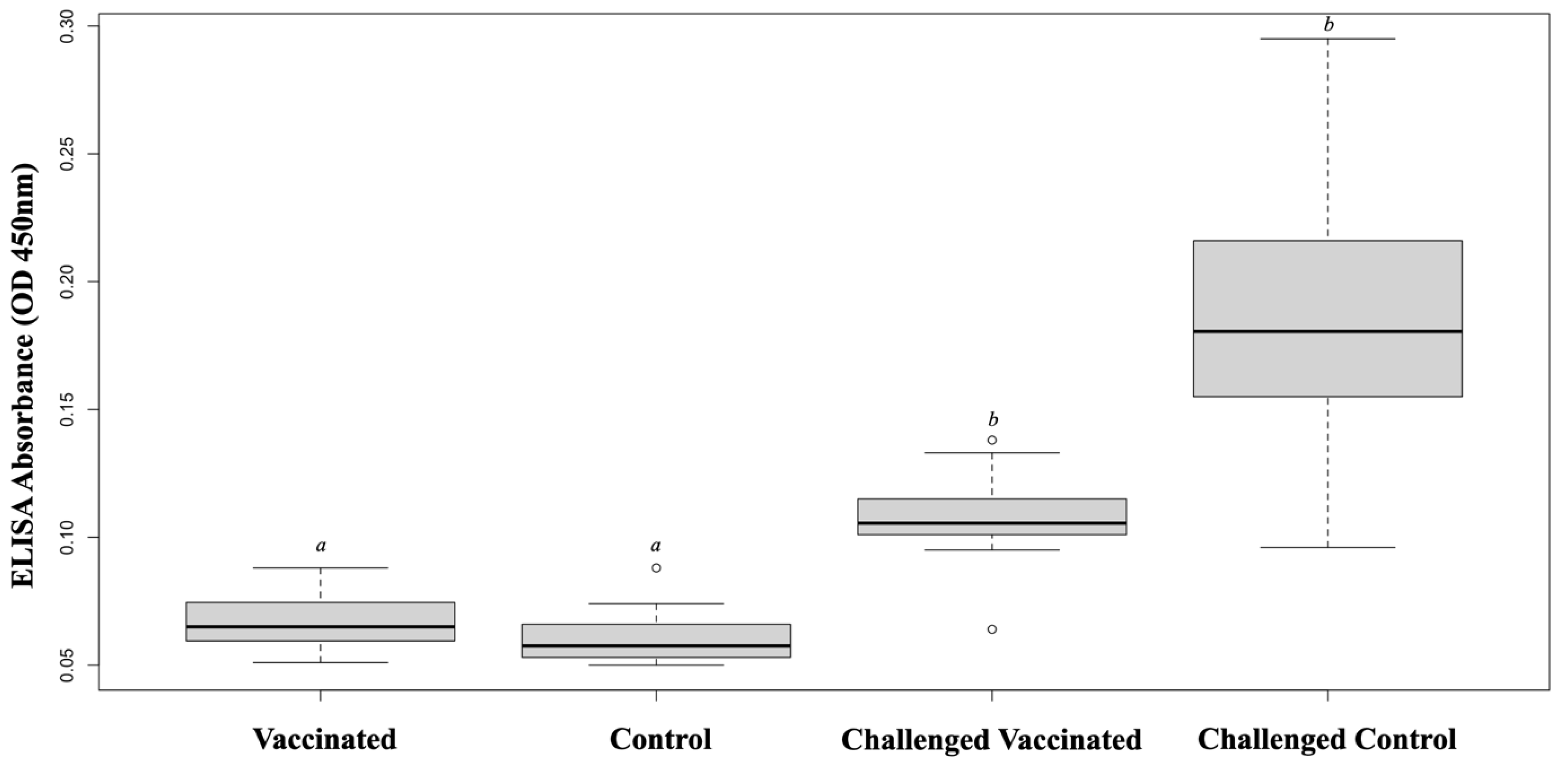
3.2. IgM Antibody Response

ELISA was used to evaluate the specific antibody response in the vaccinated and control fish at before and after challenging. ELISA absorbance values (OD 450 nm) for sera (1:1500 dilution) were 0.067 ± 0.01 and 0.060 ± 0.01 in the vaccinated and control fish, respectively. However, significantly higher ELISA values were observed in the vaccinated and control survivors compared with those in the nonchallenged groups (p < 0.05). The antibody titers of challenged survivors were not different between the vaccinated and control groups (Figure 2).

3.3. Immune-Related Gene Expression

The vaccinated fish showed significantly high CCL4 expression at 12 h and MHC II, IL-1β, and CD4 expression at 48 h (p < 0.05) (Figure 3). At 4 weeks post-vaccination, the vaccinated fish showed higher expression levels of MHC I, MHC II, IL-1β, IL-4/13B, and IL-10 at 12 h post-challenge (p < 0.05) compared with the challenged control fish. Moreover, the CD8-α gene representing cytotoxic T cells was upregulated in the challenged vaccinated fish at 24 h after challenge but was not prominent in the challenged control (p < 0.05). On the contrary, CCL4 was upregulated in the challenged control but not in the challenged vaccinated fish at 24 and 48 h (p < 0.05) (Figure 4).

4. Discussion

Inactivated vaccines or bacterins against S. iniae have been used in rainbow trout (O. mykiss) since 1997 [18,22] and have been applied via several routes, including injection, oral, and immersion. Administration via injection is generally the major route to achieve a high efficacy of vaccines, primarily because the fish can receive appropriate antigen dosage [18,33]. Klesius et al. [15] reported that for inactivated vaccines, a homologous vaccine administered via the intraperitoneal route showed better efficacy than that given via the intramuscular route, with a significantly higher relative percentage of survival (45.6% from IP route and 17.7% from IM route) for tilapia (O. niloticus). Karami et al. Ref. [34] explained that the good efficacy of IP route was because the bioavailability of the antigens was 4.5 times higher via IP route than via IM route. Thus, we focused on inactivated vaccines given via the intraperitoneal route.
In the challenge experiment, no mortality or morbidity were found among the vaccinated fish. Meanwhile, the control fish showed a 10–43.33% mortality rate with significant clinical illness and high infection rates at all time points. This finding indicated the effectiveness of S. iniae bacterin in protecting fish against disease. In addition, most of the vaccinated fish had no remnants of S. iniae at 2 weeks post-challenge, as indicated by their negative PCR result. Although some of the control fish survived, the presence of whitish feces may imply the pathological condition of their intestines that subsequently impaired their growth performance [35]. Moreover, the control survivors may become a reservoir for the pathogen because a high infection rate was prominent. Therefore, the vaccine may not only prevent mortality but also reduce disease spreading from infected individuals. Previous studies also reported the efficacy of inactivated vaccines against S. iniae in seabass. The vaccination provided 50% survivals following intraperitoneally challenge [23] and 70% survivals after 106 CFU Fish−1 intraperitoneally challenge [24]. The S. iniae bacterin has also been investigated in other fish species, including rainbow trout [22], tilapia [15], hybrid striped bass (Morone chrysops × M. saxatilis) [36], grouper [18], and red seabream (Pagrus major) [37], with more than 80% survival rate recorded in the challenged vaccinated fish. Even though inactivated vaccine might stimulate a weaker immune response than other kinds of vaccine [11], it is cost-efficient, and sufficiently protects the fish from disease throughout the culture period. As shown in our study, the formalin-killed vaccine provides up to 7 months of protection against S. iniae infection.
The susceptibility to streptococcosis caused by S. iniae may be age-dependent. The vaccinated and control fish at 7 months post-vaccination (130 ± 12 g body weight) showed no mortality upon the challenge. Múzquiz et al. [38] suggested that large rainbow trout fish can eliminate the pathogen with no mortality because of their stronger immunity response compared with the younger fish. Even though some reports showed the mass mortality of Asian seabass in grown-out farm caused by streptococcosis [3,4,39], the severity of the outbreak may be associated with disease conditions, such as water quality, density of fish, and husbandry management.
The specific antibody response against S. iniae remarkably increased in the challenged vaccinated and control survivors 1 week after challenging compared with that in the nonchallenged fish. Nevertheless, the challenged control survivors showed variations in their antibody levels, as indicated by high standard deviation due to each individual immune status. Similar to our study, the increasing antibody level of vaccinated fish after challenging was noted in Japanese flounder (Paralichthys olivaceus) receiving multivalent vaccine against S. iniae, Edwardsiella tarda, Vibrio anguillarum, and Vibrio harveyi [40]. In the present work, we observed serum antibody at 8 weeks post-vaccination because the size of the fish was suitable for blood collection. We also found a slightly higher but comparable amount of antibody titers in the vaccinated group relative to that in the control fish. However, once the fish immunity was stimulated by a challenge, the antibody levels increased immediately within 1 week. This finding may support the presence of immunoglobulin memory in the vaccinated population.
Several studies observed the alteration of immune-related genes following vaccination or challenge as a surrogate marker for protective immunity [41]. In our study, the vaccination of seabass with S. iniae bacterin activated the expression of immune genes in the pooled spleen and kidney samples. RT-PCR revealed the significant upregulation of IL-1β, MHC II, and CD4 genes in the fish at 48 h after vaccination. This phenomenon might have occurred due to the early immune response and inflammatory process in fish. The early increase in IL-1β as a pro-inflammatory cytokine induced a cascade of reactions, leading to inflammatory process, and migration of innate cellular immunity, such as neutrophils and macrophages [42,43]. MHC II is a member protein of antigen-presenting cells that interact with T cells for stimulating immune response [29]. The increased expression of these genes after vaccination might be the immunological basis of the vaccine [44]. Post-challenge, MHC I, MHC II, IL-1β, IL-4/13B, and IL-10 expression levels were significantly elevated in the vaccinated fish at 12 h, indicating the role of vaccine in stimulating the innate immune response to recognize and control the pathogen after exposure. Similar to MHC II, MHC I is also a member protein of antigen-presenting cells, and its expression might be increased upon macrophage activation. IL-4/13B is a pleiotropic cytokine secreted by T cells to regulate Th2 differentiation and mediate antibody response [43]; therefore, the increase in IL-4/13B in this study may be associated with B cell maturation and antibody production. The upregulation of IL-10, an anti-inflammatory cytokine, may serve as negative feedback to control the inflammation process following the challenge. A similar result was found in vaccinated flounder that received monovalent and divalent DNA vaccine against S. iniae and V. anguillarum and showed increasing levels of MHC I and II after challenge at 4 and 24 h, respectively [45,46]. Liu et al. [47] also showed the elevation of IL−1β after 24 h post-challenge from flounder immunized with DNA vaccine against S. iniae. Sivasankar et al. [48] observed the increased expression of IL−10 in the spleen of Asian seabass receiving inactivated vaccine against damselfish virus at 12 h to 48 h after challenge. Even though the change in gene expression level was not evident at post-vaccination compared with that at post-challenge, the potency of the vaccine in enhancing innate immune system was manifested by the prompt expression of many genes within 12 h after the pathogen invasion. Together with adaptive response, these quick responses may play an important role in keeping the fish alive.
Adaptive immunity is the main point of interest in vaccine development. At the time of our observation, the expression of adaptive immune response genes, including IgM, CD4, and CD8-α, is not yet accepted as evidence. We detected their expression within 48 h after stimulation, which may not be an appropriate time point. The expression of IgM gene in European seabass (Dicentrarchus labrax) was not detected at 48 h following Noda virus challenge [49]. The earliest response of IgM gene was recorded at 72 h after exposure to betanodavirus in European seabass [32]. Similarly, Yang et al. [50] reported an increase in CD8-α gene expression in rainbow trout at 5 days post-challenge with Yersinia ruckeri. Although the detection of the expression of adaptive immune gene was unsuccessful in our study, evidence of an adaptive immune response was demonstrated with outstanding ELISA antibody titer detected from the challenged vaccinated fish.

5. Conclusions

The formalin-killed S. iniae vaccine in this study provides substantial protection against experimental challenge and causes no mortality and significantly low infection rate in vaccinated fish. Its immunoprotective efficacy is supported by the prompt response of innate and adaptive immunities as shown by the increased antibody titer and gene expression levels. This work calls attention to the importance of vaccine application in juvenile Asian seabass for protection against severe infectious diseases.

Supplementary Materials

The following supporting information can be downloaded at: https://www.mdpi.com/article/10.3390/vaccines11020351/s1, Table S1: The immune related gene expression assessed as mean of threshold cycle (Ct) value.

Author Contributions

Conceptualization, P.T., P.P. and J.W.; Methodology, P.T. and P.P.; Validation, P.T., P.P. and J.W.; Formal Analysis, P.T. and P.P.; Investigation, P.T., P.P. and J.W.; Resources, P.T., S.C. and P.P.; Data Curation, P.T., S.S. and P.P.; Writing—original draft preparation, P.T.; Writing—review and editing, P.T., S.S., P.P. and J.W.; Visualization, P.T., S.C., P.P. and J.W.; Supervision, J.W.; Project administration, P.T., P.P. and J.W.; Funding acquisition, P.T. and J.W. All authors have read and agreed to the published version of the manuscript.

Funding

This study was funded by the 100th Anniversary Chulalongkorn University Fund for Doctoral Scholarship and the 90th Anniversary of Chulalongkorn University Fund (Ratchadaphiseksomphot Endowment Fund) Chulalongkorn University, Bangkok, Thailand.

Institutional Review Board Statement

The study was conducted according to the guideline on the design of studies to evaluate the safety and efficacy of fish vaccines, approved by the animal use ethic protocol of the Chulalongkorn University Animal Care and Use Committee (CU-ACUC: Approval No. 2231042).

Informed Consent Statement

Not applicable.

Data Availability Statement

The data that support the findings of the study are available on request from the corresponding author.

Acknowledgments

The authors would like to thank the staffs at the WiseWater Laboratory, Chachoengsao, Thailand and K. Pimsannil (Centex Shrimp) for their skilled technical assistance.

Conflicts of Interest

The authors declare no conflict of interest.

References

  1. Department of Fisheries Statistics of Marine Fish Farms SURVEY. 2020. Available online: https://www4.fisheries.go.th/local/pic_activities/202112281419571_pic.pdf (accessed on 9 November 2022).
  2. El-Noby, G.A.; Hassanin, M.; El-Hady, M.; Aboshabana, S. Streptococcus: A review article on an emerging pathogen of farmed fishes. Egypt. J. Aquat. Biol. Fish. 2021, 25, 123–139. [Google Scholar] [CrossRef]
  3. Suanyuk, N.; Sukkasame, N.; Tanmark, N.; Yoshida, T.; Itami, T.; Thune, R.L.; Tantikitti, C.; Supamattaya, K. Streptococcus iniae infection in cultured asian sea bass (Lates calcarifer) and red tilapia (Oreochromis sp.) in southern Thailand. Songklanakarin J. Sci. Technol. 2010, 32, 341–348. [Google Scholar]
  4. Bromage, E.S.; Thomas, A.; Owens, L. Streptococcus iniae, a bacterial infection in barramundi Lates calcarifer. Dis. Aquat. Org. 1999, 36, 177–181. [Google Scholar] [CrossRef] [PubMed]
  5. Fu, G.H.; Bai, Z.Y.; Xia, J.H.; Liu, F.; Liu, P.; Yue, G.H. Analysis of two lysozyme genes and antimicrobial functions of their recombinant proteins in Asian seabass. PLoS ONE 2013, 8, e79743. [Google Scholar] [CrossRef] [PubMed]
  6. Van Khang, P.; Van Nha, V.; Nguyen, N.H. Resistance to Streptococcus iniae and its genetic associations with traits of economic importance in Asian seabass (Lates calcarifer). J. Fish Dis. 2019, 42, 1657–1666. [Google Scholar] [CrossRef] [PubMed]
  7. Erfanmanesh, A.; Beikzadeh, B.; Mohseni, F.A.; Nikaein, D.; Mohajerfar, T. Ulcerative dermatitis in barramundi due to coinfection with Streptococcus iniae and Shewanella algae. Dis. Aquat. Org. 2019, 134, 89–97. [Google Scholar] [CrossRef] [PubMed]
  8. Heckman, T.I.; Griffin, M.J.; Camus, A.C.; LaFrentz, B.R.; Morick, D.; Smirnov, R.; Ofek, T.; Soto, E. Multilocus sequence analysis of diverse Streptococcus iniae isolates indicates an underlying genetic basis for phenotypic heterogeneity. Dis. Aquat. Org. 2020, 141, 53–69. [Google Scholar] [CrossRef]
  9. Piamsomboon, P.; Thanasaksiri, K.; Murakami, A.; Fukuda, K.; Takano, R.; Jantrakajorn, S.; Wongtavatchai, J. Streptococcosis in freshwater farmed seabass Lates calcarifer and its virulence in Nile tilapia Oreochromis niloticus. Aquaculture 2020, 523, 735189. [Google Scholar] [CrossRef]
  10. Kayansamruaj, P.; Dong, H.T.; Nguyen, V.V.; Le, H.D.; Pirarat, N.; Rodkhum, C. Susceptibility of freshwater rearing Asian seabass (Lates calcarifer) to pathogenic Streptococcus iniae. Aquac. Res. 2017, 48, 711–718. [Google Scholar] [CrossRef]
  11. Ma, J.; Bruce, T.J.; Jones, E.M.; Cain, K.D. A review of fish vaccine development strategies: Conventional methods and modern biotechnological approaches. Microorganisms 2019, 7, 569. [Google Scholar] [CrossRef]
  12. Nguyen, H.T.; Thu Nguyen, T.T.; Tsai, M.A.; Ya-Zhen, E.; Wang, P.C.; Chen, S.C. A formalin-inactivated vaccine provides good protection against Vibrio harveyi infection in orange-spotted grouper (Epinephelus coioides). Fish Shellfish Immunol. 2017, 65, 118–126. [Google Scholar] [CrossRef]
  13. Nor, N.A.; Zamri-Saad, M.; Yasin, I.S.M.; Salleh, A.; Mustaffa-Kamal, F.; Matori, M.F.; Azmai, M.N.A. Efficacy of whole cell inactivated vibrio harveyi vaccine against vibriosis in a marine red hybrid tilapia (Oreochromis niloticus × O. mossambicus) model. Vaccines 2020, 8, 734. [Google Scholar] [CrossRef] [PubMed]
  14. Lim, J.; Hong, S. Characterization of Aeromonas salmonicida and A. sobria isolated from cultured salmonid fish in Korea and development of a vaccine against furunculosis. J. Fish Dis. 2020, 43, 609–620. [Google Scholar] [CrossRef] [PubMed]
  15. Klesius, P.H.; Shoemaker, C.A.; Evans, J.J. Efficacy of single and combined Streptococcus iniae isolate vaccine administered by intraperitoneal and intramuscular routes in tilapia (Oreochromis niloticus). Aquaculture 2000, 188, 237–246. [Google Scholar] [CrossRef]
  16. Wang, Q.; Zhang, C.; Xu, L.; Chen, J.; Wang, X. Characterization of Streptococcus iniae ghost vaccine and its immunization in Nile tilapia (Oreochromis niloticus). Aquac. Res. 2021, 52, 1359–1368. [Google Scholar] [CrossRef]
  17. Hayat, M.; Yusoff, M.S.M.; Samad, M.J.; Razak, I.S.A.; Yasin, I.S.M.; Thompson, K.D.; Hasni, K. Efficacy of feed-based formalin-killed vaccine of streptococcus iniae stimulates the gut-associated lymphoid tissues and immune response of red hybrid tilapia. Vaccines 2021, 9, 51. [Google Scholar] [CrossRef]
  18. Huang, H.Y.; Chen, Y.C.; Wang, P.C.; Tsai, M.A.; Yeh, S.C.; Liang, H.J.; Chen, S.C. Efficacy of a formalin-inactivated vaccine against Streptococcus iniae infection in the farmed grouper Epinephelus coioides by intraperitoneal immunization. Vaccine 2014, 32, 7014–7020. [Google Scholar] [CrossRef]
  19. Membrebe, J.D.; Yoon, N.K.; Hong, M.; Lee, J.; Lee, H.; Park, K.; Seo, S.H.; Yoon, I.; Yoo, S.; Kim, Y.C.; et al. Protective efficacy of Streptococcus iniae derived enolase against Streptococcal infection in a zebrafish model. Vet. Immunol. Immunopathol. 2016, 170, 25–29. [Google Scholar] [CrossRef]
  20. Halimi, M.; Alishahi, M.; Abbaspour, M.R.; Ghorbanpoor, M.; Tabandeh, M.R. High efficacy and economical procedure of oral vaccination against Lactococcus garvieae/Streptococcus iniae in rainbow trout (Oncorhynchus mykiss). Fish Shellfish Immunol. 2020, 99, 505–513. [Google Scholar] [CrossRef]
  21. Halimi, M.; Alishahi, M.; Abbaspour, M.R.; Ghorbanpoor, M.; Tabandeh, M.R. Valuable method for production of oral vaccine by using alginate and chitosan against Lactococcus garvieae/Streptococcus iniae in rainbow trout (Oncorhynchus mykiss). Fish Shellfish Immunol. 2019, 90, 431–439. [Google Scholar] [CrossRef]
  22. Eldar, A.; Horovitcz, A.; Bercovier, H. Development and efficacy of a vaccine against Streptococcus iniae infection in farmed rainbow trout. Vet. Immunol. Immunopathol. 1997, 56, 175–183. [Google Scholar] [CrossRef] [PubMed]
  23. Mohammadi, Y.; Mesbah, M.; Dezfoulnejad, M.C.; Mehrgan, M.S.; Islami, H.R. Growth performance, blood biochemical parameters, immune response, and antioxidant defense of Asian seabass (Lates calcarifer) fingerlings exposed to monovalent and bivalent vaccines against Streptococcus iniae and Vibrio harveyi. Aquac. Int. 2021, 29, 2751–2767. [Google Scholar] [CrossRef]
  24. Thu Lan, N.G.; Salin, K.R.; Longyant, S.; Senapin, S.; Dong, H.T. Systemic and mucosal antibody response of freshwater cultured Asian seabass (Lates calcarifer) to monovalent and bivalent vaccines against Streptococcus agalactiae and Streptococcus iniae. Fish Shellfish Immunol. 2021, 108, 7–13. [Google Scholar] [CrossRef] [PubMed]
  25. Jantrakajorn, S.; Lukkana, M.; Wongtavatchai, J. Serological and molecular characterization of Streptococcus iniae in cultured Nile tilapia (Oreochromis niloticus) in Thailand. Thai J. Vet. Med. 2014, 44, 49–58. [Google Scholar]
  26. Zhou, S.M.; Fan, Y.; Zhu, X.Q.; Xie, M.Q.; Li, A.X. Rapid identification of Streptococcus iniae by specific PCR assay utilizing genetic markers in ITS rDNA. J. Fish Dis. 2011, 34, 265–271. [Google Scholar] [CrossRef]
  27. Soonthonsrima, T.; Wangman, P.; Pengsuk, C.; Sithigorngul, P.; Longyant, S. Production of Monoclonal Antibody Specific to Immunoglobulin of Asian Sea Bass Lates calcarifer. Burapha Sci. J. 2019, 24, 1237–1249. [Google Scholar]
  28. Wang, Q.; Fu, T.; Li, X.; Luo, Q.; Huang, J.; Sun, Y.; Wang, X. Cross-immunity in Nile tilapia vaccinated with Streptococcus agalactiae and Streptococcus iniae vaccines. Fish Shellfish Immunol. 2020, 97, 382–389. [Google Scholar] [CrossRef]
  29. Silvaraj, S.; Yasin, I.S.M.; Karim, M.M.A.; Saad, M.Z. Elucidating the efficacy of vaccination against vibriosis in lates calcarifer using two recombinant protein vaccines containing the outer membrane protein k (R-ompk) of vibrio alginolyticus and the dna chaperone j (r-dnaj) of vibrio harveyi. Vaccines 2020, 8, 660. [Google Scholar] [CrossRef]
  30. Mohd-Shaharuddin, N.; Mohd-Adnan, A.; Kua, B.C.; Nathan, S. Expression profile of immune-related genes in Lates calcarifer infected by Cryptocaryon irritans. Fish Shellfish Immunol. 2013, 34, 762–769. [Google Scholar] [CrossRef]
  31. Buonocore, F.; Gerdol, M.; Pallavicini, A.; Stocchi, V.; Randelli, E.; Belardinelli, M.C.; Miccoli, A.; Saraceni, P.R.; Secombes, C.J.; Scapigliati, G.; et al. Identification, molecular characterization and functional analysis of interleukin (IL)-2 and IL-2like (IL-2L) cytokines in sea bass (Dicentrarchus labrax L.). Cytokine 2020, 126, 154898. [Google Scholar] [CrossRef]
  32. Scapigliati, G.; Buonocore, F.; Randelli, E.; Casani, D.; Meloni, S.; Zarletti, G.; Tiberi, M.; Pietretti, D.; Boschi, I.; Manchado, M.; et al. Cellular and molecular immune responses of the sea bass (Dicentrarchus labrax) experimentally infected with betanodavirus. Fish Shellfish Immunol. 2010, 28, 303–311. [Google Scholar] [CrossRef] [PubMed]
  33. Bøgwald, J.; Dalmo, R.A. Review on immersion vaccines for fish: An update 2019. Microorganisms 2019, 7, 627. [Google Scholar] [CrossRef] [PubMed]
  34. Karami, A.; Christianus, A.; Ishak, Z.; Syed, M.A.; Courtenay, S.C. The effects of intramuscular and intraperitoneal injections of benzo[a]pyrene on selected biomarkers in Clarias gariepinus. Ecotoxicol. Environ. Saf. 2011, 74, 1558–1566. [Google Scholar] [CrossRef]
  35. Piamsomboon, P.; Tanpichai, P.; Wongtavatchai, J. Enteritis associated with subclinical infection of Streptococcus iniae in juvenile Asian seabass Lates calcarifer (Bloch, 1790). J. Fish Dis. 2021, 44, 1879–1882. [Google Scholar] [CrossRef]
  36. Locke, J.B.; Vicknair, M.R.; Ostland, V.E.; Nizet, V.; Buchanan, J.T. Evaluation of Streptococcus iniae killed bacterin and live attenuated vaccines in hybrid striped bass through injection and bath immersion. Dis. Aquat. Org. 2010, 89, 117–123. [Google Scholar] [CrossRef]
  37. Thanasaksiri, K.; Fukuda, K.; Tsubone, S.; Miyadai, H.; Murakami, T.; Murakami, A.; Takano, R. Efficacy of a bivalent inactivated vaccine against red seabream iridovirus and Streptococcus iniae in red seabream, Pagrus major. Aquaculture 2018, 492, 132–136. [Google Scholar] [CrossRef]
  38. Múzquiz, J.L.; Royo, F.M.; Ortega, C.; De Blas, I.; Ruiz, I.; Alonso, J.L. Pathogenicity of streptococcosis in rainbow trout(Oncorhynchus mykiss): Dependence on age of diseased fish. Bull. Eur. Assoc. Fish Pathol. 1999, 19, 114–119. [Google Scholar]
  39. Hassani, F.; Peyghan, R.; Alishahi, M.; Ghorbanpour, M.; Ahangarzadeh, M. Molecular and biochemical investigation of the role of streptococcus iniae in mortality of Lates calcarifer cages culturing in the Persian Gulf. Exp. Anim. Biol. 2021, 9, 19–27. [Google Scholar] [CrossRef]
  40. Sun, Y.; Liu, C.S.; Sun, L. A multivalent killed whole-cell vaccine induces effective protection against Edwardsiella tarda and Vibrio anguillarum. Fish Shellfish Immunol. 2011, 31, 595–599. [Google Scholar] [CrossRef]
  41. Munang’andu, H.M.; Evensen, Ø. Correlates of protective immunity for fish vaccines. Fish Shellfish Immunol. 2018, 85, 132–140. [Google Scholar] [CrossRef]
  42. Pham, T.H.; Cheng, T.C.; Wang, P.C.; Chen, S.C. Protective efficacy of four heat-shock proteins as recombinant vaccines against photobacteriosis in Asian seabass (Lates calcarifer). Fish Shellfish Immunol. 2021, 111, 179–188. [Google Scholar] [CrossRef]
  43. Reyes-Cerpa, S.; Maisey, K.; Reyes-López, F.; Toro-Ascuy, D.; Sandino, A.M.; Imarai, M. Fish Cytokines and Immune Response. In New Advances and Contributions to Fish Biology; Türker, H., Ed.; IntechOpen: Rijeka, Italy, 2012. [Google Scholar]
  44. Bedekar, M.K.; Kole, S. Fundamentals of Fish Vaccination. In Vaccine Design: Methods and Protocols, Volume 2. Vaccines for Veterinary Diseases; Thomas, S., Ed.; Springer US: New York, NY, USA, 2022; pp. 147–173. ISBN 978-1-0716-1888-2. [Google Scholar]
  45. Sun, Y.; Hu, Y.H.; Liu, C.S.; Sun, L. Construction and comparative study of monovalent and multivalent DNA vaccines against Streptococcus iniae. Fish Shellfish Immunol. 2012, 33, 1303–1310. [Google Scholar] [CrossRef] [PubMed]
  46. Sun, Y.; Zhang, M.; Liu, C.S.; Qiu, R.; Sun, L. A divalent DNA vaccine based on Sia10 and OmpU induces cross protection against Streptococcus iniae and Vibrio anguillarum in Japanese flounder. Fish Shellfish Immunol. 2012, 32, 1216–1222. [Google Scholar] [CrossRef]
  47. Liu, C.; Hu, X.; Cao, Z.; Sun, Y.; Chen, X.; Zhang, Z. Construction and characterization of a DNA vaccine encoding the SagH against Streptococcus iniae. Fish Shellfish Immunol. 2019, 89, 71–75. [Google Scholar] [CrossRef]
  48. Sivasankar, P.; John, K.R.; George, M.R.; Mansoor, M.M.; Kumar, P.M.; Selvamagheswaran, M.; Srinivasan, A.; Veerabhadran, K. Analysis of Immune Gene Expression in Seabass (Lates calcarifer) Immunized with Inactivated Vaccine against Similar Damselfish Virus. Indian J. Anim. Res. 2020, 55, 31–39. [Google Scholar] [CrossRef]
  49. Gonzalez-Silvera, D.; Guardiola, F.A.; Espinosa, C.; Chaves-Pozo, E.; Esteban, M.Á.; Cuesta, A. Recombinant nodavirus vaccine produced in bacteria and administered without purification elicits humoral immunity and protects European sea bass against infection. Fish Shellfish Immunol. 2019, 88, 458–463. [Google Scholar] [CrossRef] [PubMed]
  50. Yang, H.; Zhujin, D.; Marana, M.H.; Dalsgaard, I.; Rzgar, J.; Heidi, M.; Asma, K.M.; Per, K.W.; Kurt, B. Immersion vaccines against Yersinia ruckeri infection in rainbow trout: Comparative effects of strain differences. J. Fish Dis. 2021, 44, 1937–1950. [Google Scholar] [CrossRef]
Figure 1. Juvenile Asian seabass intraperitoneally infected with S. iniae 1010 CFU mL−1 showing bilateral lens opacity and scale loss (Left). The whitish feces from the sick fish were observed in the experimental aquaria (Right).
Vaccines 11 00351 g001
Figure 2. Serum antibody titers against S. iniae determined by ELISA at 8 weeks post-vaccination were not different between the vaccinated and control groups. Both groups showed significantly high antibody levels following a challenge with S. iniae. Significant differences (p < 0.05) are indicated with different letters.
Vaccines 11 00351 g002
Figure 3. Expression profiles of immune-related genes in the vaccinated and control fish at 12, 24, and 48 h post-vaccination (* p < 0.05).
Vaccines 11 00351 g003
Figure 4. Expression profiles of immune-related genes in challenged vaccinated survivors and challenged control survivors at 12, 24, and 48 h post-challenge (* p < 0.05).
Vaccines 11 00351 g004
Table 1. Primer sequences used for qPCR.
GenesTargetSequences (5′-3′)References
Internal control18S rRNAF: TGGTTAATTCCGATAACGAACGA
R: CGCCACTTGTCCCTCTAAGAA
Wang et al. [28]
Innate immune genesMHC IF: GGCTGTTTTTGCCGCTCTG
R: GTGGACAGGTCTGGATAAAG
Silvaraj et al. [29]
MHC IIF: GTTGGATACACTGAGTTTGG
R: GTTGGATACACTGAGTTTGG
Mohd-Shaharuddin et al. [30]
CCL4F: TCCTCGTCTACTCTGTCTGT
R: GACCTGCCACTGTCTTCAGC
Silvaraj et al. [29]
IL-1βF: ATCTGGAGGTGGTGGACAAA
R: AGGGTGCTGATGTTCAAACC
Silvaraj et al. [29]
IL-4/13BF: TCATGAAGACGCAAATCTGATGT
R: CGAGACAGGAGAACTCTTTCACACA
Buonocore et al. [31]
IL-10F: CGACCAGCTCAAGAGTGATG
R: AGAGGCTGCATGGTTTCTGT
Silvaraj et al. [29]
Adaptive immune genesIgMF: GAGCTGCAGAAGGACAGTG
R: TCAGACTGGCCTCACAGCT
Scapigliati et al. [32]
CD4F: GTGATAACGCTGAAGATCGAGCC
R: GAGGTGTGTCATCTTCCGTTG
CD8-αF: CTAAGATTCGGCAAAATAACTCGA
R: GATGAGGAGTAGAAGAGAAGGCC
Table 2. Mortality rate and infection of the vaccinated and control fish following a challenge at 4, 8, 12, 20, and 28 weeks post-vaccination.
Weeks Post-Vaccination (Bodyweight, g)GroupMortality (%)Infection
Bacterial CulturePCR Total (%)
4 (10 ±   0.7)Vaccine0000
Control13/30 (43.33)13114/17 (82.35)
8 (48 ± 3.5)Vaccine0044/30 (13.33)
Control3/30 (10.00)4610/27 (37.03)
12 (70 ± 5.8)Vaccine0123/30 (10.00)
Control8/30 (26.66)12315/22 (68.18)
20 (97 ± 9.4)Vaccine0000
Control12/30 (40.00)12214/18 (77.78)
28 (130 ± 12.6)Vaccine0000
Control0099/30 (30.00)
Disclaimer/Publisher’s Note: The statements, opinions and data contained in all publications are solely those of the individual author(s) and contributor(s) and not of MDPI and/or the editor(s). MDPI and/or the editor(s) disclaim responsibility for any injury to people or property resulting from any ideas, methods, instructions or products referred to in the content.

