Article

A Pentavalent Shigella flexneri LPS-Based Vaccine Candidate Is Safe and Immunogenic in Animal Models

by
Vladimir A. Ledov
1,2,
Marina E. Golovina
1,2,
Biana I. Alkhazova
2,3,
Vyacheslav L. Lvov
2,3,
Alexander L. Kovalchuk
1 and
Petr G. Aparin
1,2,*
1
Laboratory of Carbohydrate Vaccines, National Research Center-Institute of Immunology, Federal Medical Biological Agency of Russia, 24, Kashirskoe Shosse, 115478 Moscow, Russia
2
ATVD-TEAM Co., Ltd., 115522 Moscow, Russia
3
Laboratory of Preparative Biochemistry, National Research Center-Institute of Immunology, Federal Medical Biological Agency of Russia, 24, Kashirskoe Shosse, 115478 Moscow, Russia
*
Author to whom correspondence should be addressed.
Vaccines 2023, 11(2), 345; https://doi.org/10.3390/vaccines11020345
Submission received: 15 December 2022 / Revised: 19 January 2023 / Accepted: 31 January 2023 / Published: 3 February 2023

Abstract

:
A multivalent vaccine is much needed to achieve protection against predominant Shigella serotypes. Recently, we demonstrated the clinical applicability and immunogenic potential of tri-acylated S. flexneri 2a lipopolysaccharide (Ac3-S-LPS). Using a similar approach, we designed a pentavalent LPS candidate vaccine against S. flexneri 1b, 2a, 3a, 6, and Y (PLVF). In this study, we performed molecular and antigenic characterization of the vaccine candidate and its preclinical evaluation. There were no signs of acute toxicity after subcutaneous administration of PLVF in rabbits at a proposed human dose of 125 μg. No pyrogenic reactions and adverse effects associated with chronic toxicity after repeated administration of PLVF were revealed either. The immunization of mice with PLVF led to ≥16-fold increase in S. flexneri 1b-, 2a-, 3a-, 6-, and Y-specific antibodies. In a serum bactericidal antibody (SBA) assay, we registered 54%, 66%, 35%, 60%, and 60% killing of S. flexneri 1b, 2a, 3a, 6, and Y, respectively. In the guinea pig keratoconjunctivitis model, the efficacy was 50% to 75% against challenge with all five S. flexneri serotypes. These studies demonstrate that PLVF is safe, immunogenic over a wide range of doses, and provides protection against challenge with homologous S. flexneri strains, thus confirming the validity of pentavalent design of the combined vaccine.

1. Introduction

Dysentery is an acute infectious colitis caused by an enteric bacterial pathogen known as Shigella. Dysentery is ubiquitous. About 188 million global cases of shigellosis are reported annually [1]. The main risk groups for shigellosis are composed of children under 5 years of age, underprivileged elderly people, and travelers in endemic areas [2,3]. Shigellosis caused by Shigella flexneri is predominantly found in low- and middle-income countries [1]. Despite the high epidemiological significance of Flexner’s dysentery and progressive antibiotic resistance, a licensed vaccine against S. flexneri has not yet been developed.
Shigella vaccine development is being pursued in several directions: a live attenuated vaccine [4], a whole-cell inactivated vaccine [5], and a conjugated vaccine, including chemically conjugated or the most modern bioconjugate preparations (O-polysaccharide (O-PS) conjugated to the carrier protein, exotoxin A of P. aeruginosa, using gene engineering techniques) [6].
Live and inactivated whole-cell vaccines against S. flexneri have been immunogenic in clinical and preclinical studies [7,8,9]. However, a large number of observed adverse events and short-lived immune responses in clinical study subjects limits their regulatory approval and use in the clinic [10]. To reduce the adverse events of whole-cell vaccines, the oral route of administration [7] and/or attenuated strains have been used [4]. The main challenge for whole-cell vaccine developers is to strike a balance between reactogenicity and immunogenicity [11].
According to published clinical studies, conjugated dysentery vaccines, including bioconjugates, are safe. An increase in serum-specific antibodies of IgG and IgA classes by 2–10 times was recorded one month after the first vaccination, and levels remained unchanged for 56 days after the repeated vaccination [12].
S. flexneri can be divided into 13 serotypes: 1a, 1b, 2a, 2b, 3a, 3b, 4a, 4b, 5a, 5b, 6, X, and Y. Type antigens are determined by saccharide residues linked to the main chain. Additional sugars or conformation of the chemical structure form group antigens (serotypes X and Y lack type antigens and have only group antigens) [13]. Serotypes/serogroups isolated from approximately 90% of patients with dysentery include S. flexneri 1b, 2a, 2b, 3a, and 6 [14].
Thus, the morbidity of shigellosis caused by S. flexneri, in contrast to shigellosis caused by S. sonnei, is due to the presence of multiple serotypes of the pathogen. When developing a vaccine against shigellosis caused by S. flexneri, in addition to solving the problem of creating an effective vaccine immunogen, the choice and technological development of a set of antigenic vaccine components that provide protection against infection in a given geographic area are equally important.
Protective immunity to Shigella is induced by O-polysaccharide part of lipopolysaccharide (LPS), but LPS is highly endotoxic [15]. Therefore, the use of LPS in its native form (nLPS) as an active component of a vaccine is impossible. In an earlier study, we applied the LPS detoxification method to create a modified tri-acylated highly homogenous LPS form (Ac3-S-LPS) from S. flexneri 2a [16]. This paper describes a novel Shigella vaccine based on original pentavalent LPS preparation that, in addition to Ac3-S-LPS from S. flexneri 2a, includes four other Ac3-S-LPS compounds from S. flexneri 1b, 3a, 6, and Y (PLVF). The preclinical safety profile of PLVF was studied in rabbits. Antibody levels after immunization of mice with PLVF were assessed. Functional evaluation of anti-PLVF antibodies’ potency for lysis of virulent Shigella of selected serotypes was also performed. The protective potency of PLVF was assessed in a Sereni test.

2. Materials and Methods

2.1. Bacterial Strains and Growth Conditions

The biomass of strains of S. flexneri 1b 1818, 2a 1605, 3a 2167, 6 281-55, and Y 2643, which represented the smooth (S) form of bacteria each serotyped with a homologous Shigella serum, was obtained through fermentation using Hottinger’s broth (Nutrient Media, State Research Center for Applied Biotechnology and Microbiology, Obolensk Russia) in a 250 L fermenter (BioR 250, Prointech-bio, Pushchino, Russia) with stirring and forced aeration. Bacterial cells were separated from the liquid phase by flow centrifugation (Z-41, CEPA, Lahr, Germany). Wet cells were subsequently washed with sterile saline and water, and then lyophilized.
For serum bactericidal antibody (SBA) assay, S. flexneri 1b, 2a, 3a, 6, and Y were streaked on tryptone soy agar plates and incubated overnight at 37 °C. Then, bacterial cells were washed off plate surfaces using sterile 0.9% NaCl and further diluted in phosphate-buffered saline.

2.2. Isolation and Degradation of S-LPS

Freeze-dried bacterial cells were extracted with 45% aqueous phenol (Sigma-Aldrich, St. Louis, MO, USA) at 68–70 °C. The aqueous phase was separated, dialyzed, and lyophilized to give a crude LPS preparation. The preparation was dissolved in TRIS buffer containing 0.01% (w/w) CaCl2 and MgCl2 solution (pH 7.2). RNAse (Sigma-Aldrich, St. Louis, MO, USA) (100 μg mL−1) and DNAse (Sigma-Aldrich, St. Louis, MO, USA) (10 μg mL−1) were added, and the solution was stirred at 37 °C for 16 h. The reaction mixture was treated with proteinase K (Sigma-Aldrich, St. Louis, MO, USA) (20 μg mL−1) for 2 h at 55 °C and then dialyzed using ultrafiltration with a 50 kDa cut-off membrane (Vladisart, Vladimir, Russia), concentrated, and lyophilized to give a purified LPS preparation. S-LPS was obtained using gel-permeation chromatography on Sephadex G-150 (Sigma-Aldrich, St. Louis, MO, USA) in the presence of Na-deoxycholate (Sigma-Aldrich, St. Louis, MO, USA) as detergent. Fractions that contained S-LPS were combined and freeze-dried.
After the removal of lipid A (2% AcOH, 100 °C, 1 h), the carbohydrate portions of nLPS and S-LPS were profiled by an Agilent 1260 HPLC system with UV- and RI-detection on a TSK gel G3000PW (Toyopearl, Mainz, Germany) (7.8 mm I.D. 30 cm) column using 0.2 M NaCl elution solution and a flow rate of 0.5 mL min−1. The content of low molecular mass compounds in S-LPS did not exceed 5–10%.
Partial de-acetylation of S-LPS was performed when S-LPS (300 mg) solution was heated with stirring in 8.3% aqueous ammonia and water (100 mL) containing 100 mg of Na-deoxycholate at 30 °C for 8 h, then cooled to 5–10 °C, diluted with 200 mL of water, neutralized with AcOH, and freeze-dried. The product was treated with 100% ethanol (200 mL), the precipitate was separated by centrifugation, washed with 100% ethanol (2 × 200 mL), vacuum-dried, dissolved in water, and freeze-dried to give Ac3-S-LPS (213 mg) [17].

2.3. Production of PLVF

The candidate vaccine product was manufactured based on Ac3-S-LPS compounds from S. flexneri 1b, 2a, 3a, 6, and Y (PLVF) as the active substances at a dose of 125 μg (25 μg of each antigen compound) and contained the following formulation excipients: phenol (preservative) 0.75 mg, NaCl 4.15 mg, Na2HPO4 0.052 mg, and NaH2PO4 0.017 mg (all Sigma-Aldrich, St. Louis, MO, USA), and 0.5 mL sterile pyrogen-free water for injection [16]. The final form of PLVF product was formulated and dispensed aseptically in ampoules at a GMP-compliant manufacturing suite of vaccine enterprise of the Chumakov Institute of Poliomyelitis and Viral Encephalitides, Russian Academy of Sciences.

2.4. Chemical and Physical Analyses

SDS-PAGE was performed on a 12% acrylamide gel according to the Laemmli method using a Bio-Rad Mini-Protean electrophoresis system. Gels were stained with silver nitrate reagent (Sigma-Aldrich, St. Louis, MO, USA).
Electrospray ionization high-resolution mass spectra were recorded in the negative ion mode using a micrOTOF II instrument (Bruker Daltonics, Billerica, MA, USA). A sample (~50 ng μL−1) was dissolved in a 1:1 (v/v) water–acetonitrile mixture and sprayed at a flow rate of 3 μLmin−1. End plate offset voltage was set to 0.5 kV and capillary voltage to 4.5 kV. Drying gas temperature was 180 °C. Mass range was from m/z 50 to 3000 Da.
1H- and 13C-NMR spectroscopy were performed for solutions in 99.95% D2O at 323 K on a Bruker DRX-500 spectrometer (Bruker Daltonics, Billerica, MA, USA) using sodium3-trimethylsilylpropanoate-2,2,3,3-d4H0) and acetone (δC 31.45) as references for calibration. Prior to analysis, samples were freeze-dried from 99.5% D2O. The Bruker Topspin 2.1 program was employed to acquire and process the NMR data.

2.5. Animal Studies

The study on animal models was carried out in accordance with the ethical principles approved by the order of the Ministry of Health of the Russian Federation No. 199n from 4 January 2016. The study protocols were approved by the local ethics committees of the research organizations. Animals were housed in accredited animal facilities with free access to food and water. Before the start of a study, animals were placed in a separate room for a period of quarantine (14–21 days, depending on the animal species) and health-monitored.

2.6. Pyrogenicity

The standard method for determining the pyrogenicity is the rabbit pyrogen test (RPT). RPT was conducted on 21 adult Chinchilla rabbits (aged 3 months at the start of the experiment and weighing 2.8–3.05 g). Rabbits were randomized by weight into 7 groups. Three rabbits per group were intravenously injected with PLVF, each Ac3-S-LPS vaccine component or unmodified nLPS of S. flexneri 2a (as pyrogenicity control) at a dose of 0.025 µg kg−1.
A substance was considered apyrogenic if the cumulative temperature rise of three rabbits did not exceed 1.15 °C in accordance with the European Pharmacopoeia requirements [18]. The PLVF doze used for RPT was chosen by analogy to the WHO-approved Vi-vaccine pyrogenicity test dose of 0.025 µg kg−1.

2.7. Toxicology Study

A standard PLVF toxicity study was performed in rabbits. Chinchilla male and female rabbits were purchased from the Federal State Budgetary Institution of Science “Scientific Center for Biomedical Technologies of the Federal Medical and Biological Agency”, Russia. Acute toxicity studies were performed with a single subcutaneous injection of 125 μg/0.5 mL of PLVF (5 male and 5 female rabbits per group, aged 3 months at the start of experiment and weighing 2708 ± 71 g), followed by observation for 14 days. Animals in the control group were injected with 0.5 mL of PBS. Chronic toxicity studies were performed with daily subcutaneous immunization of rabbits with 125 μg/0.5 mL PLVF for a week (5 males and 5 females per group), followed by observation for 7 days. Animals in the control group were injected with 0.5 mL of PBS. Blood samples for hematology and biochemistry were collected before vaccination, and 7 and 14 days after vaccination. On the 14th day after vaccination, necropsy, histological examination of 16 internal organs and tissues of each rabbit, and the blood differential test were performed. The local irritation was studied by histopathological evaluation of the site of repeated subcutaneous injection of the vaccine preparation (macro- and microscopic description of the skin at the injection site).
Tissue samples for histological examination were fixed in 10% neutral buffered formalin, dehydrated in ascending concentrations of alcohol, and embedded in paraffin. Paraffin sections 5 μm thick were cut on a SM 2000R microtome (Leica, Wetzlar, Germany), stained with hematoxylin and eosin, and examined using a DM1000 microscope (Leica, Wetzlar, Germany).
The study of acute and chronic toxicity, as well as the study of the local tolerance of PLVF, was carried out in accordance with the recommendations and the requirements of local legislation (Federal Law of 12.04.10 N 61 “On the Circulation of Medicines”).

2.8. Immunochemical Identification of PLVF

We utilized standard sandwich ELISA with monovalent rabbit antisera against type antigens I, II, III, and VI, and group antigen 3,4 (Agnolla®, SPbSRIVS, Saint Petersburg, Russia) and PLVF-coated microwells for identification of PLVF components 1b, 2a, 3a, 6, and Y, respectively, and intact S. flexneri S-LPS 1b-, 2a-, 3a-, 6-, and Y-coated microwells for control antigenic activity.

2.9. Immunogenicity in Mice

Sixty (CBA × C57BL/6) F1 female mice (5 mice per group, 8-weeks-old at the start of the experiment and weighing 18 ± 0.3 g) were purchased from the Federal State Budgetary Institution of Science “Scientific Center for Biomedical Technologies of the Federal Medical and Biological Agency”, Russia and immunized intraperitoneally with 125 μg per mouse of PLVF or with two doses of 25 μg and 50 μg of Ac3-S-LPS 1b, 2a, 3a, 6, and Y. Two weeks after the primary injection, the mice were boosted with the same dose. At day 15 after the secondary immunization, serum samples were collected and levels of LPS-specific total IgG and IgM were evaluated by ELISA (using a standard protocol) with native S. flexneri LPS (nLPS) 1b, 2a, 3a, 6, and Y adsorbed on microplates (Greiner, Kremsmünster, Austria). Control animals were given saline.

2.10. SBA Assay

Heat-inactivated mouse serum samples were diluted 1:3000 in PBS and 100 µL were added into 96-well U-bottom plates (Medpolymer, Saint Petersburg, Russia). A 100 µL aliquot containing 104 CFU of each S. flexneri serotype 1b, 2a, 3a, 6, and Y separately and 25 µL of guinea pig complement (made in-house) were added into the wells. The final volume was 300 µL and serum dilution 1:9000. Plates were incubated for 1.5 h at 37 °C in a shaker at 200 rpm. Serum bactericidal activity was assessed individually for each mouse. The assay included complement control wells containing S. flexneri bacteria with guinea pig complement with no serum. This complement control was used to define 0% killing in the SBA killing rate calculation.
The percentage of bacteria killing (SBA rate) was determined by the equation [1- (colony forming units (CFU) of surviving bacteria/total CFU)] × 100.

2.11. Efficacy Evaluation of PLVF

Male (Agouti strain) guinea pigs were purchased from the Federal State Budgetary Institution of Science “Scientific Center for Biomedical Technologies of the Federal Medical and Biological Agency”, Russia. To examine the PLVF for eye protection against virulent S. flexneri stains, groups of 10 guinea pigs (aged 3 months at the start of the experiment and weighing 275 ± 3 g) were twice immunized subcutaneously dorsally with a dose of 125 µg of vaccine at an interval of 10 days. Control animals were given saline. Ten days after the last immunization, S. flexneri keratoconjunctivitis was induced in experimental and control groups of animals by inoculation into the mucosal surface of the conjunctiva of both eyes with a suspension of a virulent strain at a dose of 2 × 10⁹ cells in 30 µL of sterile saline. Each group was inoculated with one of S. flexneri 1b, 2a, 3a, 6, and Y serotypes. Keratoconjunctivitis was assessed 7 days after challenge by visual inspection. The efficacy of PLVF was calculated by the formula: PLVF Efficacy = 100 × (Control attack rate – PLVF attack rate)/Control attack rate, where attack rate = number of infected eyes/total eyes [4].

2.12. Statistical Analysis

Statistical analyses of immunogenicity data were performed using GraphPad Prism 7 software (GraphPad Software Inc., La Jolla, CA, USA). For statistical significance while comparing groups of mice, the Student’s t test was performed. Statistical significance was defined as p < 0.05.

3. Results

3.1. Chemical Structure and Specific Epitope Identification of PLVF Compounds

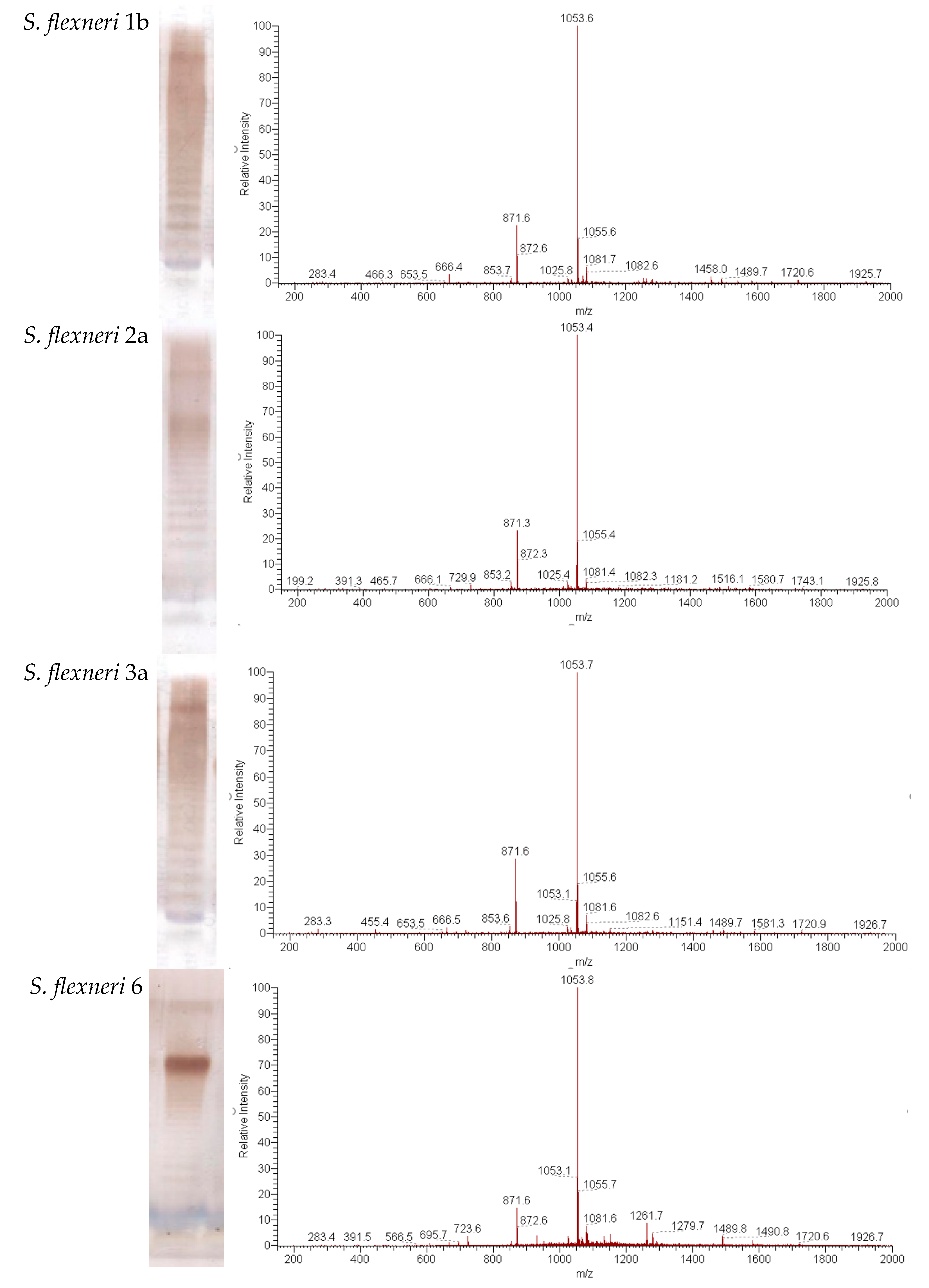
PLVF contains modified long-chain S-LPS compounds with a length of O-PS chain of 15–25 repeating units (Figure 1a). Mass spectrum analysis demonstrates that lipid A of 1b, 2a, 3a, 6, and Y represents highly homogenous Ac3-S-LPS and contains mainly three (the highest peak at m/z = 1053) residues of unsaturated fatty acids (Figure 1b). It should be noted that no peaks for penta- and hexa-acyl derivatives were present in the mass spectra of the modified lipid A from S. flexneri serotypes 1b, 2a, 3a, 6, and Y.
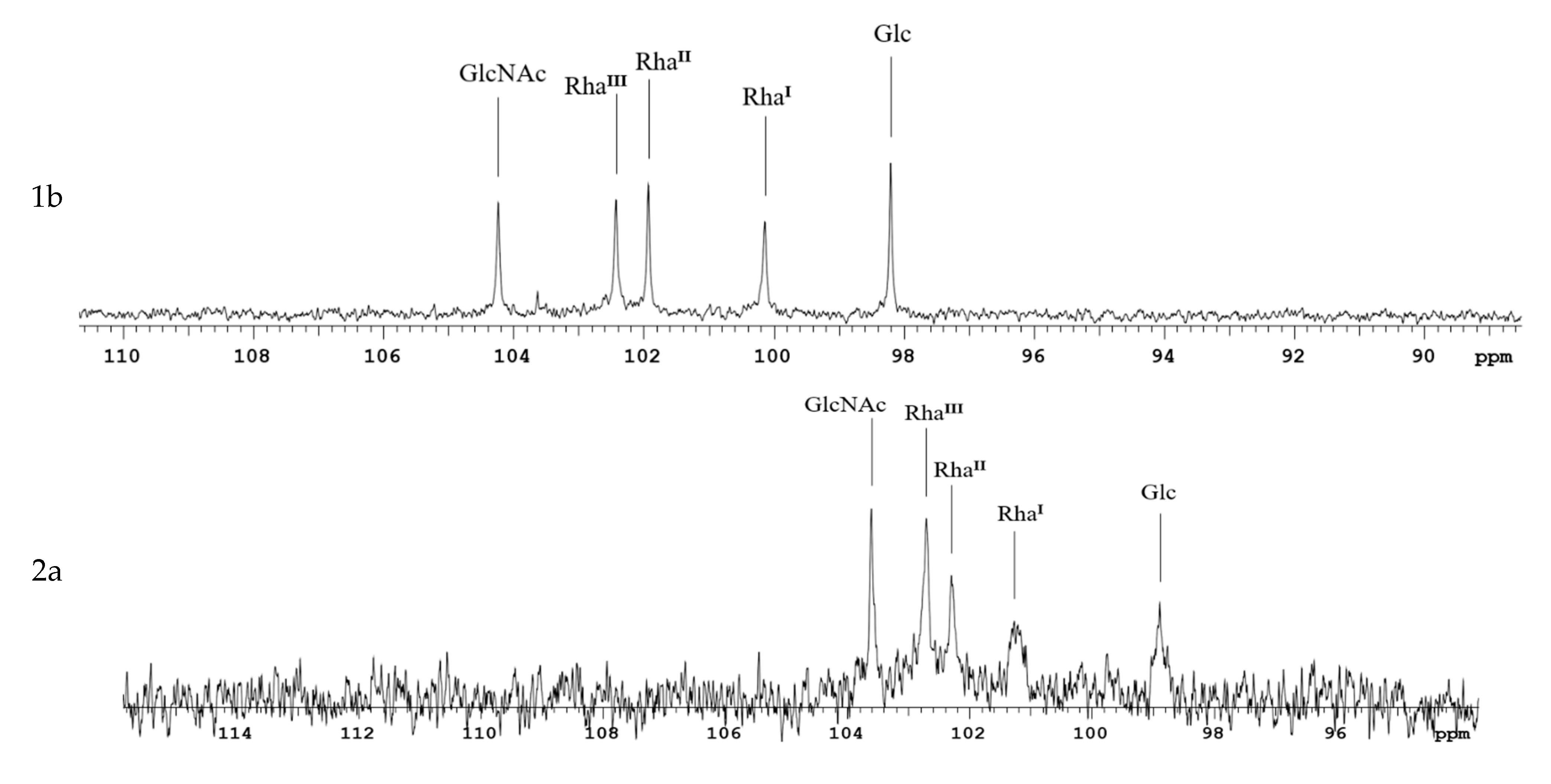
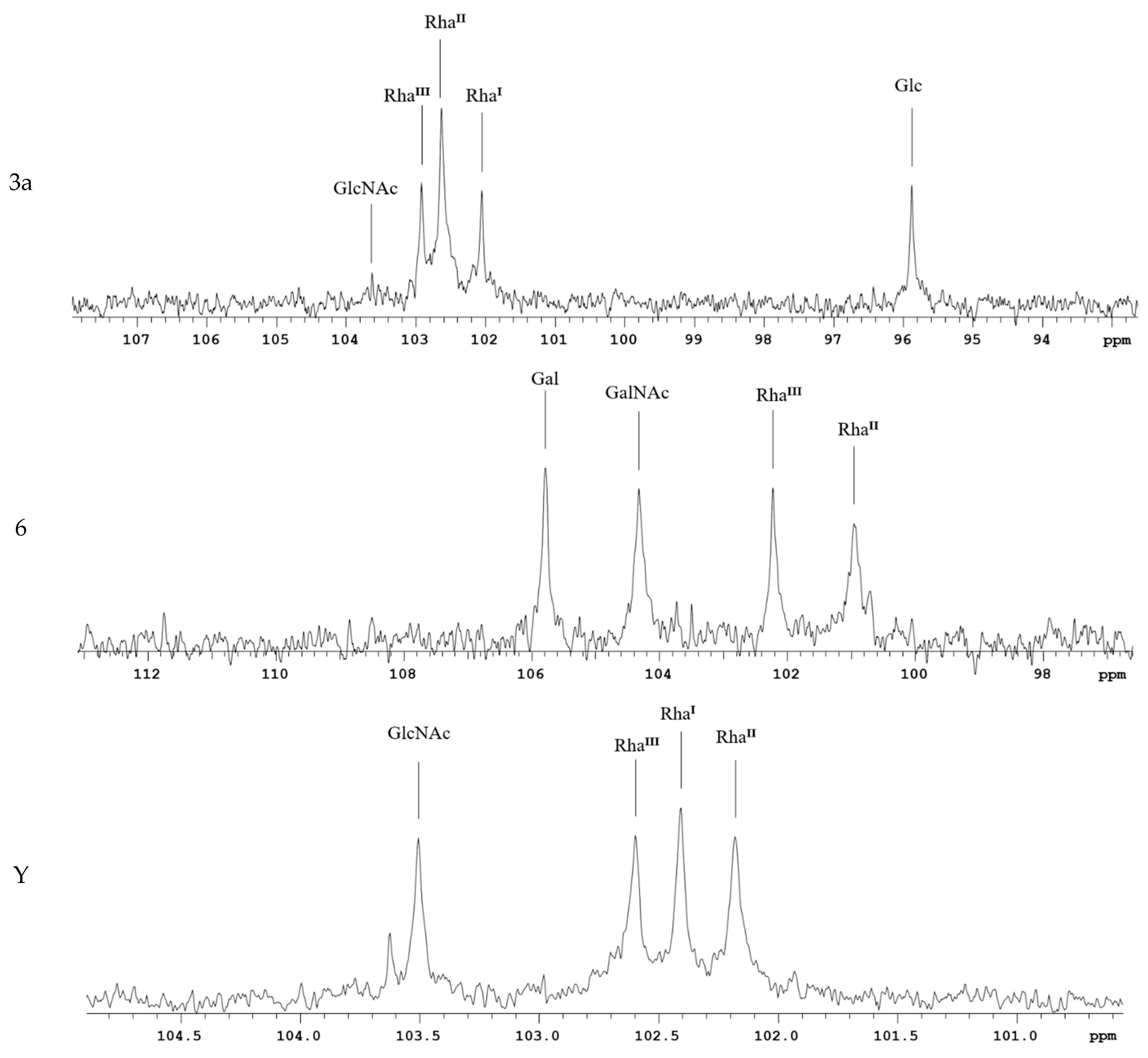
The characteristic signals in the anomeric carbon region of the 13C NMR spectra of S. flexneri Ac3-S-LPS do not overlap and are a distinctive feature for each serotype, notably the characteristic rhamnose signals associated with different degrees and positions of O-acetylation. The shifts of anomeric protons were analyzed by comparing the NMR spectra of S. flexneri 1b, 2a, 3, Y, and 6 Ac3-S-LPS with the literature data (Figure 2).
For immunochemical detection of PLVF components 1b, 2a, 3a, 6, and Y, we used monovalent Shigella antisera against type-specific antigens I, II, III, and VI, and group-specific antigen 3,4, respectively. Each Ac3-S-LPS component in PLVF composition was antigenic (or serologically active) and reacted with the corresponding specific antisera, thus confirming the presence of type- or group-specific epitopes (Figure 3). The serological activity of Ac3-S-LPS components of the vaccine was comparable to that of nLPS of the respective serotypes.

3.2. Pyrogenicity

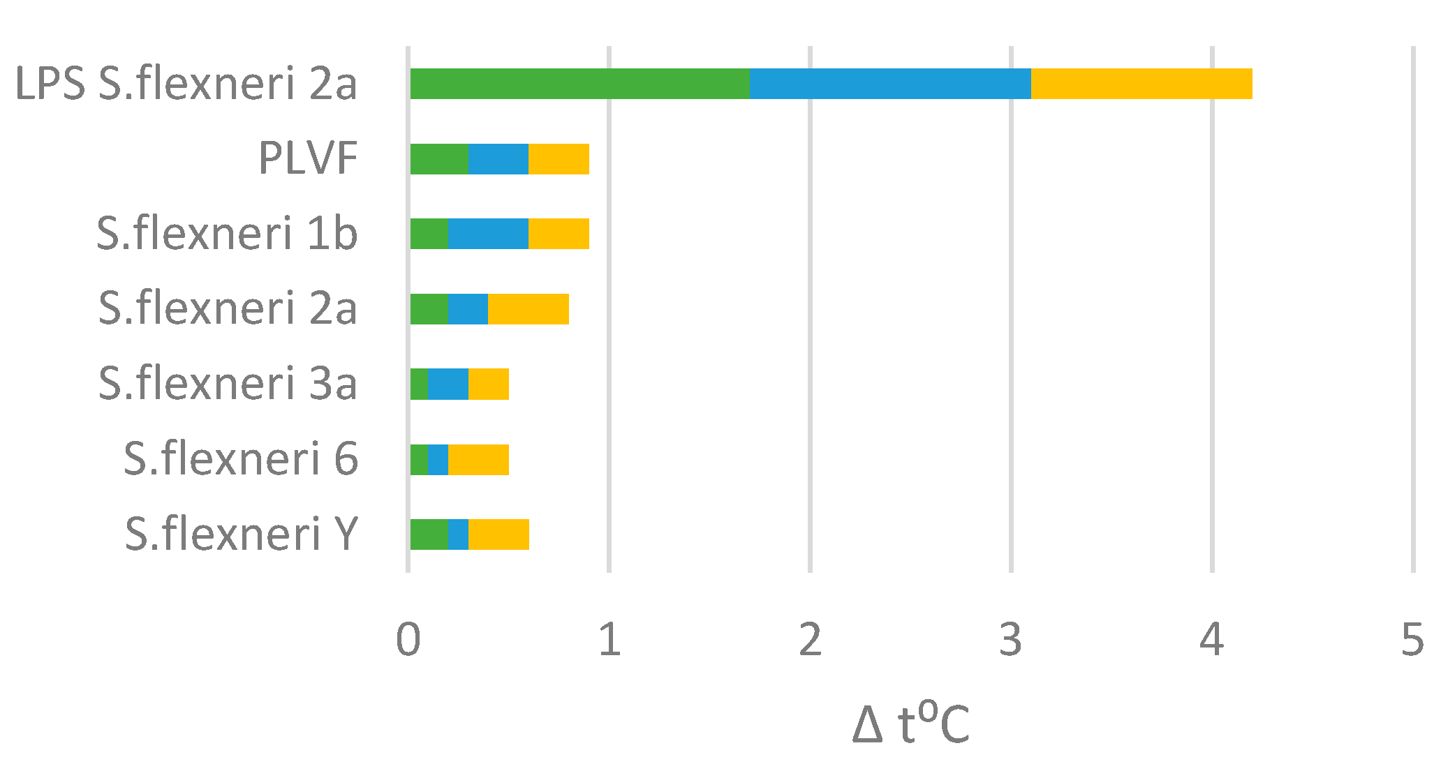
Pyrogenicity of PLVF or PLVF’s individual components 1b, 2a, 3a, 6, and Y was examined in rabbits and compared with S. flexneri nLPS. PLVF (∑Δ t °C = 0.9 °C) or PLVF’s components were apyrogenic − ∑Δ t °C < 1.15 °C when injected intravenously into rabbits (Figure 4). At the same time, unmodified nLPS induced a strong pyrogenic reaction in rabbits − ∑Δt °C (nLPS) = 4.2 °C.

3.3. Immunogenicity in Mice

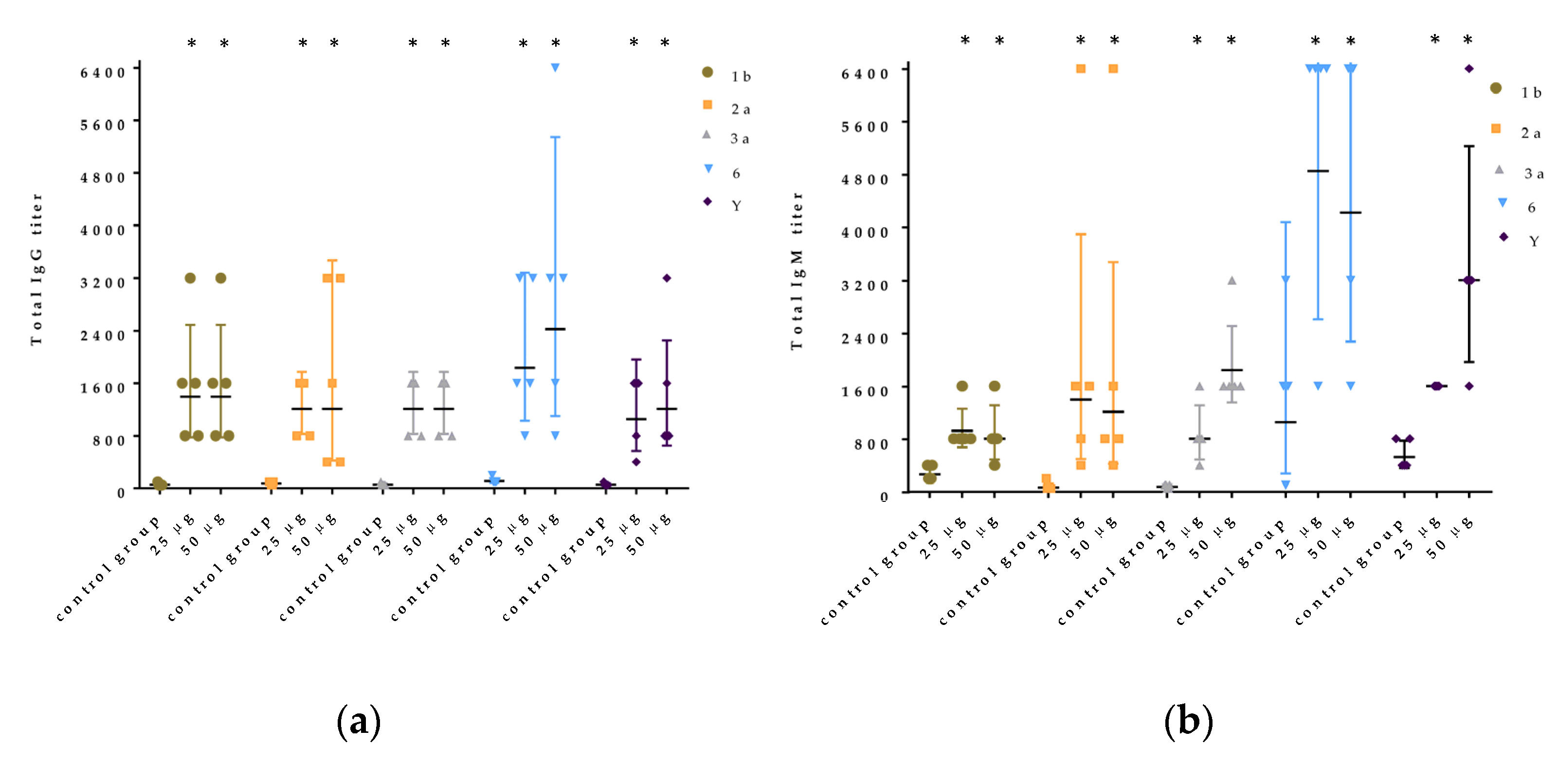
In all groups of mice immunized with Ac3-S-LPS 1b, 2a, 3a, 6, and Y, a significant increase in the titer of specific IgG and IgM was observed compared with the intact animals. The antibody response to five Ac3-S-LPS compounds did not change with an increase in immunization dose from 25 μg to 50 μg (Figure 5a,b). The immunogenicity in mice of low-endotoxic Ac3-S-LPS from S. flexneri 1b, 2a, 3a, and Y was almost equal. The level of serum antibody response in mice immunized with Ac3-S-LPS serotype 6 was higher.
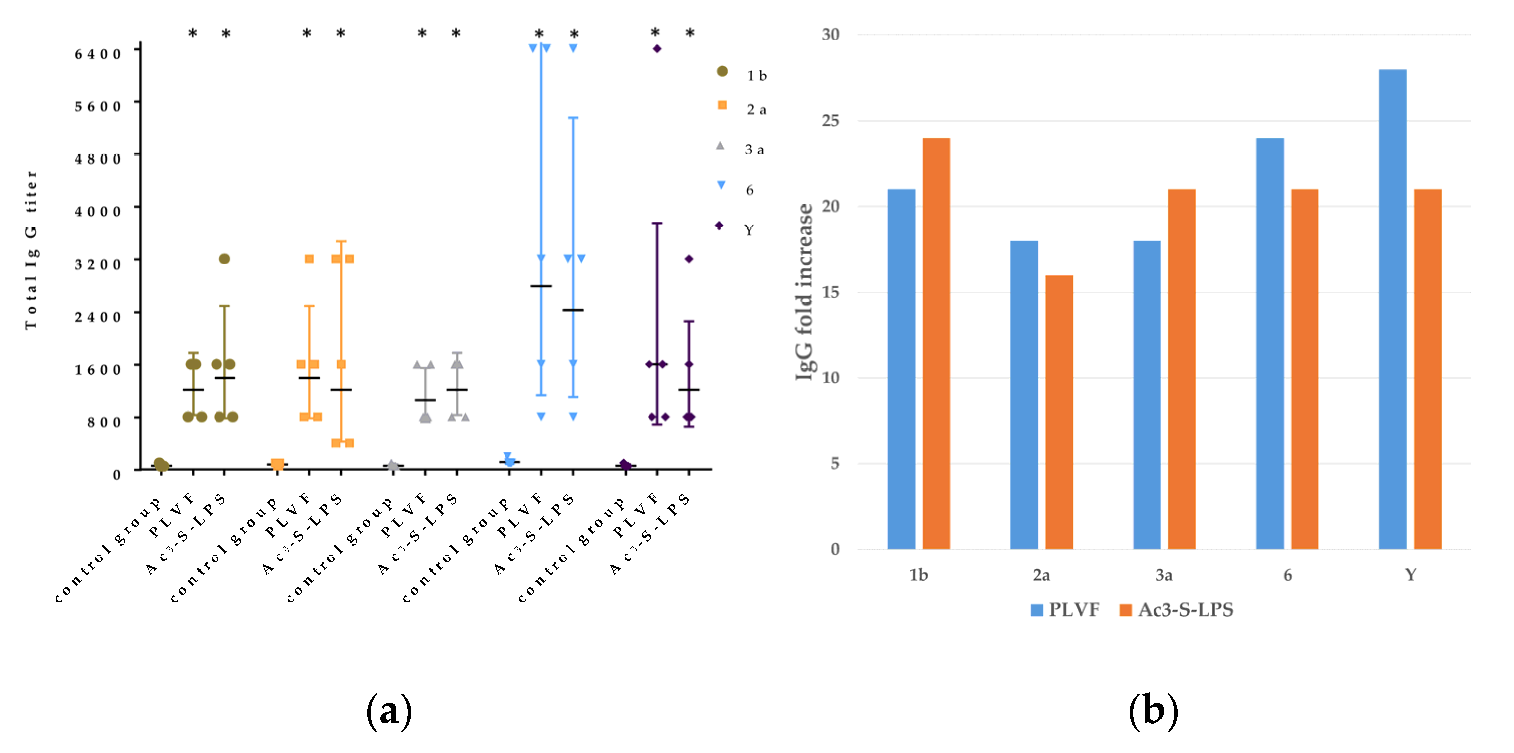
In the group of mice immunized with PLVF, an increase in the titer of specific IgG antibodies was also observed (p ≤ 0.05). Titers of IgG to LPS S. flexneri 1b, 2a, 3a, Y, and 6 were 1213 ± 438, 1393± 980, 1056± 438, 2786 ± 2629, and 1600 ± 2360, respectively (Figure 6a).
In the groups of mice immunized with only one component of PLVF, the titer of IgG was 1393 ± 980 for 1b, 1213 ± 1403 for 2a, 1213 ± 438 for 3a, 2425 ± 2147 for 6, and 1213 ± 1043 for Y. These results were not significantly different from those in the group of mice immunized with PLVF. At the same time, titers in both experimental groups were at least 16 times higher than in the control group (Figure 6b). We did not register the occurrence of antigenic competition and suppression of the response when closely related polysaccharide antigens were combined into a single preparation.
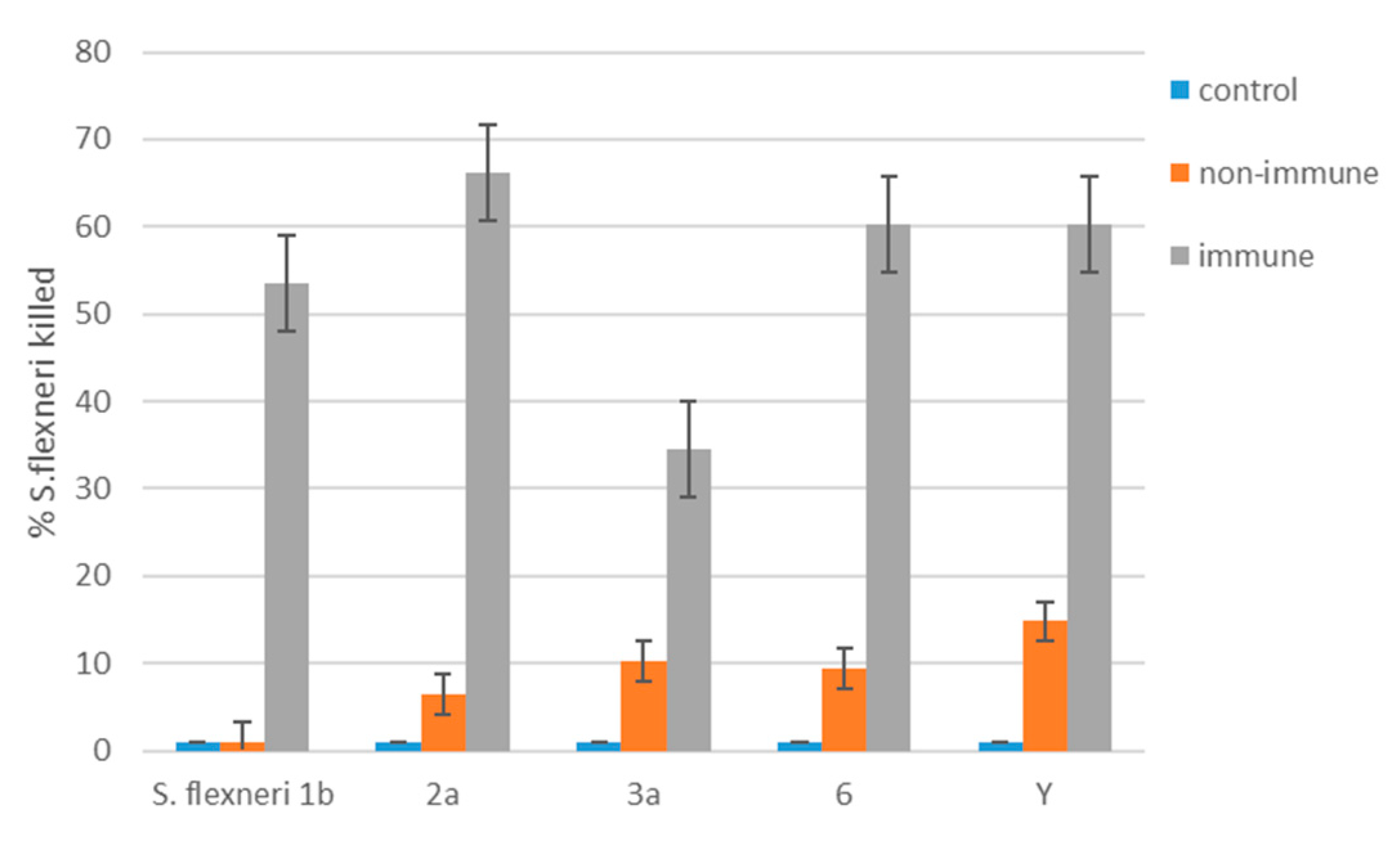
Next, we evaluated the functional activity of S. flexneri 1b-, 2a-, 3a-, 6-, and Y-specific antibodies in sera from mice immunized with PLVF at a dose of 125 μg using the SBA assay. Insignificant killing (<15%) of S. flexneri of different serotypes was observed using non-immune sera (Figure 7). After PLVF immunization, we registered significant rises in mouse immune sera bactericidal activity against virulent S. flexneri strains. The estimated killing rate was 54%, 66%, 35%, 60%, and 60% for S. flexneri 1b, 2a, 3a, 6, and Y serotype/serogroup, respectively. These data directly demonstrate the ability of modified S-LPS from different strains of S. flexneri to act as an effective protective antigen.

3.4. PLVF Protects against Keratoconjunctivitis in a Guinea Pig Model after Separate Infection with Each of S. flexneri 1b, 2a, 3a, 6, and Y Serotypes

To protect against conjunctivitis, guinea pigs were immunized with PLVF twice subcutaneously with an interval of 10 days. On day 10 after the last immunization, groups of animals were challenged with ID80 of virulent strains of S. flexneri 1b, 2a, 3a, 6, and Y. On day seven after infection, visual assessment showed that in groups of immunized animals the efficacy was 69%, 75%, 50%, 50%, and 69% against S. flexneri serotypes 1b, 2a, 3a, 6, and Y, respectively (Figure 8). In unimmunized control animals, at least 80% of the eyes were infected depending on the serotype of the virulent strain of S. flexneri.

3.5. Toxicology Study

To assess the general toxicity, the effect of the vaccine preparation was studied in rabbits using single and repeated subcutaneous administration. The results of the acute toxicity study showed that PLVF was non-toxic at a dose of 125 μg. Animal mortality was not observed, there were no signs of toxicity, and there were no changes in body weight or hematological and biochemical tests. No pathological changes in bone marrow hematopoiesis were observed in all groups. There were no significant histological differences found in organs from PLVF and control group (Table S1, Figure S1).
The results of the chronic toxicity study showed that daily subcutaneous application of PLVF to rabbits did not cause disturbances in the functional state of the main organs and systems of the body.
There was no local irritating effect in the area of PLVF administration after repeated subcutaneous application to rabbits.

4. Discussion

Flexner’s shigellosis represents a longstanding, difficult public health problem in many countries around the world. The achievement of field protection after vaccination against S. flexneri is an extremely difficult task.
Shigella-specific antibodies play an important role in promoting host defense against shigellosis. Children and adults living in areas endemic for shigellosis develop circulating antibody secreting cells and serum antibodies specific for Shigella LPS and invasion plasmid antigen. Sera of volunteers infected with S. flexneri 2a have a pronounced bactericidal efficacy [19].
Recent studies of functional antibodies after immunization with protein-lipopolysaccharide outer membrane vesicles (OMV) S. sonnei vaccine based on generalized modules for membrane antigen (GMMA) have shown that anti-LPS antibodies are the main drivers of bactericidal activity. On the contrary, anti-protein antibodies had limited ability to either bind to Shigella cells or kill them in the presence of complement [20].
In modern LPS-enriched Shigella vaccines (InvaplexAR-Detox, GMMA) genetically modified Shigella strains (ΔmsbB orΔhtrB) are being used to prevent production of the most endotoxic form of LPS with hexa-acylated lipid A domain [21,22]. However, in these vaccines, LPS remains heterogeneous like classical endotoxin and contains penta- and tetra-acylated lipid A and, therefore, is still relatively endotoxic and pyrogenic. As a result, the doses of preparations containing genetically modified LPS proposed for the safe parenteral administration by the authors of the abovementioned studies are relatively low (in the range of 0.1–15 μg). Additional fractions of protein antigens, Ipa molecules (InvaplexAR-Detox) or OMV proteins (GMMA), were also present in these vaccine preparations. Further studies aiming to increase the content of LPS, and thus change the design of GMMA-based vaccines to enhance their immunogenicity and efficacy, have been announced [20].
Our previous studies have shown that Ac3-S-LPS is apyrogenic and is the most immunogenic form of S. flexneri 2a S-LPS [16]. As we have already demonstrated the possibility of safe clinical use of S. flexneri 2a Ac3-S-LPS, it was reasonable to use this approach to obtain Ac3-S-LPS components of other serotypes/serogroups for the development of PLVF. Therefore, PLVF was designed to include in the vaccine dose the maximum number of S. flexneri antigens. Thus, PLVF contains five Ac3-S-LPS of S. flexneri, namely 1b, 2a, 3a, 6, and Y.
Each Ac3-S-LPS is a macromolecule, and at the same time is a full-fledged bioconjugate vaccine. In this respect, the O-PS conjugate is naturally produced by the bacterial cell and contains a built-in adjuvant-lipid A domain, the structure of which is deacylated to an apyrogenic form. The low content of pyrogens in PLVF preparation was confirmed using the rabbit pyrogenicity test. Preclinical toxicology studies in rabbits demonstrated the complete absence of signs of acute and chronic toxicity, local irritant action, damage to internal organs, and changes in biochemical and hematological parameters when PLVF was administered at a dose of 125 μg in 0.5 mL intended for human administration.
Thus, we achieved a significant extension of the dose range for a single parenteral administration to animals of Ac3-S-LPS as an active substance of PLVF to over 100 μg. Under the chronic toxicity regimen, multiple administration up to 875 μg of Ac3-S-LPS did not cause any endotoxic effects. These data once again demonstrate the critical difference between Ac3-S-LPS and natural endotoxin, the safe parenteral dose of which is sharply lower—just a few nanograms.
We used SDS-PAGE and mass spectrometry to determine the structure of Ac3-S-LPS of S. flexneri 1b, 2a, 3a, 6, and Y. Ac3-S-LPS from PLVF contains the long-chain S-LPS (O-PS chain length is about 20 repeating units) and has a triacylated lipid A. It should be emphasized that such vaccine grade Ac3-S-LPS is completely free from the most endotoxic highly acylated molecules of LPS—its hexa- and penta-acylated derivatives. The structure of the polysaccharide component of Ac3-S-LPS was determined using 13C NMR and was distinct for each serotype.
Each Ac3-S-LPS of PLVF was identified using a homologous monovalent antiserum. The serological properties of PLVF components and nLPS of the same S. flexneri serotypes were similar. Thus, the methods used to obtain Ac3-S-LPS did not cause the alteration of antigenic determinants.
In double immunized mice, we have registered high IgG levels (16-28-fold rise) to all components of PLVF without an aluminum hydroxide adjuvant. High immune response in mice correlated with protection against dysentery eye infection in guinea pigs. We have registered 50–75% efficacy against dysenteric keratoconjunctivitis after double immunization of guinea pigs. In a recent paper [21], a 75% protection rate in the S. flexneri 2a Sereny test after triple subcutaneous immunization with Invaplex AR-Detox vaccine was reported. Thus, in addition to the 2a component, the simultaneous induction of a systemic immune response in mice and mucosal protection in guinea pigs has now been confirmed for four more S. flexneri O-antigens.
The correctness of the choice of Ac3-S-LPS with long-chain OPS as a protective antigen of PLVF was confirmed by the presence in the sera of mice immunized with the vaccine of significant amounts of bactericidal antibodies to each of the homologous virulent strains of S. flexneri, serotypes 1b, 2a, 3a, 6, and Y.
The results of this preclinical study substantiated a wide dose interval for the safe administration of the candidate vaccine product and, thus, successfully validated the design of a pentavalent vaccine combination. If the goal of protection in the field against Flexner’s shigellosis using Ac3-S-LPS vaccine is achieved, the problem of comprehensive protection against shigellosis may be solved by the combined use of PLVF and the first available vaccine against Sonne’s shigellosis. This vaccine is based on S. sonnei exopolysaccharide, which elicits protective antibodies, and has been successfully used for prophylactic immunization for 15 years. In addition, the comprehensive data on the safety of PLVF offers the possibility of extending our vaccine construction strategy to various serotypes of Enterobacteriaceae.
Supplementary Materials

The following supporting information can be downloaded at: https://www.mdpi.com/article/10.3390/vaccines11020345/s1, Figure S1: Hematoxylin and eosin stained sections of the internal organs of rabbits from the control group (a) and 7 days after the last injection of PLVF (b). PLVF was administered subcutaneously at a dose of 125 µg daily for a week. Representative images from 5 groups; Figure S2: Chemical structure of the repeating unit of the O-specific polysaccharide chain of S. flexneri serotypes 1b, 2a, 3a, 6, and Y; Table S1: Histopathological changes of internal organs after single or multiple subcutaneous injections with 125 µg dose of PLVF.

Author Contributions

V.A.L., M.E.G., B.I.A., V.L.L., A.L.K. and P.G.A.—conceptualization; V.A.L., M.E.G., B.I.A. and V.L.L.—methodology; V.A.L.—formal analysis; V.A.L., M.E.G., B.I.A. and V.L.L.—investigation; V.A.L., A.L.K. and P.G.A.—data curation; V.A.L., A.L.K. and P.G.A.—writing, original draft preparation; V.A.L., A.L.K. and P.G.A.—writing, review and editing. All authors have read and agreed to the published version of the manuscript.

Funding

This study was funded in part by the government grant No. 14.N08.11.2009 for carrying out research and development, based on the decision of the Competition Commission of the Ministry of Education and Science of the Russian Federation No. 2013-14-241/01 (protocol dated July 26, 2013 No. 0173100003713000245-3-03).

Institutional Review Board Statement

Studies on animal models were carried out in accordance with the ethical principles approved by the order of the Ministry of Health of the Russian Federation No. 199n of 04/01/2016. The study protocols were approved by the local ethical committees of the organizations conducting the studies.

Informed Consent Statement

Not applicable.

Data Availability Statement

The data presented in this study are available on request from the corresponding author.

Acknowledgments

We are grateful to Olesia I. Naumenko for assistance in data presentation.

Conflicts of Interest

V.A.L., M.E.G., V.L.L., and P.G.A. are inventors on a US patent US20180043004A1, entitled “Modified endotoxic bacteria lipopolysaccharide (variants), combination of modified lipopolysaccharides (variants) and, containing same, a vaccine (variants) and a pharmaceutical composition (variants)”. All other authors declare no competing interest.

References

  1. Kotloff, K.L.; Riddle, M.S.; Platts-Mills, J.A.; Pavlinac, P.; Zaidi, A.K. Shigellosis. Lancet 2018, 391, 801–812. [Google Scholar] [CrossRef] [PubMed]
  2. Kotloff, K.L.; Nataro, J.P.; Blackwelder, W.C.; Nasrin, D.; Farag, T.H.; Panchalingam, S.; Wu, Y.; Sow, S.O.; Sur, D.; Breiman, R.F.; et al. Burden and aetiology of diarrhoeal disease in infants and young children in developing countries (the Global Enteric Multicenter Study, GEMS): A prospective, case-control study. Lancet 2013, 382, 209–222. [Google Scholar] [CrossRef] [PubMed]
  3. Shah, N.; Ldupont, H.; Ramsey, D.J. Global etiology of travelers’ diarrhea: Systematic review from 1973 to the present. Am. J. Trop. Med. Hyg. 2009, 80, 609–614. [Google Scholar] [CrossRef] [PubMed]
  4. DeLaine, B.O.C.; Wu, T.; Grassel, C.L.; Shimanovich, A.; Pasetti, M.F.; Levine, M.M.; Barry, E.M. Characterization of a multicomponent live, attenuated Shigella flexneri vaccine. Pathog. Dis. 2016, 74, ftw034. [Google Scholar] [CrossRef] [PubMed]
  5. Pastor, Y.; Camacho, A.I.; Zúñiga-Ripa, A.; Merchán, A.; Rosas, P.; Irache, J.M.; Gamazo, C. Towards a subunit vaccine from a Shigella flexneri ΔtolR mutant. Vaccine 2018, 36, 7509–7519. [Google Scholar] [CrossRef] [PubMed]
  6. Ravenscroft, N.; Braun, M.; Schneider, J.; Dreyer, A.M.; Wetter, M.; Haeuptle, M.A.; Kemmler, S.; Steffen, M.; Sirena, D.; Herwig, S.; et al. Characterization and immunogenicity of a Shigella flexneri 2a O-antigen bioconjugate vaccine candidate. Glycobiology 2019, 29, 669–680. [Google Scholar] [CrossRef] [PubMed]
  7. Mel, D.M.; Terzin, A.L.; Vuksić, L. Studies on vaccination against bacillary dysentery. 3. Effective oral immunization against Shigella flexneri 2a in a field trial. Bull. World Health Organ. 1965, 32, 647–655. [Google Scholar] [PubMed]
  8. Oeding, P. Vaccination against bacillary dysentery. Nord. Med. 1966, 76, 1068. [Google Scholar] [PubMed]
  9. Chakraborty, S.; Harro, C.; DeNearing, B.; Bream, J.; Bauers, N.; Dally, L.; Flores, J.; Van De Verg, L.; Sack, D.A.; Walker, R. Evaluation of the safety, tolerability, and immunogenicity of an oral, inactivated whole-cell shigella flexneri 2a vaccine in healthy adult subjects. Clin. Vaccine Immunol. 2016, 23, 315–325. [Google Scholar] [CrossRef] [PubMed]
  10. Levine, M.M.; Gangarosa, E.J.; Barrow, W.B.; Morris, G.K.; Wells, J.G.; Weiss, C.F. Shigellosis in Custodial Institutions. IV. In Vivo Stability and Transmissibility of Oral Attenuated Streptomycin-Dependent Shigella Vaccines. J. Infect. Dis. 1975, 131, 704–707. [Google Scholar] [CrossRef]
  11. Chen, W.H.; Kotloff, K.L. Shigella vaccine development: Finding the path of least resistance. Clin. Vaccine Immunol. 2016, 23, 904–907. [Google Scholar] [CrossRef]
  12. Riddle, M.S.; Kaminski, R.W.; Di Paolo, C.; Porter, C.K.; Gutierrez, R.L.; Clarkson, K.A.; Weerts, H.E.; Duplessis, C.; Castellano, A.; Alaimo, C.; et al. Safety and immunogenicity of a candidate bioconjugate vaccine against Shigella flexneri 2a administered to healthy adults: A single-blind, randomized phase i study. Clin. Vaccine Immunol. 2016, 23, 908–917. [Google Scholar] [CrossRef]
  13. Van De Verg, L.L.; Bendiuk, N.O.; Kotloff, K.; Marsh, M.M.; Ruckert, J.L.; Puryear, J.L.; Taylor, D.N.; Hartman, A.B. Cross-reactivity of Shigella flexneri serotype 2a O antigen antibodies following immunization or infection. Vaccine 1996, 14, 1062–1068. [Google Scholar] [CrossRef] [PubMed]
  14. Livio, S.; Strockbine, N.A.; Panchalingam, S.; Tennant, S.M.; Barry, E.M.; Marohn, M.E.; Antonio, M.; Hossain, A.; Mandomando, I.; Ochieng, J.B.; et al. Shigella isolates from the global enteric multicenter study inform vaccine development. Clin. Infect. Dis. 2014, 59, 933–941. [Google Scholar] [CrossRef]
  15. Robbins, J.B.; Chu, C.; Schneerson, R. Hypothesis for vaccine development: Protective immunity to enteric diseases caused by nontyphoidal salmonellae and shigellae may be conferred by serum igg antibodies to the o-specific polysaccharide of their lipopolysaccharides. Clin. Infect. Dis. 1992, 15, 346–361. [Google Scholar] [CrossRef] [PubMed]
  16. Ledov, V.A.; Golovina, M.E.; Markina, A.A.; Knirel, Y.A.; L’vov, V.L.; Kovalchuk, A.L.; Aparin, P.G. Highly homogenous tri-acylated S-LPS acts as a novel clinically applicable vaccine against Shigella flexneri 2a infection. Vaccine 2019, 37, 1062–1072. [Google Scholar] [CrossRef] [PubMed]
  17. Aparin, P.G.; Lvov, V.L.; Elkina, S.I.; Golovina, M.E. Modified lipopolysaccharide of endotoxic bacteria (options), a combination of modified lipopolysaccharides (options) and a vaccine (options) including them and a pharmaceutical composition (options); WIPO|PCT: Geneva, Switzerland, 2014; WO2014196887A1. [Google Scholar]
  18. Pyrogens (2.6.8.). In PhEu. Eruropean Pharmacopoeia, 8th ed.; Council of Europe: Strasbourg, France, 2013; pp. 183–184.
  19. Shimanovich, A.A.; Buskirk, A.D.; Heine, S.J.; Blackwelder, W.C.; Wahid, R.; Kotloff, K.L.; Pasetti, M.F. Functional and antigen-specific serum antibody levels as correlates of protection against shigellosis in a controlled human challenge study. Clin. Vaccine Immunol. 2017, 24, e00412-16. [Google Scholar] [CrossRef] [PubMed]
  20. Micoli, F.; Nakakana, U.N.; Scorza, F.B. Towards a Four-Component GMMA-Based Vaccine against Shigella. Vaccines 2022, 10, 328. [Google Scholar] [CrossRef] [PubMed]
  21. Turbyfill, K.R.; Clarkson, K.A.; Oaks, E.V.; Kaminski, R.W. From Concept to Clinical Product: A Brief History of the Novel Shigella Invaplex Vaccine’s Refinement and Evolution. Vaccines 2022, 10, 548. [Google Scholar] [CrossRef] [PubMed]
  22. Gerke, C.; Colucci, A.M.; Giannelli, C.; Sanzone, S.; Vitali, C.G.; Sollai, L.; Rossi, O.; Martin, L.B.; Auerbach, J.; Di Cioccio, V.; et al. Production of a Shigella sonnei vaccine based on generalized modules for membrane antigens (GMMA), 1790GAHB. PLoS ONE 2015, 10, e0134478. [Google Scholar] [CrossRef] [PubMed]
Figure 1. Structural analysis of S. flexneri Ac3-S-LPS isolated from serotypes 1b, 2a, 3a, 6, and Y. (a) Silver-stained SDS-PAGE was used to determine the length of the O-PS chain and (b) negative ion mass spectrometry was used to determine the amount of fatty acids. Lipid A samples were released from the modified S-LPS of S. flexneri by mild acid hydrolysis (2% AcOH, 100 °C, 1 h).
Vaccines 11 00345 g001aVaccines 11 00345 g001b
Figure 2. Position of the characteristic signals in the anomeric carbon region of the 13C NMR spectra of S. flexneri Ac3-S-LPS isolated from serotypes 1b, 2a, 3a, 6, and Y. The region for CO resonances is not shown. The assignment of rhamnose signals corresponds to their numbers in the structures (Figure S2).
Vaccines 11 00345 g002aVaccines 11 00345 g002b
Figure 3. Immunochemical characterization of PLVF and nLPS of S. flexneri. Specific activity was investigated by ELISA of: (a) PLVF and (b) nLPS of S. flexneri 1b, 2a, 3a, 6, and Y using monovalent antisera against type-specific antigens I, II, III, and VI, and group-specific antigen 3,4, respectively. Type IV-specific antiserum was used as a negative control. (n − starting serum dilution).
Vaccines 11 00345 g003
Figure 4. Comparative study of febrile response to PLVF, each Ac3-S-LPS vaccine component, and nLPS of S. flexneri 2a in the rabbit pyrogen test. For each group, the cumulative temperature rise was calculated using three rabbits. The stacked color bars represent each individual rabbit febrile response within a group.
Vaccines 11 00345 g004
Figure 5. Immunogenicity in mice of individual components of PLVF—five Ac3-S-LPSs from S. flexneri 1b, 2a, 3a, Y, and 6. (a) Total IgG responses after vaccination with a dose of 25 or 50 μg, (b) IgM responses after vaccination with a dose of 25 or 50 μg. Results shown as mean ± SD. Significant difference between control and experimental group values is indicated (* p < 0.05).
Vaccines 11 00345 g005
Figure 6. Comparative analysis of serotype-specific total IgG responses against PLVF at a dose of 125 μg per mouse and its individual Ac3-S-LPS components at a dose of 25 μg per mouse (Ac3-S-LPS 1b, 2a, 3a, 6, and Y). The immunogenicity of each Ac3-S-LPS was tested using wells coated with the corresponding antigen. (a) Total IgG titers in mice after immunization with PLVF or its individual components, (b) increase in specific IgG titers relative to unimmunized control group. (* p ≤ 0.05 compared with unimmunized control).
Vaccines 11 00345 g006
Figure 7. Serum bactericidal antibody assay on immune sera from mice immunized with PLVF at a dose of 125 μg against virulent S. flexneri strains, serotypes 1b, 2a, 3a, 6, and Y. Wells containing S. flexneri bacteria with guinea pig complement with no serum were used as complement control. Non-immune wells contained sera from naïve mice. Data represent mean SBA rate for each serotype ± SEM.
Vaccines 11 00345 g007
Figure 8. Protective immunity against separate infection with each of S. flexneri 1b, 2a, 3a, 6, and Y serotype was induced in guinea pigs by systemic immunization with PLVF. The control group of guinea pigs was injected with saline.
Vaccines 11 00345 g008
Disclaimer/Publisher’s Note: The statements, opinions and data contained in all publications are solely those of the individual author(s) and contributor(s) and not of MDPI and/or the editor(s). MDPI and/or the editor(s) disclaim responsibility for any injury to people or property resulting from any ideas, methods, instructions or products referred to in the content.