Article

Immunization with Anaplasma centrale Msp2 HVRs Is Less Effective than the Live A. centrale Vaccine against Anaplasmosis

1
Department of Veterinary Microbiology and Pathology, Washington State University, Pullman, WA 99164, USA
2
College of Sciences, Sirte University, Sirte P.O. Box 674, Libya
3
College of Medical Technology, Aljufra University, Hun P.O. Box 61602, Libya
*
Author to whom correspondence should be addressed.
Vaccines 2023, 11(10), 1544; https://doi.org/10.3390/vaccines11101544
Submission received: 11 August 2023 / Revised: 22 September 2023 / Accepted: 25 September 2023 / Published: 29 September 2023
(This article belongs to the Special Issue Vaccines against Arthropods and Arthropod-Borne Pathogens)

Abstract

:
Bovine anaplasmosis, caused by Anaplasma marginale, is the most prevalent tick-transmitted pathogen of livestock globally. In many parts of the world, Anaplasma centrale, a related organism, is used as a live blood-borne vaccine as it causes either no or only a mild clinical disease. Anaplasma centrale does not prevent infection with A. marginale but does prevent acute disease. Anaplasma centrale is prohibited from being used in the U.S. due to the risk of transmitting emerging pathogens. Both of these organisms encode proteins known as major surface protein 2 (Msp2), which is the most immunodominant protein for the organism. Both organisms persist in their host by evading clearance, i.e., the adaptive immune response, by recombining the hypervariable region (HVR) of msp2 with pseudogene alleles. The study goal was to test whether the Msp2 HVRs encoded by A. centrale are a sufficient source of immune stimulation to provide the clinical protection exhibited by the blood-borne vaccine. Calves were inoculated with recombinantly expressed A. centrale HVRs. Control groups were inoculated with saponin or infected with the A. centrale live vaccine and compared with the test group. A Western blot analysis demonstrated that the HVR immunizations and A. centrale live vaccine stimulated an immune response. All animals in the study became infected upon challenge with A. marginale-infected ticks. The saponin-immunized control group had a high PPE (5.4%) and larger drops in PCVs (14.6%). As expected, the A. centrale-immunized animals were protected from acute disease with lower (0.6%) parasitemia and lower drops in PCV (8.6%). The HVR-immunized group had intermediate results that were not statistically significantly different from either the negative or positive controls. This suggests that the HVR immunogen does not fully recapitulate the protective capacity of the live vaccine.

1. Introduction

Anaplasma marginale is an intra-erythrocytic rickettsial hemoparasite that is transmitted by over 20 different species of ticks, including Dermacentor andersoni, and causes bovine anaplasmosis, a globally distributed disease that is of great economic impact to the cattle industry [1]. Clinical symptoms can include fever, anemia, abortions, weight loss, decreased milk production, and even death in some cases. Animals that survive acute disease remain persistently infected and serve as reservoirs for ongoing tick infection [2]. The pathogen evades the developing host immune response by means of two surface proteins, Msp2 and Msp3. Msp2 is the most immunodominant A. marginale protein, eliciting a strong primary and anamnestic response [2,3]. The single full-length msp2 gene contains a central hypervariable region (HVR) that recombines with pseudogenes/donor alleles using gene conversion to generate new expressed variants that are not templated in the genome [4,5]. Over the course of infection, Msp2 has been observed to evolve from simple to complex variants that are mosaics of oligonucleotide segments from two or more pseudogenes that have recombined into the expression site [6]. These complex variants are thought to provide continued immune escape and sustain lifelong persistence in the host.
Control methods for anaplasmosis include treatment with tetracycline-based antibiotics, which have been ineffective at clearing infections [7,8]; and dipping with acaricides, which can lead to resistance in ticks and environmental contamination [9,10,11]. In the laboratory, outer membrane protein preparations have shown to be protective. However, these require technical expertise, are costly to produce, and are not viable for large-scale production [12,13,14]. Recently, a recombinant vaccine has been reported, but this requires further development prior to deployment [15]. Vaccines for anaplasmosis were reviewed in [16]. The most successful vaccine has been in use for over a century: a live blood-based vaccine using Anaplasma centrale, a close relative of A. marginale, which is used in many parts of the world to reduce severe symptoms and protect animals from death [17,18]. This vaccine has not been licensed in the United States due to the risk of spreading emerging agents [19]. Importantly, A. centrale has an msp2 gene family that works similarly to the that of A. marginale: there is a single expression site and eight pseudogene/donor alleles that are used to generate variation and immune escape. The repertoire of pseudogenes is similar but distinct from those found in A. marginale. Due to the segmental gene conversion mechanism, the overall Msp2 HVR sequence identity is not a good measure of similarity; for example, within strain and between strain identities range from ~35 to 100%, and in the absence of an identical sequence, the identities top out at ~75% (see [20] Supplementary Information for full details). When comparing Msp2 HVR sequences between A. marginale and A. centrale, we see a range of similarity scores ranging between 31 and 72%, which is seemingly not that different when comparing only within A. marginale; however, for a key region in the HVR that is known as the Block I region, there is only one pseudogene in the A. centrale repertoire that contains the same sequence as any of the six known sequences found in this region for A. marginale [5,20,21,22,23]. In an effort to move towards a subunit vaccine, this study sought to test whether the protective capacity of the live A. centrale vaccine was primarily due to the Msp2 HVRs.

2. Materials and Methods

2.1. Msp2-HVR Vaccine Constructs

All unique pseudogene HVRs from the fully sequenced Israel strain of A. centrale, AcA1, AcF, AcB1, AcAF, AcG2, AcC, and AcA22 (AcG1 is identical to AcG2; Genbank accession no. CP001759) [21] were cloned into the entry vector pDONR221 and then into pDEST17, an expression vector with a N-terminal 6XHis tag (Invitrogen, Carlsbad, CA, USA). The primers are shown in Table 1. The clones were grown in LB media supplemented with 100 μg/mL carbenicillin; before reaching 0.4 OD600, the cultures were induced with 1 mM IPTG. Cells were lysed with buffer containing 8 M urea, 50 mM sodium phosphate, and 0.3 M sodium chloride, pH 7.4, and the cells were washed with 8 M urea, 50 mM sodium phosphate, 0.3 M sodium chloride, and 10 mM imidazole, pH 7.4. Recombinant proteins were then purified by using HisPurTM Cobalt Resin (Thermo Scientific, Waltham, MA, USA). The proteins were eluted with8 M urea, 50 mM sodium phosphate, 0.3 M sodium chloride, and 150 mM imidazole, pH 7.4. Purified recombinant proteins were loaded into a Slide-A-Lyzer dialysis cassette with a 3000 molecular weight cut-off (MWCO) (Thermo Scientific, Waltham, MA, USA) and dialyzed against phosphate-buffered saline (PBS). The eluted proteins were then dialyzed and concentrated by using Amicon Ultra filter units with a 3000 MWCO (Millipore, Burlington, MA, USA). Finally, the concentrated proteins were purified by size exclusion on a Superdex 75 column XK16 using an ÄKTA FPLC system (GE Healthcare, Chicago, IL, USA).

2.2. Animal Immunization

Nine Holstein calves were confirmed to be negative for Anaplasma antibodies using c-ELISA (VMRD, Pullman, WA, USA) prior to experimental infection [24]. The first group (animal nos. C42238, C42301, C42302) was immunized three times in 21-day intervals with a cocktail containing 20 ug of Msp2-HVR suspended in 2.5 mg of saponin at a total volume of 1 mL administered subcutaneously. The second group (animal nos. C42239, C42262, and C42299) was immunized with the A. centrale vaccine by intravenous inoculation with 2 mL of blood stabilate (1 mL of A. centrale stabilate mixed with 1 mL of FBS). The negative control group (animal nos.# C42236, C42324, C42331) received three injections of saponin adjuvant subcutaneously every 21 days.

2.3. Immunoblotting

To determine whether the immunizations were stimulating an immune response, Western blots were conducted, which were timed after the third immunization (for the HVR group) and prior to the challenge with A. marginale St. Maries (StM). Proteins were separated by SDS polyacrylamide gel electrophoresis using 4–20% precast gels (BioRad, Hercules, CA, USA) and were transferred to polyvinylidene fluoride (PVDF) membranes, which were placed in blocking buffer and shaken for 1 h. Bovine serum was collected from each animal and then diluted to 1:200. A total of 100 µL of antibody was added, and the membrane was shaken for an additional 1 h. The membranes were washed according to the established protocol and incubated with a 1:20,000 dilution of anti-bovine Ig2:HRP for 1 h with shaking, and the membranes were detected using the Amersham ECL Prime Western Blotting System (GE Healthcare, Chicago, IL, USA).

2.4. Infection of D. andersoni Ticks and Challenge

Dermacentor andersoni from Reynolds Creek stock were fed on an A. marginale StM-infected calf (C1401b1) to obtain infected ticks for the challenge. Ticks were acquisition-fed for 7 days and then held at 26 °C for 7 days to allow for digestion of the blood meal. Twenty ticks were allowed to attach and feed on both the immunized and control group cattle for an additional 7 days to transmit A. marginale StM. The challenge strain, A. marginale StM, was chosen because it is known to be tick-transmissible, virulent, and has had its genome completely sequenced (Genbank accession no. CP000030). The challenge was performed 21 days after receiving the third injection of the desired treatment.

2.5. Animal Monitoring

Following immunization and challenge with A. marginale StM, blood samples were collected daily from each animal throughout peak bacteremia. The level of bacteremia is expressed as the percent of parasitized erythrocytes (PPE). The levels of anemia were measured by capillary tube centrifugation to determine decreases in the packed cell volume (PCV) from baseline, which is approximately 35%. All protocols of the animal experiments were approved by the Institutional Animal Care and Use Committee at Washington State University, USA, in accordance with institutional guidelines based on the U.S. National Institutes of Health (NIH) Guide for the Care and Use of Laboratory Animals (ASAF 3386).

2.6. DNA Extraction, Cloning, and Sequencing of msp2

The Genomic DNA of A. marginale was extracted from bovine blood samples using the DNeasy Blood and Tissue kit from Qiagen (Germantown, MD, USA). Amplification was then conducted using forward 5′-TGG AGG AGC AAG GGT TGA AGT and reverse 5′-TTA CCA CCG ATA CCA GCA CAA primers to amplify the HVR region of the msp2 gene for cloning and sequencing. PCR products were cloned in the PCR4-Topo vector (Invitrogen, Carlsbad, CA, USA). At least 30 colonies were selected from each cloning experiment (from each host animal) and grown overnight in LB broth for use in plasmid extraction. The GeneJET plasmid miniprep kit from Thermo Scientific was used to extract plasmid DNA. Vector NTI (Invitrogen, Carlsbad, CA, USA) and Snapgene (Mission City, CA, USA; available at snapgene.com) software were used to analyze the sequence data (Eurofins Genomics, Bothell, WA, USA) that were generated from the cloning experiment of each animal. Variants that differed by a single amino acid were counted as the same. All novel HVR variant sequences have been deposited in Genbank under accession numbers OR340832–OR340842.

2.7. Statistical Analysis

Data were analyzed using the Prism 8.0 software package (GraphPad, San Diego, CA, USA). Unpaired Student’s t-tests were used to test for statistical significance among immunized groups and the control. All statistical analyses were considered significant at p < 0.05.

3. Results

3.1. Antibody Response

Holstein calves were immunized with a cocktail of seven recombinant A. centrale Msp2 HVRs. As a positive control, calves were immunized with the live, blood-borne A. centrale vaccine while, as a negative control, calves were immunized with the adjuvant alone. All calves (three/group) were seronegative prior to immunization. Both A. centrale- and Msp2-HVR-immunized groups generated antibodies that recognized the recombinant proteins after immunization. A Western blot analysis showed that animals immunized with the cocktail of Msp2-HVRs appeared to have a higher titer of antibodies for the seven recombinant proteins and recognized all of the immunizing proteins (Figure 1). It is not surprising that the A. centrale-immunized animals did not detect all of the HVRs because the live vaccine may not have expressed all HVR variants for sufficient enough periods to induce a robust immune response. The adjuvant-immunized animals elicited no response to the recombinant proteins.

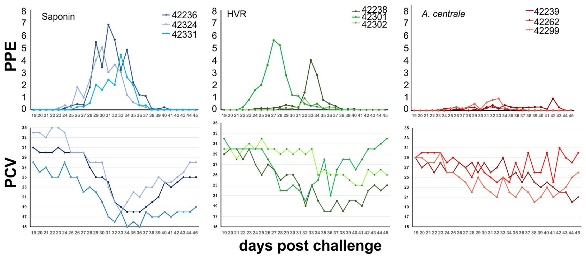
3.2. Clinical Response after Challenge

To challenge the cattle, 20 A. marginale exposed ticks were allowed to feed on each of the immunized animals for 7 days. All nine animals, regardless of vaccination group, became infected with A. marginale (Figure 2A). Infection parameters were assessed and compared between groups. There was not a significant difference between the days to peak parasitemia between groups. As expected, the negative control group animals had the highest bacteremia, with a mean (±SD) maximum peak percentage of infected erythrocytes of 5.4% (±1.25), while the A. centrale vaccinated group had the lowest bacteremia, with a mean (±SD) maximum peak percentage of infected erythrocytes of 0.52% (±0.003), which was significantly different from the negative control group (p = 0.003). Interestingly, the HVR-immunized animals had an intermediate level of bacteremia (PPE = 3.51% ± 2.37), which was not significantly different from either control group, reflecting incomplete protection with this vaccination regime.
Similarly, when examining the nadir of the PCV, the negative control group had the biggest drop in PCV, the A. centrale-immunized group had the smallest drop in PCV, and the HVR-immunized animals showed an intermediate result (Figure 2B). The difference between the negative control group and the A. centrale group was significantly different (p = 0.03), while the HVR group was not significantly different from either of the other two groups.
This intermediate result is because one of the HVR-immunized animals was protected in a manner that was indistinguishable from the A. centrale-immunized animals, while the other two acted similarly to the adjuvant control group (Figure 3).

3.3. Sequence Analysis of HVR Regions

Because one of the HVR-immunized animals (C42302) behaved differently from the other two upon challenge, we sought to determine if this was due to a different pattern of A. marginale Msp2 usage during the challenge. We examined which Msp2 alleles were expressed by amplifying, cloning, and sequencing expression site variants at peak parasitemia, rationalizing that these variants are the ones being expressed before an immune response is mounted to control the first wave of infection. A deduced protein alignment of the HVR sequences shows that there are 11 unique HVR variants represented in the study (Figure 4 and Table 2). The variants were scored for complexity [6], with complex variants typically being seen as a result of immune selection. Only four complex variants were identified, with an overall prevalence of 9% (Table 2) in the dataset. Most of the variants that were detected were simple variants; that is, the HVR was composed of two or fewer distinct pseudogene segments. Simple variant 1 was the most common variant and was detected in all animals. Variant 1 had a prevalence of 45% in the saponin control group, while in the A. centrale- and HVR-immunized groups, it was detected as 27% and 29% of the variants, respectively.
Although the overall prevalence of V1 in the unprotected animals is statistically significant compared with the protected animals, the numbers of this variant were identical between an HVR-immunized animal that was protected (C42302) and one that was not (C 42238).
One statistically significant difference that appears to be associated with protection is higher levels of expressed variant E6/F7 (corresponding to the HVR of pseudogene E6/F7 having recombined into the expression site). This variant was detected in 23–57% of the variants detected in the A. centrale-immunized animals and in animal C42302 (HVR-immunized, protected), while it only made up 1.4% of the detected variants in animals that were not protected in the saponin control group or the other two HVR-immunized animals (Table 2).
Variant 1, which was prevalent in all the animals after challenge, had identity scores ranging from 28 to 52% with the A. centrale Msp2 HVRs that were used for immunization, while the other variants had identity scores ranging from 30 to 72% (Supplemental Table S1). In other words, it appeared to be more diverse compared with the A. centrale HVRs than the other variants that appeared in the challenge.
Variant E6/F7 was found at higher levels in protected animals. This variant had an identity range of 40–57% with the A. centrale Msp2 HVRs, which was slightly more similar to the A. centrale Msp2 HVRs than the other variants detected at challenge (See Supplemental Table S1).

4. Discussion

We reject the hypothesis that the protective capacity of the A. centrale live vaccine is primarily due to immunity that is engendered by Msp2. While all of the A. centrale HVR vaccinates developed a strong humoral response, two of the three animals responded in a manner that was indistinguishable from the adjuvant only control, i.e., they were not protected. In the A. centrale live vaccine group, although all of the animals became infected, they were protected from acute disease.
We next examined whether the different treatment groups (i.e., A. centrale Msp2-HVR and A. centrale live vaccine) skewed the expression of Msp2 upon challenge with A. marginale. Interestingly, the most common variant detected was V1, which had the lowest similarity of any of the variants that were expressed during challenge when comparing across the HVR when compared with the A. centrale HVRs (Supplemental Table S1). This variant has been detected in previous A. marginale St. Maries strain infections and has been examined to show that it retains similar porin functionality as Msp2 molecules that are derived from full-length pseudogene HVRs [25]. The pseudogenes that are retained in the genome are subject to selection, whereas the transient recombined expression site mosaics that arise during the course of infection are not; therefore, it is notable that variant V1 is known to retain functionality.
While V1 was statistically more prevalent in the unprotected animals, this is likely due to the fact that the E6/F7 variant was able to establish in the protected animals. Overall, the E6/F7 variant is more similar to A. centrale Msp2 sequences than V1; however, it has a novel Block 1 sequence, which is likely a key factor in allowing organisms that express E6/F7 to be established.
However, robust protection as elicited in the animals that received the A. centrale live vaccine is likely to be multifactorial and continually boosted by the presence of the organisms that present more than just Msp2. It is known that both outer membrane protein (OMP) preparations and OMP complexes (a subset of the OMP preparations) from A. marginale are protective and can even elicit sterile immunity [12,13,26]. When immunizing with the OMP preparations or complexes, some of the proteins are naturally linked due to their context in the membrane. It has been demonstrated that linkage can be important in inducing immune responses; it was shown that a T-cell epitope in one protein enhanced a B-cell response in another linked protein [27]. In the study by Noh et al. [26], the authors used A. marginale OMP complexes that were covalently linked or unlinked to immunize cattle. Upon challenge, both groups were protected in a manner that was indistinguishable from each other. One difference between the two groups was that the linked immunogen group developed higher IgG2 titers prior to challenge. As protection was equivalent, this indicates that IgG2 is not a robust correlate of protection for A. marginale, as has been reported [14,28]. While Msp2 is the most immunodominant molecule on the surface of the organism, the subdominant epitopes of other surface proteins are also detected by the immune response [13,26]. In the search for a subunit vaccine, none of the recombinant proteins found in the OMP complexes that have been tested as vaccine candidates have appeared to be promising immunogens [29,30]. Vaccine formulation and or presentation may be just as important as having the right immunogen.
The challenge of working with the immunodominant Msp2 is that it has the ability to undergo gene conversion and evade the developing immune response. With the availability of genome sequence data, we have tracked infections and msp2 usage [6] and we see that typically, the whole pseudogene HVRs are used early in infection. Therefore, the rationale to immunize with this set of HVRs and develop immunity against them and most variants that would arise in early infection was tested. Despite using HVR sequences that would provide the immunostimulatory capacity provided by A. centrale Msp2 early in infection, the Msp2-HVR vaccine did not provide protection that was equivalent to the live A. centrale vaccine. It may be that epitopes from the conserved region of Msp2 are required or that, as with other candidates that have been presented in recombinant form (mentioned above), the presentation is not appropriate to induce a protective response. Proteins presented in their native form appear to perform better in immunization trials, i.e., the OMP preparations and others [31,32,33].
This study makes it clear that Msp2 alone is insufficient for a robust protective response and that multivalent vaccines must be considered when moving away from the live A. centrale vaccine.

Supplementary Materials

The following supporting information can be downloaded at: https://www.mdpi.com/article/10.3390/vaccines11101544/s1, Table S1: Msp2 HVR identities.

Author Contributions

Conceptualization, K.A.B.; methodology, A.F. and P.-S.K.; validation, A.F. and K.A.B.; resources, K.A.B.; data curation, A.F.; writing—original draft preparation, A.F. and K.A.B.; visualization, A.F. and K.A.B.; supervision, K.A.B.; project administration, K.A.B.; funding acquisition, K.A.B. All authors have read and agreed to the published version of the manuscript.

Funding

This research was supported by BARD grant no. US-3315-02C.

Institutional Review Board Statement

All protocols for the animal experiments were approved by the Institutional Animal Care and Use Committee at Washington State University, USA, in accordance with institutional guidelines based on the U.S. National Institutes of Health (NIH) Guide for the Care and Use of Laboratory Animals (ASAF 3386).

Informed Consent Statement

Not applicable.

Data Availability Statement

The data presented in this study are available in the body of this article as well as Supplementary Table S1. Sequence data has been deposited in Genbank under accession numbers OR340832–OR340842.

Acknowledgments

We acknowledge Xiaoya Cheng, Ralph Horn, and Benjamin Alfrejd for expert technical assistance.

Conflicts of Interest

The authors declare no conflict of interest. The funders had no role in the design of the study; in the collection, analyses, or interpretation of data; in the writing of the manuscript; or in the decision to publish the results.

References

  1. Aubry, P.; Geale, D.W. A review of bovine anaplasmosis. Transbound. Emerg. Dis. 2011, 58, 1–30. [Google Scholar] [CrossRef] [PubMed]
  2. French, D.M.; McElwain, T.F.; McGuire, T.C.; Palmer, G.H. Expression of Anaplasma marginale major surface protein 2 variants during persistent cyclic rickettsemia. Infect. Immun. 1998, 66, 1200–1207. [Google Scholar] [CrossRef] [PubMed]
  3. Eid, G.; French, D.M.; Lundgren, A.M.; Barbet, A.F.; McElwain, T.F.; Palmer, G.H. Expression of major surface protein 2 antigenic variants during acute Anaplasma marginale rickettsemia. Infect. Immun. 1996, 64, 836–841. [Google Scholar] [CrossRef] [PubMed]
  4. Brayton, K.A.; Knowles, D.P.; McGuire, T.C.; Palmer, G.H. Efficient use of a small genome to generate antigenic diversity in tick-borne ehrlichial pathogens. Proc. Natl. Acad. Sci. USA 2001, 98, 4130–4135. [Google Scholar] [CrossRef]
  5. Brayton, K.A.; Palmer, G.H.; Lundgren, A.; Yi, J.; Barbet, A.F. Antigenic variation of Anaplasma marginale msp2 occurs by combinatorial gene conversion. Mol. Microbiol. 2002, 43, 1151–1159. [Google Scholar] [CrossRef]
  6. Futse, J.E.; Brayton, K.A.; Knowles, D.P., Jr.; Palmer, G.H. Structural basis for segmental gene conversion in generation of Anaplasma marginale outer membrane protein variants. Mol. Microbiol. 2005, 57, 212–221. [Google Scholar] [CrossRef]
  7. Reinbold, J.B.; Coetzee, J.F.; Hollis, L.C.; Nickell, J.S.; Riegel, C.M.; Christopher, J.A.; Ganta, R.R. Comparison of iatrogenic transmission of Anaplasma marginale in Holstein steers via needle and needle-free injection techniques. Am. J. Vet. Res. 2010, 71, 1178–1188. [Google Scholar] [CrossRef]
  8. Reinbold, J.B.; Coetzee, J.F.; Sirigireddy, K.R.; Ganta, R.R. Detection of Anaplasma marginale and A. phagocytophilum in bovine peripheral blood samples by duplex real-time reverse transcriptase PCR assay. J. Clin. Microbiol. 2010, 48, 2424–2432. [Google Scholar] [CrossRef]
  9. Toillion, A.R.; Reppert, E.J.; Amachawadi, R.G.; Olson, K.C.; Coetzee, J.F.; Kang, Q.; Reif, K.E. Effect of Protracted Free-Choice Chlortetracycline-Medicated Mineral for Anaplasmosis Control on Escherichia coli Chlortetracycline Resistance Profile from Pastured Beef Cattle. Microorganisms 2021, 9, 2495. [Google Scholar] [CrossRef]
  10. George, J.E.; Pound, J.M.; Davey, R.B. Chemical control of ticks on cattle and the resistance of these parasites to acaricides. Parasitology 2004, 129 (Suppl. S1), S353–S366. [Google Scholar] [CrossRef]
  11. Edvantoro, B.B.; Naidu, R.; Megharaj, M.; Singleton, I. Changes in microbial properties associated with long-term arsenic and DDT contaminated soils at disused cattle dip sites. Ecotoxicol. Environ. Saf. 2003, 55, 344–351. [Google Scholar] [CrossRef] [PubMed]
  12. Tebele, N.; McGuire, T.C.; Palmer, G.H. Induction of protective immunity by using Anaplasma marginale initial body membranes. Infect. Immun. 1991, 59, 3199–3204. [Google Scholar] [CrossRef] [PubMed]
  13. Noh, S.M.; Brayton, K.A.; Brown, W.C.; Norimine, J.; Munske, G.R.; Davitt, C.M.; Palmer, G.H. Composition of the surface proteome of Anaplasma marginale and its role in protective immunity induced by outer membrane immunization. Infect. Immun. 2008, 19, 2219–2226. [Google Scholar] [CrossRef] [PubMed]
  14. Brown, W.C.; Shkap, V.; Zhu, D.; McGuire, T.C.; Tuo, W.; McElwain, T.F.; Palmer, G.H. CD4(+) T-lymphocyte and immunoglobulin G2 responses in calves immunized with Anaplasma marginale outer membranes and protected against homologous challenge. Infect. Immun. 1998, 66, 5406–5413. [Google Scholar] [CrossRef]
  15. Hove, P.; Madesh, S.; Nair, A.; Jaworski, D.; Liu, H.; Ferm, J.; Kleinhenz, M.D.; Highland, M.A.; Curtis, A.K.; Coetzee, J.F.; et al. Targeted mutagenesis in Anaplasma marginale to define virulence and vaccine development against bovine anaplasmosis. PLoS Pathog. 2022, 18, e1010540. [Google Scholar] [CrossRef]
  16. Salinas-Estrella, E.; Amaro-Estrada, I.; Cobaxin-Cardenas, M.E.; Preciado de la Torre, J.F.; Rodriguez, S.D. Bovine Anaplasmosis: Will there ever be an almighty effective vaccine? Front. Vet. Sci. 2022, 9, 946545. [Google Scholar] [CrossRef]
  17. Abdala, A.A.; Pipano, E.; Aguirre, D.H.; Gaido, A.B.; Zurbriggen, M.A.; Mangold, A.J.; Guglielmone, A.A. Frozen and fresh Anaplasma centrale vaccines in the protection of cattle against Anaplasma marginale infection. Rev. Elev. Med. Vet. Pays Trop. 1990, 43, 155–158. [Google Scholar] [CrossRef]
  18. Pipano, E. Live vaccines against hemoparasitic diseases in livestock. Vet. Parasitol. 1995, 57, 213–231. [Google Scholar] [CrossRef]
  19. Palmer, G.H.; McElwain, T.F. Molecular basis for vaccine development against anaplasmosis and babesiosis. Vet. Parasitol. 1995, 57, 233–253. [Google Scholar] [CrossRef]
  20. Futse, J.E.; Brayton, K.A.; Dark, M.J.; Knowles, D.P., Jr.; Palmer, G.H. Superinfection as a driver of genomic diversification in antigenically variant pathogens. Proc. Natl. Acad. Sci. USA 2008, 105, 2123–2127. [Google Scholar] [CrossRef]
  21. Herndon, D.R.; Palmer, G.H.; Shkap, V.; Knowles, D.P., Jr.; Brayton, K.A. Complete genome sequence of Anaplasma marginale subsp. centrale. J. Bacteriol. 2010, 192, 379–380. [Google Scholar] [CrossRef] [PubMed]
  22. Dark, M.J.; Herndon, D.R.; Kappmeyer, L.S.; Gonzales, M.P.; Nordeen, E.; Palmer, G.H.; Knowles, D.P., Jr.; Brayton, K.A. Conservation in the face of diversity: Multistrain analysis of an intracellular bacterium. BMC Genom. 2009, 10, 16. [Google Scholar] [CrossRef]
  23. Brayton, K.A.; Kappmeyer, L.S.; Herndon, D.R.; Dark, M.J.; Tibbals, D.L.; Palmer, G.H.; McGuire, T.C.; Knowles, D.P., Jr. Complete genome sequencing of Anaplasma marginale reveals that the surface is skewed to two superfamilies of outer membrane proteins. Proc. Natl. Acad. Sci. USA 2005, 102, 844–849. [Google Scholar] [CrossRef] [PubMed]
  24. Knowles, D.; Torioni de Echaide, S.; Palmer, G.; McGuire, T.; Stiller, D.; McElwain, T. Antibody against an Anaplasma marginale MSP5 epitope common to tick and erythrocyte stages identifies persistently infected cattle. J. Clin. Microbiol. 1996, 34, 2225–2230. [Google Scholar] [CrossRef] [PubMed]
  25. Graca, T.; Silva, M.G.; Kostyukova, A.S.; Palmer, G.H. Structural Basis for Recombinatorial Permissiveness in the Generation of Anaplasma marginale Msp2 Antigenic Variants. Infect. Immun. 2016, 84, 2740–2747. [Google Scholar] [CrossRef]
  26. Noh, S.M.; Turse, J.E.; Brown, W.C.; Norimine, J.; Palmer, G.H. Linkage between Anaplasma marginale outer membrane proteins enhances immunogenicity but is not required for protection from challenge. Clin. Vaccine Immunol. 2013, 20, 651–656. [Google Scholar] [CrossRef]
  27. Macmillan, H.; Norimine, J.; Brayton, K.A.; Palmer, G.H.; Brown, W.C. Physical linkage of naturally complexed bacterial outer membrane proteins enhances immunogenicity. Infect. Immun. 2007, 76, 1223–1229. [Google Scholar] [CrossRef]
  28. Barigye, R.; Garcia-Ortiz, M.A.; Rojas Ramirez, E.E.; Rodriguez, S.D. Identification of IgG2-specific antigens in Mexican Anaplasma marginale strains. Ann. N. Y Acad. Sci. 2004, 1026, 84–94. [Google Scholar] [CrossRef]
  29. Ducken, D.R.; Brown, W.C.; Alperin, D.C.; Brayton, K.A.; Reif, K.E.; Turse, J.E.; Palmer, G.H.; Noh, S.M. Subdominant Outer Membrane Antigens in Anaplasma marginale: Conservation, Antigenicity, and Protective Capacity Using Recombinant Protein. PLoS ONE 2015, 10, e0129309. [Google Scholar] [CrossRef]
  30. Albarrak, S.M.; Brown, W.C.; Noh, S.M.; Reif, K.E.; Scoles, G.A.; Turse, J.E.; Norimine, J.; Ueti, M.W.; Palmer, G.H. Subdominant antigens in bacterial vaccines: AM779 is subdominant in the Anaplasma marginale outer membrane vaccine but does not associate with protective immunity. PLoS ONE 2012, 7, e46372. [Google Scholar] [CrossRef]
  31. Palmer, G.H.; Barbet, A.F.; Cantor, G.H.; McGuire, T.C. Immunization of cattle with the MSP-1 surface protein complex induces protection against a structurally variant Anaplasma marginale isolate. Infect. Immun. 1989, 57, 3666–3669. [Google Scholar] [CrossRef] [PubMed]
  32. Palmer, G.H.; Barbet, A.F.; Davis, W.C.; McGuire, T.C. Immunization with an isolate-common surface protein protects cattle against anaplasmosis. Science 1986, 231, 1299–1302. [Google Scholar] [CrossRef] [PubMed]
  33. Palmer, G.H.; Oberle, S.M.; Barbet, A.F.; Goff, W.L.; Davis, W.C.; McGuire, T.C. Immunization of cattle with a 36-kilodalton surface protein induces protection against homologous and heterologous Anaplasma marginale challenge. Infect. Immun. 1988, 56, 1526–1531. [Google Scholar] [CrossRef] [PubMed]
Figure 1. Western blots demonstrating antibody responses in vaccinates. Animals that were immunized with a cocktail of seven recombinant A. centrale Msp2 HVRs (A) or with live, blood-borne A. centrale (B) had an antibody response that recognized the recombinant proteins after immunization. There was no response to these proteins in the negative control group that was sham immunized with adjuvant (similar results from all animals; only the results from calf no. 42324 are shown) (C). All animals had no response prior to immunization (Pre).
Vaccines 11 01544 g001
Figure 2. Clinical parameters for animals challenged with A. marginale StM. (A) The adjuvant-immunized group developed a statistically significantly higher infection based on the percent of parasitized erythrocytes (PPE) compared with the A. centrale-immunized group. (B) The A. centrale-immunized group had the smallest drop in the packed cell volume (PCV), which was statistically different from the adjuvant immunized group (*, p < 0.05; ns, not significant). Error bars represent the standard errors of the means (SEM) calculated from three animals in each group.
Vaccines 11 01544 g002
Figure 3. Clinical parameters in the challenged animals over time. Top graphs show recorded values of the percent of parasitized erythrocytes (PPE) for each animal plotted by days post challenge. The protected animal from the HVR-vaccinated group (# 42302) had similar values of daily counted infected erythrocytes to that of the A. centrale-vaccinated animal group (red). The adjuvant-immunized group (blue) and the two other unprotected HVR-immunized animals developed the highest PPE. The lower graphs show the packed cell volume for each animal over time. The animals are color-coded the same in both sets of graphs.
Vaccines 11 01544 g003
Figure 4. Alignment of Msp2 HVR sequences. Unique HVR sequences from A. centrale (Ac) are shown in the top group. The middle group are the expression site variants detected in the study that are not templated in the genome. The final group is composed of sequences that represent the unique pseudogene HVRs found in the A. marginale St. Maries strain genome, all of which were expressed and detected during the challenge phase of the study.
Vaccines 11 01544 g004
Table 1. Primers used to clone A. centrale pseudogene HVRs.
Locus-Specific Primers
ψ namePrimer NamePrimer Sequence *
AcA1
AcF
A1, F-Fx attB2GGGGACAAGTTTGTACAAAAAAGCAGGCTATCAGGCAGTAGAGGCTAAT
AcA1A1-Rx attB2GGGGACCACTTTGTACAAGAAAGCTGGGTACTTCACGCTGATGGTGGC
AcB1B1-Fx attB1GGGGACAAGTTTGTACAAAAAAGCAGGCTATAAAGCAGTAGAAGGTGTT
AcB1
AcAF
B1, AF-Rx attB2GGGGACCACTTTGTACAAGAAAGCTGGGTACTGCAAGCTGATGGTGTT
AcAF
AcG2
AcC
AF, G2, C-Fx attB1GGGGACAAGTTTGTACAAAAAAGCAGGCTATACTGCAGTAGAGGCTGCC
AcG2 G2-Rx attB2GGGGACCACTTTGTACAAGAAAGCTGGGTACTGCACGTCGATGGTGGG
AcA22A22-Fx attB1GGGGACAAGTTTGTACAAAAAAGCAGGCTATACTGCAGTAGAGGCTGCT
A22-Rx attB2GGGGACCACTTTGTACAAGAAAGCTGGGTACTTCACGCTGATGGTGCT
AcF
AcC
F, C-Rx attB2GGGGACCACTTTGTACAAGAAAGCTGGGTATGACACACTGATGGTAGC
* msp2-specific sequences are underlined.
Table 2. Sequence analysis of Msp2 usage and HVR unique variants detected during the challenge.
Number Clones from Each Animal
SaponinHVRA. centraleVariant NameType of Variant
20, 22, 17 *,a14, 12, 12 *,b13, 7, 15 *,cV1Simple
0, 0, 11, 1, 00, 0, 1V2Complex
6, 1, 01, 2, 30, 0, 1V3Complex
0, 3, 0ND #1, 0, 1V4Complex
1, 1, 0NDNDV5Simple
0, 0, 2NDNDV6Simple
0, 0, 1NDNDV7Complex
ND1, 0, 0NDV8Simple
ND0, 4, 0NDV9Simple
NDND1, 0, 0V10Simple
NDND0, 0, 1V11Simple
1, 0, 24, 2, 02, 0, 1ψ 1A. marginale StM
2, 1, 31, 0, 03, 0, 2ψ 2A. marginale StM
0, 2, 28, 8, 22, 0, 1ψ G 11A. marginale StM
0, 0, 10, 1, 98, 17, 7ψ E6 F7A. marginale StM
0, 0, 10, 0, 40, 1, 0ψ 9H 1A. marginale StM
NDND0, 5, 0ψ A22A. centrale
* Calf numbers (in order): a Saponin group C# 42324, C# 42236, C#42331; b A. centrale Msp2 HVR-immunized group C# 42238, C# 42301, C# 42302; c A. centrale-immunized group C# 42239, C# 42262, C# 42299; # ND = not detected.
Disclaimer/Publisher’s Note: The statements, opinions and data contained in all publications are solely those of the individual author(s) and contributor(s) and not of MDPI and/or the editor(s). MDPI and/or the editor(s) disclaim responsibility for any injury to people or property resulting from any ideas, methods, instructions or products referred to in the content.

