
Nanoclays: Promising Materials for Vaccinology

 , , ,
, , , 

Abstract
1. Introduction
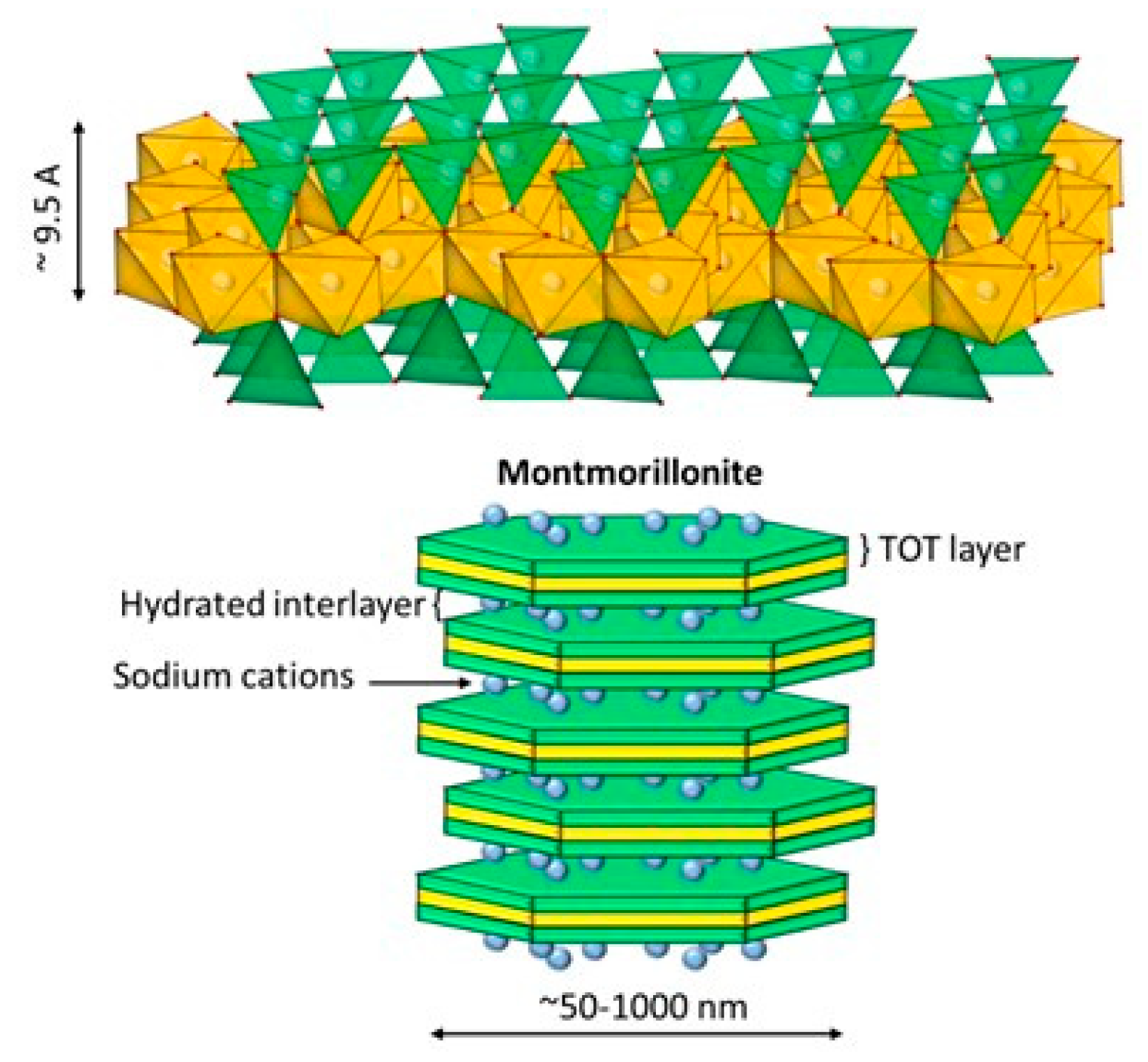
2. Clays Minerals and Nanoclays
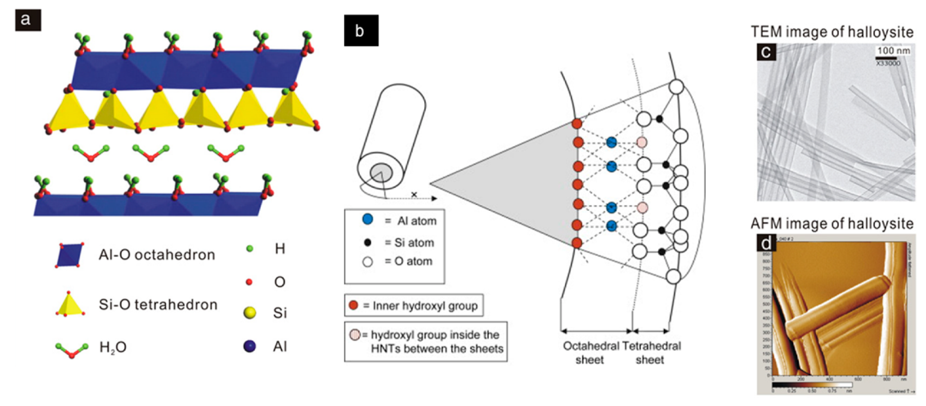
2.1. Halloysite
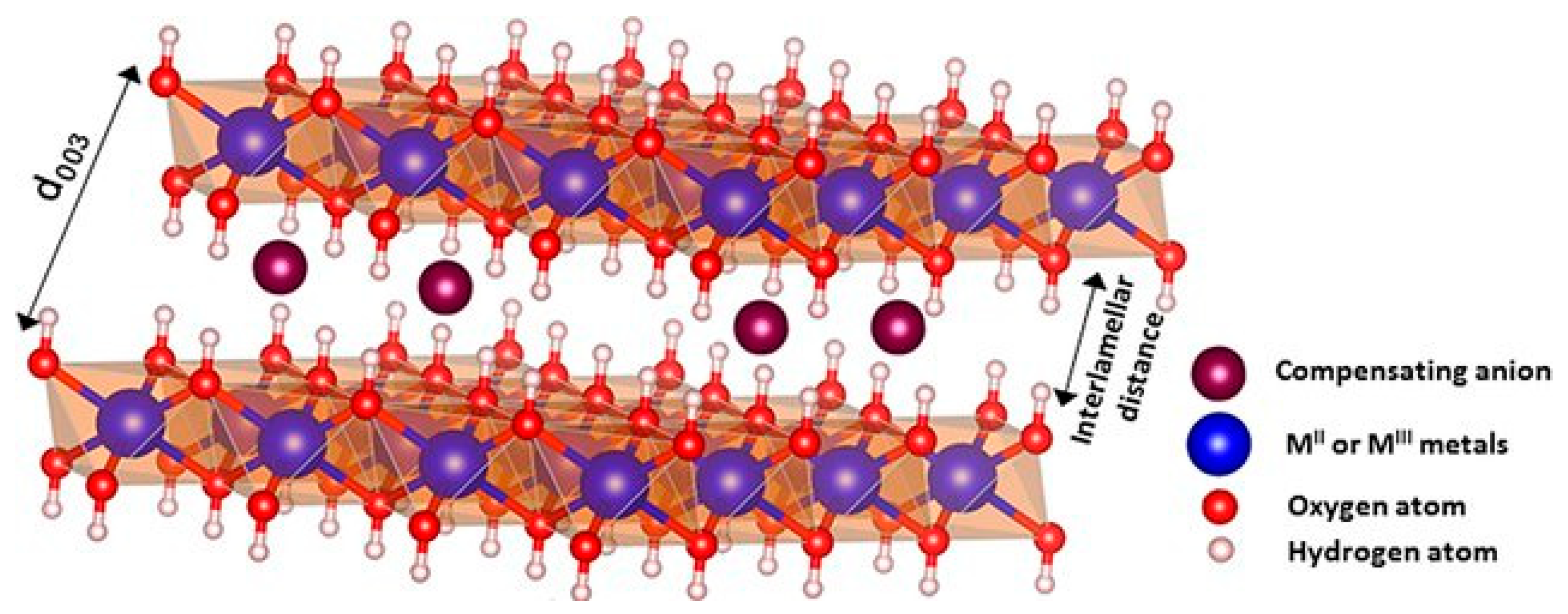
2.2. Layered Double Hydroxides
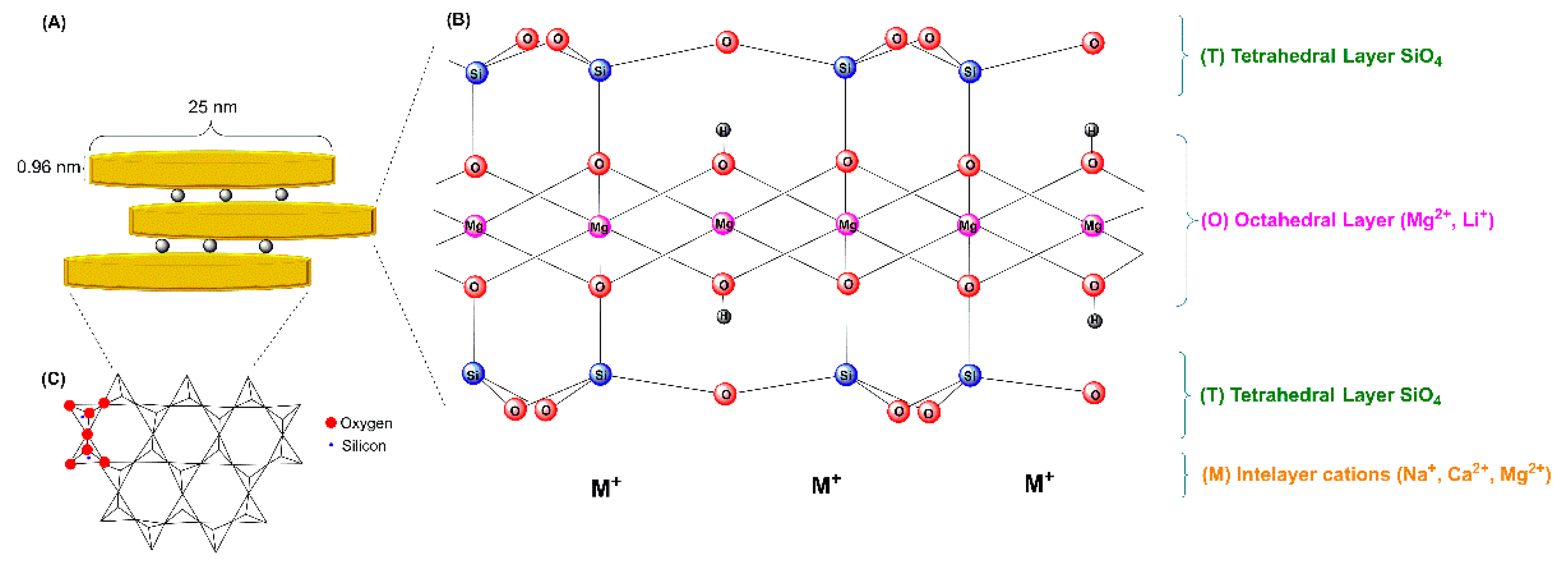
2.3. Hectorite and Laponite®
3. Functionalization of Nanoclays
3.1. Passive Adsorption
3.2. Active Adsorption
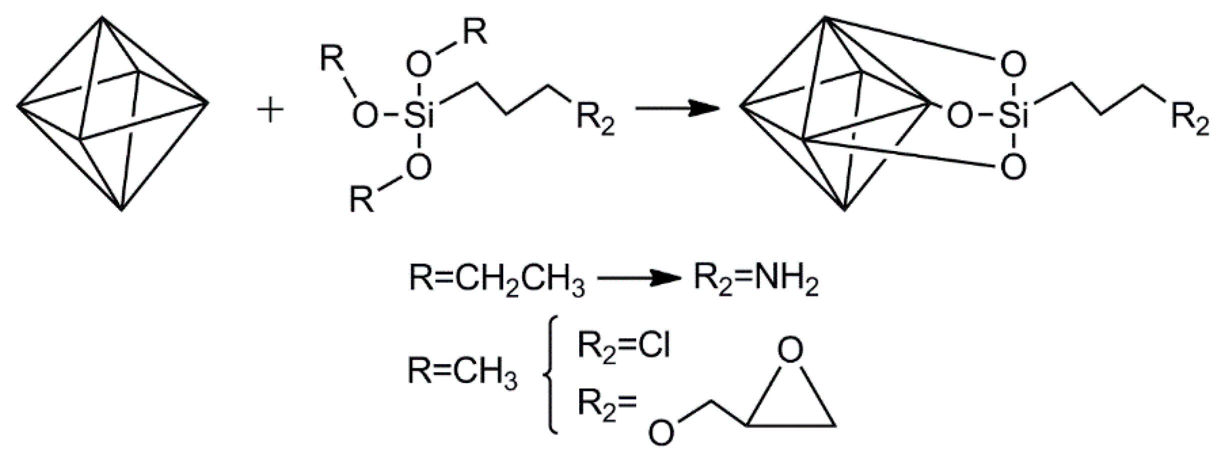
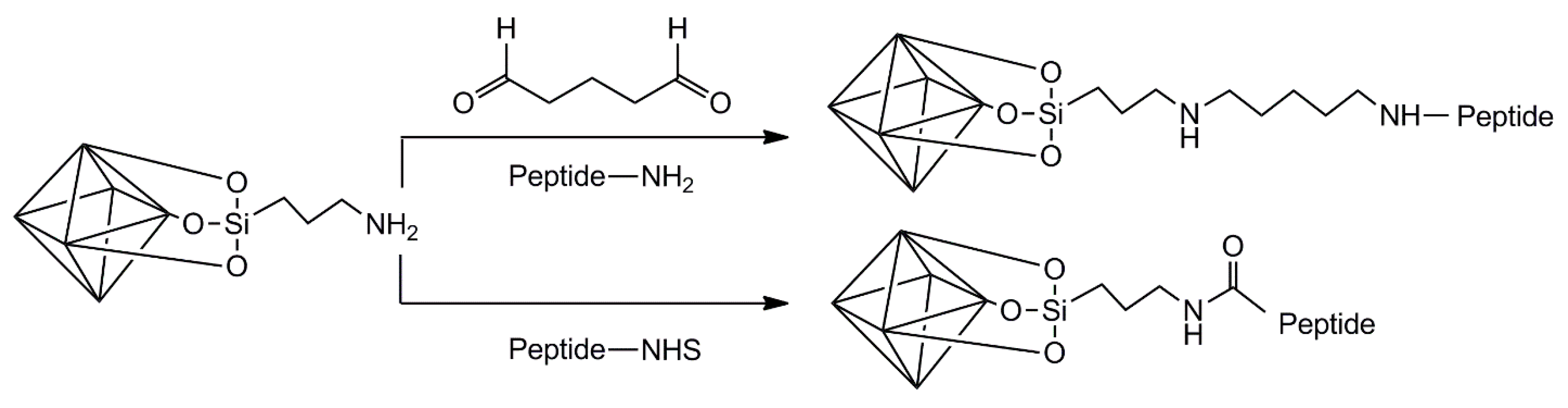
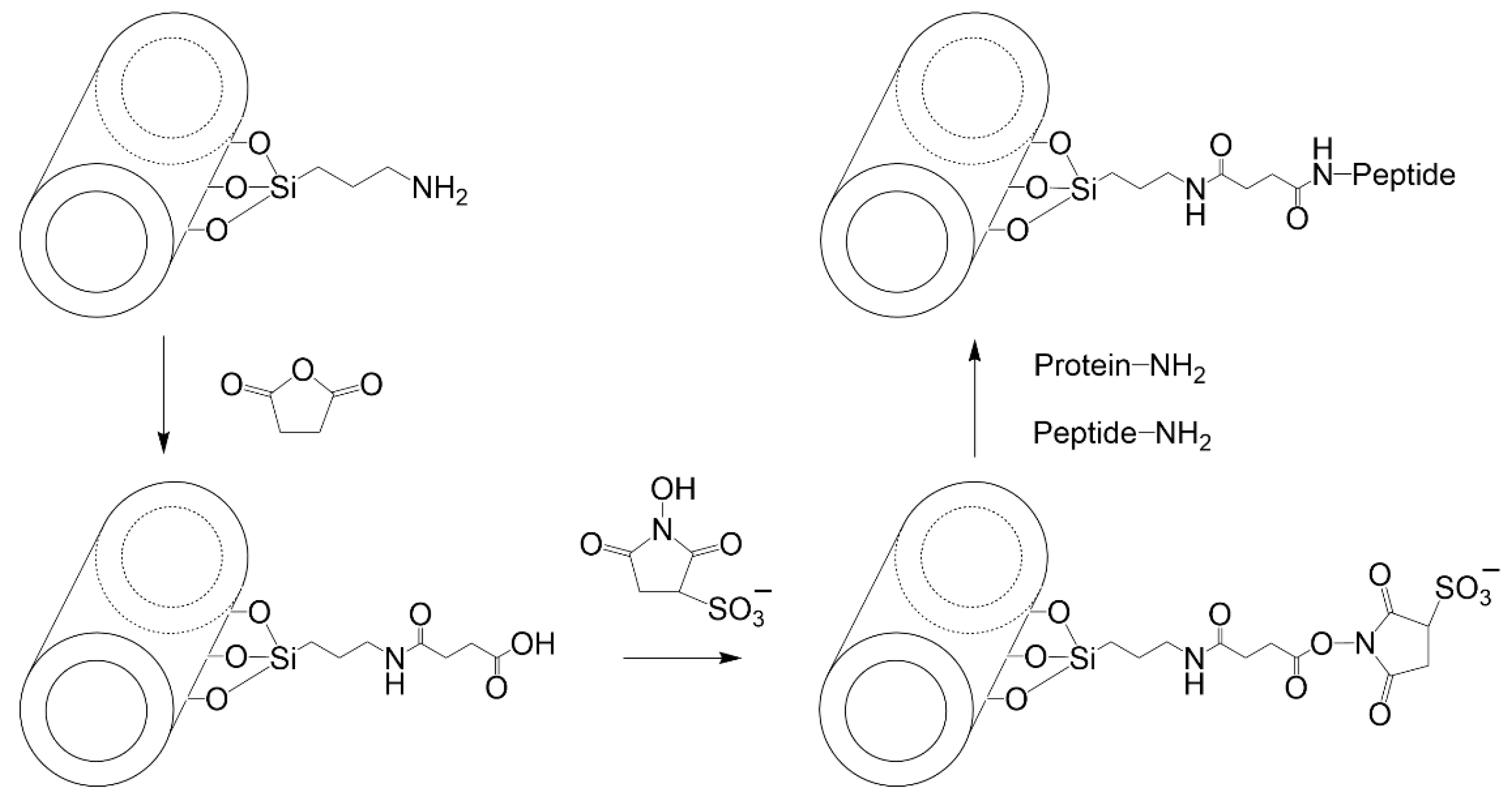
3.2.1. LDH
3.2.2. Halloysite
3.2.3. Hectorite (Laponite®)
4. Nanovaccine Candidates Based on Nanoclays
5. Nanovaccine Candidates Based on Complex Formulations Containing Nanoclays
6. Discussion and Perspectives
Author Contributions
Funding
Conflicts of Interest
References
- Sheerin, D.; Openshaw, P.J.; Pollard, A.J. Issues in vaccinology: Present challenges and future directions. Eur. J. Immunol. 2017, 47, 2017–2025. [Google Scholar] [CrossRef] [PubMed]
- Moyle, P.M.; Toth, I. Modern subunit vaccines: Development, components, and research opportunities. ChemMedChem 2013, 8, 360–376. [Google Scholar] [CrossRef] [PubMed]
- Bansal, A.; D’Souza, B.; Kapoor, D.N.; Singh, P.; Starr, G.; Muppireddy, K.K. Crafting Immunological Response Using Particulate Vaccines. Crit. Rev. Ther. Drug Carr. Syst. 2022, 39, 49–82. [Google Scholar] [CrossRef] [PubMed]
- Wibowo, D.; Jorritsma, S.H.; Gonzaga, Z.J.; Evert, B.; Chen, S.; Rehm, B.H. Polymeric nanoparticle vaccines to combat emerging and pandemic threats. Biomaterials 2021, 268, 120597. [Google Scholar] [CrossRef]
- Shah, S.M.; Alsaab, H.O.; Rawas-Qalaji, M.M.; Uddin, M.N. A review on current COVID-19 vaccines and evaluation of particulate vaccine delivery systems. Vaccines 2021, 9, 1086. [Google Scholar] [CrossRef]
- Rosales-Mendoza, S.; González-Ortega, O. PLGA-Based Mucosal Nanovaccines. In Nanovaccines; Springer: Cham, Switzerland, 2019; pp. 61–103. [Google Scholar]
- Nazarizadeh, A.; Staudacher, A.H.; Wittwer, N.L.; Turnbull, T.; Brown, M.P.; Kempson, I. Aluminium Nanoparticles as Efficient Adjuvants Compared to Their Microparticle Counterparts: Current Progress and Perspectives. Int. J. Mol. Sci. 2022, 23, 4707. [Google Scholar] [CrossRef]
- Manjaiah, K.M.; Mukhopadhyay, R.; Paul, R.; Datta, S.C.; Kumararaja, P.; Sarkar, B. Clay minerals and zeolites for environmentally sustainable agriculture. In Modified Clay and Zeolite Nanocomposite Materials; Elsevier: Amsterdam, The Netherlands, 2019; pp. 309–329. [Google Scholar]
- Schlegel, M.L.; Descostes, M. Uranium uptake by hectorite and montmorillonite: A solution chemistry and polarized EXAFS study. Environ. Sci. Technol. 2009, 43, 8593–8598. [Google Scholar] [CrossRef]
- Zvulunov, Y.; Radian, A. Alginate composites reinforced with polyelectrolytes and clay for improved adsorption and bioremediation of formaldehyde from water. ACS EST Water 2021, 1, 1837–1848. [Google Scholar] [CrossRef]
- García-Villén, F.; Carazo, E.; Borrego-Sánchez, A.; Sánchez-Espejo, R.; Cerezo, P.; Viseras, C.; Aguzzi, C. Clay minerals in drug delivery systems. In Modified Clay and Zeolite Nanocomposite Materials; Elsevier: Amsterdam, The Netherlands, 2019; pp. 129–166. [Google Scholar]
- García-Villén, F.; Ruiz-Alonso, S.; Lafuente-Merchan, M.; Gallego, I.; Sainz-Ramos, M.; Saenz-del-Burgo, L.; Pedraz, J.L. Clay minerals as bioink ingredients for 3D printing and 3D bioprinting: Application in tissue engineering and regenerative medicine. Pharmaceutics 2021, 13, 1806. [Google Scholar] [CrossRef]
- Naumenko, E.A.; Guryanov, I.D.; Yendluri, R.; Lvov, Y.M.; Fakhrullin, R.F. Clay nanotube–biopolymer composite scaffolds for tissue engineering. Nanoscale 2016, 8, 7257–7271. [Google Scholar] [CrossRef]
- Rytwo, G.; Mendelovits, A.; Eliyahu, D.; Pitcovski, J.; Aizenshtein, E. Adsoption of two vaccine-related proteins to montmorillonite and organo-montmorillonite. Appl. Clay Sci. 2010, 50, 569–575. [Google Scholar] [CrossRef]
- Wicklein, B.; Martín del Burgo, M.Á.; Yuste, M.; Darder, M.; Llavata, C.E.; Aranda, P.; Ortin, J.; del Real, G.; Ruiz-Hitzky, E. Lipid-based bio-nanohybrids for functional stabilisation of influenza vaccines. Eur. J. Inorg. Chem. 2012, 2012, 5186–5191. [Google Scholar] [CrossRef]
- Dong, J.; Cheng, Z.; Tan, S.; Zhu, Q. Clay nanoparticles as pharmaceutical carriers in drug delivery systems. Expert Opin. Drug Deliv. 2021, 18, 695–714. [Google Scholar] [CrossRef]
- Wicklein, B.; Darder, M.; Aranda, P.; Del Burgo, M.A.M.; Del Real, G.; Esteban, M.; Ruiz-Hitzky, E. Clay-lipid nanohybrids: Towards influenza vaccines and beyond. Clay Miner. 2016, 51, 529–538. [Google Scholar] [CrossRef]
- Padilla-Ortega, E.; Medellín-Castillo, N.; Robledo-Cabrera, A. Comparative study of the effect of structural arrangement of clays in the thermal activation: Evaluation of their adsorption capacity to remove Cd(II). J. Environ. Chem. Eng. 2020, 8, 103850. [Google Scholar] [CrossRef]
- Dominguez, J.M.; Schifter, I. Las Arcillas: El Barro Noble; Fondo de Cultura Económica: Mexico City, Mexico, 1992. [Google Scholar]
- Velde, B. Introduction to Clay Minerals; Editorial Chapman & Hall: London, UK, 1992. [Google Scholar]
- Guo, F.; Aryana, S.; Han, Y.; Jiao, Y. A Review of the Synthesis and Applications of Polymer–Nanoclay Composites. Appl. Sci. 2018, 8, 1696. [Google Scholar] [CrossRef]
- Katschnig, M.; Battisti, M. Chapter 3—Processing of polymer–nanoclay composites. In Polymer Nanoclay Composites; William Andrew Publishing: Norwich, NY, USA, 2015; pp. 53–91. [Google Scholar]
- Nieto, S.; Toro, N.; Robles, P.; Gálvez, E.; Gallegos, S.; Jeldres, R.I. Flocculation of Clay-Based Tailings: Differences of Kaolin and Sodium Montmorillonite in Salt Medium. Materials 2022, 15, 1156. [Google Scholar] [CrossRef]
- Zhang, T.; Wang, W.; Zhao, Y.; Bai, H.; Wen, T.; Kang, S.; Song, G.; Song, S.; Komarneni, S. Removal of heavy metals and dyes by clay-based adsorbents: From natural clays to 1D and 2D nano-composites. Chem. Eng. J. 2021, 420, 127574. [Google Scholar] [CrossRef]
- Joussein, E.; Petit, S.; Churchman, J.; Theng, B.; Righi, D.; Delvaux, B. Halloysite clay minerals—A review. Clay Miner. 2005, 40, 383–426. [Google Scholar] [CrossRef]
- Vergaro, V.; Abdullayev, E.; Lvov, Y.M.; Zeitoun, A.; Cingolani, R.; Rinaldi, R.; Leporatti, S. Cytocompatibility and uptake of halloysite clay nanotubes. Biomacromolecules 2010, 11, 820–826. [Google Scholar] [CrossRef]
- Słomkiewicz, P.; Szczepanik, B.; Czaplicka, M. Adsorption of phenol and chlorophenols by HDTMA modified halloysite nanotubes. Materials 2020, 13, 3309. [Google Scholar] [CrossRef]
- Kamble, R.; Ghag, M.; Gaikawad, S.; Panda, J.K. Halloysite Nanotubes and Applications: A Review. J. Adv. Sci. Res. 2012, 3, 25–29. [Google Scholar]
- Jamshidzadeh, F.; Mohebali, A.; Abdouss, M. Three-ply biocompatible pH-responsive nanocarriers based on HNT sandwiched by chitosan/pectin layers for controlled release of phenytoin sodium. Int. J. Mol. Sci. 2020, 150, 336–343. [Google Scholar] [CrossRef]
- Massaro, M.; Noto, R.; Riela, S. Past, present and future perspectives on halloysite clay minerals. Molecules 2020, 25, 4863. [Google Scholar] [CrossRef] [PubMed]
- Kostić, M.; Radović, M.; Velinov, N.; Najdanović, S.; Bojić, D.; Hurt, A.; Bojić, A. Synthesis of mesoporous triple-metal nanosorbent from layered double hydroxide as an efficient new sorbent for removal of dye from water and wastewater. Ecotoxicol. Environ. Saf. 2018, 159, 332–341. [Google Scholar] [CrossRef] [PubMed]
- Mourid, E.H.; Lakraimi, M.; Benaziz, L.; Elkhattabi, E.H.; Legrouri, A. Wastewater treatment test by removal of the sulfamethoxazole antibiotic by a calcined layered double hydroxide. Appl. Clay Sci. 2019, 168, 87–95. [Google Scholar] [CrossRef]
- Jitianu, M.; Gunness, D.C.; Aboagye, D.E.; Zaharescu, M.; Jitianu, A. Nanosized Ni–Al layered double hydroxides–structural characterization. Mater. Res. Bull. 2013, 48, 1864–1873. [Google Scholar] [CrossRef]
- Liu, Q.; Huang, J.; Zhao, Y.; Cao, L.; Li, K.; Zhang, N.; Yang, D.; Feng, L.; Feng, L. Tuning coupling interface of ultrathin Ni3S2@NiV-LDH heterogeneous nanosheet electrocatalysts for improved overall water splitting. Nanoscale 2019, 11, 8855–8863. [Google Scholar] [CrossRef]
- Liu, Q.; Huang, J.; Zhang, X.; Cao, L.; Yang, D.; Kim, J.; Feng, L. Controllable conversion from single-crystal nanorods to polycrystalline nanosheets of NiCoV-LTH for oxygen evolution reaction at large current density. ACS Sustain. Chem. Eng. 2020, 8, 16091–16096. [Google Scholar] [CrossRef]
- Wu, X.; Du, Y.; An, X.; Xie, X. Fabrication of NiFe layered double hydroxides using urea hydrolysis—Control of interlayer anion and investigation on their catalytic performance. Catal. Commun. 2014, 50, 44–48. [Google Scholar] [CrossRef]
- Karim, A.V.; Hassani, A.; Eghbali, P.; Nidheesh, P.V. Nanostructured modified layered double hydroxides (LDHs)-based catalysts: A review on synthesis, characterization, and applications in water remediation by advanced oxidation processes. Curr. Opin. Solid State Mater. Sci. 2022, 26, 100965. [Google Scholar] [CrossRef]
- Xu, Z.P.; Stevenson, G.S.; Lu, C.Q.; Lu, G.Q.; Bartlett, P.F.; Gray, P.P. Stable suspension of layered double hydroxide nanoparticles in aqueous solution. J. Am. Chem. Soc. 2006, 128, 36–37. [Google Scholar] [CrossRef]
- Laine, M.; Liao, Y.; Varenne, F.; Picot, P.; Michot, L.J.; Barruet, E.; Geertsen, V.; Thill, A.; Pelletier, M.; Brubach, J.B.; et al. Tuning the nature of the anion in hydrated layered double hydroxides for H2 production under ionizing radiation. ACS Appl. Nano Mater. 2018, 1, 5246–5257. [Google Scholar] [CrossRef]
- Zhang, J.; Zhou, C.H.; Petit, S.; Zhang, H. Hectorite: Synthesis, modification, assembly and applications. Appl. Clay Sci. 2019, 177, 114–138. [Google Scholar] [CrossRef]
- Strese, H.; Hoffman, U. Synthesis of magnesium silicate gels with two-dimensional regular structure. Zeit. Anorg. Allg. Chem. 1941, 247, 65–95. [Google Scholar] [CrossRef]
- Vicente, I.; Salagre, P.; Cesteros, Y.; Guirado, F.; Medina, F.; Sueiras, J.E. Fast microwave synthesis of hectorite. Appl. Clay Sci. 2019, 43, 103–107. [Google Scholar] [CrossRef]
- Kalo, H.; Möller, M.W.; Ziadeh, M.; Dolejš, D.; Breu, J. Large scale melt synthesis in an open crucible of Na-fluorohectorite with superb charge homogeneity and particle size. Appl. Clay Sci. 2010, 48, 39–45. [Google Scholar] [CrossRef]
- Daab, M.; Eichstaedt, N.J.; Edenharter, A.; Rosenfeldt, S.; Breu, J. Layer charge robust delamination of organo-clays. RSC Adv. 2018, 8, 28797–28803. [Google Scholar] [CrossRef]
- Dawson, J.I.; Kanczler, J.M.; Yang, X.B.; Attard, G.S.; Oreffo, R.O. Clay gels for the delivery of regenerative microenvironments. Adv. Mater. 2011, 23, 3304–3308. [Google Scholar] [CrossRef]
- Chen, W.; Zuo, H.; Mahony, T.J.; Zhang, B.; Rolfe, B.; Xu, Z.P. Efficient induction of comprehensive immune responses to control pathogenic E. coli by clay nano-adjuvant with the moderate size and surface charge. Sci. Rep. 2017, 7, 13367. [Google Scholar] [CrossRef]
- Singer, J.; Plotz, C. The latex fixation test: I. Application to the serologic diagnosis of rheumatoid arthritis. Am. J. Med. 1956, 21, 888–892. [Google Scholar] [CrossRef]
- Faulk, W.; Taylor, G. An immunocolloid method for the electron microscope. Immunochemistry 1971, 8, 1081–1083. [Google Scholar] [PubMed]
- Romano, E.; Stolinski, C.; Hughes-Jones, N. An antiglobulin reagent labelled with colloidal gold for use in electron microscopy. Immunochemistry 1974, 11, 521–522. [Google Scholar] [CrossRef]
- Zsigmondy, R. Die hochrothe Goldlösung als Reagens auf Colloide. Z. Anal. Chem. 1901, 40, 697–719. [Google Scholar] [CrossRef]
- Pauli, W.; Dessauer, P. Zur allgemeinen Chemie der Kolloid-Kolloid-Reaktionen. IX. Reinste hydrophobe Kolloide und Proteine, ihr Schutzmechanismus. Helv. Chim. Acta 1942, 26, 1225–1250. [Google Scholar] [CrossRef]
- Jaber, M.; Lambert, J.; Balme, S. Protein adsorption on clay minerals. In Developments in Clay Science; Schoonheydt, R., Johnson, C., Bergaya, F., Eds.; Elsevier: Amsterdam, The Netherlands, 2018; pp. 255–288. [Google Scholar]
- Butler, J. Solid supports in enzyme-linked immunosorbent assay and other solid-phase immunoassays. Methods 2000, 22, 4–23. [Google Scholar] [CrossRef]
- Gu, Z.; Zuo, H.; Li, L.; Wu, A.; Xu, Z.P. Pre-coating layered double hydroxide nanoparticles with albumin to improve colloidal stability and cellular uptake. J. Mater. Chem. B 2015, 3, 3331–3339. [Google Scholar] [CrossRef]
- Dong, H.; Parekh, H.S.; Xu, Z.P. Particle size-and number-dependent delivery to cells by layered double hydroxide nanoparticles. J. Colloid Interface Sci. 2015, 437, 10–16. [Google Scholar] [CrossRef]
- Chen, W.; Zhang, B.; Mahony, T.; Gu, W.; Rolfe, B.; Xu, Z.P. Efficient and durable vaccine against intimin β of diarrheagenic E. coli induced by clay nanoparticles. Small 2016, 12, 1627–1639. [Google Scholar] [CrossRef]
- Chen, W.; Zuo, H.; Li, B.; Duan, C.; Rolfe, B.; Zhang, B.; Mahony, T.J.; Xu, Z.P. Clay Nanoparticles Elicit Long-Term Immune Responses by Forming Biodegradable Depots for Sustained Antigen Stimulation. Small 2018, 14, 1704465. [Google Scholar] [CrossRef]
- Bejoy, N. Hydrotalcite. Resonance 2001, 6, 57–61. [Google Scholar] [CrossRef]
- Evans, D.G.; Slade, R.C. Structural aspects of layered double hydroxides. In Layered Double Hydroxides; Springer: Berlin/Heidelberg, Germany, 2006; pp. 1–87. [Google Scholar]
- Ádok-Sipiczki, M.; Szilagyi, I.; Pálinkó, I.; Pavlovic, M.; Sipos, P.; Nardin, C. Design of nucleic acid-layered double hydroxide nanohybrids. Colloid Polym. Sci. 2017, 295, 1463–1473. [Google Scholar] [CrossRef]
- Dinari, M.; Neamat, S. In Situ Polymerization of Polyaniline in Silane Modified Calcium Based Layered Double Hydroxide Intercalated Tartrate. Inorg. Chem. 2020, 4, 250–260. [Google Scholar]
- Dinari, M.; Neamati, S. Surface modified layered double hydroxide/polyaniline nanocomposites: Synthesis, characterization and Pb2+ removal. Colloids Surf. A Physicochem. Eng. Asp. 2020, 589, 124438. [Google Scholar] [CrossRef]
- Vahedi, V.; Pasbakhsh, P.; Chai, S.P. Surface modification of halloysite nanotubes: Role of external hydroxyl groups. In Natural Mineral Nanotubes: Properties and Applications; Apple Academic Press: Burlington, ON, Canada, 2014; pp. 290–312. [Google Scholar]
- Li, C.; Liu, J.; Qu, X.; Yang, Z. A general synthesis approach toward halloysite-based composite nanotube. J. Appl. Polym. Sci. 2009, 112, 2647–2655. [Google Scholar] [CrossRef]
- Luo, P.; Zhang, J.S.; Zhang, B.; Wang, J.H.; Zhao, Y.F.; Liu, J.D. Preparation and characterization of silane coupling agent modified halloysite for Cr (VI) removal. Ind. Eng. Chem. Res. 2011, 50, 10246–10252. [Google Scholar] [CrossRef]
- Pan, J.; Wang, B.; Dai, J.; Dai, X.; Hang, H.; Ou, H.; Yan, Y. Selective recognition of 2,4,5-trichlorophenol by temperature responsive and magnetic molecularly imprinted polymers based on halloysite nanotubes. J. Mater. Chem. 2012, 22, 3360–3369. [Google Scholar] [CrossRef]
- Sun, P.; Liu, G.; Lv, D.; Dong, X.; Wu, J.; Wang, D. Effective activation of halloysite nanotubes by piranha solution for amine modification via silane coupling chemistry. RSC Adv. 2015, 5, 52916–52925. [Google Scholar] [CrossRef]
- Prinz Setter, O.; Movsowitz, A.; Goldberg, S.; Segal, E. Antibody-Functionalized Halloysite Nanotubes for Targeting Bacterial Cells. ACS Appl. Bio Mater. 2021, 4, 4094–4104. [Google Scholar] [CrossRef]
- Curtiss, L.K.; Witztum, J.L. A novel method for generating region-specific monoclonal antibodies to modified proteins. Application to the identification of human glucosylated low density lipoproteins. J. Clin. Investig. 1983, 72, 1427–1438. [Google Scholar] [CrossRef]
- Zhou, C.H.; Tong, D.; Li, X. Synthetic hectorite: Preparation, pillaring and applications in catalysis. In Pillared Clays and Related Catalysts; Springer: New York, NY, USA, 2010; pp. 67–97. [Google Scholar]
- Nobel, M.L.; Mendes, E.; Picken, S.J. Enhanced properties of innovative laponite-filled waterborne acrylic resin dispersions. J. Appl. Polym. Sci. 2007, 103, 687–697. [Google Scholar] [CrossRef]
- Daniel, L.M.; Frost, R.L.; Zhu, H.Y. Edge-modification of laponite with dimethyl-octylmethoxysilane. J. Colloid Interface Sci. 2008, 321, 302–309. [Google Scholar] [CrossRef]
- Bourgeat-Lami, E.; Herrera, N.N.; Putaux, J.L.; Reculusa, S.; Perro, A.; Ravaine, S.; Mingotaud, C.; Duguet, E. November. Surface assisted nucleation and growth of polymer latexes on organically-modified inorganic particles. In Macromolecular Symposia; Wiley-VCH Verlag: Weinheim, Germany, 1995; Volume 229, pp. 32–46. [Google Scholar]
- Mustafa, R.; Luo, Y.; Wu, Y.; Guo, R.; Shi, X. Dendrimer-functionalized laponite nanodisks as a platform for anticancer drug delivery. Nanomaterials 2015, 5, 1716–1731. [Google Scholar] [CrossRef]
- Felbeck, T.; Hoffmann, K.; Lezhnina, M.M.; Kynast, U.H.; Resch-Genger, U. Fluorescent nanoclays: Covalent functionalization with amine reactive dyes from different fluorophore classes and surface group quantification. J. Phys. Chem. C 2015, 119, 12978–12987. [Google Scholar] [CrossRef]
- Wheeler, P.A.; Wang, J.; Baker, J.; Mathias, L.J. Synthesis and characterization of covalently functionalized laponite clay. Chem. Mater. 2005, 17, 3012–3018. [Google Scholar] [CrossRef]
- Gonzalez, B.; da Silva, T.H.; Ciuffi, K.J.; Vicente, M.A.; Trujillano, R.; Rives, V.; de Faria, E.H.; Korili, S.A.; Gil, A. Laponite functionalized with biuret and melamine—Application to adsorption of antibiotic trimethoprim. Microporous Mesoporous Mater. 2017, 253, 112–122. [Google Scholar] [CrossRef]
- Guerra, D.L.; Viana, R.R.; Airoldi, C. Use of raw and chemically modified hectorites as adsorbents for Th (IV), U (VI) and Eu (III) uptake from aqueous solutions. Desalination 2010, 260, 161–171. [Google Scholar] [CrossRef]
- Guimarães, A.D.M.F.; Ciminelli, V.S.T.; Vasconcelos, W.L. Surface modification of synthetic clay aimed at biomolecule adsorption: Synthesis and characterization. Mater. Res. 2007, 10, 37–41. [Google Scholar] [CrossRef]
- Colletti, C.G.; Massaro, M.; Lazzara, G.; Cavallaro, G.; Milioto, S.; Pibiri, I.; Noto, R.; Riela, S. Synthesis, characterization and study of covalently modified triazole laponite® edges. Appl. Clay Sci. 2020, 187, 105489. [Google Scholar] [CrossRef]
- Brennan, J.L.; Hatzakis, N.S.; Tshikhudo, T.R.; Dirvianskyte, N.; Resumes, V.; Patkar, S.; Vind, J.; Svendsen, A.; Nolte, R.J.; Rowan, A.E.; et al. Bionanoconjugation via click chemistry: The creation of functional hybrids of lipases and gold nanoparticles. Bioconj. Chem. 2006, 17, 1373–1375. [Google Scholar] [CrossRef]
- Li, A.; Qin, L.; Wang, W.; Zhu, R.; Yu, Y.; Liu, H.; Wang, S. The use of layered double hydroxides as DNA vaccine delivery vector for enhancement of anti-melanoma immune response. Biomaterials 2011, 32, 469–477. [Google Scholar] [CrossRef] [PubMed]
- Yan, S.; Rolfe, B.E.; Zhang, B.; Mohammed, Y.H.; Gu, W.; Xu, Z.P. Polarized immune responses modulated by layered double hydroxides nanoparticle conjugated with CpG. Biomaterials 2014, 35, 9508–9516. [Google Scholar] [CrossRef] [PubMed]
- Hartwig, D.D.; Bacelo, K.L.; Oliveira, T.L.; Schuch, R.; Seixas, F.K.; Collares, T.; Rodrigues, O.; Hartleben, C.P.; Dellagostin, O.A. The use of halloysite clay and carboxyl-functionalised multi-walled carbon nanotubes for recombinant LipL32 antigen delivery enhanced the IgG response. Mem. Inst. Oswaldo Cruz 2015, 110, 134–137. [Google Scholar] [CrossRef] [PubMed]
- Zhang, L.X.; Liu, D.Q.; Wang, S.W.; Yu, X.L.; Ji, M.; Xie, X.X.; Liu, S.Y.; Liu, R.T. MgAl-layered double hydroxide nanoparticles co-delivering siIDO and Trp2 peptide effectively reduce IDO expression and induce cytotoxic T-lymphocyte responses against melanoma tumor in mice. J. Mater. Chem. B 2017, 5, 6266–6276. [Google Scholar] [CrossRef]
- Chen, W.; Zuo, H.; Rolfe, B.; Schembri, M.A.; Cobbold, R.N.; Zhang, B.; Mahony, T.J.; Xu, Z.P. Clay nanoparticles co-deliver three antigens to promote potent immune responses against pathogenic Escherichia coli. J. Control. Release 2018, 292, 196–209. [Google Scholar] [CrossRef]
- Yan, S.; Gu, W.; Zhang, B.; Rolfe, B.E.; Xu, Z.P. High adjuvant activity of layered double hydroxide nanoparticles and nanosheets in anti-tumour vaccine formulations. Dalton Trans. 2018, 47, 2956–2964. [Google Scholar] [CrossRef]
- Oliveira, T.L.; Bacelo, K.L.; Forster, K.M.; Ilha, V.; Rodrigues, O.E.; Hartwig, D.D. DNA nanovaccines prepared using LemA antigen protect Golden Syrian hamsters against Leptospira lethal infection. Mem. Inst. Oswaldo Cruz 2020, 115. [Google Scholar] [CrossRef]
- Wu, P.; Zhang, Y.; Yin, X.; He, Y.; Zhang, Q.; Chen, C. Layered double hydroxide nanoparticles as an adjuvant for inactivated foot-and-mouth disease vaccine in pigs. BMC Vet. Res. 2020, 16, 474. [Google Scholar] [CrossRef]
- Zhang, L.X.; Jia, Y.B.; Huang, Y.R.; Liu, H.N.; Sun, X.M.; Cai, T.; Liu, R.T.; Xu, Z.P. Efficient delivery of clay-based nanovaccines to the mouse spleen promotes potent anti-tumor immunity for both prevention and treatment of lymphoma. Nano Res. 2021, 14, 1326–1334. [Google Scholar] [CrossRef]
- Pumchan, A.; Cheycharoen, O.; Unajak, S.; Prasittichai, C. An oral biologics carrier from modified halloysite nanotubes. New J. Chem. 2021, 45, 9130–9136. [Google Scholar] [CrossRef]
- Sinclair, J.F.; Obrien, A.D. Intimin types α, β, and γ bind to nucleoin with equivalent affinity but lower avidity than to the translocated intimin receptor. J. Biol. Chem. 2004, 279, 33751–33758. [Google Scholar] [CrossRef]
- Yan, S.; Xu, K.; Li, L.; Gu, W.; Rolfe, B.E.; Xu, Z.P. The pathways for layered double hydroxide nanoparticles to enhance antigen (cross)-presentation on immune cells as adjuvants for protein vaccines. Front. Pharmacol. 2018, 9, 1060. [Google Scholar] [CrossRef]
- Yu, X.; Wen, T.; Cao, P.; Shan, L.; Li, L. Alginate-chitosan coated layered double hydroxide nanocomposites for enhanced oral vaccine delivery. J. Colloid Interface Sci. 2019, 556, 258–265. [Google Scholar] [CrossRef]
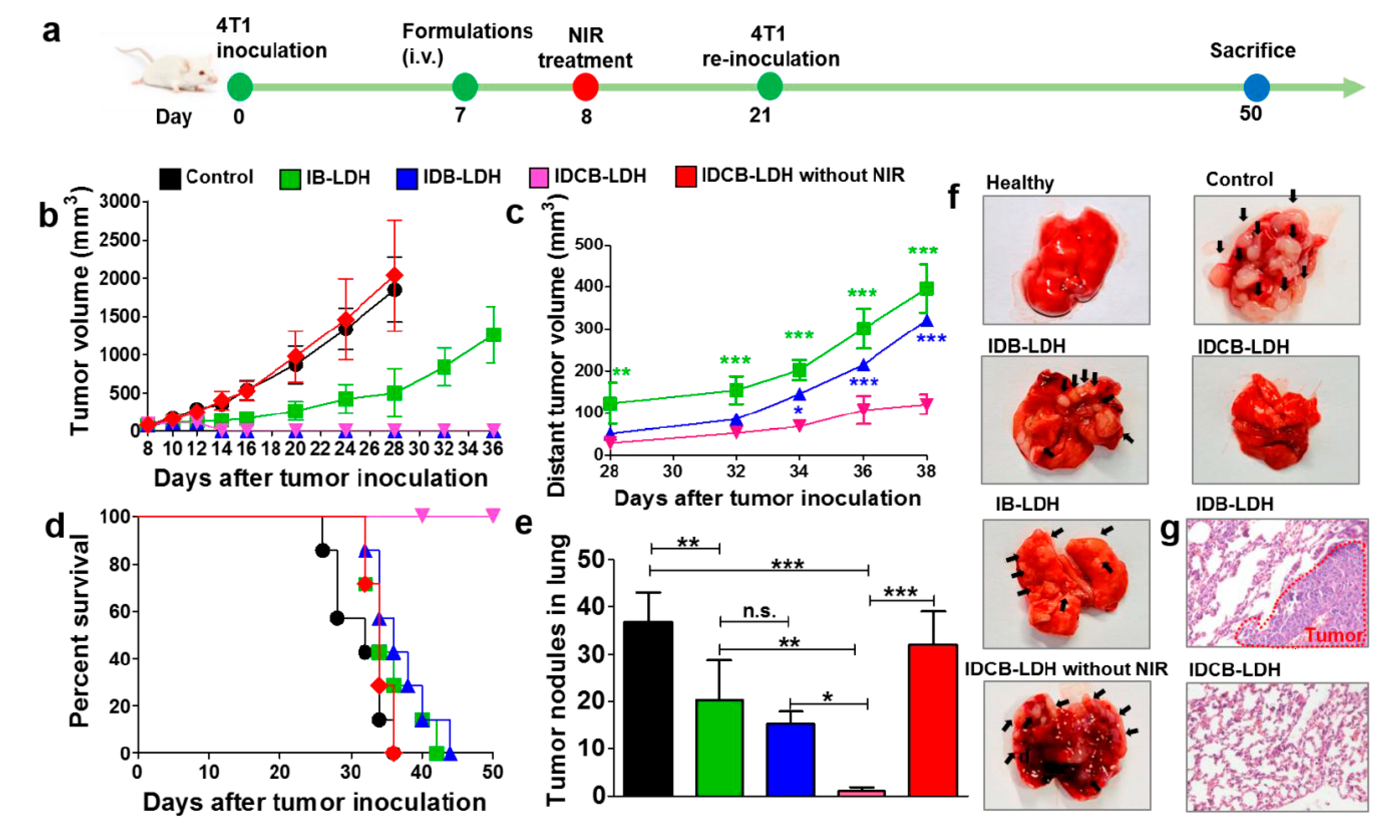
- Zhang, L.X.; Sun, X.M.; Xu, Z.P.; Liu, R.T. Development of multifunctional clay-based nanomedicine for elimination of primary invasive breast cancer and prevention of its lung metastasis and distant inoculation. ACS Appl. Mater. Interfaces. 2019, 11, 35566–35576. [Google Scholar] [CrossRef]
- Byadgi, O.; Puteri, D.; Lee, J.W.; Chang, T.C.; Lee, Y.H.; Chu, C.Y.; Cheng, T.C. The effect of TLR9 agonist CpG oligodeoxynucleotides on the intestinal immune response of cobia (Rachycentron canadum). J. Immunol. Res. 2014, 2014, 273284. [Google Scholar] [CrossRef]
- Raeesi, V.; Chou, L.Y.; Chan, W.C. Tuning the Drug Loading and Release of DNA-Assembled Gold-Nanorod Superstructures. Adv. Mat. 2016, 28, 8511–8518. [Google Scholar] [CrossRef]
- Hartwig, D.D.; Seixas, F.K.; Cerqueira, G.M.; McBride, A.J.; Dellagostin, O.A. Characterization of the immunogenic and antigenic potential of putative lipoproteins from Leptospira interrogans. Curr. Microbiol. 2011, 62, 1337–1341. [Google Scholar] [CrossRef]
- Zhao, K.; Rong, G.; Guo, C.; Luo, X.; Kang, H.; Sun, Y.; Dai, C.; Wang, X.; Wang, X.; Jin, Z.; et al. Synthesis, characterization, and immune efficacy of layered double hydroxide@ SiO2 nanoparticles with shell-core structure as a delivery carrier for Newcastle disease virus DNA vaccine. Int. J. Nanomed. 2015, 10, 2895. [Google Scholar] [CrossRef][Green Version]
- Lee, S.B.; Kim, J.Y.; Kim, K.; Ahn, K.J.; Kim, T.I.; Oh, J.M. Encapsulation and release control of fish pathogen utilizing cross-linked alginate networks and clay nanoparticles for use with a potential oral vaccination. Appl. Sci. 2020, 10, 2679. [Google Scholar] [CrossRef]
- Haines, A.; Nebergall, E.; Besong, E.; Council, K.; Lambert, O.; Gauthier, D. Draft genome sequences for seven Streptococcus parauberis isolates from wild fish in the Chesapeake Bay. Genome Announc. 2016, 4, e00741-16. [Google Scholar] [CrossRef]
- Dwari, R.K.; Mishra, B.K. E valuation of flocculation characteristics of kaolinite dispersion system using guar gum: A green flocculant. J. Min. Sci. Technol. 2019, 29, 745–755. [Google Scholar] [CrossRef]
- Willson, O.C.; Olorunyolemi, T.; Jaworski, A.; Borum, L.; Young, D.; Siriwat, A.; Dickens, E.; Oriakhi, C.; Lerner, M. Surface and interfacial properties of polymer-intercalated layered double hydroxide nanocomposites. Appl. Clay Sci. 1999, 15, 265–279. [Google Scholar] [CrossRef]
- Miörner, H.; Albertsson, P.A.; Kronvall, G. Isoelectric points and surface hydrophobicity of Gram-positive cocci as determined by cross-partition and hydrophobic affinity partition in aqueous two-phase systems. Infect. Immun. 1982, 36, 227–234. [Google Scholar] [CrossRef]
- Meng, Z.; Zhang, Y.; She, J.; Zhou, X.; Xu, J.; Han, X.; Wang, C.; Zhu, M.; Liu, Z. Ultrasound-mediated remotely controlled nanovaccine delivery for tumor vaccination and individualized cancer immunotherapy. Nano Lett. 2021, 21, 1228–1237. [Google Scholar] [CrossRef]
- Spyvee, M.; Hawkins, L.D.; Ishizaka, S.T. Modulators of Toll-like receptor (TLR) signaling. In Annual Reports in Medicinal Chemistry; Academic Press: Cambridge, MA, USA, 2010; Volume 45, pp. 191–207. [Google Scholar]
- Wu, S.; Huang, J.; Zhang, Z.; Wu, J.; Zhang, J.; Hu, H.; Zhu, T.; Zhang, J.; Luo, L.; Fan, P.; et al. Safety, tolerability, and immunogenicity of an randomized adenovirus type-5 vector-based COVID-19 vaccine (Ad5-nCoV) in adults: Preliminary report of an open-label and randomized phase 1 clinical trial. Lancet Infect. Dis. 2021, 21, 1654–1664. [Google Scholar] [CrossRef]
- Hwang, H.S.; Puth, S.; Tan, W.; Verma, V.; Jeong, K.; Lee, S.E.; Rhee, J.H. More robust gut immune responses induced by combining intranasal and sublingual routes for prime-boost immunization. Hum. Vaccines Immunother. 2018, 14, 2194–2202. [Google Scholar] [CrossRef]


| Antigen | Clay | IR | Achieved Immunogenicity | Reference |
|---|---|---|---|---|
| Ovalbumin (OVA) | LDH | i.d. | The candidate nanovaccine induced a higher humoral response than the only-DNA vaccine. In addition, it showed protective immunity against a challenge. Moreover, it induced effective CTL activation and a Th1 immune response. | [82] |
| OVA and CpG ODN 1826 | LDH | s.c. | The candidate induced significant antibody response, and promoted a switch from Th2 to Th1 response. In addition, it retarded tumor growth in a challenge after immunization. | [83] |
| rLipL32 from Leptospira interrogans | HNT | - | The nanovaccine candidate induced a significantly higher IgG response in comparison with the negative control. No protective immunity was provided against a challenge. | [84] |
| Intimin β (IB) from E. coli | LDH, HEC | s.c. | The nanovaccine candidates induced anti-IB IgG levels comparable to those induced with adjuvant. Interestingly, these IgG levels were maintained up to day 120. In addition, higher humoral immunity was recorded for LDH-IB. | [56] |
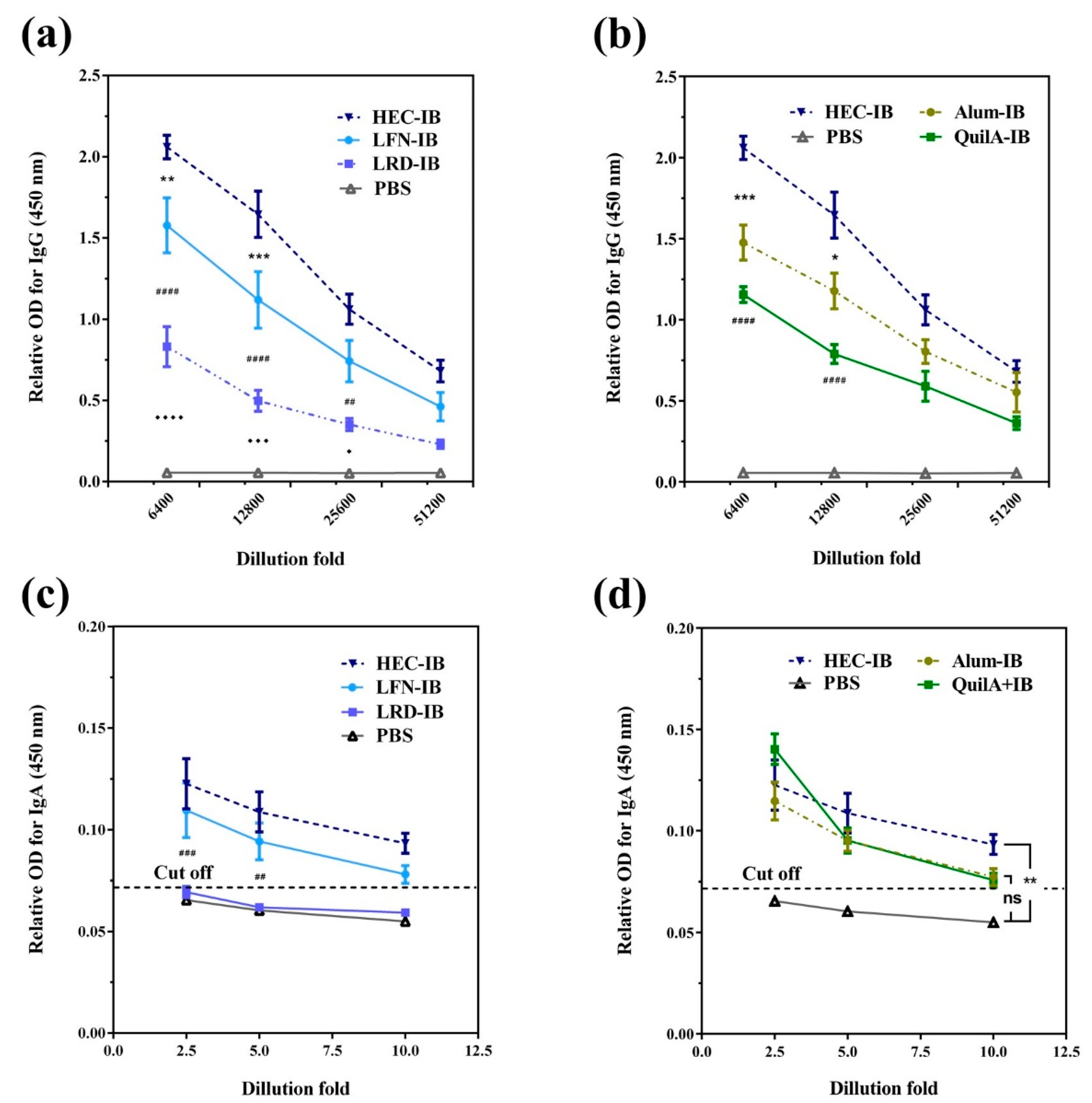
| (IB) from E. coli | HEC | s.c. | The candidate induced the strongest IgG immune response; in addition, it promoted the strongest sIgA secretion. Interestingly, HEC-IB can induce a cellular immune response. | [46] |
| Tyrosinase-related protein 2 (Trp2) and indoleamine 2,3-dioxygenase siRNA (siIDO) | LDH | s.c. | The candidate induced better tumor growth inhibition in mice immunized with the complete nanovaccine Trp2+LDH+siIDO (TLI) than other groups tested, including positive control groups. In addition, TLI induced significantly higher CTL. | [85] |
| IB from E. coli | LDH, HEC | s.c. | Potent cellular and humoral immune responses were induced in groups immunized with LDH-IB or HEC-IB in comparison with positive control groups. The candidate vaccines induced memory T-cell responses. | [57] |
| IB, proprietary antigen 1 (Pag1) and proprietary antigen 2 (Pag2) from E. coli | LDH, HEC | s.c. | The induction of IgG was more efficient in groups receiving LDH or HEC associated with the three antigens, sIgA antigen-specific levels increased at day 108. Similarly, an efficient cell immune response was induced in immunized groups receiving LDH or HEC associated with the three antigens. | [86] |
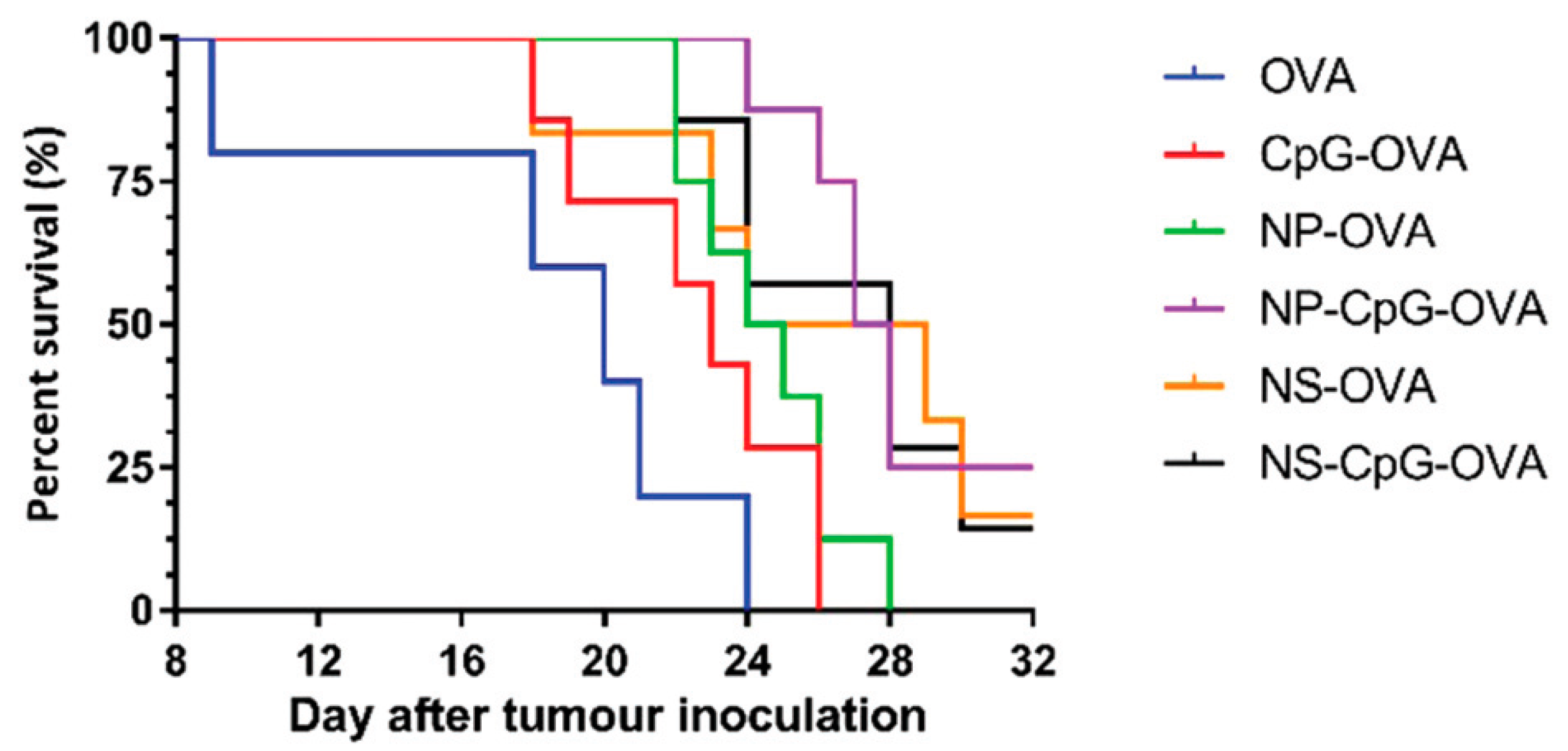
| CpG | LDH | i.v. | The candidate induced DC activation, CTL, and Th2 cells in situ and significantly inhibited the growth of primary and distant tumors. In addition, it significantly increased proinflammatory cytokine levels. | [87] |
| OVA and CpG | LDH, LDH NS | s.c. | The candidate nanovaccines induced strong Th1 and CTL immune responses, promoting inhibition of tumor growth and survivability. | [88] |
| rLemA from Leptospira interrogans | HNT | i.m. | After a challenge, the study revealed the induction of significantly higher IgG antibody response than the control groups. In addition, protective immune responses were observed. | [89] |
| Foot-and-mouth disease virus (FMDV) | LDH | s.c. | The candidate induced higher production of IFN-γ and anti-FMDV IgG antibodies than the positive control group. In pigs, similar levels of neutralizing antibodies were observed in LDH+FMDV and the positive control group, but higher than the negative control group. | [90] |
| OVA | LDH | i.v. and s.c. | The production of IgG antibodies was size-dependent; interestingly, similar levels of IgG1 and IgG2a were induced by the nanovaccine. The candidate showed total protection, mainly by CTL. In addition, the i.v. administration revealed efficient tumor inhibition. | [91] |
| Formalin-killed whole cells (FKC) of Streptococcus agalactiae | HNT | p.o. | HNT loaded with FKC induced an augmented humoral immune response in comparison with the control group, in a time dependent manner. | [92] |
Publisher’s Note: MDPI stays neutral with regard to jurisdictional claims in published maps and institutional affiliations. |
© 2022 by the authors. Licensee MDPI, Basel, Switzerland. This article is an open access article distributed under the terms and conditions of the Creative Commons Attribution (CC BY) license (https://creativecommons.org/licenses/by/4.0/).
Share and Cite
Govea-Alonso, D.O.; García-Soto, M.J.; Betancourt-Mendiola, L.; Padilla-Ortega, E.; Rosales-Mendoza, S.; González-Ortega, O. Nanoclays: Promising Materials for Vaccinology. Vaccines 2022, 10, 1549. https://doi.org/10.3390/vaccines10091549
Govea-Alonso DO, García-Soto MJ, Betancourt-Mendiola L, Padilla-Ortega E, Rosales-Mendoza S, González-Ortega O. Nanoclays: Promising Materials for Vaccinology. Vaccines. 2022; 10(9):1549. https://doi.org/10.3390/vaccines10091549
Chicago/Turabian StyleGovea-Alonso, Dania O., Mariano J. García-Soto, Lourdes Betancourt-Mendiola, Erika Padilla-Ortega, Sergio Rosales-Mendoza, and Omar González-Ortega. 2022. "Nanoclays: Promising Materials for Vaccinology" Vaccines 10, no. 9: 1549. https://doi.org/10.3390/vaccines10091549
APA StyleGovea-Alonso, D. O., García-Soto, M. J., Betancourt-Mendiola, L., Padilla-Ortega, E., Rosales-Mendoza, S., & González-Ortega, O. (2022). Nanoclays: Promising Materials for Vaccinology. Vaccines, 10(9), 1549. https://doi.org/10.3390/vaccines10091549