Article

A New Live Auxotrophic Vaccine Induces Cross-Protection against Klebsiella pneumoniae Infections in Mice

1
Department of Microbiology, University Hospital A Coruña (CHUAC)—Biomedical Research Institute A Coruña (INIBIC), 15006 A Coruña, Spain
2
Technical Services Department, CZ Vaccines S.A.U., 36400 O Porriño, Pontevedra, Spain
3
Centro de Investigación Biomédica en Red de Enfermedades Infecciosas (CIBERINFEC), Instituto de Salud Carlos III, 28029 Madrid, Spain
4
Vaxdyn S.L., 41704 Sevilla, Spain
5
Intrahospital Infections Laboratory, National Centre for Microbiology, Instituto de Salud Carlos III, 28220 Madrid, Spain
*
Author to whom correspondence should be addressed.
These authors contributed equally to this work.
Vaccines 2022, 10(6), 953; https://doi.org/10.3390/vaccines10060953
Submission received: 30 May 2022 / Revised: 13 June 2022 / Accepted: 14 June 2022 / Published: 16 June 2022
(This article belongs to the Special Issue Development of Attenuated Vaccine)

Abstract

:
The development of a whole-cell vaccine from bacteria auxotrophic for D-amino acids present in the bacterial cell wall is considered a promising strategy for providing protection against bacterial infections. Here, we constructed a prototype vaccine, consisting of a glutamate racemase-deficient mutant, for preventing Klebsiella pneumoniae infections. The deletion mutant lacks the murI gene and requires exogenous addition of D-glutamate for growth. The results showed that the K. pneumoniae ΔmurI strain is attenuated and includes a favourable combination of antigens for inducing a robust immune response and conferring an adequate level of cross-protection against systemic infections caused by K. pneumoniae strains, including some hypervirulent serotypes with elevated production of capsule polysaccharide as well as multiresistant K. pneumoniae strains. The auxotroph also induced specific production of IL-17A and IFN-γ. The rapid elimination of the strain from the blood of mice without causing disease suggests a high level of safety for administration as a vaccine.

1. Introduction

Klebsiella pneumoniae, a Gram-negative bacterium with a high genetic diversity, is widely distributed in multiple environmental and host-associated niches. This microorganism acts as an opportunistic pathogen that can asymptomatically colonize the human gastrointestinal tract and the nasopharynx. The colonization of the gastrointestinal tract by opportunistic K. pneumoniae strains could be considered the first step in the nosocomial infection development, from which bacteria may disseminate to other tissues, causing life-threatening infections [1]. K. pneumoniae strains are generally surrounded by a thick hydrophilic polysaccharide capsule (CPS) and at least 77 antigenically distinct CPSs have been recognized (K-antigens). Moreover, nine O-antigen types that are distinguished in the lipopolysaccharide of K. pneumoniae protect this bacterium from complement-mediated killing. Both K- and O-antigens are important virulence factors used to differentiate K. pneumoniae isolates. K. pneumoniae, which is included in the ESKAPE pathogens group responsible for the majority of nosocomial infections worldwide, has become resistant to most antimicrobial agents [2]. Additionally, according to the WHO, extended-spectrum β-lactam (ESBL)-producing and carbapenem-resistant K. pneumoniae are of major concern in global public health [3].
K. pneumoniae has evolved into two pathotypes: classical and hypervirulent (hvKp) [4]. The classical K. pneumoniae is the leading cause of opportunistic healthcare-associated infections, such as urinary tract infections, pneumonia, wound and surgical site infections, and bacteraemia [5]. However, severe community-acquired infections, including pyogenic liver abscess, endophthalmitis, meningitis and community-onset pneumonia, are associated with hvKp strains considered to be strict pathogens [6]. Populations at greatest risk of K. pneumoniae infection are neonates and elderly and immunocompromised individuals, including those with diabetes, chronic lung conditions, HIV-positive individuals and hospitalized patients. Most hvKp clones associated with invasive disease express a hypermucoid CPS type K1 or K2 and produce increased levels of siderophores [7]. Although, to date, these hvKp clones have predominantly infected individuals in Southeast Asia and South Africa [5], these infections are now increasing worldwide [6].
Some multidrug-resistant (MDR) K. pneumoniae clones cause localized infections within a single hospital (e.g., sequence type (ST) 70 or ST323). However, a subset of these MDR clones are widely distributed and causing outbreaks in hospital settings, especially in long-term care and pediatric units, and they have become global problematic clones. These include clonal groups (CGs) 258, CG15, ST17, CG29, CG37, ST101, CG147 and CG307. Furthermore, hvKp infections are associated with other clones, such as CG23, CG65 and CG86 [5].
Most of the described mechanisms of antibiotic resistance in K. pneumoniae are associated with the acquisition of large conjugative plasmids: ESBLs that provide resistance to third-generation cephalosporins and monobactams and to various serine carbapenemases (e.g., KPC, OXA-48) and metallo-β-lactamases (e.g., NMD-1, VIM, IMP) that confer resistance on almost all available β-lactams, including the carbapenem family [1,8,9,10]. In contrast to the classical K. pneumoniae strains, the hvKp variants are usually sensitive to most antimicrobial agents, but convergence of MDR and hypervirulence has nevertheless also been reported [5,11]. The global increase in MDR, particularly carbapenem-resistant K. pneumoniae, has led to the reuse of polymyxin/colistin to treat these infections (despite the reported neurotoxic and nephrotoxic side effects) or to the use of combinations with β-lactamase inhibitors, such as ceftazidime/avibactam [12]. However, the emergence and spread of plasmid-mediated resistance to colistin [13] and plasmid mutations during treatment with β-lactamase inhibitor combinations [14] have severely limited the available antimicrobial therapy options in the few last years, leading to renewed interest in vaccines against K. pneumoniae infections.
Currently, there is no approved vaccine for preventing K. pneumoniae infections, although different vaccine strategies have been explored: Uromune®, a whole-cell inactivated polybacterial sublingual vaccine [15]; a live attenuated vaccine based on tonB gene deletion, encoding an iron-uptake protein [16]; Klebvax®, a 24-valent Klebsiella CPS vaccine, which was not developed further, partly due to the wide range of clinically relevant CPS types [17,18]; conjugated vaccines linking polysaccharides to different carrier peptides (BSA, KLH, CRM197) [19,20]; and bioconjugate vaccines targeting the capsule of hvKp [21]. Vaccines based on the four most prevalent Klebsiella O serotypes related to human infections (O1, O2, O3 and O5) have recently been developed as conjugate and multiantigen-presenting system vaccines eliciting good immunogenicity in mice. Additional vaccine antigens (e.g., outer membrane proteins, type 3 fimbriae) and novel vaccine platforms (e.g., nanoparticles, liposomes) were also considered, as well as the addition of adjuvants [22,23].
In this study, we designed and developed a prototype vaccine consisting of a deletion mutant of K. pneumoniae MGH 78578 that results in D-glutamate auxotrophy. This strain shows promising potential as a live vaccine for the prevention of Klebsiella-induced sepsis.

2. Materials and Methods

2.1. Bacterial Strains, Growth Conditions and Plasmids

The bacterial strains and plasmids used in this study are listed in Table 1. All strains were grown in Luria–Bertani (LB) broth (10 g/L tryptone, 5 g/L yeast extract and 10 g/L NaCl) or low-salt LB (reducing NaCl to 5 g/L when hygromycin was needed) with aeration at 37 °C. For plasmid selection in Escherichia coli, apramycin (Duchefa Biochemie), ampicillin and hygromycin (Sigma-Aldrich, Merck Life Science S.L.U., Madrid, Spain) were added at concentrations of 50, 50 and 100 µg/mL, respectively. Apramycin and hygromycin were added to the medium at 200 µg/mL and 1250 µg/mL, respectively, to select the recombinants in K. pneumoniae. MGH 78578 ΔmurI was cultivated in LB medium supplemented with 10 mM D-glutamate (Sigma-Aldrich).

2.2. Construction of the murI Deletion Mutant of K. pneumoniae MGH 78578

The procedure used to construct the ΔmurI mutant strain has been reported previously [28]. The strategy was based on the use of the E. coli λ-Red recombinase system to replace the target gene and FLP recombinase for final excision of the antibiotic marker. The primers used to generate the knockout cassette were murIKOFW and murIKORV, including homology arms of 60 nt immediately upstream and downstream of the region to be deleted (Table 1). Hygromycin-sensitive single colonies that grew only on D-glutamate-containing plates were the mutant candidates. The murI deletion was confirmed by PCR with specific primers (EXTMURIFW and EXTMURIRV, Table 1) and sequence analysis.
The murI gene expression was analyzed by qRT-PCR in MGH 78578 and ΔmurI strains with the primers listed in Table 1. The qRT-PCR protocol was performed as previously described [29]. The expression levels were normalized relative to the transcription levels of the rpoB housekeeping gene.

2.3. Growth and Viability Curves

Growth and viability of K. pneumoniae MGH 78578 and its murI-deficient mutant were determined as previously described [29]. Samples were taken at different times to measure the culture turbidity (optical density at 600 nm, OD600) and to determine the colony-forming units (CFUs) on LB agar supplemented or not with 10 mM D-glutamate. All cultures were prepared in triplicate.

2.4. Electronic Microscopy Analysis

Samples for scanning and transmission electron microscopy (SEM and TEM) were prepared in the absence or presence of 10 mM D-glutamate, as previously described [29]. Briefly, for SEM, the bacterial overnight cultures were washed with 0.9% NaCl, diluted 1:100 in LB and incubated at 37 °C with agitation for 2 h. Then, the cultures were washed again, diluted 1:100 in LB in the presence or absence of D-glutamate and incubated for another 2 h before fixation with 4% paraformaldehyde. After fixation, samples were washed and dehydrated in an ethanol series. For TEM, 2 or 3 colonies of each strain were plated onto LB agar supplemented or not with D-glutamate and incubated overnight at 37 °C. Then, the first streak of each plate was dissolved in PBS buffer, washed with cacodylate buffer and the cells were prefixed with 2.5% glutaraldehyde for 4 h and fixed with 1% osmium acetate. After dehydration with acetone, cells were embedded in SPURR, and ultrathin sections of these samples were obtained and examined.

2.5. Control of Phenotypic Stability

Overnight cultures of K. pneumoniae MGH 78578 ΔmurI were diluted (1:100) in 100 mL of LB supplemented with 10 mM D-glutamate and incubated at 37 °C under agitation (180 rpm) for up to 8 days. Samples from the cultures were taken on different days, washed twice and plated on LB agar in the presence or absence of 10 mM D-glutamate for determination of cell viability. Agar plates were incubated at 37 °C for 4 days. Cultures were prepared in triplicate.

2.6. Water Survival Assay

The viability of K. pneumoniae strains in water was determined as previously described [29]. Samples of cultures were taken on different days until day 79 to determine the number of CFUs in LB agar (wild-type strain) and LB agar with 10 mM D-glutamate (ΔmurI mutant strain). Cultures were prepared in triplicate.

2.7. In Silico Molecular Typing, Capsular Type and Resistance Profile Predictions

Multilocus Sequence Typing (MLST) was determined in silico using bacterial genomic data and the MLST 2.0 server available on www.genomicepidemiology.org (28 March 2022) and Kleborate software v2.2.0 (Melbourne, VIC, Australia) [30]. Antibiotic-resistance genes were screened using the Resfinder v4.1 software available from the Center for Genomic Epidemiology (https://cge.cbs.dtu.dk/services/ResFinder/ accessed on 28 March 2022) [31]. Capsular type (K-antigen) and O-typing were predicted in silico using Kaptive v2.0.0 (Melbourne, VIC, Australia) [32].

2.8. Ethics Statement

Animal experiments were performed according to the recommendations and the guidelines of the European Union (Directive 2010/63/EU) and current national legislation (RD 53/2013) on the protection of animals used for scientific purposes. Animals were bred and maintained under specific pathogen-free conditions in the facilities at the A Coruña University Hospital (CTF-XXIAC) and were provided with free access to food and water.

2.9. Mouse Experiments

Female BALB/c mice aged 6 to 9 weeks were used in this study. For inoculations, the K. pneumoniae MGH 78578 ΔmurI strain was prepared as previously described [29], with minor modifications. In brief, the bacterial strain was grown in LB supplemented with 10 mM D-glutamate at 37 °C with shaking at 180 rpm until an OD600 of 0.7 was reached. Then, cells were harvested by centrifugation, washed and suspended in saline solution. The bacterial suspension was intraperitoneally (i.p.) injected (0.1 mL containing 4–6 × 10⁶ CFU unless otherwise indicated) into mice in a two-dose schedule (days 0 and 14). Serial dilutions of the inoculum were plated to verify actual CFUs delivered to the mice. For passive immunization tests, pools of sera were obtained from BALB/c mice (n = 5) i.p. administered three times with the vaccine candidate (2.6 × 10⁷ CFU) at 7-day intervals. Blood samples were taken from the submandibular vein or by puncture of the retro-orbital plexus as described in [29].
The protective efficacy of the vaccine was evaluated by i.p. challenge (0.1 mL) of control and immunized BALB/c mice (n = 6–8) with a lethal dose of virulent strains of K. pneumoniae on day 21. Clinical signs were examined twice-daily for a period of 7 days to measure disease severity and survival. Mice were euthanized 36 h post-infection, and the organs (spleens, livers and lungs) were aseptically removed, homogenized in sterile NaCl 0.9% and plated on LB agar for determination of CFU counts, in order to estimate bacterial dissemination to organs.

2.10. ELISA

The levels of total IgG and of subclasses IgG1, IgG2a, IgG2b and IgG3, and IgM antibodies were quantified in mouse serum using a whole-bacterial cell ELISA as previously described [29]. Cytokines IL-2, IL-4 and IL-17A, and IFN-γ were evaluated in the cellular supernatant of splenocytes isolated from immunized and control mice on day 55 after the second immunization. Mouse spleens were removed aseptically and mechanically disrupted. The cell suspension was enriched for lymphocytes by a gradient centrifugation. Then, splenocytes were ex vivo restimulated with the vaccine strain and incubated at 37 °C, 5% CO2 for 48 h. The cytokine levels were measured with a commercial ELISA kit (Affymetrix) according to the manufacturer’s instructions, with minor modifications [33].

2.11. Statistical Analysis

Mean values were compared using a Student’s t-test. Survival analysis was conducted using Kaplan–Meier curves and the Mantel–Cox log-rank test. Comparisons between pairs of groups were analyzed using the nonparametric and unpaired Mann–Whitney U test. p-values < 0.05 were considered statistically significant. Statistical analyses were performed using GraphPad Prism software package (version 6.01, GraphPad Prism Software Inc., San Diego CA, USA). Lethal doses were calculated using the probit analysis tool in the R package “ecotox” v1.4.4.

3. Results

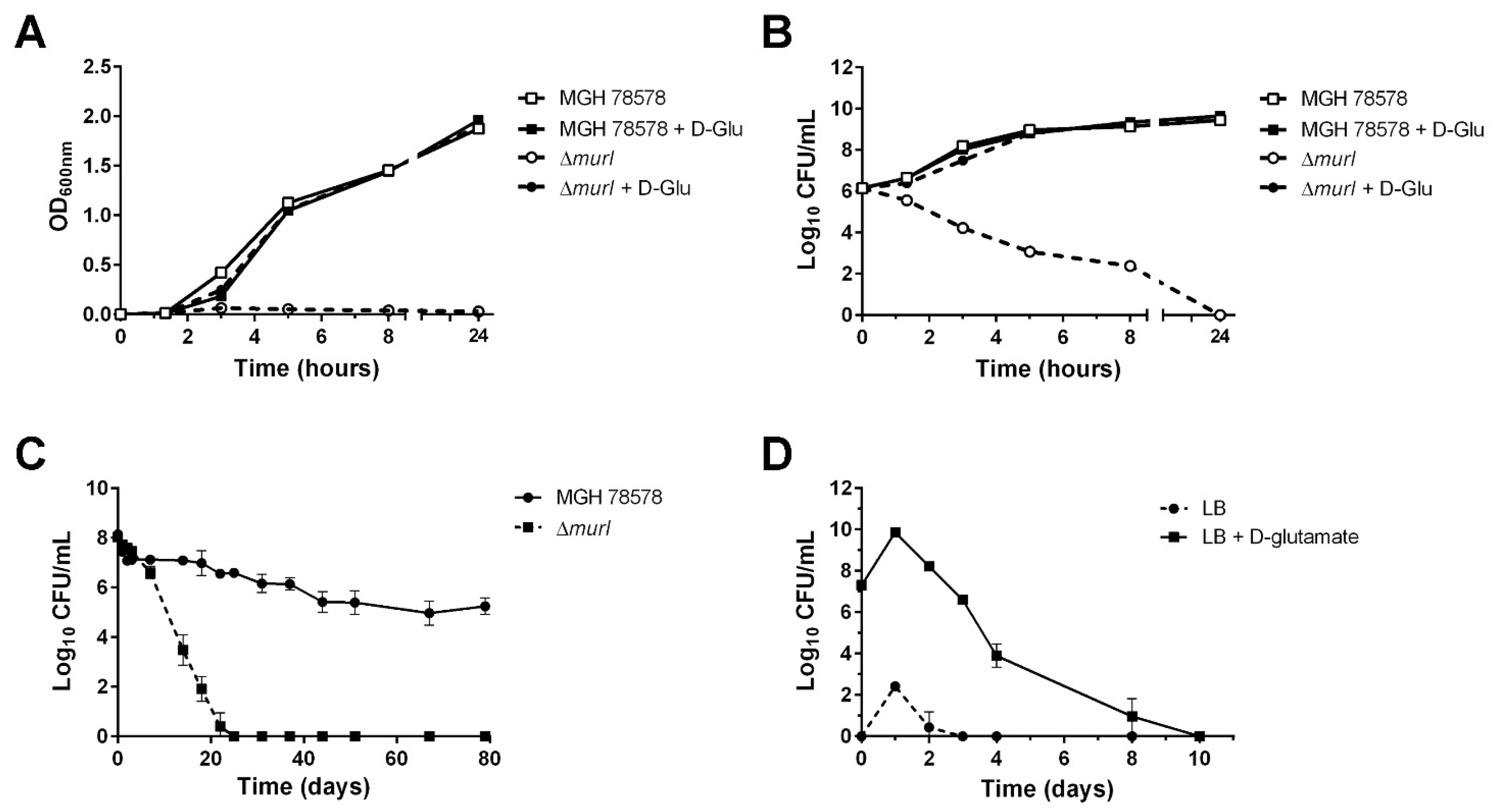
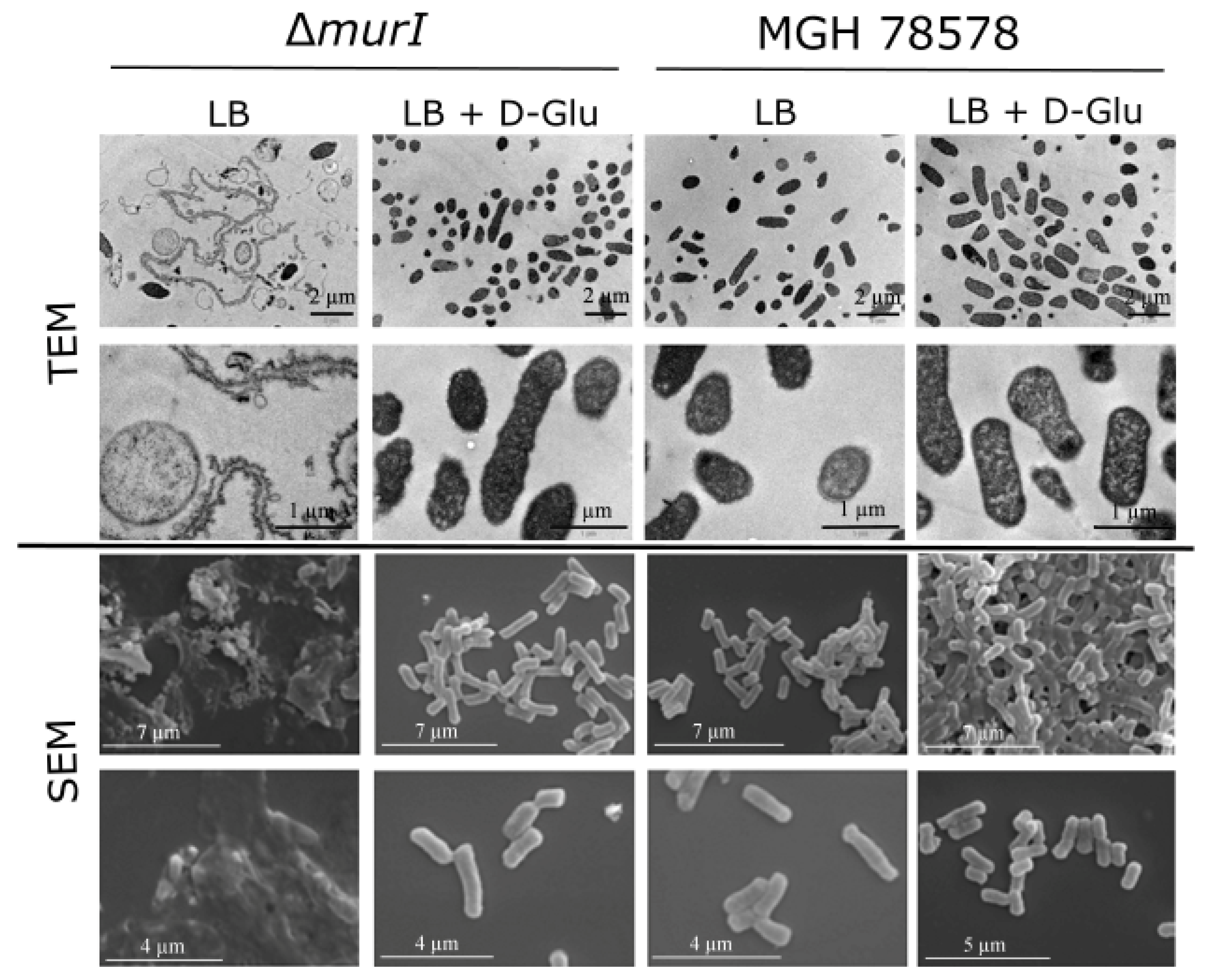
3.1. Characterization of the K. pneumoniae MGH 78578 Glutamate Racemase-Deficient Mutant

Analysis of the genome sequence of the K. pneumoniae MGH 78578 strain revealed the existence of a single putative D-glutamate racemase gene: murI (KPN_04256). In order to produce D-glutamate auxotrophs, the murI gene was deleted from the chromosome of MGH 78578 via a λ-Red knockout system [28]. The murI deletion was confirmed by PCR with primers located upstream and downstream of the murI gene (Table 1): a 1189 bp fragment from the strain carrying the wild-type allele and a 470 bp fragment from the strain carrying the ΔmurI allele (Figure S1). Moreover, the absence of murI gene mRNA in the mutant strain was confirmed by qRT-PCR (data not shown). MGH 78578 grew normally on LB agar without D-glutamate, while the MGH 78578 ΔmurI mutant required D-glutamate supplementation for growth (Figure 1A). Moreover, a 5-log reduction in viable cell counts was observed after 5 h during incubation of the deletion mutant in LB without D-glutamate, and no viable bacteria were recovered from the culture after 24 h (Figure 1B).
The persistence of the D-glutamate auxotrophic strain in the environment was determined by survival analysis in water: the viability of the ΔmurI mutant strain was considerably reduced, and no viable bacteria were recovered after 22 days in water. By contrast, the wild-type counterpart was still viable on day 79 (Figure 1C). There was a significant difference in water survival between the two strains (p = 0.0006, Student’s t-test).
To test the phenotypic stability of the nutritional auxotrophy of K. pneumoniae MGH 78578 ΔmurI, cultures were grown in LB with 10 mM D-glutamate for 10 days and samples were taken on different days and plated on LB agar either with or without D-glutamate. Bacterial counts were significantly higher in supplemented plates at the initial incubation stage (day 0) and over the following days (Figure 1D) (p = 0.0059, Student’s t-test). The recovery of a few colonies in the medium without D-glutamate during the first days may be due to the use of accumulated D-glutamate in the cytoplasm during the initial growth in supplemented media. This difference shows that the ΔmurI strain remains auxotrophic for D-glutamate over time.
Inspection of TEM and SEM micrographs showed that the ΔmurI mutant is unable to divide in the absence of D-glutamate and has filamentous aggregates, protoplast-like structures and cellular debris (Figure 2). However, after D-glutamate supplementation at 10 mM, the ΔmurI mutant cells had a similar appearance to their wild-type homologue in terms of both cell density and morphology.

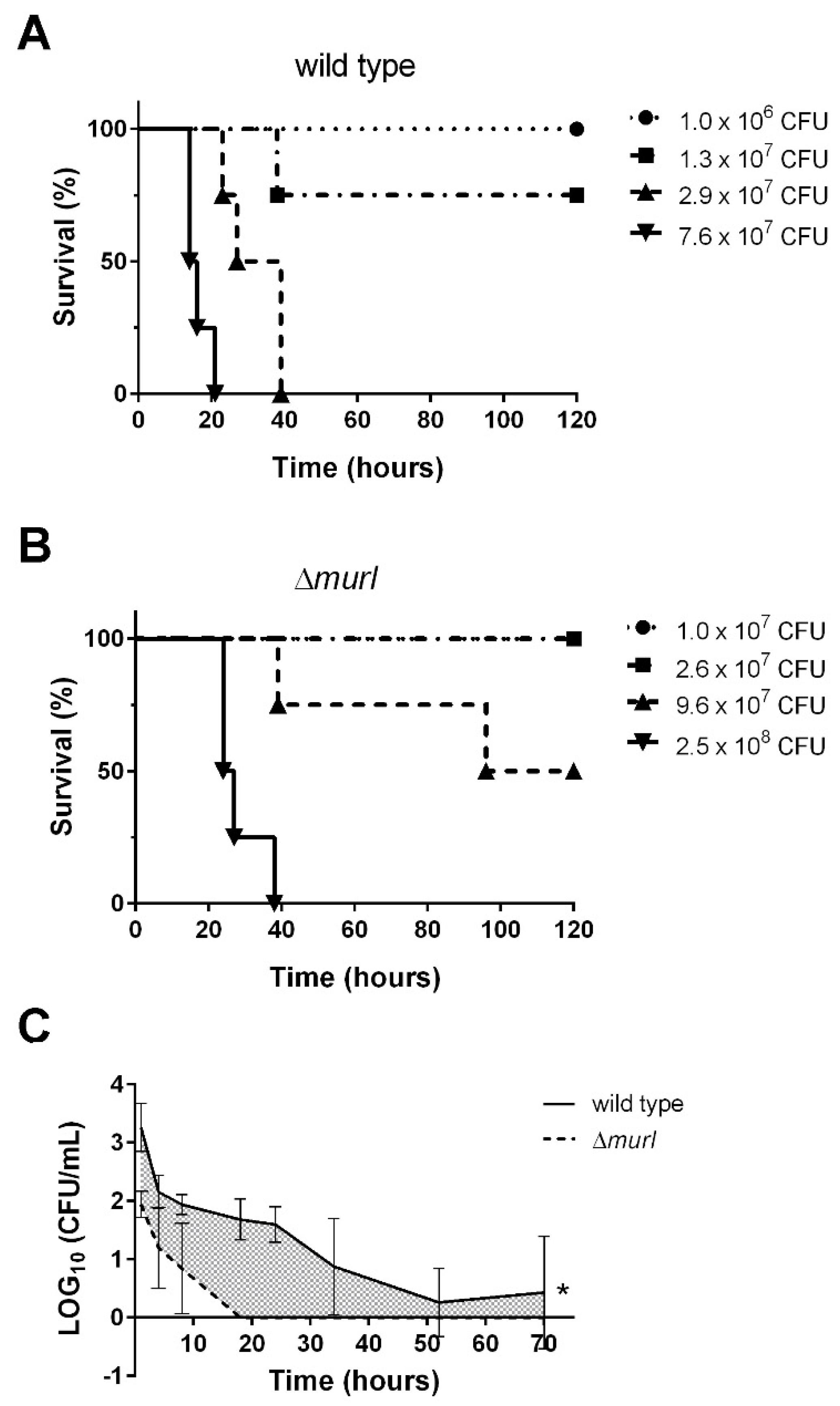
3.2. The D-glutamate Auxotroph of K. pneumoniae Is Attenuated in BALB/c Mice

Assessment of the impact on K. pneumoniae virulence revealed a marked decrease in the survival of mice inoculated with doses of 1.8 × 10⁷ CFU and higher of the wild-type strain. The lethal dose at which 50% of susceptible mice will die (LD50) of the wild-type strain MGH 78578 was calculated by the probit analysis to be 1.40 × 10⁷ CFU (Figure 3A). However, the estimated LD50 of the mutant strain ΔmurI was 9.6 × 10⁷ CFU (Figure 3B).
In order to evaluate the safety of K. pneumoniae ΔmurI as a vaccine, BALB/c mice either received an intravenous dose of the wild-type (1.2 × 10⁷ CFU; 0.1 mL) or ΔmurI (8.9 × 10⁶ CFU; 0.1 mL) strains prepared in saline media. Blood samples were then collected from the mice at different times. The ΔmurI strain was completely cleared from blood, with no colonies recovered beyond 18 h (Figure 3C), which suggests a high margin of safety for administration of the strain as a vaccine.

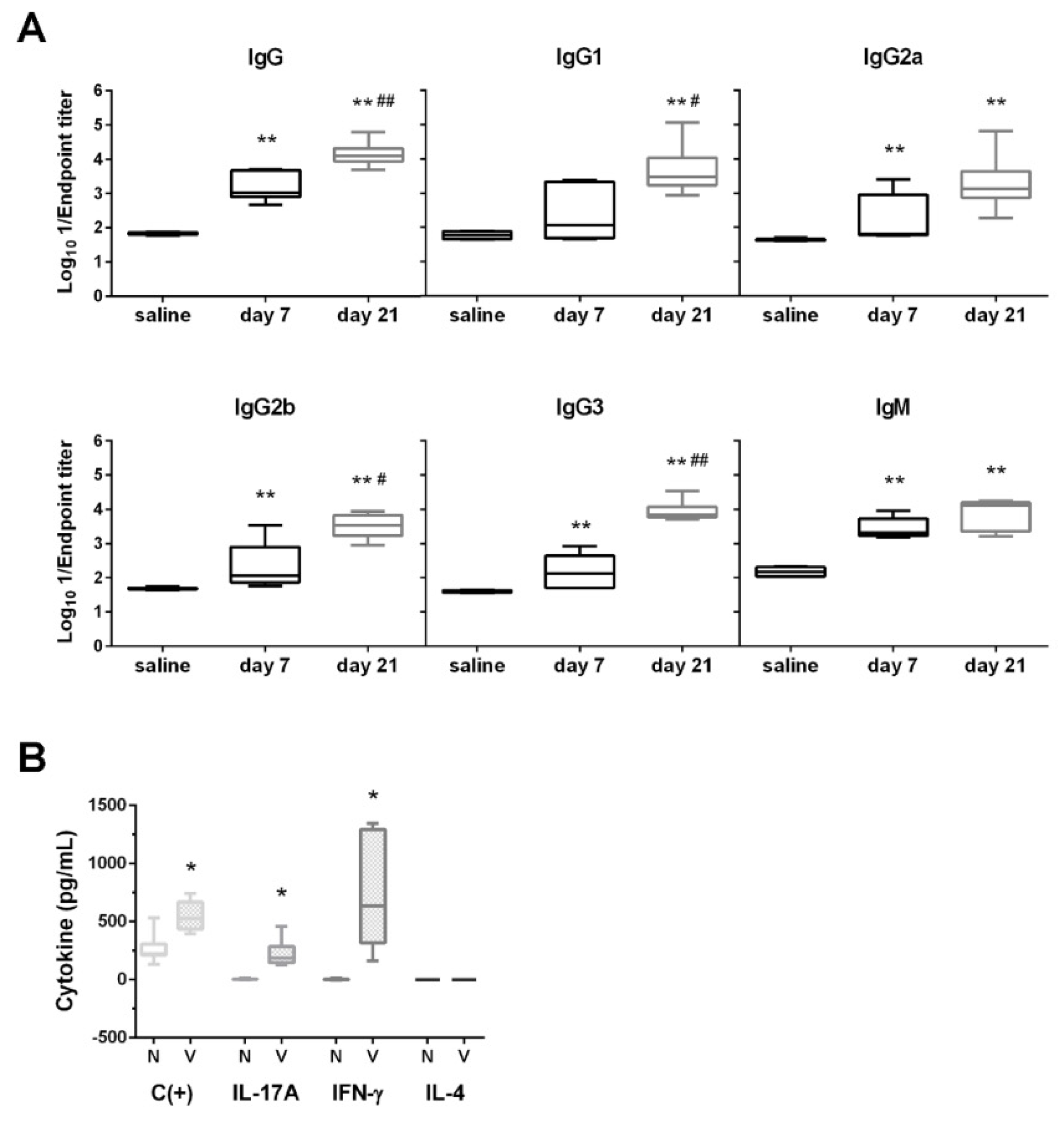
3.3. D-glutamate Auxotrophic Strain Generates Robust Humoral and Cellular Immune Responses against K. pneumoniae

Antibody-mediated immunity was evaluated after inoculation with the D-glutamate auxotroph to determine the minimum immunizing dose. As shown in Figure S2, significantly higher levels of antibodies against the wild-type strain were detected in all immunized mice compared with unvaccinated mice on days 7 and 14 (after one injection) and 21 (after two injections) (p < 0.005, Mann–Whitney U test). In contrast, on day 21 significant differences in IgG levels were observed between the group of mice inoculated with the dose of 7.2 × 10⁴ CFU and those in the control group (p < 0.005, Mann–Whitney U test), although to a lesser extent than with greater doses of vaccine. This demonstrates that a very low dose (100-fold lower than the dose of 7.9 × 10⁶ CFU) is adequate to trigger IgG production in mice, showing the immunogenic potential of the strain.
In addition, on day 21 post-vaccination, significant levels of IgG and IgM immunoglobulins against the parent strain were present in all mice injected with the ΔmurI strain (** p < 0.005, Mann–Whitney U test) and high levels of all IgG isotypes were determined (Figure 4A). Levels of all antibodies, except for IgG2a and IgM, were significantly higher after two immunizations (# p < 0.05 and ## p < 0.005, Mann–Whitney U test) than with one inoculation.
The ability of the aforementioned vaccine to stimulate cellular immune responses was assessed by measuring the secretion of cytokines in cellular supernatants of splenocytes. A strong production of IL-17A and IFNγ was observed after ex vivo antigen-specific restimulation with the vaccine strain, whereas no IL-4 was detected (Figure 4B).

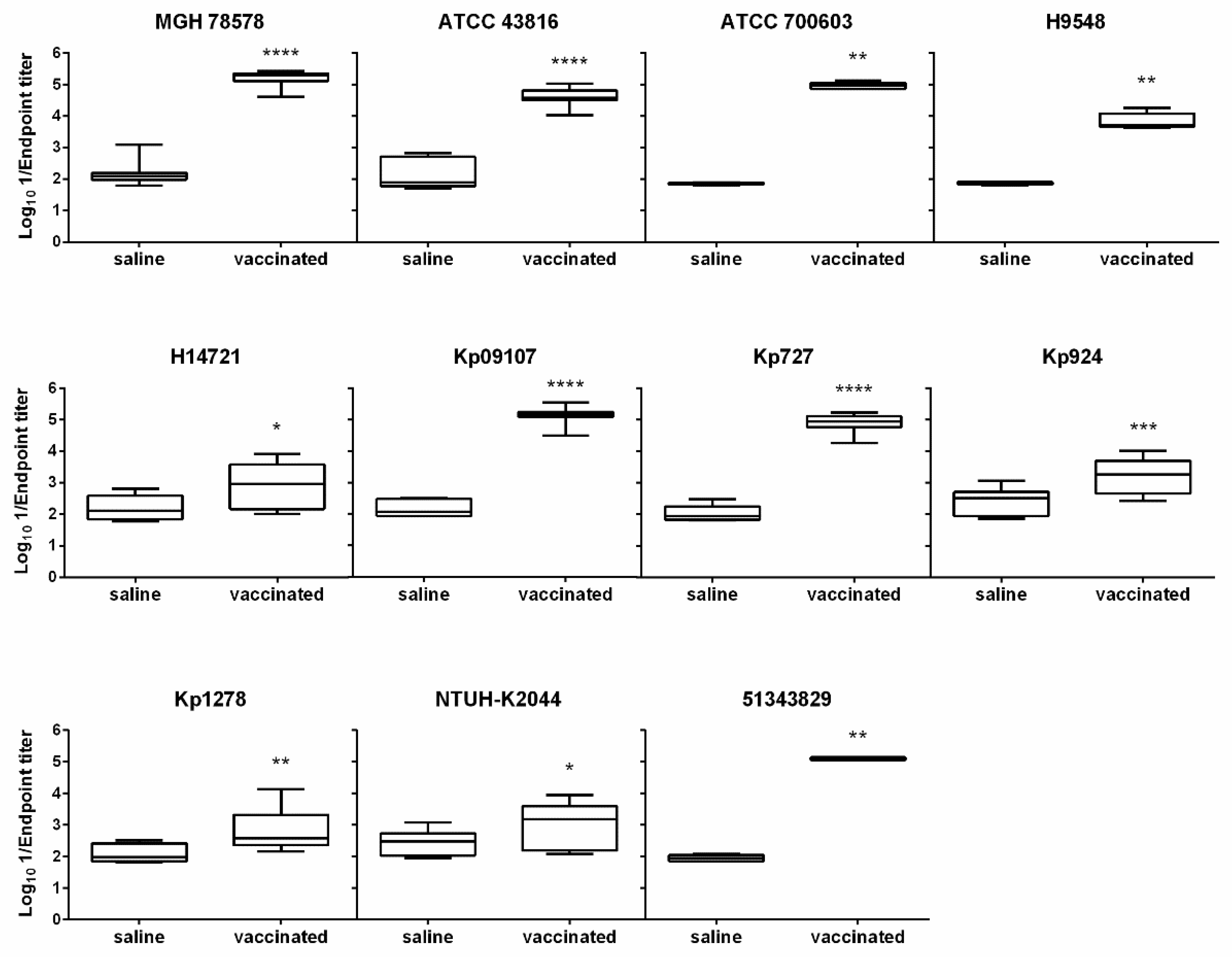
3.4. The D-glutamate Auxotroph Vaccine Generates Cross-Protective Antibodies against K. pneumoniae Heterologous Strains

The capacity of the vaccine strain to generate a broad immune response against the parental and several unrelated Klebsiella spp. strains, including MDR or hypervirulent clones belonging to different ST and K-types, was determined (Table 1, Tables S1 and S2). Significant IgG antibody titers were detected against all heterologous K. pneumoniae strains tested (p < 0.005, Mann–Whitney U test) (Figure 5). Highly significant IgG antibody titers were detected against five of these strains (ATCC 43816, ATCC 700603, Kp09107, Kp727 and 51343829), similar to that determined against the isogenic strain MGH 78578. These data demonstrate that inoculation with the ΔmurI strain not only produces antibodies against the isogenic strain, but also induces IgG antibodies that cross-react with other unrelated K. pneumoniae and K. quasipneumoniae strains.

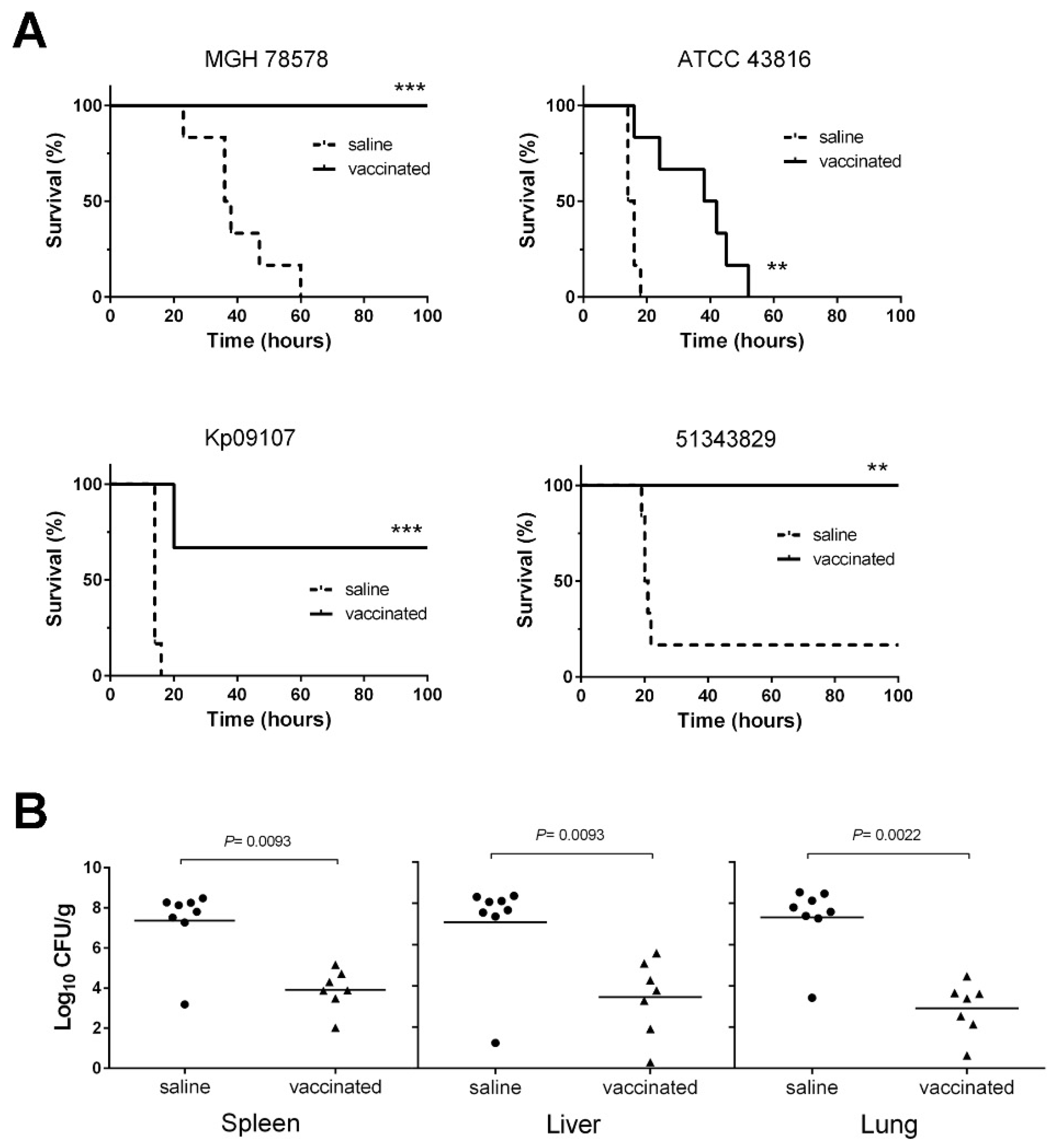
3.5. D-glutamate Auxotrophic Strain of K. pneumoniae MGH 78578 Elicits Protection against Infection with Different K. pneumoniae Strains

To investigate whether vaccination with the ΔmurI strain was sufficient to provide protection against K. pneumoniae lethal infections, BALB/c mice were challenged with other heterologous K. pneumoniae strains, including highly virulent strains and clinical isolates. When infected with 2.3 × 10⁷ CFU of the parental strain MGH 78578, all mice (n = 6) succumbed within the first 60 h after infection. By contrast, all vaccinated mice (n = 6) survived after overcoming the infection (p = 0.0005, Mantel–Cox log-rank test) (Figure 6A).
In the case of challenge with K. pneumoniae Kp09107, six deaths were reported in the group of unvaccinated mice during the first 16 h (n = 6; 100% mortality rate). By contrast, only two vaccinated mice died 20 h after infection (n = 6; 67% survival rate) Statistical analysis showed that survival differences between the two groups were highly significant (p = 0.0005, Mantel–Cox log-rank test). In the challenge with the ATCC 43816 hypervirulent strain, we found that all unvaccinated mice died within the first 18 h. Furthermore, the vaccinated mice inoculated with the ATCC 43816 strain survived for significantly longer (p < 0.005, Mantel–Cox test) and the death rate was slightly lower than in the unvaccinated mice. After challenge with the K. pneumoniae 51343829 strain (5.2 × 10⁷ CFU), five deaths were observed in the group of unvaccinated mice (83.3% mortality rate; p < 0.005, Mantel–Cox test). By contrast, all vaccinated mice survived this challenge and overcame infection (100% survival rate). For all of the strains tested, immunization with ΔmurI significantly decreased or delayed mortality, relative to sham-immunization (log-rank Mantel–Cox test) (Figure 6A). Vaccination with this modified strain could therefore elicit protective immunity against infections caused by a diverse group of K. pneumoniae strains.
The passive transfer of anti-K. pneumoniae sera (anti-Kp) to naive mice by i.p. injection 3 h before challenge with MGH 78578 (2.6 × 10⁸ CFU) resulted in a significant level of survival in mice (62.5%; p < 0.05, log-rank test), while 87.5% of control mice that received naive serum succumbed to infection (Figure S3).
The bacterial load in different organs (spleens, livers and lungs) obtained from vaccinated mice after challenge with K. pneumoniae MGH 78578 (3.2 × 10⁸ CFU) was significantly lower than in the control group (at least a 3-log-unit reduction; p < 0.01, log-rank test), indicating that vaccination prevented bacterial dissemination to internal organs and the spread of systemic infection (Figure 6B).

4. Discussion

The increase in the occurrence of MDR, particularly ESBL-producing and carbapenem-resistant K. pneumoniae, is often associated with significant morbidity and high mortality rates among patients with bacteraemia [34,35]. In addition, the recent emergence of convergence of MDR and hypervirulent isolates and the lack of an effective vaccine against this pathogen [36,37,38] demands urgent efforts to accelerate research and development of new treatments and prevention strategies. The development of vaccines containing live attenuated strains auxotrophic for D-amino acids present in cell wall peptidoglycan is considered a promising approach to fight both Gram-positive and Gram-negative bacterial pathogens [29,33]. Like all whole-cell attenuated vaccines, our auxotrophic vaccines also exhibit complete coverage of bacterial epitopes, lower manufacturing costs and longer-term preservation; additionally, they are also safer, as replication is self-limited in the absence of specific requirements for growth. In this study, we constructed a glutamate racemase-deficient mutant of K. pneumoniae MGH 78578 for use as a potential vaccine against Klebsiella spp. infections. This ΔmurI deletion mutant exhibited an absolute requirement of D-glutamate for growth and featured a stable auxotrophic phenotype. Interestingly, this vaccine candidate was shown to be less virulent than the parental wild-type strain and did not require an adjuvant to elicit a protective immune response. Importantly, this strain does not represent a risk for causing disease, as it is rapidly eliminated from the blood of mice in vivo.
We have shown that inoculation of mice with D-glutamate auxotrophic K. pneumoniae strain leads to high production of IgG and IgM antibodies. However, the IgG subclass distribution did not elicit predominance of the particular IgG isotype, suggesting a balanced Th1/Th2 profile. Higher levels of IgG3-specific antibodies were also induced in all the mice receiving the D-glutamate auxotrophic K. pneumoniae strain relative to the levels induced by DNA vaccines [39]. IgG1 and IgG3 responses are often linked during infection, and both antibodies can efficiently trigger the classical route of complement activation and promote opsonization [40,41]. Early IgG3 responses against protein antigens upon infection may be beneficial for the rapid clearance of pathogens [41]. The D-glutamate auxotrophic vaccine generates cross-reactive antibodies and induces a protective immune response against several heterologous K. pneumoniae strains, MDR or hypervirulent clones with different STs and K-types (Tables S1 and S2, Figure S4). However, lower serum IgG levels against isolates with CPS K1 and K24 were reached relative to the other strains tested. This result suggests that both the antibody production and a vaccine-induced T-cell response would be needed to fully protect against K. pneumoniae infections. In fact, we demonstrated an increase in IL-17A and IFN-γ production in response to immunization with the D-glutamate auxotroph vaccine. Th1 effector cytokines such as IFN-γ may play a crucial role in the resolution of K. pneumoniae lung infection by enhancing the antimicrobial activities of alveolar macrophages, resulting in bacterial clearance [42,43], while IL-17A may collaborate in promoting neutrophil recruitment and local control of pulmonary infection [44]. Vaccination with K. pneumoniae-derived extracellular vesicles was previously shown to elicit specific production of IFN-γ [45]. Likewise, a T-cell immune response mediated by IFN-γ, in addition to IL-4 and IL-17A, was also reported in mice vaccinated with some recombinant outer membrane proteins of K. pneumoniae [46].
In this study, the passive transfer of anti-Kp sera, elicited in response to inoculation with the D-glutamate auxotrophic strain, provided good levels of protection against systemic infection with the wild-type parental strain. In contrast, in the mid-1980s and the 1990s, other studies reported limited protection using anti-CPS antibody administered passively, e.g., optimal protection against fatal burn-wound sepsis was obtained using a combined antibiotic and passive anti-K1 CPS therapy regimen, and passive transfer of anti-K2 CPS reduced severity and inflammatory reactions in the lungs but did not prevent the invasion of virulent bacteria into the interalveolar space [47]. However, more recently, the protective effect against K. pneumoniae-induced lethality of adoptive serum and splenocyte transfers from mice vaccinated with extracellular vesicles was reported [45].
To our knowledge, this is the first time that a K. pneumoniae D-glutamate auxotroph has been tested as an experimental live vaccine against systemic Klebsiella spp. infections. Additional studies will be needed to demonstrate the ability of the D-glutamate auxotrophic mutant to stimulate protective immunity by using different routes of administration and K. pneumoniae infection models.

5. Conclusions

We have developed a vaccine prototype for the prevention of Klebsiella-induced sepsis. The vaccine consists of a live attenuated bacterial strain with auxotrophy for D-glutamate, a key structural component of bacterial cell walls. Our findings demonstrated that the candidate vaccine strain is safe and induces a protective immune response against systemic infections caused by K. pneumoniae strains, including MDR strains and some hypervirulent serotypes.

Supplementary Materials

The following supporting information can be downloaded at: https://www.mdpi.com/article/10.3390/vaccines10060953/s1, Figure S1: PCR confirmation of the deletion in the ΔmurI mutant of K. pneumoniae MGH 78578; Figure S2: Humoral immune response after inoculation; Figure S3: Passive anti-Kp sera transfer from immunized mice protects against K. pneumoniae infection in naive mice; Figure S4: Heatmap of in silico resistances detected using Resfinder; Table S1: Virulence, serotype, assembly metrics and ST predicted by Kleborate; Table S2: Chromosomic and acquired antimicrobial resistance (AMR) genes detected by Kleborate’s AMR module.

Author Contributions

Conceptualization, G.B., M.M. and J.A.V.; methodology, G.B., M.M., J.A.V., M.P.C. and P.G.; software, E.G. and P.A.-M.; validation, G.B., M.M. and J.A.V.; formal analysis, M.M., J.A.V., E.G. and P.A.-M.; investigation, M.M., J.A.V., M.P.C., P.G., V.F.-V., E.G., J.A.-S. and P.A.-M.; resources, G.B., E.G. and P.A.-M.; writing—original draft preparation, M.M. and J.A.V.; writing—review and editing, G.B., M.M., J.A.V., M.P.C., P.G. and V.F.-V.; visualization, M.M., J.A.V., E.G. and P.A.-M.; supervision, G.B.; project administration, G.B.; funding acquisition, G.B. All authors have read and agreed to the published version of the manuscript.

Funding

This work was supported by a grant from the SERGAS-Galician Healthcare Service (Program “Innova Saúde”), the Spanish Network for Research in Infectious Diseases (RD16/0016/0006) and CIBERINFEC. This study has been funded by Instituto de Salud Carlos III (ISCIII) through the projects PI18/00501 (Co-funded by European Regional Development Fund/European Social Fund “A way to make Europe”/“Investing in your future”) and PI21/00704 to GB and co-funded by the European Union. MPC was supported by a Ph.D. scholarship (SFRH/BD/64740/2009) from Portugal and POPH/FSE. VFV was funded with a predoctoral fellowship from Conselleria de Cultura, Xunta de Galicia (IN606A-2019/012).

Institutional Review Board Statement

The animal study protocol was approved by the Ethics Committee of University Hospital A Coruña (CHUAC), and the Consellería do Medio Rural of the Xunta de Galicia approved all the experiments involving animals in this study (project ID number: 15002/2020/010).

Informed Consent Statement

Not applicable.

Data Availability Statement

Data available upon request.

Acknowledgments

The skillful technical assistance of M. Fernández is gratefully acknowledged. We also thank Pep Charusanti (University of California, USA) for kindly providing the plasmids pIJ773, pACBSR-Hyg and pFLP-Hyg. In addition, we thank Carmen Ardanuy (Universitary Hospital Bellvitge, Barcelona, Spain) for supplying K. pneumoniae H14721 and H9548, and Jesús Oteo (National Microbiology Center, Institute of Health Carlos III, Madrid, Spain) for supplying K. pneumoniae Kp09107, Kp727, Kp924 and Kp1278.

Conflicts of Interest

The authors declare no conflict of interest. M.P.C. is now an employee of CZ Vaccines S.A.U.; E.G. is now an employee of Vaxdyn S.L.

References

  1. Paczosa, M.K.; Mecsas, J. Klebsiella pneumoniae: Going on the Offense with a Strong Defense. Microbiol. Mol. Biol. Rev. 2016, 80, 629–661. [Google Scholar] [CrossRef] [PubMed] [Green Version]
  2. Rice, L.B. Federal funding for the study of antimicrobial resistance in nosocomial pathogens: No ESKAPE. J. Infect. Dis. 2008, 197, 1079–1081. [Google Scholar] [CrossRef] [PubMed]
  3. Tacconelli, E.; Carrara, E.; Savoldi, A.; Harbarth, S.; Mendelson, M.; Monnet, D.L.; Pulcini, C.; Kahlmeter, G.; Kluytmans, J.; Carmeli, Y.; et al. Discovery, research, and development of new antibiotics: The WHO priority list of antibiotic-resistant bacteria and tuberculosis. Lancet Infect. Dis. 2018, 18, 318–327. [Google Scholar] [CrossRef]
  4. Opoku-Temeng, C.; Malachowa, N.; Kobayashi, S.D.; DeLeo, F.R. Innate Host Defense against Klebsiella pneumoniae and the Outlook for Development of Immunotherapies. J. Innate Immun. 2021, 14, 167–181. [Google Scholar] [CrossRef] [PubMed]
  5. Wyres, K.L.; Lam, M.M.C.; Holt, K.E. Population genomics of Klebsiella pneumoniae. Nat. Rev. Microbiol. 2020, 18, 344–359. [Google Scholar] [CrossRef] [PubMed]
  6. Russo, T.A.; Marr, C.M. Hypervirulent Klebsiella pneumoniae. Clin. Microbiol. Rev. 2019, 32, e00001-19. [Google Scholar] [CrossRef] [Green Version]
  7. Russo, T.A.; Olson, R.; Fang, C.T.; Stoesser, N.; Miller, M.; MacDonald, U.; Hutson, A.; Barker, J.H.; La Hoz, R.M.; Johnson, J.R. Identification of Biomarkers for Differentiation of Hypervirulent Klebsiella pneumoniae from Classical K. pneumoniae. J. Clin. Microbiol. 2018, 56, e00776-18. [Google Scholar] [CrossRef] [Green Version]
  8. Fodah, R.A.; Scott, J.B.; Tam, H.H.; Yan, P.; Pfeffer, T.L.; Bundschuh, R.; Warawa, J.M. Correlation of Klebsiella pneumoniae comparative genetic analyses with virulence profiles in a murine respiratory disease model. PLoS ONE 2014, 9, e107394. [Google Scholar] [CrossRef] [Green Version]
  9. Pitout, J.D.; Nordmann, P.; Poirel, L. Carbapenemase-Producing Klebsiella pneumoniae, a Key Pathogen Set for Global Nosocomial Dominance. Antimicrob. Agents Chemother. 2015, 59, 5873–5884. [Google Scholar] [CrossRef] [Green Version]
  10. Martin, R.M.; Bachman, M.A. Colonization, Infection, and the Accessory Genome of Klebsiella pneumoniae. Front. Cell. Infect. Microbiol. 2018, 8, 4. [Google Scholar] [CrossRef] [Green Version]
  11. Gonzalez-Ferrer, S.; Penaloza, H.F.; Budnick, J.A.; Bain, W.G.; Nordstrom, H.R.; Lee, J.S.; Van Tyne, D. Finding Order in the Chaos: Outstanding Questions in Klebsiella pneumoniae Pathogenesis. Infect. Immun. 2021, 89, e00693-20. [Google Scholar] [CrossRef] [PubMed]
  12. van Duin, D.; Lok, J.J.; Earley, M.; Cober, E.; Richter, S.S.; Perez, F.; Salata, R.A.; Kalayjian, R.C.; Watkins, R.R.; Doi, Y.; et al. Colistin Versus Ceftazidime-Avibactam in the Treatment of Infections Due to Carbapenem-Resistant Enterobacteriaceae. Clin. Infect. Dis. 2018, 66, 163–171. [Google Scholar] [CrossRef] [PubMed] [Green Version]
  13. Du, H.; Chen, L.; Tang, Y.W.; Kreiswirth, B.N. Emergence of the mcr-1 colistin resistance gene in carbapenem-resistant Enterobacteriaceae. Lancet Infect. Dis. 2016, 16, 287–288. [Google Scholar] [CrossRef] [Green Version]
  14. Shields, R.K.; Chen, L.; Cheng, S.; Chavda, K.D.; Press, E.G.; Snyder, A.; Pandey, R.; Doi, Y.; Kreiswirth, B.N.; Nguyen, M.H.; et al. Emergence of Ceftazidime-Avibactam Resistance Due to Plasmid-Borne blaKPC-3 Mutations during Treatment of Carbapenem-Resistant Klebsiella pneumoniae Infections. Antimicrob. Agents Chemother. 2017, 61, e02097-16. [Google Scholar] [PubMed] [Green Version]
  15. Lorenzo-Gomez, M.F.; Padilla-Fernandez, B.; Garcia-Criado, F.J.; Miron-Canelo, J.A.; Gil-Vicente, A.; Nieto-Huertos, A.; Silva-Abuin, J.M. Evaluation of a therapeutic vaccine for the prevention of recurrent urinary tract infections versus prophylactic treatment with antibiotics. Int. Urogynecol. J. 2013, 24, 127–134. [Google Scholar] [CrossRef] [PubMed] [Green Version]
  16. Hsieh, P.F.; Lin, T.L.; Lee, C.Z.; Tsai, S.F.; Wang, J.T. Serum-induced iron-acquisition systems and TonB contribute to virulence in Klebsiella pneumoniae causing primary pyogenic liver abscess. J. Infect. Dis. 2008, 197, 1717–1727. [Google Scholar] [CrossRef] [PubMed] [Green Version]
  17. Edelman, R.; Taylor, D.N.; Wasserman, S.S.; McClain, J.B.; Cross, A.S.; Sadoff, J.C.; Que, J.U.; Cryz, S.J. Phase 1 trial of a 24-valent Klebsiella capsular polysaccharide vaccine and an eight-valent Pseudomonas O-polysaccharide conjugate vaccine administered simultaneously. Vaccine 1994, 12, 1288–1294. [Google Scholar] [CrossRef]
  18. Donta, S.T.; Peduzzi, P.; Cross, A.S.; Sadoff, J.; Haakenson, C.; Cryz, S.J., Jr.; Kauffman, C.; Bradley, S.; Gafford, G.; Elliston, D.; et al. Immunoprophylaxis against Klebsiella and Pseudomonas aeruginosa infections. The Federal Hyperimmune Immunoglobulin Trial Study Group. J. Infect. Dis. 1996, 174, 537–543. [Google Scholar] [CrossRef] [Green Version]
  19. Zigterman, J.W.; van Dam, J.E.; Snippe, H.; Rotteveel, F.T.; Jansze, M.; Willers, J.M.; Kamerling, J.P.; Vliegenthart, J.F. Immunogenic properties of octasaccharide-protein conjugates derived from Klebsiella serotype 11 capsular polysaccharide. Infect. Immun. 1985, 47, 421–428. [Google Scholar] [CrossRef] [Green Version]
  20. Seeberger, P.H.; Pereira, C.L.; Khan, N.; Xiao, G.; Diago-Navarro, E.; Reppe, K.; Opitz, B.; Fries, B.C.; Witzenrath, M. A Semi-Synthetic Glycoconjugate Vaccine Candidate for Carbapenem-Resistant Klebsiella pneumoniae. Angew. Chem. Int. Ed. Engl. 2017, 56, 13973–13978. [Google Scholar] [CrossRef] [Green Version]
  21. Feldman, M.F.; Mayer Bridwell, A.E.; Scott, N.E.; Vinogradov, E.; McKee, S.R.; Chavez, S.M.; Twentyman, J.; Stallings, C.L.; Rosen, D.A.; Harding, C.M. A promising bioconjugate vaccine against hypervirulent Klebsiella pneumoniae. Proc. Natl. Acad. Sci. USA 2019, 116, 18655–18663. [Google Scholar] [CrossRef] [PubMed] [Green Version]
  22. Choi, M.; Tennant, S.M.; Simon, R.; Cross, A.S. Progress towards the development of Klebsiella vaccines. Expert. Rev. Vaccines 2019, 18, 681–691. [Google Scholar] [CrossRef] [PubMed]
  23. Lopez-Siles, M.; Corral-Lugo, A.; McConnell, M.J. Vaccines for multidrug resistant Gram negative bacteria: Lessons from the past for guiding future success. FEMS Microbiol. Rev. 2021, 45, fuaa054. [Google Scholar] [CrossRef] [PubMed]
  24. Cubero, M.; Grau, I.; Tubau, F.; Pallares, R.; Dominguez, M.A.; Linares, J.; Ardanuy, C. Hypervirulent Klebsiella pneumoniae clones causing bacteraemia in adults in a teaching hospital in Barcelona, Spain (2007–2013). Clin. Microbiol. Infect. 2016, 22, 154–160. [Google Scholar] [CrossRef] [PubMed] [Green Version]
  25. Gato, E.; Vazquez-Ucha, J.C.; Rumbo-Feal, S.; Alvarez-Fraga, L.; Vallejo, J.A.; Martinez-Guitian, M.; Beceiro, A.; Ramos Vivas, J.; Sola Campoy, P.J.; Perez-Vazquez, M.; et al. Kpi, a chaperone-usher pili system associated with the worldwide-disseminated high-risk clone Klebsiella pneumoniae ST-15. Proc. Natl. Acad. Sci. USA 2020, 117, 17249–17259. [Google Scholar] [CrossRef] [PubMed]
  26. Fang, C.T.; Chuang, Y.P.; Shun, C.T.; Chang, S.C.; Wang, J.T. A novel virulence gene in Klebsiella pneumoniae strains causing primary liver abscess and septic metastatic complications. J. Exp. Med. 2004, 199, 697–705. [Google Scholar] [CrossRef] [Green Version]
  27. Gust, B.; Challis, G.L.; Fowler, K.; Kieser, T.; Chater, K.F. PCR-targeted Streptomyces gene replacement identifies a protein domain needed for biosynthesis of the sesquiterpene soil odor geosmin. Proc. Natl. Acad. Sci. USA 2003, 100, 1541–1546. [Google Scholar] [CrossRef] [PubMed] [Green Version]
  28. Huang, T.W.; Lam, I.; Chang, H.Y.; Tsai, S.F.; Palsson, B.O.; Charusanti, P. Capsule deletion via a lambda-Red knockout system perturbs biofilm formation and fimbriae expression in Klebsiella pneumoniae MGH 78578. BMC Res. Notes 2014, 7, 13. [Google Scholar] [CrossRef] [Green Version]
  29. Cabral, M.P.; Garcia, P.; Beceiro, A.; Rumbo, C.; Perez, A.; Moscoso, M.; Bou, G. Design of live attenuated bacterial vaccines based on D-glutamate auxotrophy. Nat. Commun. 2017, 8, 15480. [Google Scholar] [CrossRef] [Green Version]
  30. Lam, M.M.C.; Wick, R.R.; Watts, S.C.; Cerdeira, L.T.; Wyres, K.L.; Holt, K.E. A genomic surveillance framework and genotyping tool for Klebsiella pneumoniae and its related species complex. Nat. Commun. 2021, 12, 4188. [Google Scholar] [CrossRef]
  31. Bortolaia, V.; Kaas, R.S.; Ruppe, E.; Roberts, M.C.; Schwarz, S.; Cattoir, V.; Philippon, A.; Allesoe, R.L.; Rebelo, A.R.; Florensa, A.F.; et al. ResFinder 4.0 for predictions of phenotypes from genotypes. J. Antimicrob. Chemother. 2020, 75, 3491–3500. [Google Scholar] [CrossRef] [PubMed]
  32. Wyres, K.L.; Wick, R.R.; Gorrie, C.; Jenney, A.; Follador, R.; Thomson, N.R.; Holt, K.E. Identification of Klebsiella capsule synthesis loci from whole genome data. Microb. Genom. 2016, 2, e000102. [Google Scholar] [CrossRef] [PubMed]
  33. Moscoso, M.; Garcia, P.; Cabral, M.P.; Rumbo, C.; Bou, G. A D-Alanine auxotrophic live vaccine is effective against lethal infection caused by Staphylococcus aureus. Virulence 2018, 9, 604–620. [Google Scholar] [CrossRef] [PubMed] [Green Version]
  34. Lee, C.C.; Lee, C.H.; Hong, M.Y.; Hsieh, C.C.; Tang, H.J.; Ko, W.C. Propensity-matched analysis of the impact of extended-spectrum beta-lactamase production on adults with community-onset Escherichia coli, Klebsiella species, and Proteus mirabilis bacteremia. J. Microbiol. Immunol. Infect. 2018, 51, 519–526. [Google Scholar] [CrossRef]
  35. Tzouvelekis, L.S.; Markogiannakis, A.; Psichogiou, M.; Tassios, P.T.; Daikos, G.L. Carbapenemases in Klebsiella pneumoniae and other Enterobacteriaceae: An evolving crisis of global dimensions. Clin. Microbiol. Rev. 2012, 25, 682–707. [Google Scholar] [CrossRef] [Green Version]
  36. Yao, B.; Xiao, X.; Wang, F.; Zhou, L.; Zhang, X.; Zhang, J. Clinical and molecular characteristics of multi-clone carbapenem-resistant hypervirulent (hypermucoviscous) Klebsiella pneumoniae isolates in a tertiary hospital in Beijing, China. Int. J. Infect. Dis. 2015, 37, 107–112. [Google Scholar] [CrossRef] [Green Version]
  37. Gu, D.; Dong, N.; Zheng, Z.; Lin, D.; Huang, M.; Wang, L.; Chan, E.W.; Shu, L.; Yu, J.; Zhang, R.; et al. A fatal outbreak of ST11 carbapenem-resistant hypervirulent Klebsiella pneumoniae in a Chinese hospital: A molecular epidemiological study. Lancet Infect. Dis. 2018, 18, 37–46. [Google Scholar] [CrossRef]
  38. Karlsson, M.; Stanton, R.A.; Ansari, U.; McAllister, G.; Chan, M.Y.; Sula, E.; Grass, J.E.; Duffy, N.; Anacker, M.L.; Witwer, M.L.; et al. Identification of a Carbapenemase-Producing Hypervirulent Klebsiella pneumoniae Isolate in the United States. Antimicrob. Agents Chemother. 2019, 63, e00519-19. [Google Scholar] [CrossRef] [Green Version]
  39. Kurupati, P.; Ramachandran, N.P.; Poh, C.L. Protective efficacy of DNA vaccines encoding outer membrane protein A and OmpK36 of Klebsiella pneumoniae in mice. Clin. Vaccine Immunol. 2011, 18, 82–88. [Google Scholar] [CrossRef] [Green Version]
  40. Vidarsson, G.; Dekkers, G.; Rispens, T. IgG subclasses and allotypes: From structure to effector functions. Front. Immunol. 2014, 5, 520. [Google Scholar] [CrossRef] [Green Version]
  41. Damelang, T.; Rogerson, S.J.; Kent, S.J.; Chung, A.W. Role of IgG3 in Infectious Diseases. Trends Immunol. 2019, 40, 197–211. [Google Scholar] [CrossRef]
  42. Moore, T.A.; Perry, M.L.; Getsoian, A.G.; Newstead, M.W.; Standiford, T.J. Divergent role of gamma interferon in a murine model of pulmonary versus systemic Klebsiella pneumoniae infection. Infect. Immun. 2002, 70, 6310–6318. [Google Scholar] [CrossRef] [PubMed] [Green Version]
  43. Ivin, M.; Dumigan, A.; de Vasconcelos, F.N.; Ebner, F.; Borroni, M.; Kavirayani, A.; Przybyszewska, K.N.; Ingram, R.J.; Lienenklaus, S.; Kalinke, U.; et al. Natural killer cell-intrinsic type I IFN signaling controls Klebsiella pneumoniae growth during lung infection. PLoS Pathog. 2017, 13, e1006696. [Google Scholar] [CrossRef] [PubMed] [Green Version]
  44. Bengoechea, J.A.; Sa Pessoa, J. Klebsiella pneumoniae infection biology: Living to counteract host defences. FEMS Microbiol. Rev. 2019, 43, 123–144. [Google Scholar] [CrossRef] [PubMed] [Green Version]
  45. Lee, W.H.; Choi, H.I.; Hong, S.W.; Kim, K.S.; Gho, Y.S.; Jeon, S.G. Vaccination with Klebsiella pneumoniae-derived extracellular vesicles protects against bacteria-induced lethality via both humoral and cellular immunity. Exp. Mol. Med. 2015, 47, e183. [Google Scholar] [CrossRef] [Green Version]
  46. Zhang, B.Z.; Hu, D.; Dou, Y.; Xiong, L.; Wang, X.; Hu, J.; Xing, S.Z.; Li, W.; Cai, J.P.; Jin, M.; et al. Identification and Evaluation of Recombinant Outer Membrane Proteins as Vaccine Candidates Against Klebsiella pneumoniae. Front. Immunol. 2021, 12, 730116. [Google Scholar] [CrossRef] [PubMed]
  47. Ahmad, T.A.; El-Sayed, L.H.; Haroun, M.; Hussein, A.A.; El Ashry el, S.H. Development of immunization trials against Klebsiella pneumoniae. Vaccine 2012, 30, 2411–2420. [Google Scholar] [CrossRef] [PubMed]
Figure 1. Growth (A) and viability (B) of K. pneumoniae MGH 78578 (squares) and the ΔmurI mutant (circles) strain. Turbidity of cultures (OD at 600 nm) at different times and viability (Log10 CFU/mL) on LB agar containing 10 mM D-glutamate after growth with (solid symbols) or without (open symbols) D-glutamate. (C) Low persistence of D-glutamate auxotrophic strain in the environment. Viable counts (Log10 CFU/mL) of MGH 78578 (circles) and ΔmurI mutant (squares) strains maintained in distilled water at 37 °C with agitation for 79 days. p = 0.0006, according to Student’s t-test. (D) Phenotypic stability of D-glutamate auxotroph MGH 78578. Viable counts recovered from LB agar (circles) or LB agar supplemented with 10 mM D-glutamate (squares) when this strain was grown on LB supplemented with 10 mM D-glutamate at 37 °C with shaking for 10 days. p = 0.0059, Student’s t-test.
Vaccines 10 00953 g001
Figure 2. Morphological and structural changes of K. pneumoniae MGH 78578 ΔmurI in the absence of D-glutamate. Transmission electron microscopy (TEM, upper panels) and scanning electron microscopy (SEM, lower panels) micrographs, obtained at different magnifications. Images of the wild-type strain are shown as controls.
Vaccines 10 00953 g002
Figure 3. D-glutamate auxotrophic strain of K. pneumoniae MGH 78578 is attenuated for virulence in mice. Survival of BALB/c mice (n = 4) after i.p. injection with different doses of the MGH 78578 wild-type strain (A) and the ΔmurI mutant strain (B). Mouse survival was monitored for 7 days. (C) Blood clearance of the D-glutamate auxotroph vaccine candidate after intravenous injection. Log10 CFU per mL of K. pneumoniae recovered from the blood of mice intravenously inoculated with 0.1 mL of the MGH 78578 wild-type (1.2 × 10⁷ CFU) and ΔmurI strains (8.9 × 10⁶ CFU) over time. * p < 0.05, unpaired t-test.
Vaccines 10 00953 g003
Figure 4. Humoral and cellular immune responses after inoculation. (A) Log10 1/Endpoint titer of the IgG isotypes and IgM antibodies against the parental strain and produced in BALB/c mice (n = 6) on days 7 and 21 after one or two injections with MGH 78578 ΔmurI (4.6 × 10⁶ CFU), and in non-vaccinated control mice (saline control). The antibody titers were determined by indirect ELISA. ** p < 0.005 relative to the uninoculated mice; # p < 0.05 and ## p < 0.005 relative to the preceding condition (Mann–Whitney U test). (B) Inoculation with the K. pneumoniae MGH 78578 D-glutamate auxotrophic strain triggered IFN-γ and IL-17A cytokine–secreting T-cells. BALB/c mice (n = 7) were immunized twice (days 0 and 14) with the auxotrophic strain (5 × 10⁶ CFU) or administered saline. On day 55 after the second inoculation, splenocytes were isolated and ex vivo restimulated (9 × 10⁵ cells per well) with the vaccine strain (4 × 10⁶ CFU per well) for 48 h. As a positive control (C+), mouse splenocytes were cultured with 1X Cell Stimulation Cocktail. Levels of IFN-γ and IL-17A in splenocyte supernatants differed considerably between vaccinated mice and naive mice (* p = 0.0001, Mann–Whitney U test).
Vaccines 10 00953 g004
Figure 5. Inoculation with the D-glutamate auxotrophic vaccine elicited cross-reactive antibodies. Cross-reactivity (Log10 1/Endpoint titer) of IgG antibodies produced by BALB/c mice (n = 6–12) on day 21 post-inoculation and in uninoculated control mice against the parental strain MGH 78578 and ten different Klebsiella spp. heterologous strains was observed. The antibody titers were determined by indirect ELISA. * p < 0.05, ** p < 0.01, *** p < 0.001 and **** p < 0.0001 relative to the group of uninoculated mice (Mann–Whitney U test).
Vaccines 10 00953 g005
Figure 6. Vaccinated mice were protected from infection by K. pneumoniae. (A) Survival of BALB/c mice (n = 6) following i.p. infection with a lethal dose of different strains of K. pneumoniae: MGH 78578 (2.3 × 10⁷ CFU), Kp09107 (7.2 × 10⁶ CFU), ATCC 43816 (8.6 × 10⁴ CFU) and 51343829 (5.2 × 10⁷ CFU). Vaccinated mice were inoculated on days 0 and 14 with the MGH 78578 ΔmurI strain (6.2 × 10⁶ CFU), while uninoculated mice were administered saline on the same days. Both mouse groups were infected with the wild-type strain on day 21. ** p < 0.005, *** p = 0.0005 survival of vaccinated group compared to the control group; p-values according to the Mantel–Cox log-rank test. (B) Bacterial loads in the spleens, livers and lungs obtained from immunized and control mice (n = 7–8) 36 h after infection with MGH 78578 (3.2 × 10⁸ CFU). Each symbol represents an individual mouse, and the horizontal lines show the averages for each group. p-values for the Mantel–Cox log-rank test.
Vaccines 10 00953 g006
Table 1. Bacterial strains, plasmids and primers used in this study.
Strain, Plasmid or
Primer
Relevant Features or Sequence (5′ → 3′)Reference
K. pneumoniaestrains
MGH 78578ATCC 700721, isolate from the sputum of a patient with pneumonia (ST38, K52)American Type Culture Collection (ATCC)
MGH 78578 ΔmurIMGH 78578 derivative, ΔKPN_04256This study
ATCC 43816A K2 clinical pneumonia isolate (ST493, K2)ATCC
ATCC 43816 KPPR1A rifampin-resistant mutant of ATCC 43816ATCC
ATCC 700603Klebsiella quasipneumoniae (formerly known as K. pneumoniae K6), a clinical isolate from a hospitalized patient with urinary tract infection; ESBL reference strainATCC
H9548A hypervirulent strain isolated from a patient with bacteraemia in Barcelona (ST493, K2)[24]
H14721A hypermucoviscous strain causing bacteraemia in adults in Barcelona (ST23, K1)[24]
Kp09107Isolate from rectal swabs of patients hospitalized in Spain (ST101, K17)[25]
Kp727Clinical isolate recovered from blood cultures in Spain (ST405)[25]
Kp924Clinical isolate from bronchoalveolar lavage fluid samples of patients in Spain (ST11, K24)[25]
Kp1278Isolate from multiple urine cultures in a hospital outbreak in Spain (ST15, K24)[25]
NTUH-K2044Isolate from a patient with liver abscess and meningitis in Taiwan (ST23, K1)[26]
51343829Hypermucoviscous strain from rectal swabs of patients in the A Coruña University Hospital (ST15)Laboratory collection
E. colistrains
E. coliDH5α competent cells (F φ80lacZΔM15 Δ(lacZYA-argF)U169 recA1 endA1 hsdR17(rK, mK+) phoA supE44 λthi-1 gyrA96 relA1)Thermo Fisher Scientific
Plasmids
pIJ773pBluescript II SK(+) derivative containing an apramycin-resistance cassette (aac(3)IV) and the oriT from RK2 flanked by FLP recognition target (FRT) sites[27]
pACBSR-HygA p15A replicon plasmid containing an arabinose-inducible λ-Red recombinase and a hygromycin-resistance marker (hph)[28]
pFLP-HygA p15A replicon plasmid bearing a heat shock-inducible FLP recombinase and a hygromycin-resistance marker (hph)[28]
Primer
murIKOFWCTGCAGGACGGGAATACACCTTGTCTGGCAGCTACACCTTCTGATCCACGTCCCACCATGATTCCGGGGATCCGTCGACC
murIKORVTGACAAGCCCTGTTTTCAAAAAATTATTCAACCGTCTCTTTTTTGTGCAAAACGCCCTTATGTAGGCTGGAGCTGCTTC
EXTMURIFWATGATGATACTGGCAAGG
EXTMURIRVATAGGGAGTTCTGAGACGT
RT(KbrpoB) FwGGTGAAACTGCCTCCTTCG
RT(KbrpoB) RvACGGCCTTTCTCAACGTACA
RT(KbMurI) FwGTGGGTTGTCGGTTTATAATGAG
RT(KbMurI) RvGGCAACGTTATCGAAAGCATA
Publisher’s Note: MDPI stays neutral with regard to jurisdictional claims in published maps and institutional affiliations.

