Immunoinformatics and Biophysics Approaches to Design a Novel Multi-Epitopes Vaccine Design against Staphylococcus auricularis
Abstract
1. Introduction
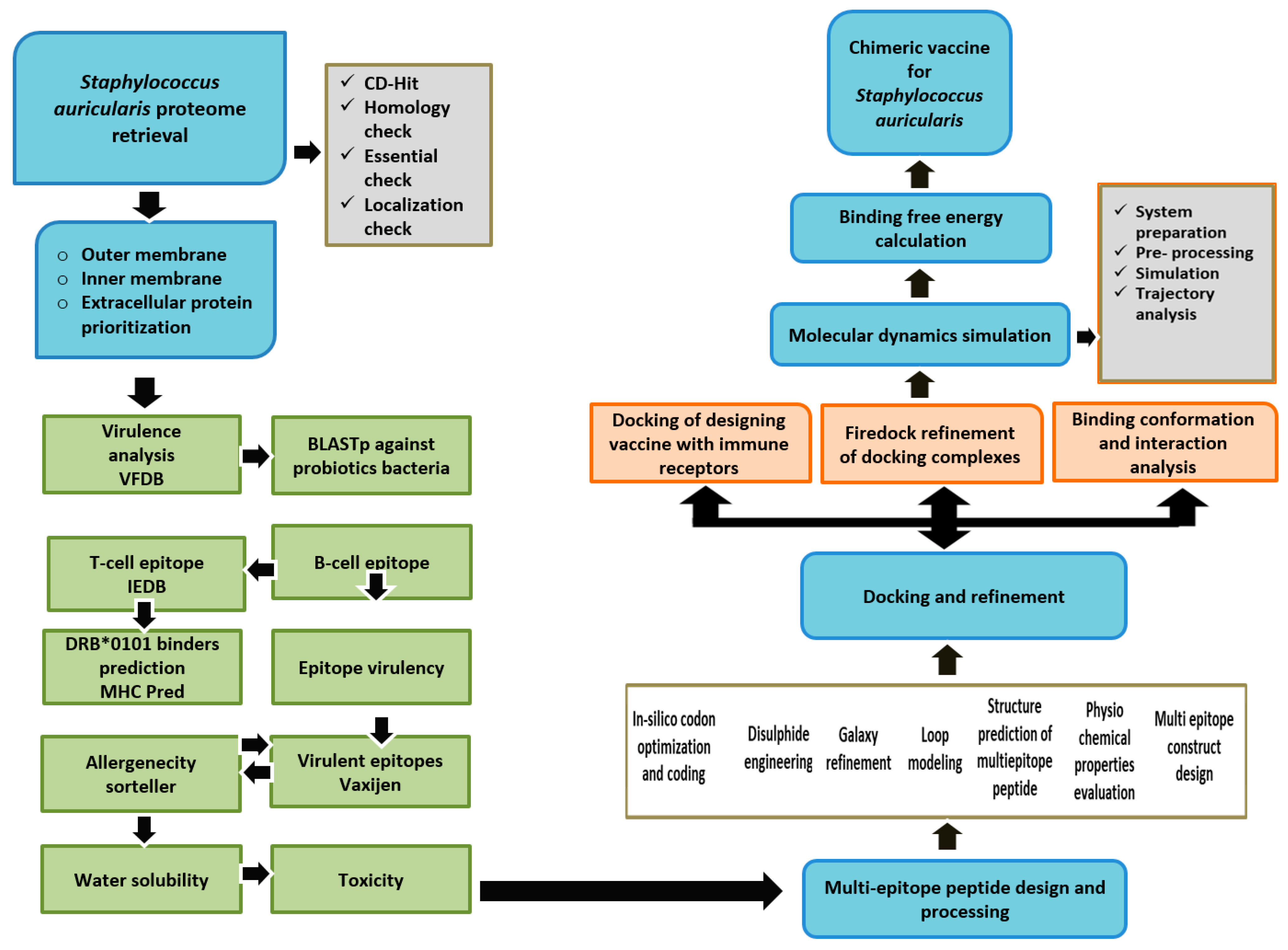
2. Research Methodology
2.1. Bacterial Pan-Genomics, Subtractive Proteomics, and Reverse Vaccinology
2.2. Pre-Selection Stage
2.3. CD-Hit Analysis
2.4. Subcellular Localization Phase
2.5. Vaccine Candidate’s Prioritization Phase
2.6. Prediction of Immune Cell Epitopes
2.7. MHCPred 2.0 Analysis
2.8. Antigenicity, Allergenicity, and Adhesion Probability Prediction
2.9. Multi-Epitopes Peptide Designing
2.10. Codon Optimization
2.11. Docking and Refinement
2.12. Molecular Dynamics (MD) Simulation Assay
2.13. Free Energy of Immune Receptors and Vaccine Design
3. Results
3.1. Genomes Retrieval of S. auricularis
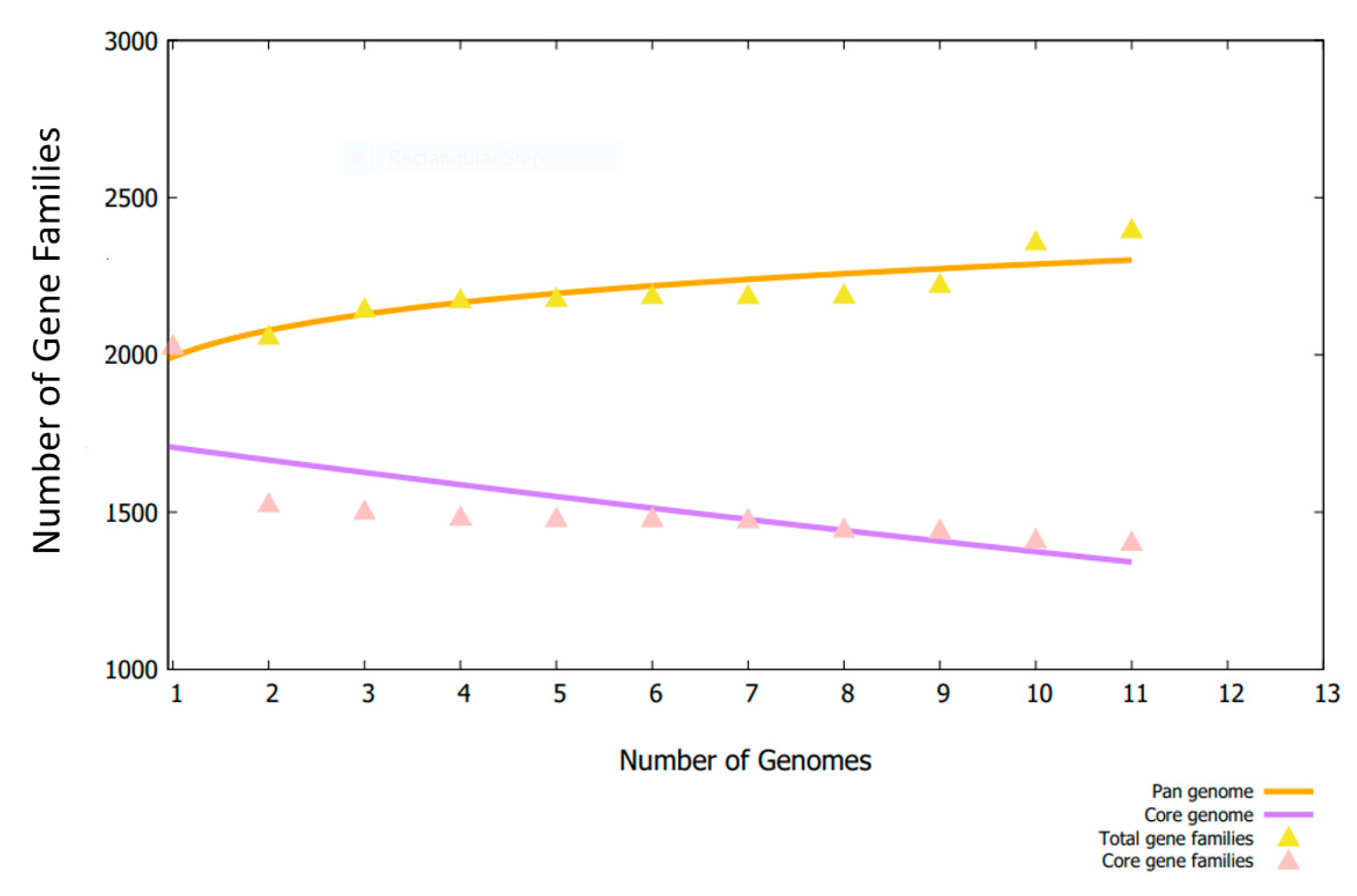
3.2. Bacterial Pan-Genome Analysis
3.3. CD-HIT Analysis and Proteins Subcellular Localization
3.4. VFDB Analysis
3.5. B-Cell Epitopes Prediction
3.6. MHC-I and MHC-II Epitopes Prediction
3.7. Epitope Prioritization Phase
3.8. MHCPred Analysis
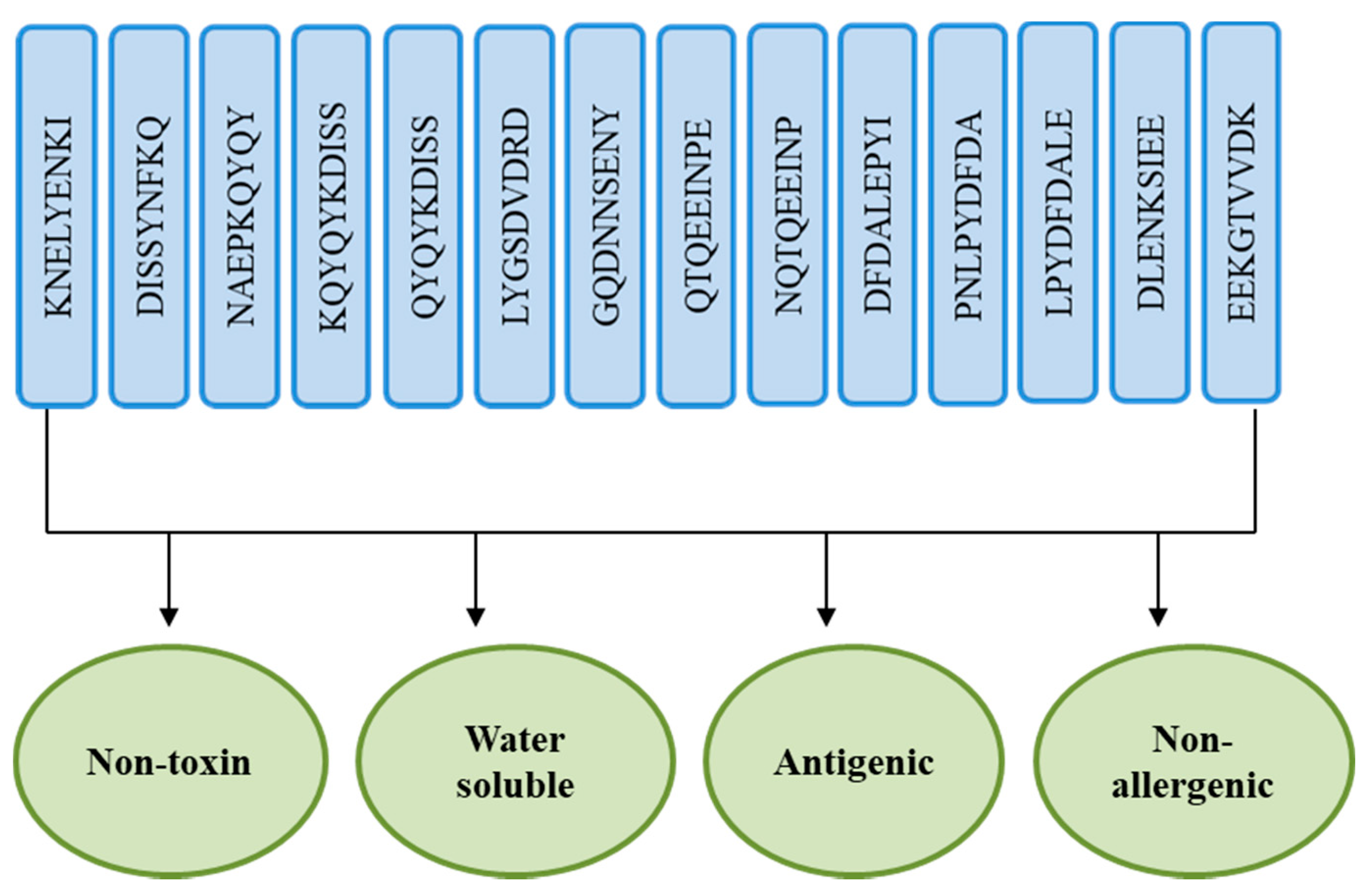
3.9. Allergenicity and Antigenicity
3.10. Analysis of Solubility and Toxicity
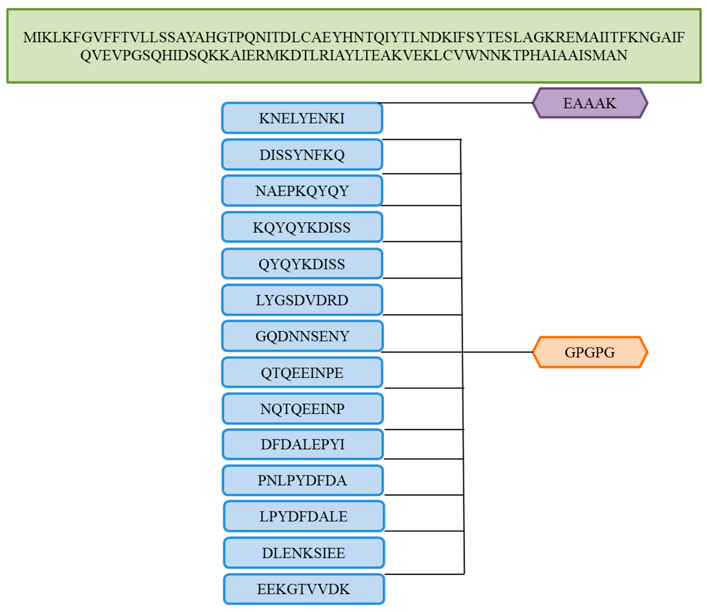
3.11. Multi-Epitopes Vaccine Designing
3.12. Vaccine Structure Modeling
3.13. Loop Modeling and Refinement
3.14. Disulfide Engineering
3.15. Optimizing Codon Sequences
3.16. Analysis of Molecular Docking
3.17. Docked Complexes Refinement
3.18. Docked Conformation of Vaccine with Immune Receptors
3.19. Interactions of Vaccine to Immune Receptors
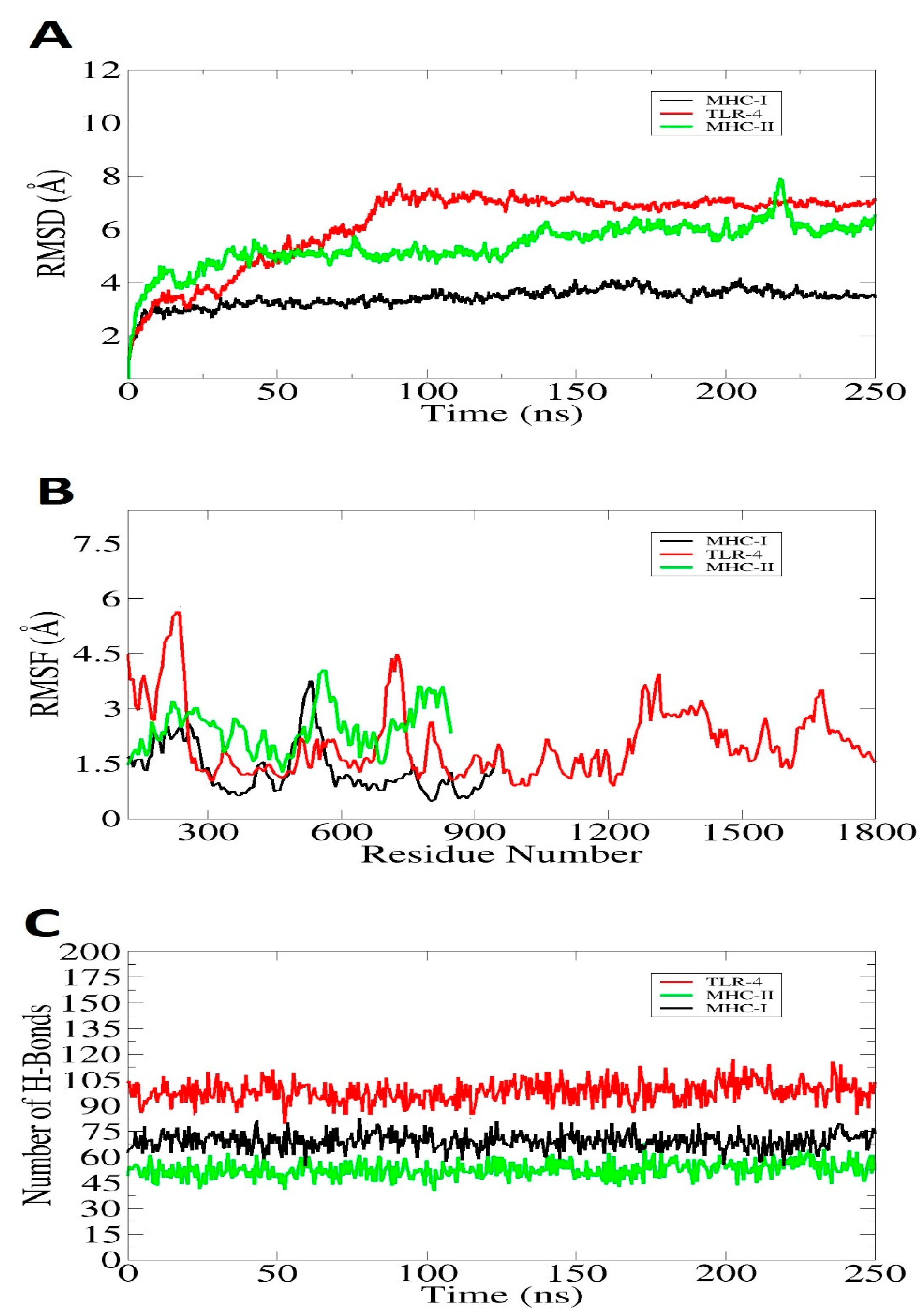
3.20. Molecular Dynamic Simulation
3.21. Estimation of Binding Free Energy
4. Discussion
5. Conclusions and Limitations
Author Contributions
Funding
Institutional Review Board Statement
Informed Consent Statement
Data Availability Statement
Acknowledgments
Conflicts of Interest
References
- Hutchings, M.; Truman, A.; Wilkinson, B. Antibiotics: Past, present and future. Curr. Opin. Microbiol. 2019, 51, 72–80. [Google Scholar] [CrossRef] [PubMed]
- Bennett, J.W.; Chung, K.-T. Alexander Fleming and the Discovery of Penicillin; New Orleans, LA, USA, 2001; Available online: https://www.acs.org/content/acs/en/education/whatischemistry/landmarks/flemingpenicillin.html#alexander-fleming-penicillin (accessed on 11 March 2022).
- Hasan, T.H.; Al-Harmoosh, R.A. Mechanisms of antibiotics resistance in bacteria. Sys. Rev. Pharm. 2020, 11, 817–823. [Google Scholar]
- O’Neill, J. Tackling Drug-Resistant Infections Globally: Final Report and Recommendations; Wellcome Trust: London, UK, 2016. [Google Scholar]
- Adu-Bobie, J.; Capecchi, B.; Serruto, D.; Rappuoli, R.; Pizza, M. Two years into reverse vaccinology. Vaccine 2003, 21, 605–610. [Google Scholar] [CrossRef]
- Clem, A.S. Fundamentals of vaccine immunology. J. Glob. Infect. Dis. 2011, 3, 73. [Google Scholar] [CrossRef]
- Excler, J.L.; Saville, M.; Berkley, S.; Kim, J.H. Vaccine development for emerging infectious diseases. Nat. Med. 2021, 27, 591–600. [Google Scholar] [CrossRef]
- Malonis, R.J.; Lai, J.R.; Vergnolle, O. Peptide-Based Vaccines: Current Progress and Future Challenges. Chem. Rev. 2019, 120, 3210–3229. [Google Scholar] [CrossRef]
- Lombard, M.; Pastoret, P.P.; Moulin, A.M. A brief history of vaccines and vaccination. OIE Rev. Sci. Tech. 2007, 26, 29–48. [Google Scholar] [CrossRef]
- Kavey, R.-E.W.; Kavey, A.B. Viral Pandemics: From Smallpox to COVID-19; Routledge: London, UK, 2020; Available online: https://www.taylorfrancis.com/books/mono/10.4324/9781003006800/viral-pandemics-rae-ellen-kavey-allison-kavey (accessed on 11 March 2022).
- Okafor, C.N.; Rewane, A.; Momodu, I.I. Bacillus Calmette Guerin; StatePearls: Treasure Island, FL, USA, 2019. Available online: https://medlineplus.gov/druginfo/meds/a682809.html (accessed on 11 March 2022).
- Naz, A.; Awan, F.M.; Obaid, A.; Muhammad, S.A.; Paracha, R.Z.; Ahmad, J.; Ali, A. Identification of putative vaccine candidates against Helicobacter pylori exploiting exoproteome and secretome: A reverse vaccinology based approach. Infect. Genet. Evol. 2015, 32, 280–291. [Google Scholar] [CrossRef]
- Bidmos, F.A.; Siris, S.; Gladstone, C.A.; Langford, P.R. Bacterial Vaccine Antigen Discovery in the Reverse Vaccinology 2.0 Era: Progress and Challenges. Front. Immunol. 2018, 9, 2315. [Google Scholar] [CrossRef]
- Buckland, B.C. The process development challenge for a new vaccine. Nat. Med. 2005, 11, S16. [Google Scholar] [CrossRef]
- Sette, A.; Rappuoli, R. Reverse vaccinology: Developing vaccines in the era of genomics. Immunity 2010, 33, 530–541. [Google Scholar] [CrossRef] [PubMed]
- Suleman, M.; ul Qamar, M.T.; Rasool, S.; Rasool, A.; Albutti, A.; Alsowayeh, N.; Alwashmi, A.S.S.; Aljasir, M.A.; Ahmad, S.; Hussain, Z. Immunoinformatics and Immunogenetics-Based Design of Immunogenic Peptides Vaccine against the Emerging Tick-Borne Encephalitis Virus (TBEV) and Its Validation through In Silico Cloning and Immune Simulation. Vaccines 2021, 9, 1210. [Google Scholar] [CrossRef]
- Ul Qamar, M.T.; Shahid, F.; Aslam, S.; Ashfaq, U.A.; Aslam, S.; Fatima, I.; Fareed, M.M.; Zohaib, A.; Chen, L.-L. Reverse vaccinology assisted designing of multiepitope-based subunit vaccine against SARS-CoV-2. Infect. Dis. Poverty 2020, 9, 1–14. [Google Scholar] [CrossRef] [PubMed]
- Alharbi, M.; Alshammari, A.; Alasmari, A.F.; Alharbi, S.; Tahir ul Qamar, M.; Abbasi, S.W.; Shaker, B.; Ahmad, S. Whole Proteome-Based Therapeutic Targets Annotation and Designing of Multi-Epitope-Based Vaccines against the Gram-Negative XDR-Alcaligenes faecalis Bacterium. Vaccines 2022, 10, 462. [Google Scholar] [CrossRef] [PubMed]
- Serruto, D.; Bottomley, M.J.; Ram, S.; Giuliani, M.M.; Rappuoli, R. The new multicomponent vaccine against meningococcal serogroup B, 4CMenB: Immunological, functional and structural characterization of the antigens. Vaccine 2012, 30, B87–B97. [Google Scholar] [CrossRef] [PubMed]
- Alharbi, M.; Alshammari, A.; Alasmari, A.F.; Alharbi, S.M.; Tahir ul Qamar, M.; Ullah, A.; Ahmad, S.; Irfan, M.; Khalil, A.A.K. Designing of a Recombinant Multi-Epitopes Based Vaccine against Enterococcus mundtii Using Bioinformatics and Immunoinformatics Approaches. Int. J. Environ. Res. Public Health 2022, 19, 3729. [Google Scholar] [CrossRef]
- Natsis, N.E.; Cohen, P.R. Coagulase-negative staphylococcus skin and soft tissue infections. Am. J. Clin. Dermatol. 2018, 19, 671–677. [Google Scholar] [CrossRef]
- Roth, R.R.; James, W.D. Microbial ecology of the skin. Annu. Rev. Microbiol. 1988, 42, 441–464. [Google Scholar] [CrossRef]
- Hoffman, D.J.; Brown, G.D.; Lombardo, F.A. Early-onset sepsis with Staphylococcus auricularis in an extremely low-birth weight infant-An uncommon pathogen. J. Perinatol. 2007, 27, 519–520. [Google Scholar] [CrossRef]
- Ha, E.T.; Heitner, J.F. Staphylococcus Auricularis Endocarditis: A Rare Cause of Subacute Prosthetic Valve Endocarditis with Severe Aortic Stenosis. Cureus 2021, 13, e12738. [Google Scholar] [CrossRef]
- Hajighahramani, N.; Nezafat, N.; Eslami, M.; Negahdaripour, M.; Rahmatabadi, S.S.; Ghasemi, Y. Immunoinformatics analysis and in silico designing of a novel multi-epitope peptide vaccine against Staphylococcus aureus. Infect. Genet. Evol. 2017, 48, 83–94. [Google Scholar] [CrossRef] [PubMed]
- Tahir ul Qamar, M.; Ahmad, S.; Fatima, I.; Ahmad, F.; Shahid, F.; Naz, A.; Abbasi, S.W.; Khan, A.; Mirza, M.U.; Ashfaq, U.A.; et al. Designing multi-epitope vaccine against Staphylococcus aureus by employing subtractive proteomics, reverse vaccinology and immuno-informatics approaches. Comput. Biol. Med. 2021, 132, 104389. [Google Scholar] [CrossRef] [PubMed]
- Szczuka, E.; Jabłońska, L.; Kaznowski, A. Coagulase-negative staphylococci: Pathogenesis, occurrence of antibiotic resistance genes and in vitro effects of antimicrobial agents on biofilm-growing bacteria. J. Med. Microbiol. 2016, 65, 1405–1413. [Google Scholar] [CrossRef] [PubMed]
- Khan, M.M.; Faiz, A.; Ashshi, A.M. Clinically significant Coagulase Negative Staphylococci and their antibiotic resistance pattern in a tertiary care hospital. J. Pak. Med. Assoc. 2014, 64, 1171–1174. [Google Scholar]
- Morgenstern, M.; Erichsen, C.; Hackl, S.; Mily, J.; Militz, M.; Friederichs, J.; Hungerer, S.; Bühren, V.; Moriarty, T.F.; Post, V.; et al. Antibiotic resistance of commensal Staphylococcus aureus and coagulase-negative staphylococci in an international cohort of surgeons: A prospective point-prevalence study. PLoS ONE 2016, 11, e0148437. [Google Scholar] [CrossRef]
- Koksal, F.; Yasar, H.; Samasti, M. Antibiotic resistance patterns of coagulase-negative staphylococcus strains isolated from blood cultures of septicemic patients in Turkey. Microbiol. Res. 2009, 164, 404–410. [Google Scholar] [CrossRef]
- Blast, N. Basic local alignment search tool. Natl. Libr. Med. Natl. Cent. Biotechnol. Inf. 2015. Available online: https://blast.ncbi.nlm.nih.gov/Blast.cgi (accessed on 11 March 2022).
- Ali, A.; Naz, A.; Soares, S.C.; Bakhtiar, M.; Tiwari, S.; Hassan, S.S.; Hanan, F.; Ramos, R.; Pereira, U.; Barh, D. Pan-genome analysis of human gastric pathogen H. pylori: Comparative genomics and pathogenomics approaches to identify regions associated with pathogenicity and prediction of potential core therapeutic targets. Biomed Res. Int. 2015, 2015, 139580. [Google Scholar] [CrossRef]
- Tahir Ul Qamar, M.; Zhu, X.; Xing, F.; Chen, L.-L. ppsPCP: A plant presence/absence variants scanner and pan-genome construction pipeline. Bioinformatics 2019, 35, 4156–4158. [Google Scholar] [CrossRef]
- Enayatkhani, M.; Hasaniazad, M.; Faezi, S.; Gouklani, H.; Davoodian, P.; Ahmadi, N.; Einakian, M.A.; Karmostaji, A.; Ahmadi, K. Reverse vaccinology approach to design a novel multi-epitope vaccine candidate against COVID-19: An in silico study. J. Biomol. Struct. Dyn. 2021, 39, 2857–2872. [Google Scholar] [CrossRef]
- Ahmad, S.; Raza, S.; Uddin, R.; Azam, S.S. Comparative subtractive proteomics based ranking for antibiotic targets against the dirtiest superbug: Acinetobacter baumannii. J. Mol. Graph. Model. 2018, 82, 74–92. [Google Scholar] [CrossRef] [PubMed]
- Chaudhari, N.M.; Gupta, V.K.; Dutta, C. BPGA-an ultra-fast pan-genome analysis pipeline. Sci. Rep. 2016, 6, 24373. [Google Scholar] [CrossRef] [PubMed]
- Ismail, S.; Shahid, F.; Khan, A.; Bhatti, S.; Ahmad, S.; Naz, A.; Almatroudi, A.; ul Qamar, M.T. Pan-Vaccinomics Approach Towards a Universal Vaccine Candidate Against WHO Priority Pathogens to Address Growing Global Antibiotic Resistance. Comput. Biol. Med. 2021, 136, 104705. [Google Scholar] [CrossRef] [PubMed]
- Zhang, R.; Ou, H.-Y.; Zhang, C.-T. DEG: A database of essential genes. Nucleic Acids Res. 2004, 32, D271–D272. [Google Scholar] [CrossRef]
- Azam, S.S.; Shamim, A. An insight into the exploration of druggable genome of Streptococcus gordonii for the identification of novel therapeutic candidates. Genomics 2014, 104, 203–214. [Google Scholar] [CrossRef]
- Rehman, A.; Ahmad, S.; Shahid, F.; Albutti, A.; Alwashmi, A.S.S.; Aljasir, M.A.; Alhumeed, N.; Qasim, M.; Ashfaq, U.A.; Tahir ul Qamar, M. Integrated Core Proteomics, Subtractive Proteomics, and Immunoinformatics Investigation to Unveil a Potential Multi-Epitope Vaccine against Schistosomiasis. Vaccines 2021, 9, 658. [Google Scholar] [CrossRef]
- Abbas, G.; Zafar, I.; Ahmad, S.; Azam, S.S. Immunoinformatics design of a novel multi-epitope peptide vaccine to combat multi-drug resistant infections caused by Vibrio vulnificus. Eur. J. Pharm. Sci. 2020, 142, 105160. [Google Scholar] [CrossRef]
- Fu, L.; Niu, B.; Zhu, Z.; Wu, S.; Li, W. CD-HIT: Accelerated for clustering the next-generation sequencing data. Bioinformatics 2012, 28, 3150–3152. [Google Scholar] [CrossRef]
- Li, W.; Godzik, A. Cd-hit: A fast program for clustering and comparing large sets of protein or nucleotide sequences. Bioinformatics 2006, 22, 1658–1659. [Google Scholar] [CrossRef]
- Solanki, V.; Tiwari, M.; Tiwari, V. Prioritization of potential vaccine targets using comparative proteomics and designing of the chimeric multi-epitope vaccine against Pseudomonas aeruginosa. Sci. Rep. 2019, 9, 5240. [Google Scholar] [CrossRef]
- Barh, D.; Barve, N.; Gupta, K.; Chandra, S.; Jain, N.; Tiwari, S.; Leon-Sicairos, N.; Canizalez-Roman, A.; Rodrigues dos Santos, A.; Hassan, S.S. Exoproteome and secretome derived broad spectrum novel drug and vaccine candidates in Vibrio cholerae targeted by Piper betel derived compounds. PLoS ONE 2013, 8, e52773. [Google Scholar] [CrossRef] [PubMed]
- Liu, B.; Zheng, D.; Jin, Q.; Chen, L.; Yang, J. VFDB 2019: A comparative pathogenomic platform with an interactive web interface. Nucleic Acids Res. 2019, 47, D687–D692. [Google Scholar] [CrossRef] [PubMed]
- Jespersen, M.C.; Peters, B.; Nielsen, M.; Marcatili, P. BepiPred-2.0: Improving sequence-based B-cell epitope prediction using conformational epitopes. Nucleic Acids Res. 2017, 45, W24–W29. [Google Scholar] [CrossRef] [PubMed]
- Dhanda, S.K.; Mahajan, S.; Paul, S.; Yan, Z.; Kim, H.; Jespersen, M.C.; Jurtz, V.; Andreatta, M.; Greenbaum, J.A.; Marcatili, P. IEDB-AR: Immune epitope database—Analysis resource in 2019. Nucleic Acids Res. 2019, 47, W502–W506. [Google Scholar] [CrossRef] [PubMed]
- Pandey, R.K.; Bhatt, T.K.; Prajapati, V.K. Novel immunoinformatics approaches to design multi-epitope subunit vaccine for malaria by investigating anopheles salivary protein. Sci. Rep. 2018, 8, 1125. [Google Scholar] [CrossRef]
- Jones, E.Y.; Fugger, L.; Strominger, J.L.; Siebold, C. MHC class II proteins and disease: A structural perspective. Nat. Rev. Immunol. 2006, 6, 271. [Google Scholar] [CrossRef]
- Hewitt, E.W. The MHC class I antigen presentation pathway: Strategies for viral immune evasion. Immunology 2003, 110, 163–169. [Google Scholar] [CrossRef]
- Guan, P.; Doytchinova, I.A.; Zygouri, C.; Flower, D.R. MHCPred: A server for quantitative prediction of peptide—MHC binding. Nucleic Acids Res. 2003, 31, 3621–3624. [Google Scholar] [CrossRef]
- Vakili, B.; Eslami, M.; Hatam, G.R.; Zare, B.; Erfani, N.; Nezafat, N.; Ghasemi, Y. Immunoinformatics-aided design of a potential multi-epitope peptide vaccine against Leishmania infantum. Int. J. Biol. Macromol. 2018, 120, 1127–1139. [Google Scholar] [CrossRef]
- Doytchinova, I.A.; Flower, D.R. VaxiJen: A server for prediction of protective antigens, tumour antigens and subunit vaccines. BMC Bioinform. 2007, 8, 4. [Google Scholar] [CrossRef]
- Azim, K.F.; Hasan, M.; Hossain, M.N.; Somana, S.R.; Hoque, S.F.; Bappy, M.N.I.; Chowdhury, A.T.; Lasker, T. Immunoinformatics approaches for designing a novel multi epitope peptide vaccine against human norovirus (Norwalk virus). Infect. Genet. Evol. 2019, 74, 103936. [Google Scholar] [CrossRef] [PubMed]
- Dimitrov, I.; Bangov, I.; Flower, D.R.; Doytchinova, I. AllerTOP v. 2—A server for in silico prediction of allergens. J. Mol. Model. 2014, 20, 2278. [Google Scholar] [CrossRef] [PubMed]
- Hossain, M.S.; Hossan, M.I.; Mizan, S.; Moin, A.T.; Yasmin, F.; Akash, A.-S.; Powshi, S.N.; Hasan, A.K.R.; Chowdhury, A.S. Immunoinformatics approach to designing a multi-epitope vaccine against Saint Louis Encephalitis Virus. Inform. Med. Unlocked 2021, 22, 100500. [Google Scholar] [CrossRef]
- Wizemann, T.M.; Adamou, J.E.; Langermann, S. Adhesins as targets for vaccine development. Emerg. Infect. Dis. 1999, 5, 395. [Google Scholar] [CrossRef] [PubMed]
- Sajjad, R.; Ahmad, S.; Azam, S.S. In silico screening of antigenic B-cell derived T-cell epitopes and designing of a multi-epitope peptide vaccine for Acinetobacter nosocomialis. J. Mol. Graph. Model. 2020, 94, 107477. [Google Scholar] [CrossRef]
- Nain, Z.; Abdulla, F.; Rahman, M.M.; Karim, M.M.; Khan, M.S.A.; Sayed, S.B.; Mahmud, S.; Rahman, S.M.R.; Sheam, M.M.; Haque, Z. Proteome-wide screening for designing a multi-epitope vaccine against emerging pathogen Elizabethkingia anophelis using immunoinformatic approaches. J. Biomol. Struct. Dyn. 2020, 38, 4850–4867. [Google Scholar] [CrossRef]
- Saadi, M.; Karkhah, A.; Nouri, H.R. Development of a multi-epitope peptide vaccine inducing robust T cell responses against brucellosis using immunoinformatics based approaches. Infect. Genet. Evol. 2017, 51, 227–234. [Google Scholar] [CrossRef]
- Naz, S.; Ahmad, S.; Walton, S.; Abbasi, S.W. Multi-epitope based vaccine design against Sarcoptes scabiei paramyosin using immunoinformatics approach. J. Mol. Liq. 2020, 319, 114105. [Google Scholar] [CrossRef]
- Qamar, M.T.U.; Saba Ismail, S.A.; Mirza, M.U.; Abbasi, S.W.; Ashfaq, U.A.; Chen, L.-L. Development of a Novel Multi-Epitope Vaccine Against Crimean-Congo Hemorrhagic Fever Virus: An Integrated Reverse Vaccinology, Vaccine Informatics and Biophysics Approach. Front. Immunol. 2021, 12, 669812. [Google Scholar] [CrossRef]
- Dorosti, H.; Eskandari, S.; Zarei, M.; Nezafat, N.; Ghasemi, Y. Design of a multi-epitope protein vaccine against herpes simplex virus, human papillomavirus and Chlamydia trachomatis as the main causes of sexually transmitted diseases. Infect. Genet. Evol. 2021, 96, 105136. [Google Scholar] [CrossRef]
- Craig, D.B.; Dombkowski, A.A. Disulfide by Design 2.0: A web-based tool for disulfide engineering in proteins. BMC Bioinform. 2013, 14, 346. [Google Scholar] [CrossRef]
- Hossan, M.I.; Chowdhury, A.S.; Hossain, M.U.; Khan, M.A.; Mahmood, T.B.; Mizan, S. Immunoinformatics aided-design of novel multi-epitope based peptide vaccine against Hendra henipavirus through proteome exploration. Inform. Med. Unlocked 2021, 25, 100678. [Google Scholar] [CrossRef]
- Shankar, U.; Jain, N.; Mishra, S.K.; Sk, M.F.; Kar, P.; Kumar, A. Mining of Ebola virus genome for the construction of multi-epitope vaccine to combat its infection. J. Biomol. Struct. Dyn. 2021, 1–17. [Google Scholar] [CrossRef] [PubMed]
- Khatoon, N.; Pandey, R.K.; Prajapati, V.K. Exploring Leishmania secretory proteins to design B and T cell multi-epitope subunit vaccine using immunoinformatics approach. Sci. Rep. 2017, 7, 8285. [Google Scholar] [CrossRef]
- Kar, P.P.; Srivastava, A. Immuno-informatics analysis to identify novel vaccine candidates and design of a multi-epitope based vaccine candidate against Theileria parasites. Front. Immunol. 2018, 9, 2213. [Google Scholar] [CrossRef]
- Mugunthan, S.P.; Harish, M.C. Multi-epitope-based vaccine designed by targeting cytoadherence proteins of Mycoplasma gallisepticum. ACS Omega 2021, 6, 13742–13755. [Google Scholar] [CrossRef] [PubMed]
- Mashiach, E.; Schneidman-Duhovny, D.; Andrusier, N.; Nussinov, R.; Wolfson, H.J. FireDock: A web server for fast interaction refinement in molecular docking. Nucleic Acids Res. 2008, 36, W229–W232. [Google Scholar] [CrossRef] [PubMed]
- Pettersen, E.F.; Goddard, T.D.; Huang, C.C.; Couch, G.S.; Greenblatt, D.M.; Meng, E.C.; Ferrin, T.E. UCSF Chimera—A visualization system for exploratory research and analysis. J. Comput. Chem. 2004, 25, 1605–1612. [Google Scholar] [CrossRef]
- Chukwudozie, O.S.; Gray, C.M.; Fagbayi, T.A.; Chukwuanukwu, R.C.; Oyebanji, V.O.; Bankole, T.T.; Adewole, R.A.; Daniel, E.M. Immuno-informatics design of a multimeric epitope peptide based vaccine targeting SARS-CoV-2 spike glycoprotein. PLoS ONE 2021, 16, e0248061. [Google Scholar] [CrossRef]
- Rafi, S.; Yasmin, S.; Uddin, R. A molecular dynamic simulation approach: Development of dengue virus vaccine by affinity improvement techniques. J. Biomol. Struct. Dyn. 2022, 40, 61–76. [Google Scholar] [CrossRef]
- Case, D.A.; Belfon, K.; Ben-Shalom, I.; Brozell, S.R.; Cerutti, D.; Cheatham, T.; Cruzeiro, V.W.D.; Darden, T.; Duke, R.E.; Giambasu, G.; et al. USA Amber 2020. 2020. Available online: https://ambermd.org/doc12/Amber20.pdf (accessed on 11 March 2022).
- Kräutler, V.; Van Gunsteren, W.F.; Hünenberger, P.H. A fast SHAKE algorithm to solve distance constraint equations for small molecules in molecular dynamics simulations. J. Comput. Chem. 2001, 22, 501–508. [Google Scholar] [CrossRef]
- Khan, M.; Khan, S.; Ali, A.; Akbar, H.; Sayaf, A.M.; Khan, A.; Wei, D.-Q. Immunoinformatics approaches to explore Helicobacter Pylori proteome (Virulence Factors) to design B and T cell multi-epitope subunit vaccine. Sci. Rep. 2019, 9, 13321. [Google Scholar] [CrossRef] [PubMed]
- Majee, P.; Jain, N.; Kumar, A. Designing of a multi-epitope vaccine candidate against Nipah virus by in silico approach: A putative prophylactic solution for the deadly virus. J. Biomol. Struct. Dyn. 2021, 39, 1461–1480. [Google Scholar] [CrossRef] [PubMed]
- Roe, D.R.; Cheatham, T.E., III. PTRAJ and CPPTRAJ: Software for processing and analysis of molecular dynamics trajectory data. J. Chem. Theory Comput. 2013, 9, 3084–3095. [Google Scholar] [CrossRef] [PubMed]
- Miller, B.R.; McGee, T.D.; Swails, J.M.; Homeyer, N.; Gohlke, H.; Roitberg, A.E. MMPBSA.py: An efficient program for end-state free energy calculations. J. Chem. Theory Comput. 2012, 8, 3314–3321. [Google Scholar] [CrossRef] [PubMed]
- Sanober, G.; Ahmad, S.; Azam, S.S. Identification of plausible drug targets by investigating the druggable genome of MDR Staphylococcus epidermidis. Gene Rep. 2017, 7, 147–153. [Google Scholar] [CrossRef]
- Ali, S.; Ali, S.; Javed, S.O.; Shoukat, S.; Ahmad, S.; Ali, S.S.; Hussain, Z.; Waseem, M.; Rizwan, M.; Suleman, M. Proteome wide vaccine targets prioritization and designing of antigenic vaccine candidate to trigger the host immune response against the Mycoplasma genitalium infection. Microb. Pathog. 2021, 152, 104771. [Google Scholar] [CrossRef]
- Ahmad, S.; Azam, S.S. A novel approach of virulome based reverse vaccinology for exploring and validating peptide-based vaccine candidates against the most troublesome nosocomial pathogen: Acinetobacter baumannii. J. Mol. Graph. Model. 2018, 83, 1–11. [Google Scholar] [CrossRef]
- Fatima, I.; Ahmad, S.; Abbasi, S.W.; Ashfaq, U.A.; Shahid, F.; ul Qamar, M.T.; Rehman, A.; Allemailem, K.S. Designing of a multi-epitopes-based peptide vaccine against rift valley fever virus and its validation through integrated computational approaches. Comput. Biol. Med. 2021, 141, 105151. [Google Scholar] [CrossRef]
- Gupta, S.; Kapoor, P.; Chaudhary, K.; Gautam, A.; Kumar, R.; Raghava, G.P.S. Peptide toxicity prediction. In Computational Peptidology; Springer: Berlin/Heidelberg, Germany, 2015; pp. 143–157. [Google Scholar]
- Javadi, M.; Oloomi, M.; Bouzari, S. In Silico Design of a Poly-epitope Vaccine for Urinary Tract Infection Based on Conserved Antigens by Modern Vaccinology. Int. J. Pept. Res. Ther. 2021, 27, 909–921. [Google Scholar] [CrossRef]
- Ullah, A.; Ahmad, S.; Ismail, S.; Afsheen, Z.; Khurram, M.; Tahir ul Qamar, M.; AlSuhaymi, N.; Alsugoor, M.H.; Allemailem, K.S. Towards A Novel Multi-Epitopes Chimeric Vaccine for Simulating Strong Immune Responses and Protection against Morganella morganii. Int. J. Environ. Res. Public Health 2021, 18, 10961. [Google Scholar] [CrossRef] [PubMed]
- Naz, S.; Ahmad, S.; Abbasi, S.W.; Ismail, S.; Waseem, S.; Ul Qamar, M.T.; Almatroudi, A.; Ali, Z. Identification of immunodominant epitopes in allelic variants VK210 and VK247 of Plasmodium Vivax Circumsporozoite immunogen. Infect. Genet. Evol. 2021, 96, 105120. [Google Scholar] [CrossRef]
- Zakeri, B.; Fierer, J.O.; Celik, E.; Chittock, E.C.; Schwarz-Linek, U.; Moy, V.T.; Howarth, M. Peptide tag forming a rapid covalent bond to a protein, through engineering a bacterial adhesin. Proc. Natl. Acad. Sci. USA 2012, 109, E690–E697. [Google Scholar] [CrossRef]
- Ali, M.; Pandey, R.K.; Khatoon, N.; Narula, A.; Mishra, A.; Prajapati, V.K. Exploring dengue genome to construct a multi-epitope based subunit vaccine by utilizing immunoinformatics approach to battle against dengue infection. Sci. Rep. 2017, 7, 9232. [Google Scholar] [CrossRef] [PubMed]
- Li, W.; Joshi, M.D.; Singhania, S.; Ramsey, K.H.; Murthy, A.K. Peptide vaccine: Progress and challenges. Vaccines 2014, 2, 515–536. [Google Scholar] [CrossRef] [PubMed]
- Aslam, S.; Ahmad, S.; Noor, F.; Ashfaq, U.A.; Shahid, F.; Rehman, A.; Tahir ul Qamar, M.; Alatawi, E.A.; Alshabrmi, F.M.; Allemailem, K.S. Designing a Multi-Epitope Vaccine against Chlamydia trachomatis by Employing Integrated Core Proteomics, Immuno-Informatics and In Silico Approaches. Biology 2021, 10, 997. [Google Scholar] [CrossRef] [PubMed]
- Elliott, S.L.; Suhrbier, A.; Miles, J.J.; Lawrence, G.; Pye, S.J.; Le, T.T.; Rosenstengel, A.; Nguyen, T.; Allworth, A.; Burrows, S.R. Phase I trial of a CD8+ T-cell peptide epitope-based vaccine for infectious mononucleosis. J. Virol. 2008, 82, 1448–1457. [Google Scholar] [CrossRef] [PubMed]
- Gul, S.; Ahmad, S.; Ullah, A.; Ismail, S.; Khurram, M.; Tahir ul Qamar, M.; Hakami, A.R.; Alkhathami, A.G.; Alrumaihi, F.; Allemailem, K.S. Designing a Recombinant Vaccine against Providencia rettgeri Using Immunoinformatics Approach. Vaccines 2022, 10, 189. [Google Scholar] [CrossRef]
- Greenwood, B. The contribution of vaccination to global health: Past, present and future. Philos. Trans. R. Soc. B Biol. Sci. 2014, 369, 20130433. [Google Scholar] [CrossRef]
- Peschel, A.; Otto, M. Phenol-soluble modulins and staphylococcal infection. Nat. Rev. Microbiol. 2013, 11, 667–673. [Google Scholar] [CrossRef]
- Mukherjee, S.; Karmakar, S.; Babu, S.P.S. TLR2 and TLR4 mediated host immune responses in major infectious diseases: A review. Braz. J. Infect. Dis. 2016, 20, 193–204. [Google Scholar] [CrossRef] [PubMed]
- Chintoan-Uta, C.; Cassady-Cain, R.L.; Al-Haideri, H.; Watson, E.; Kelly, D.J.; Smith, D.G.E.; Sparks, N.H.C.; Kaiser, P.; Stevens, M.P. Superoxide dismutase SodB is a protective antigen against Campylobacter jejuni colonisation in chickens. Vaccine 2015, 33, 6206–6211. [Google Scholar] [CrossRef] [PubMed][Green Version]
- Oertli, M.; Noben, M.; Engler, D.B.; Semper, R.P.; Reuter, S.; Maxeiner, J.; Gerhard, M.; Taube, C.; Müller, A. Helicobacter pylori γ-glutamyl transpeptidase and vacuolating cytotoxin promote gastric persistence and immune tolerance. Proc. Natl. Acad. Sci. USA 2013, 110, 3047–3052. [Google Scholar] [CrossRef] [PubMed]


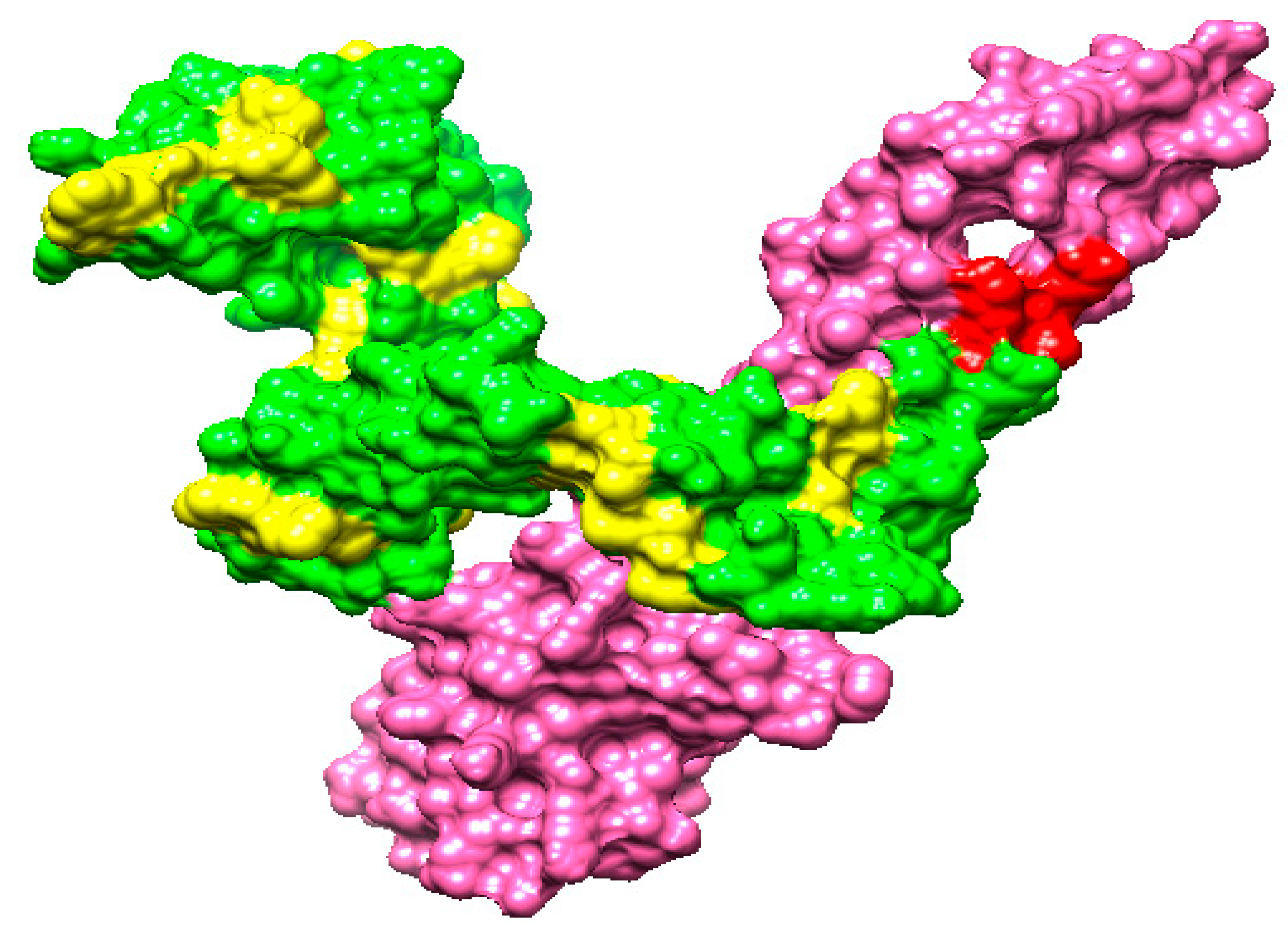
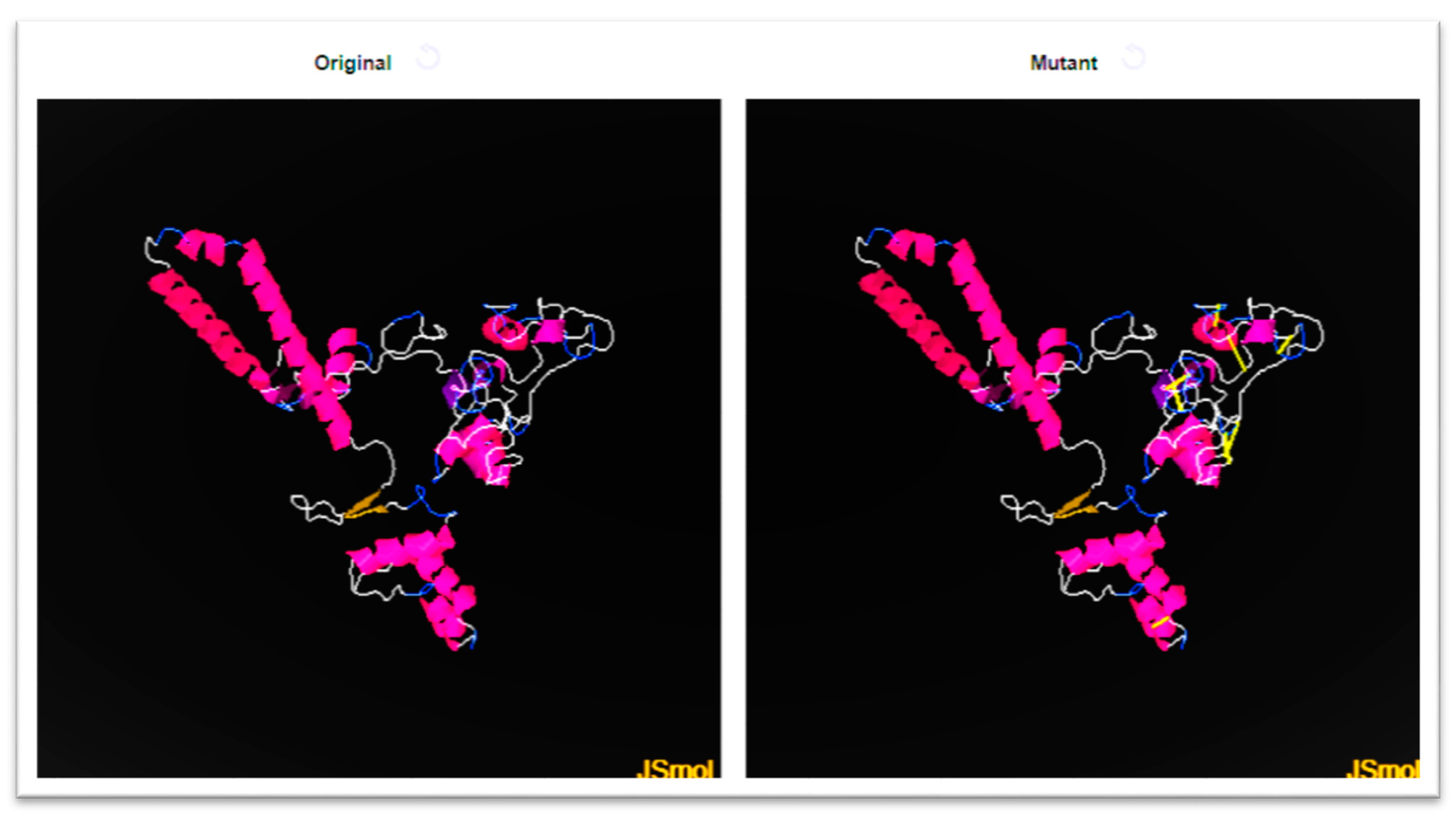

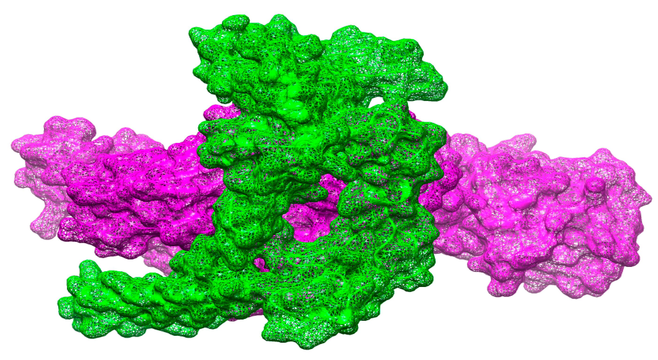
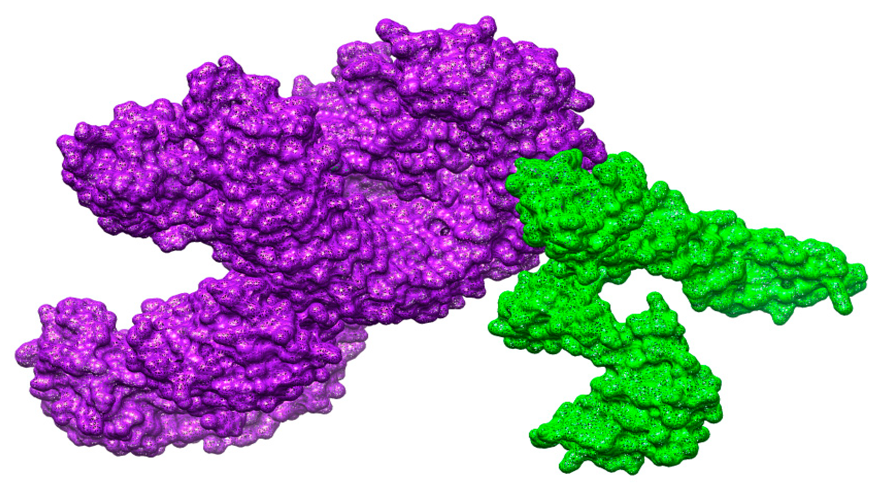
| Organism Name | Strain | Size (Mb) | GC% |
|---|---|---|---|
| S. auricularis | FDAARGOS_882 | 2.22 | 37.40 |
| S. auricularis | NCTC12101 | 2.22 | 37.40 |
| S. auricularis | JCM 2421 | 2.26 | 37.40 |
| S. auricularis | DSM 20609 | 2.20 | 37.30 |
| S. auricularis | NCTC 12101 | 2.20 | 37.20 |
| S. auricularis | DE0381 | 2.42 | 37.40 |
| S. auricularis | S52 | 2.28 | 37.10 |
| S. auricularis | 1H20 | 2.25 | 37.10 |
| S. auricularis | SNUC 3034 | 2.28 | 37.10 |
| S. auricularis | SNUC 993 | 2.30 | 37.10 |
| S. auricularis | CHK138−4784 | 1.68 | 37.10 |
| Proteins | Subcellular Localization | Bit Score | Sequence Identity |
|---|---|---|---|
| core/231/1/Org1_Gene1151 gamma-glutamyl transferase | Extracellular | 244 bits | 34% |
| core/1478/1/Org1_Gene484 superoxide dismutase | Periplasmic | 179 bits | 43% |
| B-Cell Epitopes | Peptides |
|---|---|
| core/231/1/Org1_Gene1151 gamma-glutamyl transferase | PDTYDKNELYENKIAGHTQSASNQ |
| QNAEPKQYQYKDISSYNFKQ | |
| LYGSDVDRDSPFFDGSRTKREGDVVK | |
| KQLDNKLTKKDFQDYEMT | |
| VNGQDNNSENYLSQE | |
| NEVNQTQEEINPEGIDN | |
| SDPNSPNYGEKHKQ | |
| IDVFKNMGYNVEEKRNDP | |
| core/1478/1/Org1_Gene484 superoxide dismutase | LPNLPYDFDALEPYIDKE |
| DLENKSIEEIVANLDSVPED | |
| TPNSEEKGTVVDKIKEQWGSL | |
| LKYQNKRPEYIE |
| MHC-II | Percentile Score | MHC-I | Percentile Score |
|---|---|---|---|
| KIAGHTQSASN | 4.3 | KIAGHTQSA | 0.15 |
| AGHTQSASN | 28 | ||
| PDTYDKNELYENKI | 28 | PDTYDKNELY | 0.28 |
| DKNELYENKI | 20 | ||
| KQYQYKDISSYNFKQ | 4.8 | QYQYKDISSY | 0.01 |
| DISSYNFKQ | 4.4 | ||
| NAEPKQYQYKDISS | 28 | NAEPKQYQY | 0.03 |
| KQYQYKDISS | 7.4 | ||
| LYGSDVDRDSPFFD | 7 | DVDRDSPFF | 0.41 |
| LYGSDVDRD | 24 | ||
| DVDRDSPFFD | 9.9 | ||
| SPFFDGSRTKREGDV | 37 | FFDGSRTKR | 0.17 |
| SPFFDGSRTK | 0.31 | ||
| GSRTKREGDV | 13 | ||
| LTKKDFQDYEM | 7.2 | LTKKDFQDY | 0.2 |
| KKDFQDYEM | 2.9 | ||
| KQLDNKLTKKDFQDY | 33 | KQLDNKLTK | 0.05 |
| LTKKDFQDY | 0.2 | ||
| GQDNNSENYLSQE | 45 | GQDNNSENY | 0.28 |
| NSENYLSQE | 7.8 | ||
| NEVNQTQEEINPE | 13 | EVNQTQEEI | 0.1 |
| NEVNQTQEEI | 0.79 | ||
| QTQEEINPE | 6.3 | ||
| VNQTQEEINPEGIDN | 22 | QEEINPEGI | 0.63 |
| VNQTQEEINP | 37 | ||
| SDPNSPNYGEKHKQ | 74 | NSPNYGEKHK | 3.4 |
| DPNSPNYGEK | 3.5 | ||
| SDPNSPNYGE | 6.8 | ||
| IDVFKNMGYNV | 0.82 | DVFKNGYNV | 0.34 |
| IDVFKNMGY | 1.4 | ||
| VFKNMGYNVEEKRND | 12 | MGYNVEEKR | 0.62 |
| VFKNMGYNV | 1.3 | ||
| YNVEEKRND | 65 | ||
| PNLPYDFDALEPYI | 1.1 | DFDALEPYI | 1.9 |
| PNLPYDFDAL | 38 | ||
| LPYDFDALEPYIDKE | LPYDFDALE | 1.7 | |
| DALEPYIDKE | 14 | ||
| IEEIVANLDSVPE | 3.2 | IEEIVANLD | 18 |
| VANLDSVPE | 9.3 | ||
| DLENKSIEEIVANLD | 11 | DLENKSIEEI | 1.4 |
| SIEEIVANLD | 17 | ||
| TVVDKIKEQWGSL | 22 | TVVDKIKEQW | 0.08 |
| KIKEQWGSL | 0.26 | ||
| TPNSEEKGTVVDKI | 43 | EEKGTVVDKI | 0.57 |
| TPNSEEKGTV | 0.89 | ||
| LKYQNKRPEYI | 3 | YQNKRPEYI | 0.21 |
| MHC- Pred | DRB*0101 IC50 Score | Antigenicity | Allergenicity | Solubility | Toxin Pred |
|---|---|---|---|---|---|
| KNELYENKI | 7.768 | 0.5035 | |||
| DISSYNFKQ | 57.15 | 1.5642 | |||
| NAEPKQYQY | 2.93 | 1.0100 | |||
| KQYQYKDISS | 83.37 | 0.5079 | |||
| QYQYKDISS | 11.02 | 0.5677 | |||
| LYGSDVDRD | 37.24 | 0.9838 | |||
| GQDNNSENY | 64.86 | 1.5275 | Non-allergen | Good water solubility | Non-toxin |
| QTQEEINPE | 6.34 | 1.1296 | |||
| NQTQEEINP | 22.39 | 0.9309 | |||
| DFDALEPYI | 9.38 | 1.2216 | |||
| PNLPYDFDA | 18.54 | 1.0231 | |||
| LPYDFDALE | 56.36 | 0.7775 | |||
| DLENKSIEE | 32.43 | 1.3709 | |||
| EEKGTVVDK | 42.27 | 0.8901 |
| Sequence Number | Amino Acid | Sequence Number | Amino Acid | Chi3 | Energy | Sum B-Factors |
|---|---|---|---|---|---|---|
| 7 | GLY | 34 | HIS | 67.21 | 2.37 | 0 |
| 15 | SER | 24 | GLN | 88.65 | 5.88 | 0 |
| 18 | TYR | 23 | PRO | 74.33 | 4.57 | 0 |
| 209 | ASP | 218 | ASN | 122.68 | 5.37 | 0 |
| 216 | GLN | 220 | SER | 122.93 | 4.95 | 0 |
| 225 | PRO | 229 | GLN | 113.02 | 3.01 | 0 |
| 238 | GLY | 245 | THR | 110.79 | 5.25 | 0 |
| 285 | LEU | 288 | ASP | 106.06 | 3.04 | 0 |
| 301 | GLU | 305 | ILE | 106.28 | 3.68 | 0 |
| Solution No | Score | Area | ACE | Transformation |
|---|---|---|---|---|
| 1 | 20132 | 3445.10 | 405.18 | −0.44 0.33 1.63 99.21 38.67 29.15 |
| 2 | 19844 | 3938.00 | 311.82 | −0.92 0.91 0.60 22.26–27.15 9.21 |
| 3 | 19282 | 2805.20 | 378.13 | −2.47–0.49 3.11–18.74 12.00 37.06 |
| 4 | 19272 | 3180.70 | 371.95 | −0.55–0.04 2.27 92.68 70.24 41.81 |
| 5 | 18218 | 2781.40 | 283.06 | −0.95 0.40–3.05 42.02 21.11 3.99 |
| 6 | 17986 | 2731.90 | 493.30 | −1.97 0.09 0.80 2.95 47.68 44.62 |
| 7 | 17968 | 3816.80 | 300.18 | −3.01 1.42 1.28–14.91–40.50–6.83 |
| 8 | 17940 | 2845.80 | 427.92 | −2.81 0.37–2.54 46.23–29.01 36.16 |
| 9 | 17936 | 3052.60 | 86.01 | −2.91–0.90 2.92–3.45 14.79 55.77 |
| 10 | 17934 | 3182.80 | 498.19 | 2.21 0.23–1.45 52.65–6.49 5.15 |
| 11 | 17904 | 2907.60 | 493.90 | −0.97 0.93 0.88 33.16–26.75 13.49 |
| 12 | 17688 | 2908.80 | 139.47 | 2.77–0.08–0.59 64.71 52.46 56.37 |
| 13 | 17674 | 2832.70 | 465.57 | 0.17–0.26 2.33 14.30–1.73–34.33 |
| 14 | 17502 | 2380.50 | 477.76 | −0.85–0.07 0.54 22.31 1.39 66.68 |
| 15 | 17168 | 2920.20 | 471.12 | 2.24 0.17–0.67 28.98 58.30–17.67 |
| 16 | 16922 | 2557.70 | 389.82 | −0.39 1.28 2.15 47.56 2.78–27.80 |
| 17 | 16866 | 3903.90 | 336.25 | −1.45 0.79 1.08 27.04–33.05 26.94 |
| 18 | 16826 | 2201.00 | 447.60 | −0.07–0.17–2.61–7.26 71.44 30.42 |
| 19 | 16782 | 3966.20 | 232.52 | −0.83–0.06–1.50 2.64 41.63 19.99 |
| 20 | 16776 | 3039.60 | 442.93 | −0.33 0.92–2.63–9.19 69.57 23.36 |
| Solution No | Score | Area | ACE | Transformation |
|---|---|---|---|---|
| 1 | 20570 | 3565.40 | 271.97 | −2.17 –0.39 –2.54 108.70 52.17 53.60 |
| 2 | 20382 | 3218.70 | −25.73 | –1.94 –0.37 2.71 87.91 105.99 58.18 |
| 3 | 19196 | 3055.90 | 213.63 | 1.68 0.11 1.94 111.16 36.90 –54.35 |
| 4 | 18992 | 3096.60 | 143.82 | 0.14 –0.74 –1.31 39.48 110.23 –5.13 |
| 5 | 18592 | 2957.80 | 177.57 | 1.48 –0.90 1.70 138.56 40.32 –13.49 |
| 6 | 18488 | 3716.80 | 101.29 | –1.83 –0.24 2.67 94.90 105.50 59.00 |
| 7 | 17942 | 2977.00 | 343.89 | −0.38 –0.06 0.02 117.98 14.99 –19.99 |
| 8 | 17780 | 2450.90 | 395.91 | 0.82 0.11 –1.88 125.52 53.90 –48.68 |
| 9 | 17736 | 3020.90 | 355.92 | 1.52 0.18 1.93 115.95 39.51 –55.51 |
| 10 | 17612 | 4129.70 | 451.99 | 1.30 –0.16 1.77 110.37 47.24 –52.75 |
| 11 | 17540 | 2462.60 | 404.36 | −1.18 0.51 –1.31 117.85 80.56 6.15 |
| 12 | 17310 | 2888.70 | 355.39 | −3.03 0.48 –1.16 105.35 115.94 –6.39 |
| 13 | 17232 | 2316.70 | 477.49 | −2.65 –0.81 –2.79 76.34 64.59 43.98 |
| 14 | 17136 | 2466.10 | 424.10 | −2.22 0.14 –0.88 133.03 42.01 49.26 |
| 15 | 17096 | 2801.00 | 425.08 | 1.22 –0.46 1.74 94.04 58.55 –31.78 |
| 16 | 17074 | 2166.70 | 249.61 | −3.05 0.31 0.14 88.68 108.92 35.75 |
| 17 | 16878 | 2952.00 | 489.28 | −1.91 0.06 2.00 123.27 28.22 11.90 |
| 18 | 16832 | 2913.60 | 347.82 | −0.77 –0.12 1.64 149.12 75.77 44.31 |
| 19 | 16808 | 2452.30 | 270.53 | −0.36 0.38 –1.74 81.35 107.07 26.55 |
| 20 | 16784 | 3289.80 | 440.55 | −1.76 –0.04 1.19 102.77 90.46 58.32 |
| Solution No | Score | Area | ACE | Transformation |
|---|---|---|---|---|
| 1 | 20378 | 3608.40 | 443.05 | 3.04 −0.64 1.00 −38.53 51.32 −52.20 |
| 2 | 20200 | 3636.60 | 420.46 | 2.82 0.21 −0.74 −50.55 43.33 −63.93 |
| 3 | 19730 | 3020.20 | 148.51 | 2.05 0.19 2.66 −27.29 20.10 −80.32 |
| 4 | 19420 | 3354.30 | 328.45 | −0.55 0.18 −2.68 −51.45 45.12 17.38 |
| 5 | 18960 | 3190.80 | 253.67 | −1.65 −0.25 −0.03 −36.03 32.54 −32.10 |
| 6 | 18942 | 3010.20 | 418.13 | −0.74 −0.23 1.98 −0.03 −42.39 −0.40 |
| 7 | 18908 | 3164.90 | 159.14 | 2.21 0.07 0.42 −62.64 46.15 −9.60 |
| 8 | 18654 | 2702.20 | 457.75 | 2.89 0.54 −1.86 −6.36 25.96 8.73 |
| 9 | 18428 | 2381.70 | 407.50 | 0.19 0.20 2.02 26.92 22.52 −8.30 |
| 10 | 18120 | 2812.80 | 185.16 | −0.76 −0.73 −1.94 −27.68 −21.12 14.81 |
| 11 | 17936 | 2628.00 | −81.00 | −0.16 −0.13 2.43 39.24 44.49 −72.70 |
| 12 | 17846 | 2217.50 | 249.64 | 1.79 −0.07 −1.37 30.39 33.31 −87.83 |
| 13 | 17816 | 2887.90 | 356.04 | −0.07 −0.95 2.66 46.97 46.18 −40.96 |
| 14 | 17780 | 2755.00 | 288.75 | 1.14 0.48 1.45 4.04 −19.81 −27.50 |
| 15 | 17736 | 2691.70 | 452.35 | 0.96 1.15 −3.01 −7.89 −5.87 −90.50 |
| 16 | 17596 | 3641.90 | 295.19 | −2.03 −0.23 −1.28 0.76 48.92 7.22 |
| 17 | 17324 | 2388.50 | 215.65 | 0.78 −0.70 −2.12 16.08 33.32 −89.40 |
| 18 | 17006 | 3484.90 | 158.11 | 1.30 −0.00 2.52 10.41 6.75 −92.90 |
| 19 | 17004 | 2233.20 | 413.44 | 1.14 0.40 1.64 5.01 −17.91 −26.25 |
| 20 | 16972 | 2286.50 | 443.84 | 0.89 −0.21 −2.41 −72.64 11.88 −109.83 |
| Rank | Solution Number | Global Energy | Attractive VdW | Repulsive VdW | ACE | HB |
|---|---|---|---|---|---|---|
| 1 | 4 | 4.17 | −31.16 | 19.84 | 11.47 | −5.30 |
| 2 | 5 | 6.15 | −0.68 | 0.00 | −0.75 | 0.00 |
| 3 | 3 | 19.06 | −22.15 | 22.69 | 19.85 | −0.33 |
| 4 | 8 | 52.95 | −14.47 | 49.91 | 11.17 | −3.19 |
| 5 | 10 | 455.54 | −48.39 | 574.75 | 33.65 | −7.69 |
| 6 | 6 | 743.07 | −36.55 | 965.01 | 16.58 | −8.90 |
| 7 | 9 | 1611.59 | −20.94 | 2061.10 | −6.63 | −5.60 |
| 8 | 1 | 3726.53 | −70.15 | 4711.56 | 12.36 | −7.89 |
| 9 | 2 | 8261.20 | −123.35 | 10537.72 | 22.74 | −27.64 |
| 10 | 7 | 10978.99 | −103.14 | 13898.94 | 7.02 | −12.71 |
| Rank | Solution Number | Global Energy | Attractive VdW | Repulsive VdW | ACE | HB |
|---|---|---|---|---|---|---|
| 1 | 9 | −5.24 | −4.69 | 3.43 | 0.93 | 0.00 |
| 2 | 8 | 19.99 | −16.03 | 44.33 | 3.41 | −2.16 |
| 3 | 4 | 87.59 | −20.16 | 93.00 | 12.23 | −0.25 |
| 4 | 3 | 266.04 | −22.59 | 342.76 | 5.64 | −2.57 |
| 5 | 5 | 323.56 | −29.38 | 438.65 | 9.04 | −1.39 |
| 6 | 6 | 411.02 | −30.17 | 555.41 | 6.83 | −4.74 |
| 7 | 1 | 869.72 | −55.34 | 1144.92 | 15.69 | −6.62 |
| 8 | 7 | 1648.44 | −43.84 | 2070.98 | 19.56 | −4.58 |
| 9 | 10 | 3313.72 | −59.53 | 4183.44 | 30.29 | −4.02 |
| 10 | 2 | 3450.81 | −76.67 | 4459.19 | 9.78 | −10.96 |
| Rank | Solution Number | Global Energy | Attractive VdW | Repulsive VdW | ACE | HB |
|---|---|---|---|---|---|---|
| 1 | 3 | −10.12 | −30.06 | 23.12 | 5.72 | −1.33 |
| 2 | 10 | −3.55 | −36.23 | 23.67 | 16.26 | −1.65 |
| 3 | 8 | 10.03 | −0.01 | 0.00 | 0.05 | 0.00 |
| 4 | 6 | 17.42 | −19.98 | 5.60 | 15.01 | −0.67 |
| 5 | 9 | 28.73 | −25.53 | 41.89 | 14.33 | −3.45 |
| 6 | 5 | 31.17 | −27.30 | 8.09 | 18.01 | −3.54 |
| 7 | 1 | 33.43 | −21.47 | 21.40 | 22.06 | −2.42 |
| 8 | 4 | 183.74 | −33.69 | 303.96 | 2.38 | −4.28 |
| 9 | 2 | 508.87 | −22.98 | 645.31 | 15.89 | −7.35 |
| 10 | 7 | 1460.30 | −73.92 | 1947.38 | −1.23 | −6.66 |
| Vaccine Complex | Interactive Residues |
|---|---|
| MHC-I | ASN 42, ALA 59, ASN 131, ASP 276, ASN 218, ASN 110, GLU 104, GLY 182, GLN 187, GLY 240, GLU 307, HIS 34, ILE 138, ILE 234, LYS162, LYS 5, LEU 13, MET 58, PHE 150, PRO 170, PRO 269, TYR 148, TYR 33, THR 49, VAL 206, VAL 71 |
| MHC-II | ASP 43, ASP 178, ALA 159, ASP 290, ALA 53, ARG 56, GLY 203, GLY 268, GLU 247, GLN 77, GLY 200, GLN 175, HIS 20, LEU 52, LYS 84, PRO 251, SER 195, SER 81, TYR 223, TYR 166, TYR 287 |
| TLR-4 | ASP 28, ALA 59, ARG 94, ALA 119, ASP 144, ASN 302, ASP 320, CYS 30, CYS 107, GLY 184, GLY 256, GLY 296, ILE 45, ILE 68, ILU 133, LYS 162, LYS 315, LEU 200, PHE 10, THR 99, TRP 109, TYR 275VAL 71, VAL 206 |
| Energy Parameter | TLR-4-Vaccine Complex | MHC-I-Vaccine Complex | MHC-II-Vaccine Complex |
|---|---|---|---|
| MM-GBSA | |||
| VDWAALS | −280.47 | −214.36 | −194.85 |
| Electrostatic | −172.96 | −146.39 | −105.61 |
| Delta G solv | 40.00 | 49.74 | 30.00 |
| Delta Total | −413.43 | −311.01 | −270.46 |
| MM-PBSA | |||
| VDWAALS | −280.47 | −214.36 | −194.85 |
| EEL | −172.96 | −146.39 | −105.61 |
| Delta G solv | 39.20 | 46.20 | 28.64 |
| Delta Total | −414.23 | −314.55 | −271.82 |
Publisher’s Note: MDPI stays neutral with regard to jurisdictional claims in published maps and institutional affiliations. |
© 2022 by the authors. Licensee MDPI, Basel, Switzerland. This article is an open access article distributed under the terms and conditions of the Creative Commons Attribution (CC BY) license (https://creativecommons.org/licenses/by/4.0/).
Share and Cite
Attar, R.; Alatawi, E.A.; Aba Alkhayl, F.F.; Alharbi, K.N.; Allemailem, K.S.; Almatroudi, A. Immunoinformatics and Biophysics Approaches to Design a Novel Multi-Epitopes Vaccine Design against Staphylococcus auricularis. Vaccines 2022, 10, 637. https://doi.org/10.3390/vaccines10050637
Attar R, Alatawi EA, Aba Alkhayl FF, Alharbi KN, Allemailem KS, Almatroudi A. Immunoinformatics and Biophysics Approaches to Design a Novel Multi-Epitopes Vaccine Design against Staphylococcus auricularis. Vaccines. 2022; 10(5):637. https://doi.org/10.3390/vaccines10050637
Chicago/Turabian StyleAttar, Roba, Eid A. Alatawi, Faris F. Aba Alkhayl, Khloud Nawaf Alharbi, Khaled S. Allemailem, and Ahmad Almatroudi. 2022. "Immunoinformatics and Biophysics Approaches to Design a Novel Multi-Epitopes Vaccine Design against Staphylococcus auricularis" Vaccines 10, no. 5: 637. https://doi.org/10.3390/vaccines10050637
APA StyleAttar, R., Alatawi, E. A., Aba Alkhayl, F. F., Alharbi, K. N., Allemailem, K. S., & Almatroudi, A. (2022). Immunoinformatics and Biophysics Approaches to Design a Novel Multi-Epitopes Vaccine Design against Staphylococcus auricularis. Vaccines, 10(5), 637. https://doi.org/10.3390/vaccines10050637


