Review

Advances in the Lung Cancer Immunotherapy Approaches

by
Hafiza Padinharayil
1,
Reema Rose Alappat
1,
Liji Maria Joy
1,
Kavya V. Anilkumar
1,
Cornelia M. Wilson
2,
Alex George
1,*,
Abilash Valsala Gopalakrishnan
3,
Harishkumar Madhyastha
4,
Thiyagarajan Ramesh
5,
Ezhaveni Sathiyamoorthi
6,
Jintae Lee
6 and
Raja Ganesan
7,*
1
Jubilee Centre for Medical Research, Jubilee Mission Medical College and Research Institute, Thrissur 680005, Kerala, India
2
Life Sciences Industry Liaison Lab, School of Psychology and Life Sciences, Canterbury Christ Church University, Sandwich CT13 9ND, UK
3
Department of Biomedical Sciences, School of Biosciences and Technology, Vellore Institute of Technology (VIT), Vellore 632014, Tamil Nadu, India
4
Department of Cardiovascular Physiology, Faculty of Medicine, University of Miyazaki, Miyazaki 889-1692, Japan
5
Department of Basic Medical Sciences, College of Medicine, Prince Sattam bin Abdulaziz University, P.O. Box 173, Al-Kharj 11942, Saudi Arabia
6
School of Chemical Engineering, Yeungnam University, Gyeongsan 38541, Republic of Korea
7
Institute for Liver and Digestive Diseases, College of Medicine, Hallym University, Chuncheon 24253, Republic of Korea
*
Authors to whom correspondence should be addressed.
Vaccines 2022, 10(11), 1963; https://doi.org/10.3390/vaccines10111963
Submission received: 18 October 2022 / Revised: 13 November 2022 / Accepted: 17 November 2022 / Published: 19 November 2022
(This article belongs to the Special Issue Cancer Immunology Focus: Cellular & Molecular Immunology)

Abstract

:
Despite the progress in the comprehension of LC progression, risk, immunologic control, and treatment choices, it is still the primary cause of cancer-related death. LC cells possess a very low and heterogeneous antigenicity, which allows them to passively evade the anticancer defense of the immune system by educating cytotoxic lymphocytes (CTLs), tumor-infiltrating lymphocytes (TILs), regulatory T cells (Treg), immune checkpoint inhibitors (ICIs), and myeloid-derived suppressor cells (MDSCs). Though ICIs are an important candidate in first-line therapy, consolidation therapy, adjuvant therapy, and other combination therapies involving traditional therapies, the need for new predictive immunotherapy biomarkers remains. Furthermore, ICI-induced resistance after an initial response makes it vital to seek and exploit new targets to benefit greatly from immunotherapy. As ICIs, tumor mutation burden (TMB), and microsatellite instability (MSI) are not ideal LC predictive markers, a multi-parameter analysis of the immune system considering tumor, stroma, and beyond can be the future-oriented predictive marker. The optimal patient selection with a proper adjuvant agent in immunotherapy approaches needs to be still revised. Here, we summarize advances in LC immunotherapy approaches with their clinical and preclinical trials considering cancer models and vaccines and the potential of employing immunology to predict immunotherapy effectiveness in cancer patients and address the viewpoints on future directions. We conclude that the field of lung cancer therapeutics can benefit from the use of combination strategies but with comprehension of their limitations and improvements.

1. Introduction

Lung cancer (LC) is the primary reason for cancer-related death worldwide. Over the previous ten years, progress in overall survival (OS) has been reported as a result of new, efficient medicines that have developed in part due to advancements in the management of non-small cell lung cancer (NSCLC) [1]. Numerous authorized targeted drugs are the therapy of choice for NSCLC patients with genetic alterations [2]. NSCLC is caused by a biologically complex collection of several oncogenes. A downside of targeted therapy is the inability to identify suitable oncogenes to target cancer cells. Immune checkpoint inhibitors (ICIs), the current leading candidate in immunotherapy, are currently a vital component for NSCLC therapy. Biomarkers that can indicate how well a patient will respond to checkpoint inhibitors include Programmed cell death ligand-1 (PD-L1) and CTLA-4-cytotoxic T-lymphocyte antigen 4 (CTLA-4). Although PD-L1 expression is not ideal, it is currently the most reliable clinical biomarker. Patients in advanced LC stages exhibit disease regression with first-line treatment, as well as the second-line choices are restricted in patients without a targetable gene mutation [3]. Additionally, the clinical utility of ICIs in the administration of small cell lung cancer (SCLC) is competitively less than that of NSCLC [4]. It is still imperative to increase immune system activation, broaden the range of available therapies, and prevent ICI resistance to improve the clinical efficacy of LC [5].
In this review, we discuss the epidemiologic trend of LC immunotherapy approaches using data curated from The Cancer Genome Atlas (TCGA)-cBioportal. We also focus on the current trends and advances of LC immunotherapy, importantly LC-immunoprofiling, preclinical and clinical studies in NSCLC and SCLC considering cancer models and vaccines, and also the emerging combinatorial approaches using immunotherapy.

2. Current Lung Cancer Epidemiology

Substantial changes have been experienced in LC epidemiology, considering incidence, prevalence, and mortality during the past several decades [6]. The WHO mortality predictions and the epidemiologic trends of different malignancies reported on the Global Cancer Observatory (GLOBOCAN)-2020 revision are in agreement [7]. The frequent occurrence, prevalence, and mortality rates of LC highlight the need for efficient therapeutic approaches. Cancer in its early-stage identification is vital because the localized stage has a competitive benefit in terms of relative five-year survival over regional and distant stages. Over NSCLC, which makes up around 85% of all LC types, SCLC is the most prevalent epithelial LCs [8]. According to Surveillance, Epidemiology and End Result program (SEER), distant stage LC has a greater diagnosis rate (56%) and a lower relative five-year survival rate (6.3%) than regional and local stages [9]. About 15% of all cases of LCs are SCLC, in which most patients (60–70%) have advanced stages of their diagnoses. Less than 10% of patients with advanced illnesses survive for five years [8]. We have curated TCGA-cBioportal data with the keywords LC, clinical prognosis, and immunotherapy and have plotted them in Figure 1. The OS rate of LC patients is poor and is associated with the stage of LC initially diagnosed (Figure 1a). The PD-L1 expression in various tissue sites (Figure 1d) of LC cases has given a higher frequency for lymph nodes followed by bone, pleura, liver, brain, and pleura, using the protein expression data from 31 studies (Figure 1c), and various histotypes (Figure 1b). We have compared the number of cases that have undergone radiation therapy, chemotherapy, targeted therapy, and immunotherapy, and it is higher in chemotherapy (683/175) and lower in radiation therapy (158/822) followed by immunotherapy (191/666) (Table 1) [10]. The data represent that only 28.6% of LC patients underwent immunotherapy.

3. Five Pillars of Lung Cancer Therapy

Traditional cancer treatment approaches can be divided into five main categories: surgery, chemotherapy, radiation therapy (also known as external radionuclide therapy (ERT)), targeted therapy, and immunotherapy which has recently been introduced as a fifth category [11]. Clinical cancer care has undergone a profound transformation with specific flaws in each of these pillars that have now been targeted by designer customized medicines intended to enhance survival rates and lessen side effects. The entire lung tumor, as well as any adjacent lymph nodes, are to be removed during surgery [12]. Radiation therapy can be used to treat LC in its early stages, but like surgery, it cannot be applied in invasive cases. The cancer cells that are in the exposure to radiation are killed by radiation therapy. The healthy cells in its path are likewise harmed [13]. Neoadjuvant and adjuvant therapies are preferred due to disease recurrence in LC patients, respectively, before and after the surgery [7]. Chemotherapy, targeted therapy, and immunotherapy fall into adjuvant therapy subtypes wherein medications kill cancer cells (Table 2) [14]. Cancer cells are prevented from proliferating, dividing, and producing new cells by chemotherapy [15]. It has been demonstrated to enhance OS in patients with LC at all stages. A chemotherapy regimen normally comprises a specific number of cycles delivered over a predefined duration of time. The medications suggested for chemotherapy depends on cancer histotypes, such as adenocarcinoma (ADC) or squamous cell carcinoma (SQCC). The chemotherapy side effects vary depending on the dosage and the patient. A treatment known as targeted therapy specifically targets the unique proteins, genes, or tissue environment that promotes the survival of the disease. This form of therapy prevents the proliferation and invasion of cancer cells while minimizing harm to healthy cells [16]. Immunotherapy, often known as biological therapy, aims to strengthen the inherent defenses against cancer. It uses materials that can be produced in vivo or in vitro to restore immune system function. Immunotherapy for NSCLC patients may be administered as a single medication, in combination with other immunotherapy medications, or in conjunction with chemotherapy. Immunotherapy or combinatorial approaches can be considered in cases where targeted therapy cannot be applied [17]. For instance, immunotherapy outperformed chemotherapy in NSCLC patients with high expression of PD-L1. For example, Phase III trial KEYNOTE-042 trial compared the efficacy of immunotherapy (pembrolizumab) over chemotherapy (platinum-based) in 1274 patients with PD-L1 expression of greater than 1% Tumor Percentage Score (TPS). Pembrolizumab had better OS comparing chemotherapy (16.7 months, 12.1 months, respectively), but the PFS was more significant for chemotherapy (5.4 months, 6.5 months, respectively) [18].

4. Lung Cancer Agonistic and Antagonistic Immune Cells

Although LC immunosurveillance may be successful in the early stages of oncogenesis, it is hindered when a clinically detectable tumor has formed. LC cells have a very low and heterogeneous antigenicity, allowing them to passively evade the anti-cancer defense of the immune system. Mesenchymal, epithelial, and endothelial cells, in addition to a substantial number of immune cells tangled in a sophisticated cytokine network, serve the lung’s immune system. The cytokines and the cellular components in the immune system interact and are stably integrated into healthy individuals [45] but are distorted in cancer conditions [46,47]. The antitumor response requires two major immune cells: non-specific cells and cytotoxic T lymphocytes (CTLs). Non-specific cells can recognize and present the tumor cell antigen to CTLs [45,48,49]. The malignant tissue should be invaded by activated T lymphocytes that can identify the neoplastic antigens. The intracellular cytotoxic proteins from CTLs are released at the site of neoantigens and in combination with general mechanisms offered by natural killer (NK) cells or macrophages. The interactions between the tumor microenvironment (TME) and the immune system are obviously diverse and dynamic, impacting carcinogenesis [45,50,51]. LC agonistic and antagonistic immune cells in terms of ICI effectiveness and immune activation can be explained with (a) tumor with an immunological exclusion, where the immune cells are limited to the stroma or the margin of the tumor; (b) cold tumors, where a significant inflammatory response is not present in tumor cells; (c) hot tumors, in which T lymphocytes are heavily infiltrated, and various inflammatory signals are activated [52,53,54].
Immunological cells that lack the ability to homing to the tumor bed are another definition of tumors with immune exclusion [55]. Angiogenesis, elevated transforming growth factor (TGF)-β signaling, and myeloid polarization are all characteristics of immune-excluded tumors [55,56]. Additionally, T cell distribution at the boundary of the tumor is also a characteristic. Pai et al. hypothesized different mechanisms most likely in charge of immunological exclusion. There are physical barriers that stop T cells from encountering cancer cells directly as an initial anti-tumor response [55]. The primary contributing factors include increased Vascular Endothelial Growth Factor (VEGF) secretion and their cross-talks [55,57]. Second, there might be functional barriers made up of biochemical and metabolic cross-talk between immune, stromal, and cancer cells [58,59]. The deregulation of Wnt/β-catenin signaling and phosphatase and tensin homolog (PTEN) leads to the inhibition of CTL from invading tumor tissue [55,59,60]. Additionally, TME is enriched with metabolites, including indoleamine 2,3-Dioxygenase (IDO), nitric oxide (NO), and arginase [61].
Cold tumors are characterized by the absence of tumor-inducing immune cells inside the tumor tissue [52]. In addition, large amounts of pro-inflammatory cytokines or other metabolic factors, such as NO, IDO, and arginase, are not seen in the microenvironment of this kind of neoplasm. A single-driver mutation is thought to be more prevalent in cold tumors because neither a significant TMB nor the presence of neoantigens are seen in these tumors [62]. However, Treg cells and MDSC may be present in the TME around cold tumors [63,64,65,66]. A low concentration of adhesion molecules such as CD34, intercellular adhesion molecule (ICAM), vascular cell adhesion molecule (VCAM), and E-selectin, together with MDSC and Treg cells, stimulate angiogenesis and metastasis. MDSC and Treg cells also inhibit the tumor-suppressing activity of dendritic cells [52,63,67].
Hot tumors are characterized by significant inflammatory signals inside the tissue, both pro-inflammatory cytokines and inflammatory cell infiltration [54,68]. However, the immune reaction in this form of cancer is incredibly ineffectual even though the tumor cells are very heavily infiltrated by tumor-suppressing immune cells [53,68]. Tumor-associated macrophages (TAMs) with their pro-tumor characteristics are strongly infiltrated in hot tumors along with non-specific immune cells [69,70]. TGF-β, IL-10, and other anti-inflammatory cytokines secreted by M2 macrophages have immunosuppressive effects in TME. T cell and NK cell anti-tumor activities are less stimulated because of the inefficiency of TAM to present tumor-associated antigens [63,69,70]. In particular, interferon (IFN)-γ, which mediates PD-L1 expression on tumor cells will be masked in lymphocytes [64,65].
Macrophages, lymphocytes, and granulocytes are the major antagonistic candidates in cancer progression. TAM [71,72], tumor-infiltrating lymphocytes (TIL) [73,74], tumor-associated tissue eosinophilia (TATE) [75], and tumor-associated neutrophils (TAN) [76] are the acronyms for the domains of macrophages, lymphocytes, eosinophils, and neutrophils respectively [75]. CTLs are the predominant cell population with activity in the immune response against cancer [77]. Natural killer T cells (NKT), CD8+ and CD4+ lymphocytes, and B lymphocytes make up the lymphocyte population [78,79]. Cancer cells are often destroyed by the cytolytic reaction or by apoptosis. Tumor cells and antigen-presenting cells (APC) must effectively offer antigens for the cytotoxic onslaught to be successful. Dendritic Cells (DCs) and macrophages play a major role in doing this [80]. After coming into touch with cancer antigens, APC delivers neoantigens to activate effector cells. Co-stimulatory molecules on APC and associated receptors on lymphocytes serve a critical role in the propagation of APC-lymphocyte signaling [77,81]. As major candidates in the CTL candidates, cytotoxic CD4+ lymphocytes, and CD8+ cells, the signal pathway B7-CD28 is extensively studied [77]. APC-CTL interaction was found to be blocked in malignancies. It is generally known that tumor inhibitory response is inadequate in larger solid tumor masses [82]. Low costimulatory molecule expression and low antigen presentation help LC cells conceal themselves from the cytotoxic onslaught. The various epigenetic and genetic changes during oncogenesis also result in the instability and poor definition of the LC antigen. Together, these factors cause cancer cells to passively evade immunosurveillance. However, many other aspects of this escape involve the intentional control and inhibition of the anti-cancer response of the immune system. Interactions between PD-1/PD-L1, FAS/FASL, and the secreted cytokines, including TGF-β, IL-6, and IL-10, induce CTL inhibition [83]. Cytotoxic T cells (Tc), Treg, Helper T cells (Th), NK cells, and B lymphocytes express PD-1. High levels of the PD1 ligands B7-H1 (CD274) and PD-L2 (CD273, B7-DC) are expressed by immune cells in TME. The interaction between PD1/PD-L1 and FAS/FASL has potent immunosuppressive properties [84,85,86]. Along with a higher percentage of FAS-positive lymphocytes and CTLs expressing a lot of the FAS receptor, cancer cells are also known to express a lot of the FAS-L protein. NSCLC has also been associated with changes in the amount of the soluble forms of FASL and FAS, which downstream regulate apoptosis. As a result, this receptor pathway is crucial to CTL number reduction [87]. There is presently no treatment being tested in a clinical setting that targets this pathway. Co-stimulatory molecules on APC and cancer cells can get modified as a mechanism for reducing anticancer resistance and masking cancer cells from CTL attack [88].
The CD28 receptor on lymphocytes (co-stimulatory molecule) and the B7 molecule (CD80/CD86) on the APC are required to initiate the cytotoxic action. B7 molecules can, however, also deliver a suppressive effect when they are linked to CTLA-4 [89,90,91]. CTLA-4 can block the T cell receptor (TCR) through sharing homology with CD28. By inhibiting CD28, CTLA-4 prevents the progression of the cell cycle and reduces IL-2 secretion while increasing the secretion of TGF-β. CTLA-4 expressed on Treg cells can induce cancer regulatory effects via interaction with forkhead box P3 (FOXP3) [92,93,94]. There are two ways that CTLA-4 is expressed: intracellularly as storage and on the cell surface following activation. T cells express CTLA-4 differently in LC than in healthy individuals. The intracellular domain of CTLA-4 is altered in cancer patients, while the surface expression is noticeably higher in cancer patients [95,96].

5. Immunophenotyping of Lung Cancer

A favorable correlation exists between the cancer prognosis and the intensity of immune cell infiltration into neoplastic tissue [97,98,99]. LC patient prognosis may be significantly influenced by the knowledge about the immune system state in tumor tissue [100,101,102]. CD8+ cell infiltration, non-Treg infiltration, IL12Rβ2 expression, CD69 expression on T cells, and the granzyme secretion contribute to a better prognosis, while neutrophil infiltration, IL-7R expression, and Treg cell infiltration represent worse prognosis. ICI therapy efficacy is in association with infiltration of CD8+, CD3+, CD19+ T cells, CD68+ macrophages, and PD-L1 expression [103]. Rather than the cytotoxic actions of T lymphocytes, they may be functionally inhibited in TME (for instance, tumor cell mutations in the janus kinase (JAK)1 and JAK2 genes may result in incorrect antigen presentation) [66,104,105]. The presence of TIL is a positive prognostic indicator in LC [106,107,108,109]. The presence of a high density of FOXP3+ lymphocytes and a higher proportion of stromal FOXP3+ cells to CD3+ cells is a reliable indication of stage II ADC recurrence [109]. Additionally, high expression of IL-12R is linked to a better outcome for patients with early-stage IA and IB, whereas IL-7R expression is a significant marker for the poor OS. Patients might even be classed for immunotherapeutic approaches focusing on the biology of TME [109]. Immunohistochemical analysis of TIM and TIL infiltration and c-Kit+ mast cells in cancer stroma in advanced ADC cases who received chemotherapy was performed. Patients who had much higher levels of TIM and TIL found in cancer tissue had a significantly better prognosis than those whose infiltration was predominately stromal. However, the quantity of immune cells in either tumor clusters or stroma and treatment response were not significantly correlated. On the other hand, Kinoshita et al. showed that CD8+ T cells are classified as a poor predictive marker in the TME of non-smoking LC patients. These cells expressed numerous immunoregulation genes at high levels and were immunodysfunctional in phenotype. On the other hand, high levels of IFNγ and granzyme-secreting activated CD8+ T cells were associated with postoperative survival in those patients [107].

Impact of Immune Profiling and Scoring on Lung Cancer Prognosis

More PD-1 and CD8+ T cells were identified within the tumor and tumor borders of samples from pembrolizumab-treated patients [110,111], which indicates the predictive value of ICIs in cancer patients. Immunophenotyping of cancer cells is not an ICI predictor in prospective trials, including LC patients receiving immunotherapy [112,113]. The POPLAR research is one of the few retrospective experiments that correspond to immune system functional studies in tumor tissues. The effectiveness of atezolizumab and docetaxel in treating patients with locally progressed or metastatic NSCLC was compared. Immune-related gene signature profiles by using NGS were analyzed for the mechanisms corresponding to immune cell activation, like IFNγ signaling and immune cytolytic activity, and the results were compared. The genes linked to T-effector cell activation (GZMB, CD8A, IFNγ, CXCL10, GZMA, CXCL9, TBX2, and EOMES) was associated significantly with therapeutic significance in individuals who underwent anti-PD-L1 immunotherapy. Anti-PD1 inhibition is more effective against hot tumors, which are linked to higher gene expression for pro-inflammatory markers. The relationship between immunotherapy effectiveness and the immune environment in LC tissue was shown by Hwang et al. [114,115]. Using an Oncomine Immune Response Research Assay, 395 immune-related genes were examined in the pretreatment tumor sample. Patients with advanced NSCLC availing of anti-PD1 immunotherapy were classified into a non-durable clinical benefit (NDCB) and durable clinical benefit (DCB). DCB greatly outperformed NDCB in terms of the percentage of lymphocytes that infiltrated the center of the tumor. Gene profiles of peripheral T cells and M1 macrophages had the greatest influence on discriminating between NDCB and DCB patients. Genes with strong expression in the group M1 include CD48, c-c motif chemokine receptor 7 (CCR7), CD27, major histocompatibility complex class I G (HLA-G), forkhead box O1 (FOXO1), HLA-B, lysosomal associated membrane protein 3 (LAMP3), and NFκB inhibitor alpha (NFKBIA), whereas genes with high expression in the peripheral T cell signature include HLA-DOA, G protein-coupled receptor 18 (GPR18), and signal transducer and activator of transcription 1 (STAT1). Additionally, the authors discovered that the highest expression of proteasome subunit beta type-9 (PSMB9) and CD137 among the several examined genes were indicative of a long-lasting therapeutic benefit from anti-PD1 immunotherapy. Hwang et al. amply show the excessive complexity of the interactions necessary for an efficient immune response in neoplastic disorders. It signifies the accuracy of multigene analysis over TMB status evaluation or PD-L1 expression alone. Moreover, this study demonstrated the need for major efforts to be made to gather accurate information regarding the specific (peripheral T cell) and non-specific (M1) signatures of the immune response [115]. Automated image analysis, as carried out by Althammer et al., is a new method to identify immunological predictive markers, such as the presence of CD8+ T cells and PD-L1 expression. Anti-PDL1 expressing NSCLC cases who received immunotherapy were digitally graded for the density of CD8- and PD-L1 + ve cells. The median OS for durvalumab-treated PD-L1 and CD8 double +ve tumors and CD8 and PD-L1 negative tumors were 21 and 7.8 months, respectively. Only in ICI-treated patients, PD-L1-and CD8- double positive signatures offered a more accurate classification of OS than single high levels of PD-L1 or CD8+ cells. A single dense population of CD8+ cells, however, was substantially related to longer median OS (67 months) for immunotherapy-naive patients compared to the group with reduced CD8+ cell concentration [116].

6. Immunotherapy-Based Clinical Studies in Lung Cancer

Immunotherapy, particularly ICIs, has replaced chemotherapy as the primary treatment due to its improved survival rates and manageable adverse effects. Moreover, immunotherapy has the potential for better OS comparing other LC therapies. Both PD-1 and PD-L1 inhibitors are now considered a crucial component of managing unresectable and locally advanced LC treatment options [81]. In this section, we will discuss major NSCLC and SCLC clinical trials corresponding to immunotherapy.

6.1. Immunotherapy in Non-Small Cell Lung Cancer

The therapeutic potential of nivolumab (PD-1 inhibitor) was examined in the CA209-003 Phase I study in multiple cancer types, which consisted of 122 patients with NSCLC, and has resulted in RR of 17%, 5-year OS rate of 16%, and a median response of 17 months [117]. KEYNOTE001, a phase I investigation of pembrolizumab, was analyzed in 495 NSCLC patients, which involved immunohistochemical detection of PD-L1 using a 22C3 clone [118]. It resulted in an RR of 19.4%. Moreover, 53 patients with NSCLC were considered in the atezolizumab (targets PD-L1) phase I study, which revealed an RR of 23% [119]. Another PD-L1 clone, SP142, was used to measure PD-L1 expression, however, the outcomes were consistent. Randomly selected phase III trials (CheckMate017) were used to test the effectiveness of three ICIs against the docetaxel-based standard second-line treatment. Docetaxel 75 mg or Nivolumab 3 mg monotherapy was the second-line treatment option for 272 patients with squamous NSCLC [120]. The study showed significant OS and outperformed second-line docetaxel in almost all significant outcomes. Nivolumab had an RR of 20% compared to docetaxel’s RR of 9%. Nivolumab was related to a longer median PFS of 3.5 months and reduced toxicity.
In the OAK study, 850 NSCLC patients with any histology were randomized to receive either 1200 mg of atezolizumab monotherapy or 75 mg of docetaxel every three weeks. Atezolizumab provided better chances of survival. Atezolizumab and docetaxel showed a median OS of 13.8 months and 9.6 months, respectively. The OS of non-squamous and squamous LC had similar outcomes across histologic subtypes. Additionally, atezolizumab showed OS benefits in all PD-L1 strata, including those with low, moderate, high, and unfavorable outcomes. Atezolizumab (15%) had a lower incidence of grades 3–4 TRAEs than conventional docetaxel (43%) [121].
In individuals with advanced NSCLC, anti-PD-L1 treatments do not work similarly. The JAVELIN Lung 200 study compared the anti-PD-L1 antibody avelumab to docetaxel [122]. In this phase III randomized trial, 792 NSCLC patients’ recurrence after receiving platinum-doublet chemotherapy and with a minimum of 1% of cells expressing PD-L1 were enrolled. Despite having fewer Grade 3 or higher TRAEs than docetaxel (10% vs. 49%), a significant OS was not achieved. Avelumab and docetaxel had a median OS of 11.4 and 10.3 months, respectively.
Ipilimumab (anti-CTLA-4 antibody) is a typical candidate for renal cell carcinoma (RCC) and melanoma therapy. CheckMate 227 study shows the effectiveness of CTLA-4 in immunotherapy. It is a common first-line choice in NSCLC. In the MYSTIC study, 1118 NSCLC cases with previously untreated EGFR and ALK wild-type NSCLC were categorized into treatment: chemotherapy alone, durvalumab, durvalumab plus four doses of tremelimumab, or durvalumab plus tremelimumab [123]. Although there was no PD-L1 selection for study enrollment, the results were analyzed in 488 NSCLC cases with a minimum of 25% PD-L1 expression. There was a numerical but not statistically relevant increase in survival with immunotherapy in the primary efficacy analysis cohort. Compared to 12.9 months with chemotherapy, the median OS was 16.3 months with durvalumab. Durvalumab with tremelimumab resulted in a median OS of 11.9 months. Durvalumab, durvalumab plus tremelimumab, and chemotherapy had median PFS of 4.6, 3.9, and 5.4 months, respectively.

6.2. Immunotherapy in Small Cell Lung Cancer

The goal of immunotherapy and their combinatorial approaches involving chemotherapy is to maximize the efficiency of the immune system fight cancer by fostering a favorable environment [124]. In the phase 2 STIMULI study, which included 153 limited-stage small cell lung cancer (LS- SCLC) cases, the treatment with nivolumab and ipilimumab showed a one-year OS rate of 79%, while 89% in the observational group. The 3-year OS was 54% in the consolidation group but 41% in the other arm, with no significant difference in PFS in both arms [125].
The phase 3 CASPIAN trial demonstrated superior survival results in durvalumab-platinum etoposide (PE) (another PD-L1 inhibitor) treated patient groups [126]. In this trial, patients in the control arm were compared to the test arm who received 4–6 rounds of durvalumab plus tremelimumab, carboplatin, or cisplatin in combination with etoposide and durvalumab. In comparison to the conventional chemotherapy arm, durvalumab contributed to OS of 2.4 months. Contrary to IMPOWER 133 study, durvalumab raised RR by 10% (67.9% in the test arm vs. 58% control arm) and significantly favored immunotherapy in all populations with or without brain metastases. Tremelimumab in combination with durvalumab and chemotherapy, and these results indicated a trend toward increased survival.
The phase 2 ECOGACRIN EA5161 research used nivolumab as the first-line therapy for ES-SCLC. Every three weeks, nivolumab was used with four cycles of chemotherapy. In responding patients, nivolumab 240 mg was given in every two weeks. In terms of OS (11.3 vs. 8.5 months) and PFS (5.5 vs. 4.7 months), nivolumab showed statistically significant improvement. Moreover, the nivolumab arm with no significant toxicities was noted [127].
The efficiency of nivolumab alone and in combination with ipilimumab in 834 SCLC patients after four dosages of chemotherapy was examined in the phase III CHECKMATE 451 trial [128]. The test arm was treated with nivolumab every two weeks and four sessions of nivolumab with ipilimumab every three weeks, and the results were compared with the placebo arm. Both ipilimumab and nivolumab (9.2 vs. 9.6 months) and nivolumab alone (10.4 vs. 9.6 months) did not significantly differ from the placebo in OS. Relative to placebo, immunotherapy was associated with a reduced rate of recurrence in both treatment groups (1.7 vs. 1.4 months, 1.9 vs. 1.4 months, respectively). In patients with TMB of 13 mutations per mega base, nivolumab with ipilimumab showed a trend toward OS advantage. The rate of adverse events in the consolidation arm of nivolumab plus ipilimumab, in the nivolumab arm, and the placebo arm was 52.2%, 11.5%, and 8.4%, respectively.
CTLA-4 inhibitor (ipilimumab) efficiency in ES-SCLC in combination with first-line therapy was studied in ICE and CA184-041 phase II studies and CA184-156 phase 3 study. ICE study consisted of 42 patients in the single arm who received ipilimumab 10 mg/kg together with etoposide and carboplatin. PFS was the main objective of the study, but it was not successfully achieved (median PFS 6.9 months). The chemotherapy drug paclitaxel plus carboplatin was used in the CA184-041 phase II study, which involved 130 patients, and they were randomly grouped into three: the first group received ipilimumab (10 mg/kg) together with chemotherapy; the second group (the staged arm) received ipilimumab along with chemotherapy; the third group received chemotherapy plus placebo. Only a PFS difference (6.4 months vs. 5.3 months) and a statistically insignificant OS difference (12.9 months vs. 9.9 months) were seen between the second arm of the study and the placebo group. There was no discernible difference between the third arm and the placebo. CheckMate 032 phase I/II comprises patients with disease recurrence after first-line platinum-based therapy. Both randomized and non-randomized arms were included in the study. In the non-randomized arm, four cycles of nivolumab in combination with ipilimumab were administered to 61 patients, and nivolumab was tested on 98 patients every two weeks. Interestingly, the randomized arm contained 95 and 147 patients, respectively. Interestingly, nivolumab alone had RR for the second line or later that were approximately 11–12% in the pooled cohort, whereas a doubled response in the combination arm was noted [129].

7. Cancer Immunotherapy Approaches

Various immunotherapy approaches include cytokines, ICIs, monoclonal antibodies (mAbs), vaccinations, and adoptive cell transfer (ACT) [130]. Immunotherapy shows a competitive advantage over other LC therapeutic approaches but determining personalized treatment strategy and when and how to target tumor cells remains a challenge [131]. The immunotherapy-based clinical trials with their central design and descriptions are listed in Table 3.

7.1. Checkpoint Inhibitors

The range of standard therapeutic options available to patients with metastatic NSCLC has recently been expanded by the use of ICIs that are used alone or in combination with therapeutic strategies including anti-angiogenic antibodies and chemother-apy [133]. ICIs that target PD-L1 or PD-1 and the CTLA-4 to treat advanced NSCLC are approved by FDA [134,135]. Immune checkpoint drugs work primarily by blocking the immune inhibitory signal pathways activated by the interplay of PD-1 and its ligand PD-L1, restoring the normal capacity of T lymphocytes to destroy tumor cells [136].
It was verified that for 12 types of human malignancies, ICIs have a significant curative effect (melanoma, colorectal, head and neck, esophagus, bladder, gastric, hematology, breast, ovarian, lung, and pediatric cancers). The likelihood of high neoantigen content and the TMB vary greatly between various cancer types. Studies have addressed that combining PD-L1 and TMB as composite biomarkers possess a better predictive capacity for the combination strategy [137,138]. Tumors with a high TMB are more susceptible to anti-PD-1 therapy because it can increase T cell response efficacy [139]. IgG1 and IgG4 antibodies that can target the PD-L1/PD-1 axis for NSCLC in combinational first line therapy includes atezolizumab, avelumab, durvalumab, cemiplimab, nivolumab, and pembrolizumab [140]. Recently, well-studied immune checkpoint inhibitions including blockade with mAbs, are utilized as a treatment for several illnesses, including malignancies with diverse origins. Combinatorial therapy, which uses mAbs and other medications at a tolerable dose can prolong survival in NSCLC patients [141].
BMS-936559 is the first IgG4 mAb that was directed against PD-L1 and showed potential therapeutic effects in NSCLC. In a phase I study to test BMS-936559 in 49 NSCLC patients, an ORR of 10% was demonstrated, out of which; 12% had stable disease (SD) at 6 months; 31% had PFS at 24 weeks, and an RR that was irrespective of histology [142]. NSCLC patients at stage IV received the PD-1 inhibitor nivolumab in the second line and pembrolizumab in the first line, had up to 16% and 31.9% of patients living for five years, respectively [117,143]. PD-L1 is better used as a biomarker in clinical practice, yet its accuracy in predicting outcomes is not 100% [144].
CD28 possesses competitive binding with CTLA-4 on the activated CD8+/CD4+ T cells for their natural interaction with B7 molecules [136]. An anti-CTLA-4 mAb (IgG1), Ipilimumab, inhibits the binding of CTLA-4 to its ligand. Patients who underwent chemotherapy with ipilimumab had a greater OS than those who had chemotherapy alone (12.2 vs. 8.3 months) [145]. Nonetheless, blockade of CTLA-4 pathway causes more toxicity than compared to siege of PD-1/PD-L1 pathway [146].
Phase I Keynote 001 trial, showed that patients with treatment-naive NSCLC and PD-L1 TPS of not more than 50% benefited from pembrolizumab, achieving a 58.3% RR, 12.5 months of median PFS, and a 24-month OS rate of 60.6% [147]. Pembroli-zumab was approved by FDA for patients that exhibit a higher TMB with any tumor histotypes as a response to the KEY-NOTE-158 trial [138]. In phase II trial of 21 advanced NSCLC patients who were refractory or had advanced to at least first-line treatment, cemiplimab was assessed and observed to have an ORR (6/21) of 28.6% and disease control rate (DCR) (12/21) of 57.1% along with grade 3 TEAEs. Within both non-squamous and SQCC, the ICI, nivolumab enhanced the median OS when compared to docetaxel [148].
Access to non-invasive cancer-specific mutations is possible through the new liquid biopsy method, which can be used to profile circulating tumor DNA (ctDNA) and RNA (ctRNA) released into the blood by tumor cells [149]. A low-risk category can be identified by decreased ctDNA levels or its degradation with ICI therapy [150]. 34 NSCLC patients receiving ICI treatment were monitored using ctDNA analysis and blood kirsten rat sarcoma virus (KRAS) mutation detection. Before treatment, finding a KRAS mutation had no obvious prognostic significance, but later on, significantly shorter PFS and a shorter OS were linked to a novel KRAS mutation that emerged after 3 to 4 weeks of therapy [151].

7.2. Monoclonal Antibodies

Monoclonal antibody therapy has proven to be a successful alternative that produces good results with reduced adverse effects. Four mAbs, pembrolizumab, cetuximab, bevacizumab, and nivolumab are licensed by FDA to treat NSCLC in recent years. These clinical trials showed potential benefits for NSCLC. Patients with malignancies linked to EGFR mutations are given erlotinib afatinib, and gefitinib; while a kinase inhibitor, crizotinib has been authenticated to treat tumors containing ALK changes [152]. Cetuximab targets the EGFR, which is present in 80%–85% of people with NSCLC [153,154,155]. According to earlier research, adding chemotherapy to this treatment increases the likelihood of survival. For early-stage NSCLC, cetuximab has shown hope as neoadjuvant therapy when combined with other medications, like docetaxel and cisplatin [156]. Bevacizumab, a humanized anti-VEGF monoclonal, is the first medicine to receive approval against tumor angiogenesis. In NSCLC, the expression of EGFR /human epidermal growth factor receptor 1 (HER-1) and VEGF are linked to poor prognosis. Advanced NSCLC can be treated more effectively by combining various mAbs that have direct effects on tumor cells. Phase I of Pembrolizumab clinical trials produced positive outcomes and reduced tumor size in 18% of advanced NSCLC patients who no more responded to chemotherapy [157]. In 2015, FDA authorized pembrolizumab for LC patients as second-line therapy [158]. According to combination trials primarily involving NSCLC patients, Figitumumab, an anti-IGF-1 receptor (IGF-1R) mAb was shown to be effective in a phase I and randomized phase II research when combined with paclitaxel and carboplatin [159].

7.3. CAR-T Cell Therapy

Chimeric Antigen Receptor- T cells (CAR-T) are genetically modified T cells that express synthetic CAR vectors to recognize and attach to antigens (like CD19) on tumor cells [160,161]. The exact costimulatory molecules vary most significantly between CAR generations, and the fifth generation of CARs is now being tested [162,163]. NSCLC is the solid tumor type on which most CAR-T cell research is concentrated. In the EGFR based CAR-T therapy of NSCLC, two patients demonstrated partial response, and five patients displayed stable illness; has demonstrated additional need for CAR-T cells to treat NSCLC in the future. EGFR, prostate stem cell antigen (PSCA), mesothelin (MSLN), carcinoembryonic antigen (CEA), mucin 1 (MUC1), PD-L1, inactive tyrosine-protein kinase transmembrane receptor (ROR1), CD80/CD86, HER2 are the antigens most frequently targeted in NSCLC. More than 60% of NSCLC mutations are related to neovascularization, tumor growth, and metastasis. Recombinant anti-EGFR CAR-T cells exhibits cytolytic activity specifically against tumor cells that are EGFR-positive [164].
At Sun Yat-sen University, C-X-C chemokine receptor (CXCR) type 5-modified anti-EGFR CAR-T cells are evaluated in a phase I clinical trial to test their effectiveness and reliability in treating advanced NSCLC patients with EGFR mutations (NCT04153799). It was found that patients can easily administer anti-EGFR CAR-T cell perfusions three to five days at a time. Therefore, although more clinical research is required to support these findings, anti-EGFR CAR-T cells have turned up to be effective in treating NSCLC patients who possess the EGFR mutation [164]. Lower chance of survivability and tumor aggressiveness are linked with high expression of MSLN in individuals with early-stage lung ADC [165]. Benefits of anti-MSLN CAR-T cell treatment for NSCLC were evidenced by the management of mRNA-engineered T cells intravenously that allowed for transient expression of an anti-MSLN CAR, but metastatic tumors in NSCLC were not revealed [164]. A transmembrane glycoprotein called MUC1 is overexpressed in numerous cancer forms, including NSCLC. The effectiveness and security of anti-MUC1 CAR-T cell therapy combined with PD-1 deletion are being evaluated in Phase I/II clinical trial in advanced NSCLC patients (NCT03525782) [164]. In patient-derived xenograft (PDX) model, anti-MUC1 CAR-T cells were unable to effectively slow down the development of an NSCLC tumor mass [166]. Anti-MUC1 and anti-PSCA CAR-T cells can potentially work together to treat NSCLC more efficaciously. A phase I research (NCT03330834) to assess the safety, tolerance, and engraftment potential of autologous CAR-T cells that target CD80/CD86 and PD-L1 is used to treat recurrent or refractory NSCLC. In a phase I study of patients with PD-L1-positive advanced NSCLC, anti-PD-L1 CAR-T-cell treatments are tested for their dependability and effectivities [164]. Anti-ROR1 CAR-T cells used organoid tumor models and successfully killed NSCLC and triple-negative breast cancer (TNBC) cells. Moreover, tyrosine kinase-like ROR1 is an orphan receptor found in both NSCLC and TNBC. On that account, anti-ROR1 CAR-T-cell therapy emerges as an advanced strategy for the treatment of NSCLC [167].
The toxicity of CAR-T cells in clinical settings is still a concern, although they offer a potential method for treating NSCLC. As many TAAs are not tumor-specific, CAR-T cell nonspecific interaction with normal cells may lead to toxicity. A longer extent of in vivo efficacy, stronger ability to bind to targets, and a faster curative effect on NSCLC are the advantages of CAR-T over conventional therapy. The safety and effectiveness of conventional CARs have also been enhanced by the development of multi-target, drug-inducible, dual-target switchable, universal, and inhibitory CARs [168,169,170,171].

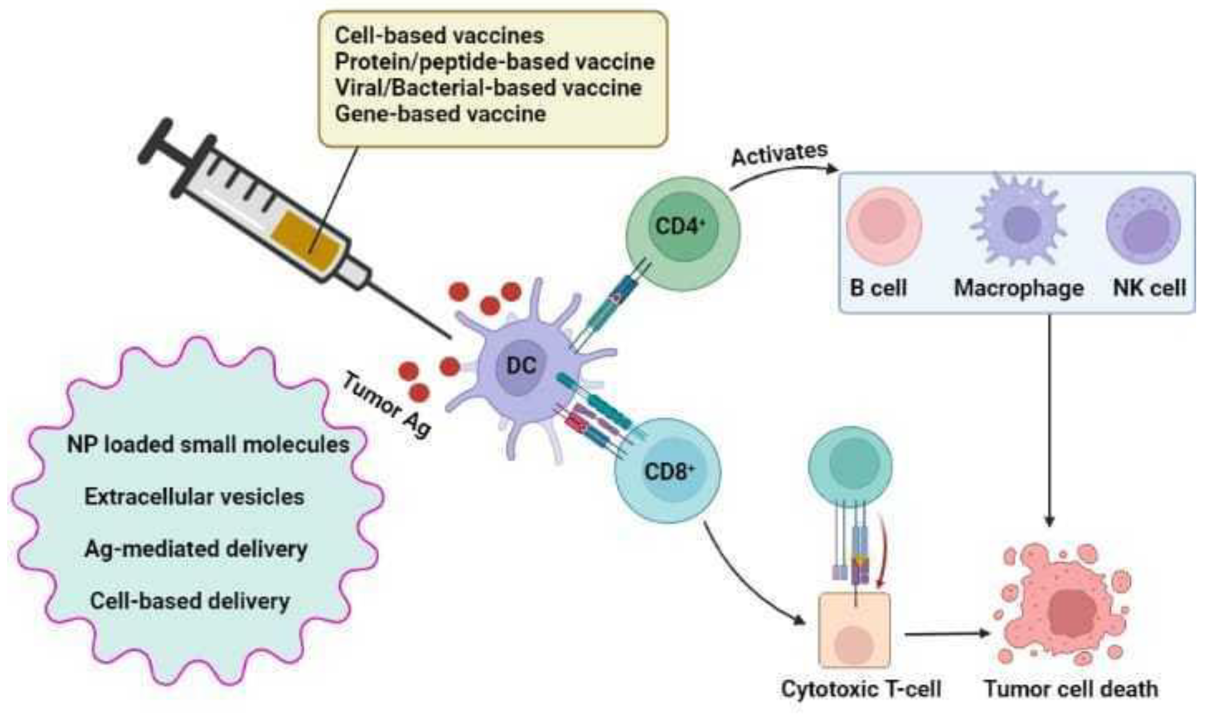
7.4. Lung Cancer Vaccines

Patients receiving chemotherapy for advanced-stage disease frequently possess drug-induced harsh side effects and have short-lived responses, especially when combination chemotherapy is given for an extended period. Vaccines may offer a therapeutic potential when incorporated into treatment plans soon after the first round of chemotherapy. A few phase III trials are being conducted to evaluate these vaccinations [172]. After definitive treatment, patients with minimally recurrent disease are likely to benefit more from vaccinations and may have long-lasting therapeutic effects (Figure 2).

7.4.1. Belagenpumatucel-L Vaccine (Lucanix)

One of the negative diagnostic factors of NSCLC, TGF-β, is identified to present in elevated levels in various malignan-cies including LC [173]. Preclinical research has demonstrated that the immunogenicity of tumor vaccines is raised by TGF-β2 suppression which serves as a repository for many TAAs [174]. An allogeneic tumor cell, gene-modified vaccine called Lu-canixTM (NovaRx, San Diego, CA, USA) is tested for four distinct NSCLC cancer cell lines that suppress TGF-β2 expression and boost immunogenicity by producing a TGF-β2 antisense gene [175,176]. At the injection site, the cause of immune suppression for vaccine is expected to be due to reduced TGF-β2 expression in the vaccine. The idea is that injecting downregulated TGF-β2 into allogeneic tumor cells will improve local immune identification and activate effector cells, triggering a systemic immune response that can target the patient’s original tumor [177]. Lucanix to placebo, a phase III trial as maintenance therapy did not find any differences in OS or PFS, documenting 96 NSCLC [178,179].

7.4.2. MAGE-A3

MAGE-A family consists of more than 60 genes and most of them are positioned on X chromosome to which belongs the melanoma antigen A3 (MAGE-A3) [180]. TAA which are expressed explicitly on tumor cells include MAGE protein. It is expressed in male germ cell lines, but not in other normal cells, and due to the lack of MHC they are incapable to hand-out MAGE-A antigens to the immune system [181]. The MAGE-A3 expression is considered directly proportional to the cancer progression. As the disease spread, MAGE-A3, whose normal expression level was 35% in NSCLC changed from 30% in stage I patients to 50% in stage II patients indicating their correlation with poor prognosis [182,183]. Cancer germline genes are important factors in determining the immortality, tumorigenesis, invasiveness, metastatic, and immune evasion capacity of tumor cells [184]. The tumor cell morphology, adhesion and migration can be altered by downregulating expression of cancer-germline gene [185,186]. Comparison of postoperative injections of MAGE-A3 recombinant protein coupled with an adjuvant system in 182 patients of a randomized phase II trial resulted in an outcome that was statistically insignificant, although the survival benefits of MAGE-A3 were sufficiently robust to initiate a Phase III assessment [181,187]. To establish a predictive signature that matches up with the treatment of MAGE-A3 antigen-specific immunotherapy, the gene expression profiling of tumors before treatment was analyzed [188]. In a population with a predictive genetic signature, the risk of recurrence was reduced by 43% after treatment with MAGE-A3. Another phase III LC trial commenced in 2007 with NSCLC patients recruited from 33 countries. In patients with MAGE-A3–positive stages IB, II, and IIIA, the efficacy of MAGE-A3 antigen-specific cancer immunotherapeutic (ASCI) agents were tested [189]. A total of 13,849 patients were screened, of which, 4210 had a MAGE-A3 positive tumor and 2272 patients were selected by randomization and then treated. A gene signature that aided in clinical benefit to MAGE-A3 was not observed because disease free survival (DFS) did not escalate on NSCLC in either the overall popula-tion or in NSCLC patients who did not receive ACT after treatment.

7.4.3. CIMAvax-EGF

CIMAvax-EGF is EGF based recombinant vaccine conjugated chemically onto p64K and emulsified in Montanide ISA 51 as adjuvant [190,191]. Antibodies against EGF are produced as part of mechanism of action of CIMAvax EGF, blocking EGF-EGFR interaction, and inhibit EGFR phosphorylation that results in EGF withdrawal [192]. A total of five phase I/II, a randomized controlled phase II and another randomized controlled phase III clinical trials have been per-formed since 1995. The first five trials were modeled to optimize the formulation, dosage and timing of the vaccine concerning immunogenicity and safety [193,194]. The pioneer step that attested the immunogenicity and the feasibility of inducing an antibody titer against autologous EGF was conducted in patients with colon, lung, prostate cancer or stomach [190,195,196]. The phase II randomized trial could not show that the vaccinated cohort had an advantage in OS [197]. In patients younger than 60 years of age and with GAR compared to PAR in the vaccinated group, a significant trend toward a survival advantage was seen. However, an advantage in OS for vaccinated patients in the phase III trial was not observed. As a possible diagnostic and predictive factor, baseline serum EGF levels were revealed [198]. The link between CIMAvax-EGF and chemo-combination therapy was disclosed by the Pilot 5 study, regardless of the small sample size. It revealed the potential rise in vaccine immunogenicity when conjugated to a cytotoxic systemic treatment [197].

7.4.4. Racotumomab

Racotumomab, the monoclonal anti-idiotypic murine IgG1 vaccine [199], causes the N-glycolyl GM3 (NeuGcGM3) ganglioside, almost undetectable in normal cells but seen in some tumor cells to provoke a particular humoral and cellular immune response. Their overexpression remains a target for cancer therapy, and is linked to altered cell proliferation, tumor spread, angiogenesis, and immunological tolerance [200,201]. According to a meta-analysis of stage III and IV NSCLC comprising 26 studies and 7839 patients, racotumomab and pemetrexed maintenance therapy were found to be successful in terms of DFS and OS [202]. A phase II/III trial with advanced NSCLC in 87 patients led to the conclusion that the product is effective, safe, and showed an increase in OS and PFS. The trial reached a certain conclusion from the results: drug induced only minor adverse effects like localized injection site responses, bone pain, coughing, and asthenia [203]. In 71 NSCLC patients for whom surgical treatment was not a choice, racotumomab was used as a second line therapy, and has shown to enhance OS in stage IV NSCLC patients [204].

7.4.5. TG4010

MUC1 is a TAA expressed by many solid tumors, including NSCLC. TG4010 targets MUC1 TAA and IL-2 [205]. The MUC1 protein is upregulated in LC and appears abnormally glycosylated, making it an immune target as glycosylation is the reservoir for new antigens. In a randomized, multicenter, phase II study with advanced NSCLC patients, TG4010 was administered in combination with platinum-based chemotherapy (cisplatin/vinorelbine) wherein the ORR and time to progression were 29.5% and 4.8 month in arm 1, respectively. In 2011, the study was further extended to open-label randomized trial III, which comprised 100 and 48 patients expressing MUC1 in their tumor with stage IIIB and stage IV NSCLC, respectively [206]. In this study, to predict the response to TG4010, frequency of CD56+, CD16+, and CD69+ cells were considered as biomarkers as they correspond to the phenotype of activated NK cells in PBMCs.

7.4.6. Vaccine Delivery Vehicles

Even though immunotherapy is a successful treatment option for NSCLC, there are still several limitations such as tumor penetration, low efficacy, high toxicity, issue with optimization of synergistic treatment, and off-target effects re-main. Effective delivery methods can overcome all of these restrictions [207,208]. A crucial technique for creating a com-prehensive therapeutic approach is making of an effective delivery system. For instance, therapeutic nanoparticles (NPs), are used to jointly administer chemo-immunotherapy regimens such as IL-12 and doxorubicin to achieve effective delivery into the tumor [209].

Nanoparticle-Based Delivery

Small molecules and their antagonists, for example, CpG oligodeoxynucleotides, inhibitors of TGF-β, IL-2, anti-PD-1 mAbs, antibodies and their fragments, peptides and proteins, can be delivered by NPs [210]. A few options available for NPs are nucleic acid nanotechnology, liposomes, dendrimers, inorganic nanocarriers polymeric systems, and exosomes. Delivery of cancer immunotherapies by NPs improves drug penetration, antitumor efficacy, synergetic effect of treatments and drug retention [211,212]. Additionally, by employing techniques like drug efflux pump modulation and administering numerous medications, NPs can overcome chemotherapeutic resistance.

NP-Loaded Small Molecules

Advanced LC tumors require a standard of care of combination chemotherapy and radiotherapy and additional cut-ting-edge treatments like immunotherapy or tailored medication. The most recent advancements in nanomedicine for the treatment of LC are magnetic NPs (such as: GastromarkVR, LumirenVR, Feridex, EndoremVR, and I.V.VR), liposomes and solid lipid vaccines (metal NPs (zinc oxide NPs, titanium dioxide NPs, gold NPs, iron NPs, silver NPs, and cerium NPs), cisplatin-loaded lipid-chitosan hybrid NPs, taxane class of NPs, virus NPs (TMV-cisplatin VNP complex, DOX-PhMV-PEG, Trop2CD40L VLPs) and polymer NPs (doxorubicin-conjugated InP/ZnS QDs) [213].

Extracellular Vesicles

Tiny, membrane-enclosed vesicles released by living cells that can carry a variety of lipids, nucleic acid species, and proteins are called extracellular vesicles (EVs) [214]. Anti-tumor immune responses are regulated by EVs, which assist in cell-cell interaction within TME [215]. Recently EVs have been identified as vehicles for various bioactive agents of cancer immunotherapy. For instance, designed EVs developed from fibroblast-like mesenchymal cells carrying short hairpin RNAs (shRNAs) or small interfering RNA (siRNAs), that target KRAS, are found to improve anti-pancreatic cancer activity and also raise mouse OS rates. The advantages of using EVs as a delivery platform are numerous and include their capacity to overcome environmental obstacles, inherent cell-targeting abilities, and circulatory stability [216].

Antigen-Mediated Delivery

CHP-NY-ESO-1 vaccine system aids in enhanced immune response by providing targeted delivery and is one of the important antigen delivery approaches in cancer immunotherapy. The polysaccharide nanogels of cholesteryl group-modified pullulan (CHP), that regulates TAMs are used to deliver the cancer-testis antigen, New York esophageal squamous cell carcinoma (NY-ESO)-1. The survival rate in patients with NSCLC found to be dose dependent on CHP-NY-ESO-1 in which a higher dose produces a longer survival rate. Targeted delivery and an improved immune response are possible with this approach [217].

Cell-Based Delivery

A common procedure of cell-based therapy involves ACT, CAR-T, and TIL therapy [218]. CAR-T cell treatment is a highly successful immunotherapy in the fight against blood malignancies and cancers that are refractory. The FDA has ap-proved a number of CAR-T cell-mediated immunotherapy products. Breyanzi can be given to patients with refractory or relapsed large B-cell lymphoma, Abecma and Carvykti for refractory or relapsed multiple myeloma patients, Yescarta for large B-cell lymphoma patients, and Tecartus for patients with refractory or relapsed mantle cell lymphoma [219]. In addition, drugs can act as nanoscale medicines that do not possess carriers advised for cancer care. Types of nanomedi-cines free of cargo are drug nanocrystals, antibody-drug conjugates (ADCs), drug-drug conjugate NPs and prodrug self-assembled NPs. For instance, to boost drug lodgment to tumors and kill both non-cancer stem cells (non-CSC) and CSCs, the medication SN38 (7-ethyl-10-hydroxycamptothecin) has been combined with the pH-responsive prodrug (PEG-CH=N-Doxorubicin (DOX) [213].

8. Combinatorial Approaches to Lung Cancer-Immunotherapy

The traditional therapeutic methods continue to be the first choice of cancer treatment; however, the success of these conventional regimens is limited by their unpleasant side effects, late illness detection, and deadly reversion as a resistant micro-metastatic disease. Hence, the gold standard of care for patients whose disease is not caused by a genomic change in NSCLC is PD-1 and PD-L1 inhibitors alone or given along with chemotherapy, radiation, or other ICIs [220].

8.1. a. Targeted therapy

Targeted therapy is the best option for those with irreversible LC and a driver gene mutation [221]. FDA-approved first-line medicines for metastatic NSCLC patients whose tumors possess EGFR exon 21 L858R mutations or EGFR exon 19 deletions are gefitinib, erlotinib, afatinib, dacomitinib, and osimertinib. EGFR/HER2 exon 20 mutations containing patients responded well to the two newly developed targeted medications such as TAK-788 and poziotinib [148]. A novel fusion oncogene echinoderm microtubule-associated protein-like (EML4)-ALK is seen in young and non-smokers, and 2–7% of patients with advanced NSCLC [221,222]. AMG510 is a tiny, effective drug that locks KRAS G12C in an inactive, GDP-bound state. Another KRAS G12C inhibitor, MRTX849, has shown promise in treating advanced solid tumors with KRAS G12C mutations [148]. IFN, which activates a variety of immune cell types, is found to be produced more abundantly by combining systemic IL-12 with trastuzumab and paclitaxel in a phase I clinical trial, and this further enhanced NK cell activation [223]. Their favorable correlation with clinical response potentiates the use of combinatorial strategies.

8.2. b. Chemotherapy

Platinum-based chemotherapy, with roughly one-year median survival, is crucial in treatment methods for patients without a driver gene mutation [224]. In multiple randomized controlled trials in NSCLC and SCLC, chemotherapy plus ICI showed better results than chemotherapy alone [144]. The CheckMate-9LA study showed that chemotherapy for a shorter period, along with inhibitors of CTLA-4 and PD-1 is feasible without sacrificing efficacy [225,226]. Ipilimumab, a CTLA-4 inhibitor, was given to NSCLC patients included in the phase II study either in a steady regimen in cycles 3 to cycle 6 of chemotherapy or concurrently in cycles 1 to 4 of six chemotherapy cycles, followed by ipilimumab maintenance [227]. In the KEYNOTE-021 study, the carboplatin-pemetrexed-pembrolizumab combination had an ORR of almost 70%, testing the activity of pembrolizumab with carboplatin and paclitaxel, vs. carboplatin, paclitaxel, and bevacizumab, or carboplatin and pemetrexed [228]. In a recent phase Ib study, the ICI durvalumab (PD-L1) and tremelimumab (CTLA-4) were coupled with the chemotherapy drug cisplatin-pemetrexed. Two ICIs combined with conventional chemotherapy appeared to be well tolerated, which revealed an ORR of almost 50% without specific hazard effects [229]. Quadruple combination therapy using bevacizumab, platinum-based chemotherapy, and atezolizumab was effective in treating patients with NSCLC who were oncogene-dependent [142,230].

8.3. c. Radiotherapy

Immunomodulatory effects of radiation include tumor-associated dendritic cell activation, improved T-cell action on tumors, altered TAM polarization, reduced immunosuppressive stromal cells, and induced immunogenic cell death [231]. The possibility of toxicity, particularly pneumonitis, when radiation and ICI are used together is a significant issue [232]. It has also been demonstrated that high-dose ionizing radiation increases PD-L1 expression, and inhibition of PD-L1 improves the effectiveness of radiation by a CTL-dependent mechanism [233]. Anti-CTLA-4 and anti-PD-1 therapy with combined radiation enhance the number of tumor-specific T cells, and it is noted that radiation-induced immune responses can have anticancer effects [145]. Radiation, when combined with CTLA-4 inhibition, produced diversification of the T cell receptor repertoire of TILs and molded the repertoire of T cell clones that are expanded, which is consistent with an effective vaccination [234]. It was discovered that overexpression of PD-L1, which causes T-cell exhaustion, is a mediator of radiation resistance and CTLA-4 blocking [235].

8.4. Emerging Combination Strategies in Adoptive Cell Therapy

Reactive T cells are extracted from patients, amplified ex vivo, and returned to the patient, which will aid host immunity for battling the disease [236] and is a more direct, less expensive, and effective technique to produce potent immunotherapeutic molecules [237,238]. Adoptive immunotherapy comes in a variety of forms that are employed in clinical trials, including lymphokine-activated killer (LAK), cytokine-induced killer (CIK), γ δ T-cells, TAA, TIL, and specific CTL [239]. NK-resistant tumor cells can be killed by LAK cells produced by lymphocytes exposed to IL-2. For patients with metastatic melanoma, TIL-based adoptive treatment has shown promising anticancer activity in a great number of clinical trials [240]. LAK possesses a powerful ability to lyse tumor cells that have survived after receiving cisplatin treatment and impart cytotoxic effects on A549 cells [241]. IL-2 and LAK cells coupled with chemotherapy or radiotherapy enhanced the survival of patients following surgical resection of primary LC, with 5- 9 years of survival rates in the immunotherapy group. Immunochemotherapy and immunoradiotherapy have shown OS of 54.4% and 52.0% in patient groups and 33.4% and 24.2% in the control group.
PD-L1 expression and the degree of CD8+ T cell infiltration were affected by concurrent chemoradiotherapy, and both affected the prognosis of NSCLC patients [242]. 22.4 months and 14.1 months was the median survival in the immunotherapy group and in the control group in a randomized trial, respectively, that added IL-2 and TIL to standard chemotherapy/radiotherapy after surgery. For stage III cancer patients, local relapse was significantly decreased in the immunotherapy group but not in the distant relapse [243]. After concurrent chemoradiotherapy (CCRT) treatment, Yoneda et al. noted that CD8+ TIL with increased density is a good prognostic indicator for locally progressed NSCLC [244]. The heterogeneous T-lymphocytes, known as CIK cells, have unlimited MHC-wide anticancer activity and a mixed NKT phenotype [245]. Seven ongoing CIK trials are being conducted, and a recent study among early-stage patients revealed that the 2-year OS rate could be increased when chemotherapy is coupled with DC-activated CIK cells, but the DFS rate does not get affected [246]. In patients receiving CIK in addition to chemotherapy, Wu et al. noticed that PFS (p = 0.042) and OS (p = 0.029) were longer [247]. Anti-γ δ TCR antibodies can be used to grow γ δ T-cells in vitro; this method may be more effective than employing phosphoantigen-expanded γ δ T-cells because the expanded γ δ T-cells have a longer in vivo survival time [248]. The effectiveness of CAR-NK cells to precisely identify tumor antigens has been demonstrated in numerous preclinical and clinical investigations, and therefore, adaptive NK-cell-based therapies can benefit from the features of recombinant CARs [249]. The phase-I trials, which were performed on patients with advanced and recurrent NSCLC revealed that activated NK cells are safe, well tolerated, and exhibit no substantial toxicity. Patients with LC were found to benefit clinically from repeated infusions of in vitro-activated HLA-mismatched NK cells when combined with conventional treatment [250]. In late-stage LC patients receiving vinorelbine-platinum chemotherapy, administration of a combination of immune cells, peptide-pulsed DCs and CIK were found to lessen the side effects of chemotherapy and increase patient survival [251].
Due to the viability and functionality of transplanted cells, which are instantly rejected after injection, adoptive treatment applications are restricted [252]. Nanocarrier systems based on innovative technologies can overcome these constraints. With tailored NPs, better adoptive T-cell treatment against prostate cancer was described [253,254]. Maleimide was surface conjugated to liposomes before being evaluated in a metastatic melanoma murine model. It was discovered that animals treated with NP-conjugated T-cells completely cleared the tumors, but non-conjugated T-cells slightly improved the survival of untreated mice [253]. These studies report that we can improve the effectiveness of ACT by delivering T cells using biomaterials [252]. Hence, according to recent clinical research, adoptive immunotherapy is effective for treating NSCLC and aid in an improved overall outcome and minimal toxicity.

9. Lung Cancer Preclinical Models in Testing Immunotherapy
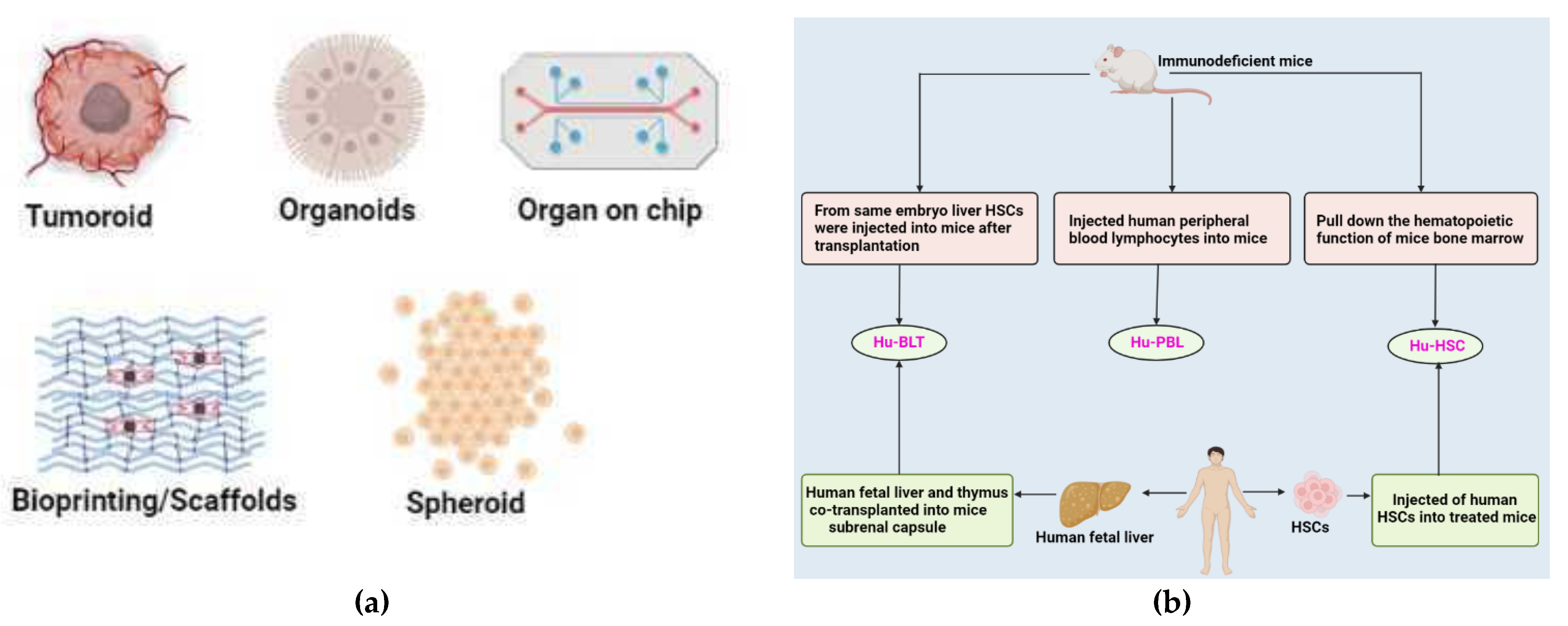
9.1. 3D Models

TME contain many factors that can affect the evolution and development of tumor. It can affect the metabolism, vascularization, and immune system of tumor tissues. It is comprehensive to study tumors using 2D culture models due to their inability to mimic the whole TME. 3D models can exhibit the proliferation, activation, and immuno-modulatory abilities of tumor tissues more than 2D culture models [255] (Table 4).

9.1.1. Cell-Lines

For the past 40 to 50 years, the spheroid model has been regarded as the gold standard among 3D in vitro models. Spheroids are 3D-based collections of cells along with ECM. To a certain extent, they can mimic the structure and metabolism of the tissue from where they originated. Based on cell genesis and culture methodologies, several spheroids may be defined, including multicellular tumor spheroids (MTCS), tumorospheres, tissue-derived tumor spheres (TDTSs), and organotypic multicellular spheroid/organoid (OMS) [256]. The most popular and thoroughly defined model, MTCS, is frequently produced from primary cell or cell line suspension. The MTCS model accurately depicts the diffusion and exchange of oxygen, nutrients, and other soluble factors. Due to its remarkable repeatability and relatively low cost, it is now the most used model for evaluating immunotherapeutic techniques [257].
Recently, inhibitors for immune checkpoints have been used for therapy in the fourth stage of NSCLC. In several studies, patient-derived NSCLC spheroids were used as a model to predict immune checkpoint treatment sensitivity. Spheroid heterotypic models are frequently employed to simulate the immunological microenvironment. The anticancer effect of CTLA4, PD L1, and PD1 blockage are mediated by immune cells in spheroids; also, they are characterized by increased CD8+ T cells and pro-inflammatory cytokine expression. There was no correlation analysis performed with patients’ outcomes in this model. Only a single clinical study on PD 1 therapeutic effect shows consistency in this model. Therefore, the predictive values are unclear, and this model cannot be considered a valid model in immunotherapy. New studies in this area are necessary to evaluate patient-derived spheroids’ prognostic role in testing immunotherapy [258].

9.1.2. Patient-Derived Lung Cancer Organoid

Scientific attention has switched to 3D cell culture techniques since 2D cell cultures only preserve a minimal similarity to their parent tissue. Patient-derived cell populations may be expanded in a 3D ECM to form organoids, distinguished by their ability to form structures that resemble the tissue from which they were obtained. Numerous tissues, most notably the lung, have been used to create tumor organoids [259]. LCOs require a specific concoction of growth factors and inhibitors, as well as a 3D matrix for support, most frequently Matrigel. Although the formulation of this growing medium differs between laboratories, all formulations include components that support the preservation of lung stem cells. Growth factors included are either EGF or members of the fibroblast growth factor (FGF) family, as well as inhibitors/activators of specific pathways, namely TGF-β and Rho-associated protein kinase and Wnt. The B27 supplement used as a substituted serum in neuronal cell cultures is the only factor common to all reported LCO-specific media formulations. LCOs may be created from tumor biopsies or resections that show a high rate of success in both short- and long-term cultures [260,261]. Li et al. developed a patient-derived tumoroid assay and the whole sequence to screen afatinib’s effect in EGFR-mutated patients [262]. Using transcriptomics, Peng et al. compared the primary tissues of SQCC or ADC to the equivalent tumoroids. Comparing tumoroid models to cancer cell lines or PDXs, tumoroid models showed a greater transcriptional accuracy [263]. Another study conducted by Ma et al. in these same tumoroids identified the genes (CDK1, CCNB2, and CDC25A) implicated in the NSCLC tumor formation [264] (Table 5).

9.1.3. Patient-Derived 3D Models

PDOs enable the 3D cultivation of malignant cells from the primary tissue, resulting in stromal destruction and immunological compartments. Tumorospheres are clonal representations of spheroids or organoids. However, this spheroid model is only suitable for CSC research since it cannot capture the diversity of cell types found in the TME. TDTSs are made from the patient tumor tissue that enzymes have digested. This model can preserve only the interactions between tumor cells, not between the stroma and tumor cells. TDTSs produce small replicas of vascularized tumor regions. OMS preserve the cellular heterogeneity and origin tissue architecture, making them the most accurate replicas of the parental tumor. OMS is useful for biomarker and medication testing since they are reliable preclinical patient models. PDOs replicate the gene and protein expression of the real biopsies. Several models are available, including those developed from NSCLC, clear cell renal cell carcinoma (CCRC), melanoma, and glioblastoma [255].
Table 4. The main studies conducted in the field of lung cancer using spheroids and organoids models.
ModelsSourceApplicationReference
spheroidsResectionLung cancer stem cells’ identification and description; production of
Xenografts that replicate the parental tumor’s histology
[265]
Resection, pleural effusionTechnique to expand
cancer cells from the lungs of patients
[266]
Core needle biopsy, surgical biopsy, pleural effusionDrug testing[267]
OrganoidsbiopsyDrug testing and evaluation of immune cell populations penetrating cultured tissues[268]
Resection/PDXLCOs’ long-term growth, confirmation, and drug screening[269,270]
biopsyFor use in immuno-oncology research and testing for customized immunotherapy, a new approach for maintaining endogenous tumor-infiltrating cells has been developed.[271]
Resection/biopsyBiobanking, drug testing[272,273,274]
Resection/biopsyExamining and blocking regulators of mitochondrial fission in various tumor organoids[275]
Pleural effusiondrug testing and the establishment of an LCO culture system from pleural effusions[276]
Resection/biopsyAnalyzing several techniques to determine the tumor purity of organoids created from intrapulmonary tumors[277]
PDX derived from biopsiesDrug testing and organoid creation using PDXs from SCLC biopsies[278]
Resection/biopsyAnalyses of pathway inhibitors found by single-cell proteomics[279]
pleural effusionTargeted drug testing and LCO production and characterization[280]
Table 5. Currently used lung organoids and tumoroids models in NSCLC research.
Tumor TypeModelApplicationReference
NSCLC
(Non-small cell lung carcinoma)
Lung cancer organoidsDrug screening[269]
NSCLC organoidsDrug screening[270]
Patient-derived organoids modelGenomics, production of treatment outcomes[274]
Lung ADK (LADC)-derived organoid model Drug screening, biomarker development, and living biobank[273,274]
Patient-derived tumoroids (PDTs)PDTs are developed to be used in microfluidic devices for drug screening and mimic the cancer vascular network.[281]
Patient-derived lung cancer organoidsPatient-specific medication screening and support for the xenograft model from a living biobank[272]
Patient-derived tumoroids
(PDTs)
creating new cell lines[282]
Patient lung-derived
tumoroids (PLDTs)
Drug screening[283]
Lung cancer organoidsPersonalized medicine[277]

9.1.4. Organ-On-Chip

Although there have been in vitro models of LC for many years, most of them have consisted of monolayer cultures of lung epithelial cells or planar 3D tissue models with air-liquid interfaces. The model can be used to trace pharmacological treatments and immune cells [284]. NSCLC-derived cell lines can be employed in microfluidic models of LC, where the A549 cell line is most often used. A549 cells often acquire the spheroid shape when cultivated in microfluidic devices because they cannot establish intercellular connections. These platforms have been utilized to research a variety of significant issues [285]. The effectiveness of photodynamic treatment was analyzed using A549 spheroids cultivated in microfluidic devices. The ability to precisely manipulate and characterize cell-cell communication in the LC TME has been made possible using microfluidic devices, for instance, to segregate different cell types seen in LC such as CAFs and ECs. NSCLC cell lines were cultured in microfluidic models in various studies to investigate the effects of tumor growth, medication response, and mechanical forces found in the lung, including blood and interstitial fluid flow. The interaction of a lung tumor and bacteria has also been studied using NSCLC cells cultured in microfluidic devices [284]. Using autologous TILs in two studies from the Borenstein group, small primary tumor organoids from NSCLC patient tumor biopsies were cultured. This allowed for the characterization of tumor-immune interactions and the prediction of patient-specific responses to ICI therapies [286,287]. Future developments in microfluidic models of LC should carefully consider the tumor cell type and their source; the inclusion of different cell types such as immune cells, epithelial cells, and/or CAFs; and the biomarkers to represent ECM and hypoxia. Furthermore, the impact of mechanical strain, ECM composition, immune cell phenotype and infiltration, and response to treatment regarding this model is still poorly understood [284].

9.1.5. 3D Bioprinting

3D bioprinting is a biomanufacturing technology that enables loading live cells, signaling molecules, and biomaterials to create tissue-engineered constructions with precisely regulated tissue architecture. The use of bioprinting technology can produce gradated macroscale designs that imitate the ECM, also boosting both the adhesion and proliferation of various cell types. Constructs created via 3D bioprinting can successfully imitate the TME. Additionally, the capacity to incorporate perusable vascular networks, spatial control of matrix characteristics, and automatic and high-level testing capabilities for identifying metabolic toxins and metabolic parameters are crucial components of bioprinted models. According to their deposition process, droplet, extrusion, and laser-based bioprinting techniques can be categorized based on instrumentation methodologies [288]. A substantial number of biopsy samples from patients with lung cancer were utilized by Mazzocchi et al. to create 3D tumor models containing pleural effusion aspirates and lung cancer spheroids implanted within hydrogel scaffolds [276]. Another study showed that sodium alginate-gelatin hydrogel provides better printability and viability to the cells of NSCLC from patient-derived xenografts and associated fibroblast coculture [289]. The printed tumor models can also be manufactured using spheroids that exhibit tumor-specific markers and are used for drug screening. Wang et al. used A549/95-D lung cancer cells to create 3D bioprinted scaffolds to study metastasis [290]. The bioprinted tumor models developed using patient-derived cancer cells have in vivo-like anatomy [288].

9.2. In Vivo Animal Models

The mouse genome is quite like the human genome and possesses the ease of gene editing, cheap cost, and straightforward feeding. It can imitate several biological properties, including cancer development and metastasis in vivo. Four approaches are often used to create animal cancer models, including chemical induction, PDX, genetically engineered mouse models (GEMM), and cell line-derived xenograft (CDX). The chemical induction model is a model induced by chemical carcinogens that can mimic the onset of cancer from the beginning of its carcinogenic process. However, the major drawback is that it takes about 30 to 50 weeks for a tumor to grow. The CDX model, also known as the xenotransplantation model, is created by delivering cancer cell lines subcutaneously to immunocompromised mice. The key merit and demerit of this model include convenience to handle, but the prolonged in vitro culture alters the behavior of original tumor cells. The PDX model was developed as an animal model by introducing patient tumor tissues directly into mice. This model faithfully replicates the histology and genetics of the original tumor [291]. These current models, however, are unable to precisely anticipate how the tumor and immune system of humans would interact among different species of mammals. The humanlike mouse immune system model rebuilds the human immune system by grafting lymphocytes, hematopoietic cells, or organs of humans into mice with a compromised immune system [292] (Figure 3b). By implanting human tumor cells or tissues, it is feasible to understand tumor growth, the human immune system setting, and assess anti-tumor therapy, specifically the impact of immunotherapy and associated processes. The humanized mouse immune system models are categorized into three groups based on how the human immune system was recreated, and it includes human hematopoietic stem cells (Hu-HSCs), human peripheral blood lymphocyte (Hu-PBL), and human bone marrow, liver, and thymus (Hu-BLT) models [291].

9.2.1. Zebrafish Model

Zebrafish is the most popular vertebrate model due to their genome similarity with humans [293]. Transgenic and immunodeficient zebrafish can grow very fast, remain transparent in the adult stage, and have simple gene operations. Translucent embryos can detect and trail cancer cell propagation, metastasis, and spread [291]. Shen et al. developed the LINC00152 knockout xenotransplantation model and demonstrated that LINC00152 silencing might lessen LC cell proliferation and spread [294]. The effectiveness and safety of anti-LC medications may also be assessed using the zebrafish model to determine a more effective course of therapy. Huang et al. employed the zebrafish LC models to assess the impact of DFIQ (quinoline derivative anti-cancer agent) on NSCLC in vivo. Monitoring cell migration, apoptosis, and proliferation revealed that DFIQ might suppress cancer cells. Zebrafish models can accurately reproduce the interactions between TME though they lack lungs. The efficacy and safety of three anti-angiogenic medications have been investigated using a human NSCLC xenograft model. All molecules examined had anti-angiogenic properties and reduced the development of tumors in zebrafish [295].

9.2.2. Patient-Derived Tumor Xenograft (PDX) Model

The Hu-PDX model has been used in several tumor research studies. Lin et al. demonstrated PD-L1/PD-1 targeted immunotherapy in immunodeficient mice using patients’ peripheral blood cells [296]. Rosato and his colleagues constructed a PDX model derived from TNBC patients to test anti-PD-1 immunotherapy as a TNBC preclinical trial [291]. Sanmamed et al. administered immunodeficient mice with gastric cancer patient lymphocytes, then administered mice with transplanted gastric cancer tissue, and then with nivolumab (inhibitor of PD-1) and urelumab (anti-CD137 agonist). This study demonstrated that these drugs could resist tumor growth through the induction of T cells [297]. The present Hu-PDX model does have significant flaws, such as a low modeling success rate, limited lifespan for the humanized immune system, and poor immunological response. The advancement of humanized mouse modeling technologies and the effectiveness and longevity of the immune system should be the main areas of future study [291].

9.2.3. Patient-Derived Orthotropic Xenograft (PDOX) Model

Comparable to the primary site of cancer, an in vivo environment that is favorable for the growth of the tumor can be created through orthotopic transfer of tumor tissue into animal organs. As a result, the PDX model serves as the foundation for the PDOX model. This model is more objective and accurate than the conventional PDX model in simulating the development of human cancers in vivo [298]. Using cervical cancer tissue, Hiroshima et al. created ten cases of the PDX model and eight cases of PDOX models by subcutaneous injection of cervical cancer tissues. The result demonstrated that in 50% of the PDOX models, tumor metastasis was shown but not in the PDX model. According to the data, the PDOX model is more likely to have characteristics of malignant tumors, such as invasion and metastasis, than the PDX model [299]. Hiroshima et al. conducted a study by treating these two models with entinostat medication and found that tumor growth inhibition took place only in PDOX models [291]. In the PDOX model, growth was found in vivo, making it challenging to monitor their progress using conventional detection techniques and even more challenging to identify their metastatic paths [300].

9.2.4. Mini Patient-Derived Xenograft (Mini-PDX) Model

This model was created by infusing the patient’s tumor tissue’s digested cell suspension into a microcapsule and implanting the capsule into the animal. The major advantages of this model in the area of drug screening include consistent outcomes with the PDX model, economic, and short time to approach the results [291]. Zhang et al. created Mini-PDX models of pancreatic, gastric, and lung cancers, and PDX model was used as a reference model to assess drug sensitivity. The results indicate that both the Mini-PDX model and the conventional PDX model produce results that are 92% consistent; however, the Mini-PDX methodology requires substantially less time than the PDX model does [301]. This demonstrates that the Mini-PDX model might serve as a useful alternative to the PDX model for assessing cancer treatment. Due to these benefits, the Mini-PDX model can be anticipated as a device that aids in the individualized treatment of cancer patients [291].

10. Conclusions and Future Perspectives

LC continues to pose a serious threat to global health, despite significant advancements in early LC prevention, diagnosis, and treatment. Within the next 40 years, it is anticipated to overtake IHD as the leading cause of death. The GLOBOCAN-2020 revision on epidemiologic trends of different malignancies agrees with WHO mortality projections. LC’s increased prevalence, incidence, and mortality rates highlight the need for efficient intervention and therapy approaches. The TCGA-c Bioportal data on the number of LC patients who have undergone radiation therapy, targeted therapy, and immunotherapy is 19.2%, 58.9%, and 28.6%, respectively, where cases undergone chemotherapy outnumber the data that do not undergo therapy. The data availability for cases with various therapy to that of ’data does not available’ was 13.2%, 12.1%, 12.1%, and 12% for radiotherapy, chemotherapy, targeted therapy, and immunotherapy, respectively, which raises concerns about the validity of the data.
The use of methods like combination therapy and the exploration of predictive treatment biomarkers and prognosis have resulted from clinical trials of immunotherapy. The therapeutic tactics employed to take advantage of the immunosuppressive properties of TME in the present immunotherapy clinical trial landscape have created difficulties in treatment, clinical testing, and monitoring across diverse tumors. Immunotherapy as a cancer treatment strategy poses difficulties due to the immunosuppression brought on by TME. Microbiome-targeted cancer immunotherapy and the combination of genetic biomarkers with immune-related indicators for more individualized treatment are the new immunotherapy-based strategies. Immune organization in TME is a potent predictor of response to immunotherapy. The basic pathomorphological diagnosis of NSCLC patients could be supplemented with a quick immunological study of the immune response already present in the malignant tissue, such as the presence of lymphocytes and CD8+ macrophages, including PDL1, CXCR3, CCLA4, JAK, TGFβ, and IFNγ expressions, and their placement inside the tumor. The complexity of immune system-tumor communication is complicated, and our understanding of key molecular mechanisms helps us to provide a suitable therapeutic strategy and predict the course of the disease.
The FDA has approved a few combination medicines to boost the clinical effectiveness of ICIs. Combinations of ACT, newer ICIs, cancer vaccines, and small molecule inhibitors are anticipated as action-driven reliable biomarkers for clinical immuno-oncology decision-making well-understood. In this sense, a truly patient-centered, tailored approach is what cancer immunotherapy needs to succeed in the future. Trials on combination therapy using chemotherapeutic drugs with PD1 and CTLA4 checkpoint inhibitors are in clinical trials now. Vaccinations in LC have limited clinical trial evidence to demonstrate definite therapeutic advantages. Observable immune responses may not always correspond to clinically significant responses. While many trials concentrate on patients with distant-stage cancers, patients with stage I or stage II LC with potential recurrence after surgery may be the leading candidates for LC vaccinations. The set of best tumor antigens to target and tackle the different tumor escape mechanisms and many epitopes of a broad set of genes are to be traced for an effective immunotherapy-based LC treatment.
The multidisciplinary approaches of preclinical cancer research, with contributions from the field of stem cell biology, immunology, and developmental biology, help to decipher the interaction between cancer and stroma. Currently, available models can fully recapitulate patient-tumor phenotypes and responses though various cancer models are available for cancer research. The application of multiple approaches, along with an idea of their limitations, can make improvements in the field of cancer therapeutics.

Author Contributions

Conceptualization, H.P., R.R.A., L.M.J., K.V.A., and A.G.; software, H.P. and C.M.W.; resources, H.P.; data curation, H.P.; writing—original draft preparation, H.P., K.V.A., L.M.J., and R.R.A., writing—review and editing, A.G., H.P., K.V.A., L.M.J., R.R.A., and C.M.W.; visualization, H.P., K.V.A., R.R.A., H.M., T.R., E.S., and J.L.; supervision, A.G., A.V.G., and R.G.; project administration, A.G., A.V.G., and R.G. All authors contributed to conceptualizing, drafting, and revising the manuscript. All authors have read and agreed to the published version of the manuscript.

Funding

This research received no external funding.

Institutional Review Board Statement

Not applicable.

Informed Consent Statement

Not applicable.

Data Availability Statement

Not applicable.

Conflicts of Interest

The authors declare no conflict of interest.

References

  1. Howlader, N.; Forjaz, G.; Mooradian, M.J.; Meza, R.; Kong, C.Y.; Cronin, K.A.; Mariotto, A.B.; Lowy, D.R.; Feuer, E.J. The Effect of Advances in Lung-Cancer Treatment on Population Mortality. N. Engl. J. Med. 2020, 383, 640–649. [Google Scholar] [CrossRef] [PubMed]
  2. Chevallier, M.; Borgeaud, M.; Addeo, A.; Friedlaender, A. Oncogenic Driver Mutations in Non-Small Cell Lung Cancer: Past, Present and Future. World J. Clin. Oncol. 2021, 12, 217. [Google Scholar] [CrossRef] [PubMed]
  3. Pawelczyk, K.; Piotrowska, A.; Ciesielska, U.; Jablonska, K.; Gletzel-Plucinska, N.; Grzegrzolka, J.; Podhorska-Okolow, M.; Dziegiel, P.; Nowinska, K. Role of PD-L1 Expression in Non-Small Cell Lung Cancer and Their Prognostic Significance According to Clinicopathological Factors and Diagnostic Markers. Int. J. Mol. Sci. 2019, 20, 824. [Google Scholar] [CrossRef] [PubMed] [Green Version]
  4. Small Cell Lung Cancer Stages | Stages of Small Cell Lung Cancer. Available online: https://www.cancer.org/cancer/lung-cancer/detection-diagnosis-staging/staging-sclc.html (accessed on 13 October 2022).
  5. Wang, C.; Li, J.; Zhang, Q.; Wu, J.; Xiao, Y.; Song, L.; Gong, H.; Li, Y. The Landscape of Immune Checkpoint Inhibitor Therapy in Advanced Lung Cancer. BMC Cancer 2021, 21, 1–18. [Google Scholar] [CrossRef] [PubMed]
  6. Mathers, C.D.; Loncar, D. Projections of Global Mortality and Burden of Disease from 2002 to 2030. PLoS Med. 2006, 3, 2011–2030. [Google Scholar] [CrossRef] [Green Version]
  7. Padinharayil, H.; Varghese, J.; John, M.C.; Rajanikant, G.K.; Wilson, C.M.; Al-Yozbaki, M.; Renu, K.; Dewanjee, S.; Sanyal, R.; Dey, A.; et al. Non-Small Cell Lung Carcinoma (NSCLC): Implications on Molecular Pathology and Advances in Early Diagnostics and Therapeutics. Genes Dis. 2022, 2352–3042. [Google Scholar] [CrossRef]
  8. Registry, P.C. Global Cancer Observatory. Available online: https://gco.iarc.fr/ (accessed on 10 October 2022).
  9. Conger, A.B. Cancer Statistics. Available online: https://seer.cancer.gov/statistics/ (accessed on 10 October 2022).
  10. Gao CBioPortal for Cancer Genomics. Available online: https://www.cbioportal.org/%0Ahttps://www.cbioportal.org/%0Ahttp://www.cbioportal.org/public-portal/sci_signal_reprint.jsp (accessed on 10 October 2022).
  11. Siamof, C.M.; Goel, S.; Cai, W. Moving Beyond the Pillars of Cancer Treatment: Perspectives From Nanotechnology. Front. Chem. 2020, 8, 598100. [Google Scholar] [CrossRef]
  12. Hoy, H.; Lynch, T.; Beck, M. Surgical Treatment of Lung Cancer. Crit. Care Nurs. Clin. N. Am. 2019, 31, 303–313. [Google Scholar] [CrossRef]
  13. Bogart, J.A.; Waqar, S.N.; Mix, M.D. Radiation and Systemic Therapy for Limited-Stage Small-Cell Lung Cancer. J. Clin. Oncol. 2022, 40, 661–670. [Google Scholar] [CrossRef]
  14. Lung Cancer-Non-Small Cell: Types of Treatment | Cancer.Net. Available online: https://www.cancer.net/cancer-types/lung-cancer-non-small-cell/types-treatment (accessed on 13 October 2022).
  15. Nagasaka, M.; Gadgeel, S.M. Role of Chemotherapy and Targeted Therapy in Early-Stage Non-Small Cell Lung Cancer. Expert Rev. Anticancer. Ther. 2018, 18, 63–70. [Google Scholar] [CrossRef]
  16. Ruiz-Cordero, R.; Devine, W.P. Targeted Therapy and Checkpoint Immunotherapy in Lung Cancer. Surg. Pathol. Clin. 2020, 13, 17–33. [Google Scholar] [CrossRef] [PubMed]
  17. Doroshow, D.B.; Sanmamed, M.F.; Hastings, K.; Politi, K.; Rimm, D.L.; Chen, L.; Melero, I.; Schalper, K.A.; Herbst, R.S. Immunotherapy in Non-Small Cell Lung Cancer: Facts and Hopes. Clin Cancer Res 2019, 25, 4592. [Google Scholar] [CrossRef] [PubMed] [Green Version]
  18. Mok, T.S.K.; Wu, Y.L.; Kudaba, I. Pembrolizumab versus Chemotherapyfor Previously Untreated, PD-L1-expressing, Locally Advancedor Metastatic Non-small-cell Lung Cancer (KEYNOTE-042): Arandomised, Open-label, Controlled, Phase 3 Trial. Lancet 2019, 393, 1819–1830. [Google Scholar] [CrossRef]
  19. Non-Small Cell Lung Cancer Chemotherapy | Chemo Side Effects. Available online: https://www.cancer.org/cancer/lung-cancer/treating-non-small-cell/chemotherapy.html (accessed on 13 November 2022).
  20. Chen, L.; Smith, D.A.; Somarouthu, B.; Gupta, A.; Gilani, K.A.; Ramaiya, N.H. A Radiologist’s Guide to the Changing Treatment Paradigm of Advanced Non–Small Cell Lung Cancer: The ASCO 2018 Molecular Testing Guidelines and Targeted Therapies. Am. J. Roentgenol. 2019, 213, 1047–1058. [Google Scholar] [CrossRef]
  21. Pakkala, S.; Ramalingam, S.S. Personalized Therapy for Lung Cancer: Striking a Moving Target. JCI Insight 2018, 3, e120858. [Google Scholar] [CrossRef] [Green Version]
  22. Soria, J.-C.; Ohe, Y.; Vansteenkiste, J.; Reungwetwattana, T.; Chewaskulyong, B.; Lee, K.H.; Dechaphunkul, A.; Imamura, F.; Nogami, N.; Kurata, T.; et al. Osimertinib in Untreated EGFR-Mutated Advanced Non-Small-Cell Lung Cancer. N Engl J Med 2018, 378, 113–125. [Google Scholar] [CrossRef] [PubMed]
  23. Ramalingam, S.S.; Vansteenkiste, J.; Planchard, D.; Cho, B.C.; Gray, J.E.; Ohe, Y.; Zhou, C.; Reungwetwattana, T.; Cheng, Y.; Chewaskulyong, B.; et al. Overall Survival with Osimertinib in Untreated, EGFR -Mutated Advanced NSCLC. N. Engl. J. Med. 2020, 382, 41–50. [Google Scholar] [CrossRef]
  24. Kong, X.; Zhang, K.; Wang, X.; Yang, X.; Li, Y.; Zhai, J.; Xing, Z.; Qi, Y.; Gao, R.; Feng, X.; et al. Mechanism of Trastuzumab Resistance Caused by HER-2 Mutation in Breast Carcinomas. Cancer Manag. Res. 2019, 11, 5971–5982. [Google Scholar] [CrossRef] [Green Version]
  25. Home-ClinicalTrials.Gov. Available online: https://clinicaltrials.gov/ (accessed on 4 March 2022).
  26. Camidge, D.R.; Dziadziuszko, R.; Peters, S.; Mok, T.; Noe, J.; Nowicka, M.; Gadgeel, S.M.; Cheema, P.; Pavlakis, N.; de Marinis, F.; et al. Updated Efficacy and Safety Data and Impact of the EML4-ALK Fusion Variant on the Efficacy of Alectinib in Untreated ALK-Positive Advanced Non–Small Cell Lung Cancer in the Global Phase III ALEX Study. J. Thorac. Oncol. 2019, 14, 1233–1243. [Google Scholar] [CrossRef]
  27. Peters, S.; Mok, T.S.K.; Gadgeel, S.M.; Rosell, R.; Dziadziuszko, R.; Kim, D.-W.; Perol, M.; Ou, S.-H.I.; Shaw, A.T.; Bordogna, W.; et al. Updated Overall Survival (OS) and Safety Data from the Randomized, Phase III ALEX Study of Alectinib (ALC) versus Crizotinib (CRZ) in Untreated Advanced ALK + NSCLC. J. Clin. Oncol. 2020, 38, 9518. [Google Scholar] [CrossRef]
  28. Camidge, R.; Kim, H.; Ahn, M.; Al, E.; Al, E.; Al, E. Brigatinib versus Crizotinib in ALK-Positive Non–Small-Cell Lung Cancer [Published Online September 25, 2018]. N. Engl. J. Med. 2018, 379, 2027–2039. [Google Scholar] [CrossRef] [PubMed]
  29. Soria, J.C.; Tan, D.S.W.; Chiari, R.; Wu, Y.L.; Paz-Ares, L.; Wolf, J.; Geater, S.L.; Orlov, S.; Cortinovis, D.; Yu, C.J.; et al. First-Line Ceritinib versus Platinum-Based Chemotherapy in Advanced ALK-Rearranged Non-Small-Cell Lung Cancer (ASCEND-4): A Randomised, Open-Label, Phase 3 Study. Lancet 2017, 389, 917–929. [Google Scholar] [CrossRef]
  30. Kumar, M.; Ernani, V.; Owonikoko, T.K. Biomarkers and Targeted Systemic Therapies in Advanced Non-Small Cell Lung Cancer. Mol. Asp. Med. 2015, 45, 55–66. [Google Scholar] [CrossRef] [Green Version]
  31. Drilon, A.; Siena, S.; Dziadziuszko, R. Entrectinib in ROS1 Fusion-Positive Non-Small-Cell Lung Cancer: Integrated Analysis of Three Phase 1–2 Trials. Lancet Oncol. 2020, 21, 261–270. [Google Scholar] [CrossRef]
  32. Shaw, A.T.; Ou, S.H.; Bang, Y.J. Crizotinib in ROS1-Rearranged Non-Small-Cell Lung Cancer. N. Engl. J. Med. 2014, 371, 1963–1971. [Google Scholar] [CrossRef] [Green Version]
  33. Shaw, A.T.; Riely, G.J.; Bang, Y.J.; Kim, D.W.; Camidge, D.R.; Solomon, B.J.; Varella-Garcia, M.; Iafrate, A.J.; Shapiro, G.I.; Usari, T.; et al. Crizotinib in ROS1-Rearranged Advanced Non-Small-Cell Lung Cancer (NSCLC): Updated Results, Including Overall Survival, from PROFILE 1001. Ann. Oncol. 2019, 30, 1121–1126. [Google Scholar] [CrossRef] [PubMed]
  34. Kim, S.; Kim, T.M.; Kim, D.W.; Kim, S.; Kim, M.; Ahn, Y.O.; Keam, B.; Heo, D.S. Acquired Resistance of MET-Amplified Non-Small Cell Lung Cancer Cells to the MET Inhibitor Capmatinib. Cancer Res. Treat. 2019, 51, 951–962. [Google Scholar] [CrossRef]
  35. Wolf, J.; Seto, T.; Han, J.-Y.; Reguart, N.; Garon, E.B.; Groen, H.J.M.; Tan, D.S.W.; Hida, T.; de Jonge, M.; Orlov, S.V.; et al. Capmatinib in MET Exon 14–Mutated or MET -Amplified Non–Small-Cell Lung Cancer. N. Engl. J. Med. 2020, 383, 944–957. [Google Scholar] [CrossRef]
  36. Drilon, A.; Oxnard, G.R.; Tan, D.S.W. Efficacy of Selpercatinib in RET Fusion-Positive Non-Small-Cell Lung Cancer. N. Engl. J. Med. 2020, 383, 813–824. [Google Scholar] [CrossRef]
  37. Gainor, J.F.; Curigliano, G.; Kim, D.-W.; Lee, D.H.; Besse, B.; Baik, C.S.; Doebele, R.C.; Cassier, P.A.; Lopes, G.; Tan, D.S.-W.; et al. Registrational Dataset from the Phase I/II ARROW Trial of Pralsetinib (BLU-667) in Patients (Pts) with Advanced RET Fusion+ Non-Small Cell Lung Cancer (NSCLC). J. Clin. Oncol. 2020, 38, 9515. [Google Scholar] [CrossRef]
  38. Nowicki, T.S.; Hu-Lieskovan, S.; Ribas, A. Mechanisms of Resistance to PD-1 and PD-L1 Blockade. Cancer J. 2018, 24, 47–53. [Google Scholar] [CrossRef] [PubMed]
  39. Skov, B.G.; Rørvig, S.B.; Jensen, T.H.L.; Skov, T. The Prevalence of Programmed Death Ligand-1 (PD-L1) Expression in Non-Small Cell Lung Cancer in an Unselected, Consecutive Population. Mod. Pathol. 2019, 33, 109–117. [Google Scholar] [CrossRef] [PubMed]
  40. Reck, M.; Rodríguez-Abreu, D.; Robinson, A.G.; Hui, R.; Csoszi, T.; Fülöp, A.; Gottfried, M.; Peled, N.; Tafreshi, A.; Cuffe, S.; et al. Updated Analysis of KEYNOTE-024: Pembrolizumab versus Platinum-Based Chemotherapy for Advanced Non–Small-Cell Lung Cancer with PD-L1 Tumor Proportion Score of 50% or Greater. J. Clin. Oncol. 2019, 37, 537–546. [Google Scholar] [CrossRef]
  41. Herbst, R.; de Marinis, F.; Giaccone, G.; Reinmuth, N.; Vergnenegre, A.; Barrios, C.; Morise, M.; Font, E.; Andric, Z.; Geater, S.; et al. O81 IMpower110: Interim Overall Survival (OS) Analysis of a Phase III Study of Atezolizumab (ATEZO) Monotherapy vs Platinum-Based Chemotherapy (CHEMO) as First-Line (1L) Treatment in PD-L1–Selected NSCLC. Ann. Oncol. 2020, 30, v915. [Google Scholar] [CrossRef]
  42. Tony, S.K.; Mok, M.; Yi-Long Wu, M.; Iveta Kudaba, M.; Dariusz, M.; Kowalski, M.; Byoung Chul Cho, M.; Hande, Z.; Turna, M.; Gilberto Castro, P., Jr.; et al. Pembrolizumab versus Chemotherapy for Previously Untreated, PD-L1-Expressing, Locally Advanced or Metastatic Non-Small-Cell Lung Cancer (KEYNOTE-042): A Randomised, Open-Label, Controlled, Phase 3 Trial. Lancet 2019, 393, 1819–1830. [Google Scholar]
  43. Hellmann, M.D.; Paz-Ares, L.; Bernabe Caro, R.; Zurawski, B.; Kim, S.-W.; Carcereny Costa, E.; Park, K.; Alexandru, A.; Lupinacci, L.; de la Mora Jimenez, E.; et al. Nivolumab plus Ipilimumab in Advanced Non–Small-Cell Lung Cancer. N. Engl. J. Med. 2019, 381, 2020–2031. [Google Scholar] [CrossRef]
  44. Qin, H.; Wang, F.; Liu, H.; Zeng, Z.; Wang, S.; Pan, X.; Gao, H. New Advances in Immunotherapy for Non-Small Cell Lung Cancer. Am. J. Transl. Res. 2018, 10, 2234. [Google Scholar] [CrossRef]
  45. Galon, J.; Bruni, D. Tumor Immunology and Tumor Evolution: Intertwined Histories. Immunity 2020, 52, 55–81. [Google Scholar] [CrossRef]
  46. Domagala-Kulawik, J. The Role of the Immune System in Non-Small Cell Lung Carcinoma and Potential for Therapeutic Intervention. Transl. Lung Cancer Res. 2015, 4, 177–190. [Google Scholar] [CrossRef]
  47. Franceschi, C.; Bonafè, M.; Valensin, S.; Olivieri, F.; de Luca, M.; Ottaviani, E.; de Benedictis, G. Inflamm-Aging. An Evolutionary Perspective on Immunosenescence. Ann. N. Y. Acad. Sci. 2000, 908, 244–254. [Google Scholar] [CrossRef]
  48. Chen, D.S.; Mellman, I. Oncology Meets Immunology: The Cancer-Immunity Cycle. Immunity 2013, 39, 1–10. [Google Scholar] [CrossRef] [PubMed]
  49. Chen, D.S.; Mellman, I. Elements of Cancer Immunity and the Cancer-Immune Set Point. Nature 2017, 541, 321–330. [Google Scholar] [CrossRef] [PubMed]
  50. Dunn, G.P.; Bruce, A.T.; Ikeda, H.; Old, L.J.; Schreiber, R.D. Cancer Immunoediting: From Immunosurveillance to Tumor Escape. Nat. Immunol. 2002, 3, 991–998. [Google Scholar] [CrossRef] [PubMed]
  51. Schreiber, R.D.; Old, L.J.; Smyth, M.J. Cancer Immunoediting: Integrating Immunity’s Roles in Cancer Suppression and Promotion. Science 2011, 331, 1565–1570. [Google Scholar] [CrossRef] [Green Version]
  52. Bonaventura, P.; Shekarian, T.; Alcazer, V.; Valladeau-Guilemond, J.; Valsesia-Wittmann, S.; Amigorena, S.; Caux, C.; Depil, S. Cold Tumors: A Therapeutic Challenge for Immunotherapy. Front. Immunol. 2019, 10, 168. [Google Scholar] [CrossRef] [Green Version]
  53. Chi, A.; He, X.; Hou, L.; Nguyen, N.P.; Zhu, G.; Cameron, R.B.; Lee, J.M. Classification of Non-Small Cell Lung Cancer’s Tumor Immune Micro-Environment and Strategies to Augment Its Response to Immune Checkpoint Blockade. Cancers 2021, 13, 2924. [Google Scholar] [CrossRef]
  54. Gajewski, T.F.; Corrales, L.; Williams, J.; Horton, B.; Sivan, A.; Spranger, S. Cancer Immunotherapy Targets Based on Understanding the t Cell-Inflamed versus Non-t Cell-Inflamed Tumor Microenvironment. Adv. Exp. Med. Biol. 2017, 1036, 19–31. [Google Scholar] [CrossRef]
  55. Pai, S.I.; Cesano, A.; Marincola, F.M. The Paradox of Cancer Immune Exclusion: Immune Oncology Next Frontier. Cancer Treat Res. 2020, 180, 173–195. [Google Scholar] [CrossRef]
  56. Li, Y.L.; Zhao, H.; Ren, X.B. Relationship of VEGF/VEGFR with Immune and Cancer Cells: Staggering or Forward? Cancer Biol. Med. 2016, 13, 206–214. [Google Scholar] [CrossRef] [Green Version]
  57. Yang, J.; Yan, J.; Liu, B. Targeting VEGF/VEGFR to Modulate Antitumor Immunity. Front. Immunol. 2018, 9, 978. [Google Scholar] [CrossRef] [Green Version]
  58. Dillon, L.; Miller, T. Therapeutic Targeting of Cancers with Loss of PTEN Function. Curr. Drug Targets 2014, 15, 65–79. [Google Scholar] [CrossRef] [PubMed]
  59. Peng, W.; Chen, J.Q.; Liu, C.; Malu, S.; Creasy, C.; Tetzlaff, M.T.; Xu, C.; McKenzie, J.A.; Zhang, C.; Liang, X.; et al. Loss of PTEN Promotes Resistance to T Cell–Mediated Immunotherapy. Cancer Discov. 2016, 6, 202–216. [Google Scholar] [CrossRef] [PubMed] [Green Version]
  60. Gil-Julio, H.; Perea, F.; Rodriguez-Nicolas, A.; Cozar, J.M.; González-Ramirez, A.R.; Concha, A.; Garrido, F.; Aptsiauri, N.; Ruiz-Cabello, F. Tumor Escape Phenotype in Bladder Cancer Is Associated with Loss of HLA Class I Expression, T-Cell Exclusion and Stromal Changes. Int. J. Mol. Sci. 2021, 22, 7248. [Google Scholar] [CrossRef] [PubMed]
  61. Sim, S.H.; Ahn, Y.O.; Yoon, J.; Kim, T.M.; Lee, S.H.; Kim, D.W.; Heo, D.S. Influence of Chemotherapy on Nitric Oxide Synthase, Indole-Amine-2,3-Dioxygenase and CD124 Expression in Granulocytes and Monocytes of Non-Small Cell Lung Cancer. Cancer Sci. 2012, 103, 155–160. [Google Scholar] [CrossRef]
  62. Su, P.L.; Chen, J.Y.; Chu, C.Y.; Chen, Y.L.; Chen, W.L.; Lin, K.Y.; Ho, C.L.; Tsai, J.S.; Yang, S.C.; Chen, C.W.; et al. The Impact of Driver Mutation on the Treatment Outcome of Early-Stage Lung Cancer Patients Receiving Neoadjuvant Immunotherapy and Chemotherapy. Sci. Rep. 2022, 12, 1–9. [Google Scholar] [CrossRef]
  63. Broz, M.L.; Binnewies, M.; Boldajipour, B.; Nelson, A.E.; Pollack, J.L.; Erle, D.J.; Barczak, A.; Rosenblum, M.D.; Daud, A.; Barber, D.L.; et al. Dissecting the Tumor Myeloid Compartment Reveals Rare Activating Antigen-Presenting Cells Critical for T Cell Immunity. Cancer Cell 2014, 26, 938. [Google Scholar] [CrossRef]
  64. Gocher, A.M.; Workman, C.J.; Vignali, D.A.A. Interferon-γ: Teammate or Opponent in the Tumour Microenvironment? Nat. Rev. Immunol. 2022, 22, 158–172. [Google Scholar] [CrossRef]
  65. Zhang, Y.; Guan, X.Y.; Jiang, P. Cytokine and Chemokine Signals of T-Cell Exclusion in Tumors. Front. Immunol. 2020, 11, 594609. [Google Scholar] [CrossRef]
  66. Jensen, H.K.; Donskov, F.; Nordsmark, M.; Marcussen, N.; von Maase, H. der Increased Intratumoral FOXP3-Positive Regulatory Immune Cells during Interleukin-2 Treatment in Metastatic Renal Cell Carcinoma. Clin. Cancer Res. 2009, 15, 1052–1058. [Google Scholar] [CrossRef] [Green Version]
  67. Weber, R.; Fleming, V.; Hu, X.; Nagibin, V.; Groth, C.; Altevogt, P.; Utikal, J.; Umansky, V. Myeloid-Derived Suppressor Cells Hinder the Anti-Cancer Activity of Immune Checkpoint Inhibitors. Front. Immunol. 2018, 9, 1310. [Google Scholar] [CrossRef] [Green Version]
  68. Maby, P.; Bindea, G.; Mlecnik, B.; Galon, J. License to Kill: Microsatellite Instability and Immune Contexture. Oncoimmunology 2021, 10, 1905935. [Google Scholar] [CrossRef] [PubMed]
  69. Jayasingam, S.D.; Citartan, M.; Thang, T.H.; Mat Zin, A.A.; Ang, K.C.; Ch’ng, E.S. Evaluating the Polarization of Tumor-Associated Macrophages Into M1 and M2 Phenotypes in Human Cancer Tissue: Technicalities and Challenges in Routine Clinical Practice. Front. Oncol. 2020, 9, 1512. [Google Scholar] [CrossRef] [Green Version]
  70. Najafi, M.; Hashemi Goradel, N.; Farhood, B.; Salehi, E.; Nashtaei, M.S.; Khanlarkhani, N.; Khezri, Z.; Majidpoor, J.; Abouzaripour, M.; Habibi, M.; et al. Macrophage Polarity in Cancer: A Review. J. Cell. Biochem. 2019, 120, 2756–2765. [Google Scholar] [CrossRef] [PubMed]
  71. Allavena, P.; Mantovani, A. Immunology in the Clinic Review Series; Focus on Cancer: Tumour-Associated Macrophages: Undisputed Stars of the Inflammatory Tumour Microenvironment. Clin. Exp. Immunol. 2012, 167, 195–205. [Google Scholar] [CrossRef] [PubMed]
  72. Través, P.G.; Luque, A.; Hortelano, S. Macrophages, Inflammation, and Tumor Suppressors: ARF, a New Playerin the Game. Mediators. Inflamm. 2012, 2012, 1–11. [Google Scholar] [CrossRef] [PubMed]
  73. Yoshino, I.; Yano, T.; Murata, M.; Miyamoto, M.; Ishida, T.; Sugimachi, K.; Kimura, G.; Nomoto, K. Phenotypes of Lymphocytes Infiltrating Non-Small Cell Lung Cancer Tissues and Its Variation with Histological Types of Cancer. Lung Cancer 1993, 10, 13–19. [Google Scholar] [CrossRef]
  74. Al-Shibli, K.I.; Donnem, T.; Al-Saad, S.; Persson, M.; Bremnes, R.M.; Busund, L.T. Prognostic Effect of Epithelial and Stromal Lymphocyte Infiltration in Non-Small Cell Lung Cancer. Clin. Cancer Res. 2008, 14, 5220–5227. [Google Scholar] [CrossRef] [Green Version]
  75. Gatault, S.; Legrand, F.; Delbeke, M. Involvement Ofeosinophils in the Anti-Tumor Response. Cancer Immunol. Immunother. 2012, 61, 1527–1534. [Google Scholar] [CrossRef]
  76. Fridlender, Z.G.; Albelda, S.M. Tumor-Associated Neutrophils: Friend or Foe? Carcinogenesis 2012, 33, 949–955. [Google Scholar] [CrossRef] [Green Version]
  77. Aerts, J.G.; Hegmans, J.P. Tumor-Specific Cytotoxic t Cells Are Crucial for Efficacy of Immunomodulatory Antibodies in Patients with Lung Cancer. Cancer Res. 2013, 73, 2381–2388. [Google Scholar] [CrossRef] [Green Version]
  78. Salagianni, M.; Baxevanis, C.N.; Papamichail, M.; Perez, S.A. New Insights into the Role of NK Cells in Cancer Immunotherapy. Oncoimmunology 2012, 1, 205–207. [Google Scholar] [CrossRef] [PubMed]
  79. Motohashi, S.; Okamoto, Y.; Yoshino, I. Antitumorimmune Responses Induced by INKT Cell-Basedimmunotherapy for Lung Cancer and Head and Neck Cancer. Clin. Immunol. 2011, 140, 167–176. [Google Scholar] [CrossRef] [PubMed]
  80. Cook, D.N.; Nakano, H. Pulmonary Dendritic Cells. Comp. Biol. Norm. Lung Second. Ed. 2015, 172, 651–664. [Google Scholar] [CrossRef]
  81. Montenegro, G.B.; Farid, S.; Liu, S.V. Immunotherapy in Lung Cancer. J. Surg. Oncol. 2021, 123, 718–729. [Google Scholar] [CrossRef]
  82. Tartour, E.; Zitvogel, L. Lung Cancer: Potential Targets Forimmunotherapy. Lancet Respir. Med. 2013, 1, 551–563. [Google Scholar] [CrossRef]
  83. Brahmer, J.R. PD-1-Targeted Immunotherapy: Recentclinical Findings. Clin. Adv. Hematol. Oncol. 2012, 10, 674–675. [Google Scholar] [PubMed]
  84. Dasanu, C.A.; Sethi, N.; Ahmed, N. Immune Alterations Andemerging Immunotherapeutic Approaches in Lung Cancer. Expert Opin. Biol. Ther. 2012, 12, 923–937. [Google Scholar] [CrossRef] [PubMed]
  85. Declerck, S.; Vansteenkiste, J. Immunotherapy for Lung Cancer: Ongoing Clinical Trials. Future Oncol. 2014, 10, 91–105. [Google Scholar] [CrossRef]
  86. Shih, K.; Arkenau, H.T.; Infante, J.R. Clinical Impact of Checkpoint Inhibitors as Novel Cancer Therapies. Drugs 2014, 74, 1993–2013. [Google Scholar] [CrossRef] [Green Version]
  87. Hoser, G.; Wasilewska, D.; Domagała-Kulawik, J. Expression of FAs Receptor on Peripheral Blood Lymphocytes from Patients with Non-Small Cell Lung Cancer. Folia Histochem. Cytobiol. 2004, 42, 249–252. [Google Scholar]
  88. Gajewski, T.F.; Meng, Y.; Harlin, H. Immune Suppression in the Tumor Microenvironment. J. Immunother. 2006, 29, 233–240. [Google Scholar] [CrossRef] [PubMed]
  89. Erfani, N.; Khademi, B.; Haghshenas, M.R.; Mojtahedi, Z.; Khademi, B.; Ghaderi, A. Intracellular CTLA4 and Regulatory T Cells in Patients with Laryngeal Squamous Cell Carcinoma. Immunol. Invest. 2013, 42, 81–90. [Google Scholar] [CrossRef] [PubMed]
  90. Erfani, N.; Mehrabadi, S.M.; Ghayumi, M.A.; Haghshenas, M.R.; Mojtahedi, Z.; Ghaderi, A.; Amani, D. Increase of Regulatory T Cells in Metastatic Stage and CTLA-4 over Expression in Lymphocytes of Patients with Non-Small Cell Lung Cancer (NSCLC). Lung Cancer 2012, 77, 306–311. [Google Scholar] [CrossRef] [PubMed]
  91. Mocellin, S.; Nitti, D. CTLA-4 Blockade and the Renaissance of Cancer Immunotherapy. Biochim. Biophys. Acta Rev. Cancer 2013, 1836, 187–196. [Google Scholar] [CrossRef] [PubMed]
  92. Baecher-Allan, C.; Viglietta, V.; Hafler, D.A. Human CD4+CD25+ Regulatory T Cells. Semin. Immunol. 2004, 16, 89–98. [Google Scholar] [CrossRef]
  93. Chambers, C.A.; Kuhns, M.S.; Egen, J.G. CTLA-4- Mediated Inhibition in Regulation of T Cell Responses. Mech. Manip. Tumor Immunotherapy. Annu. Rev. Immunol. 2001, 19, 565–594. [Google Scholar]
  94. Egen, J.G.; Kuhns, M.S.; Allison, J.P. CTLA-4: New Insights into Its Biological Function and Use in Tumor Immunotherapy. Nat. Immunol. 2002, 3, 611–618. [Google Scholar] [CrossRef]
  95. Iida, T.; Ohno, H.; Nakaseko, C.; Sakuma, M.; Takeda-Ezaki, M.; Arase, H.; Kominami, E.; Fujisawa, T.; Saito, T. Regulation of Cell Surface Expression of CTLA-4 by Secretion of CTLA-4-Containing Lysosomes Upon Activation of CD4 + T Cells. J. Immunol. 2000, 165, 5062–5068. [Google Scholar] [CrossRef] [Green Version]
  96. Wang, X.B.; Zheng, C.Y.; Giscombe, R.; Lefvert, A.K. Regulation of Surface and Intracellular Expression of CTLA-4 on Human Peripheral T Cells. Scand. J. Immunol. 2001, 54, 453–458. [Google Scholar] [CrossRef]
  97. Binnewies, M.; Roberts, E.W.; Kersten, K.; Chan, V.; Fearon, D.F.; Merad, M.; Coussens, L.M.; Gabrilovich, D.I.; Ostrand-Rosenberg, S.; Hedrick, C.C.; et al. Understanding the Tumor Immune Microenvironment (TIME) for Effective Therapy. Nat. Med. 2018, 24, 541–550. [Google Scholar] [CrossRef]
  98. Ness, N.; Andersen, S.; Valkov, A.; Nordby, Y.; Donnem, T.; Al-Saad, S.; Busund, L.T.; Bremnes, R.M.; Richardsen, E. Infiltration of CD8+ Lymphocytes Is an Independent Prognostic Factor of Biochemical Failure-Free Survival in Prostate Cancer. Prostate 2014, 74, 1452–1461. [Google Scholar] [CrossRef] [PubMed]
  99. Siddiqui, S.A.; Frigola, X.; Bonne-Annee, S.; Mercader, M.; Kuntz, S.M.; Krambeck, A.E.; Sengupta, S.; Dong, H.; Cheville, J.C.; Lohse, C.M.; et al. Tumor-Infiltrating Foxp3-CD4+CD25+ T Cells Predict Poor Survival in Renal Cell Carcinoma. Clin. Cancer Res. 2007, 13, 2075–2081. [Google Scholar] [CrossRef] [Green Version]
  100. Chen, D.; Wang, Y.; Zhang, X.; Ding, Q.; Wang, X.; Xue, Y.; Wang, W.; Mao, Y.; Chen, C.; Chen, Y. Characterization of Tumor Microenvironment in Lung Adenocarcinoma Identifies Immune Signatures to Predict Clinical Outcomes and Therapeutic Responses. Front. Oncol. 2021, 11, 356. [Google Scholar] [CrossRef] [PubMed]
  101. Galli, F.; Aguilera, J.V.; Palermo, B.; Markovic, S.N.; Nisticò, P.; Signore, A. Relevance of Immune Cell and Tumor Microenvironment Imaging in the New Era of Immunotherapy. J. Exp. Clin. Cancer Res. 2020, 39, 1–21. [Google Scholar] [CrossRef] [PubMed]
  102. Seo, J.S.; Kim, A.; Shin, J.Y.; Kim, Y.T. Comprehensive Analysis of the Tumor Immune Micro-Environment in Non-Small Cell Lung Cancer for Efficacy of Checkpoint Inhibitor. Sci. Rep. 2018, 8, 14576. [Google Scholar] [CrossRef] [Green Version]
  103. Wojas-Krawczyk, K.; Paśnik, I.; Kucharczyk, T.; Wieleba, I.; Krzyżanowska, N.; Gil, M.; Krawczyk, P.; Milanowski, J. Immunoprofiling: An Encouraging Method for Predictive Factors Examination in Lung Cancer Patients Treated with Immunotherapy. Int. J. Mol. Sci. 2021, 22, 9133. [Google Scholar] [CrossRef]
  104. Mella, M.; Kauppila, J.H.; Karihtala, P.; Lehenkari, P.; Jukkola-Vuorinen, A.; Soini, Y.; Auvinen, P.; Vaarala, M.H.; Ronkainen, H.; Kauppila, S.; et al. Tumor Infiltrating CD8+ T Lymphocyte Count Is Independent of Tumor TLR9 Status in Treatment Na�ve Triple Negative Breast Cancer and Renal Cell Carcinoma. Oncoimmunology 2015, 4, 1002726. [Google Scholar] [CrossRef] [Green Version]
  105. Anderson, A.C.; Joller, N.; Kuchroo, V.K. Lag-3, Tim-3, and TIGIT: Co-Inhibitory Receptors with Specialized Functions in Immune Regulation. Immunity 2016, 44, 989–1004. [Google Scholar] [CrossRef] [Green Version]
  106. Brambilla, E.; le Teuff, G.; Marguet, S.; Lantuejoul, S.; Dunant, A.; Graziano, S.; Pirker, R.; Douillard, J.Y.; le Chevalier, T.; Filipits, M.; et al. Prognostic Effect of Tumor Lymphocytic Infiltration in Resectable Non-Small-Cell Lung Cancer. J. Clin. Oncol. 2016, 34, 1223–1230. [Google Scholar] [CrossRef] [Green Version]
  107. Kinoshita, T.; Kudo-Saito, C.; Muramatsu, R.; Fujita, T.; Saito, M.; Nagumo, H.; Sakurai, T.; Noji, S.; Takahata, E.; Yaguchi, T.; et al. Determination of Poor Prognostic Immune Features of Tumour Microenvironment in Non-Smoking Patients with Lung Adenocarcinoma. Eur. J. Cancer 2017, 86, 15–27. [Google Scholar] [CrossRef]
  108. Lizotte, P.H.; Ivanova, E.V.; Awad, M.M.; Jones, R.E.; Keogh, L.; Liu, H.; Dries, R.; Almonte, C.; Herter-Sprie, G.S.; Santos, A.; et al. Multiparametric Profiling of Non–Small-Cell Lung Cancers Reveals Distinct Immunophenotypes. JCI Insight 2016, 1, 89014. [Google Scholar] [CrossRef] [PubMed]
  109. Suzuki, K.; Kadota, K.; Sima, C.S.; Nitadori, J.I.; Rusch, V.W.; Travis, W.D.; Sadelain, M.; Adusumilli, P.S. Clinical Impact of Immune Microenvironment in Stage i Lung Adenocarcinoma: Tumor Interleukin-12 Receptor Β2 (IL-12Rβ2), IL-7R, and Stromal FoxP3/CD3 Ratio Are Independent Predictors of Recurrence. J. Clin. Oncol. 2013, 31, 490–498. [Google Scholar] [CrossRef] [Green Version]
  110. Riaz, N.; Havel, J.J.; Makarov, V.; Desrichard, A.; Urba, W.J.; Sims, J.S.; Hodi, F.S.; Martín-Algarra, S.; Mandal, R.; Sharfman, W.H.; et al. Tumor and Microenvironment Evolution during Immunotherapy with Nivolumab. Cell 2017, 171, 934–949.e15. [Google Scholar] [CrossRef]
  111. Kümpers, C.; Jokic, M.; Haase, O.; Offermann, A.; Vogel, W.; Grätz, V.; Langan, E.A.; Perner, S.; Terheyden, P. Immune Cell Infiltration of the Primary Tumor, Not PD-L1 Status, Is Associated With Improved Response to Checkpoint Inhibition in Metastatic Melanoma. Front. Med. 2019, 6, 27. [Google Scholar] [CrossRef] [Green Version]
  112. Lam, V.K.; Zhang, J. Blood-Based Tumor Mutation Burden: Continued Progress toward Personalizing Immunotherapy in Non-Small Cell Lung Cancer. J. Thorac. Dis. 2019, 11, 2208–2211. [Google Scholar] [CrossRef]
  113. van Campenhout, C.; Meléndez, B.; Remmelink, M.; Salmon, I.; D’Haene, N. Blood Tumor Mutational Burden: Are We Ready for Clinical Implementation? J. Thorac. Dis. 2019, 11, S1906–S1908. [Google Scholar] [CrossRef]
  114. Kooshkaki, O.; Derakhshani, A.; Hosseinkhani, N.; Torabi, M.; Safaei, S.; Brunetti, O.; Racanelli, V.; Silvestris, N.; Baradaran, B. Combination of Ipilimumab and Nivolumab in Cancers: From Clinical Practice to Ongoing Clinical Trials. Int J Mol Sci 2020, 21, 4427. [Google Scholar] [CrossRef]
  115. Hwang, S.; Kwon, A.Y.; Jeong, J.Y.; Kim, S.; Kang, H.; Park, J.; Kim, J.H.; Han, O.J.; Lim, S.M.; An, H.J. Immune Gene Signatures for Predicting Durable Clinical Benefit of Anti-PD-1 Immunotherapy in Patients with Non-Small Cell Lung Cancer. Sci. Rep. 2020, 10, 1–10. [Google Scholar] [CrossRef] [Green Version]
  116. Althammer, S.; Tan, T.H.; Spitzmüller, A.; Rognoni, L.; Wiestler, T.; Herz, T.; Widmaier, M.; Rebelatto, M.C.; Kaplon, H.; Damotte, D.; et al. Automated Image Analysis of NSCLC Biopsies to Predict Response to Anti-PD-L1 Therapy. J. Immunother. Cancer 2019, 7, 121. [Google Scholar] [CrossRef] [Green Version]
  117. Gettinger, S.; Horn, L.; Jackman, D.; Spigel, D.; Antonia, S.; Hellmann, M.; Powderly, J.; Heist, R.; Sequist, L.V.; Smith, D.C.; et al. Five-Year Follow-up of Nivolumab in Previously Treated Advanced Non–Small-Cell Lung Cancer: Results from the CA209-003 Study. J. Clin. Oncol. 2018, 36, 1675–1684. [Google Scholar] [CrossRef]
  118. Garon, E.B.; Rizvi, N.A.; Hui, R. Pembrolizumab for the Treatmentof Non-small-cell Lung Cancer. N. Engl. J. Med. 2015, 372, 2018–2028. [Google Scholar] [CrossRef] [PubMed]
  119. Herbst, R.S.; Gordon, M.S.; Fine, G.D. A Study of MPDL3280A, Anengineered PD-L1 Antibody in Patients with Locally Advanced Ormetastatic Tumors. J. Clin. Oncol. 2013, 31, 3000. [Google Scholar] [CrossRef]
  120. Brahmer, J.; Reckamp, K.L.; Baas, P. Nivolumab versus Docetaxelin Advanced Squamous-cell Non-small-cell Lung Cancer. N. Engl. J. Med. 2015, 373, 123–135. [Google Scholar] [CrossRef] [PubMed]
  121. Vansteenkiste, J.; Wauters, E.; Park, K.; Rittmeyer, A.; Sandler, A.; Spira, A. Prospects and Progress of Atezolizumab in Non-Small Cell Lung Cancer. Expert Opin. Biol. Ther. 2017, 17, 781–789. [Google Scholar] [CrossRef]
  122. Barlesi, F.; Vansteenkiste, J.; Spigel, D. Avelumab versus Docetaxelin Patients with Platinum-treated Advanced Non-small-celllung Cancer (JAVELIN Lung 200): An Open-label, Randomised, Phase3 Study. Lancet Oncol. 2018, 19, 1468–1479. [Google Scholar] [CrossRef]
  123. Rizvi, N.A.; Cho, B.C.; Reinmuth, N. Durvalumab with or Withouttremelimumab vs Standard Chemotherapy in First-line Treatment Ofmetastatic Non-small Cell Lung Cancer: The MYSTIC Phase 3 Randomizedclinical Trial. JAMA Oncol. 2020, 6, 661–674. [Google Scholar] [CrossRef] [Green Version]
  124. Araz, M.; Karakurt Eryilmaz, M. Immunotherapy Era in the Treatment of Small Cell Lung Cancer. Med. Oncol. 2021, 38, 1–7. [Google Scholar] [CrossRef]
  125. Peters, S.; Pujol, J.-L.; Dafni, U. Consolidation Ipilimumab Andnivolumab vs Observation in Limited Stage SCLC after Chemoradiotherapy:Results from the ETOP/IFCT 4–12. STIMULI Trial. Ann. Oncol. 2021, 10, 1016. [Google Scholar]
  126. Paz-Ares, L.; Dvorkin, M.; Chen, Y. Durvalumab plus Platinum-Etoposide versus Platinum-Etoposide in First-Line Treatmentof Extensive-Stage Small-Cell Lung Cancer (CASPIAN):A Randomised, Controlled, Open-Label, Phase 3 Trial. Lancet 2019, 394, 1929–1939. [Google Scholar] [CrossRef]
  127. Leal, T.; Wang, Y.; Afshin Dowlati, A. Randomized Phase IIclinical Trial of Cisplatin/Carboplatin and Etoposide (CE) Alone Orin Combination with Nivolumab as Frontline Therapy for Extensivestagesmall Cell Lung Cancer (ES-SCLC): ECOG-ACRIN EA5161. J. Clin. Oncol. 2020, 38. Available online: https://ascopubs.org/doi/abs/10.1200/JCO.2020.38.15_suppl.9000 (accessed on 1 July 2022). [CrossRef]
  128. Ready, N.; Owonikoko, T.K.; Postmus, P.E.; Reck, M.; Peters, S.; Pieters, A.; Selvaggi, G.; Fairchild, J.P.; Govindan, R. CheckMate 451: A Randomized, Double-Blind, Phase III Trial of Nivolumab (Nivo), Nivo plus Ipilimumab (Ipi), or Placebo as Maintenance Therapy in Patients (Pts) with Extensive-Stage Disease Small Cell Lung Cancer (ED-SCLC) after First-Line Platinum-Based d. J. Clin. Oncol. 2016, 34, TPS8579. [Google Scholar] [CrossRef]
  129. Antonia, S.J.; Lopez-Martin, J.A.; Bendell, J. Nivolumab Aloneand Nivolumab plus Ipilimumab in Recurrent Small-Cell Lung Cancer(CheckMate 032): A Multicentre, Open-Label, Phase 1/2 Trial. Lancet Oncol. 2016, 17, 883–895. [Google Scholar] [CrossRef] [Green Version]
  130. Zhang, H.; Chen, J. Current Status and Future Directions of Cancer Immunotherapy. J. Cancer 2018, 9, 1773–1781. [Google Scholar] [CrossRef] [PubMed] [Green Version]
  131. Taefehshokr, S.; Parhizkar, A.; Hayati, S.; Mousapour, M.; Mahmoudpour, A.; Eleid, L.; Rahmanpour, D.; Fattahi, S.; Shabani, H.; Taefehshokr, N. Cancer Immunotherapy: Challenges and Limitations. Pathol. Res. Pract. 2022, 229, 153723. [Google Scholar] [CrossRef]
  132. Home-ClinicalTrials.Gov. Available online: https://clinicaltrials.gov/ct2/results?cond=Non+Small+Cell+Lung+Cancer (accessed on 14 October 2022).
  133. Qu, J. Mechanism and Potential Predictive Biomarkers of Immune Checkpoint Inhibitors in NSCLC. Biomed. Pharmacother. 2020, 127, 109996. [Google Scholar] [CrossRef]
  134. Jain, P. Role of Immune-Checkpoint Inhibitors in Lung Cancer. Ther. Adv. Respir. Dis. 2018, 12, 1753465817750075. [Google Scholar] [CrossRef] [Green Version]
  135. Sharon, E. Immune Checkpoint Inhibitors in Clinical Trials. Chin. J. Cancer 2014, 33, 434–444. [Google Scholar] [CrossRef]
  136. Sławiński, G.; Wrona, A.; Dabrowska-Kugacka, A.; Raczak, G.; Lewicka, E. Immune Checkpoint Inhibitors and Cardiac Toxicity in Patients Treated for Non-Small Lung Cancer: A Review. Int. J. Mol. Sci. 2020, 21, 7195. [Google Scholar] [CrossRef]
  137. Carbone, D.P. First-Line Nivolumab in Stage IV or Recurrent Non-Small Cell Lung Cancer. Oncol. Times 2017, 39, 28–29. [Google Scholar] [CrossRef]
  138. Marabelle, A.; Fakih, M.; Lopez, J.; Shah, M.; Shapira-Frommer, R.; Nakagawa, K.; Chung, H.C.; Kindler, H.L.; Lopez-Martin, J.A.; Miller, W.H.; et al. Association of Tumour Mutational Burden with Outcomes in Patients with Advanced Solid Tumours Treated with Pembrolizumab: Prospective Biomarker Analysis of the Multicohort, Open-Label, Phase 2 KEYNOTE-158 Study. Lancet Oncol. 2020, 21, 1353–1365. [Google Scholar] [CrossRef]
  139. Rizvi, N.A.; Hellmann, M.D.; Snyder, A.; Kvistborg, P.; Makarov, V.; Havel, J.J.; Lee, W.; Yuan, J.; Wong, P.; Ho, T.S.; et al. Cancer Immunology. Mutational Landscape Determines Sensitivity to PD-1 Blockade in Non-Small Cell Lung Cancer. Science 2015, 348, 124–128. [Google Scholar] [CrossRef] [PubMed] [Green Version]
  140. Akinleye, A.; Rasool, Z. Immune Checkpoint Inhibitors of PD-L1 as Cancer Therapeutics. J. Hematol. Oncol. 2019, 12, 92. [Google Scholar] [CrossRef] [PubMed] [Green Version]
  141. Rosenblatt, J.; Avigan, D. Targeting the PD-1/PD-L1 Axis in Multiple Myeloma: A Dream or a Reality? Blood 2017, 129, 275–279. [Google Scholar] [CrossRef] [Green Version]
  142. Brahmer, J.R. Safety and Activity of Anti–PD-L1 Antibody in Patients with Advanced Cancer. N. Engl. J. Med. 2012, 366, 2455–2465. [Google Scholar] [CrossRef] [PubMed]
  143. LoConte, N.; Sahasrabudhe, K. Faculty Opinions Recommendation of Pembrolizumab versus Chemotherapy for PD-L1-Positive Non-Small-Cell Lung Cancer. Fac. Opin.-Post-Publ. Peer Rev. Biomed. Lit. 2018, 375, 1823–1833. [Google Scholar] [CrossRef]
  144. Mamdani, H. Immunotherapy in Lung Cancer: Current Landscape and Future Directions. Front. Immunol. 2022, 13, 823618. [Google Scholar] [CrossRef]
  145. Anagnostou, V.K.; Brahmer, J.R. Cancer Immunotherapy: A Future Paradigm Shift in the Treatment of Non-Small Cell Lung Cancer. Clin. Cancer Res. 2015, 21, 976–984. [Google Scholar] [CrossRef] [Green Version]
  146. Yang, J. Management of Adverse Events in Cancer Patients Treated With PD-1/PD-L1 Blockade: Focus on Asian Populations. Front. Pharmacol. 2019, 10, 726. [Google Scholar] [CrossRef] [Green Version]
  147. Theelen, W.S.M.E.; Baas, P. Pembrolizumab Monotherapy for PD-L1 ≥50% Non-Small Cell Lung Cancer, Undisputed First Choice? Ann. Transl. Med. 2019, 7, S140. [Google Scholar] [CrossRef]
  148. Chen, R. Emerging Therapeutic Agents for Advanced Non-Small Cell Lung Cancer. J. Hematol. Oncol. 2020, 13, 58. [Google Scholar] [CrossRef]
  149. Cabel, L. Clinical Potential of Circulating Tumour DNA in Patients Receiving Anticancer Immunotherapy. Nat. Rev. Clin. Oncol. 2018, 15, 639–650. [Google Scholar] [CrossRef] [PubMed]
  150. Brueckl, W.M.; Ficker, J.H.; Zeitler, G. Clinically Relevant Prognostic and Predictive Markers for Immune-Checkpoint-Inhibitor (ICI) Therapy in Non-Small Cell Lung Cancer (NSCLC). BMC Cancer 2020, 20, 1–16. [Google Scholar] [CrossRef] [PubMed]
  151. Zulato, E. Early Assessment of KRAS Mutation in CfDNA Correlates with Risk of Progression and Death in Advanced Non-Small-Cell Lung Cancer. Br. J. Cancer 2020, 123, 81–91. [Google Scholar] [CrossRef] [PubMed]
  152. Reungwetwattana, T.; Dy, G.K. Targeted Therapies in Development for Non-Small Cell Lung Cancer. J. Carcinog. 2013, 12, 22. [Google Scholar] [PubMed]
  153. Bonomi, P.D.; MacE, J.; Mandanas, R.A.; Min, M.; Olsen, M.; Youssoufian, H.; Katz, T.L.; Sheth, G.; Lee, H.J. Randomized Phase II Study of Cetuximab and Bevacizumab in Combination with Two Regimens of Paclitaxel and Carboplatin in Chemonaive Patients with Stage IIIB/IV Non-Small-Cell Lung Cancer. J. Thorac. Oncol. 2013, 8, 338–345. [Google Scholar] [CrossRef] [Green Version]
  154. Garrett, C.R.; Siu, L.L.; El-Khoueiry, A.; Buter, J.; Rocha-Lima, C.M.; Marshall, J.; Lorusso, P.; Major, P.; Chemidlin, J.; Mokliatchouk, O.; et al. Phase I Dose-Escalation Study to Determine the Safety, Pharmacokinetics and Pharmacodynamics of Brivanib Alaninate in Combination with Full-Dose Cetuximab in Patients with Advanced Gastrointestinal Malignancies Who Have Failed Prior Therapy. Br. J. Cancer 2011, 105, 44–52. [Google Scholar] [CrossRef] [Green Version]
  155. Janjigian, Y.Y.; Azzoli, C.G.; Krug, L.M.; Pereira, L.K.; Rizvi, N.A.; Pietanza, M.C.; Kris, M.G.; Ginsberg, M.S.; Pao, W.; Miller, V.A.; et al. Phase I/II Trial of Cetuximab and Erlotinib in Patients with Lung Adenocarcinoma and Acquired Resistance to Erlotinib. Clin. Cancer Res. 2011, 17, 2521–2527. [Google Scholar] [CrossRef] [Green Version]
  156. Hilbe, W.; Pall, G.; Kocher, F.; Pircher, A.; Zabernigg, A.; Schmid, T.; Schumacher, M.; Jamnig, H.; Fiegl, M.; Gächter, A.; et al. Multicenter Phase II Study Evaluating Two Cycles of Docetaxel, Cisplatin and Cetuximab as Induction Regimen Prior to Surgery in Chemotherapy-Naive Patients with NSCLC Stage IB-IIIA (INN06-Study). PLoS ONE 2015, 10, e0125364. [Google Scholar] [CrossRef]
  157. Silva, A.P.S.; Coelho, P.V.; Anazetti, M.; Simioni, P.U. Targeted Therapies for the Treatment of Non-Small-Cell Lung Cancer: Monoclonal Antibodies and Biological Inhibitors. Hum. Vaccin. Immunother. 2017, 13, 843–853. [Google Scholar] [CrossRef] [Green Version]
  158. Sul, J. FDA Approval Summary: Pembrolizumab for the Treatment of Patients With Metastatic Non-Small Cell Lung Cancer Whose Tumors Express Programmed Death-Ligand 1. Oncologist 2016, 21, 643–650. [Google Scholar] [CrossRef] [Green Version]
  159. Langer, C.J.; Olszanski, A.J.; Karp, D.D.; Benner, R.J.; Scranton, J.R.; Novello, S.; Park, K.; Krzakowski, M.; Jassem, J.; Mok, T. Randomized, Phase III Trial of First-Line Figitumumab in Combination With Paclitaxel and Carboplatin Versus Paclitaxel and Carboplatin Alone in Patients With Advanced Non–Small-Cell Lung Cancer. J. Clin. Oncol. 2014, 32, 2059. [Google Scholar] [CrossRef] [PubMed] [Green Version]
  160. Srivastava, S.; Riddell, S.R. Engineering CAR-T Cells: Design Concepts. Trends Immunol. 2015, 36, 494–502. [Google Scholar] [CrossRef] [PubMed] [Green Version]
  161. Ying, Z.; Huang, X.F.; Xiang, X.; Liu, Y.; Kang, X.; Song, Y.; Guo, X.; Liu, H.; Ding, N.; Zhang, T.; et al. A Safe and Potent Anti-CD19 CAR T Cell Therapy. Nat. Med. 2019, 25, 947–953. [Google Scholar] [CrossRef]
  162. Kim, D.W.; Cho, J.Y. Recent Advances in Allogeneic CAR-T Cells. Biomolecules 2020, 10, 263. [Google Scholar] [CrossRef] [PubMed]
  163. Tokarew, N.; Ogonek, J.; Endres, S.; von Bergwelt-Baildon, M.; Kobold, S. Teaching an Old Dog New Tricks: Next-Generation CAR T Cells. Br. J. Cancer 2019, 120, 26–37. [Google Scholar] [CrossRef] [PubMed] [Green Version]
  164. Qu, J. Chimeric Antigen Receptor (CAR)-T-Cell Therapy in Non-Small-Cell Lung Cancer (NSCLC): Current Status and Future Perspectives. Cancer Immunol. Immunother. 2020, 70, 619–631. [Google Scholar] [CrossRef] [PubMed]
  165. Kachala, S.S. Mesothelin Overexpression Is a Marker of Tumor Aggressiveness and Is Associated with Reduced Recurrence-Free and Overall Survival in Early-Stage Lung Adenocarcinoma. Clin. Cancer Res. 2014, 20, 1020–1028. [Google Scholar] [CrossRef] [Green Version]
  166. Wei, X. PSCA and MUC1 in Non-Small-Cell Lung Cancer as Targets of Chimeric Antigen Receptor T Cells. Oncoimmunology 2017, 6, 1284722. [Google Scholar] [CrossRef] [Green Version]
  167. Wallstabe, L. ROR1-CAR T Cells Are Effective against Lung and Breast Cancer in Advanced Microphysiologic 3D Tumor Models. JCI Insight 2019, 4, 18. [Google Scholar] [CrossRef] [Green Version]
  168. Feldmann, A.; Arndt, C.; Koristka, S.; Berndt, N.; Bergmann, R.; Bachmann, M.P. Conventional CARs versus Modular CARs. Cancer Immunol. Immunother. 2019, 68, 1713–1719. [Google Scholar] [CrossRef] [Green Version]
  169. Rafiq, S.; Hackett, C.S.; Brentjens, R.J. Engineering Strategies to Overcome the Current Roadblocks in CAR T Cell Therapy. Nat. Rev. Clin. Oncol. 2020, 17, 147–167. [Google Scholar] [CrossRef] [PubMed]
  170. MacKay, M.; Afshinnekoo, E.; Rub, J.; Hassan, C.; Khunte, M.; Baskaran, N.; Owens, B.; Liu, L.; Roboz, G.J.; Guzman, M.L.; et al. The Therapeutic Landscape for Cells Engineered with Chimeric Antigen Receptors. Nat. Biotechnol. 2020, 38, 233–244. [Google Scholar] [CrossRef] [PubMed]
  171. Lindner, S.E.; Johnson, S.M.; Brown, C.E.; Wang, L.D. Chimeric Antigen Receptor Signaling: Functional Consequences and Design Implications. Sci. Adv. 2020, 6, eaaz3223. [Google Scholar] [CrossRef] [PubMed]
  172. Mokhtari, R.B.; Homayouni, T.S.; Baluch, N.; Morgatskaya, E.; Kumar, S.; Das, B.; Yeger, H. Combination Therapy in Combating Cancer. Oncotarget 2017, 8, 38022. [Google Scholar] [CrossRef]
  173. Fischer, J.R. Constitutive Secretion of Bioactive Transforming Growth Factor Beta 1 by Small Cell Lung Cancer Cell Lines. Eur. J. Cancer 1994, 30, 2125–2129. [Google Scholar] [CrossRef]
  174. Simmons, O.; Magee, M.; Nemunaitis, J. Current Vaccine Updates for Lung Cancer. Expert Rev. Vaccines 2010, 9, 323–335. [Google Scholar] [CrossRef] [PubMed]
  175. Fakhrai, H.; Mantil, J.C.; Liu, L.; Nicholson, G.L.; Murphy-Satter, C.S.; Ruppert, J.; Shawler, D.L. Phase I Clinical Trial of a TGF-Beta Antisense-Modified Tumor Cell Vaccine in Patients with Advanced Glioma. Cancer Gene Ther. 2006, 13, 1052–1060. [Google Scholar] [CrossRef]
  176. Nemunaitis, J.; Dillman, R.O.; Schwarzenberger, P.O.; Senzer, N.; Cunningham, C.; Cutler, J.; Tong, A.; Kumar, P.; Pappen, B.; Hamilton, C.; et al. Phase II Study of Belagenpumatucel-L, a Transforming Growth Factor Beta-2 Antisense Gene-Modified Allogeneic Tumor Cell Vaccine in Non-Small-Cell Lung Cancer. J Clin Oncol 2006, 24, 4721–4730. [Google Scholar] [CrossRef]
  177. Kelly, R.J.; Giaccone, G. Lung Cancer Vaccines. Cancer J. 2011, 17, 302–308. [Google Scholar] [CrossRef]
  178. Giaccone, G. A Phase III Study of Belagenpumatucel-L, an Allogeneic Tumour Cell Vaccine, as Maintenance Therapy for Non-Small Cell Lung Cancer. Eur. J. Cancer 2015, 51, 2321–2329. [Google Scholar] [CrossRef] [Green Version]
  179. Nemunaitis, J.; Nemunaitis, M.; Senzer, N.; Snitz, P.; Bedell, C.; Kumar, P.; Pappen, B.; Maples, P.B.; Shawler, D.; Fakhrai, H. Phase II Trial of Belagenpumatucel-L, a TGF-Β2 Antisense Gene Modified Allogeneic Tumor Vaccine in Advanced Non Small Cell Lung Cancer (NSCLC) Patients. Cancer Gene Ther. 2009, 16, 620–624. [Google Scholar] [CrossRef] [PubMed] [Green Version]
  180. Esfandiary, A.; Ghafouri-Fard, S. MAGE-A3: An Immunogenic Target Used in Clinical Practice. Immunotherapy 2015, 7, 683–704. [Google Scholar] [CrossRef] [PubMed]
  181. Vansteenkiste, J.F.; Cho, B.C.; Vanakesa, T.; de Pas, T.; Zielinski, M.; Kim, M.S.; Jassem, J.; Yoshimura, M.; Dahabreh, J.; Nakayama, H.; et al. Efficacy of the MAGE-A3 Cancer Immunotherapeutic as Adjuvant Therapy in Patients with Resected MAGE-A3-Positive Non-Small-Cell Lung Cancer (MAGRIT): A Randomised, Double-Blind, Placebo-Controlled, Phase 3 Trial. Lancet Oncol. 2016, 17, 822–835. [Google Scholar] [CrossRef]
  182. Takahashi, K.; Shichijo, S.; Noguchi, M.; Hirohata, M.; Itoh, K. Identification of MAGE-1 and MAGE-4 Proteins in Spermatogonia and Primary Spermatocytes of Testis. Cancer Res. 1995, 55, 3478–3482. [Google Scholar]
  183. Tomita, Y.; Kimura, M.; Tanikawa, T.; Nishiyama, T.; Morishita, H.; Takeda, M.; Fujiwara, M.; Sato, S. Immunohistochemical Detection of Intercellular Adhesion Molecule-1 (ICAM- 1) and Major Histocompatibility Complex Class I Antigens in Seminoma. J. Urol. 1993, 149, 659–663. [Google Scholar] [CrossRef]
  184. Weon, J.L.; Potts, P.R. The MAGE Protein Family and Cancer. Curr. Opin. Cell Biol. 2015, 37, 1. [Google Scholar] [CrossRef] [Green Version]
  185. Koop, A.; Sellami, N.; Adam-Klages, S.; Lettau, M.; Kabelitz, D.; Janssen, O.; Heidebrecht, H.J. Down-Regulation of the Cancer/Testis Antigen 45 (CT45) Is Associated with Altered Tumor Cell Morphology, Adhesion and Migration. Cell Commun. Signal. 2013, 11, 4. [Google Scholar] [CrossRef] [Green Version]
  186. Caballero, O.L.; Cohen, T.; Gurung, S.; Chua, R.; Lee, P.; Chen, Y.T.; Jat, P.; Simpson, A.J.G. Effects of CT-Xp Gene Knock down in Melanoma Cell Lines. Oncotarget 2013, 4, 531–541. [Google Scholar] [CrossRef] [Green Version]
  187. Vansteenkiste, J.; Zielinski, M.; Linder, A.; Dahabreh, J.; Gonzalez, E.E.; Malinowski, W.; Lopez-Brea, M.; Vanakesa, T.; Jassem, J.; Kalofonos, H.; et al. Adjuvant MAGE-A3 Immunotherapy in Resected Non–Small-Cell Lung Cancer: Phase II Randomized Study Results. J. Clin. Oncol. 2013, 31, 2396–2403. [Google Scholar] [CrossRef]
  188. Vansteenkiste, J.F.; Zielinski, M.; Dahabreh, I.J.; Linder, A.; Lehmann, F.; Gruselle, O.; Therasse, P.; Louahed, J.; Brichard, V.G. Association of Gene Expression Signature and Clinical Efficacy of MAGE-A3 Antigen-Specific Cancer Immunotherapeutic (ASCI) as Adjuvant Therapy in Resected Stage IB/II Non-Small Cell Lung Cancer (NSCLC). J. Clin. Oncol. 2008, 26, 7501. [Google Scholar] [CrossRef]
  189. Tyagi, P.; Mirakhur, B. MAGRIT: The Largest-Ever Phase III Lung Cancer Trial Aims to Establish a Novel Tumor-Specific Approach to Therapy. Clin. Lung Cancer 2009, 10, 371–374. [Google Scholar] [CrossRef] [PubMed]
  190. Ramos, T.C.; Vinageras, E.N.; Ferrer, M.C.; Verdecia, B.G.; Rupalé, I.L.; Pérez, L.M.; Marinello, G.G.; Rodríguez, R.P.; Dávila, A.L. Treatment of NSCLC Patients with an EGF-Based Cancer Vaccine: Report of a Phase I Trial. Cancer Biol. Ther. 2006, 5, 145–149. [Google Scholar] [CrossRef] [PubMed] [Green Version]
  191. Massarelli, E.; Papadimitrakopoulou, V.; Welsh, J.; Tang, C.; Tsao, A.S. Immunotherapy in Lung Cancer. Transl. Lung Cancer Res. 2014, 3, 53. [Google Scholar] [CrossRef] [PubMed]
  192. Saavedra, D.; Crombet, T. CIMAvax-EGF: A New Therapeutic Vaccine for Advanced Non-Small Cell Lung Cancer Patients. Front. Immunol. 2017, 8, 269. [Google Scholar] [CrossRef] [PubMed] [Green Version]
  193. Crombet Ramos, T.; Rodríguez, P.C.; Neninger Vinageras, E.; Garcia Verdecia, B.; Lage Davila, A. CIMAvax EGF (EGF-P64K) Vaccine for the Treatment of Non-Small-Cell Lung Cancer. Expert Rev. Vaccines 2015, 14, 1303–1311. [Google Scholar] [CrossRef] [PubMed]
  194. Rodríguez, P.C.; Rodríguez, G.; González, G.; Lage, A. Clinical Development and Perspectives of CIMAvax EGF, Cuban Vaccine for Non-Small-Cell Lung Cancer Therapy. MEDICC Rev. 2010, 12, 17–23. [Google Scholar] [CrossRef]
  195. González, G.; Crombet, T.; Catalá, M.; Mirabal, V.; Hernández, J.C.; González, Y.; Marinello, P.; Guillén, G.; Lage, A. A Novel Cancer Vaccine Composed of Human-Recombinant Epidermal Growth Factor Linked to a Carrier Protein: Report of a Pilot Clinical Trial. Ann. Oncol. 1998, 9, 431–435. [Google Scholar] [CrossRef]
  196. Gonzalez, G.; Crombet, T.; Torres, F.; Catala, M.; Alfonso, L.; Osorio, M.; Neninger, E.; Garcia, B.; Mulet, A.; Perez, R.; et al. Epidermal Growth Factor-Based Cancer Vaccine for Non-Small-Cell Lung Cancer Therapy. Ann. Oncol. 2003, 14, 461–466. [Google Scholar] [CrossRef]
  197. Neninger, E.; Verdecia, B.G.; Crombet, T.; Viada, C.; Pereda, S.; Leonard, I.; Mazorra, Z.; Fleites, G.; González, M.; Wilkinson, B.; et al. Combining an EGF-Based Cancer Vaccine with Chemotherapy in Advanced Nonsmall Cell Lung Cancer. J. Immunother. 2009, 32, 92–99. [Google Scholar] [CrossRef]
  198. Rodriguez, P.C.; Popa, X.; Martínez, O.; Mendoza, S.; Santiesteban, E.; Crespo, T.; Amador, R.M.; Fleytas, R.; Acosta, S.C.; Otero, Y.; et al. A Phase III Clinical Trial of the Epidermal Growth Factor Vaccine CIMAvax-EGF as Switch Maintenance Therapy in Advanced Non-Small Cell Lung Cancer Patients. Clin. Cancer Res. 2016, 22, 3782–3790. [Google Scholar] [CrossRef] [Green Version]
  199. Gabri, M.R.; Cacciavillano, W.; Chantada, G.L.; Alonso, D.F. Racotumomab for Treating Lung Cancer and Pediatric Refractory Malignancies. Expert Opin. Biol. Ther. 2016, 16, 573–578. [Google Scholar] [CrossRef] [PubMed]
  200. Samraj, A.N.; Pearce, O.M.T.; Läubli, H.; Critten-den, A.N.; Bergfeld, A.K.; Banda, K. A Red Meat-Derived Glycan Promotes Infl Ammation and Cancer Progression. Proc. Natl. Acad. Sci. USA 2015, 112, 542–547. [Google Scholar] [CrossRef] [PubMed] [Green Version]
  201. Segatori, V.I.; Cuello, H.A.; Gulino, C.A.; Albertó, M.; Venier, C.; Guthmann, M.D.; Demarco, I.A.; Alonso, D.F.; Gabri, M.R. Antibody-Dependent Cell-Mediated Cytotoxicity Induced by Active Immunotherapy Based on Racotumomab in Non-Small Cell Lung Cancer Patients. Cancer Immunol. Immunother. 2018, 67, 1285–1296. [Google Scholar] [CrossRef] [PubMed]
  202. Wang, Q.; Huang, H.; Zeng, X.; Ma, Y.; Zhao, X.; Huang, M. Single-Agent Maintenance Therapy for Advanced Non-Small Cell Lung Cancer (NSCLC): A Systematic Review and Bayesian Network Meta-Analysis of 26 Randomized Controlled Trials. PeerJ 2016, 2016, e2550. [Google Scholar] [CrossRef] [Green Version]
  203. Alfonso, S.; Valdés-Zayas, A.; Santiesteban, E.R.; Flores, Y.I.; Areces, F.; Hernández, M.; Viada, C.E.; Mendoza, I.C.; Guerra, P.P.; García, E.; et al. A Randomized, Multicenter, Placebo-Controlled Clinical Trial of Racotumomab-Alum Vaccine as Switch Maintenance Therapy in Advanced Non-Small Cell Lung Cancer Patients. Clin. Cancer Res. 2014, 20, 3660–3671. [Google Scholar] [CrossRef]
  204. Cáceres-Lavernia, H.H.; Nenínger-Vinageras, E.; Varona-Rodríguez, L.M.; Olivares-Romero, Y.A.; Sánchez-Rojas, I.; Mazorra-Herrera, Z.; Basanta-Bergolla, D.; Duvergel-Calderín, D.; Torres-Cuevas, B.L.; del Castillo-Carrillo, C. Racotumomab in Non-Small Cell Lung Cancer as Maintenance and Second-Line Treatment. MEDICC Rev. 2021, 23, 21–28. [Google Scholar] [CrossRef]
  205. Limacher, J.M.; Quoix, E. TG4010: A Therapeutic Vaccine against MUC1 Expressing Tumors. Oncoimmunology 2012, 1, 791–792. [Google Scholar] [CrossRef] [Green Version]
  206. Ramlau, R.; Quoix, E.; Rolski, J.; Pless, M.; Lena, H.; Lévy, E.; Krzakowski, M.; Hess, D.; Tartour, E.; Chenard, M.P.; et al. A Phase II Study of Tg4010 (Mva-Muc1-Il2) in Association with Chemotherapy in Patients with Stage III/IV Non-Small Cell Lung Cancer. J. Thorac. Oncol. 2008, 3, 735–744. [Google Scholar] [CrossRef]
  207. Rosenblum, D.; Joshi, N.; Tao, W.; Karp, J.M.; Peer, D. Progress and Challenges towards Targeted Delivery of Cancer Therapeutics. Nat. Commun. 2018, 9, 9. [Google Scholar] [CrossRef] [Green Version]
  208. Kudling, T.V.; Clubb, J.H.A.; Quixabeira, D.C.A.; Santos, J.M.; Havunen, R.; Kononov, A.; Heiniö, C.; Cervera-Carrascon, V.; Pakola, S.; Basnet, S.; et al. Local Delivery of Interleukin 7 with an Oncolytic Adenovirus Activates Tumor-Infiltrating Lymphocytes and Causes Tumor Regression. Oncoimmunology 2022, 11, 20965–20972. [Google Scholar] [CrossRef]
  209. Zhang, P.; Meng, J.; Li, Y.; Yang, C.; Hou, Y.; Tang, W.; McHugh, K.J.; Jing, L. Nanotechnology-Enhanced Immunotherapy for Metastatic Cancer. Innovation 2021, 2, 100174. [Google Scholar] [CrossRef] [PubMed]
  210. Patra, J.K.; Das, G.; Fraceto, L.F.; Campos, E.V.R.; Rodriguez-Torres, M.D.P.; Acosta-Torres, L.S.; Diaz-Torres, L.A.; Grillo, R.; Swamy, M.K.; Sharma, S.; et al. Nano Based Drug Delivery Systems: Recent Developments and Future Prospects. J. Nanobiotechnology 2018, 16, 71. [Google Scholar] [CrossRef] [Green Version]
  211. Li, L.; Zhang, Y.; Zhou, Y.; Hu, H.; Hu, Y.; Georgiades, C.; Mao, H.Q.; Selaru, F.M. Quaternary Nanoparticles Enable Sustained Release of Bortezomib for Hepatocellular Carcinoma. Hepatology 2022, 76, 1660–1672. [Google Scholar] [CrossRef] [PubMed]
  212. Reda, M.; Ngamcherdtrakul, W.; Nelson, M.A.; Siriwon, N.; Wang, R.; Zaidan, H.Y.; Bejan, D.S.; Reda, S.; Hoang, N.H.; Crumrine, N.A.; et al. Development of a Nanoparticle-Based Immunotherapy Targeting PD-L1 and PLK1 for Lung Cancer Treatment. Nat. Commun. 2022, 13, 4261. [Google Scholar] [CrossRef] [PubMed]
  213. Carrasco-Esteban, E.; Domínguez-Rullán, J.A.; Barrionuevo-Castillo, P.; Pelari-Mici, L.; Leaman, O.; Sastre-Gallego, S.; López-Campos, F. Current Role of Nanoparticles in the Treatment of Lung Cancer. J. Clin. Transl. Res. 2021, 7, 140. [Google Scholar] [CrossRef]
  214. Wiklander, O.P.B.; Brennan, M.; Lötvall, J.; Breakefield, X.O.; Andaloussi, S.E.L. Advances in Therapeutic Applications of Extracellular Vesicles. Sci. Transl. Med. 2019, 11, 8521. [Google Scholar] [CrossRef]
  215. Yang, P.; Peng, Y.; Feng, Y.; Xu, Z.; Feng, P.; Cao, J.; Chen, Y.; Chen, X.; Cao, X.; Yang, Y.; et al. Immune Cell-Derived Extracellular Vesicles—New Strategies in Cancer Immunotherapy. Front. Immunol. 2021, 12, 771551. [Google Scholar] [CrossRef]
  216. Ma, Y.; Dong, S.; Li, X.; Kim, B.Y.S.; Yang, Z.; Jiang, W. Extracellular Vesicles: An Emerging Nanoplatform for Cancer Therapy. Front. Oncol. 2021, 10, 606906. [Google Scholar] [CrossRef]
  217. Kageyama, S.; Wada, H.; Muro, K.; Niwa, Y.; Ueda, S.; Miyata, H.; Takiguchi, S.; Sugino, S.H.; Miyahara, Y.; Ikeda, H.; et al. Dose-Dependent Effects of NY-ESO-1 Protein Vaccine Complexed with Cholesteryl Pullulan (CHP-NY-ESO-1) on Immune Responses and Survival Benefits of Esophageal Cancer Patients. J. Transl. Med. 2013, 11, 246. [Google Scholar] [CrossRef] [Green Version]
  218. Morotti, M.; Albukhari, A.A.; Artibani, A.M.; Brenton, J.D. Promises and Challenges of Adoptive T-Cell Therapies for Solid Tumours. Br. J. Cancer 2021, 124, 1759–1776. [Google Scholar] [CrossRef]
  219. Cellular & Gene Therapy Products | FDA. Available online: https://www.fda.gov/vaccines-blood-biologics/cellular-gene-therapy-products (accessed on 12 October 2022).
  220. Santarpia, M. Recent Developments in the Use of Immunotherapy in Non-Small Cell Lung Cancer. Expert Rev. Respir. Med. 2016, 10, 781–798. [Google Scholar] [CrossRef] [PubMed]
  221. Kwak, E.L. Anaplastic Lymphoma Kinase Inhibition in Non-Small-Cell Lung Cancer. N. Engl. J. Med. 2010, 363, 1693–1703. [Google Scholar] [CrossRef] [PubMed] [Green Version]
  222. Gainor, J.F. ALK Rearrangements Are Mutually Exclusive with Mutations in EGFR or KRAS: An Analysis of 1,683 Patients with Non–Small Cell Lung Cancer. Clin. Cancer Res. 2013, 19, 4273–4281. [Google Scholar] [CrossRef] [PubMed] [Green Version]
  223. Bekaii-Saab, T.S.; Roda, J.M.; Guenterberg, K.D.; Ramaswamy, B.; Young, D.C.; Ferketich, A.K.; Lamb, T.A.; Grever, M.R.; Shapiro, C.L.; Carson, W.E. A Phase I Trial of Paclitaxel and Trastuzumab in Combination with Interleukin-12 in Patients with HER2/Neu-Expressing Malignancies. Mol. Cancer Ther. 2009, 8, 2983–2991. [Google Scholar] [CrossRef] [Green Version]
  224. Gerber, D.E.; Schiller, J.H. Maintenance Chemotherapy for Advanced Non-Small-Cell Lung Cancer: New Life for an Old Idea. J. Clin. Oncol. 2013, 31, 1009–1020. [Google Scholar] [CrossRef]
  225. Paz-Ares, L. First-Line Nivolumab plus Ipilimumab Combined with Two Cycles of Chemotherapy in Patients with Non-Small-Cell Lung Cancer (CheckMate 9LA): An International, Randomised, Open-Label, Phase 3 Trial. Lancet Oncol. 2021, 22, 198–211. [Google Scholar] [CrossRef]
  226. Pabani, A.; Butts, C.A. Current Landscape of Immunotherapy for the Treatment of Metastatic Non-Small-Cell Lung Cancer. Curr. Oncol. 2018, 25, 94–102. [Google Scholar] [CrossRef] [Green Version]
  227. Lynch, T.J. Ipilimumab in Combination with Paclitaxel and Carboplatin as First-Line Treatment in Stage IIIB/IV Non-Small-Cell Lung Cancer: Results from a Randomized, Double-Blind, Multicenter Phase II Study. J. Clin. Oncol. 2012, 30, 2046–2054. [Google Scholar] [CrossRef]
  228. Gadgeel, S.M. Pembrolizumab (Pembro) plus Chemotherapy as Front-Line Therapy for Advanced NSCLC: KEYNOTE-021 Cohorts A-C. J. Clin. Oncol. 2016, 34, 9016. [Google Scholar] [CrossRef]
  229. Juergens, R. MA09.03 Cisplatin/Pemetrexed Durvalumab /- Tremelimumab in Pts with Advanced Non-Squamous NSCLC: A CCTG Phase IB Study-IND.226. J. Thorac. Oncol. 2017, 12, 392–393. [Google Scholar] [CrossRef] [Green Version]
  230. Reck, M. Atezolizumab plus Bevacizumab and Chemotherapy in Non-Small-Cell Lung Cancer (IMpower150): Key Subgroup Analyses of Patients with EGFR Mutations or Baseline Liver Metastases in a Randomised, Open-Label Phase 3 Trial. Lancet Respir. Med. 2019, 7, 387–401. [Google Scholar] [CrossRef]
  231. Azghadi, S.; Daly, M.E. Radiation and Immunotherapy Combinations in Non-Small Cell Lung Cancer. Cancer Treat. Res. Commun. 2020, 26, 100298. [Google Scholar] [CrossRef] [PubMed]
  232. Jabbour, S.K.; Lee, K.H.; Frost, N.; Breder, V.; Kowalski, D.M.; Pollock, T.; Levchenko, E.; Reguart, N.; Martinez-Marti, A.; Houghton, B.; et al. Pembrolizumab plus Concurrent Chemoradiation Therapy in Patients with Unresectable, Locally Advanced, Stage III Non-Small Cell Lung Cancer: The Phase 2 KEYNOTE-799 Nonrandomized Trial. JAMA Oncol. 2021, 7, 1351–1359. [Google Scholar] [CrossRef] [PubMed]
  233. Deng, L.; Liang, H.; Burnette, B.; Beckett, M.; Darga, T.; Weichselbaum, R.R.; Fu, Y.X. Irradiation and Anti-PD-L1 Treatment Synergistically Promote Antitumor Immunity in Mice. J. Clin. Investig. 2014, 124, 687–695. [Google Scholar] [CrossRef] [PubMed]
  234. Victor, T.-S.; Rech, A.J.; Maity, A.; Rengan, R.; Pauken, K.E.; Stelekati, E.; Benci, J.L.; Xu, B.; Dada, H.; Odorizzi, P.M.; et al. Radiation and Dual Checkpoint Blockade Activate Non-Redundant Immune Mechanisms in Cancer. Nature 2015, 520, 373–377. [Google Scholar] [CrossRef]
  235. Ott, P.A.; Hodi, F.S.; Kaufman, H.L.; Wigginton, J.M.; Wolchok, J.D. Combination Immunotherapy: A Road Map. J. Immunother. Cancer 2017, 5, 16. [Google Scholar] [CrossRef] [Green Version]
  236. Hung, L.V.M.; Ngo, H.T.; van Pham, P. Clinical Trials with Cytokine-Induced Killer Cells and CAR-T Cell Transplantation for Non-Small Cell Lung Cancer Treatment. Adv. Exp. Med. Biol. 2020, 1292, 113–130. [Google Scholar] [CrossRef]
  237. Vera, J.F.; Brenner, L.J.; Gerdemann, U.; Ngo, M.C.; Sili, U.; Liu, H.; Wilson, J.; Dotti, G.; Heslop, H.E.; Leen, A.M.; et al. Accelerated Production of Antigen-Specific T Cells for Preclinical and Clinical Applications Using Gas-Permeable Rapid Expansion Cultureware (G-Rex). J. Immunother. 2010, 33, 305–315. [Google Scholar] [CrossRef] [Green Version]
  238. Barrett, J.; Bollard, C.M. T-Cell Therapy for Cancer. Immunotherapy 2012, 4, 347–350. [Google Scholar] [CrossRef]
  239. Zheng, Y.W.; Li, R.M.; Zhang, X.W.; Ren, X.B. Current Adoptive Immunotherapy in Non-Small Cell Lung Cancer and Potential Influence of Therapy Outcome. Cancer Investig. 2013, 31, 197–205. [Google Scholar] [CrossRef]
  240. Rosenberg, S.A. Durable Complete Responses in Heavily Pretreated Patients with Metastatic Melanoma Using T-Cell Transfer Immunotherapy. Clin. Cancer Res. 2011, 17, 4550–4557. [Google Scholar] [CrossRef] [PubMed] [Green Version]
  241. Zhukova, O.S. Combined Effect of Cisplatin and Lymphokine-Activated Killer Cells on A549 Cells of Non-Small Cell Lung Cancer. Bull. Exp. Biol. Med. 2007, 144, 231–234. [Google Scholar] [CrossRef] [PubMed]
  242. Choe, E.-A. Dynamic Changes in PD-L1 Expression and CD8 T Cell Infiltration in Non-Small Cell Lung Cancer Following Chemoradiation Therapy. Lung Cancer 2019, 136, 30–36. [Google Scholar] [CrossRef] [PubMed]
  243. Ratto, G.B. A Randomized Trial of Adoptive Immunotherapy with Tumor-Infiltrating Lymphocytes and Interleukin-2 versus Standard Therapy in the Postoperative Treatment of Resected Nonsmall Cell Lung Carcinoma. Cancer 1996, 78, 244–251. [Google Scholar] [CrossRef]
  244. Yoneda, K. Alteration in Tumoural PD-L1 Expression and Stromal CD8-Positive Tumour-Infiltrating Lymphocytes after Concurrent Chemo-Radiotherapy for Non-Small Cell Lung Cancer. Br. J. Cancer 2019, 121, 490–496. [Google Scholar] [CrossRef]
  245. Mesiano, G. Cytokine-Induced Killer (CIK) Cells as Feasible and Effective Adoptive Immunotherapy for the Treatment of Solid Tumors. Expert Opin. Biol. Ther. 2012, 12, 673–684. [Google Scholar] [CrossRef]
  246. Li, H.; Wang, C.; Yu, J.; Cao, S.; Wei, F.; Zhang, W.; Han, Y.; Ren, X.B. Dendritic Cell-Activated Cytokine-Induced Killer Cells Enhance the Anti-Tumor Effect of Chemotherapy on Non-Small Cell Lung Cancer in Patients after Surgery. Cytotherapy 2009, 11, 1076–1083. [Google Scholar] [CrossRef]
  247. Wu, C. Prospective Study of Chemotherapy in Combination with Cytokine-Induced Killer Cells in Patients Suffering from Advanced Non-Small Cell Lung Cancer. Anticancer Res. 2009, 28, 3997–4002. [Google Scholar]
  248. Zhou, J. Anti-Γδ TCR Antibody-Expanded Γδ T Cells: A Better Choice for the Adoptive Immunotherapy of Lymphoid Malignancies. Cell Mol. Immunol. 2012, 9, 34–44. [Google Scholar] [CrossRef] [Green Version]
  249. Murakami, T. Novel Human NK Cell Line Carrying CAR Targeting EGFRvIII Induces Antitumor Effects in Glioblastoma Cells. Anticancer Res. 2018, 38, 5049–5056. [Google Scholar] [CrossRef]
  250. Iliopoulou, E.G. A Phase I Trial of Adoptive Transfer of Allogeneic Natural Killer Cells in Patients with Advanced Non-Small Cell Lung Cancer. Cancer Immunol. Immunother. 2010, 59, 1781–1789. [Google Scholar] [CrossRef] [PubMed]
  251. Zhong, R. Dendritic Cells Combining with Cytokine-Induced Killer Cells Synergize Chemotherapy in Patients with Late-Stage Non-Small Cell Lung Cancer. Cancer Immunol. Immunother. 2011, 60, 1497–1502. [Google Scholar] [CrossRef] [PubMed]
  252. Bhargava, A. Immune Cell Engineering: Opportunities in Lung Cancer Therapeutics. Drug Deliv. Transl. Res. 2020, 10, 1203–1227. [Google Scholar] [CrossRef] [PubMed]
  253. Stephan, M.T.; Moon, J.J. Therapeutic Cell Engineering with Surface-Conjugated Synthetic Nanoparticles. Nat. Med. 2010, 16, 1035–1041. [Google Scholar] [CrossRef] [Green Version]
  254. Stephan, M.T.; Stephan, S.B. Synapse-Directed Delivery of Immunomodulators Using T-Cell-Conjugated Nanoparticles. Biomaterials 2012, 33, 5776–5787. [Google Scholar] [CrossRef] [PubMed] [Green Version]
  255. Boucherit, N.; Gorvel, L.; Olive, D. 3D Tumor Models and Their Use for the Testing of Immunotherapies. Front. Immunol. 2020, 11, 603640. [Google Scholar] [CrossRef]
  256. Weiswald, L.-B.; Bellet, D.; Dangles-Marie, V. Spherical Cancer Models in Tumor Biology. Neoplasia 2015, 17, 1–15. [Google Scholar] [CrossRef] [Green Version]
  257. Nunes, A.S.; Barros, A.S.; Costa, E.C.; Moreira, A.F.; Correia, I.J. 3D Tumor Spheroids as in Vitro Models to Mimic In Vivo Human Solid Tumors Resistance to Therapeutic Drugs. Biotechnol. Bioeng. 2019, 116, 206–226. [Google Scholar] [CrossRef] [Green Version]
  258. Rozenberg, J.M.; Filkov, G.I.; Trofimenko, A.V.; Karpulevich, E.A.; Parshin, V.D.; Royuk, V.V.; Sekacheva, M.I.; Durymanov, M.O. Biomedical Applications of Non-Small Cell Lung Cancer Spheroids. Front Oncol 2021, 11. [Google Scholar] [CrossRef]
  259. Huo, K.-G.; D’Arcangelo, E.; Tsao, M.-S. Patient-Derived Cell Line, Xenograft and Organoid Models in Lung Cancer Therapy. Transl. Lung Cancer Res. 2020, 9, 2214–2232. [Google Scholar] [CrossRef]
  260. Lê, H.; Seitlinger, J.; Lindner, V.; Olland, A.; Falcoz, P.-E.; Benkirane-Jessel, N.; Quéméneur, E. Patient-Derived Lung Tumoroids-An Emerging Technology in Drug Development and Precision Medicine. Biomedicines 2022, 10, 1677. [Google Scholar] [CrossRef]
  261. Shi, R.; Radulovich, N.; Ng, C.; Liu, N.; Notsuda, H.; Cabanero, M.; Martins-Filho, S.N. Organoid Cultures as Preclinical Models of Non-Small Cell Lung Cancer. Clin. Cancer Res. 2020, 26, 1162–1174. [Google Scholar] [CrossRef] [Green Version]
  262. Liu, L.; Yu, L.; Li, Z.; Li, W.; Huang, W. Patient-Derived Organoid (PDO) Platforms to Facilitate Clinical Decision Making. J. Transl. Med. 2021, 19, 40. [Google Scholar] [CrossRef]
  263. Peng, D.; Gleyzer, R.; Tai, W.-H.; Kumar, P.; Bian, Q.; Isaacs, B.; Rocha, E.L. Evaluating the Transcriptional Fidelity of Cancer Models. Genome Med. 2021, 13, 73. [Google Scholar] [CrossRef]
  264. Ma, X.; Yang, S.; Jiang, H.; Wang, Y.; Xiang, Z. Transcriptomic Analysis of Tumor Tissues and Organoids Reveals the Crucial Genes Regulating the Proliferation of Lung Adenocarcinoma. J. Transl. Med. 2021, 19, 368. [Google Scholar] [CrossRef]
  265. Eramo, A. Identification and Expansion of the Tumorigenic Lung Cancer Stem Cell Population. Cell Death Differ. 2007, 15, 504–514. [Google Scholar] [CrossRef]
  266. Endo, H. Spheroid Culture of Primary Lung Cancer Cells with Neuregulin 1/HER3 Pathway Activation. J. Thorac. Oncol. 2013, 8, 131–139. [Google Scholar] [CrossRef] [Green Version]
  267. Ivanova, E. Use of Ex Vivo Patient-Derived Tumor Organotypic Spheroids to Identify Combination Therapies for HER2 Mutant Non–Small Cell Lung Cancer. Clin. Cancer Res. 2020, 26, 2393–2403. [Google Scholar] [CrossRef] [Green Version]
  268. Finnberg, N.K.; Gokare, P.; Lev, A.; Grivennikov, S.I.; MacFarlane, A.W.; Campbell, K.S.; Winters, R.M.; Kaputa, K.; Farma, J.M.; Abbas, A.E.-S.; et al. Application of 3D Tumoroid Systems to Define Immune and Cytotoxic Therapeutic Responses Based on Tumoroid and Tissue Slice Culture Molecular Signatures. Oncotarget 2017, 8, 66747–66757. [Google Scholar] [CrossRef] [Green Version]
  269. Sachs, N. Long-Term Expanding Human Airway Organoids for Disease Modeling. EMBO J. 2019, 38, e100300. [Google Scholar] [CrossRef]
  270. Shi, J.; Hua, X.; Zhu, B.; Ravichandran, S.; Wang, M.; Nguyen, C.; Brodie, S.A.; Palleschi, A.; Alloisio, M.; Pariscenti, G.; et al. Somatic Genomics and Clinical Features of Lung Adenocarcinoma: A Retrospective Study. PLoS Med. 2016, 13, e1002162. [Google Scholar] [CrossRef] [PubMed]
  271. Neal, J.T.; Li, X.; Zhu, J.; Giangarra, V.; Grzeskowiak, C.L.; Ju, J.; Liu, I.H. Organoid Modeling of the Tumor Immune Microenvironment. Cell 2018, 175, 1972–1988.e16. [Google Scholar] [CrossRef] [PubMed] [Green Version]
  272. Kim, M. Patient-Derived Lung Cancer Organoids as in Vitro Cancer Models for Therapeutic Screening. Nat. Commun. 2019, 10, 3991. [Google Scholar] [CrossRef] [PubMed] [Green Version]
  273. Li, Z. Human Lung Adenocarcinoma-Derived Organoid Models for Drug Screening. iScience 2020, 23, 101411. [Google Scholar] [CrossRef]
  274. Chen, J. Genomic Characteristics and Drug Screening among Organoids Derived from Non-small Cell Lung Cancer Patients. Thorac. Cancer 2020, 11, 2279–2290. [Google Scholar] [CrossRef]
  275. Seo, J.H. MFF Regulation of Mitochondrial Cell Death Is a Therapeutic Target in Cancer. Cancer Res. 2019, 79, 6215–6226. [Google Scholar] [CrossRef]
  276. Mazzocchi, A.; Devarasetty, M.; Herberg, S.; Petty, W.J.; Marini, F.; Miller, L.; Kucera, G. Pleural Effusion Aspirate for Use in 3D Lung Cancer Modeling and Chemotherapy Screening. ACS Biomater. Sci. Eng. 2019, 5, 1937–1943. [Google Scholar] [CrossRef]
  277. Dijkstra, K.K. Challenges in Establishing Pure Lung Cancer Organoids Limit Their Utility for Personalized Medicine. Cell Rep. 2020, 31, 107588. [Google Scholar] [CrossRef]
  278. Gmeiner, W.H. Dysregulated Pyrimidine Biosynthesis Contributes to 5-FU Resistance in SCLC Patient-Derived Organoids but Response to a Novel Polymeric Fluoropyrimidine, CF10. Cancers 2020, 12, 788. [Google Scholar] [CrossRef] [Green Version]
  279. Taverna, J.A.; Hung, C.N.; DeArmond, D.T.; Chen, M.; Lin, C.L.; Osmulski, P.A.; Gaczynska, M.E.; Wang, C.M.; Lucio, N.D.; Chou, C.W.; et al. Single-Cell Proteomic Profiling Identifies Combined AXL and JAK1 Inhibition as a Novel Therapeutic Strategy for Lung Cancer. Cancer Res 2020, 80, 1551–1563. [Google Scholar] [CrossRef] [Green Version]
  280. Kim, S.-Y. Modeling Clinical Responses to Targeted Therapies by Patient-Derived Organoids of Advanced Lung Adenocarcinoma. Clin. Cancer Res. 2021, 27, 4397–4409. [Google Scholar] [CrossRef] [PubMed]
  281. Seitlinger, J. Vascularization of Patient-Derived Tumoroid from Non-Small-Cell Lung Cancer and Its Microenvironment. Biomedicines 2022, 10, 1103. [Google Scholar] [CrossRef] [PubMed]
  282. Yokota, E. Clinical Application of a Lung Cancer Organoid (Tumoroid) Culture System. NPJ Precis. Oncol. 2021, 5, 29. [Google Scholar] [CrossRef]
  283. Delom, F. Patients Lung Derived Tumoroids (PLDTs) to Model Therapeutic Response. Biochim. Biophys. Acta Mol. Cell Res. 2020, 1867, 118808. [Google Scholar] [CrossRef] [PubMed]
  284. Piccolo, D.; Nuala, V.S.S.; Ye Bi, S.P.G.; Gholami, S.; Hughes, C.C.W.; Fields, R.C.; George, S.C. Tumor-on-Chip Modeling of Organ-Specific Cancer and Metastasis. Adv. Drug Deliv. Rev. 2021, 175, 113798. [Google Scholar] [CrossRef]
  285. Ying, L.; Zhu, Z.; Xu, Z.; He, T.; Li, E.; Guo, Z.; Liu, F.; Jiang, C.; Wang, Q. Cancer Associated Fibroblast-Derived Hepatocyte Growth Factor Inhibits the Paclitaxel-Induced Apoptosis of Lung Cancer A549 Cells by Up-Regulating the PI3K/Akt and GRP78 Signaling on a Microfluidic Platform. PLoS ONE 2015, 10, 129593. [Google Scholar] [CrossRef]
  286. Moore, N.; Doty, D.; Zielstorff, M.; Kariv, I.; Moy, L.Y.; Gimbel, A.; Chevillet, J.R. A Multiplexed Microfluidic System for Evaluation of Dynamics of Immune-Tumor Interactions. Lab Chip 2018, 18, 1844–1858. [Google Scholar] [CrossRef]
  287. Beckwith, A.L.; Velásquez-García, L.F.; Borenstein, J.T. Microfluidic Model for Evaluation of Immune Checkpoint Inhibitors in Human Tumors. Adv. Healthc. Mater. 2019, 8, 1900289. [Google Scholar] [CrossRef]
  288. Augustine, R.; Kalva, S.N.; Ahmad, R.; Zahid, A.A.; Hasan, S.; Nayeem, A.; McClements, L.; Hasan, A. 3D Bioprinted Cancer Models: Revolutionizing Personalized Cancer Therapy. Transl. Oncol. 2021, 14, 101015. [Google Scholar] [CrossRef]
  289. Mondal, A.; Gebeyehu, A.; Miranda, M.; Bahadur, D.; Patel, N.; Ramakrishnan, S.; Rishi, A.K.; Singh, M. Characterization and Printability of Sodium Alginate -Gelatin Hydrogel for Bioprinting NSCLC Co-Culture. Sci. Rep. 2019, 9, 19914. [Google Scholar] [CrossRef] [Green Version]
  290. Wang, X. Tumor-like Lung Cancer Model Based on 3D Bioprinting. 3 Biotech 2018, 8, 501. [Google Scholar] [CrossRef] [PubMed]
  291. Li, Z.; Zheng, W.; Wang, H.; Cheng, Y.; Fang, Y.; Wu, F.; Sun, G.; Sun, G.; Lv, C.; Hui, B. Application of Animal Models in Cancer Research: Recent Progress and Future Prospects. Cancer Manag. Res. 2021, 13, 2455–2475. [Google Scholar] [CrossRef]
  292. Shultz, L.D.; Brehm, M.A.; Garcia-Martinez, J.V.; Greiner, D.L. Humanized Mice for Immune System Investigation: Progress, Promise and Challenges. Nat. Rev. Immunol. 2012, 12, 786–798. [Google Scholar] [CrossRef] [PubMed]
  293. Konantz, M.; Balci, T.B.; Hartwig, U.F.; Dellaire, G.; André, M.C.; Berman, J.N.; Lengerke, C. Zebrafish Xenografts as a Tool for in Vivo Studies on Human Cancer. Ann. New York Acad. Sci. 2012, 1266, 124–137. [Google Scholar] [CrossRef] [PubMed]
  294. Shen, W.; Pu, J.; Sun, J.; Tan, B.; Wang, W.; Wang, L.; Cheng, J.; Zuo, Y. Zebrafish Xenograft Model of Human Lung Cancer for Studying the Function of LINC00152 in Cell Proliferation and Invasion. Cancer Cell Int. 2020, 20, 1–11. [Google Scholar] [CrossRef]
  295. Hason, H.; Bartůněk, P. Zebrafish Models of Cancer—New Insights on Modeling Human Cancer in a Non-Mammalian Vertebrate. Genes 2019, 10, 935. [Google Scholar] [CrossRef]
  296. Lin, S.; Huang, G.; Cheng, L.; Li, Z.; Xiao, Y.; Deng, Q.; Jiang, Y. Establishment of Peripheral Blood Mononuclear Cell-Derived Humanized Lung Cancer Mouse Models for Studying Efficacy of PD-L1/PD-1 Targeted Immunotherapy. MAbs 2018, 10, 1301–1311. [Google Scholar] [CrossRef] [Green Version]
  297. Sanmamed, M.F.; Rodriguez, I.; Schalper, K.A.; Oñate, C.; Azpilikueta, A.; Rodriguez-Ruiz, M.E.; Morales-Kastresana, A. Nivolumab and Urelumab Enhance Antitumor Activity of Human T Lymphocytes Engrafted in Rag2-/-IL2Rγnull Immunodeficient Mice. Cancer Res. 2015, 75, 3466–3478. [Google Scholar] [CrossRef] [Green Version]
  298. Sicklick, J.K.; Leonard, S.Y.; Babicky, M.L.; Tang, C.-M.; Mose, E.S.; French, R.P.; Jaquish, D.V. Generation of Orthotopic Patient-Derived Xenografts from Gastrointestinal Stromal Tumor. J. Transl. Med. 2014, 12, 1–11. [Google Scholar] [CrossRef] [Green Version]
  299. Hiroshima, Y.; Zhang, Y.; Zhang, N.; Maawy, A.; Mii, S.; Yamamoto, M.; Uehara, F. Establishment of a Patient-Derived Orthotopic Xenograft (PDOX) Model of HER-2-Positive Cervical Cancer Expressing the Clinical Metastatic Pattern. PLoS ONE 2015, 10, 117417. [Google Scholar] [CrossRef]
  300. Igarashi, K.; Kawaguchi, K.; Kiyuna, T.; Murakami, T.; Miwa, S.; Nelson, S.D.; Dry, S.M. Patient-Derived Orthotopic Xenograft (PDOX) Mouse Model of Adult Rhabdomyosarcoma Invades and Recurs after Resection in Contrast to the Subcutaneous Ectopic Model. Cell Cycle 2017, 16, 91–94. [Google Scholar] [CrossRef] [PubMed]
  301. Zhang, F.; Wang, W.; Long, Y.; Liu, H.; Cheng, J.; Guo, L.; Li, R.; Meng, C.; Yu, S.; Zhao, Q.; et al. Characterization of Drug Responses of Mini Patient-Derived Xenografts in Mice for Predicting Cancer Patient Clinical Therapeutic Response. Cancer Commun. 2018, 38, 1–12. [Google Scholar] [CrossRef] [PubMed]
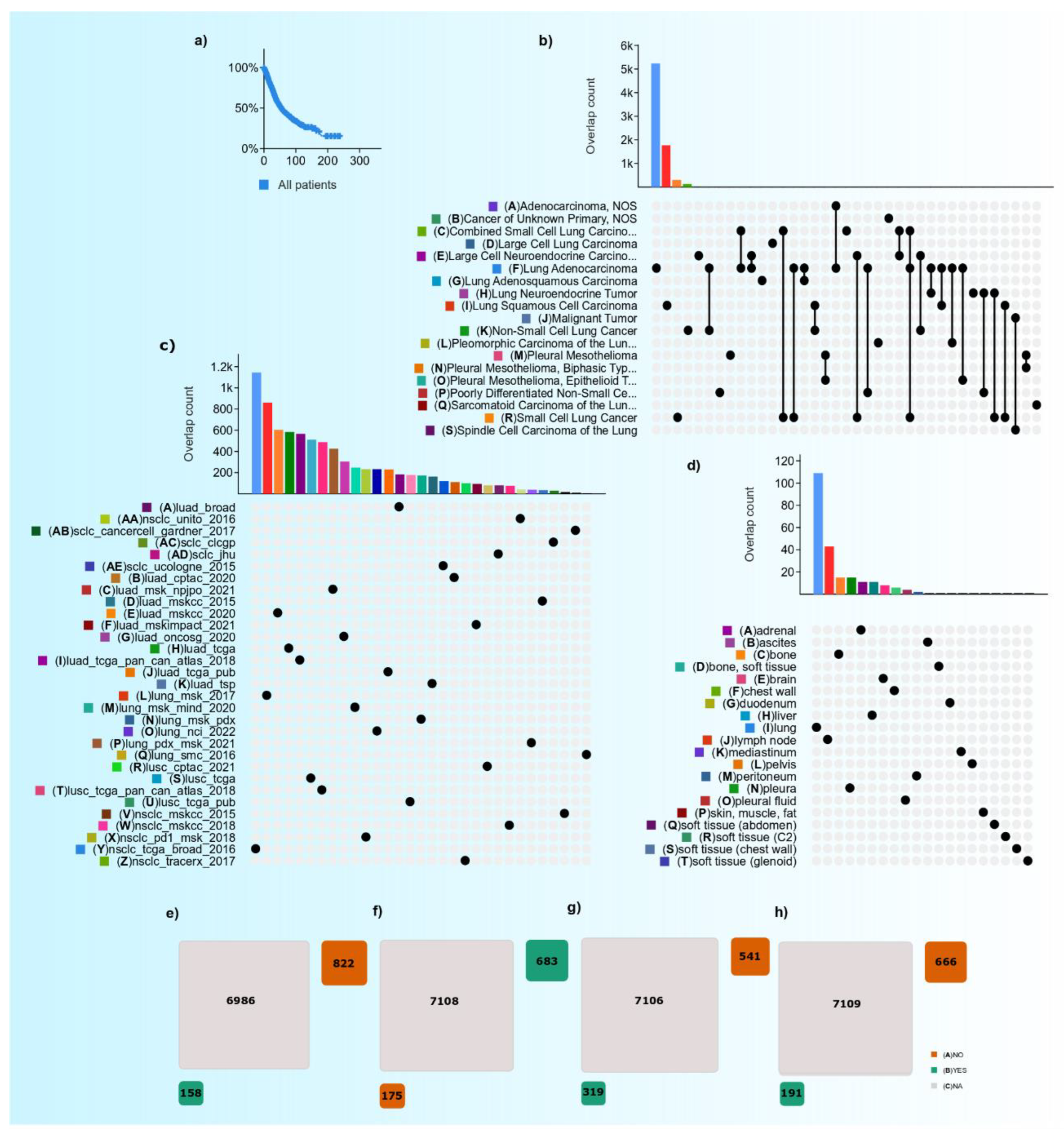
Figure 1. TCGA-cbioportal data on LC. (a) KM plot of the LC cases with five-year overall survival: x-axis (% event free), y-axis (time of event in months). (b) Frequency of LC histotypes with the overlap in different studies. (c) LC projects registered with TCGA. (d) PD-L1 tissue site expression in LC cases. (eh) Patient records who availed therapies, including radiation therapy, chemotherapy, targeted therapy, and immunotherapy, respectively (Data source: https://www.cbioportal.org (accessed on 1 July 2022)).
Vaccines 10 01963 g001
Figure 2. DCs get activated upon vaccine injection and move to the lymph node, wherein antigens are presented to T cells, which are then activated. As CD8+ cells mature because of cytokines released by CD4+ cells, they travel to the cells displaying the target antigen and execute a cytotoxic antitumor response.
Vaccines 10 01963 g002
Figure 3. Lung cancer models: (a) 3D models currently used to study Lung cancer, cancer stroma, and immunotherapy effects. (b) The humanized mouse model of the human immune system involves implanting human hematopoietic cells, lymphocytes, or organs into immunodeficient mice to recreate the human immune system for cancer studies. Hu-BLT, human bone marrow, liver, and thymus; Hu-HSC, human hematopoietic stem cell; Hu-PBL, human peripheral blood lymphocyte.
Vaccines 10 01963 g003
Table 1. TCGA-cBioportal data comparison on various LC therapy from 31 studies (Data source: https://www.cbioportal.org/ (accessed on 1 July 2022)).
TherapyNo. of Patients Undergone TherapyNo. of Patients Does Not Undergo TherapyNot ApplicableThe Ratio between No. of Patients Undergone to Do Not Undergone (%)Total Number of Cases with LC Therapy-Data AvailabilityThe Proportion of Subjects with Data Availability and ‘Not Applicable’ (%)
Radiotherapy158822698619.292013.2%
Chemotherapy6831757108_85812.1%
Targeted therapy319541710658.986012.1%
Immunotherapy191666710928.685712.0%
Table 2. FDA-approved drugs used in chemotherapy, targetable therapies, and immunotherapy against major oncogenes in LC (Gene names are italicized).
TherapyDrugsTargetEstimated Frequency of Mutation in LC (%)References
ChemotherapyCarboplatin, cisplatin, docetaxel, etoposide, gemcitabine, nab-paclitaxel, paclitaxel, pemetrexed, vinorelbine [19]
Targeted therapyAfatinib, dacomitinib, entrectinib, erlotinib, gefitinib, osimertinibEGFR (receptor protein)15[20,21,22,23]
Amivantamab, mobocertinibEGFR (exon 20 insertion)15[20,21,22,23]
Fam-trastuzumab deruxtecan-nxkiHER22[21,24,25]
Alectinib, brigatinib, ceritinib, crizotinib, loralitinibALK5[20,21,26,27,28,29]
Ceritinib, crizotinib, entrectinibROS12[21,30,31,32,33]
SotorasibKRAS G12C25–33[20,21,25]
LarotrectinibNTRK
Dabrafenib, trametinibBRAF V600E2[20,21,25]
Capmatinib, tepotinibMET (exon 14 skipping)3[21,34,35]
Pralsetinib, selpercatinibRET2[20,21,36,37]
ImmunotherapyAtezolizumab, durvalumab, cemiplimab, nivolumab, pembrolizumabPD1/PDL1 pathway33[21,38,39,40,41,42,43]
IpilimumabCTLA4 pathway [25,44]
The italic represent the gene.
Table 3. Current and completed clinical studies of PD-1 and CTLA-4 ICIs (Data source: https://clinicaltrials.gov/ (accessed on 1 July 2022)) [132].
AgentPhaseStudy PopulationDesign and DescriptionPrimary EndpointEnrolmentNCT
DurvalumabIIAdvanced NSCLCEvaluating efficacy and safety of the PD-L1 inhibitor durvalumab as first-line therapyOS50NCT02879617
NiraparibIINSCLCNiraparib + Pembrolizumab
Niraparib alone
Niraparib + Dostarlimab
ORR53NCT03308942
IpilimumabIIIStage IV/Recurrent NSCLCIpilimumab + Paclitaxel/Carboplatin
Placebo + Paclitaxel/Carboplatin
OS1289NCT01285609
PembrolizumabIIAdvanced NSCLCPembrolizumab + Physician’s choice chemotherapyPFS35NCT03083808
AK104IIAdvanced NSCLCAK104 +DocetaxelORR40NCT05215067
PembrolizumabIIMetastatic NSCLCPembrolizumab + chemotherapy vs. Placebo + chemotherapyPFS98 NCT03656094
AK105IIIMetastatic Nonsquamous NSCLC Stage IVAK105 + Carboplatin and Pemetrexed vs. Placebo + Carboplatin and PemetrexedPFS360 NCT03866980
ONC-392I & IIadvanced or metastatic solid tumors and NSCLCONC-392 Treatment as a single agent vs. ONC-392 in combination with pembrolizumabDLT, MTD, RP2D, TRAE468NCT04140526
mRNA VaccineI & IIMetastatic NSCLCBI 1361849 mRNA Vaccine + durvalumab
BI 1361849 mRNA Vaccine + durvalumab + tremelimumab
TEAE61NCT03164772
KN046IIAdvanced NSCLCKN046 + AxitinibORR54NCT05420220
Publisher’s Note: MDPI stays neutral with regard to jurisdictional claims in published maps and institutional affiliations.

