Article

Heterologous Systemic Prime–Intranasal Boosting Using a Spore SARS-CoV-2 Vaccine Confers Mucosal Immunity and Cross-Reactive Antibodies in Mice as well as Protection in Hamsters

by
Paidamoyo M. Katsande
1,
Leira Fernández-Bastit
2,3,
William T. Ferreira
1,4,
Júlia Vergara-Alert
2,3,
Mateusz Hess
1,
Katie Lloyd-Jones
1,
Huynh A. Hong
1,
Joaquim Segales
2,5 and
Simon M. Cutting
1,*
1
Department of Biological Sciences, Royal Holloway University of London, Egham TW20 0EX, UK
2
Unitat Mixta d’investigació IRTA-UAB en Sanitat Animal, Centre de Recerca en Sanitat Animal (CReSA), Campus de la Universitat Autònoma de Barcelona (UAB), 08193 Bellaterra, Spain
3
Institut de Recerca i Tecnologia Agroalimentàries (IRTA), Programa de Sanitat Animal, Centre de Recerca en Sanitat Animal (CReSA), Campus de la Universitat Autònoma de Barcelona (UAB), 08193 Bellaterra, Spain
4
SporeGen Ltd., London Bioscience Innovation Centre, 2 Royal College Street, London NW1 0NH, UK
5
Departament de Sanitat i Anatomia Animals, Facultat de Veterinària, UAB, 08193 Bellaterra, Spain
*
Author to whom correspondence should be addressed.
Vaccines 2022, 10(11), 1900; https://doi.org/10.3390/vaccines10111900
Submission received: 11 October 2022 / Revised: 4 November 2022 / Accepted: 7 November 2022 / Published: 10 November 2022
(This article belongs to the Special Issue COVID-19 Vaccine Candidate Development)

Abstract

:
Background: Current severe acute respiratory syndrome coronavirus 2 (SARS-CoV-2) vaccines are administered systemically and typically result in poor immunogenicity at the mucosa. As a result, vaccination is unable to reduce viral shedding and transmission, ultimately failing to prevent infection. One possible solution is that of boosting a systemic vaccine via the nasal route resulting in mucosal immunity. Here, we have evaluated the potential of bacterial spores as an intranasal boost. Method: Spores engineered to express SARS-CoV-2 antigens were administered as an intranasal boost following a prime with either recombinant Spike protein or the Oxford AZD1222 vaccine. Results: In mice, intranasal boosting following a prime of either Spike or vaccine produced antigen-specific sIgA at the mucosa together with the increased production of Th1 and Th2 cytokines. In a hamster model of infection, the clinical and virological outcomes resulting from a SARS-CoV-2 challenge were ameliorated. Wuhan-specific sIgA were shown to cross-react with Omicron antigens, suggesting that this strategy might offer protection against SARS-CoV-2 variants of concern. Conclusions: Despite being a genetically modified organism, the spore vaccine platform is attractive since it offers biological containment, the rapid and cost-efficient production of vaccines together with heat stability. As such, employed in a heterologous systemic prime–mucosal boost regimen, spore vaccines might have utility for current and future emerging diseases.

1. Introduction

The coronavirus infectious disease 2019 (COVID-19) pandemic will be remembered both for its impact on global health and society but also for the remarkable speed at which prophylactic vaccines were implemented. Vaccines (mRNA, viral-vectored and subunit) have all shown high levels of protection against the most severe forms of disease, reducing morbidity and mortality [1]. On the other hand, it has become apparent that current COVID-19 vaccines are unable to prevent person-to-person transmission of its causal agent, severe acute respiratory syndrome coronavirus 2 (SARS-CoV-2), with breakthrough infections observed in fully vaccinated individuals supporting this [2,3]. The dissemination of the virus will undoubtedly continue as a) immunity wanes and b) the reality that a significant portion of the world’s population (~40–45%) has yet to receive their first vaccine dose [1]. The longer the virus continues to circulate, the greater the probability that new variants of concern (VOCs) will emerge [1]. The poor durability of current vaccines with protection of about six months may necessitate repeated booster campaigns. Taken together, the COVID-19 pandemic cannot yet be considered closed and will continue to represent a major burden to public health authorities until better vaccines are developed.
SARS-CoV-2 transmits person-to-person by respiratory droplets and infects the upper respiratory tract (URT). Mucosal immunity is required to neutralize SARS-CoV-2 at the mucosal surfaces of the URT, either by tissue-resident memory T cells (TRMs) or a secretory IgA (sIgA) that can neutralize or prevent virus attachment within the epithelial cells of the mucosa. [4,5]. Vaccines administered by a parenteral route (noting that all COVID-19 vaccines so far are delivered by intra-muscular injection) fail to evoke a significant mucosal immune response, probably accounting for their failure to prevent the transmission and dissemination of the virus [6].
Accordingly, there is now a focus on developing mucosal COVID-19 vaccines of which intranasal is the optimal route of administration [7,8]. Mucosal vaccination has the potential to provide robust protective immunity at the mucosal site of pathogen entry by recruiting antigen-presenting cells (APCs) and/or engaging other innate immune cells. This requires safe and effective mucosal vaccination strategies that utilize both mucosal adjuvants and appropriate delivery systems [7]. SARS-CoV-2 vaccine peptides and a combination of flagellin (KF) and cyclic GMP-AMP (cgGAMP) as an adjuvant delivered intranasally have been shown to elicit strong systemic and mucosal immune responses that provide protection against SARS-CoV-2 challenge in a transgenic mouse model [9]. Early clinical data have suggested that without pre-existing immunity to SARS-CoV-2, immunity resulting from intranasal vaccination is suboptimal [7,10]. One promising approach might be that of boosting a current COVID-19 vaccine with an intranasal vaccine [11]. Adenoviral 5 and 19a vectored vaccines administered intranasally after a systemic mRNA vaccine prime were shown to induce high levels of both mucosal IgA and TRM, be fully protective in a mouse model of SARS-CoV-2 infection and neutralize VOCs [11].
Bacterial endospores of Bacillus subtilis engineered to display heterologous antigens have been shown to provide several advantages for mucosal immunization with efficacy against a number of bacterial and viral pathogens [12,13,14]. Spores carry natural mucosal adjuvant properties [15] and induce innate immunity [16]. Using a recently developed system (THY-X-CISE®), engineered spores can be rendered unable to proliferate in the environment ensuring, biological containment [17]. Spores are of uniform size (1 μm diameter), can be produced rapidly in large quantities and are currently manufactured at medicinal grade by several pharmaceutical companies [18]. Most importantly, spores are heat stable (<70 °C), and with a few exceptions, antigens expressed on their surface are stable both to heat and gastric enzymes [19].
Here, we engineered B. subtilis spores to express SARS-CoV-2 antigens and used these to nasally boost a systemic prime consisting of either a recombinant Spike protein or the Oxford AZD1222 vaccine. Our work shows that this heterologous systemic prime–mucosal spore boost strategy evokes mucosal immunity and is potentially protective.

2. Materials and Methods

2.1. General Methods

Methods for B. subtilis, including the preparation of spores and the extraction of spore coat proteins, are described elsewhere [20]. B. subtilis strain PY79 is a prototrophic laboratory strain.

2.2. Construction of B. subtilis Spores Expressing SARS-CoV-2 Antigens

A cloning method referred to as THY-X-CISE® [17] was used to introduce heterologous genes into the chromosome of B. subtilis. First, chimeric genes were synthesized (Azenta Life Sciences), carrying the 5′ segment (including promoter) of either the cotB or cotC genes of B. subtilis fused at their 3′-end to DNA-encoding SARS-CoV-2 domains. Gene sequences are shown in Supplementary Figure S1, which included the receptor-binding domain (RBD) (Wuhan-Hu-1 2019-nCov and Omicron B.1.1.529 variants) and the HR1-HR2 domain (Wuhan-Hu-1 2019-nCov) of the Spike protein. For clarity, these are referred to henceforth as SWuh, SOmi and HR1-HR2Wuh. Chimeras were then subcloned into the plasmid pThyA [17]. This placed the chimeric gene between the left and right arms of the thyA gene that encodes thymidine synthetase A (Supplementary Figure S1). The linearization of the resulting plasmid and introduction into the B. subtilis chromosome (strain PY79) by double-crossover recombination generated trimethoprim-resistant transformants that were thymine-dependent. The resulting thyA::insertion strain was then used as a recipient in a second transformation where an empty pThyB vector was introduced, thus disrupting the thyB locus (thyB::Δ). The resulting strain (thyA::insertion thyB::Δ) was thymine-dependent but resistant to a higher concentration of trimethoprim. The strains constructed were PK120 thyA::cotB-RBDWuh thyB::Δ, PK122 thyA::cotC-HR1-HR2Wuh thyB::Δ and PK230 thyA::cotB-RBDOmi thyB::Δ. An isogenic strain, PK118 (thyA::Δ thyB::Δ), which carries no gene insertions was made using the same procedure with empty pThyA and pThyB vectors that carry no gene insertions. PK118 (thyA::Δ thyB::Δ), referred to henceforth as WT, was used as an isogenic control strain that produces “naked” spores devoid of SARS-CoV-2 antigens. Strains were confirmed by preparing spores, extracting spore coat proteins and probing Western blots with antibodies to the relevant SARS-CoV-2 domains: anti-SWuh PAbs raised against the entire SWuh polypeptide (SinoBiological, 40589-T62) and anti-SOmi MAbs that were raised against the RBD of the SOmi (SinoBiological, 40592-MM117).

2.3. Immunogens

Recombinant Spike protein rSWuh and rSOmi (baculovirus expressed) were obtained from SinoBiological (Cat Nos. 40589-V08B1 and 40589-V08B33, respectively). Prior to intramuscular injection, 5 μg of protein was suspended in PBS and complexed (1:1) with the adjuvant AddaVax (Invivogen, vac-adx-10). The COVID-19 vaccine ChAdOx1 nCoV-19 (AZD1222) with a concentration of 3.8 × 10⁹ infectious units (IU)/mL (3.7 × 10¹¹ virus particles/mL) was provided by Prof. Sarah Gilbert (Jenner Institute, Oxford University) and referred to henceforth as AZD1222. Prior to use, AZD1222 was diluted in PBS to give a working concentration of 2 × 10⁹ IU/mL.

2.4. Prime Boost Vaccination Studies

BALB/c female mice were obtained from Charles River, UK (aged 8–10 weeks before study start) and allocated into groups (n = 5–6) for recombinant prime and n = 6 for the AZD1222 prime.
(a) Recombinant prime boost vaccination (Supplementary Figure S2A)
Group 1 received no immunizations and served as a naïve control group. Groups 2–4 each received an intramuscular injection (i.m.) of 50 μL (5 μg) of formulated rSWuh protein in each hind quadricep muscle. Group 2 was primed (i.m.) with rSWuh on day 1 and culled on day 48. Groups 3 and 4 were primed (i.m.) with rSWuh on day 1 and then intranasally (i.n.) boosted three and five weeks later with spores (1 × 10⁹ CFU) of either WT (‘naked’) spores (Group 3) or a 1:1 mixture of PK120 (CotB-RBDWuh) and PK122 (CotC-HR1-HR2Wuh) referred to as SporCoVax (Group 4). Each intranasal administration (10 μL total/animal, 5 μL/nare, no anesthesia) consisted of three administrations given on consecutive days (Boost 1, days 21, 22 and 23 and Boost 2, days 35, 36 and 37).
(b) AZD1222 prime boost vaccination (Supplementary Figure S2B)
Group 1 was a naïve group and received no immunizations. Groups 2–5 each received 50 μL of AZD1222 (1.0 × 10⁸ IU) in phosphate-buffered saline (PBS) in the right hind quadricep muscle. Group 2 was culled on day 48. Groups 3–5 were boosted (intranasal; 5 μL/nare, no anesthesia) three and five weeks later with spores (1 × 10⁹ CFU) of WT (‘naked’; Group 3), SporCoVax (a 1:1 mixture of CotB-RBDWuh and CotC-HR1-HR2Wuh spores; Group 4) or PK230 (CotB-RBDOmi; Group 5). Each intranasal administration (10 μL total/animal, 5 μL/nare, no anesthesia) consisted of three administrations given on consecutive days (Boost 1, days 21, 22 and 23 and Boost 2, days 35, 36 and 37).

2.5. Determination of Mucosal and Systemic Antibody Titers by Indirect ELISA

For the analysis of immunological responses, saliva was collected on days −1, 20, 34 and 48, serum was taken on day 48, and lungs were collected on day 48 and stored at −80 °C. For lungs, sample extractions were made at a one-fifth (w/v) dilution in extraction buffer (2% (v/v) fetal calf serum) containing protease inhibitors, EDTA (0.05 mg/mL), as previously described [21]. Saliva extractions were made at a one-tenth (w/v) dilution in PBS. Samples were gently shaken for 2 h at 4 °C to disrupt solid material, centrifuged (8000× g, 15 min.) and the supernatant used for analysis. Antibody levels in saliva (sIgA), lungs (IgA) and serum (IgG) were quantified by indirect enzyme-linked immunosorbent assay (ELISA). Greiner 96-well plates (MaxiSorp) were coated with 2 µg/mL of either rSWuh or rSOmi (50 μL/well) in PBS overnight at 4 °C, followed by blocking for 1 h at RT with PBS containing 2% (w/v) bovine serum albumin (BSA). Saliva samples were diluted 1:20 in PBS. Lung and serum samples were diluted 1:10 and 1:1000, respectively, in diluent buffer (0.01 M PBS, 1% (w/v) BSA, 2% (v/v) FBS, 0.1% (v/v) Triton X-100, 0.05% (v/v) Tween 20). Samples were added to plates and 2-fold serially diluted. Plates containing saliva and lung samples were incubated for 2 h at 30 °C, and those containing serum samples were incubated for 2 h at RT. Levels of IgA and IgG were detected using the appropriate horseradish peroxidase-conjugated anti-mouse IgA (Sigma Cat No. A4789-1) or anti-mouse IgG (Dako Cat No. P0447) in conjugate buffer (2% (v/v) FBS, 1% (v/v) BSA, 0.05% (v/v) Tween 20 in 0.01 PBS). Plates were incubated for 1 h at RT and then developed using tetramethyl benzidine (TMB) substrate (0.1 mg/mL 3.3′,5.5′-tetramethylbenzidine in 0.1 M sodium acetate buffer (pH 5.5)). Reactions were stopped using 2 M H2SO4, and the ODs were read at 450 nm. Dilution curves were created for each sample, and endpoint titers were estimated as the maximum dilution that gave an absorbance reading above the average naïve sample.

2.6. Cytokine Analysis

Mouse spleens were harvested, homogenized and treated with ACK lysis buffer (Sigma Cat. No 11814389001) to remove erythrocyte contamination. Splenocytes were cultured at 1 × 10⁶/well in 96-well plates. Cells were stimulated with 2.5 µg/mL of rSWuh for 72 h. IL-2, IL-5 and TNF-α in culture supernatants were measured using an 8-plex multiplex immunoassay (Biolegend Cat No. 741053) according to the manufacturer’s instructions. Data were acquired using a BD FACSCanto™ II, and analysis was performed using Legendplex software (Biolegend, San Diego, CA, USA).

2.7. Hamster Challenge Study

Golden Syrian hamsters (age 6–8 weeks, 80–120 g) were allocated into groups (50% male, 50% females). Group 1 (n = 6) was a naïve, ‘unchallenged’ control group. Animals were dosed with PBS buffer on day 1 (i.m.), day 22 (i.n.) and day 35 (i.n.) and culled on day 49. Group 2 (n = 12) was a negative control group dosed as Group 1 but challenged with SARS-CoV-2 on day 42 and culled on day 49. Group 3 (n = 12) was primed (i.m.) on day 1 with rSWuh (10 μg, formulated 1:1 with the AddaVax adjuvant) and then boosted (intranasal) on days 22 and 35 with SporCoVax (a 1:1 mixture of CotB-RBDWuh and CotC-HR1-HR2Wuh spores at 2.5 × 10⁹ CFU), after which, animals were challenged with SARS-CoV-2 on day 42 and culled on day 49. For intranasal administration, a total volume of 0.1 mL/dose (50 μL/nare) was used, and procedures were conducted under anesthesia (isofluorane).
A SARS-CoV-2 Cat02 isolate (variant D614G, ID EPI_ISL-47147) was used for challenge. It was isolated from a patient in a laboratory-confirmed COVID-19 case in Barcelona, Spain, and propagated in Vero E6 cells (ATCC CRL-1586). The inoculum was prepared on the same day of the challenge at a concentration of 105 TCID50/mL and kept refrigerated until use. Then, 0.1 mL was administered per challenge dose (50 μL/nare under isofluorane anesthesia), giving a challenge dose of 104 TCID50. At 2 and 4 days post challenge, 4 animals/group were sacrificed for necropsy, and at day 7, all remaining animals were culled. Methods for qPCR in nasal turbinates (NT), oropharyngeal (OP) swabs, etc., were performed as described elsewhere [22].

2.8. Statistical Analysis

Statistical significance was assessed by Mann–Whitney U test. All statistical analyses were performed using Prism (GraphPad, San Diego, CA, USA). Flow cytometry data were analyzed using LEGENDplex™ Data Analysis Software.

3. Results

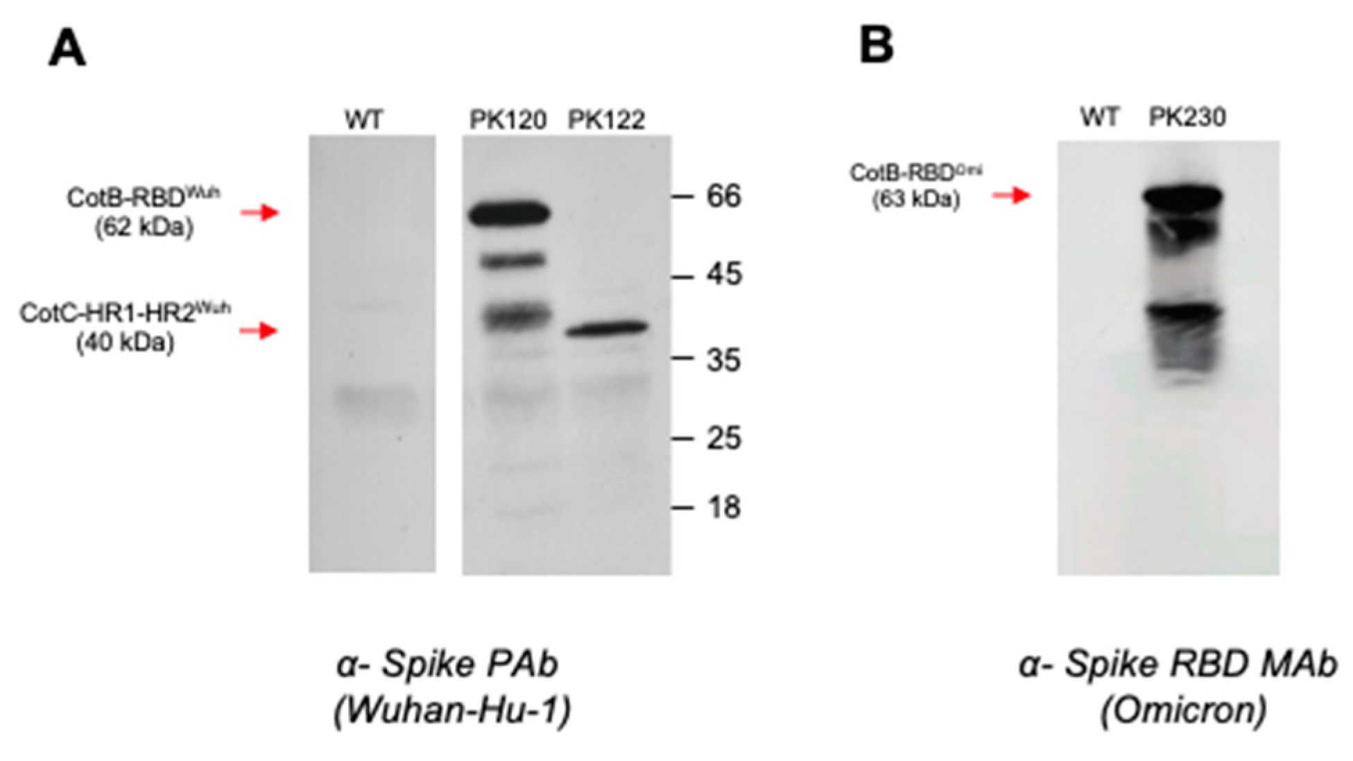
3.1. Construction of B. subtilis Spores Displaying SARS-CoV-2 Domains

Isogenic strains of B. subtilis were engineered to express SARS-CoV-2 (Wuhan-Hu-1 variant 2019-nCov) antigens on the spore surface as chimeric fusions to the spore coat proteins CotB or CotC. This included two segments of the Spike (S) protein, the RBD and the HR1-HR2 domains fused to CotB (PK120, CotB-RBDWuh) and CotC (PK122; CotC-HR1-HR2Wuh). A third strain, PK230, was made which expressed the RBD of the Omicron variant (B.1.1.529) fused to CotB (CotB-RBDOmi).
B. subtilis vaccine strains carrying insertions at the thyA loci were examined by Western blotting of SDS-PAGE size-fractionated spore coat proteins extracted from preparations of pure spores (approx. 2 × 10⁹ spores/extraction). Blots were probed with PAbs or MAbs as shown.
The use of anti-SWuh-specific PAbs Western blotting of proteins extracted from purified spores of each strain showed that the RBD (primary CotB-RBDWuh species of ~62 kDa) and HR1-HR2 (CotC-HR1-HR2Wuh, 40 kDa) domains were expressed on the spore surface. Proteins extracted from spores of an isogenic WT strain devoid of SARS-CoV-2 antigens exhibited no cross-reaction (Figure 1A). Using an Omicron-specific anti-SOmi MAb expression of RBD was apparent in spores of PK230 as a primary CotB-RBDOmi species of ~63 kDa (Figure 1B).

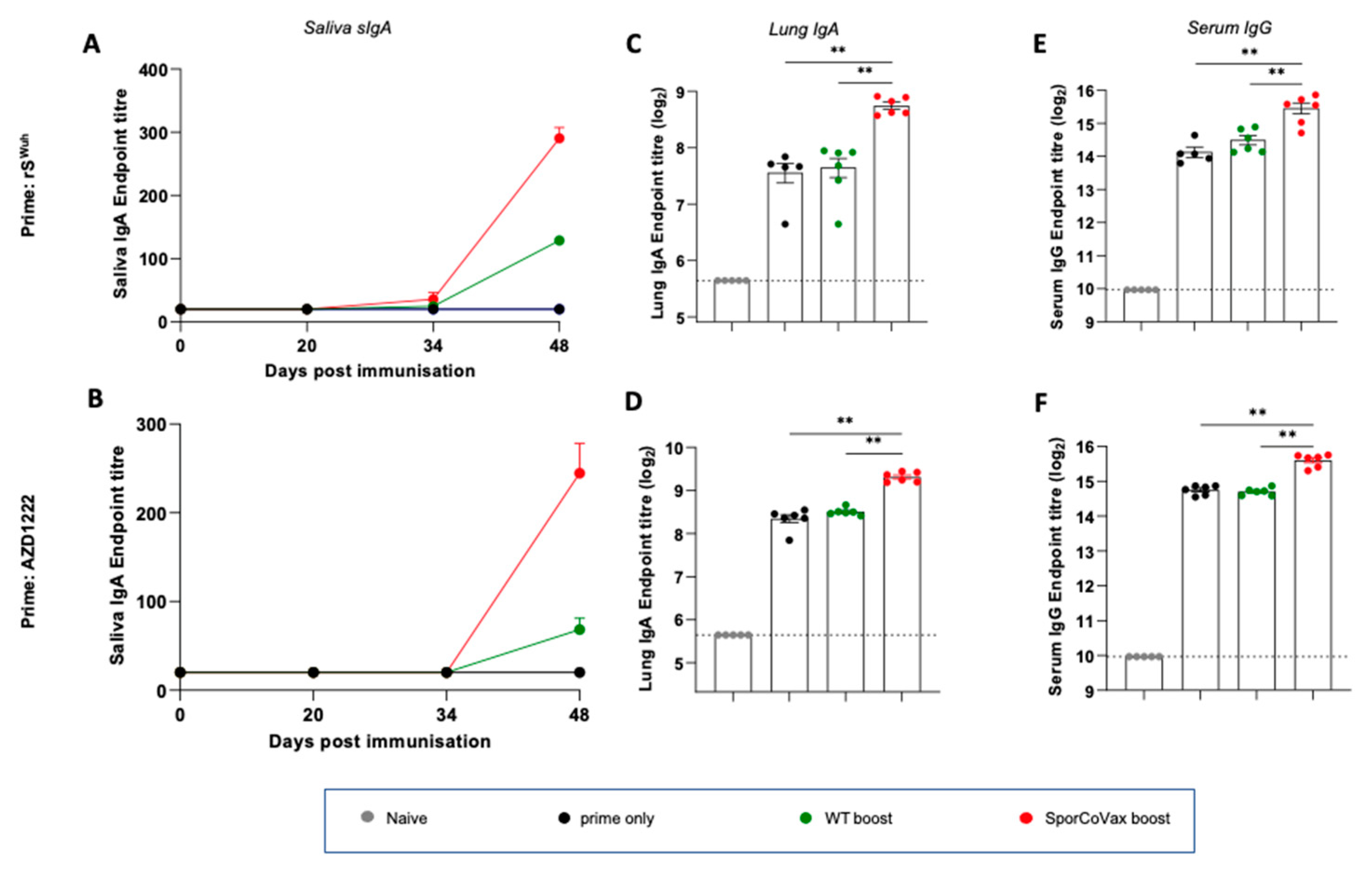
3.2. Intranasal Boosting with a Spore Vaccine following a Systemic Prime Evokes Mucosal Immune Responses

We used a heterologous systemic prime–mucosal boost regimen to determine whether the intranasal administration of spore vaccines expressing SARS-CoV-2 antigens could evoke mucosal immunity. Mice were first dosed by intra-muscular injection (i.m.) with either the rSWuh protein or the AZD1222 vaccine and then, 21 and 35 days later, administered spore vaccines as an intranasal boost. For murine studies, a single dose consisted of three daily intranasal administrations (e.g., days 21, 22 and 23) and resulted from the regulatory dosing limitations required for mice. Following a prime of rSWuh or AZD1222 animals administered SporCoVax, a 1:1 mixture of CotB-RBDWuh and CotC-HR1-HR2Wuh, spores exhibited antigen-specific sIgA in the saliva together with seroconversion (Figure 2A,B). sIgA levels were significantly (p < 0.001) greater than the group receiving only an rSWuh or AZD1222 prime, each of which yielded essentially no detectable sIgA (Figure 2A,B). Interestingly, isogenic spores (WT) carrying no SARS-CoV-2 antigens also evoked antigen-specific sIgA but at levels significantly lower (p < 0.01) than in animals dosed with SporCoVax (Figure 2A,B). Antigen-specific IgA was also detectable in lung extracts (day 48) of animals primed only with rSWuh or AZD1222, as well as those primed and then boosted with WT spores, but levels were significantly higher (p < 0.01) in mice boosted with SporCoVax (Figure 2C,D). The presence of IgA in lung extracts of mice dosed only with rSWuh or AZD1222 (i.m.) most probably results from contamination with serum IgA and should be considered here as a baseline. Serum IgG levels measured (Figure 2E,F) at day 48 showed significantly (p < 0.01) higher levels of rSWuh-specific IgG in animals primed and then boosted with SporCoVax spores compared to animals primed only or primed and then boosted with WT spores (p < 0.01).
Using a similar systemic prime–mucosal boost regimen, we primed (i.m.) mice with the AZD1222 vaccine and boosted with either WT spores (no antigens) or PK230, a spore vaccine expressing CotB-RBDOmi. As reported above for boosting with spores displaying Wuhan-specific antigens, boosting with CotB-RBDOmi spores evoked antigen (rSOmi)-specific sIgA in saliva and serum which was at significantly higher levels than in groups dosed with WT spores (p < 0.001 and p < 0.01, respectively) or in animals primed only (p < 0.01) (Supplementary Figure S3).
Together, these results show that intranasal boosting with spores expressing SARS-CoV-2 antigens following a systemic prime evokes mucosal immunity.

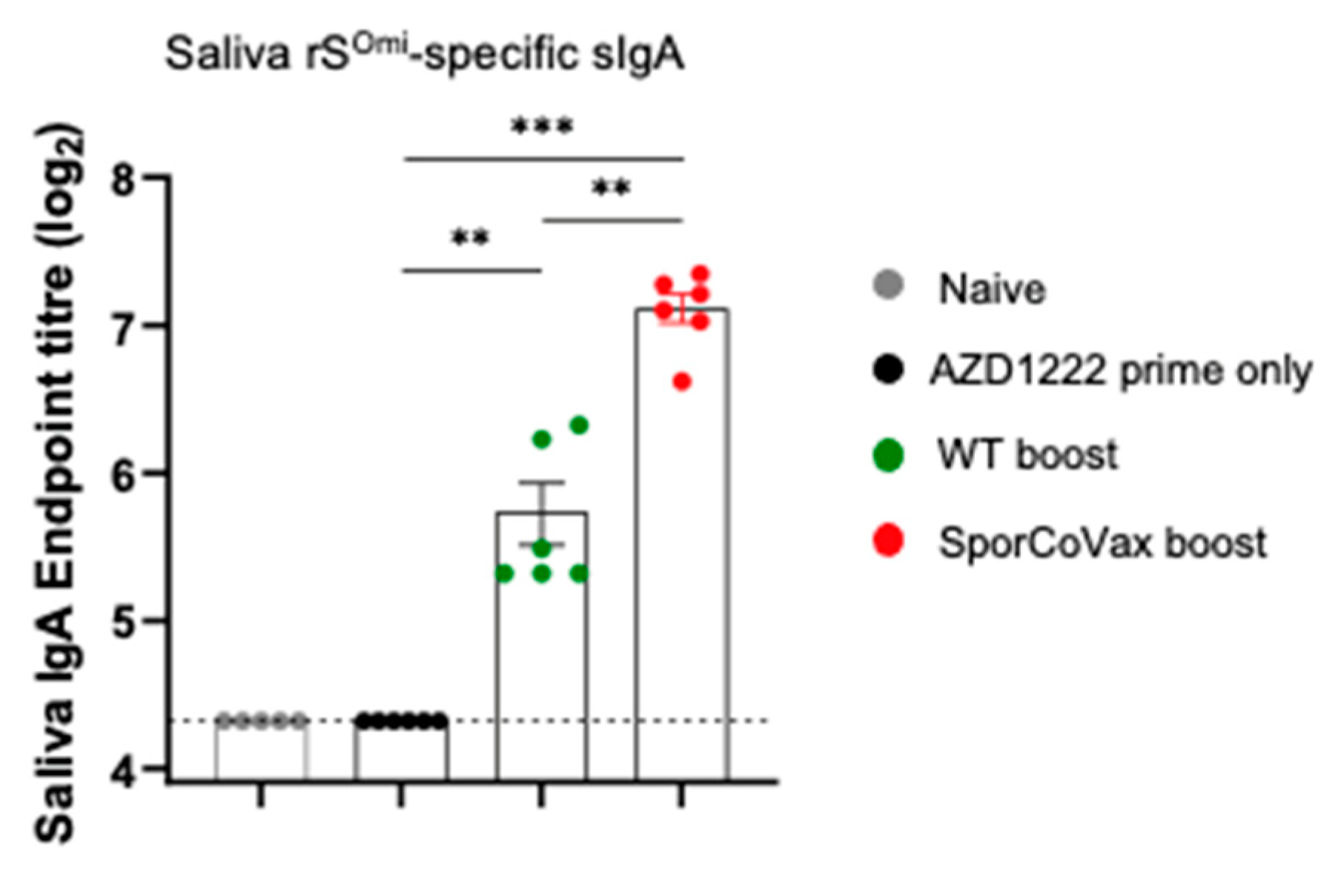
3.3. Boosting with Spores Evokes Cross-Reactive Antibodies

From the study above in which animals had been primed with AZD1222 and then boosted with SporCoVax, we determined whether saliva samples carried antibodies that cross-reacted with the Omicron variant. Using ELISA plates coated with rSOmi, we showed that SporCoVax-boosted animals exhibited antigen-specific sIgA in saliva samples which was significantly greater (p < 0.01 and p < 0.001, respectively) than in animals dosed with WT spores or rSWuh only (Figure 3). As shown in the previous section, WT spores used alone as a boost also enhanced antigen-specific sIgA.

3.4. Intranasal Boosting with a Spore Vaccine Results in a Mixed T Cell Cytokine Profile

We next studied which subsets of T helper (Th) cells and cytotoxic T cells were responding to the rSWuh protein. Animals (n = 3/group) were either primed with AZD1222 or primed (AZD1222) and then boosted with WT or SporCoVax spores. Splenocytes from immunized animals were cultured with rSWuh and assessed for the production of IL-2, TNF-α and IL-5, which are induced in Th1 (IL-2, TNF-α) and Th2 (IL-5) T-cell responses (Figure 4). SporCoVax-boosted animals exhibited marked increases in the expression of all three cytokines compared to other groups. Together, this shows that intranasal spore boosting confers both Th1 and Th2 responses.

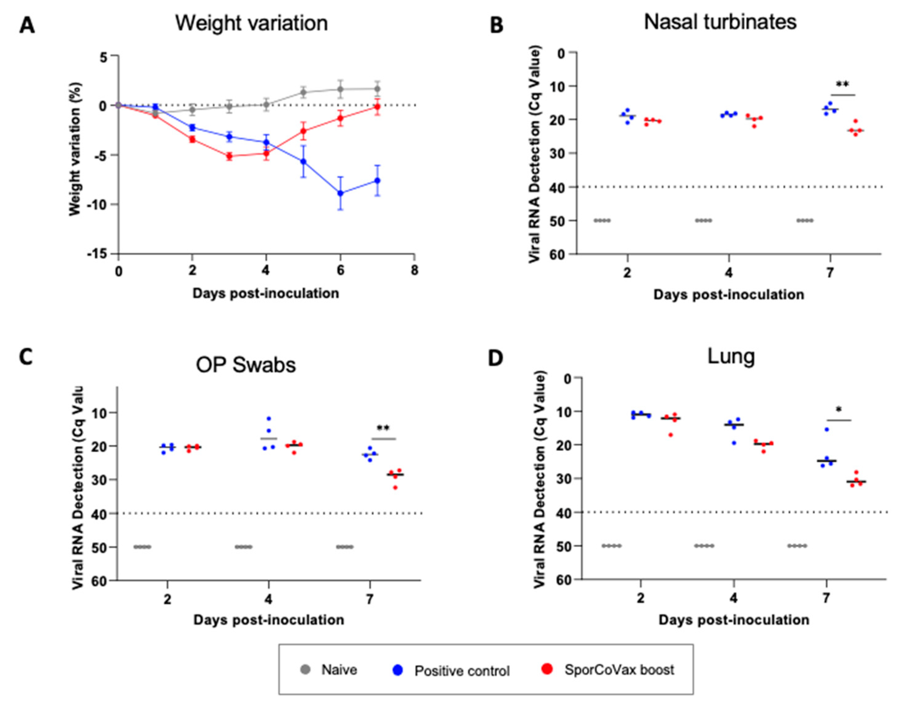
3.5. Intranasal Boosting with Spores Is Protective in the Hamster SARS-CoV-2 Model of Infection

The golden Syrian hamster model is currently considered one of the best models to study the effects of SARS-CoV-2 infection because it permits high levels of SARS-CoV-2 replication and generates significant clinical signs (weight loss) and moderate to severe pneumonia [23,24]. To determine whether intranasal boosting with spores expressing SARS-CoV-2 antigens conferred protection, we first primed animals (n = 12; Group 3) with rSWuh protein (i.m.) and then administered two intranasal boosts of SporCoVax. Seven days after the last boost, animals were challenged (intranasal) with SARS-CoV-2 (104 TCID50/animal). Two additional groups, Group 1, being a naïve, unchallenged group (n = 6), and Group 2, a negative control group (n = 12) which received PBS in place of immunogens (i.m. prime and two i.n. boosts) and challenged with SARS-CoV-2 seven days after the last dose, were used for comparison.
Golden Syrian hamsters were primed (i.m.) with recombinant Spike (rSWuh) protein followed by two intranasal boosts with 2.5 × 10⁹ spores of SporCoVax (1:1 mixture of CotB-RBDWuh and CotC-HR1-HR2Wuh spores) (Group 3). Negative, unvaccinated (Group 2) and naïve (Group 1) control groups received PBS in place of immunogens. Seven days after the final boost, animals in Groups 2 and 3 were intranasally challenged with SARS-CoV-2 (D614G variant, 104 TCID50/animal). Weight loss post challenge was shown (panel A). Animals were sequentially euthanized (2, 4 and 7 dpi), necropsies were performed, and viral loads of SARS-CoV-2 were determined by qPCR in nasal turbinate (panel B), OP swabs (panel C) and lungs (panel D). Mann–Whitney, * p < 0.05, ** p < 0.01.
Weight loss was used to track the progression of the disease (Figure 5A). Naïve (Group 1) animals showed no reduction in weight, whereas non-vaccinated animals (Group 2) challenged with SARS-CoV-2 showed progressive reductions in weight 2–7 dpi. By contrast, animals from Group 3 (SporCoVax boost) lost weight at 2 dpi and started to recover at 4 dpi, and finally regained 100% of their initial weight at 7 dpi (M = −7.4, SE = 1.73, 95%. CI [−11.65, −3.2], p < 0.01). For operational reasons, we were unable to assign animals to a prime-only group. We acknowledge this as a major flaw in the experimental design and interpretation. On the other hand, we note that it is extensively reported that a single systemic dose of S protein fails to induce protective antibodies [25,26,27].
To assess the impact of intranasal boosting on viral replication, we examined SARS-CoV-2 RNA (gRNA) in NT (Figure 5B), OP swabs (Figure 5C) and lungs (Figure 5D) at 2, 4 and 7 dpi. After SARS-CoV-2 inoculation (Groups 2 and 3), viral gRNA was detected at 2 dpi in NT, OP swabs and lungs in both groups. The viral load decreased progressively in OP swabs and both tissues in animals receiving intranasal boosts with SporCoVax (PK220 boost; Group 3), compared to the unimmunised–infected control group (Group 2), which was statistically significant at 7 dpi (p < 0.01). Taken together, we conclude that the heterologous systemic prime–intranasal boost regimen ameliorates the clinical and virological outcome of SARS-CoV-2 challenge.

4. Discussion

The continued dissemination of SARS-CoV-2 ensures that COVID-19 is likely to persist. To halt transmission, vaccine strategies must focus on blocking interaction of the virus at the mucosa. Accomplishing this requires mucosal immunity and more specifically induction of sIgA that can block viral adhesion. We present evidence that bacterial spores engineered to express SARS-CoV-2 antigens and delivered as an intranasal boost following a systemic prime (either a S protein subunit or the AZD1222 COVID-19 vaccine) induce antigen-specific sIgA. This is further supported by a robust IL-5 response often associated with sIgA induction [5]. Moreover, when this strategy was employed in hamsters, they showed signs of protection from viral challenge including recovery of body weight and a reduction in viral shedding. This is encouraging for a number of reasons but most importantly a reduction in viral shedding would lead to reduced transmission and dissemination ultimately providing a means to controlling this disease. A heterologous systemic prime – mucosal boost vaccination strategy is being considered as one of the most promising routes forward to enhance existing COVID-19 vaccines [11,28]. The underlying basis for how a mucosal boost induces a localized response is not yet fully understood but a “prime-pull” mechanism has been proposed where systemic memory cells (induced by the prime) are expanded during the recall response and which then migrate to the mucosa followed by differentiation into tissue-resident memory cells [29].
Spores are dormant entities that are found in soil and air and as such in humans and animals also [30]. As dominant members of the aerobiome (~10%) humans are continuously exposed to Bacillus spores with approximately 10–105 spores inhaled daily [31]. In the gastro-intestinal tract they are present at constant levels of ~104 spores/g of feces [32]. The spore vaccine platform has been well documented, and spores genetically engineered or, alternatively, adsorbed with heterologous antigens, evoke balanced Th1/Th2 responses as well as augmenting mucosal antibody responses [15]. This is supported by the studies here using a genetically engineered spore vaccine and resulting antibody and cytokine responses. Using the THY-X-CISE® cloning, spores, should they germinate, are unable to proliferate in the environment since live cells die immediately due to a ‘thymine-less’ death [17]. Biological containment is thus ensured, and a similar strategy has been used to enable clinical evaluation of a Lactococcus lactis IL-10 delivery system [33]. Spore vaccines can be constructed rapidly on a case-by-case basis although, as shown here, antibodies resulting from a Wuhan-delivered antigen cross react with the Omicron variant suggesting that the vaccines described here might be protective against emerging VOCs. Spores are themselves heat stable and can be stored indefinitely in an aqueous or desiccated form. Of course, this may not necessarily apply to a heterologous protein displayed on the spore surface. Other spore vaccines we have generated do indeed show stability, but this would need to be addressed by an extensive program of formulation and stability studies.
Intriguingly, nasal boosting with isogenic spores displaying no antigens also showed a clear augmentation of mucosal antigen-specific responses. The underlying mechanism is not fully understood but other studies have shown that B. subtilis spores carry natural adjuvant properties [15,34]. Spores can bind antigens including intact virions and adjuvanticity is observed when spores and adjuvants are administered together or by separate routes (systemic-mucosal) [15,16,19]. Spores have also been shown to act as immunomodulators and specifically by inducing innate immunity [16]. Spores can be taken up by M-cells [35], persist within phagocytes [36,37] and intriguingly, inactivated spores are also able to stimulate the innate immune system when delivered nasally [16]. This is achieved by interaction of spores with Toll-like receptors (TLR2 and TLR4), cytokine induction, recruitment of NK cells and maturation of Dendritic cells [16,37]. What is interesting is that here we show that ’naked’ spores administered three weeks after the systemic prime are able to augment mucosal immunity suggesting that spore-induced innate immunity may be a factor. Although the mucosal responses are clearly greater when spores-displaying antigens are delivered the use of non-recombinant spores may, long-term, have greater utility. It is worthwhile noting that in previous work we have shown that nasal administration of spores alone is sufficient to confer protection against influenza (H5N2) using both murine and ferret models of infection [16,38]. Based on the observed seroconversion of sIgA in mice we suspect that a single dose of spore vaccine using this regimen (3-weeks post-prime) is unlikely to be protective but further empirical analysis of dosing regimens might facilitate use of a single nasal dose.
That nasal boosting with spores displaying no antigens (i.e., WT spores) evoke mucosal sIgA in primed animals is intriguing. Although the levels of induction are not as high as those in animals boosted with antigen-presenting spores this observation is important since it is possible that spores alone might provide a mechanism for evoking mucosal immunity. We are now addressing this in expanded animal and human studies.

Supplementary Materials

The following supporting information can be downloaded at: https://www.mdpi.com/article/10.3390/vaccines10111900/s1, Figure S1: Recombinant spore vaccines, Figure S2: Animal study schedules, Figure S3: Intranasal boost of the AZD1222 vaccine with spores expressing Omicron-specific antigens.

Author Contributions

S.M.C., W.T.F. and H.A.H. wrote the manuscript. H.A.H., J.S., L.F.-B. and P.M.K. designed the experimental study. P.M.K. and L.F.-B. conducted the statistical analysis. P.M.K., L.F.-B., K.L.-J., W.T.F., M.H. and J.V.-A. conducted experimental studies. All authors have read and agreed to the published version of the manuscript.

Funding

This work was supported in part by funding from the Medical Research Council (MR/R026262/1) to SMC, a BBSRC CASE studentship co-funded by SporeGen Ltd. to P.K., and a transnational access (TNA-08-COVID) of the European Project TRANSVAC2–730964–INFRAIA–2016-1 granted to SporeGen Ltd.

Institutional Review Board Statement

Murine studies were conducted with approval from the Royal Holloway University of London Ethics Committee under an approved UK Home Office animal project license PB9FA6ABB. Hamster studies were approved by the Institutional Animal Welfare Committee of the Institut de Recerca i Tecnologia Agroalimentàries (CEEA-IRTA, registration number CEEA 262/2021) and by the Ethical Commission of Animal Experimentation of the Autonomous Government of Catalonia (registration number CEA-OH/11579/2) and conducted by certified staff. Experiments with SARS-CoV-2 were performed at the Biosafety Level-3 (BSL-3) facilities of the Biocontainment Unit of IRTA-CReSA (Barcelona, Spain).

Informed Consent Statement

Not applicable.

Data Availability Statement

All data generated or analyzed during this study are included in this published article (and its supplementary information files).

Acknowledgments

Authors also acknowledge Xavier Abad, Guillermo Cantero, Núria Roca and all the BSL3 animal caretakers of IRTA-CReSA for their contribution to the golden Syrian hamster study.

Conflicts of Interest

SMC is CEO of SporeGen Ltd., HAH is a shareholder of SporeGen Ltd.

References

  1. Mouro, V.; Fischer, A. Dealing with a mucosal viral pandemic: Lessons from COVID-19 vaccines. Mucosal Immunol. 2022, 15, 584–594. [Google Scholar] [CrossRef] [PubMed]
  2. Brosh-Nissimov, T.; Orenbuch-Harroch, E.; Chowers, M.; Elbaz, M.; Nesher, L.; Stein, M.; Maor, Y.; Cohen, R.; Hussein, K.; Weinberger, M.; et al. BNT162b2 vaccine breakthrough: Clinical characteristics of 152 fully vaccinated hospitalized COVID-19 patients in Israel. Clin. Microbiol. Infect. 2021, 27, 1652–1657. [Google Scholar] [CrossRef] [PubMed]
  3. Farinholt, T.; Doddapaneni, H.; Qin, X.; Menon, V.; Meng, Q.; Metcalf, G.; Chao, H.; Gingras, M.C.; Avadhanula, V.; Farinholt, P.; et al. Transmission event of SARS-CoV-2 delta variant reveals multiple vaccine breakthrough infections. BMC Med. 2021, 19, 255. [Google Scholar] [CrossRef] [PubMed]
  4. Okuya, K.; Yoshida, R.; Manzoor, R.; Saito, S.; Suzuki, T.; Sasaki, M.; Saito, T.; Kida, Y.; Mori-Kajihara, A.; Kondoh, T.; et al. Potential Role of Nonneutralizing IgA Antibodies in Cross-Protective Immunity against Influenza A Viruses of Multiple Hemagglutinin Subtypes. J. Virol. 2020, 94, e00408-20. [Google Scholar] [CrossRef] [PubMed]
  5. Mantis, N.J.; Rol, N.; Corthesy, B. Secretory IgA’s complex roles in immunity and mucosal homeostasis in the gut. Mucosal Immunol. 2011, 4, 603–611. [Google Scholar] [CrossRef] [Green Version]
  6. Moldoveanu, Z.; Clements, M.L.; Prince, S.J.; Murphy, B.R.; Mestecky, J. Human immune responses to influenza virus vaccines administered by systemic or mucosal routes. Vaccine 1995, 13, 1006–1012. [Google Scholar] [CrossRef]
  7. Alu, A.; Chen, L.; Lei, H.; Wei, Y.; Tian, X.; Wei, X. Intranasal COVID-19 vaccines: From bench to bed. EBioMedicine 2022, 76, 103841. [Google Scholar] [CrossRef]
  8. Tiboni, M.; Casettari, L.; Illum, L. Nasal vaccination against SARS-CoV-2: Synergistic or alternative to intramuscular vaccines? Int. J. Pharm. 2021, 603, 120686. [Google Scholar] [CrossRef]
  9. Jiang, W.; Shi, L.; Cai, L.; Wang, X.; Li, J.; Li, H.; Liang, J.; Gu, Q.; Ji, G.; Li, J.; et al. A two-adjuvant multiantigen candidate vaccine induces superior protective immune responses against SARS-CoV-2 challenge. Cell Rep. 2021, 37, 110112. [Google Scholar] [CrossRef]
  10. Wu, S.; Huang, J.; Zhang, Z.; Wu, J.; Zhang, J.; Hu, H.; Zhu, T.; Zhang, J.; Luo, L.; Fan, P.; et al. Safety, tolerability, and immunogenicity of an aerosolised adenovirus type-5 vector-based COVID-19 vaccine (Ad5-nCoV) in adults: Preliminary report of an open-label and randomised phase 1 clinical trial. Lancet Infect. Dis. 2021, 21, 1654–1664. [Google Scholar] [CrossRef]
  11. Lapuente, D.; Fuchs, J.; Willar, J.; Vieira Antao, A.; Eberlein, V.; Uhlig, N.; Issmail, L.; Schmidt, A.; Oltmanns, F.; Peter, A.S.; et al. Protective mucosal immunity against SARS-CoV-2 after heterologous systemic prime-mucosal boost immunization. Nat. Commun. 2021, 12, 6871. [Google Scholar] [CrossRef] [PubMed]
  12. Permpoonpattana, P.; Hong, H.A.; Phetcharaburanin, J.; Huang, J.M.; Cook, J.; Fairweather, N.F.; Cutting, S.M. Immunization with Bacillus spores expressing toxin A peptide repeats protects against infection with Clostridium difficile strains producing toxins A and B. Infect. Immun. 2011, 79, 2295–2302. [Google Scholar] [CrossRef] [PubMed] [Green Version]
  13. Reljic, R.; Sibley, L.; Huang, J.M.; Pepponi, I.; Hoppe, A.; Hong, H.A.; Cutting, S.M. Mucosal vaccination against tuberculosis using inert bioparticles. Infect. Immun. 2013, 81, 4071–4080. [Google Scholar] [CrossRef] [PubMed] [Green Version]
  14. Lee, S.; Belitsky, B.R.; Brinker, J.P.; Kerstein, K.O.; Brown, D.W.; Clements, J.D.; Keusch, G.T.; Tzipori, S.; Sonenshein, A.L.; Herrmann, J.E. Development of a Bacillus subtilis-based rotavirus vaccine. Clin. Vaccine Immunol. 2010, 17, 1647–1655. [Google Scholar] [CrossRef] [PubMed] [Green Version]
  15. Barnes, A.G.; Cerovic, V.; Hobson, P.S.; Klavinskis, L.S. Bacillus subtilis spores: A novel microparticle adjuvant which can instruct a balanced Th1 and Th2 immune response to specific antigen. Eur. J. Immunol. 2007, 37, 1538–1547. [Google Scholar] [CrossRef] [PubMed]
  16. Song, M.; Hong, H.A.; Huang, J.M.; Colenutt, C.; Khang, D.D.; Nguyen, T.V.; Park, S.M.; Shim, B.S.; Song, H.H.; Cheon, I.S.; et al. Killed Bacillus subtilis spores as a mucosal adjuvant for an H5N1 vaccine. Vaccine 2012, 30, 3266–3277. [Google Scholar] [CrossRef]
  17. Hosseini, S.; Curilovs, A.; Cutting, S.M. Biological Containment of Genetically Modified Bacillus subtilis. Appl. Environ. Microbiol. 2018, 84, e02334-17. [Google Scholar] [CrossRef] [Green Version]
  18. Ianiro, G.; Rizzatti, G.; Plomer, M.; Lopetuso, L.; Scaldaferri, F.; Franceschi, F.; Cammarota, G.; Gasbarrini, A. Bacillus clausii for the Treatment of Acute Diarrhea in Children: A Systematic Review and Meta-Analysis of Randomized Controlled Trials. Nutrients 2018, 10, 1074. [Google Scholar] [CrossRef] [Green Version]
  19. Huang, J.M.; Hong, H.A.; Van Tong, H.; Hoang, T.H.; Brisson, A.; Cutting, S.M. Mucosal delivery of antigens using adsorption to bacterial spores. Vaccine 2010, 28, 1021–1030. [Google Scholar] [CrossRef]
  20. Harwood, C.R.; Cutting, S.M. (Eds.) Molecular Biological Methods for Bacillus; John Wiley & Sons Ltd.: Chichester, UK, 1990; p. 581. [Google Scholar]
  21. Christensen, D.; Mortensen, R.; Rosenkrands, I.; Dietrich, J.; Andersen, P. Vaccine-induced Th17 cells are established as resident memory cells in the lung and promote local IgA responses. Mucosal Immunol. 2017, 10, 260–270. [Google Scholar] [CrossRef]
  22. Brustolin, M.; Rodon, J.; Rodriguez de la Concepcion, M.L.; Avila-Nieto, C.; Cantero, G.; Perez, M.; Te, N.; Noguera-Julian, M.; Guallar, V.; Valencia, A.; et al. Protection against reinfection with D614- or G614-SARS-CoV-2 isolates in golden Syrian hamster. Emerg. Microbes Infect. 2021, 10, 797–809. [Google Scholar] [CrossRef] [PubMed]
  23. Munoz-Fontela, C.; Dowling, W.E.; Funnell, S.G.P.; Gsell, P.S.; Riveros-Balta, A.X.; Albrecht, R.A.; Andersen, H.; Baric, R.S.; Carroll, M.W.; Cavaleri, M.; et al. Animal models for COVID-19. Nature 2020, 586, 509–515. [Google Scholar] [CrossRef] [PubMed]
  24. Sia, S.F.; Yan, L.M.; Chin, A.W.H.; Fung, K.; Choy, K.T.; Wong, A.Y.L.; Kaewpreedee, P.; Perera, R.; Poon, L.L.M.; Nicholls, J.M.; et al. Pathogenesis and transmission of SARS-CoV-2 in golden Syrian hamsters. Nature 2020, 583, 834–838. [Google Scholar] [CrossRef] [PubMed]
  25. Tabynov, K.; Turebekov, N.; Babayeva, M.; Fomin, G.; Yerubayev, T.; Yespolov, T.; Li, L.; Renukaradhya, G.J.; Petrovsky, N.; Tabynov, K. An adjuvanted subunit SARS-CoV-2 spike protein vaccine provides protection against COVID-19 infection and transmission. NPJ Vaccines 2022, 7, 24. [Google Scholar] [CrossRef] [PubMed]
  26. Meseda, C.A.; Stauft, C.B.; Selvaraj, P.; Lien, C.Z.; Pedro, C.; Nunez, I.A.; Woerner, A.M.; Wang, T.T.; Weir, J.P. MVA vector expression of SARS-CoV-2 spike protein and protection of adult Syrian hamsters against SARS-CoV-2 challenge. NPJ Vaccines 2021, 6, 145. [Google Scholar] [CrossRef] [PubMed]
  27. Johnson, S.; Martinez, C.I.; Tedjakusuma, S.N.; Peinovich, N.; Dora, E.G.; Birch, S.M.; Kajon, A.E.; Werts, A.D.; Tucker, S.N. Oral Vaccination Protects Against Severe Acute Respiratory Syndrome Coronavirus 2 in a Syrian Hamster Challenge Model. J. Infect. Dis. 2022, 225, 34–41. [Google Scholar] [CrossRef] [PubMed]
  28. Zhou, R.; Wang, P.; Wong, Y.C.; Xu, H.; Lau, S.Y.; Liu, L.; Mok, B.W.; Peng, Q.; Liu, N.; Woo, K.F.; et al. Nasal prevention of SARS-CoV-2 infection by intranasal influenza-based boost vaccination in mouse models. EBioMedicine 2022, 75, 103762. [Google Scholar] [CrossRef]
  29. Cuburu, N.; Kim, R.; Guittard, G.C.; Thompson, C.D.; Day, P.M.; Hamm, D.E.; Pang, Y.S.; Graham, B.S.; Lowy, D.R.; Schiller, J.T. A Prime-Pull-Amplify Vaccination Strategy To Maximize Induction of Circulating and Genital-Resident Intraepithelial CD8(+) Memory T Cells. J. Immunol. 2019, 202, 1250–1264. [Google Scholar] [CrossRef] [Green Version]
  30. Nicholson, W.L. Roles of Bacillus endospores in the environment. Cell. Mol. Life Sci. 2002, 59, 410–416. [Google Scholar] [CrossRef]
  31. Shaffer, B.T.; Lighthart, B. Survey of Culturable Airborne Bacteria at Four Diverse Locations in Oregon: Urban, Rural, Forest, and Coastal. Microb. Ecol. 1997, 34, 167–177. [Google Scholar] [CrossRef]
  32. Hong, H.A.; To, E.; Fakhry, S.; Baccigalupi, L.; Ricca, E.; Cutting, S.M. Defining the natural habitat of Bacillus spore-formers. Res. Microbiol. 2009, 160, 375–379. [Google Scholar] [CrossRef] [PubMed]
  33. Steidler, L.; Neirynck, S.; Huyghebaert, N.; Snoeck, V.; Vermeire, A.; Goddeeris, B.; Cox, E.; Remon, J.P.; Remaut, E. Biological containment of genetically modified Lactococcus lactis for intestinal delivery of human interleukin 10. Nat. Biotechnol. 2003, 21, 785–789. [Google Scholar] [CrossRef] [PubMed]
  34. de Souza, R.D.; Batista, M.T.; Luiz, W.B.; Cavalcante, R.C.; Amorim, J.H.; Bizerra, R.S.; Martins, E.G.; Ferreira, L.C. Bacillus subtilis spores as vaccine adjuvants: Further insights into the mechanisms of action. PLoS ONE 2014, 9, e87454. [Google Scholar] [CrossRef] [PubMed]
  35. Rhee, K.J.; Sethupathi, P.; Driks, A.; Lanning, D.K.; Knight, K.L. Role of commensal bacteria in development of gut-associated lymphoid tissues and preimmune antibody repertoire. J. Immunol. 2004, 172, 1118–1124. [Google Scholar] [CrossRef] [Green Version]
  36. Huang, J.M.; La Ragione, R.M.; Cooley, W.A.; Todryk, S.; Cutting, S.M. Cytoplasmic delivery of antigens, by Bacillus subtilis enhances Th1 responses. Vaccine 2008, 26, 6043–6052. [Google Scholar] [CrossRef]
  37. Huang, J.M.; La Ragione, R.M.; Nunez, A.; Cutting, S.M. Immunostimulatory activity of Bacillus spores. FEMS Immunol. Med. Microbiol. 2008, 53, 195–203. [Google Scholar] [CrossRef] [Green Version]
  38. James, J.; Meyer, S.M.; Hong, H.A.; Dang, C.; Linh, H.T.Y.; Ferreira, W.; Katsande, P.M.; Vo, L.; Hynes, D.; Love, W.; et al. Intranasal Treatment of Ferrets with Inert Bacterial Spores Reduces Disease Caused by a Challenging H7N9 Avian Influenza Virus. Vaccines 2022, 10, 1559. [Google Scholar] [CrossRef]
Figure 1. Spore coat expression of SARS-CoV-2 proteins. Panel (A) PK120 (thyA::cotB-RBDWuh) and PK122 (thyA::cotC-HR1-HR2Wuh) express the RBD and HR1-HR2 domains of Spike from the Wuhan-Hu-1 (2019-nCoV) variant when probed with anti-S (Wuhan-Hu-1) PAbs. Bands corresponding to CotB-RBDWuh (62 kDa) and CotC-HR1-HR2Wuh (40 kDa) are shown. No bands were detectable in spores of the isogenic strain WT which expresses no heterologous antigens. Panel (B) PK230 (thyA::cotB-RBDOmi) expresses the RBD domain of Omicron (B.1.1.529) fused to CotB (63 kDa) when probed with anti-S (Omicron) PAbs. No bands were detectable in spores of the strain WT.
Vaccines 10 01900 g001
Figure 2. Intranasal boosting of a subunit prime or AZD1222. Female BALB/C mice were primed (i.m.) with recombinant Spike (rSWuh) or AZD1222 (1.0 × 10⁸ IU) followed by two intranasal boosts with 1 × 10⁹ spores of either WT (naked spores, no antigen expression) or SporCoVax (1:1 mixture of CotB-RBDWuh) and CotC-HR1-HR2Wuh spores) three and five weeks post prime. Panels show SWuh-specific responses determined by ELISA (OD450 nm) 48 days post immunization. Panel (A,B) rSWuh-specific sIgA in longitudinal saliva samples, (C,D) rSWuh-specific IgA in the lungs, (E,F) rSWuh-specific IgG in serum. Mann–Whitney, ** p < 0.01.
Vaccines 10 01900 g002
Figure 3. Spore-induced mucosal Spike-specific sIgA cross-reacts with Wuhan and Omicron variants. Mice were primed (i.m.) with 1.0 × 10⁸ IU of AZD1222 followed by two intranasal boosts with spores (1 × 10⁹ CFU) of WT (naked) or SporCoVax (1:1 mixture of CotB-RBDWuh and CotC-HR1-HR2Wuh spores) at three and five weeks post prime. rSOmi-specific sIgA in the saliva 48 days post immunization is shown. Mann–Whitney, ** p < 0.01, *** p < 0.001.
Vaccines 10 01900 g003
Figure 4. Cytokine profiles. BALB/C mice (female, aged 8 weeks; n = 3/gp) were immunized with AZD1222 (i.m.; 1.0 × 10⁸ IU) followed by two intranasal boosts with spores (1 × 10⁹ CFU/dose) of WT or SporCoVax (a 1:1 mixture of CotB-RBDWuh and CotC-HR1-HR2Wuh) three and five weeks post prime immunization. Control groups included untreated animals (naïve) and animals primed only (AZD1222). All mice were sacrificed 48 days post prime, and their spleens dissected and stimulated with 2.5 µg/mL rSWuh protein for 72 h, and levels of the cytokines IL-2 (panel (A)), TNF-α (panel (B)) and IL-5 (panel ((C)) determined by flow cytometry.
Vaccines 10 01900 g004
Figure 5. Protection in golden Syrian hamsters. Golden Syrian Hamsters were primed (i.m.) with recombinant spike (rSWuh) protein followed by two intranasal boosts with 2.5 × 10⁹ spores of SporCoVax (1:1 mixture of CotB-RBDWuh and CotC-HR1-HR2Wuh spores) (Group 3). Negative, unvaccinated, (Group 2) and naive (Group 1) control groups received PBS in place of immunogens. 7-days after the final boost animals in Groups 2 and 3 were intranasally challenged with SARS-CoV-2 (D614G variant, 104 TCID50/animal). Weight loss post-challenge are shown (panel (A)). Animals were sequentially euthanized (2, 4, and 7 dpi), necropsies were performed, and viral load of SARS-CoV-2 were determined by qPCR in NT (panel (B0), OP swabs (panel (C)) and lungs (panel (D0). Mann-Whitney, * p < 0.05, ** p < 0.01.
Vaccines 10 01900 g005
Publisher’s Note: MDPI stays neutral with regard to jurisdictional claims in published maps and institutional affiliations.

