Article

Gas Plasma Protein Oxidation Increases Immunogenicity and Human Antigen-Presenting Cell Maturation and Activation

ZIK plasmatis, Leibniz Institute for Plasma Science and Technology (INP), Felix-Hausdorff-Str. 2, 17489 Greifswald, Germany
*
Author to whom correspondence should be addressed.
Vaccines 2022, 10(11), 1814; https://doi.org/10.3390/vaccines10111814
Submission received: 12 September 2022 / Revised: 24 October 2022 / Accepted: 24 October 2022 / Published: 28 October 2022
(This article belongs to the Special Issue Studies on Strategies for Enhancing Vaccine Immunogenicity)

Abstract

:
Protein vaccines rely on eliciting immune responses. Inflammation is a prerequisite for immune responses to control infection and cancer but is also associated with disease onset. Reactive oxygen species (ROSs) are central during inflammation and are capable of inducing non-enzymatic oxidative protein modifications (oxMods) associated with chronic disease, which alter the functionality or immunogenicity of proteins that are relevant in cancer immunotherapy. Specifically, antigen-presenting cells (APCs) take up and degrade extracellular native and oxidized proteins to induce adaptive immune responses. However, it is less clear how oxMods alter the protein’s immunogenicity, especially in inflammation-related short-lived reactive species. Gas plasma technology simultaneously generates a multitude of ROSs to modify protein antigens in a targeted and controlled manner to study the immunogenicity of oxMods. As model proteins relevant to chronic inflammation and cancer, we used gas plasma-treated insulin and CXCL8. We added those native or oxidized proteins to human THP-1 monocytes or primary monocyte-derived cells (moDCs). Both oxidized proteins caused concentration-independent maturation phenotype alterations in moDCs and THP-1 cells concerning surface marker expression and chemokine and cytokine secretion profiles. Interestingly, concentration-matched H2O2-treated proteins did not recapitulate the effects of gas plasma, suggesting sufficiently short diffusion distances for the short-lived reactive species to modify proteins. Our data provide evidence of dendric cell maturation and activation upon exposure to gas plasma- but not H2O2-modified model proteins. The biological consequences of these findings need to be elucidated in future inflammation and cancer disease models.

1. Introduction

The innate immune system is the first line of defense to protect the body from pathogens and their damage. During the local inflammatory reaction of an innate immune response, antigen-presenting cells (APC), such as dendritic cells (DC), randomly engulf proteins and present peptides to adaptive immune cells, such as lymphocytes. At the same time, the APCs become stimulated and activated, leading to a change in the surface marker expression and increased cytokine secretion. Another innate immune cell type entering the tissue during the first line of defense against invading pathogens is neutrophils. Neutrophils generate reactive oxygen species (ROS) and the heme enzyme myeloperoxidase (MPO), which in the presence of hydrogen peroxide (H2O2) generates the powerful oxidant hypochlorous acid (HOCl) and chloride ions (Cl) [1,2,3,4]. H2O2 acts as a signaling molecule and chemoattractant to recruit immune cells [5,6]. Simultaneously, ROSs modify biomolecules, forming, e.g., dityrosine-containing cross-linked proteins, so-called advanced oxidation protein products (AOPP) [7]. Oxidative protein modifications in the microenvironment may simulate pro-inflammatory responses [8], and evidence has shown that ROS-treated proteins have altered immunogenicity. For instance, HOCl-treated protein ovalbumin (Ova) increases APC activation [9,10] and promotes APC-dependent activation of lymphocytes from transgenic animals [11]. In human type II-IV atherosclerotic lesions, oxidation and chlorination of laminin by MPO-derived ROS have been found [12]. However, short-lived reactive species, such as nitric oxide, peroxynitrite, and singlet oxygen, generate different oxidative modifications (oxMods) in proteins and lipids compared to long-lived species, such as H2O2 [13,14]. Hence, it is important to recapitulate the complex short- and long-lived ROS chemistry apparent during inflammation to infer about the biological consequences. Unfortunately, traditional chemical approaches allow generating only a limited number of types of ROS simultaneously.
Gas plasma technology generates various short-lived and long-lived ROSs simultaneously and without thermal harm. Gas plasma has various applications in physics, biomedical research, and medicine [15,16,17]. For instance, gas plasma was shown to have immunostimulatory properties. Gas plasma-treated APCs showed increased activation, characterized by migration and TNFα secretion [18], and upregulation of major histocompatibility complex (MHC-II) and other markers, such as CD83, CD86, CD40, CD25 [19,20], and CD63 [21]. Furthermore, successful immunization with gas plasma-treated tumor lysates exhibited increased immunogenicity compared to controls by protecting animals from tumor growth [22,23,24,25]. However, it is understudied if and to what extent gas plasma treatment affects protein immunogenicity and its inflammatory consequences in human APCs.
To this end, we investigated a human monocytic cell line (THP-1) and monocyte-derived APC (moDCs) regarding surface marker expression and secretion profile following incubation with the native, gas plasma-oxidized, and H2O2-oxidized model proteins CXCL8 (IL8) and insulin across different protein concentrations and using the well-characterized argon plasma jet kINPen [26]. H2O2 was used at the same concentration as the gas plasma treatment process. The equimolar H2O2 concentrations helped to understand whether the effects observed are unique to the gas plasma processes or can be mimicked using cheaper and more convenient chemical long-lived oxidants. Our results suggested that there are concentration-independent stimulatory effects of gas plasma-treated proteins, especially in moDCs, which could not be replicated by H2O2 treatment of the proteins.

2. Experimental Section

2.1. Monocyte Isolation and Cell Culture

Peripheral blood mononuclear cells were isolated from buffy coats dedicated to research and obtained at the Institute of Transfusion Medicine (Greifswald University Medical Center, Greifswald, Germany) via the Ficoll–Paque density gradient centrifugation method as described before [27]. Erythrocytes were lysed (RBC lysis buffer; BioLegend, Amsterdam, The Netherlands), and CD14+ monocytes were separated via positive magnetic bead separation (BioLegend). Cell purity was assessed using flow cytometry, and viable cell count was determined. A total of 2 × 10⁵ cells per well were seeded in a 24-well plate in 500 µL Roswell Park Memorial Institute medium (RPMI 1640; Corning, Kaiserslautern, Germany) containing 10% fetal bovine serum (FBS; Sigma-Aldrich, Taufkirchen, Germany), 1% L-glutamine (Corning), 1% penicillin/streptomycin (P/S; Corning), 800 international units human granulocyte-macrophage stimulating factor (GM-CSF; PeproTech, Hamburg, Germany) and 500 international units human interleukin 4 (IL4; PeproTech) to retrieve immature human monocytes-derived dendritic cells. Cells were incubated at 37 °C, 95% humidity, and 5% CO2. Human monocytic THP-1 cells (ATCC: Tib-202) were cultured in RPMI containing 10% FBS, 1% P/S, and 1% L-glutamine.

2.2. Proteins and ROS Treatment

CXC-motive-chemokine 8 (CXCL8), also referred to as interleukin 8 (IL8; BioLegend), or insulin (Sigma-Aldrich) was diluted in PBS and treated with ROS before THP-1 cells or moDCs were cultured with the proteins at indicated concentrations. PBS without proteins served as vehicle control. A total of 100 ng/mL, 1 µg/mL, and 10 µg/mL of IL8 or insulin in a total volume of 200 µL PBS were exposed to gas plasma for 60 s in a conductive mode (distance between nozzle and liquid surface: 5 mm). For gas plasma treatment, the atmospheric pressure plasma jet kINPen (neoplas, Greifswald, Germany) [26] was used with argon (purity 99.9999%; Air Liquide, Bremen, Germany) as the carrier gas. Gas flux-mediated evaporation of liquid was compensated for after the gas plasma treatment by adding a predetermined amount of double-distilled water to restore iso-osmolarity.

2.3. H2O2 Quantification

To generate so-called concentration-matched-controls (cmc), i.e., liquids containing the same concentration of H2O2 compared to a gas plasma-treated liquid in the absence of proteins, the concentration of H2O2 in gas plasma-treated PBS without protein was measured using the Amplex Ultra Red reagent kit (Thermo Fisher Scientific, Bremen, Germany) as described before [28]. For subsequent control experiments, chemically produced H2O2 (Sigma-Aldrich, Germany) was added to PBS freshly containing either IL8 or insulin on the day of experimentation to generate the cmc.

2.4. Flow Cytometry

Cells were collected in FACS tubes and washed three times with cold FACS washing buffer (Miltenyi Biotec, Teterow, Germany). Surface marker expression was investigated by incubating the cells with fluorochrome-conjugated antibodies (Table 1) after incubation with anti-CD16/32 Fc-block (BioLegend) for 10 min. Cell viability was determined using 1µM 4′,6-diamidino-2-phenylindole dihydrochloride (DAPI; BioLegend) or iFluor860 (Thermo Fisher Scientific). Flow cytometry experiments were performed using a CytoFLEX LX device (Beckman-Coulter, Krefeld, Germany), and data were analyzed using Kaluza software 2.1.3 (Beckman-Coulter).

2.5. Chemokine and Cytokine Measurements

Supernatants of stimulated cells were collected after 24 h incubation with proteins and stored at −20 °C until longitudinal analysis. Chemokines and cytokines were measured using multiplex cytokine detection technology according to the manufacturer’s instructions (BioLegend) and as described before [29]. Parallel quantification of a set of cytokines and chemokines (IFNα, IFNγ, IL1β, IL6, IL10, IL12p10, IL17A, IL18, IL23, IL33, MCP, and TNFα) was carried out using flow cytometry (CytoFLEX S; Beckman-Coulter). Data analysis was performed using LEGENDplex online software yielding absolute analyte quantities after normalization against a 5-log fitting of a standard.

2.6. Statistical Analysis

Graphing and statistical analysis were performed using prism 9.4.1 (GraphPad Software, San Diego, CA, USA). Comparison was made with different types of tests, as stated in the figure legends. Levels of significance (p-values) were indicated as follows: α = 0.05 (*), α = 0.01 (**), and α = 0.001 (***).

3. Results

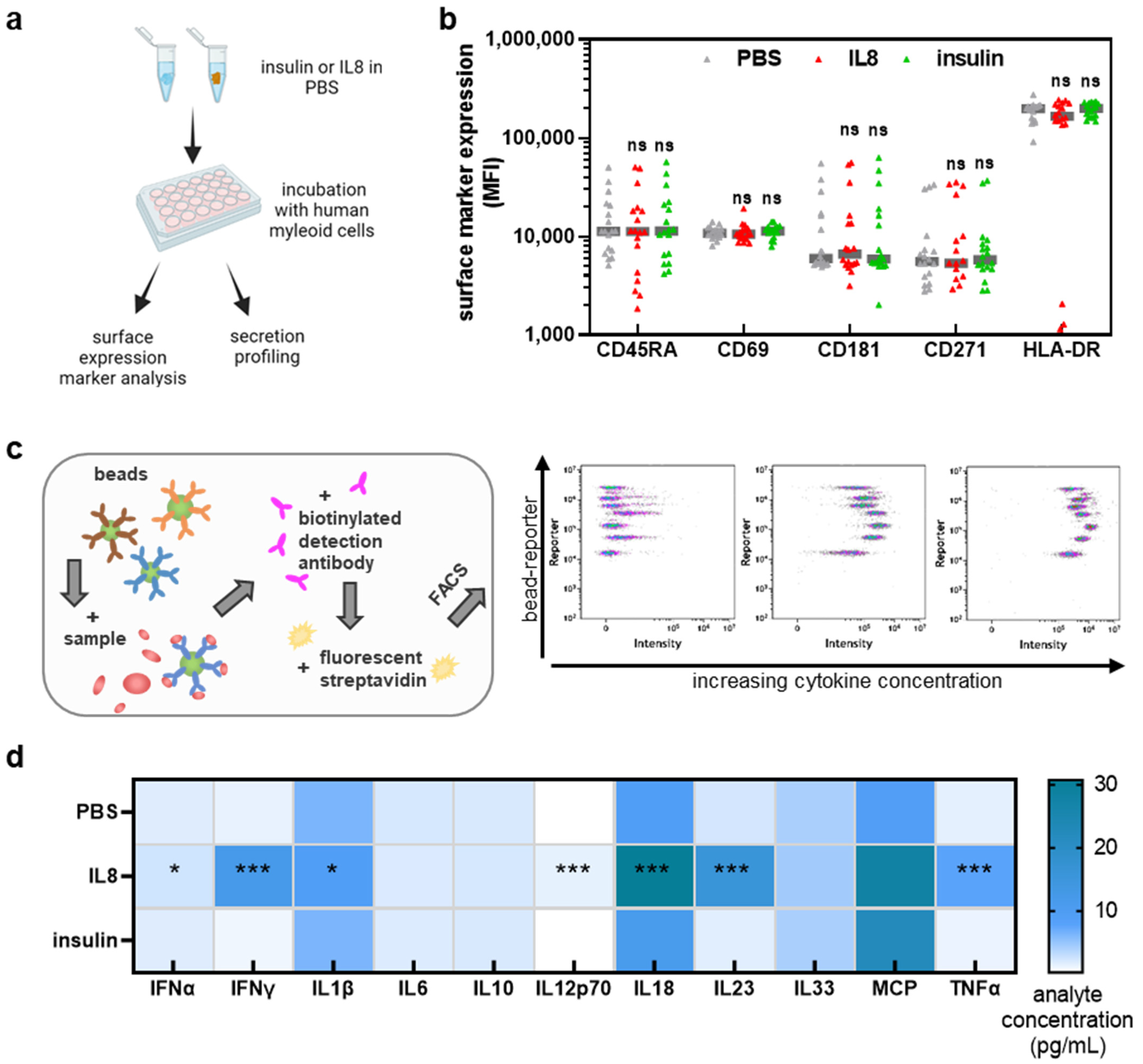
3.1. IL8, but Not Insulin, Triggered Pro-Inflammatory Cytokines Secretion in THP-1 Cells

In this study, we aimed to understand the effect of the oxidation of two physiological model proteins, IL8 and insulin, in human APCs. Accordingly, it was assessed first if the native proteins confer effects in the cells (Figure 1a). THP-1 cells were harvested and stained to determine surface marker expression by flow cytometry analysis. No significant changes in activation markers HLA-DR, CD181, and CD69 were observed after incubation with native IL8 or insulin (Figure 1b). There was also no effect on the expression of the differentiation markers CD271 and CD45RA. We further investigated the supernatants of THP-1 cells by using bead-based multiplex cytokine analysis to quantify several analytes in parallel (Figure 1c). The addition of native (untreated) IL8 to the cell culture medium led to significantly increased release levels of several cytokines, namely IFNα, IFNγ, IL1β, IL12p70, IL8, IL23, and TNFα (Figure 1d). By contrast, the addition of surplus insulin to THP-1 cell cultures did not significantly affect the secretion of any of the analytes investigated when compared to PBS vehicle alone.

3.2. THP-1 Cell Incubation with Gas Plasma- but Not H2O2-Oxidized Proteins Increased Activation-Related Surface Marker Expression

To investigate the effect of oxidative protein modifications in THP-1, we next incubated the cells with insulin and IL8 after the treatment with gas plasma or H2O2. H2O2 is one of the most abundant ROS produced in gas plasma-treated liquids (Figure 2a). To generate an H2O2 control condition, the concentration of H2O2 in gas plasma-treated PBS was determined (Figure 2b). In subsequent experiments, a concentration-matched control (cmc) was freshly prepared, containing equimolar amounts of H2O2 in PBS compared to what gas plasma would generate in such liquid in the absence of proteins. The main difference between both treatments was that gas plasma also produced a range of short-lived reactive species. In the following experiments, cells were incubated with PBS containing IL8 or insulin and either exposed to gas plasma or supplemented with H2O2, and functionally tested (Figure 2c). After THP-1 cells with different concentrations were incubated for 24 h, cell viability was tested first (Figure 2d). Interestingly, gas plasma- but not H2O2-treated IL8 (Figure 2e) and insulin (Figure 2f) at the highest (10 µg/mL) but not medium (1 µg/mL) or low (0.1 µg/mL) concentrations conferred modest but significant cytotoxic effects in THP-1 cells 24 h after exposure. This, in principle, already indicated that the gas plasma treatment altered the proteins differently than the H2O2 exposure. In addition, the cytotoxic effects can be attributed to the proteins since neither gas plasma-treated PBS nor H2O2 on their own affected the viability of THP-1 cells (Figure A1a). Subsequently, the surface expression profiles were investigated in THP-1 cells exposed to IL8 (Figure 2g). In response to gas plasma-treated IL8, four out of five surface markers were significantly increased. In comparison, there was no significant increase or decrease following incubation with H2O2-treated IL8 (Figure 2h). Gas plasma-treated insulin was tested in the following (Figure 2i). All tested surface markers were significantly increased following incubation with gas plasma-treated insulin. In contrast, H2O2-treated insulin did not significantly affect the expression of any surface markers (Figure 2j). Surface marker expression changes often come with alterations in the chemokine and cytokine secretion profiles. Hence, we determined the effect of IL8 (Figure 3a) and insulin (Figure 3b) protein oxidation on the expression levels of 11 different analytes. Patterns between IL8 and insulin conditions for each of the specific analytes did not match; both proteins seemed to elucidate individual cellular responses. Gas plasma-treated IL8 significantly increased IL10 and decreased IL23 and TNFα release, while H2O2-treated IL8 increased extracellular IL12p70 and TNFα levels. Hence, gas plasma treatment of IL8 was suggested to have anti-inflammatory properties. The same profile, although for different analytes, was observed for gas plasma-treated insulin, significantly decreasing IL1β, IL18, and IL23 release levels. For H2O2, only an MCP1 increase was found. Of those data, only the changes observed with IL12p70 could be potentially attributed to the ROS produced due to the gas plasma treatment or the H2O2 itself (Figure A1b).

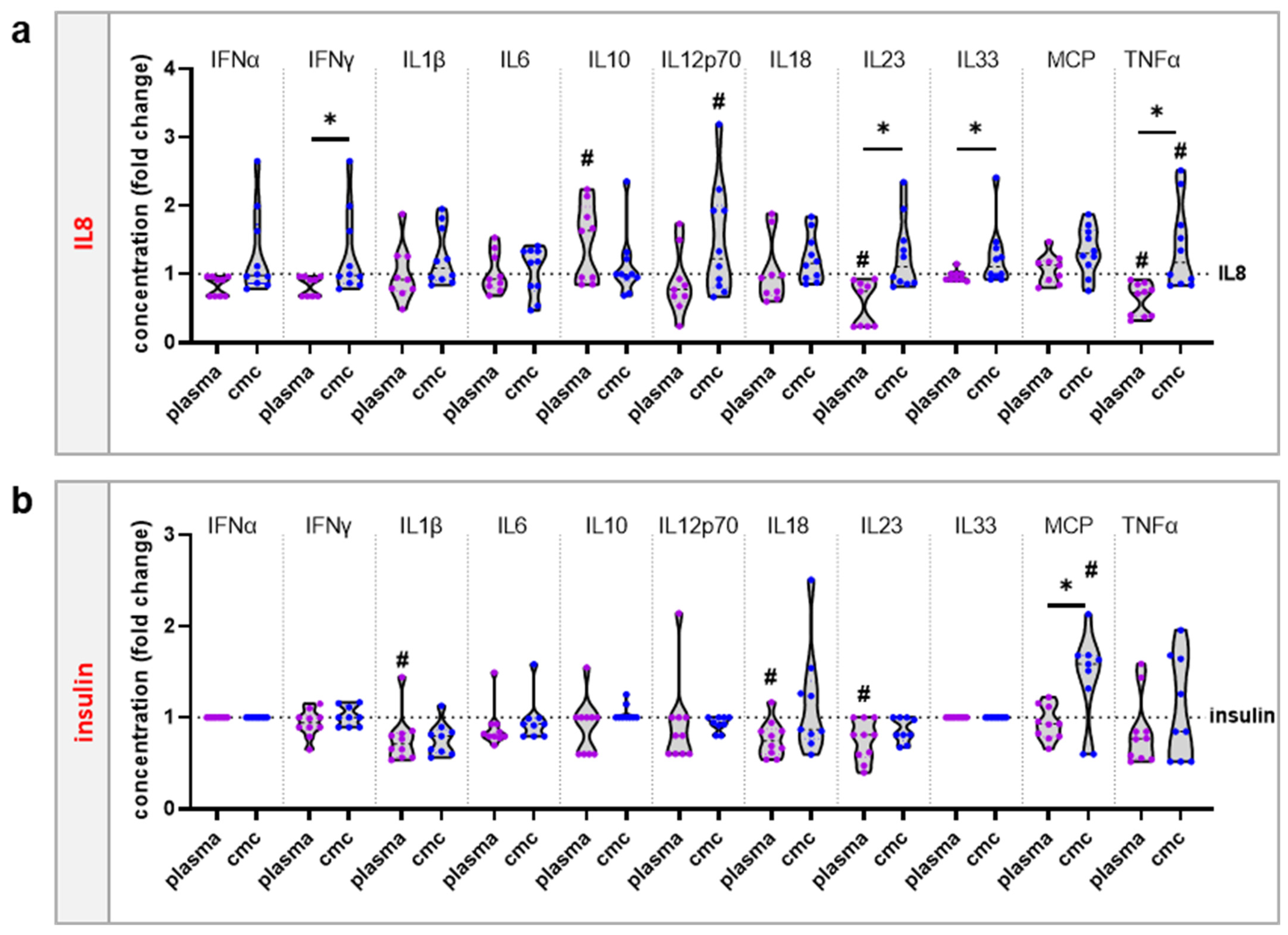
3.3. moDCs Activation and Secretion Profiles Were Profoundly Affected by Gas Plasma-Mediated Protein Oxidation

THP-1 cells are a culture model for monocytes but lack certain features of primary cells [30]. At the site of inflammation, mononuclear phagocytes, such as immature dendritic cells and macrophages that derive from monocytes (moDC and moMac), take up proteins and differentiate after activation to promote T cell response. We isolated CD14+ monocytes and cultured them in GM-CSF and IL4 before adding native or gas plasma- or H2O2-oxidized IL8 or insulin for 24 h. The viability of the cells was not markedly affected by native or oxidized proteins, regardless of the concentration investigated (Figure 4a). With regard to surface marker profiling, gas plasma-treated IL8 and insulin significantly increased the expression of activation or maturation marker CD14 (Figure 4b), CD25 (Figure 4c), CD40 (Figure 4d), CD80/86 (Figure 4e), CD83 (Figure 4f) and HLA-DR (Figure 4g). Interestingly, in principle, the same results were retrieved for H2O2-treated IL8 and insulin. The difference was, however, that the responses observed for gas plasma treatment were significantly higher than when using H2O2 to oxidize proteins. This effect was true for all surface markers regardless of IL8 or insulin being investigated. These results suggest that moDCs respond sensitively to protein oxidation. As myeloid cell responses often come with alterations in the secretion profiles, several chemokines and cytokines were quantitatively assessed for IL8 (Figure 4h) and insulin (Figure 4i) culture conditions. Gas plasma-treated IL8 reduced secretion of MCP1 and TNFα and increased IL23 compared to native IL8. Interestingly, moDCs incubated with cmc-treated IL8 showed significantly higher levels of IL12p70 and IL23 than gas plasma-treated IL8. Strikingly, gas plasma-treated insulin showed remarkable effects in human moDCs, as all chemokines and cytokines (except for TNFα) investigated were significantly decreased. This was partially recapitulated for H2O2-treated insulin, which significantly differed from responses observed with gas plasma-treated insulin for a few of the analytes investigated. In summary, these data suggest that the incubation of a monocytic cell line or primary human monocyte-derived dendritic cells with gas plasma-treated IL8 or insulin promoted significant surface marker expression changes along with altered patterns of chemokine and cytokine release, which may be attributed to the perception of oxidized proteins as a danger signal, with important implications for testing protein immunogenicity in terms of vaccination campaigns.

4. Discussion

There is significant interest in understanding the functionality and immunogenicity of ROS-modified structures and the association between such oxMods and pathology [31,32,33,34,35]. At the same time, understanding the immunogenicity of protein oxidation has important implications for vaccination. We here use gas plasma technology for generating highly reactive radicals and short-lived reactive species to investigate the effect of oxidized proteins on antigen-presenting cells.
During an inflammatory response, neutrophils generate the oxidizing hypohalous acids HOCl, HOBr, and HOSCN by the enzyme myeloperoxidase [3]. Furthermore, enzymes such as NADPH oxidases, xanthine oxidase, and 5-lipoxygenase can rapidly increase local H2O2 levels [36]. The major source of ROS in the TME is myeloid cells, such as macrophages and neutrophils [37]. They cause their effect through the so-called oxidative burst the biological system in general and local proteins in particular. Consequently, oxidative post-translational modification of proteins can lead to altered activity, functionality, and immunogenicity. For instance, the biological activity of HMGB1 depends on its redox state being either oxidized or reduced, ranging from pro- to anti-inflammatory [38]. The altered immunogenicity caused by oxidized amino acids or structural changes plays an important role in diseases by affecting immune cell activation. For instance, H2O2-oxidized β-glycoprotein I (β2-GPI) and naturally oxidized β2-GPI were shown to increase the expression of the DC activation marker HLA-DR, CD40, CD80, CD83, and CD86 [39]. In line with those results, we also observed increased surface marker expression when moDCs were incubated with oxidized proteins. A plausible mechanism involves the adjuvant effect of oxidized structures, leading to increased immunogenicity. Indeed, HOCl-modified serum albumin inhibits the uptake of other antigens and IFNγ-mediated MHC-II expression in THP-1 derived M1 macrophages [40]; the authors suggest that N-chlorinated protein competes with the other antigen for the binding to the same receptor. Biedron and colleagues observed reduced antigen uptake of bone-marrow-derived dendritic cells when the cells were previously incubated with HOCl-modified proteins [9]. We reported similar findings on the preferred uptake of gas plasma-treated proteins in a competition assay, where human moDCs were incubated with oxidized proteins and fluorescent proteins [41]. It should be mentioned here, however, that argon gas plasma was used in the present study, in which no HOCl but primarily H2O2 was generated.
Interestingly, a generalized activation by oxidized proteins was not found in our work, as H2O2 treatment did not reproduce the effects of gas plasma-treated proteins entirely and throughout all investigations in our study. Along similar lines, IL8 and insulin did not produce the same effects. The first obvious reason for this is the different physiological functions of both proteins, likely contributing in their own way to the results observed. A second reason could be their different structures and varying amino acid compositions and sequences. Several receptors were proposed for recognizing oxidized proteins, namely toll-like-receptor (TLR), mannose receptor (CD206), scavenger receptors A (CD204/ SR-A), CD36, SREC-I, and lectin-like oxidized low-density lipoprotein receptor (LOX-1), as well as RAGE [9,10,35,39,40,42]. Here, however, it should be taken into account that the different receptors were examined in other phagocytic cells. Our study also observed other effects of oxidized IL8 and oxidized insulin on THP-1 and moDCs. This is not much of a surprise, starting with the fact that we left THP-1 undifferentiated, i.e., without incubation of cytokines prior to incubation with the proteins, while moDCs were incubated with IL4 and GM-CSF, apart from intrinsic differences observed with both cell types [30]. Our data also support previous reports, where differences in phenotypic changes, activation, and maturation in moDCs, THP-1, KG-1, and MUTZ-3 after incubation with proteins or chemical sensitizers were shown before [43,44,45,46]. However, different activation and maturation abilities of the different cell types also point to the presence of different subpopulations in the organism and during an immune response that need to be considered in general. This makes studying oxidized proteins in the inflammatory context more complex, and further comparative studies are needed.
In our study, we did compare not only THP-1 versus moDCs but also different proteins and different concentrations. We did not observe a correlation between protein concentration and APC activation in both cell types. However, a correlation between ROS concentration for oxMod-generation and immunogenicity was shown previously in vivo [11]. More research must be performed to investigate the effects of ROS concentrations and a variety of ROS on proteins produced by gas plasmas; parallel studies on the effect of gas plasma-induced oxMods on immune cells help to understand the individual recognition, signaling, and immune responses associated with them. Gas plasma is tunable, and different ROSs are generated, depending on different feeding gas and gas mixtures, which were previously shown to have different effects on the immunogenicity of Ova [41]. Furthermore, treatment conditions, such as exposure time or conductive mode, influence ROS generation and potential protein oxPTMs [47].
In summary, we observed different effects of protein oxidation and immunogenicity, depending on the type of protein, exposure to H2O2 or ROS cocktail, and cell type. Indeed, we determined different effects of oxidized IL8 and oxidized insulin on THP-1 and moDCs, although different protein concentrations did not influence activation and maturation. Furthermore, oxidation by gas plasma seemed superior to using H2O2 alone, while both had specific effects on the cells. Our results suggest that different oxidative post-translational modifications induced by various ROSs lead to distinct recognition, further processing, and the activation/ maturation state in APCs, which, ultimately, also depends to a certain extent on the protein investigated.

5. Conclusions

Oxidative stress and ROS generation during inflammation lead to advanced oxidation protein products and oxidized proteins. Phagocytic immune cells take up extracellular oxidized proteins, become activated, and maturate to support an adaptive immune response. Gas plasma technology generates a variety of ROSs simultaneously and is a promising tool for investigating protein oxidation and immunogenic effects. We studied the effect of gas plasma-treated IL8 and insulin on phenotypic alterations and cytokine secretion profiles in moDCs and THP-1 compared to H2O2-tested proteins. Taken together, the results show different immunogenicities of exogenous ROS-induced oxidized proteins. Furthermore, we observed significant differences in moDCs while THP-1 cells were more reserved in response to oxMods.

Author Contributions

Conceptualization, S.B.; methodology, S.B.; software, R.C., K.A. and S.B.; validation, K.A., R.C., and S.B.; formal analysis, K.A. and R.C.; investigation, K.A.; resources, S.B.; data curation, K.A. and R.C.; writing—original draft preparation, R.C. and S.B.; writing—review and editing, S.B. and T.v.W.; visualization, R.C. and S.B.; supervision, S.B., T.v.W., and R.C.; project administration, S.B. and R.C.; funding acquisition, S.B. All authors have read and agreed to the published version of the manuscript.

Funding

This work was funded by the German Federal Ministry of Education and Research (BMBF), grant numbers 03Z22DN11 and 03Z22Di1 (to SB). The funding source had no role in the design of this study or its execution, analyses, interpretation of the data, or decision to publish results.

Institutional Review Board Statement

Not applicable.

Informed Consent Statement

Not applicable.

Data Availability Statement

The underlying data from this work can be retrieved from the author upon reasonable request.

Acknowledgments

The authors acknowledge the technical assistance of Felix Niessner, Henry Skowsky, Lea Miebach, Debora Singer, and Julia Berner (all ZIK plasmatis, INP, Greifswald, Germany). Figure 1a and Figure 2c were generated with the help of a commercial license of biorender.com.

Conflicts of Interest

The authors declare no conflict of interest.

Appendix A

Figure A1. (a) Viability of THP-1 cells after incubation with protein-free PBS or gas plasma-oxidized protein-free PBS; (b) cytokine secretion of protein-free oxPBS-treated THP-1 cells normalized to secretion following protein-free PBS. Data are the mean of at least three independent experiments. Statistical analysis was performed using two-way analysis of variances; for ANOVA: F = 2.6 (a)/3.5 (b), df = 2 (a)/2 (b). ns = not significant; * denotes significant differences (p < 0.05).
Vaccines 10 01814 g0a1

References

  1. Winterbourn, C.C.; Kettle, A.J.; Hampton, M.B. Reactive Oxygen Species and Neutrophil Function. Annu. Rev. Biochem. 2016, 85, 765–792. [Google Scholar] [CrossRef] [PubMed]
  2. Davies, M.J. Myeloperoxidase-derived oxidation: Mechanisms of biological damage and its prevention. J. Clin. Biochem. Nutr. 2011, 48, 8–19. [Google Scholar] [CrossRef] [PubMed] [Green Version]
  3. Ulfig, A.; Leichert, L.I. The effects of neutrophil-generated hypochlorous acid and other hypohalous acids on host and pathogens. Cell Mol. Life Sci. 2021, 78, 385–414. [Google Scholar] [CrossRef]
  4. Hampton, M.B.; Kettle, A.J.; Winterbourn, C.C. Inside the neutrophil phagosome: Oxidants, myeloperoxidase, and bacterial killing. Blood 1998, 92, 3007–3017. [Google Scholar] [CrossRef]
  5. Niethammer, P.; Grabher, C.; Look, A.T.; Mitchison, T.J. A tissue-scale gradient of hydrogen peroxide mediates rapid wound detection in zebrafish. Nature 2009, 459, 996–999. [Google Scholar] [CrossRef] [Green Version]
  6. Lammermann, T.; Afonso, P.V.; Angermann, B.R.; Wang, J.M.; Kastenmuller, W.; Parent, C.A.; Germain, R.N. Neutrophil swarms require LTB4 and integrins at sites of cell death in vivo. Nature 2013, 498, 371–375. [Google Scholar] [CrossRef] [PubMed]
  7. Selmeci, L. Advanced oxidation protein products (AOPP): Novel uremic toxins, or components of the non-enzymatic antioxidant system of the plasma proteome? Free Radic. Res. 2011, 45, 1115–1123. [Google Scholar] [CrossRef]
  8. Strollo, R.; Vinci, C.; Arshad, M.H.; Perrett, D.; Tiberti, C.; Chiarelli, F.; Napoli, N.; Pozzilli, P.; Nissim, A. Antibodies to post-translationally modified insulin in type 1 diabetes. Diabetologia 2015, 58, 2851–2860. [Google Scholar] [CrossRef] [Green Version]
  9. Biedron, R.; Konopinski, M.K.; Marcinkiewicz, J.; Jozefowski, S. Oxidation by neutrophils-derived HOCl increases immunogenicity of proteins by converting them into ligands of several endocytic receptors involved in antigen uptake by dendritic cells and macrophages. PLoS ONE 2015, 10, e0123293. [Google Scholar] [CrossRef] [Green Version]
  10. Prokopowicz, Z.M.; Arce, F.; Biedron, R.; Chiang, C.L.; Ciszek, M.; Katz, D.R.; Nowakowska, M.; Zapotoczny, S.; Marcinkiewicz, J.; Chain, B.M. Hypochlorous acid: A natural adjuvant that facilitates antigen processing, cross-priming, and the induction of adaptive immunity. J. Immunol. 2010, 184, 824–835. [Google Scholar] [CrossRef]
  11. Marcinkiewicz, J.; Bobek, M.; Mak, M.; Biedroń, R. Immunogenic properties of collagen and ovalbumin modified by chlorination. Cent. Eur. J. Immunol. 2004, 28, 160–166. [Google Scholar]
  12. Nybo, T.; Dieterich, S.; Gamon, L.F.; Chuang, C.Y.; Hammer, A.; Hoefler, G.; Malle, E.; Rogowska-Wrzesinska, A.; Davies, M.J. Chlorination and oxidation of the extracellular matrix protein laminin and basement membrane extracts by hypochlorous acid and myeloperoxidase. Redox Biol. 2019, 20, 496–513. [Google Scholar] [CrossRef] [PubMed]
  13. Nasri, Z.; Memari, S.; Wenske, S.; Clemen, R.; Martens, U.; Delcea, M.; Bekeschus, S.; Weltmann, K.D.; von Woedtke, T.; Wende, K. Singlet Oxygen-Induced Phospholipase A2 Inhibition: A Major Role for Interfacial Tryptophan Dioxidation. Chemistry 2021, 27, 14702–14710. [Google Scholar] [CrossRef] [PubMed]
  14. Wenske, S.; Lackmann, J.W.; Bekeschus, S.; Weltmann, K.D.; von Woedtke, T.; Wende, K. Nonenzymatic post-translational modifications in peptides by cold plasma-derived reactive oxygen and nitrogen species. Biointerphases 2020, 15, 061008. [Google Scholar] [CrossRef]
  15. Khlyustova, A.; Labay, C.; Machala, Z.; Ginebra, M.P.; Canal, C. Important parameters in plasma jets for the production of RONS in liquids for plasma medicine: A brief review. Front. Chem. Sci. Eng. 2019, 13, 238–252. [Google Scholar] [CrossRef]
  16. Adamovich, I.; Baalrud, S.D.; Bogaerts, A.; Bruggeman, P.J.; Cappelli, M.; Colombo, V.; Czarnetzki, U.; Ebert, U.; Eden, J.G.; Favia, P.; et al. The 2017 Plasma Roadmap: Low temperature plasma science and technology. J. Phys. D Appl. Phys. 2017, 50, 323001. [Google Scholar] [CrossRef]
  17. Laroussi, M.; Bekeschus, S.; Keidar, M.; Bogaerts, A.; Fridman, A.; Lu, X.; Ostrikov, K.; Hori, M.; Stapelmann, K.; Miller, V.; et al. Low-Temperature Plasma for Biology, Hygiene, and Medicine: Perspective and Roadmap. IEEE Trans. Radiat. Plasma Med. Sci. 2022, 6, 127–157. [Google Scholar] [CrossRef]
  18. Kaushik, N.K.; Kaushik, N.; Min, B.; Choi, K.H.; Hong, Y.J.; Miller, V.; Fridman, A.; Choi, E.H. Cytotoxic macrophage-released tumour necrosis factor-alpha (TNF-alpha) as a killing mechanism for cancer cell death after cold plasma activation. J. Phys. D Appl. Phys. 2016, 49, 084001. [Google Scholar] [CrossRef]
  19. Bekeschus, S.; Meyer, D.; Arlt, K.; von Woedtke, T.; Miebach, L.; Freund, E.; Clemen, R. Argon Plasma Exposure Augments Costimulatory Ligands and Cytokine Release in Human Monocyte-Derived Dendritic Cells. Int. J. Mol. Sci. 2021, 22, 3790. [Google Scholar] [CrossRef]
  20. Freund, E.; Moritz, J.; Stope, M.; Seebauer, C.; Schmidt, A.; Bekeschus, S. Plasma-Derived Reactive Species Shape a Differentiation Profile in Human Monocytes. Appl. Sci. 2019, 9, 2530. [Google Scholar] [CrossRef] [Green Version]
  21. Crestale, L.; Laurita, R.; Liguori, A.; Stancampiano, A.; Talmon, M.; Bisag, A.; Gherardi, M.; Amoruso, A.; Colombo, V.; Fresu, L. Cold Atmospheric Pressure Plasma Treatment Modulates Human Monocytes/Macrophages Responsiveness. Plasma 2018, 1, 261–276. [Google Scholar] [CrossRef]
  22. Lin, A.; Gorbanev, Y.; De Backer, J.; Van Loenhout, J.; Van Boxem, W.; Lemiere, F.; Cos, P.; Dewilde, S.; Smits, E.; Bogaerts, A. Non-Thermal Plasma as a Unique Delivery System of Short-Lived Reactive Oxygen and Nitrogen Species for Immunogenic Cell Death in Melanoma Cells. Adv. Sci. 2019, 6, 1802062. [Google Scholar] [CrossRef] [PubMed] [Green Version]
  23. Bekeschus, S.; Clemen, R.; Niessner, F.; Sagwal, S.K.; Freund, E.; Schmidt, A. Medical Gas Plasma Jet Technology Targets Murine Melanoma in an Immunogenic Fashion. Adv. Sci. 2020, 7, 1903438. [Google Scholar] [CrossRef] [PubMed] [Green Version]
  24. Lin, A.G.; Xiang, B.; Merlino, D.J.; Baybutt, T.R.; Sahu, J.; Fridman, A.; Snook, A.E.; Miller, V. Non-thermal plasma induces immunogenic cell death in vivo in murine CT26 colorectal tumors. Oncoimmunology 2018, 7, e1484978. [Google Scholar] [CrossRef] [PubMed] [Green Version]
  25. Tomic, S.; Petrovic, A.; Puac, N.; Skoro, N.; Bekic, M.; Petrovic, Z.L.; Colic, M. Plasma-Activated Medium Potentiates the Immunogenicity of Tumor Cell Lysates for Dendritic Cell-Based Cancer Vaccines. Cancers 2021, 13, 1626. [Google Scholar] [CrossRef]
  26. Reuter, S.; von Woedtke, T.; Weltmann, K.D. The kINPen-a review on physics and chemistry of the atmospheric pressure plasma jet and its applications. J. Phys. D Appl. Phys. 2018, 51, 233001. [Google Scholar] [CrossRef] [Green Version]
  27. Bekeschus, S.; Masur, K.; Kolata, J.; Wende, K.; Schmidt, A.; Bundscherer, L.; Barton, A.; Kramer, A.; Bröker, B.; Weltmann, K.-D. Human Mononuclear Cell Survival and Proliferation is Modulated by Cold Atmospheric Plasma Jet. Plasma Process. Polym. 2013, 10, 706–713. [Google Scholar] [CrossRef]
  28. Freund, E.; Liedtke, K.R.; Gebbe, R.; Heidecke, A.K.; Partecke, L.-I.; Bekeschus, S. In Vitro Anticancer Efficacy of Six Different Clinically Approved Types of Liquids Exposed to Physical Plasma. IEEE Trans. Radiat. Plasma Med. Sci. 2019, 3, 588–596. [Google Scholar] [CrossRef]
  29. Bekeschus, S.; Freund, E.; Wende, K.; Gandhirajan, R.K.; Schmidt, A. Hmox1 Upregulation Is a Mutual Marker in Human Tumor Cells Exposed to Physical Plasma-Derived Oxidants. Antioxidants 2018, 7, 151. [Google Scholar] [CrossRef] [Green Version]
  30. Bosshart, H.; Heinzelmann, M. THP-1 cells as a model for human monocytes. Ann. Transl. Med. 2016, 4, 438. [Google Scholar] [CrossRef] [Green Version]
  31. Estevez, M.; Xiong, Y. Intake of Oxidized Proteins and Amino Acids and Causative Oxidative Stress and Disease: Recent Scientific Evidences and Hypotheses. J. Food Sci. 2019, 84, 387–396. [Google Scholar] [CrossRef] [PubMed]
  32. Clemen, R.; Bekeschus, S. Oxidatively Modified Proteins: Cause and Control of Diseases. Appl. Sci. 2020, 10, 6419. [Google Scholar] [CrossRef]
  33. Davies, M.J. Protein oxidation and peroxidation. Biochem. J. 2016, 473, 805–825. [Google Scholar] [CrossRef] [PubMed] [Green Version]
  34. Winyard, P.G.; Ryan, B.; Eggleton, P.; Nissim, A.; Taylor, E.; Lo Faro, M.L.; Burkholz, T.; Szabo-Taylor, K.E.; Fox, B.; Viner, N.; et al. Measurement and meaning of markers of reactive species of oxygen, nitrogen and sulfur in healthy human subjects and patients with inflammatory joint disease. Biochem. Soc. Trans. 2011, 39, 1226–1232. [Google Scholar] [CrossRef]
  35. Ryan, B.J.; Nissim, A.; Winyard, P.G. Oxidative post-translational modifications and their involvement in the pathogenesis of autoimmune diseases. Redox Biol. 2014, 2, 715–724. [Google Scholar] [CrossRef] [PubMed] [Green Version]
  36. van der Vliet, A.; Janssen-Heininger, Y.M. Hydrogen peroxide as a damage signal in tissue injury and inflammation: Murderer, mediator, or messenger? J. Cell. Biochem. 2014, 115, 427–435. [Google Scholar] [CrossRef] [Green Version]
  37. Kennel, K.B.; Greten, F.R. Immune cell—Produced ROS and their impact on tumor growth and metastasis. Redox Biol. 2021, 42, 101891. [Google Scholar] [CrossRef] [PubMed]
  38. Kang, R.; Zhang, Q.; Zeh, H.J., 3rd; Lotze, M.T.; Tang, D. HMGB1 in cancer: Good, bad, or both? Clin. Cancer Res. 2013, 19, 4046–4057. [Google Scholar] [CrossRef] [Green Version]
  39. Buttari, B.; Profumo, E.; Mattei, V.; Siracusano, A.; Ortona, E.; Margutti, P.; Salvati, B.; Sorice, M.; Rigano, R. Oxidized beta2-glycoprotein I induces human dendritic cell maturation and promotes a T helper type 1 response. Blood 2005, 106, 3880–3887. [Google Scholar] [CrossRef]
  40. Ulfig, A.; Bader, V.; Varatnitskaya, M.; Lupilov, N.; Winklhofer, K.F.; Leichert, L.I. Hypochlorous acid-modified human serum albumin suppresses MHC class II—Dependent antigen presentation in pro-inflammatory macrophages. Redox Biol. 2021, 43, 101981. [Google Scholar] [CrossRef]
  41. Clemen, R.; Freund, E.; Mrochen, D.; Miebach, L.; Schmidt, A.; Rauch, B.H.; Lackmann, J.W.; Martens, U.; Wende, K.; Lalk, M.; et al. Gas Plasma Technology Augments Ovalbumin Immunogenicity and OT-II T Cell Activation Conferring Tumor Protection in Mice. Adv. Sci. 2021, 8, 2003395. [Google Scholar] [CrossRef] [PubMed]
  42. Chechushkov, A.V.; Kozhin, P.M.; Zaitseva, N.S.; Lemza, A.E.; Men’shchikova, E.B.; Troitskii, A.V.; Shkurupy, V.A. Oxidized Dextran Enhances Alternative Activation of Macrophages in Mice of Opposite Lines. Bull Exp. Biol. Med. 2016, 160, 783–786. [Google Scholar] [CrossRef] [PubMed]
  43. Hitzler, M.; Bergert, A.; Luch, A.; Peiser, M. Evaluation of selected biomarkers for the detection of chemical sensitization in human skin: A comparative study applying THP-1, MUTZ-3 and primary dendritic cells in culture. Toxicol. In Vitro 2013, 27, 1659–1669. [Google Scholar] [CrossRef] [PubMed]
  44. Tietze, C.; Blomeke, B. Sensitization assays: Monocyte-derived dendritic cells versus a monocytic cell line (THP-1). J. Toxicol. Environ. Health A 2008, 71, 965–968. [Google Scholar] [CrossRef]
  45. Larsson, K.; Lindstedt, M.; Borrebaeck, C.A. Functional and transcriptional profiling of MUTZ-3, a myeloid cell line acting as a model for dendritic cells. Immunology 2006, 117, 156–166. [Google Scholar] [CrossRef]
  46. Santegoets, S.J.; van den Eertwegh, A.J.; van de Loosdrecht, A.A.; Scheper, R.J.; de Gruijl, T.D. Human dendritic cell line models for DC differentiation and clinical DC vaccination studies. J. Leukoc. Biol. 2008, 84, 1364–1373. [Google Scholar] [CrossRef] [Green Version]
  47. Miebach, L.; Freund, E.; Clemen, R.; Weltmann, K.D.; Metelmann, H.R.; von Woedtke, T.; Gerling, T.; Wende, K.; Bekeschus, S. Conductivity augments ROS and RNS delivery and tumor toxicity of an argon plasma jet. Free Radic. Biol. Med. 2022, 180, 210–219. [Google Scholar] [CrossRef]
Figure 1. IL8 but not insulin triggers the secretion of pro-inflammatory cytokines in THP-1. (a) Workflow of the experimental procedure; (b) surface marker expression on IL8 or insulin-spiked THP-1 cells reveals no differences compared to unstimulated cells; (c) principle of bead-based multiplex assay; (d) absolute chemokine and cytokine levels of THP-1 cells exposed to IL8 or insulin. Data are the mean of at least three independent experiments. Statistical analysis was performed using two-way analysis of variances; for ANOVA: F = 2.8 (b)/33.2 (d), df = 2 (b)/2 (d). (* = p < 0.05; *** p < 0.001).
Vaccines 10 01814 g001
Figure 2. Gas plasma but not cmc-treated IL8 and insulin stimulates THP-1 cells. (a) Representative image of gas plasma treatment; (b) H2O2 concentration measured after exposure to gas plasma—the 30 s concentration was used as the cmc in subsequent experiments; (c) workflow for THP-1 incubation with native or oxidized proteins; (d) representative strategy of flow cytometry analysis; (e,f) quantified and normalized data of cell viability after incubation with native or oxidized IL8 (e) or insulin (f) concentrations; (g,h) representative histogram (g) and quantification of surface marker expression after incubation with native or oxidized IL8 (h); (i,j) representative histogram (i) and quantification of surface marker expression after incubation with native or oxidized IL8 (j). Data show individual technical replicates of an assay with three independent experiments. Data are the mean of at least three independent experiments. Statistical analysis was performed using one-way analysis of variances of gas plasma and H2O2 conditions against PBS (rhombi) or Mann–Whitney tests to compare gas plasma vs. H2O2 (asterisks); for ANOVA: F = 1.9 (0.1)/4.4 (1.0)/10.4 (10) and df = 2 (e); F = 3.4 (0.1)/2.4 (1.0)/23.0 (10) and df = 2 (f); F = 0.7 (CD181)/4.9 (HLA-DR)/4.2 (CD271)/4.4 (CD45RA)/4.0 (CD69) and df = 2 (h); F = 3.2 (CD181)/3.2 (HLA-DR)/4.2 (CD271)/6.1 (CD45RA)/4.1 (CD69) and df = 2 (j).
Vaccines 10 01814 g002
Figure 3. Oxidized IL8 and insulin led to modestly altered secretion profiles in THP-1 cells. (a,b) Secretion profiling was performed using bead-based multiplex assays of supernatants of THP-1 cells incubated with native or oxidized IL8 (a) or insulin (b). Data are the mean of at least three independent experiments. Statistical analysis was performed using one-way analysis of variances; for ANOVA: F = 3.0 (a)/2.6 (b), df = 2 (a)/2 (b). * denotes statistically significant differences between plasma and cmc (p < 0.05; t-test); # denotes statistically significant differences between the individual group and the gas insulin control normalized to 1 (p < 0.05; one-way analysis of variances).
Vaccines 10 01814 g003
Figure 4. Oxidized IL8 and insulin stimulate moDC activation and suppress cytokine secretion. (a) Viability of human moDCs in response to native, gas plasma-, or H2O2-treated IL8 or insulin; (bg) surface marker expression of CD14 (b), CD25 (c), CD40 (d), CD80/86 (e), CD83 (f), and HLA-DR (g) in moDCs incubated with the proteins was investigated using flow cytometry; (h,i) secretion profiling was performed using bead-based multiplex assays of supernatants of moDCs incubated with native or oxidized IL8 (h) or insulin (i). Data are the mean of at least three independent experiments. Statistical analysis was performed using one-way analysis of variances of gas plasma and H2O2 conditions against PBS (ag: asterisks; hi: rhombi) or Mann–Whitney tests to compare gas plasma vs. H2O2 (asterisks); for ANOVA: F = 2.8 (0.1)/1.5 (1.0)/3.9 (10) and df = 2 (a); F = 3.7 (0.1)/1.5 (1.0)/2.1 (10) and df = 2 (b); F = 27.4 (CD14)/64.2 (CD25)/68.6 (CD40)/88.5 (CD80/86)/13.6 (CD83)/87.0 (HLA-DR) and df = 2 (bg); F = 2.5 (h)/19.7 (i), df = 2 (h)/2 (i). For (bg): */**/***/**** denotes statistically significant differences between plasma and cmc, plasma and gas control, and cmc and gas control (* = p < 0.05; ** p < 0.01; *** p < 0.001; **** p < 0.0001); # denotes statistically significant differences between the individual group and the gas insulin control normalized to 1 (p < 0.05; one-way analysis of variances). For (h,i): * denotes statistically significant differences between plasma and cmc (p < 0.05; t-test).
Vaccines 10 01814 g004
Table 1. Fluorescently-labeled monoclonal antibodies used in this study.
LigandFluorescent LabelCloneSupplier
CD11CPE-Cy7S-HCL-3BioLegend
CD14BV650M5E2BioLegend
CD25AF488BC96BioLegend
CD40APCHB14BioLegend
CD45RAPerCP/Cy5.5HI100BD Biosciences
CD69AF488FN50BioLegend
CD80PE2D10BioLegend
CD83PerCP/Cy5.5HB15BioLegend
CD86PEBU63BioLegend
CD181PE8F1/CXCR1BioLegend
CD271PE-Cy7C40-1457BD Biosciences
HLA-A,B,CAF700W6/32BioLegend
HLA-DRAPC-Cy7L246BioLegend
Publisher’s Note: MDPI stays neutral with regard to jurisdictional claims in published maps and institutional affiliations.

