UHPLC-HESI-OT-MS-MS Biomolecules Profiling, Antioxidant and Antibacterial Activity of the “Orange-Yellow Resin” from Zuccagnia punctata Cav.


 ,
,  and
and
Abstract
1. Introduction
2. Materials and Methods
2.1. Chemicals
2.2. Plant Material
2.3. Z. punctata Orange-Yellow Resin (ZpRe)
2.4. UHPLC-DAD-MS Instrument
2.5. LC Parameters and MS Parameters
2.6. Total Phenolic (TP) and Flavonoid (F) Content
2.7. Antioxidant Activity
2.7.1. 2,2-Diphenyl-1-picrylhydrazyl Radical Scavenging Capacity Assay
2.7.2. Ferric-Reducing Antioxidant Power Assay (FRAP)
2.7.3. Trolox Equivalent Antioxidant Activity Assay (TEAC)
2.7.4. Lipid Peroxidation in Erythrocytes
2.8. Antibacterial Activity
2.8.1. Microorganisms
2.8.2. Antibacterial Susceptibility Testing
2.9. Statistical Analysis
3. Results
3.1. UHPLC-PDA-OT-MS Analysis of the Orange-Yellow Resin From San Juan Province, Argentina
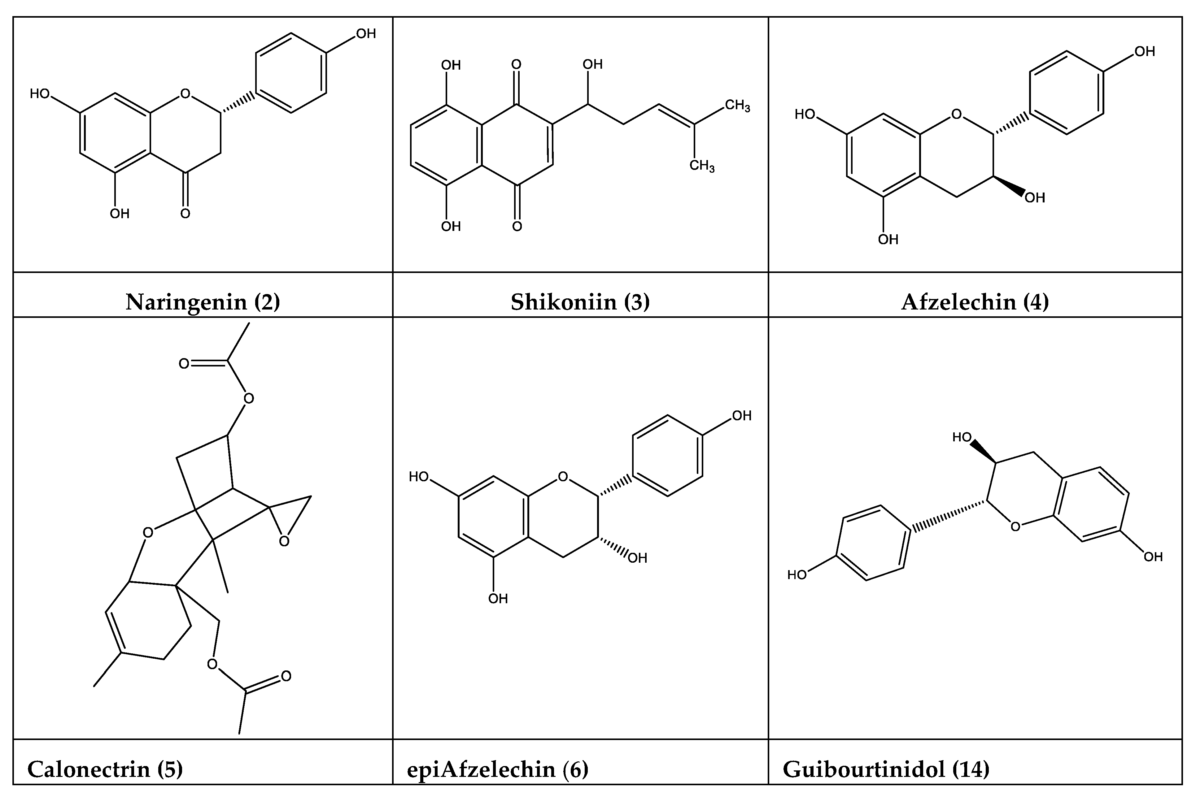
3.1.1. Flavonoids
3.1.2. Chalcones
3.1.3. Caffeic Acid Derivatives
3.1.4. Coumaric Acid Esters
3.1.5. Xanthene’s Derivatives, Trichothecenes; Vedelianin Derivatives, and Others
3.2. Total Phenolic and Flavonoid Contents and Antioxidant, and Antibacterial Activities
4. Conclusions
Supplementary Materials
Author Contributions
Funding
Acknowledgments
Conflicts of Interest
References
- Lima Licá, I.C.; Dos Santos Soares, A.M.; Silva de Mesquita, L.S.; Malik, S. Biological properties and pharmacological potential of plant exudates. Food Res. Int. 2018, 105, 1039–1053. [Google Scholar] [CrossRef]
- Modak, B.; Salina, M.; Rodilla, J.; Torres, R. Study of the chemical composition of the resinous exudate isolated from Heliotropium sclerocarpum and evaluation of the antioxidant properties of the phenolic compounds and the resin. Molecules 2009, 14, 4625–4633. [Google Scholar] [CrossRef]
- Agüero, M.B.; Gonzalez, M.; Lima, B.; Svetaz, L.; Sánchez, M.; Zacchino, S.; Feresin, G.E.; Schmeda-Hirschmann, G.; Palermo, J.A.; Wunderlin, D.A.; et al. Argentinean Propolis from Zuccagniapunctata Cav. (Caesalpinieae) Exudates: Phytochemical Characterization and Antifungal Activity. J. Agric. Food Chem. 2010, 58, 194–201. [Google Scholar] [CrossRef]
- Zampini, I.C.; Vattuone, M.A.; Isla, M.I. Antibacterial activity of Zuccagnia punctata Cav. ethanolic extracts. J. Ethnopharmacol. 2005, 102, 450–456. [Google Scholar] [CrossRef]
- Zampini, I.C.; Villarini, M.; Moretti, M.; Dominici, L.; Isla, M.I. Evaluation of genotoxic and antigenotoxic effects of hydroalcoholic extracts of Zuccagnia punctata Cav. J. Ethnopharmacol. 2007, 115, 330–335. [Google Scholar] [CrossRef]
- Svetaz, L.; Tapia, A.; López, S.N.; Furlán, R.; Petenatti, E.; Pioli, R.; Schmeda-Hirschmann, G.; Zacchino, S.A. Antifungal chalcones and new cafeic acid esters from Zuccagnia punctata acting against soybean infecting fungi. J. Agric. Food Chem. 2004, 52, 3297–3300. [Google Scholar] [CrossRef]
- Svetaz, L.; Aguero, M.B.; Alvarez, S.; Luna, L.; Feresin, G.; Derita, M.; Tapia, A.; Zacchino, S. Antifungal activity of chalcones from Zuccagnia punctata Cav. acting against clinically important fungi and studies of mechanism of action. Planta Med. 2007, 73, 1074–1080. [Google Scholar] [CrossRef]
- Butassi, E.; Svetaz, L.; Ivancovich, J.J.; Feresin, G.; Tapia, A.; Zacchino, S. Synergistic antifungal fixed-ratio combinations of Zuccagnia punctata Cav. and Larrea nitida Cav., using Mixed-Effects Loewe (MixLow) method. Phytomedicine 2015, 22, 666–678. [Google Scholar] [CrossRef]
- Butassi, E.; Svetaz, L.A.; Sortino, M.A.; Quiroga, A.D.; Carvalho, V.S.D.; Cortés, J.C.G.; Ribas, J.C.; Zacchino, S.A. Approaches to the mechanism of antifungal activity of Zuccagnia punctata-Larrea nitida bi-herbal combination. Phytomedicine 2019, 54, 291–301. [Google Scholar] [CrossRef]
- Solorzano, E.R.; Bortolini, C.; Bogialli, S.; Di Gangi, I.M.; Favaro, G.; Maldonado, L.; Pastore, P. Use of a LC-DAD-QTOF system for the characterization of the phenolic profile of the argentinean plant Zuccagnia punctata and of the related propolis: New biomarkers. J. Funct. Foods 2017, 33, 425–435. [Google Scholar] [CrossRef]
- Cornejo, A.; Salgado, F.; Caballero, J.; Vargas, R.; Simirgiotis, M.; Areche, C. Secondary metabolites in Ramalina terebrata detected by UHPLC/ESI/MS/MS and identification of parietin as tau protein inhibitor. Int. J. Mol. Sci. 2016, 17, 1303. [Google Scholar] [CrossRef]
- Quispe, C.; Bórquez, J.; Villalobos, M.; Simirgiotis, M. Chemical Composition and Antioxidant Activity of Aloe vera from the Pica Oasis (Tarapacá, Chile) by UHPLC-Q/Orbitrap/MS/MS. J. Chem. 2018, 1–12. [Google Scholar] [CrossRef]
- Simirgiotis, M.J.; Quispe, C.; Mocan, A.; Villatoro, J.M.; Areche, C.; Bórquez, J.; Sepúlveda, B.; Echiburu-Chau, C. UHPLC high resolution orbitrapmetabolomic fingerprinting of the unique species Ophryosporus triangularis meyen from the atacama desert, Northern Chile. Rev. Bras. Farmacogn. 2017, 27, 179–187. [Google Scholar] [CrossRef]
- Luna, L.; Simirgiotis, M.J.; Lima, B.; Bórquez, J.; Feresin, G.E.; Tapia, A. UHPLC-MS metabolome fingerprinting: The isolation of main compounds and antioxidant activity of the andean species Tetraglochin ameghinoi (Speg.) Speg. Molecules 2018, 23, 793. [Google Scholar] [CrossRef]
- Gómez, J.; Simirgiotis, M.J.; Lima, B.; Paredes, J.D.; Villegas Gabutti, C.M.; Gamarra-Luques, C.; Bórquez, J.; Luna, L.; Wendel, G.H.; Maria, A.O.; et al. Antioxidant, Gastroprotective, Cytotoxic Activities and UHPLC-PDA-Q Orbitrap Mass Spectrometry Identification of Metabolites in Baccharis grisebachii Decoction. Molecules 2019, 24, 1085. [Google Scholar] [CrossRef]
- Simirgiotis, M.J.; Quispe, C.; Areche, C.; Sepulveda, B. Phenolic Compounds in Chilean Mistletoe (Quintral, Tristerix tetrandus) Analyzed by UHPLC-Q/Orbitrap/MS/MS and Its Antioxidant Properties. Molecules 2016, 21, 245. [Google Scholar] [CrossRef]
- Simirgiotis, M.J.; Quispe, C.; Bórquez, J.; Schmeda-Hirschmann, G.; Avendaño, M.; Sepúlveda, B.; Winterhalter, P. Fast high resolution Orbitrap MS fingerprinting of the resin of Heliotropium taltalense Phil. from the Atacama Desert. Ind. Crop. Prod. 2016, 85, 159–166. [Google Scholar] [CrossRef]
- Gómez, J.; Simirgiotis, M.J.; Lima, B.; Gamarra-Luques, C.; Bórquez, J.; Caballero, D.; Feresin, G.E.; Tapia, A. UHPLC–Q/Orbitrap/MS/MS Fingerprinting, Free Radical Scavenging, and Antimicrobial Activity of Tessaria absinthiodes (Hook. &Arn.) DC. (Asteraceae) Lyophilized Decoction from Argentina and Chile. Antioxidants 2019, 8, 593–609. [Google Scholar]
- Cifuentes, F.; Palacios, J.; Nwokocha, C.R.; Bórquez, J.; Simirgiotis, M.J.; Norambuena, I.; Chiong, M.; Paredes, A. Polyphenolic Composition and Hypotensive Effects of Parastrephia quadrangularis (Meyen) Cabrera in Rat. Antioxidants 2019, 8, 591. [Google Scholar] [CrossRef]
- Benzie, I.F.F.; Strain, J.J. The Ferric Reducing Ability of Plasma (FRAP) as a Measure of “Antioxidant Power”: The FRAP Assay. Anal. Biochem. 1996, 239, 70–76. [Google Scholar] [CrossRef]
- Re, R.; Pellegrini, N.; Proteggente, A.; Pannala, A.; Yang, M.; Rice-Evans, C. Antioxidant activity applying an improved ABTS radical cation decolorization assay. Free Radic. Biol. Med. 1999, 26, 1231–1237. [Google Scholar] [CrossRef]
- Clinical and Laboratory Standards Institute. Performance Standards for Antimicrobial Susceptibility Testing; Twenty-Third Informational Supplement; CLSI document M100-S23 (ISBN 1-56238-865-7 [Print]; ISBN 1-56238-866-5 [Electronic]); Clinical and Laboratory Standards Institute: Wayne, PA, USA, 2013. [Google Scholar]
- Parejo, I.; Jauregui, O.; Sanchez-Rabaneda, F.; Viladomat, F.; Bastida, J.; Codina, C. Separation and Characterization of Phenolic Compounds in Fennel (Foeniculum vulgare) Using Liquid Chromatography−Negative Electrospray Ionization Tandem Mass Spectrometry. J. Agric. Food Chem. 2004, 52, 3679–3687. [Google Scholar] [CrossRef] [PubMed]
- Liao, M.; Yana, P.; Liua, X.; Dua, Z.; Jiaa, S.; Aybekc, R.; Lid, A.; Kaisac, S.; Jianga, H. Spectrum-effect relationship for anti-tumor activity of shikonins and shikonofurans in medicinal Zicao by UHPLC-MS/MS and chemometric approaches. J. Chromatogr. B 2019, 1136, 121924. [Google Scholar]
- Drewes, S.E.; Taylor, C.W.; Cunningham A., B. (+)-Afzelechin 3-Rhamnoside from Cassipourea gerrardzi. Phytochemlstry 1992, 31, 1073–1075. [Google Scholar] [CrossRef]
- King, F.E.; Clark-Lewis, J.W.; Forbes, W.F. The Chemistry of Extractives from Hardwoods. Part XXV, (–)-epiAfzelechin, a New Member of the Catechin Series. J. Chem. Soc. 1955, 2948–2956. [Google Scholar] [CrossRef]
- Narasimhachari, N.; Von Rudloff, E. Gas-liquid chromatography of some flavonoid compounds and hydroxydiphenyls. Can. J. Chem. 1962, 40, 1960–1961. [Google Scholar] [CrossRef]
- Nel, R.J.J.; Mthembua, M.; Coetzee, J.; Van Rensburga, H.; Malana, E.; Ferreira, D. The novelavan-3-ol, (2R,3S)-guibourtinidol and its diastereomers. Phytochemistry 1999, 52, 1153–1158. [Google Scholar] [CrossRef]
- Zhang, L.; Zhu, C.C.; Zhao, Z.X.; Lin, C.Z. Simultaneous determination of seven flavonoids in Nerviliafordii with HPLC. ActaPharm. Sin. B. 2011, 46, 1237–1240. [Google Scholar]
- Kitagawa, I.; Chen, W.; Hori, K.; Harada, E.; Yasuda, N.; Yoshikawa, M.; Ren, J. Chemical studies of chinese licorice-roots. I. Elucidaton of Five New Flavonoid Constituents from the roots of Glycyrrhiza glabra L. Collected in Xinjiang. Chem. Pharm. Bull. 1994, 42, 1056–1062. [Google Scholar] [CrossRef]
- Nomura, T.; Fukai, T.; Yamada, S.; Katayanagi, M. Studies on the constituents of the cultivated Mulberry tree. I. Three new prenylflavones from the root bark of Morusalba L. Chem Pharm. Bull. 1978, 26, 1394–1402. [Google Scholar] [CrossRef]
- Gardner, D.; Glen, A.T.; Turner, W.B. Calonectrin and 15-deacetylcalonectrin, new trichothecanes from Calo-nectrianivalis. J. Chem. Soc. Perkin Trans. 1972, 1, 2576–2578. [Google Scholar] [CrossRef] [PubMed]
- Yamaki, M.; Bai, L.; Inoue, K.; Takagi, S. Biphenanthrenes from Bletillastriata. Phytochemistry 1989, 28, 3503–3505. [Google Scholar] [CrossRef]
- Thoison, O.; Hnawia, E.; Guérite-Voegelein, F.; Sévenet, T. Vedelianin, a hexahydroxanthene derivative isolated from Macarangavedeliana. Phytochemistry 1992, 31, 1439–1442. [Google Scholar] [CrossRef]
- Mahidol, C.; Prawat, H.; Ruchirawat, S.; Lihkitwitayawuid, K.; Lin, L.; Coriell, G.A. Prenylatedflavanones from Derris reticulata. Phytochemistry 1997, 45, 825–829. [Google Scholar] [CrossRef]
- Kiokias, S.; Varzakas, T.; Oreopoulou, V. In vitro activity of vitamins, flavanoids, and natural phenolic antioxidants against the oxidative deterioration of oil-based systems. Crit. Rev. Food Sci. Nutr. 2008, 48, 78–93. [Google Scholar] [CrossRef]
- Joshi, R.; Kulkarni, Y.A.; Wairkar, S. Pharmacokinetic, pharmacodynamic and formulations aspects of Naringenin: An update. Life Sci. 2018, 215, 43–56. [Google Scholar] [CrossRef]
- Karim, N.; Jiac, Z.; Zhenga, X.; Cuib, S.; Chen, W. A recent review of citrus flavanone naringenin on metabolic diseases and its potential sources for high yield-production. Trends Food Sci. Technol. 2018, 79, 35–54. [Google Scholar] [CrossRef]
- Agus, S.; Achmadi, S.S.; Mubarik, N.R. Antibacterial activity of naringenin-rich fraction of pigeon pea leaves toward Salmonella thypi. Asian. Pac. J. Trop. Biomed. 2017, 7, 725–728. [Google Scholar] [CrossRef]
- Wang, L.; Wen, Q.; Zenga, X.; Hana, Z.; Brennan, C.S. Influence ofnaringenin adaptation and shockon resistance of Staphylococcus aureus and Escherichia coli to pulsed electric fields. Food Sci. Technol. 2019, 107, 308–317. [Google Scholar]
- Nair, M.S.; Ma, F.; Lauc, P.; Upadhyayad, I.; Venkitanarayanan, K. Inactivation of Escherichia coli O157:H7 in apple cider by resveratrol and naringenin. Food Microbiol. 2020, 86, 103327. [Google Scholar] [CrossRef]
- Xue, N.; Wu, X.; Wu, L.; Li, L.; Wang, F. Antinociceptive and anti-inflammatory effect of Naringenin in different nociceptive and inflammatory mice models. Life Sci. 2019, 217, 148–154. [Google Scholar] [CrossRef]
- Wojnar, W.; Zych, M.; Kaczmarczyk-Sedlak, I. Antioxidative effect of flavonoid naringenin in the lenses of type 1 diabetic rats. Biomed. Pharmacother. 2018, 108, 974–984. [Google Scholar] [CrossRef]
- Mato, E.P.M.; Essop, M.F.; Owira, P.M.O. Effects of naringenin on renal expression of organic cation transporter 1 and 2 proteins and metformin disposition in diabetic rats. J. Funct. Foods 2019, 59, 1–7. [Google Scholar] [CrossRef]
- Chandran, A.M.K.; Christina, H.; Das, S.; Mumbrekar, K.D.; Rao, B.S.S. Neuroprotective role of naringenin against methylmercury induced cognitive impairment and mitochondrial damage in a mouse model. Environ. Toxicol. Pharmacol. 2019, 71, 103224. [Google Scholar] [CrossRef]
- Andújar, I.; Ríos, J.L.; Giner, R.M.; Recio, M.C. Pharmacological Properties of Shikonin—A Review of Literature since 2002. Planta Med. 2013, 79, 1685–1697. [Google Scholar] [CrossRef]
- Kourounakisa, A.P.; Assimopoulou, A.N.; Papageorgiou, V.P.; Gavalasa, A.; Kourounakisa, P.N. Alkannin and Shikonin: Effect on Free Radical Processes and on Inflammation. A Preliminary Pharmacochemical Investigation. Arch. Pharm. Pharm. Med. Chem. 2002, 6, 262–266. [Google Scholar] [CrossRef]
- Assimopoulou, A.N.; Boskou, D.; Papageorgiou, V.P. Antioxidant activities of alkannin, shikonin and Alkanna tinctoria root extracts in oil substrates. Food Chem. 2004, 87, 433–438. [Google Scholar] [CrossRef]
- Assimopoulou, A.N.; Papageorgiou, V.P. Radical scavenging activity of Alkanna tinctoria root extracts and their main constituents, hydroxynaphthoquinones. Phytother Res. 2005, 19, 141–147. [Google Scholar] [CrossRef]
- Jin, R.; Bai, Y. Theoretical investigation of the radical scavenging activity of shikonin and acylshikonin derivatives. J. Mol. Model. 2012, 18, 1401–1408. [Google Scholar] [CrossRef]
- Wang, Z.; Liu, T.; Gan, L.; Wang, T.; Yuan, X.; Zhang, B.; Chen, H.; Zheng, Q. Shikonin protects mouse brain against cerebral ischemia/reperfusion injury through its antioxidant activity. Eur. J. Pharmacol. 2010, 643, 211–217. [Google Scholar] [CrossRef]
- Wang, Y.; Zhou, Y.; Jia, G.; Han, B.; Liu, J.; Teng, Y.; Lv, J.; Song, Z.; Li, Y.; Ji, L. Shikonin suppresses tumor growth and synergizes with gemcitabine in a pancreatic cancer xenograft model: Involvement of NF-kB signaling pathway. Biochem. Pharmacol. 2014, 88, 322–333. [Google Scholar] [CrossRef]
- Li, G.; Min, B.; Zheng, C.; Lee, J.; Oh, S.; Ahn, K.; Lee, H. Neuroprotective and Free Radical Scavenging Activities of Phenolic Compounds from Hovenia dulcis. Arch Pharm. Res. 2005, 28, 804–809. [Google Scholar] [CrossRef]
- Li, D.; Li, X.; Peng, Z.; Wang, B. Flavanol Derivatives from Rhizophora stylosa and Their DPPH Radical Scavenging Activity. Molecules 2007, 12, 1163–1169. [Google Scholar] [CrossRef]
- Ruby, K.; Chauhan, R.; Sharma, S.; Dwivedi, J. Polypharmacological activities of Bergenia species. Int. J. Pharm. Sci. Rev. Res. 2012, 13, 100–110. [Google Scholar]
- Fu, C.; Wang, H.; Ling, N.W.; Song, L.; Huang, D. Antioxidant Activity and Proanthocyanidin Profile of Selliguea feei Rhizomes. Molecules 2013, 18, 4282–4292. [Google Scholar] [CrossRef] [PubMed]
- Lee, Y.S.; Lim, S.S.; Shin, K.H.; Kim, Y.S.; Ohuchi, K.; Jung, S.H. Anti-angiogenic and Anti-tumor Activities of 2 -Hydroxy-4-Methoxychalcone. Biol. Pharm. Bull. 2006, 29, 1028–1031. [Google Scholar] [CrossRef] [PubMed]
- Özaslan, M.S.; Hatice, Y.D.; Aslan, E.; Beydemir, S.; Küfrevioglu, Ö.I. Evaluation of chalcones as inhibitors of glutathione S-transferase. J. Biochem. Mol. Toxicol. 2018, 32, e22047. [Google Scholar] [CrossRef] [PubMed]
- Liu, C.S.; Chang, C.C.; Du, Y.C.; Chang, F.R.; Wu, Y.C.; Chang, W.C.; Hsieh, T.J. 2-Hydroxy-4′-Methoxychalcone Inhibits Proliferation and Inflammation of Human Aortic Smooth Muscle Cells by Increasing the Expression of Peroxisome Proliferator–Activated Receptor Gamma. J. Cardiovasc. Pharmacol. 2012, 59, 339–351. [Google Scholar] [CrossRef]
- Topczewski, J.J.; Wiemer, D.F. First total synthesis of (+)-vedelianin, a potent antiproliferative agent. Tetrahedron Lett. 2011, 52, 1628–1630. [Google Scholar] [CrossRef]
- Según, P.A.; Ogbolea, O.O.; Ismail, F.M.D.; Nahar, L.; Evans, A.R.; Ajaiyeoba, E.O.; Sarker, S.D. Bioassay-guided isolation and structure elucidation of cytotoxic stilbenes and flavonols from the leaves of Macaranga barteri. Fitoterapia 2019, 134, 151–157. [Google Scholar] [CrossRef]
- Chang Lee, J.; Won, S.; Chao, C.; Wu, F.; Liu, H.; Ling, P.; Lin, C.; Su, C. Morusin induces apoptosis and suppresses NF-jB activity in human colorectal cancer HT-29 cells. Biochem. Biophys. Res. Commun. 2008, 372, 236–242. [Google Scholar]
- Park, H.; Min, T.; Chi, G.; Choi, Y.; Park, S. Induction of apoptosis by morusin in human non-small cell lung cancer cells by suppression of EGFR/STAT3 activation. Biochem. Biophys. Res. Commun. 2018, 505, 194–200. [Google Scholar] [CrossRef] [PubMed]
- Kikkawa, Y.; Takaki, S.; Matsuda, Y.; Okabe, K.; Taniguchi, M.; Oomachi, K.; Samejima, T.; Katagiri, F.; Hozumi, K.; Nomizu, M. The Influence of Tribenoside on Expression and Deposition of Epidermal Laminins in HaCaT Cells. Biol. Pharm. Bull. 2010, 33, 307–310. [Google Scholar] [CrossRef] [PubMed][Green Version]
- Lorenc, Z.; Gökçe, Ö. Tribenoside and lidocaine in the local treatment of hemorrhoids: An overview of clinical evidence. Eur. Rev. Med. Pharmacol. Sci. 2016, 20, 2742–2751. [Google Scholar]
- Sobeh, M.; Mahmoud, M.F.; Abdelfattah, M.A.O.; Cheng, H.; El-Shazly, A.M.; Wink, M. A proanthocyanidin-rich extract from Cassia abbreviate exhibits antioxidant and hepatoprotective activities in vivo. J. Ethnopharmacol. 2018, 213, 38–47. [Google Scholar] [CrossRef]
- Natarajan, K.; Singh, S.; Burke, T.R.; Grunberger, D.; Aggarwal, B.B. Caffeic acid phenethyl ester is a potent and specific inhibitor of activation of nuclear transcription factor NF-KB (tumor necrosis factor/okadaic acid/ceramide/phorbol ester/hydrogen peroxide). Proc. Natl. Acad. Sci. USA 1996, 93, 9090–9095. [Google Scholar] [CrossRef]
- Chen, L.; Jin, Y.; Chen, H.; Sun, C.; Fu, W.; Zheng, L.; Lu, M.; Chen, P.; Chen, G.; Zhang, Y.; et al. Discovery of caffeic acid phenethyl ester derivatives as novel myeloid differentiation protein 2 inhibitors for treatment of acute lung injury. Eur. J. Med. 2018, 14, 361–375. [Google Scholar] [CrossRef]
- Tambuwala, M.M.; Kesharwani, P.; Shukla, R.; Thompson, P.D.; McCarron, P.A. Caffeic acid phenethyl ester (CAPE) reverses fibrosis caused by chronic colon inflammation in murine model of colitis. Eur. J. Med. 2018, 143, 361–375. [Google Scholar] [CrossRef]
- Guan, Y.; Chen, H.; Zhong, Q. Nanoencapsulation of caffeic acid phenethyl ester in sucrose fatty acid esters to improve activities against cancer cells. J. Food Eng. 2019, 246, 125–133. [Google Scholar] [CrossRef]
- Murtaza, G.; Karim, S.; Akram, M.R.; Khan, S.A.; Azhar, S.; Mumtaz, A.; Bin Asad, M.H.H. Caffeic Acid Phenethyl Ester and Therapeutic Potentials. Bio. Med. Res. Int. 2014, 1–9. [Google Scholar] [CrossRef]


| Peak | Retention Time (min) | UV Max | Tentative Identification | Elemental Composition [M − H]− | Theoretical Mass (m/z) | Measured Mass (m/z) | Accuracy(δ ppm) | MSnIons |
|---|---|---|---|---|---|---|---|---|
| 1 | 1.33 | unknown | C16H15O5 | 85.00343 | ||||
| 2 | 2.77 | 279 | Naringenin a | C15H11O5 | 271.06110 | 271.0601 | 3.67 | |
| 3 | 3.35 | 279–367 | Shikoniin a | C16H15O5 | 287.09325 | 287.0923 | 3.40 | |
| 4 | 3.65 | 279 | Afzelechin a | C15H13O5 | 273.07575 | 273.0766 | 3.22 | |
| 5 | 4.05 | - | Calonectrin a | C19H25O6 | 349.16456 | 349.1654 | 2.58 | 85.00342 |
| 6 | 4.18 | 279 | EpiAfzelechin a | C15H13O5 | 273.07575 | 273.0766 | 3.10 | |
| 7 | 4.28 | 287 | Naringenin enantiomer a | C15H11O5 | 271.06195 | 271.0611 | 3.67 | 151.0394:109.0286 |
| 8 | 4.31 | 287 | 3,7-dihydroxiflavanone | C15H11O4 | 255.06519 | 255.0661 | 3.67 | 151.0394:109. 0286 |
| 9 | 4.61 | 287 | 7,8-dihydroxiflavone | C15H11O4 | 255.06519 | 255.0661 | 3.11 | 237.0553 |
| 10 | 5.12 | 287 | 5-hydroxy-4′,7-dimethoxyflavanone | C17H15O5 | 299.09140 | 299.0929 | 2.95 | 285.0403;179.0345, 135.0444 |
| 11 | 5.38 | 287 | 3,7,8-trihydroxydihydroflavanone | C15H11O5 | 271.06010 | 271.0610 | 3.33 | 253.0503;225.0552; 197.0603;151.0029 |
| 12 | 5.67 | 234-292-325 | 1-methyl-3-(3′,4′-dihydroxyphenyl)-propyl caffeic acid ester | C19H19O6 | 343.11761 | 343.1187 | 2.49 | 179.0344;161.0236; 135.0443 |
| 13 | 5.88 | 236-277-312 | 1-methyl-3-(3′,4′-dihydroxyphenyl)-propyl caffeic acid ester isomer a | C19H19O6 | 343.11761 | 343.1183 | 2.22 | 257.0818;179.0345; 151.0393;135.0444; 107.0494 |
| 14 | 5.88 | 236-277-312 | Guibourtinidol a | C15H13O4 | 257.08084 | 257.0816 | 3.10 | 179.0345;151.0393; 135.0444;107.0494 |
| 15 | 6.14 | 279–367 | 1-methyl-3-(3′,4′-dihydroxyphenil)-propyl caffeic acid ester isomer a | C19H19O6 | 343.11853 | 343.1187 | 2.66 | 287.0818;151.0393; 119.0495;107.0494 |
| 16 | 6.14 | 279–367 | Shikoniin isomer a | C16H15O5 | 287.09140 | 287.0923 | 3.18 | 151.0393;119.0495; 107.0494 |
| 17 | 6.27 | 285 | 7,4′-dihydroxy-5-methoxy-flavanone | C16H13O5 | 285.07575 | 285.0766 | 3.19 | 149.9952;119.0495 |
| 18 | 6.73 | 287 | Dihydroxyflavanone | C15H11O4 | 255.06519 | 255.0669 | 3.05 | 237.0553;209.0604; 195.0400 |
| 19 | 6.91 | 251–349 | Rhamnetin a | C16H11O7 | 315.04993 | 315. 0511 | 3.18 | 185.0034,146.93796 |
| 20 | 7.18 | 246-324-237 | 3,7-dihydroxyflavone | C15H9O4 | 253.05029 | 253.0495 | 2.99 | 208.0524;223.0326; 195.0447; 180.0576 |
| 21 | 7.31 | 277 314 | 1-methyl-3-(4′-hydroxyphenil)-propyl caffeic acid ester | C19H19O5 | 327.12357 | 327.1227 | 2.64 | 135.0443 |
| 22 | 7.55 | 242, 291–324 | 2-methyl-3-(3-hydroxy-4′-methoxyphenyl)-propyl caffeic acid ester | C20H21O6 | 357.13409 | 357.1332 | 2.32 | 343.1104;193.0500; 179.0343;161.0237;135.0440 |
| 23 | 7.70 | 249-285-323 | 1-methyl-3-(4′-hydroxyphenil)-propyl caffeic acid ester isomer a | C19H19O5 | 327.12380 | 327.1227 | 3.01 | 179.0344;163.0394; 135.0443;119.0494 |
| 24 | 8.04 | 235-343 | Pinocembrin | C15H11O4 | 255.06601 | 255.0651 | 3.23 | 227.0907;213.0503; 164.0109;151.0029; 123.0080 |
| 25 | 8.54 | 239–306 | 2′hydroxy-4-methoxychalcone a | C16H13O3 | 253.08592 | 253.0866 | 3.02 | |
| 26 | 9.00 | 291 | Pinocembrin isomer a | C15H11O4 | 255.06599 | 255.0651 | 3.17 | 227.0709;213.0553; 164.0109;145.0642; 123.0080 |
| 27 | 9.14 | 267-315-360 | Galangin(3,5,7-trihydroxyflavone) | C15H9O5 | 269.04579 | 269.0453 | 3.22 | 213.0551;169.0653 |
| 28 | 9.39 | 242-268-310-357 | Caffeic acid phenetyl esther | C17H15O14 | 283.09649 | 283.0794 | 3.38 | |
| 29 | 9.61 | 271 | 4′-terbutyloxyphenyl p-coumaric acid ester | C19H19O4 | 311.12866 | 311.1286 | 2.91 | 163.0394;119.0490 |
| 30 | 9.67 | 231-308 347 | 1-methyl-3-(4′-hydroxyphenyl)-propyl p-coumaric acid ester | C19H19O4 | 311.12779 | 311.1289 | 2.91 | 179.0344;163.0394; 134.0366;119.0490 |
| 31 | 9.88 | 231-308-347 | 1-methyl-3-(4′-hydroxyphenyl)-propyl p-coumaric acid ester isomer a | C19H19O4 | 311.12866 | 311.1289 | 2.81 | 179.0344;163.0394; 135.0444;119.0494 |
| 32 | 10.80 | 277–312 | Dunnione a | C15H13O3 | 241.08592 | 241.0866 | 2.98 | |
| 33 | 10.88 | 287 | Flavanone * | C15H11O3 | 239.07027 | 239.0709 | 2.91 | 197.0603;169.0653; 153.0186;135.0080; 121.0280 |
| 34 | 10.90 | 232–346 | 2′,4′-dihydroxychalcone | C15H11O3 | 239.07027 | 239.0710 | 197.0603;169.0653; 153.0186;135.0080; 121.0280 | |
| 35 | 11.37 | 232–345 | 2′,4′-dihydroxy-3′-methoxychalcone | C16H13O4 | 269.08167 | 269.0808 | 3.08 | |
| 36 | 11.77 | 285 | Blestriarene B a | C30H23O6 | 479.14957 | 479.14891 | 1.36 | |
| 37 | 11.91 | 280–323 | 4′-terbutyloxyphenyl p-coumaric acid ester isomer a | C19H19O4 | 311.12779 | 311.1286 | 2.71 | 179.0344;161.0237; 135.0442 |
| 38 | 12.72 | 283 | Glyvenol a | C29H33O6 | 477.22754 | 477.2271 | 0.78 | |
| 39 | 12.95 | 293 | 1-methyl-3-(3′,4′-dihydroxyphenil)-propyl caffeic acid ester isomer a | C19H19O6 | 343.11761 | 343.1183 | 179.0344; 161.0238; 135.0444: 109.0286 | |
| 40 | 13.19 | 280 | Vedelianin a | C29H35O6 | 479.24882 | 479.2433 | 1.17 | |
| 41 | 13.47 | 280 | Hidroxivedelianin a | C29H35O7 | 495.23773 | 495.2381 | 0.82 | 161.0238;135.0443; 109.0286 |
| 42 | 13.93 | 3,7-dimethyl-2-octaenyl caffeic acid ester | C19H21O4 | 313.14344 | 313.1443 | 2.91 | ||
| 43 | 14.04 | 267–357 | hidroxivedelianin isomer a | C29H35O7 | 495.23785 | 495.2377 | 0.25 | 479.2432;239.0710; 179.0345;161.0238; 135.0442 |
| 44 | 14.42 | 285 | Vedelianin reduced a | C29H33O6 | 477.22717 | 477.2276 | 1.10 | |
| 45 | 15.26 | 285–320 | 3,7-dimethyl-2,6-octadienyl caffeic acid ester (geranyl Caffeate) | C19H23O4 | 315.16993 | 315.1600 | 3.10 | 178.0265;134.0364; 133. 0289 |
| 46 | 16.58 | 289-320 | Lupinifolin | C25H25O6 | 405.16965 | 405.1754 | −1.27 | |
| 47 | 16.58 | 289 | Vedelianin isomer a | C29H35O6 | 479.24882 | 479.2433 | 1.17 | |
| 48 | 16.89 | 287 | Shinflavanone a | C25H25O4 | 389.17474 | 389.1756 | 2.37 | 371.1654 |
| 49 | 17.39 | 285 | Morusin a | C25H23O6 | 419.1502 | 419.14891 | 2.21 | 363.0873;179.0344; 151.0354;109.0286 |
| 50 | 19.23 | 291 | 8-C-Prenyl-6″,6″-dimethylpyrano [2″,3″:7,6] flavanone a | C25H25O3 | 373.19782 | 373.1806 | 2.09 | |
| 51 | 19.68 | 287 | Shinflavanone isomer a | C25H25O4 | 389.17474 | 389.1757 | 2.52 |
| Phenolics Content | ZpRe |
| Total phenolics (mg GAE/g ZpRe) | 391.40 ± 2.18 |
| Flavonoids (mg QE/g ZpRe) | 313.18 ± 3.10 |
| Antioxidant Assay | |
| DPPH (EC50 in µg ZpRe/mL) | 25.72 ± 1.51 |
| FRAP (mg TE/g ZpRe) | 1.74 ± 0.13 |
| TEAC (mg TE/g ZpRe) | 1.25 ± 0.01 |
| Percentage LP (at 100 µg ZpRe/mL) | 70.14 ± 2.26 |
| Percentage LP (at 100 µg catechin/mL) | 74.14 ± 1.25 |
© 2020 by the authors. Licensee MDPI, Basel, Switzerland. This article is an open access article distributed under the terms and conditions of the Creative Commons Attribution (CC BY) license (http://creativecommons.org/licenses/by/4.0/).
Share and Cite
Gómez, J.; Simirgiotis, M.J.; Manrique, S.; Lima, B.; Bórquez, J.; Feresin, G.E.; Tapia, A. UHPLC-HESI-OT-MS-MS Biomolecules Profiling, Antioxidant and Antibacterial Activity of the “Orange-Yellow Resin” from Zuccagnia punctata Cav. Antioxidants 2020, 9, 123. https://doi.org/10.3390/antiox9020123
Gómez J, Simirgiotis MJ, Manrique S, Lima B, Bórquez J, Feresin GE, Tapia A. UHPLC-HESI-OT-MS-MS Biomolecules Profiling, Antioxidant and Antibacterial Activity of the “Orange-Yellow Resin” from Zuccagnia punctata Cav. Antioxidants. 2020; 9(2):123. https://doi.org/10.3390/antiox9020123
Chicago/Turabian StyleGómez, Jessica, Mario J. Simirgiotis, Sofía Manrique, Beatriz Lima, Jorge Bórquez, Gabriela E. Feresin, and Alejandro Tapia. 2020. "UHPLC-HESI-OT-MS-MS Biomolecules Profiling, Antioxidant and Antibacterial Activity of the “Orange-Yellow Resin” from Zuccagnia punctata Cav." Antioxidants 9, no. 2: 123. https://doi.org/10.3390/antiox9020123
APA StyleGómez, J., Simirgiotis, M. J., Manrique, S., Lima, B., Bórquez, J., Feresin, G. E., & Tapia, A. (2020). UHPLC-HESI-OT-MS-MS Biomolecules Profiling, Antioxidant and Antibacterial Activity of the “Orange-Yellow Resin” from Zuccagnia punctata Cav. Antioxidants, 9(2), 123. https://doi.org/10.3390/antiox9020123