Article

Sex Differences in Glutathione Peroxidase Activity and Central Obesity in Patients with Type 2 Diabetes at High Risk of Cardio-Renal Disease

1
Thomas Addison Unit: St George’s University Hospitals NHS Foundation Trust, London SW17 0QT, UK
2
Institute of Infection and Immunity, St George’s University of London, London SW17 0QT, UK
3
NIHR Health Protection Research Unit in Health Impact of Environmental Hazards, King’s College London, London SE1 9NH, UK
4
Chemical Pathology and Metabolic Medicine, University Hospital Southampton NHS Foundation Trust, Southampton SO16 6YD, UK
5
Institute of Medical and Biomedical Education, St George’s University of London, London SW17 0QT, UK
*
Author to whom correspondence should be addressed.
Antioxidants 2019, 8(12), 629; https://doi.org/10.3390/antiox8120629
Submission received: 31 October 2019 / Revised: 5 December 2019 / Accepted: 5 December 2019 / Published: 7 December 2019

Abstract

:
Women with type 2 diabetes (T2DM) have an increased susceptibility of developing cardio-renal disease compared to men, the reasons and the mechanisms of this vulnerability are unclear. Since oxidative stress plays a key role in the development of cardio-renal disease, we investigated the relationship between sex, plasma antioxidants status (glutathione peroxidase (GPx-3 activity), vitamin E and selenium), and adiposity in patients with T2DM at high risk of cardio-renal disease. Women compared to men had higher GPx-3 activity (p = 0.02), bio-impedance (p ≤ 0.0001), and an increase in waist circumference in relation to recommended cut off-points (p = 0.0001). Waist circumference and BMI were negatively correlated with GPx-3 activity (p ≤ 0.05 and p ≤ 0.01, respectively) and selenium concentration (p ≤ 0.01 and p ≤ 0.02, respectively). In multiple regression analysis, waist circumference and sex were independent predictors of GPx-3 activity (p ≤ 0.05 and p ≤ 0.05, respectively). The data suggest that increased central fat deposits are associated with reduced plasma antioxidants which could contribute to the future risk of cardio-renal disease. The increased GPx-3 activity in women could represent a preserved response to the disproportionate increase in visceral fat. Future studies should be aimed at evaluating if the modulation of GPx-3 activity reduces cardio-renal risk in men and women with T2DM.

1. Introduction

Worldwide, the incidence of cardiovascular disease (CVD) is the main cause of premature death in patients with type 2 diabetes, which is more common in women than their male counterparts [1,2]. Excess body weight has attracted much attention as one of the contributing risk factors to CVD, especially with the continuous global increase, and recent epidemiological data suggest that the accumulation of central fat deposits in women contributes to this excess risk of CVD [3,4]. Clinical studies have shown strong correlations between markers of oxidative stress and obesity [5], with central fat deposits acting as a source of pro-inflammatory cytokines and reactive oxygen species (ROS) [6,7].
A strong experimental evidence base shows that the dysregulated production of ROS is central to the pathogenesis of diabetes and its vascular complications [8,9]. Reactive oxygen species have an impact on structural vascular remodeling by causing a loss of elasticity in the arterial wall [10], and impairing endothelial function by reducing the bioavailability of nitric oxide [11,12]. Some authors have reported that a greater dependency on the availability of nitric oxide for vascular responsiveness occurs in women than men with diabetes who are at risk of renal disease [13]. Moreover, evidence is accumulating that endogenous pathways involving antioxidants play a key role in limiting the effect of ROS on the development of CVD and renal disease in diabetes [9,14,15].
The antioxidant defense system is composed of different antioxidant molecules that work at different levels and milieus to neutralize free radicals. The first line of antioxidant defense against oxidative stress that removes ROS are the three antioxidant enzymes, superoxide dismutase (SOD), glutathione peroxidase (GPx), and catalase. SOD catalyzes the dismutation of the superoxide anion (O2*) into hydrogen peroxide (H2O2) intracellularly and extracellularly. Catalase and GPx both remove hydrogen peroxide, while GPx can also scavenge lipid hydroperoxides to alcohols [16]. A meta-analyses of observational cohort and case-control studies reported an inverse relationship between coronary heart disease with circulating levels of superoxide dismutase, glutathione peroxidase, and catalase activities [17].
In this research paper, we study cardio-renal disease vulnerability, hence, the focus is on the antioxidant enzyme GPx and, specifically, one of the seven isoforms, which is plasma GPx (GPx-3), because it is mainly derived from the renal tubular epithelium [18], although it is expressed in other organs, such as the lung, liver, heart, eyes, and adipose tissue [19,20,21]. Glutathione peroxidase anti-oxidative activity in the plasma is mostly owed to the GPx-3, which is a selenoprotein enzyme containing selenocysteine at its active site. GPx-3 activity is reduced in association with increased oxidative stress in various degrees of renal failure [22]. Crawford et al. (2011) reported a direct relationship between GPx-3 activity and kidney function deterioration over one year in patients with chronic kidney disease (CKD) [23]. Interestingly, the progression of CKD is associated with a two- to four-fold increased risk of dying prematurely from CVD, which is high in female diabetic patients [1]. Serum hydroperoxides levels as an index of oxidative stress were observed to be different between male and female participants [24]. Therefore, we seek to investigate whether GPx-3 activity and its co-factor selenium and vitamin E status are differentially affected by sex and how it relates to measures of obesity in patients with type 2 diabetes who are free from, but at high risk of, developing cardiovascular and renal complications.

2. Materials and Methods

We studied a cohort of 170 adult men and women with type 2 diabetes mellitus who were recruited as part of the PREVENT trial from general practices in South West London, UK (protocol previously published, Trial Registration ISRCTN 97358113 [25]. Written informed consent was obtained from all participants prior to inclusion in the study and ethical permission was granted by the National Health Service Ethics Research Committee (05/Q0803/57). Type 2 diabetes mellitus was diagnosed according to World Health Organization (WHO) criteria. Eligible patients also had hypertension (3 consecutive sitting systolic blood pressure readings >140 mmHg and/or diastolic >90 mmHg without treatment, or were receiving treatment for hypertension) and early chronic kidney disease (CKD), defined as eGFR <90 and >45 mL/min/1.73 m2 and/or a urinary albumin:creatinine ratio >3 mg/mmol. Patients were excluded if they had any of the following: history of cardiovascular disease (defined as a clinical record of ischemic heart disease, cerebrovascular disease or peripheral vascular disease); history of malignancy or other life-threatening illness, current pregnancy, systolic blood pressure >200 mmHg or diastolic blood pressure >160 mmHg, hemoglobin A1c (HbA1c) > 10% (86 mmol/mol) or nephrotic range proteinuria (total protein excretion rate >3 g/day or an albumin: creatinine ratio >300 mg/mmol). Family history, along with personal medical history, smoking status, and treatment histories were recorded on electronic proformas.

2.1. Biochemical Assessments

Venous blood was sampled after a 12-h overnight fast at entry into the study. Plasma creatinine was measured using an isotope dilution mass spectrometry reference measurement procedure and was used to estimate renal function using the Chronic Kidney Disease Epidemiology Collaboration (CKD-EPI) equations [26]. Three early morning urine samples were collected to measure the urinary albumin:creatinine ratio. The total triglycerides and total- and high-density lipoprotein (HDL)-cholesterols were estimated using an enzymatic assay (Roche system 702 on Cobas 8000/702; Roche Diagnostics, Mannheim, Germany).

2.2. Antioxidant Defense Assessment

Plasma glutathione peroxidase activity was measured using a coupled assay system based on the oxidation of reduced glutathione (GSH) leading to the generation of NADP+ which was monitored at 340 nm [27].
Vitamin E as plasma α -tocopherol was analyzed by a high-performance liquid chromatography system with ultraviolet detection using tocopherol acetate as the internal standard. α-Tocopherol concentration in the samples was calculated by relating their peak areas to that of the internal standard. The plasma contents of absolute and lipid standardized vitamin E was expressed relative to cholesterol [27].
Plasma selenium (Se) was analyzed at Southampton University Hospital Trace Element Laboratory by inductively coupled plasma mass spectrometry NexION 300D (Perkin-Elmer, Beaconsfield, UK). 78Se was measured using ammonia (0.5 mL/min) as the dynamic reaction cell gas to remove argon based isobaric interferences [28]. Samples were run against a Sigma Serum calibration curve, which was spiked with different concentrations of Se standard reference solution 1000 ppm (Fisher Chemical, Loughborough, UK). Samples were diluted with a diluent containing 0.5% butan-1-ol for calibration, test, and quality control, which increases the ionization of Se and the sensitivity of the signal [29]. Rhodium was used as the internal standard. Internal quality control material was used throughout the assay, observed values were 0.60 ± 0.04 µmol/L (target 0.55 µmol/L), 1.16 ± 0.10 µmol/L (target 1.15 µmol/L) and 2.08 ± 0.11 µmol/L (target 2.10 µmol/L).

2.3. Body Adiposity Measurements

Anthropometric measures included height in meters, weight in kilograms, and waist circumference in centimeters. Body mass index (BMI) was calculated as weight in kilograms divided by height in square meters. Systolic blood pressure (SBP) and diastolic blood pressure (DBP) were measured in the sitting position. The total body fat percentage was assessed as bio-impedance. Briefly, patients were asked to stand without shoes or outer garments on their feet on a bio-impedance foot-to-foot analyzer (Tanita BF-350, Tanita Corporation, Tokyo, Japan).

2.4. Statistical Analysis

Data were analyzed using SPSS (IBM Corp. Released 2013. IBM SPSS Statistics for Windows, Version 22.0. Armonk, NY: IBM Corp.) Sex-specific descriptive analyses of our study cohort’s characteristics and cardiovascular risk factors was performed. Continuous variables were expressed as mean ± standard deviation (SD) and categorical variables were expressed as proportions. Normal distribution was assessed using visual inspection and Shapiro-Wilk tests of normality, with significance of >0.05 accepted as indicating normal distribution. Data with skewed distribution were expressed as median (inter-quartile range) and were log-transformed before statistical analyses. Where log transformation was not possible, data were analyzed using non-parametric tests. Statistical significance was defined as p ≤ 0.05. Sex differences were assessed using the two-sample t-test. Bivariate relationships between clinical and biochemical variables was analyzed using Pearson and Spearman’s Rho correlation analysis, followed by regression analysis using generalized linear models with GPx-3 activity as the dependent variable.

3. Results

The data from 170 patients, whose clinical characteristics are shown in Table 1, were analyzed. There were no significant differences between the male and female patients in chronological age, duration of diabetes, smoking status, blood pressure, and renal function. Women had significantly higher total-, HDL-, and LDL-cholesterols than men. The proportion of men prescribed HMGCoA reductase inhibitors and antihypertensive agents was higher (79% vs 68% and 79% vs 70%, respectively) compared with the women, but this difference did not reach statistical significance. There were no differences in the use of oral hypoglycemic agents and/or insulin in the treatment of diabetes or the quality of diabetes control according to the glycated hemoglobin level.
Body mass index and waist circumference were similar in both sexes. The relative increase in waist circumference, when compared to the recommended cut off-points of the International Diabetes Federation [30] and bio-impedance assessment of total body fat was significantly greater in women (Table 1).
Plasma levels of vitamin E, selenium, and GPx-3 activity were assessed as markers of antioxidant defense. GPx-3 activity was significantly higher in women in comparison to men (Table 2 and Figure 1). There was a strong correlation between GPx-3 activity and selenium concentration (R = 0.305, p ≤ 0.001).
In the whole group, waist circumference and BMI both significantly negatively correlated with GPx-3 activity (r = −0.168; p ≤ 0.05 and r = −0.182; p ≤ 0.01, respectively) and selenium concentration (r = −0.212; p ≤ 0.01 and r = −0.184; p ≤ 0.02, respectively) but not with vitamin E. Bio-impedance had no relationship with either GPx-3 activity, vitamin E, or selenium levels (r = 0.01, p = 0.09; r = −0.04, p = 0.6; r = −0.08, p = 0.3, respectively).
In male patients, GPX-3 activity correlated negatively with BMI (r = −0.245, p ≤ 0.05), while there was no relationship between GPX-3 activity and BMI in the female cohort.
Linear regression models were used to assess the impact of sex on the above associations. In model A, GPx-3 activity was used as the dependent variable, waist circumference and sex were entered as the independent variables, these were strong predictors of GPx-3 activity (β = −1.48; p ≤ 0.05 and β = 41.0; p ≤ 0.05, respectively). In Model B, waist circumference was replaced by bio-impedance in model A, sex remained a predictor of GPx-3 activity (β = 58.01, p = 0.01) (Table 3).
We run similar regression models to model A and B using vitamin E as the dependent variable, body adiposity measures and sex as independent variables, the analyses did not show any interactions.

4. Discussion

Obesity is a major and a modifiable determining factor of cardiovascular disease that has recently been recognized as a potential promoter of renal disease in women. In this study, we found that female patients with type 2 diabetes at high risk of progressive cardio-renal disease have a relatively greater increase in central obesity and a surprisingly elevated activity of GPx-3 compared with men. While increased waist circumference was associated with lower GPx-3 activity and its co-factor selenium in the whole population. No associations were found between total body fat and vitamin E.
The NHF Risk Factor Prevalence Study of 4487 women with no history of heart disease, diabetes, or stroke unveiled that central obesity markers are far more superior than BMI in predicting women who are at risk of CVD [4]. This is in line with our study findings in which BMI did not show any sex difference, however, there was a striking difference in central obesity as defined by the ratio of waist circumference to recommended cut-off points of the International Diabetes Federation and bio-impedance.
Experimental studies have shown that obesity related oxidative stress may activate select signaling pathways in the liver, leading to lipogenesis and steatosis, and thereby exacerbates insulin resistance disease progression and contributes to cardiovascular complications [31]. Adipocytes produce a wide variety of metabolically active substances and dysregulation of these cause oxidative stress, which contribute to obesity-related complications [5]. Whilst fat accumulates in the body, the production of reactive oxygen species including superoxide anion increases [5]. Superoxide anion is dismutated by superoxide dismutase and as a result, hydrogen peroxide is produced, the latter, is converted to water by the anti-oxidative activity of glutathione peroxidase. Lower SOD and GPx activities have been reported in obese and overweight women and men with metabolic syndrome without diabetes compared to normal weight controls [32,33]. Furthermore, others have reported lower levels in patients with diabetes compared to non-diabetic control subjects, the magnitude of which was greater in the presence of obesity [34].
On the one hand, from the above-mentioned studies [5,32,33,34], it is apparent that there is an increased oxidative stress in the presence of obesity as a result of diminished antioxidant enzyme activities. On the other hand, there is a paradox with the current study that women have increased GPx-3 activity even though they have an increase in central obesity. There is overwhelming evidence that high levels of antioxidant enzyme activity may be an index of protection and also reflect transcriptional upregulation of protective mechanisms in response to high levels of reactive oxygen species [35]. In vitro studies established oxidative stress as a stimuli of selenoprotein expression, including glutathione peroxidase [36]. Pendergrass and colleagues demonstrated that cardiac progenitor cells upregulated GPx gene expression after H2O2 treatment to protect the cells from injury [37]. Recently, it was found that GPx activity and GPx-3 mRNA were upregulated in patients with acute coronary syndromes compared to patients with stable coronary artery disease and healthy controls [38]. Although the regulation of GPx-3 gene is complex and poorly understood, Bierl and colleagues (2004) demonstrated that human embryonic kidney cells exposed to hypoxia for 24 h induced a three-fold increase in GPx-3 expression levels [39].
It is of note that in this study the female patients were of menopausal age. Menopause is accompanied by increased systemic oxidative stress, which is triggered by the loss of estrogen, which has been shown to have antioxidant properties in vivo and in vitro in animal models [40,41,42,43,44]. However, epidemiologic studies did not corroborate the in vivo impact of this hormone in the systemic redox balance [45]. Menopause is associated with a change in fat distribution and the lack of estrogen is linked with an inability to reduce fat mass. Changes in fat distribution in women towards a more android phenotype could have a metabolic effect of redox balance and, hence, the risk of cardio-renal disease. Recent studies have shown that the GPx-3 gene is a target gene for estrogen α receptor activation in white adipose tissue [46]. Interestingly, it was proposed that estradiol upregulate the expression of antioxidant enzymes including GPx through MAP Kinase and NFkappaB [47] that might explain the increased expression of GPx in females compared to males [48,49], but it does not explain the findings of the current study. Although, Lee et al. (2008) observed that both white and brown adipose tissues from mice have high levels of GPx-3 mRNA expression, in the presence of obesity it decreased in the white adipose tissue only [21]. While in obese male individuals, Akl and colleagues recently reported an increase in antioxidant enzymes activities and H2O2 in visceral but not subcutaneous fat [50]. It would seem that adipose tissue types respond differently to oxidative stress. These data suggest that transitional changes in body composition that occur in menopause could itself enhance oxidative stress and points to another mechanism that does not involve estrogen.
From previous studies [36,37,38,39] and ours, it is becoming apparent that the observed higher GPx-3 activity in females may be an adaptive response to increased oxidative stress loads from abdominal and visceral fat deposits. Nevertheless, a failure to sustain antioxidant defence by the production of GPx is associated with the development of cardiovascular events and renal disease progression [51,52]. Although antioxidant therapy with micronutrients has had conflicting benefit and possible harm in the general population, there has been some clear benefit in specific, high risk groups [53,54]. Our data suggest that women with type 2 diabetes at increased risk of renal and cardiovascular disease have higher GPx-3 activity, which is abrogated by increasing central fat deposits compared to men. It will be instructive to determine whether weight loss sustains GPx-3 activity and/or conversely stimulating GPx-3 activity reduces oxidative stress and cardio-renal risk in men and women with type 2 diabetes. A recent report in patients with and without diabetes with an average reduction in body weight of 25% after bariatric surgery, compared with 9% loss with lifestyle change, had a significant decrease in mRNA expression of GPx-3 in subcutaneous and visceral adipose tissue [55]. These authors did not measure GPx-3 activity but the changes in weight and gene expression support our findings and those from other cohorts that obesity and the metabolic syndrome is associated with an increase in GPx-3 activity [56].
The current study has some limitations, as it provides only a single cross-sectional measurement and cannot provide information on patients’ disease progression. In addition, we did not include patients without diabetes to corroborate the sex differences in GPx-3 activity. Furthermore, estrogen measurement was beyond the scope of this study, but such information would be beneficial for future regulatory studies of the GPx-3 enzyme. The current study sample size was adequate with good representation from Caucasian and non-Caucasian subjects who were carefully evaluated clinically to exclude clinically evident cardiovascular disease, which would have confounded the findings. This study is the first report to provide good evidence that GPx-3 activity is determined by sex and central obesity in a well-matched population in age and diabetes duration. GPx-3 activity may play a key protective role in reducing the risk of cardio-renal disease, which may be compromised in menopausal women with diabetes.

Author Contributions

Conceptualization, K.A.E.; methodology, K.A.E., K.Z., F.J.K., and P.C.; formal analysis, M.S. and K.Z.; data curation, M.S. and K.Z.; writing—original draft preparation, M.S.; writing—review and editing, all authors; supervision, K.A.E.; project administration, K.Z.; funding acquisition, K.A.E.

Funding

This research was funded by a grant from Dialysis Clinic Inc, grant number C-3305 and The National Institutes of Health Research, grant number UKCRN ID:8432.

Acknowledgments

Some of the data in this article were presented as an abstract at the American Diabetes Association 77th Scientific Sessions, June 2017. We would like to thank John Pepe of Richmond University, Staten Island, New York, United States and J. Godbold of the Icahn School of Medicine, New York, United States for their contributions in setting up this program and developing the protocol. In addition, we would like to thank the staff at the Clinical Research Facility at St George’s Hospital NHS Foundation Trust, London, United Kingdom, for their contribution to the smooth running of this study.

Conflicts of Interest

The authors declare no conflict of interest.

References

  1. Gomez-Marcos, M.A.; Recio-Rodriguez, J.I.; Gomez-Sanchez, L.; Agudo-Conde, C.; Rodríguez-Sanchez, E.; Maderuelo-Fernandez, J.; Gomez-Sanchez, M.; García-Ortiz, L. LOD-DIABETES Group. Sex differences in the progression of target organ damage in patients with increased insulin resistance: The LOD-DIABETES study. Cardiovasc. Diabetol. 2015, 14, 132–143. [Google Scholar] [CrossRef] [PubMed] [Green Version]
  2. Huxley, R.; Barzi, F.; Woodward, M. Excess risk of fatal coronary heart disease associated with diabetes in men and women: Meta-analysis of 37 prospective cohort studies. BMJ 2006, 332, 73–78. [Google Scholar] [CrossRef] [PubMed] [Green Version]
  3. Kardassis, D.; Karason, K.; Narbro, K.; Lystig, T. Female obesity appears to reverse sex differences in cardiovascular disease incidence. Int. J. Cardiol. 2007, 119, S11. [Google Scholar] [CrossRef]
  4. Goh, L.G.H.; Dhaliwal, S.S.; Welborn, T.A.; Lee, A.H.; Della, P.R. Anthropometric measurements of general and central obesity and the prediction of cardiovascular disease risk in women: A cross-sectional study. BMJ 2014, 4, e004138. [Google Scholar] [CrossRef] [PubMed] [Green Version]
  5. Furukawa, S.; Fujita, T.; Shimabukuro, M.; Iwaki, M.; Yamada, Y.; Nakajima, Y.; Nakayama, O.; Makishima, M.; Matsuda, M.; Shimomura, I. Increased oxidative stress in obesity and its impact on metabolic syndrome. J. Clin. Investig. 2004, 114, 1752–1761. [Google Scholar] [CrossRef]
  6. Couillard, C.; Ruel, G.; Archer, W.R.; Pomerleau, S.; Bergeron, J.; Couture, P.; Lamarche, B.; Bergeron, N. Circulating levels of oxidative stress markers and endothelial adhesion molecules in men with abdominal obesity. J. Clin. Endocrinol. Metab. 2005, 90, 6454–6459. [Google Scholar] [CrossRef] [Green Version]
  7. Pou, K.M.; Massaro, J.M.; Hoffmann, U.; Vasan, R.S.; Maurovich-Horvat, P.; Larson, M.G.; Keaney, J.F., Jr.; Meigs, J.B.; Lipinska, I.; Kathiresan, S.; et al. Visceral and subcutaneous adipose tissue volumes are cross-sectionally related to markers of inflammation and oxidative stress: The Framingham Heart Study. Circulation 2007, 116, 1234–1241. [Google Scholar] [CrossRef] [Green Version]
  8. Stanton, R.C. Oxidative stress and diabetic kidney disease. Curr. Diab. Rep. 2011, 11. [Google Scholar] [CrossRef]
  9. Popolo, A.; Autore, G.; Pinto, A.; Marzocco, S. Oxidative stress in patients with cardiovascular disease and chronic renal failure. Free Radic. Res. 2013, 47, 346–356. [Google Scholar] [CrossRef]
  10. Patel, R.S.; Al Mheid, I.; Morris, A.A.; Ahmed, Y.; Kavtaradze, N.; Ali, S.; Dabhadkar, K.; Brigham, K.; Hooper, W.C.; Alexander, R.W.; et al. Oxidative stress is associated with impaired arterial elasticity. Atherosclerosis 2011, 2018, 90–95. [Google Scholar] [CrossRef] [Green Version]
  11. Correia, M.L.G.; Haynes, W.G. Arterial compliance and endothelial function. Curr. Diab. Rep. 2007, 7, 269–275. [Google Scholar] [CrossRef] [PubMed]
  12. Jain, S.; Khera, R.; Corrales-Medina, V.F.; Townsend, R.R.; Chirinos, J.A. Review: Inflammation and arterial stiffness in humans. Atherosclerosis 2014, 237, 381–390. [Google Scholar] [CrossRef] [PubMed]
  13. Cherney, D.Z.I.; Scholey, J.W.; Sochett, E.B. Sex differences in renal responses to hyperglycemia, l-Arginine, and l-NMMA in humans with uncomplicated Type 1 Diabetes. Diabetes Care 2013, 36, 1290–1296. [Google Scholar] [CrossRef] [PubMed] [Green Version]
  14. Rask-Madsen, C.; King, G.L. Vascular complications of diabetes: Mechanisms of injury and protective factors. Cell Metab. 2013, 17, 20–33. [Google Scholar] [CrossRef] [Green Version]
  15. Forbes, J.M.; Coughlan, M.T.; Cooper, M. Oxidative stress as a major culprit in kidney disease in diabetes. Diabetes 2008, 57, 1446–1454. [Google Scholar] [CrossRef] [Green Version]
  16. Kurutas, E.B. The importance of antioxidants which play the role in cellular response against oxidative/nitrosative stress: Current state. Nutr. J. 2016, 15, 71. [Google Scholar] [CrossRef] [Green Version]
  17. Covas, M.I.; Flores-Mateo, G.; Carrillo-Santisteve, P.; Elosua, R.; Guallar, E.; Marrugat, J.; Bleys, J. Antioxidant enzyme activity and coronary heart disease: Meta-analyses of observational studies. Am. J. Epidemiol. 2009, 170, 135–147. [Google Scholar]
  18. Avissar, N.; Ornt, D.B.; Yagil, Y.; Horowitz, S.; Watkins, R.H.; Kerl, E.A.; Takahashi, K.; Palmer, I.S.; Cohen, H.J. Human kidney proximal tubules are the main source of plasma glutathione peroxidase. Am. J. Physiol. 1994, 266, C367–C375. [Google Scholar] [CrossRef]
  19. Maser, R.L.; Magenheimer, B.S.; Calvet, J.P. Mouse plasma glutathione peroxidase. Cdna sequence analysis and renal proximal tubular expression and secretion. J. Biol. Chem. 1994, 269, 27066–27073. [Google Scholar]
  20. Yoshimura, S.; Watanabe, K.; Suemizu, H.; Onozawa, T.; Mizoguchi, J.; Tsuda, K.; Hatta, H.; Moriuchi, T. Tissue specific expression of the plasma glutathione peroxidase gene in rat kidney. J. Biochem. 1991, 109, 918–923. [Google Scholar] [CrossRef]
  21. Lee, Y.S.; Kim, A.Y.; Choi, J.W.; Kim, M.; Yasue, S.; Son, H.J.; Masuzaki, H.; Park, K.S.; Kim, J.B. Dysregulation of adipose glutathione peroxidase 3 in obesity contributes to local and systemic oxidative stress. Mol. Endocrinol. 2008, 22. [Google Scholar] [CrossRef] [PubMed] [Green Version]
  22. Ceballos-Picot, I.; Witko-Sarsat, V.; Merad-Boudia, M.; Nguyen, A.T.; Thévenin, M.; Jaudon, M.C.; Zingraff, J.; Verger, C.; Jungers, P.; Descamps-Latscha, B. Glutathione antioxidant system as a marker of oxidative stress in chronic renal failure. Free Radic. Biol. Med. 1996, 21, 845–853. [Google Scholar] [CrossRef]
  23. Crawford, A.; Fassett, R.G.; Coombes, J.S.; Kunde, D.A.; Ahuja, K.D.; Robertson, I.K.; Ball, M.J.; Geraghty, D.P. Glutathione peroxidase, superoxide dismutase and catalase genotypes and activities and the progression of chronic kidney disease. Nephrol. Dial. Transplant. 2011, 26, 2806–2813. [Google Scholar] [CrossRef] [PubMed] [Green Version]
  24. Vassalle, C.; Maffei, S.; Boni, C.; Zucchelli, G.C. Gender-related differences in oxidative stress levels among elderly patients with coronary artery disease. Fertil Steril. 2008, 89, 608–613. [Google Scholar] [CrossRef]
  25. Earle, K.A.; Zitouni, K.; Pepe, J.; Karaflou, M.; Godbold, J.R. Modulation of endogenous antioxidant defense and progression of kidney disease in multi-heritage groups of patients with type 2 diabetes: Prospective Evaluation of Early Nephropathy and its Treatment (PREVENT). J. Transl. Med. 2016, 14, 234–241. [Google Scholar] [CrossRef] [Green Version]
  26. Levey, A.S.; Stevens, L.A. Estimating GFR Using the CKD Epidemiology Collaboration (CKD-EPI) Creatinine Equation: More Accurate GFR Estimates, Lower CKD Prevalence Estimates, and Better Risk Predictions. Am. J. Kidney Dis. 2010, 55, 622–627. [Google Scholar] [CrossRef] [Green Version]
  27. Zitouni, K.; Nourooz-Zadeh, J.; Harry, D.; Kerry, S.M.; Betteridge, D.J.; Cappuccio, F.P.; Earle, K.A. Race-specific differences in antioxidant enzyme activity in patients with type 2 diabetes. Diabetes Care 2005, 28, 1698–1703. [Google Scholar] [CrossRef] [Green Version]
  28. Sloth, J.; Larsen, E. The application of ICP dynamic reaction cell mass spectrometry for measurement of selenium isotopes, isotope ratios and chromatographic detection of selenoamino acids. J. Anal. At. Spectrom. 2000, 15, 669–672. [Google Scholar] [CrossRef]
  29. Sieniawska, C.; Mensikov, R.; Delves, H.T. Determination of total selenium in serum, whole blood and erythrocytes by ICP-MS. J. Anal. At. Spectrom. 1999, 14, 109–112. [Google Scholar] [CrossRef]
  30. Available online: https://www.idf.org/webdata/docs/MetSyndrome_FINAL.pdf (accessed on 1 November 2016).
  31. Gurzov, E.N.; Tran, M.; Fernandez-Rojo, M.A.; Merry, T.L.; Zhang, X.; Xu, Y.; Fukushima, A.; Waters, M.J.; Watt, M.J.; Andrikopoulos, S.; et al. Hepatic oxidative stress promotes insulin-STAT-5 signaling and obesity by inactivating protein tyrosine phosphatase N2. Cell Metab. 2014, 20, 85–102. [Google Scholar] [CrossRef] [Green Version]
  32. Amirkhizi, F.; Fereydoun, S.; Mahmoud, D.; Soudabeh, H.S. Impaired enzymatic antioxidant defense in erythrocytes of women with general and abdominal obesity. Obes. Res. Clin. Pract. 2014, 8, e26–e34. [Google Scholar] [CrossRef] [PubMed]
  33. Mutakin, M.; Meiliana, A.; Wijaya, A.; Kobayashi, K.; Yamazaki, C.; Kameo, S.; Nakazawa, M.; Koyama, H. Association between selenium nutritional status and metabolic risk factors in men with visceral obesity. J. Trace Elem. Med. Biol. 2013, 27, 112–116. [Google Scholar] [CrossRef] [PubMed]
  34. Goyal, R.; Singhai, M.; Faizy, A.F. Glutathione peroxidase activity in obese and non-obese diabetic patients and role of hyperglycaemia in oxidative stress. J. Midlife Health 2011, 2, 72–76. [Google Scholar] [PubMed]
  35. Galougahi, K.K.; Antoniades, C.; Nicholls, S.J.; Channon, K.M.; Figtree, G.A. Redox biomarkers in cardiovascular medicine. Eur. Heart J. 2015, 36, 1576–1582. [Google Scholar] [CrossRef] [Green Version]
  36. Touat-Hamici, Z.; Legrain, Y.; Bulteau, A.L.; Chavatte, L. Selective up-regulation of human selenoproteins in response to oxidative stress. J. Biol. Chem. 2014, 289. [Google Scholar] [CrossRef] [Green Version]
  37. Pendergrass, K.D.; Rafferty, K.; Davis, M.E. H2O2 Acutely Upregulates Glutathione Peroxidase and Catalase mRNA levels in Cardiac Progenitor Cells. FASEB J. 2011, 25, 826.3. [Google Scholar]
  38. Holley, A.; Pitman, J.; Miller, J.; Harding, S.; Larsen, P. Glutathione peroxidase activity and expression levels are significantly increased in acute coronary syndromes. J. Investig. Med. 2017, 65, 919–925. [Google Scholar] [CrossRef]
  39. Bierl, C.; Voetsch, B.; Jin, R.C.; Handy, D.E.; Loscalzo, J. Determinants of human plasma glutathione peroxidase (GPx-3) expression. J. Biol. Chem. 2004, 279, 26839–26845. [Google Scholar] [CrossRef] [Green Version]
  40. Zhu, X.; Bonet, B.; Gillenwater, H.; Knopp, R.H. Opposing effects of estrogen and progestins on LDL oxidation and vascular wall cytotoxicity: Implications for atherogenesis. Proc. Soc. Exp. Biol. Med. 1999, 222, 214–221. [Google Scholar] [CrossRef]
  41. Shwaery, G.T.; Vita, J.A.; Keaney, J.F. Antioxidant protection of LDL by physiological concentrations of 17 beta-estradiol. Requirement for estradiol modification. Circulation 1997, 95, 1378–1385. [Google Scholar] [CrossRef]
  42. Ozacmak, V.H.; Sayan, H. The effects of 17beta estradiol, 17alpha estradiol and progesterone on oxidative stress biomarkers in ovariectomized female rat brain subjected to global cerebral ischemia. Physiol. Res. 2009, 58, 909–912. [Google Scholar] [PubMed]
  43. Ha, B.J. Oxidative stress in ovariectomy menopause and role of chondroitin sulfate. Arch. Pharm. Res. 2004, 27, 867–872. [Google Scholar] [CrossRef] [PubMed]
  44. Sack, M.N.; Rader, D.J.; Cannon, R.O. Oestrogen and inhibition of oxidation of low-density lipoproteins in postmenopausal women. Lancet 1994, 343, 269–270. [Google Scholar] [CrossRef]
  45. Cervellati, C.; Bergamini, C.M. Oxidative damage and the pathogenesis of menopause related disturbances and diseases. Clin. Chem. Lab. Med. 2016, 54, 739–753. [Google Scholar] [CrossRef] [PubMed] [Green Version]
  46. Lundholm, L.; Putnik, M.; Otsuki, M.; Andersson, S.; Ohlsson, C.; Gustafsson, J.A.; Dahlman-Wright, K. Effects of estrogen on gene expression profiles in mouse hypothalamus and white adipose tissue: Target genes include glutathione peroxidase 3 and cell death-inducing DNA fragmentation factor, alpha-subunit-like effector A. J. Endocrinol. 2008, 196, 547–557. [Google Scholar] [CrossRef] [Green Version]
  47. Blankenberg, S.; Rupprecht, H.J.; Bickel, C.; Torzewski, M.; Hafner, G.; Tiret, L.; Smieja, M.; Cambien, F.; Meyer, J.; Lackner, K.J. AtheroGene Investigators. Glutathione peroxidase 1 activity and cardiovascular events in patients with coronary artery disease. N. Engl. J. Med. 2003, 349, 1605–1613. [Google Scholar] [CrossRef] [Green Version]
  48. Borrás, C.; Gambini, J.; Gómez-Cabrera, M.C.; Sastre, J.; Pallardó, F.V.; Mann, G.E.; Viña, J. 17beta-oestradiol up-regulates longevity-related, antioxidant enzyme expression via the ERK1 and ERK2[MAPK]/NFkappaB cascade. Aging Cell 2005, 4, 113–118. [Google Scholar] [CrossRef]
  49. Viña, J.; Sastre, J.; Pallardó, F.; Borrás, C. Mitochondrial theory of aging: Importance to explain why females live longer than males. Antioxid. Redox Signal. 2003, 5, 549–556. [Google Scholar] [CrossRef]
  50. Akl, M.G.; Fawzy, E.; Deif, M.; Farouk, A.; Elshorbagy, A.K. Perturbed adipose tissue hydrogen peroxide metabolism in centrally obese men: Association with insulin resistance. PLoS ONE 2005, 12, e0177268. [Google Scholar] [CrossRef] [Green Version]
  51. Pinto, R.E.; Bartley, W. The nature of the sex-linked differences in glutathione peroxidase activity and aerobic oxidation of glutathione in male and female rat liver. Biochem. J. 1969, 115, 449–456. [Google Scholar] [CrossRef] [Green Version]
  52. Lei, X.G.; Zhu, J.; Cheng, W.; Bao, Y.; Ho, Y.S.; Reddi, A.R.; Holmgren, A.; Arnér, E.S. Paradoxical Roles of antioxidant enzymes: Basic mechanisms and health implications. Physiol. Rev. 2016, 96, 307–364. [Google Scholar] [CrossRef] [PubMed] [Green Version]
  53. Gaede, P.; Poulsen, H.E.; Parving, H.H.; Pedersen, O. Double-blind, randomised study of the effect of combined treatment with vitamin C and E on albuminuria in Type 2 diabetic patients. Diabetes Med. 2001, 18, 756–760. [Google Scholar] [CrossRef] [PubMed]
  54. Milman, U.; Blum, S.; Shapira, C.; Aronson, D.; Miller-Lotan, R.; Anbinder, Y.; Alshiek, J.; Bennett, L.; Kostenko, M.; Landau, M.; et al. Vitamin E supplementation reduces cardiovascular events in a subgroup of middle-aged individuals with both type 2 diabetes mellitus and the haptoglobin 2–2 genotype: A prospective double-blinded clinical trial. Arterioscler. Thromb. Vasc. Biol. 2008, 28, 341–347. [Google Scholar] [CrossRef] [PubMed]
  55. Langhardt, J.; Flehmig, G.; Klöting, N.; Lehmann, S.; Ebert, T.; Kern, M.; Schön, M.R.; Gärtner, D.; Lohmann, T.; Dressler, M.; et al. Effects of Weight Loss on Glutathione Peroxidase 3 Serum Concentrations and Adipose Tissue Expression in Human Obesity. Obes. Facts 2018, 11, 475–490. [Google Scholar] [CrossRef]
  56. Baez-Duarte, B.G.; Mendoza-Carrera, F.; García-Zapién, A.; Flores-Martínez, S.E.; Sánchez-Corona, J.; Zamora-Ginez, I.; Torres-Rasgado, E.; León-Chávez, B.A.; Pérez-Fuentes, R. Multidisciplinary Research Group on Diabetes of the Instituto Mexicano del Seguro Social. Glutathione peroxidase 3 serum levels and GPX3 gene polymorphisms in subjects with metabolic syndrome. Arch. Med. Res. 2014, 45, 375–382. [Google Scholar] [CrossRef]
Figure 1. Mean (SEM) of plasma glutathione peroxidase activity (GPx-3) (black bars) and percentage increase from International Diabetes Federation (IDF) waist circumference threshold (grey bars) in male (n = 84) and female (n = 81) patients with type 2 diabetes mellitus at high risk of cardio-renal disease. Female patients, compared to their male counterparts, have an increased plasma glutathione peroxidase activity and a substantial increase from IDF waist circumference threshold percentage.
Antioxidants 08 00629 g001
Table 1. Clinical, biochemical, and anthropometric characteristics of men and women with type 2 diabetes at high risk of developing progressive cardio-renal disease.
Demographic/Clinical ParametersMale (n = 86)Female (n = 85)p
Age (years)59.87 ± 8.3161.58 ± 6.960.15
Diabetes duration (years)11.06 ± 8.049.46 ± 6.970.19
Ethnicity: Caucasian (%)4933
Smoking status: current-/Ex-/ or never-smoker (%)11/46/436/20/740.0001
Systolic blood pressure (mmHg)141.3 ± 15.36138.77 ± 17.110.31
Diastolic blood pressure (mmHg)83.08 ± 9.6181.05 ± 8.900.16
HbA1c % (mmol/mol)7.3 ± 3.8
(55.89 ± 18.26)
7.6 ± 3.8
(59.07 ± 18.32)
0.22
eGFR CKD-EPI (mL/min/1.73 m2)88.76 ± 17.9589.98 ± 16.240.64
Total Cholesterol (mmol/L)3.89 ± 0.724.36 ± 0.92<0.0001
Triglyceride (mmol/L)1.62 ± 0.891.52 ± 1.080.51
HDL-cholesterol (mmol/L)1.19 ± 0.351.36 ± 0.350.002
LDL-cholesterol (mmol/L)1.98 ± 0.632.34 ± 0.780.001
Urinary ACR (mg:mol)5.48 ± 14.561.95 ± 3.060.03
Bio-impedance (%)28.99 ± 7.6840.41 ± 8.07<0.0001
BMI (kg/m2)29.87 ± 4.8831.02 ± 6.900.20
Waist Circumference (cm)102.8 ± 11.7101.3 ± 14.1]0.46
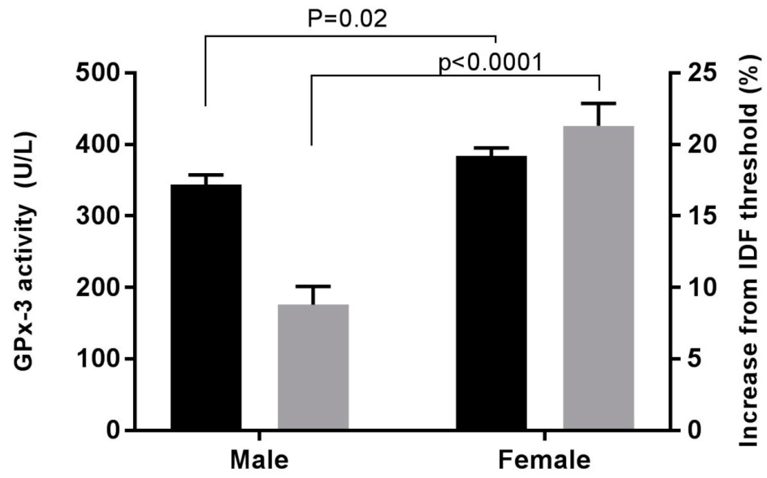
Increase in waist circumference from IDF recommended threshold (%)8.9 ± 11.721.3 ± 14.1<0.0001
Data expressed as mean ± SD or median [inter-quartile range]. eGFR, estimated glomerular filtration rate; HDL, high density lipoprotein; LDL, low density lipoprotein; ACR, albumin creatinine ratio. p ≤ 0.05 was considered significant.
Table 2. Plasma GPx-3 activity, vitamin E, and selenium levels in men and women type 2 diabetes patients at high risk of developing progressive cardio-renal disease.
Plasma AntioxidantsMaleFemalep
Vitamin E:Total-Cholesterol (µmol/mmol)8.95 ± 3.038.79 ± 2.540.72
Selenium (µmol/L)1.25 ± 0.281.26 ± 0.240.73
GPx-3 activity (U/L)343.3 ± 128.0384.1 ± 99.70.02
Data expressed as mean ± SD, p ≤ 0.05 was considered significant.
Table 3. Linear regression with GPx-3 activity as the dependent variable.
Variablesβtp
Model ASex41.02.280.02
Waist circumference−1.48−2.130.04
Model BSex58.02.530.01
Bioimpedance−1.48−1.260.21
Standardized beta (β), the t test statistic (t), and the probability value (p).

