Oxidized-Lipid Signaling and Ferroptosis as Downstream Mechanisms of Titanium-Associated Peri-Implant Bone Loss

 ,
,  , ,
, ,  ,
,  and
and
Abstract
1. Introduction
2. Materials and Methods
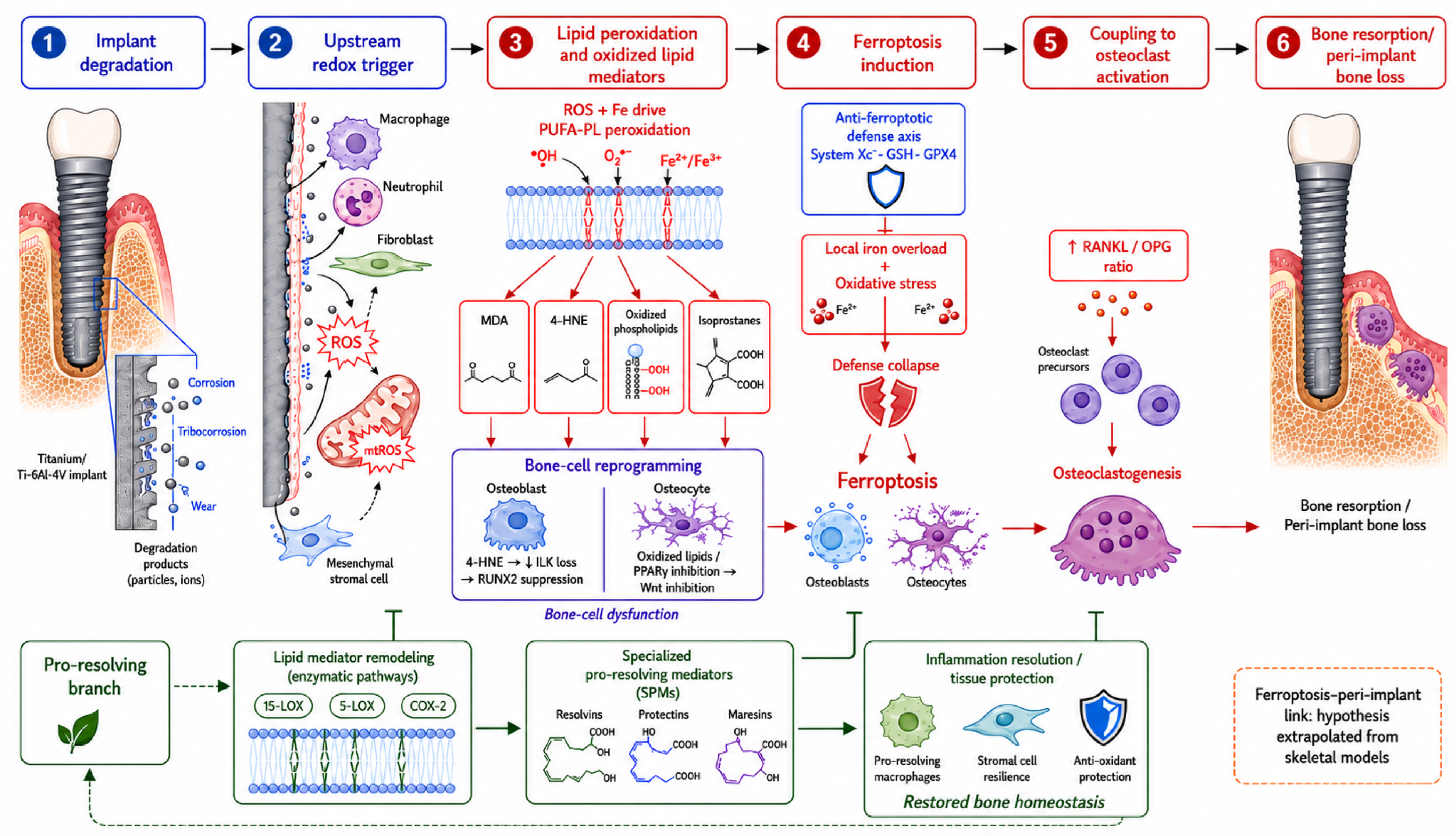
3. Titanium Degradation and the Redox Origin of Lipid Injury
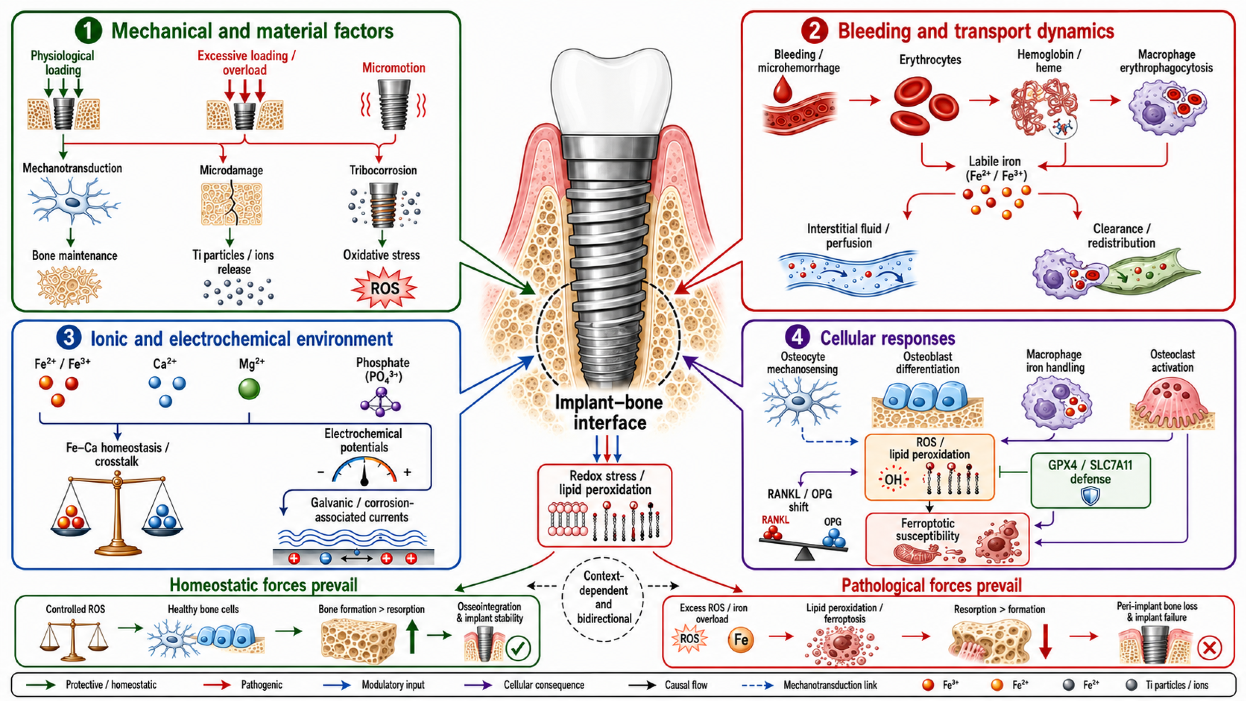
3.1. The Redox-Active Peri-Implant Interface
3.2. Mitochondria as an Important Source of Peroxidizing Radicals
3.3. Lipid Peroxidation: From Reactive Oxygen Species to Reactive Lipids
3.4. Oxidized Lipids as Signaling Molecules That Reprogram Bone Cells
3.5. Membrane Remodeling Determines Susceptibility to Ferroptotic Lipid Damage
3.6. The Dual Biology of Oxidized Lipids
4. Ferroptosis: An Iron- and Lipid-Dependent Mode of Bone-Cell Death at the Peri-Implant Interface
4.1. Definition and Mechanistic Distinctiveness
4.2. The System Xc−–GSH–GPX4 Axis and Its Collapse Under Oxidative Load
4.3. Iron Availability at the Peri-Implant Interface
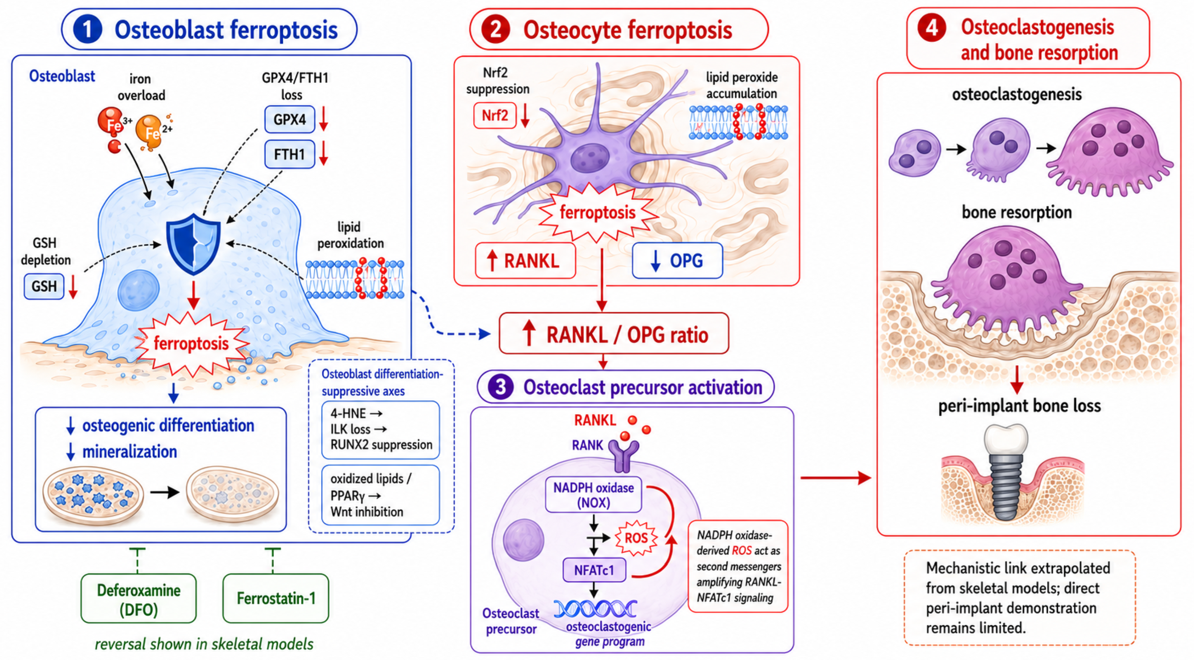
4.4. Ferroptosis of Bone Cells and the Link to the RANKL/OPG Axis
4.5. Convergence with Inflammasome and Macrophage Signaling
4.6. Osteoclastogenesis as the Endpoint
5. Microbial Contributions to the Lipid–Ferroptosis Substrate
6. Systemic and Local Correlates of the Lipid–Ferroptosis Axis
6.1. Circulating and Local Biomarkers of Lipid and Nitrosative Oxidation
6.2. The Dose Problem and Its Interpretation
6.3. Hypersensitivity Versus Redox-Driven Intolerance
6.4. Orthopedic Wear-Debris Osteolysis as a Mechanistic Model
6.5. Host Susceptibility: Iron Status and Antioxidant Reserve
6.6. A Balanced Interpretation
7. Methodological Limitations and the Evidentiary Gap
7.1. Absence of Direct Peri-Implant Ferroptosis Data
7.2. Limitations of Experimental Models
7.3. Causality and Confounding
7.4. Underrepresentation of Lipidomic and Systems-Level Approaches
7.5. Toward a Systems Framework
8. Future Directions
8.1. Redox Lipidomics and Pro-Resolving Mediators
8.2. Testing Ferroptosis Directly
8.3. Iron- and Lipid-Targeted Protective Strategies
8.4. Host-Stratified Implantology
8.5. Redox-Responsive Biomaterials
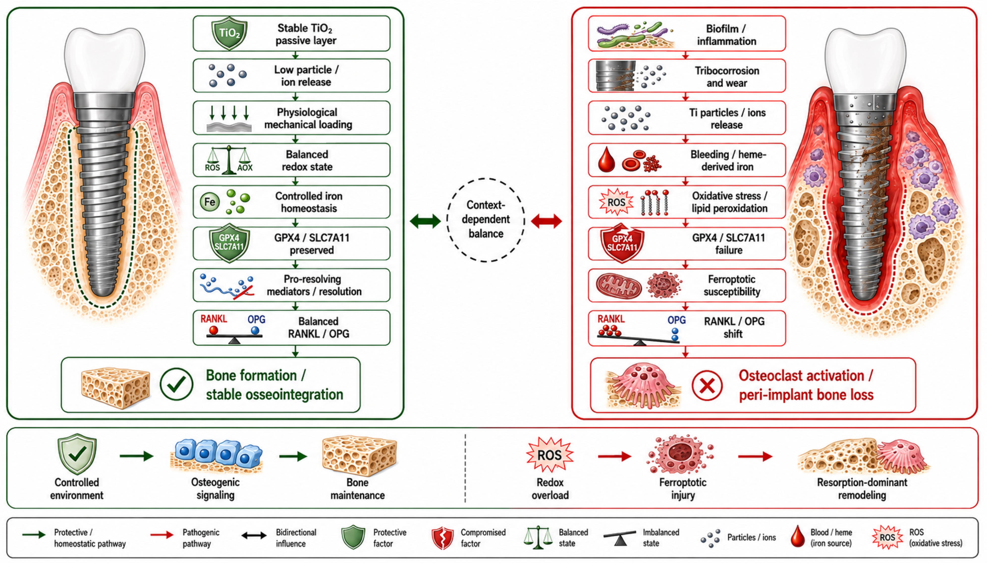
9. Discussion


10. Conclusions
Author Contributions
Funding
Institutional Review Board Statement
Informed Consent Statement
Data Availability Statement
Acknowledgments
Conflicts of Interest
References
- Mierzejewska, Ż.A.; Woźniak, Ł.; Lechien, J.R.; Borys, J.; Łukaszuk, K.; Antonowicz, B. Beyond biocompatibility: Immune dysregulation, oxidative stress, and tissue intolerance associated with Ti-6Al-4V dental implants—A critical review and perspective. Antioxidants 2026, 15, 365. [Google Scholar] [CrossRef] [Scilit] [PubMed]
- Suárez-López del Amo, F.; Garaicoa-Pazmiño, C.; Fretwurst, T.; Castilho, R.M.; Squarize, C.H. Dental implants-associated release of titanium particles: A systematic review. Clin. Oral Implant. Res. 2018, 29, 1085–1100. [Google Scholar] [CrossRef] [Scilit] [PubMed]
- Delgado-Ruiz, R.; Romanos, G. Potential causes of titanium particle and ion release in implant dentistry: A systematic review. Int. J. Mol. Sci. 2018, 19, 3585. [Google Scholar] [CrossRef] [Scilit] [PubMed]
- Mombelli, A.; Hashim, D.; Cionca, N. What is the impact of titanium particles and biocorrosion on implant survival and complications? A critical review. Clin. Oral Implant. Res. 2018, 29, 37–53. [Google Scholar] [CrossRef] [Scilit] [PubMed]
- Manke, A.; Wang, L.; Rojanasakul, Y. Mechanisms of nanoparticle-induced oxidative stress and toxicity. BioMed Res. Int. 2013, 2013, 942916. [Google Scholar] [CrossRef] [Scilit] [PubMed]
- Kotsakis, G.A.; Ganesan, S.M. Microbial dysbiosis, titanium release, and peri-implantitis. J. Dent. Res. 2025, 104, 473–480. [Google Scholar] [CrossRef] [Scilit] [PubMed]
- Fretwurst, T.; Buzanich, G.; Nahles, S.; Woelber, J.P.; Riesemeier, H.; Nelson, K. Metal elements in tissue with dental peri-implantitis: A pilot study. Clin. Oral Implant. Res. 2016, 27, 1178–1186. [Google Scholar] [CrossRef] [Scilit] [PubMed]
- Wilson, T.G., Jr.; Valderrama, P.; Burbano, M.; Blansett, J.; Levine, R.; Kessler, H.; Rodrigues, D.C. Foreign bodies associated with peri-implantitis human biopsies. J. Periodontol. 2015, 86, 9–15. [Google Scholar] [CrossRef] [Scilit] [PubMed]
- Pettersson, M.; Pettersson, J.; Johansson, A. Titanium release in peri-implantitis. J. Oral Rehabil. 2019, 46, 179–188. [Google Scholar] [CrossRef] [Scilit] [PubMed]
- Noronha Oliveira, M.; Schunemann, W.V.H.; Mathew, M.T.; Henriques, B.; Magini, R.S.; Teughels, W.; Souza, J.C.M. Can degradation products released from dental implants affect peri-implant tissues? J. Periodontal Res. 2018, 53, 1–11. [Google Scholar] [CrossRef] [Scilit] [PubMed]
- Wilson, T.G., Jr. Bone loss around implants—Is it metallosis? J. Periodontol. 2021, 92, 181–185. [Google Scholar] [CrossRef] [Scilit] [PubMed]
- Ji, C.; Chen, Y.; Si, M.; Chen, X. The impact of biocorrosion and titanium ions release on peri-implantitis. Clin. Oral Investig. 2025, 29, 155. [Google Scholar] [CrossRef] [Scilit] [PubMed]
- Ayala, A.; Muñoz, M.F.; Argüelles, S. Lipid peroxidation: Production, metabolism, and signaling mechanisms of malondialdehyde and 4-hydroxy-2-nonenal. Oxid. Med. Cell. Longev. 2014, 2014, 360438. [Google Scholar] [CrossRef] [Scilit] [PubMed]
- Sonowal, H.; Ramana, K.V. 4-Hydroxy-trans-2-nonenal in the regulation of anti-oxidative and pro-inflammatory signaling pathways. Oxid. Med. Cell. Longev. 2019, 2019, 5937326. [Google Scholar] [CrossRef] [Scilit] [PubMed]
- Dixon, S.J.; Lemberg, K.M.; Lamprecht, M.R.; Skouta, R.; Zaitsev, E.M.; Gleason, C.E.; Patel, D.N.; Bauer, A.J.; Cantley, A.M.; Yang, W.S.; et al. Ferroptosis: An iron-dependent form of nonapoptotic cell death. Cell 2012, 149, 1060–1072. [Google Scholar] [CrossRef] [Scilit] [PubMed]
- Yang, D.; Gong, G.; Song, J.; Chen, J.; Wang, S.; Li, J.; Wang, G. Ferroptosis-mediated osteoclast–osteoblast crosstalk: Signaling pathways governing bone remodeling in osteoporosis. J. Orthop. Surg. Res. 2025, 20, 888. [Google Scholar] [CrossRef] [Scilit] [PubMed]
- Ruan, B.; Dong, J.; Wei, F.; Huang, Z.; Yang, B.; Zhang, L.; Li, C.; Dong, H.; Cao, W.; Wang, H.; et al. DNMT aberration-incurred GPX4 suppression prompts osteoblast ferroptosis and osteoporosis. Bone Res. 2024, 12, 68. [Google Scholar] [CrossRef] [Scilit] [PubMed]
- Jiang, Z.; Wang, H.; Qi, G.; Jiang, C.; Chen, K.; Yan, Z. Iron overload-induced ferroptosis of osteoblasts inhibits osteogenesis and promotes osteoporosis: An in vitro and in vivo study. IUBMB Life 2022, 74, 1052–1069. [Google Scholar] [CrossRef] [Scilit] [PubMed]
- Dong, J.; Ruan, B.; Zhang, L.; Wei, A.; Li, C.; Tang, N.; Zhu, L.; Jiang, Q.; Cao, W. DNA methylation-mediated GPX4 transcriptional repression and osteoblast ferroptosis promote titanium particle-induced osteolysis. Research 2024, 7, 0457. [Google Scholar] [CrossRef] [Scilit] [PubMed]
- Serhan, C.N. Pro-resolving lipid mediators are leads for resolution physiology. Nature 2014, 510, 92–101. [Google Scholar] [CrossRef] [Scilit] [PubMed]
- Natarajan, V.; Wilson, C.L.; Hayward, S.L.; Kidambi, S. Titanium dioxide nanoparticles trigger loss of function and perturbation of mitochondrial dynamics in primary hepatocytes. PLoS ONE 2015, 10, e0134541. [Google Scholar] [CrossRef] [Scilit] [PubMed]
- Huerta-García, E.; Pérez-Arizti, J.A.; Márquez-Ramírez, S.G.; Delgado-Buenrostro, N.L.; Chirino, Y.I.; Iglesias, G.G.; López-Marure, R. Titanium dioxide nanoparticles induce strong oxidative stress and mitochondrial damage in glial cells. Free Radic. Biol. Med. 2014, 73, 84–94. [Google Scholar] [CrossRef] [Scilit] [PubMed]
- Zalewska, A.; Antonowicz, B.; Szulimowska, J.; Zieniewska-Siemieńczuk, I.; Leśniewska, B.; Borys, J.; Maciejczyk, M. Mitochondrial redox balance of fibroblasts exposed to Ti-6Al-4V microplates subjected to different types of anodizing. Int. J. Mol. Sci. 2023, 24, 12896. [Google Scholar] [CrossRef] [Scilit] [PubMed]
- Shadel, G.S.; Horvath, T.L. Mitochondrial ROS signaling in organismal homeostasis. Cell 2015, 163, 560–569. [Google Scholar] [CrossRef] [Scilit] [PubMed]
- Murphy, M.P. How mitochondria produce reactive oxygen species. Biochem. J. 2009, 417, 1–13. [Google Scholar] [CrossRef] [Scilit] [PubMed]
- Paradies, G.; Paradies, V.; Ruggiero, F.M.; Petrosillo, G. Cardiolipin and mitochondrial function in health and disease. Antioxid. Redox Signal. 2014, 20, 1925–1953. [Google Scholar] [CrossRef] [Scilit] [PubMed]
- Yin, H.; Xu, L.; Porter, N.A. Free radical lipid peroxidation: Mechanisms and analysis. Chem. Rev. 2011, 111, 5944–5972. [Google Scholar] [CrossRef] [Scilit] [PubMed]
- Gaschler, M.M.; Stockwell, B.R. Lipid peroxidation in cell death. Biochem. Biophys. Res. Commun. 2017, 482, 419–425. [Google Scholar] [CrossRef] [Scilit] [PubMed]
- Conrad, M.; Pratt, D.A. The chemical basis of ferroptosis. Nat. Chem. Biol. 2019, 15, 1137–1147. [Google Scholar] [CrossRef] [Scilit] [PubMed]
- Kagan, V.E.; Mao, G.; Qu, F.; Angeli, J.P.F.; Doll, S.; Croix, C.S.; Dar, H.H.; Liu, B.; Tyurin, V.A.; Ritov, V.B.; et al. Oxidized arachidonic and adrenic PEs navigate cells to ferroptosis. Nat. Chem. Biol. 2017, 13, 81–90. [Google Scholar] [CrossRef] [Scilit] [PubMed]
- Doll, S.; Proneth, B.; Tyurina, Y.Y.; Panzilius, E.; Kobayashi, S.; Ingold, I.; Irmler, M.; Beckers, J.; Aichler, M.; Walch, A.; et al. ACSL4 dictates ferroptosis sensitivity by shaping cellular lipid composition. Nat. Chem. Biol. 2017, 13, 91–98. [Google Scholar] [CrossRef] [Scilit] [PubMed]
- Wenzel, S.E.; Tyurina, Y.Y.; Zhao, J.; St Croix, C.M.; Dar, H.H.; Mao, G.; Tyurin, V.A.; Anthonymuthu, T.S.; Kapralov, A.A.; Amoscato, A.A.; et al. PEBP1 wardens ferroptosis by enabling lipoxygenase generation of lipid death signals. Cell 2017, 171, 628–641.e26. [Google Scholar] [CrossRef] [Scilit] [PubMed]
- Milne, G.L.; Yin, H.; Hardy, K.D.; Davies, S.S.; Roberts, L.J., II. Isoprostane generation and function. Chem. Rev. 2011, 111, 5973–5996. [Google Scholar] [CrossRef] [Scilit] [PubMed]
- Esterbauer, H.; Schaur, R.J.; Zollner, H. Chemistry and biochemistry of 4-hydroxynonenal, malonaldehyde and related aldehydes. Free Radic. Biol. Med. 1991, 11, 81–128. [Google Scholar] [CrossRef] [Scilit] [PubMed]
- Yadav, U.C.S.; Ramana, K.V. Regulation of NF-κB-induced inflammatory signaling by lipid peroxidation-derived aldehydes. Oxid. Med. Cell. Longev. 2013, 2013, 690545. [Google Scholar] [CrossRef] [Scilit] [PubMed]
- Almeida, M.; Ambrogini, E.; Han, L.; Manolagas, S.C.; Jilka, R.L. Increased lipid oxidation causes oxidative stress, increased PPARγ expression, and diminished pro-osteogenic Wnt signaling in the skeleton. J. Biol. Chem. 2009, 284, 27438–27448. [Google Scholar] [CrossRef] [Scilit] [PubMed]
- Bochkov, V.N.; Oskolkova, O.V.; Birukov, K.G.; Levonen, A.L.; Binder, C.J.; Stöckl, J. Generation and biological activities of oxidized phospholipids. Antioxid. Redox Signal. 2010, 12, 1009–1059. [Google Scholar] [CrossRef] [Scilit] [PubMed]
- Antonowicz, B.; Borys, J.; Zalewska, A.; Żendzian-Piotrowska, M.; Łukaszuk, K.; Woźniak, Ł.; Maciejczyk, M. Circulating biomarkers of nitrosative stress, protein glycoxidation and inflammation in maxillofacial surgery patients treated with titanium implants. Dent. Med. Probl. 2025, 62, 225–236. [Google Scholar] [CrossRef] [Scilit] [PubMed]
- Popa, P.Ș.; Popa, G.V.; Earar, K.; Popa-Cazacu, C.E.; Matei, M.N. Salivary oxidative stress biomarkers in peri-implant disease: A systematic review and meta-analysis. Int. J. Mol. Sci. 2025, 26, 11269. [Google Scholar] [CrossRef] [Scilit] [PubMed]
- Zhang, Q.-Y.; Gong, H.-B.; Jiang, M.-Y.; Jin, F.; Wang, G.; Yan, C.-Y.; Luo, X.; Sun, W.-Y.; Ouyang, S.-H.; Wu, Y.-P.; et al. Regulation of enzymatic lipid peroxidation in osteoblasts protects against postmenopausal osteoporosis. Nat. Commun. 2025, 16, 758. [Google Scholar] [CrossRef] [Scilit] [PubMed]
- Aschner, M.; Skalny, A.V.; Martins, A.C.; Tizabi, Y.; Zaitseva, I.P.; Santamaria, A.; Lu, R.; Gluhcheva, Y.Y.; Tinkov, A.A. The role of NLRP3 inflammasome activation in proinflammatory and cytotoxic effects of metal nanoparticles. Arch. Toxicol. 2025, 99, 1287–1314. [Google Scholar] [CrossRef] [Scilit] [PubMed]
- Milne, G.L.; Dai, Q.; Roberts, L.J., II. The isoprostanes—25 years later. Biochim. Biophys. Acta Mol. Cell Biol. Lipids 2015, 1851, 433–445. [Google Scholar] [CrossRef] [Scilit] [PubMed]
- Serhan, C.N.; Levy, B.D. Resolvins in inflammation: Emergence of the pro-resolving superfamily of mediators. J. Clin. Investig. 2018, 128, 2657–2669. [Google Scholar] [CrossRef] [Scilit] [PubMed]
- Chiang, N.; Serhan, C.N. Specialized pro-resolving mediator network: An update on production and actions. Essays Biochem. 2020, 64, 443–462. [Google Scholar] [CrossRef] [Scilit] [PubMed]
- Dalli, J.; Serhan, C.N. Identification and structure elucidation of the pro-resolving mediators provides novel leads for resolution pharmacology. Br. J. Pharmacol. 2019, 176, 1024–1037. [Google Scholar] [CrossRef] [Scilit] [PubMed]
- Freire, M.O.; Van Dyke, T.E. Natural resolution of inflammation. Periodontology 2000 2013, 63, 149–164. [Google Scholar] [CrossRef] [Scilit] [PubMed]
- Galluzzi, L.; Kepp, O.; Chan, F.K.M.; Kroemer, G. Necroptosis: Mechanisms and relevance to disease. Annu. Rev. Pathol. 2017, 12, 103–130. [Google Scholar] [CrossRef] [Scilit] [PubMed]
- Tang, D.; Chen, X.; Kang, R.; Kroemer, G. Ferroptosis: Molecular mechanisms and health implications. Cell Res. 2021, 31, 107–125. [Google Scholar] [CrossRef] [Scilit] [PubMed]
- Stockwell, B.R.; Friedmann Angeli, J.P.; Bayir, H.; Bush, A.I.; Conrad, M.; Dixon, S.J.; Fulda, S.; Gascón, S.; Hatzios, S.K.; Kagan, V.E.; et al. Ferroptosis: A regulated cell death nexus linking metabolism, redox biology, and disease. Cell 2017, 171, 273–285. [Google Scholar] [CrossRef] [Scilit] [PubMed]
- Dixon, S.J.; Olzmann, J.A. The cell biology of ferroptosis. Nat. Rev. Mol. Cell Biol. 2024, 25, 424–442. [Google Scholar] [CrossRef] [Scilit] [PubMed]
- Antonowicz, B.; Mierzejewska, Ż.A.; Borys, J.; Maciejczyk, M.; Prokopiuk, S.; Car, H. Evaluation of titanium particles, TNF-α, and caspase-3 concentrations in patients with bone fixations of the maxilla and mandible. Int. J. Mol. Sci. 2025, 26, 2316. [Google Scholar] [CrossRef] [Scilit] [PubMed]
- Li, J.; Cao, F.; Yin, H.L.; Huang, Z.J.; Lin, Z.T.; Mao, N.; Sun, B.; Wang, G. Ferroptosis: Past, present and future. Cell Death Dis. 2020, 11, 88. [Google Scholar] [CrossRef] [Scilit] [PubMed]
- Galluzzi, L.; Vitale, I.; Aaronson, S.A.; Abrams, J.M.; Adam, D.; Agostinis, P.; Alnemri, E.S.; Altucci, L.; Amelio, I.; Andrews, D.W.; et al. Molecular mechanisms of cell death: Recommendations of the Nomenclature Committee on Cell Death 2018. Cell Death Differ. 2018, 25, 486–541. [Google Scholar] [CrossRef] [Scilit] [PubMed]
- Broz, P.; Pelegrín, P.; Shao, F. The gasdermins, a protein family executing cell death and inflammation. Nat. Rev. Immunol. 2020, 20, 143–157. [Google Scholar] [CrossRef] [Scilit] [PubMed]
- Yang, W.S.; SriRamaratnam, R.; Welsch, M.E.; Shimada, K.; Skouta, R.; Viswanathan, V.S.; Cheah, J.H.; Clemons, P.A.; Shamji, A.F.; Clish, C.B.; et al. Regulation of ferroptotic cancer cell death by GPX4. Cell 2014, 156, 317–331. [Google Scholar] [CrossRef] [Scilit] [PubMed]
- Friedmann Angeli, J.P.; Schneider, M.; Proneth, B.; Tyurina, Y.Y.; Tyurin, V.A.; Hammond, V.J.; Herbach, N.; Aichler, M.; Walch, A.; Eggenhofer, E.; et al. Inactivation of the ferroptosis regulator Gpx4 triggers acute renal failure in mice. Nat. Cell Biol. 2014, 16, 1180–1191. [Google Scholar] [CrossRef] [Scilit] [PubMed]
- Bersuker, K.; Hendricks, J.M.; Li, Z.; Magtanong, L.; Ford, B.; Tang, P.H.; Roberts, M.A.; Tong, B.; Maimone, T.J.; Zoncu, R.; et al. The CoQ oxidoreductase FSP1 acts parallel to GPX4 to inhibit ferroptosis. Nature 2019, 575, 688–692. [Google Scholar] [CrossRef] [Scilit] [PubMed]
- Doll, S.; Freitas, F.P.; Shah, R.; Aldrovandi, M.; da Silva, M.C.; Ingold, I.; Goya Grocin, A.; Xavier da Silva, T.N.; Panzilius, E.; Scheel, C.H.; et al. FSP1 is a glutathione-independent ferroptosis suppressor. Nature 2019, 575, 693–698. [Google Scholar] [CrossRef] [Scilit] [PubMed]
- Mao, C.; Liu, X.; Zhang, Y.; Lei, G.; Yan, Y.; Lee, H.; Koppula, P.; Wu, S.; Zhuang, L.; Fang, B.; et al. DHODH-mediated ferroptosis defence is a targetable vulnerability in cancer. Nature 2021, 593, 586–590. [Google Scholar] [CrossRef] [Scilit] [PubMed]
- Kraft, V.A.N.; Bezjian, C.T.; Pfeiffer, S.; Ringelstetter, L.; Müller, C.; Zandkarimi, F.; Merl-Pham, J.; Bao, X.; Anastasov, N.; Kössl, J.; et al. GTP cyclohydrolase 1/tetrahydrobiopterin counteract ferroptosis through lipid remodeling. ACS Cent. Sci. 2020, 6, 41–53. [Google Scholar] [CrossRef] [Scilit] [PubMed]
- Soula, M.; Weber, R.A.; Zilka, O.; Alwaseem, H.; La, K.; Yen, F.; Molina, H.; Garcia-Bermudez, J.; Pratt, D.A.; Birsoy, K. Metabolic determinants of cancer cell sensitivity to canonical ferroptosis inducers. Nat. Chem. Biol. 2020, 16, 1351–1360. [Google Scholar] [CrossRef] [Scilit] [PubMed]
- Koleini, N.; Shapiro, J.S.; Geier, J.; Ardehali, H. Ironing out mechanisms of iron homeostasis and disorders of iron deficiency. J. Clin. Investig. 2021, 131, e148671. [Google Scholar] [CrossRef] [Scilit] [PubMed]
- Puylaert, P.; Roth, L.; Van Praet, M.; Pintelon, I.; Dumitrascu, C.; van Nuijs, A.; Klejborowska, G.; Guns, P.-J.; Berghe, T.V.; Augustyns, K.; et al. Effect of erythrophagocytosis-induced ferroptosis during angiogenesis in atherosclerotic plaques. Angiogenesis 2023, 26, 505–522. [Google Scholar] [CrossRef] [Scilit] [PubMed]
- Yang, H.; Ru, K.; Liu, S.; Zhu, C.; Qian, A.; Chen, Z. Targeting ferroptosis: Emerging insights into osteoporosis mechanisms. Biology 2025, 14, 1062. [Google Scholar] [CrossRef] [Scilit] [PubMed]
- Ru, Q.; Li, Y.; Xie, W.; Ding, Y.; Chen, L.; Xu, G.; Wu, Y.; Wang, F. Fighting age-related orthopedic diseases: Focusing on ferroptosis. Bone Res. 2023, 11, 12. [Google Scholar] [CrossRef] [Scilit] [PubMed]
- Das, B.K.; Wang, L.; Fujiwara, T.; Zhou, J.; Aykin-Burns, N.; Krager, K.J.; Lan, R.; Mackintosh, S.G.; Edmondson, R.; Jennings, M.L.; et al. Transferrin receptor 1-mediated iron uptake regulates bone mass in mice via osteoclast mitochondria and cytoskeleton. eLife 2022, 11, e73539. [Google Scholar] [CrossRef] [Scilit] [PubMed]
- Chen, H.; Zhang, Y.; Zhu, Y.; Xiao, X.; Huang, S.; Duan, X. Spectrum and functions of ion channels and transporters in osteoclasts. Bone Res. 2026, 14, 35. [Google Scholar] [CrossRef] [Scilit] [PubMed]
- Liu, K.; Xu, J.; Yang, R.; Wang, F.; Su, Y. Ion channel Piezo1 induces ferroptosis of trabecular meshwork cells: A novel observation in the pathogenesis in primary open angle glaucoma. Am. J. Physiol. Cell Physiol. 2024, 327, C1591–C1603. [Google Scholar] [CrossRef] [Scilit] [PubMed]
- Wan, S.; He, Y.; Chong, B.; Xia, Z.; Qiu, Y.; Zhang, R.; Hu, M.; An, Z.; Li, Y.; Xie, J.; et al. Osteoblast ferroptosis driven by iron overload: Implications for osteoporosis pathogenesis and FTH1/GPX4-targeted therapy. Biochem. Biophys. Res. Commun. 2025, 779, 152439. [Google Scholar] [CrossRef] [Scilit] [PubMed]
- Jiang, Z.; Qi, G.; He, X.; Yu, Y.; Cao, Y.; Zhang, C.; Zou, W.; Yuan, H. Ferroptosis in osteocytes as a target for protection against postmenopausal osteoporosis. Adv. Sci. 2024, 11, e2307388. [Google Scholar] [CrossRef] [Scilit] [PubMed]
- Chankamngoen, W.; Krungchanuchat, S.; Thongbunchoo, J.; Sirinonthanawech, N.; Teerapornpuntakit, J.; Panupinthu, N.; Charoenphandhu, N. Extracellular Fe2+ and Fe3+ modulate osteocytic viability, expression of SOST, RANKL and FGF23, and fluid flow-induced YAP1 nuclear translocation. Sci. Rep. 2023, 13, 20550. [Google Scholar] [CrossRef] [Scilit] [PubMed]
- Swanson, K.V.; Deng, M.; Ting, J.P.Y. The NLRP3 inflammasome: Molecular activation and regulation to therapeutics. Nat. Rev. Immunol. 2019, 19, 477–489. [Google Scholar] [CrossRef] [Scilit] [PubMed]
- Wen, Q.; Liu, J.; Kang, R.; Zhou, B.; Tang, D. The release and activity of HMGB1 in ferroptosis. Biochem. Biophys. Res. Commun. 2019, 510, 278–283. [Google Scholar] [CrossRef] [Scilit] [PubMed]
- Proneth, B.; Conrad, M. Ferroptosis and necroinflammation, a yet poorly explored link. Cell Death Differ. 2019, 26, 14–24. [Google Scholar] [CrossRef] [Scilit] [PubMed]
- Lee, N.K.; Choi, Y.G.; Baik, J.Y.; Han, S.Y.; Jeong, D.W.; Bae, Y.S.; Kim, N.; Lee, S.Y. A crucial role for reactive oxygen species in RANKL-induced osteoclast differentiation. Blood 2005, 106, 852–859. [Google Scholar] [CrossRef] [Scilit] [PubMed]
- Kang, I.S.; Kim, C. NADPH oxidase gp91phox contributes to RANKL-induced osteoclast differentiation by upregulating NFATc1. Sci. Rep. 2016, 6, 38014. [Google Scholar] [CrossRef] [Scilit] [PubMed]
- Murusuri, A.; Tang, D. The role of ferroptosis in osteoporosis: A cellular perspective on osteoblast, osteoclast and osteocyte dysfunction. Front. Endocrinol. 2026, 17, 1742656. [Google Scholar] [CrossRef] [Scilit] [PubMed]
- Ma, H.; Wang, X.; Zhang, W.; Li, H.; Zhao, W.; Sun, J.; Yang, M. Melatonin suppresses ferroptosis induced by high glucose via activation of the Nrf2/HO-1 signaling pathway in type 2 diabetic osteoporosis. Oxid. Med. Cell. Longev. 2020, 2020, 9067610. [Google Scholar] [CrossRef] [Scilit] [PubMed]
- Daubert, D.; Pozhitkov, A.; McLean, J.; Kotsakis, G. Titanium as a modifier of the peri-implant microbiome structure. Clin. Implant Dent. Relat. Res. 2018, 20, 945–953. [Google Scholar] [CrossRef] [Scilit] [PubMed]
- Kotsakis, G.A.; Olmedo, D.G. Peri-implantitis is not periodontitis: Scientific discoveries shed light on microbiome-biomaterial interactions that may determine disease phenotype. Periodontology 2000 2021, 86, 231–240. [Google Scholar] [CrossRef] [Scilit] [PubMed]
- Souza, J.C.M.; Mota, R.R.C.; Sordi, M.B.; Passoni, B.B.; Benfatti, C.A.M.; Magini, R.S. Biofilm formation on different materials used in oral rehabilitation. Braz. Dent. J. 2016, 27, 141–147. [Google Scholar] [CrossRef] [Scilit] [PubMed]
- Yılmaz, B.; Gürkan, A.; Afacan, B.; Atmaca, H.; Köse, T.; Emingil, G. Peri-implant hypoxia as a potential barrier against ferroptotic mechanisms during peri-implant diseases: A cross-sectional study. J. Periodontol. 2026, 97, 883–892. [Google Scholar] [CrossRef] [Scilit] [PubMed]
- Kheder, W.; Soumya, S.; Samsudin, A.R. Impact of titanium dioxide particle size on macrophage production of intracellular reactive oxygen species. Arch. Oral Biol. 2021, 125, 105077. [Google Scholar] [CrossRef] [Scilit] [PubMed]
- Swiatkowska, I.; Martin, N.; Hart, A.J. Blood titanium level as a biomarker of orthopaedic implant wear. J. Trace Elem. Med. Biol. 2019, 53, 120–128. [Google Scholar] [CrossRef] [Scilit] [PubMed]
- Valentine-Thon, E.; Schiwara, H.W. Validity of MELISA for metal sensitivity testing. Neuro Endocrinol. Lett. 2003, 24, 57–64. [Google Scholar] [PubMed]
- Chen, A.; Kurmis, A.P. Understanding immune-mediated titanium allergy to in situ orthopaedic implants: A narrative review. ANZ J. Surg. 2024, 94, 1478–1487. [Google Scholar] [CrossRef] [Scilit] [PubMed]
- St Pierre, C.A.; Chan, M.; Iwakura, Y.; Ayers, D.C.; Kurt-Jones, E.A.; Finberg, R.W. Periprosthetic osteolysis: Characterizing the innate immune response to titanium wear particles. J. Orthop. Res. 2010, 28, 1418–1424. [Google Scholar] [CrossRef] [Scilit] [PubMed]
- Boyce, B.F.; Xing, L. Functions of RANKL/RANK/OPG in bone modeling and remodeling. Arch. Biochem. Biophys. 2008, 473, 139–146. [Google Scholar] [CrossRef] [Scilit] [PubMed]
- Duyck, J.; Vandamme, K. The effect of loading on peri-implant bone: A critical review of the literature. J. Oral Rehabil. 2014, 41, 783–794. [Google Scholar] [CrossRef] [Scilit] [PubMed]
- Emam, S.M.; Moussa, N. Signaling pathways of dental implants’ osseointegration: A narrative review on two of the most relevant; NF-κB and Wnt pathways. BDJ Open 2024, 10, 29. [Google Scholar] [CrossRef] [Scilit] [PubMed]
- Labadi, C.; Gauthier, R.; Trunfio-Sfarghiu, A.-M.; Abouelleil, H.; Lafon, A.; Attik, N. Toward clinically relevant in vitro models for dental implant integration: Methodological insights and translational perspectives. Dent. Mater. 2026; in press. [CrossRef] [Scilit] [PubMed]
- Hariprasad, A.; Menon, S.S.; Kurumathur Vasudevan, A.; Nair, P.R.; Varma, R.B.; Xavier, A.M. Bio-tribocorrosion of titanium dental implants in the oral environment: A narrative review. Cureus 2026, 18, e103188. [Google Scholar] [CrossRef] [Scilit] [PubMed]
- Rovai, E.S.; Polassi, M.; Iunes da Silveira, M.; Araújo, S.L.; Van Dyke, T.E.; Castro dos Santos, N.C. Impact of specialized pro-resolving lipid mediators on craniofacial and alveolar bone regeneration: A scoping review. Braz. Dent. J. 2024, 35, e246133. [Google Scholar] [CrossRef] [Scilit] [PubMed]
- Zhang, Z.; Ji, C.; Wang, Y.N.; Liu, S.; Wang, M.; Xu, X.; Zhang, D. Maresin1 suppresses high-glucose-induced ferroptosis in osteoblasts via NRF2 activation in type 2 diabetic osteoporosis. Cells 2022, 11, 2560. [Google Scholar] [CrossRef] [Scilit] [PubMed]
- Wang, M.; Ji, J.; Zhang, X.; Zhang, J.; Guo, J. GPX4-mediated bone ferroptosis under mechanical stress decreased bone formation via the YAP-TEAD signalling pathway. J. Cell. Mol. Med. 2024, 28, e18231. [Google Scholar] [CrossRef] [Scilit] [PubMed]
- Gittens, R.A.; Olivares-Navarrete, R.; Tannenbaum, R.; Boyan, B.D.; Schwartz, Z. Electrical implications of corrosion for osseointegration of titanium implants. J. Dent. Res. 2011, 90, 1389–1397. [Google Scholar] [CrossRef] [Scilit] [PubMed]
- Amine, M.; Merdad, K.; Sabbah, W.; Al-Manei, K.; Aldegheishem, A. Electrogalvanism in oral implantology: A systematic review. Int. J. Dent. 2022, 2022, 4575416. [Google Scholar] [CrossRef] [Scilit] [PubMed]
- Rolić, T.; Yazdani, M.; Mandić, S.; Distante, S. Iron metabolism, calcium, magnesium and trace elements: A review. Biol. Trace Elem. Res. 2025, 203, 2216–2225. [Google Scholar] [CrossRef] [Scilit] [PubMed]
- Pettersen, E.; Anderson, J.; Ortiz-Catalan, M. Electrical stimulation to promote osseointegration of bone anchoring implants: A topical review. J. Neuroeng. Rehabil. 2022, 19, 31. [Google Scholar] [CrossRef] [Scilit] [PubMed]
- Wang, X.; Hu, H.; Xiong, N.; Gu, S.; Liu, J. Electrical stimulation as an emerging strategy in bone repair: Mechanisms, applications, and advances. J. Orthop. Transl. 2026, 59, 101177. [Google Scholar] [CrossRef] [Scilit] [PubMed]



| Lipid Species | Origin | Biological Action in Bone/Inflammation | Ref. |
|---|---|---|---|
| Malondialdehyde (MDA) | Terminal aldehyde of PUFA peroxidation | Protein/DNA adducts; established biomarker in peri-implant fluids | [13,39] |
| 4-Hydroxynonenal (4-HNE) | Reactive α,β-unsaturated aldehyde; Michael adducts | Degrades ILK → disrupts RUNX2; NF-κB amplification; impairs osteoblasts | [14,40] |
| Oxidized phospholipids | Enzymatic/non-enzymatic PL oxidation | PRR/scavenger-receptor ligands; NLRP3 priming | [37,41] |
| Isoprostanes | Non-enzymatic arachidonate oxidation | Stable in vivo oxidative-stress markers; vasoactive | [42] |
| Specialized pro-resolving mediators | Enzymatic oxygenation (lipoxins, resolvins, protectins) | Pro-resolving; counterbalance injury (candidate protective branch) | [20] |
| Ferroptotic lipid peroxides | Iron-dependent PUFA-PL peroxidation | Execute ferroptosis; drive osteoblast/osteocyte death | [15,16] |
| Mode | Core Mechanism | Morphology | Immune Character | Ref. |
|---|---|---|---|---|
| Ferroptosis | Iron-dependent lipid-peroxide accumulation; system Xc−–GPX4 collapse | Mitochondrial shrinkage, dense membranes, loss of cristae; intact nucleus | Context-dependent immunogenicity; may release DAMPs and oxidized lipids | [15,52] |
| Apoptosis | Caspase-3/8/9 cascade; intrinsic/extrinsic | Cell shrinkage, blebbing, apoptotic bodies, chromatin condensation | Usually immunologically silent when clearance is efficient; context dependent | [53] |
| Necroptosis | RIPK1/RIPK3/MLKL | Organelle swelling, membrane permeabilization | Pro-inflammatory | [53] |
| Pyroptosis | Inflammasome–caspase-1–gasdermin-D pores | Swelling, membrane rupture, IL-1β/IL-18 release | Strongly pro-inflammatory | [53,54] |
| Node/Molecule | Function in the Axis | Consequence for Bone | Ref. |
|---|---|---|---|
| PUFA/membrane lipids | Substrate for peroxidation | Source of MDA, 4-HNE, oxidized phospholipids | [13] |
| 4-HNE | Reactive aldehyde; ILK binding, PPARγ ligand | Degrades ILK → disrupts RUNX2; suppresses Wnt → ↓ osteogenesis | [36,40] |
| Oxidized phospholipids | PRR/scavenger-receptor ligands; NLRP3 priming | Amplify sterile inflammation and inflammasome activation | [37,41] |
| Labile iron/TFR1 | Fenton catalysis; osteoclast iron demand | Initiates peroxidation; permissive for osteoclastogenesis | [64,65] |
| System Xc−–GSH–GPX4 | Central anti-ferroptotic defense | Collapse → lethal lipid-peroxide accumulation | [16,17] |
| Nrf2/HO-1 | Sustains GPX4, SLC7A11 | Failure precipitates osteoblast ferroptosis | [77,78] |
| Osteoblast ferroptosis | Iron-overload GPX4/FTH1 loss | ↓ Mineralization; reversible by DFO/ferrostatin-1 | [18,69] |
| Osteocyte ferroptosis | Nrf2 suppression → RANKL up | ↑ RANKL/OPG → excess osteoclast activation | [70,71] |
| Strategy | Mechanism on the Axis | Evidence (Mostly Skeletal/Extrapolated) | Ref. |
|---|---|---|---|
| Iron chelation (DFO, deferiprone) | Lowers labile iron; blocks Fenton initiation | Reverses osteoblast ferroptosis and bone loss (skeletal models) | [18,64] |
| Ferrostatin-1/liproxstatin | Lipophilic radical trapping; halts lipid-peroxide propagation | Restores osteoblast viability and mineralization in vitro/in vivo | [18,77] |
| GPX4/system Xc− support | Restores phospholipid-hydroperoxide clearance | Prevents ferroptotic osteoblast/osteocyte death | [17,77] |
| Nrf2/HO-1 activation | Transcriptional antioxidant induction sustaining GPX4 | Protects osteogenesis under oxidative/diabetic stress | [78] |
| Pro-resolving mediators (SPMs) | Shift lipid balance toward resolution | Candidate; unproven in peri-implant setting | [20] |
| Redox-responsive surfaces | Reduce particle/ion release and local ROS | Readout should include lipid-peroxidation endpoints | [1] |
| Dimension | Prior Work [1] | Present Review |
|---|---|---|
| Central question | Why the interface becomes redox-dysregulated (upstream trigger) | How the redox signal destroys bone (downstream effector) |
| Organizing thesis | Oxidative stress + antioxidant failure as unifying driver | Oxidized-lipid signaling and ferroptosis as effector arm |
| Alloy focus | Ti-6Al-4V; aluminium/vanadium redox activity | Titanium broadly; lipid/iron chemistry at the interface |
| Key mechanisms | ROS generation, NADPH oxidase, NLRP3, M1/M2, intolerance vs. allergy | Lipid peroxidation, 4-HNE–RUNX2, system Xc−–GPX4, iron, osteocyte ferroptosis–RANKL |
| Therapeutic frame | Redox-aware surfaces; antioxidant strategies | Iron chelation, ferrostatin, GPX4/Nrf2 support, pro-resolving mediators |
| Relationship | – | Builds on and explicitly cites the prior work; does not repeat it |
Disclaimer/Publisher’s Note: The statements, opinions and data contained in all publications are solely those of the individual author(s) and contributor(s) and not of MDPI and/or the editor(s). MDPI and/or the editor(s) disclaim responsibility for any injury to people or property resulting from any ideas, methods, instructions or products referred to in the content. |
© 2026 by the authors. Licensee MDPI, Basel, Switzerland. This article is an open access article distributed under the terms and conditions of the Creative Commons Attribution (CC BY) license.
Share and Cite
Woźniak, Ł.; Antonowicz, B.; Mierzejewska, Ż.A.; Kosicka, E.; Lechien, J.R.; Vaira, L.A.; Borys, J. Oxidized-Lipid Signaling and Ferroptosis as Downstream Mechanisms of Titanium-Associated Peri-Implant Bone Loss. Antioxidants 2026, 15, 1174. https://doi.org/10.3390/antiox15091174
Woźniak Ł, Antonowicz B, Mierzejewska ŻA, Kosicka E, Lechien JR, Vaira LA, Borys J. Oxidized-Lipid Signaling and Ferroptosis as Downstream Mechanisms of Titanium-Associated Peri-Implant Bone Loss. Antioxidants. 2026; 15(9):1174. https://doi.org/10.3390/antiox15091174
Chicago/Turabian StyleWoźniak, Łukasz, Bożena Antonowicz, Żaneta Anna Mierzejewska, Ewelina Kosicka, Jérôme R. Lechien, Luigi Angelo Vaira, and Jan Borys. 2026. "Oxidized-Lipid Signaling and Ferroptosis as Downstream Mechanisms of Titanium-Associated Peri-Implant Bone Loss" Antioxidants 15, no. 9: 1174. https://doi.org/10.3390/antiox15091174
APA StyleWoźniak, Ł., Antonowicz, B., Mierzejewska, Ż. A., Kosicka, E., Lechien, J. R., Vaira, L. A., & Borys, J. (2026). Oxidized-Lipid Signaling and Ferroptosis as Downstream Mechanisms of Titanium-Associated Peri-Implant Bone Loss. Antioxidants, 15(9), 1174. https://doi.org/10.3390/antiox15091174