T-2 Toxin Neurotoxicity: Molecular Mechanisms and Emerging Chemoprotective Strategies




Abstract
1. Introduction
2. T-2 Toxin’s Metabolism and the Accumulation in Brain Tissues
3. An Overview of T-2 Toxin Exposure-Induced Neurotoxic Effects
4. Roles of Neurotransmitters in T-2 Toxin-Induced Neurotoxicity
5. Molecular Mechanisms of T-2 Toxin-Induced Neurotoxicity
5.1. Role of Oxidative Stress
5.2. Role of Mitochondrial Dysfunction and Apoptosis in T-2 Toxin Neurotoxicity
5.3. Role of Inflammatory Responses and Cell Pyroptosis
5.4. Role of Autophagy
5.5. The Induction of Cell Cycle Arrest and Cellular Senescence
5.6. Imbalance of Gut Microbiota
6. Chemo-Protective Agents for T-2 Toxin-Induced Neurotoxicity
6.1. Antioxidants
6.2. Small-Molecule Inhibitors
6.3. Natural Products
6.4. Other Neuroprotective Agents
7. Conclusions and Future Perspectives
Author Contributions
Funding
Institutional Review Board Statement
Informed Consent Statement
Data Availability Statement
Conflicts of Interest
References
- Ayelign, A.; De Saeger, S. Mycotoxins in Ethiopia: Current status, implications to food safety and mitigation strategies. Food Control 2020, 113, 107163. [Google Scholar] [CrossRef]
- Mukhtar, K.; Nabi, B.G.; Ansar, S.; Bhat, Z.F.; Aadil, R.M.; Mousavi Khaneghah, A. Mycotoxins and consumers’ awareness: Recent progress and future challenges. Toxicon 2023, 232, 107227. [Google Scholar] [CrossRef]
- Dai, C.; Tian, E.; Hao, Z.; Tang, S.; Wang, Z.; Sharma, G.; Jiang, H.; Shen, J. Aflatoxin B1 Toxicity and Protective Effects of Curcumin: Molecular Mechanisms and Clinical Implications. Antioxidants 2022, 11, 2031. [Google Scholar] [CrossRef]
- Dai, C.; Sharma, G.; Liu, G.; Shen, J.; Shao, B.; Hao, Z. Therapeutic detoxification of quercetin for aflatoxin B1-related toxicity: Roles of oxidative stress, inflammation, and metabolic enzymes. Environ. Pollut. 2024, 345, 123474. [Google Scholar] [CrossRef] [PubMed]
- Dai, C.; Das Gupta, S.; Wang, Z.; Jiang, H.; Velkov, T.; Shen, J. T-2 toxin and its cardiotoxicity: New insights on the molecular mechanisms and therapeutic implications. Food Chem. Toxicol. 2022, 167, 113262. [Google Scholar] [CrossRef]
- Mhlongo, T.N.; Ogola, H.J.O.; Selvarajan, R.; Sibanda, T.; Kamika, I.; Tekere, M. Occurrence and diversity of waterborne fungi and associated mycotoxins in treated drinking water distribution system in South Africa: Implications on water quality and public health. Environ. Monit. Assess. 2020, 192, 519. [Google Scholar] [CrossRef]
- Eskola, M.; Kos, G.; Elliott, C.T.; Hajšlová, J.; Mayar, S.; Krska, R. Worldwide contamination of food-crops with mycotoxins: Validity of the widely cited ‘FAO estimate’ of 25. Crit. Rev. Food Sci. Nutr. 2020, 60, 2773–2789. [Google Scholar] [CrossRef] [PubMed]
- Xu, H.; Wang, L.; Sun, J.; Wang, L.; Guo, H.; Ye, Y.; Sun, X. Microbial detoxification of mycotoxins in food and feed. Crit. Rev. Food Sci. Nutr. 2022, 62, 4951–4969. [Google Scholar] [CrossRef]
- Hao, W.; Guan, S.; Li, A.; Wang, J.; An, G.; Hofstetter, U.; Schatzmayr, G. Mycotoxin Occurrence in Feeds and Raw Materials in China: A Five-Year Investigation. Toxins 2023, 15, 63. [Google Scholar] [CrossRef]
- Zain, M.E. Impact of mycotoxins on humans and animals. J. Saudi Chem. Soc. 2011, 15, 129–144. [Google Scholar] [CrossRef]
- Hussein, H.S.; Brasel, J.M. Toxicity, metabolism, and impact of mycotoxins on humans and animals. Toxicology 2001, 167, 101–134. [Google Scholar] [CrossRef]
- Zhang, X.; Wang, Y.; Velkov, T.; Tang, S.; Dai, C. T-2 toxin-induced toxicity in neuroblastoma-2a cells involves the generation of reactive oxygen, mitochondrial dysfunction and inhibition of Nrf2/HO-1 pathway. Food Chem. Toxicol. 2018, 114, 88–97. [Google Scholar] [CrossRef]
- Omotayo, O.P.; Omotayo, A.O.; Mwanza, M.; Babalola, O.O. Prevalence of Mycotoxins and Their Consequences on Human Health. Toxicol. Res. 2019, 35, 1–7. [Google Scholar] [CrossRef] [PubMed]
- Yang, C.; Ning, X.; Wang, B.; Tian, T.; Chen, Y.; Ma, L.; Wang, L. Association between spectrum of mycotoxins and semen quality: A cross-sectional study in Beijing, China. J. Hazard. Mater. 2024, 476, 135124. [Google Scholar] [CrossRef]
- Huybrechts, I.; Jacobs, I.; Biessy, C.; Aglago, E.K.; Jenab, M.; Claeys, L.; Zavadil, J.; Casagrande, C.; Nicolas, G.; Scelo, G.; et al. Associations between dietary mycotoxins exposures and risk of hepatocellular carcinoma in a European cohort. PLoS ONE 2024, 19, e0315561. [Google Scholar] [CrossRef]
- Li, D.; Han, J.; Guo, X.; Qu, C.; Yu, F.; Wu, X. The effects of T-2 toxin on the prevalence and development of Kashin-Beck disease in China: A meta-analysis and systematic review. Toxicol. Res. 2016, 5, 731–751. [Google Scholar] [CrossRef]
- Jolly, P.E.; Shuaib, F.M.; Jiang, Y.; Preko, P.; Baidoo, J.; Stiles, J.K.; Wang, J.S.; Phillips, T.D.; Williams, J.H. Association of high viral load and abnormal liver function with high aflatoxin B1-albumin adduct levels in HIV-positive Ghanaians: Preliminary observations. Food Addit. Contam. Part A Chem. Anal. Control Expo. Risk Assess. 2011, 28, 1224–1234. [Google Scholar] [CrossRef]
- Desjardins, A.E.; Hohn, T.M.; McCormick, S.P. Trichothecene biosynthesis in Fusarium species: Chemistry, genetics, and significance. Microbiol. Rev. 1993, 57, 595–604. [Google Scholar] [CrossRef] [PubMed]
- Lei, R.H.; Jiang, N.; Zhang, Q.; Hu, S.K.; Dennis, B.S.; He, S.S.; Guo, X. Prevalence of Selenium, T-2 Toxin, and Deoxynivalenol in Kashin-Beck Disease Areas in Qinghai Province, Northwest China. Biol. Trace Elem. Res. 2016, 171, 34–40. [Google Scholar] [CrossRef]
- Sun, L.Y.; Li, Q.; Meng, F.G.; Fu, Y.; Zhao, Z.J.; Wang, L.H. T-2 Toxin Contamination in Grains and Selenium Concentration in Drinking Water and Grains in Kaschin-Beck Disease Endemic Areas of Qinghai Province. Biol. Trace Elem. Res. 2012, 150, 371–375. [Google Scholar] [CrossRef] [PubMed]
- Wang, X.C.; Liu, X.D.; Liu, J.C.; Wang, G.; Wan, K.Y. Contamination Level of T-2 and HT-2 Toxin in Cereal Crops from Aba Area in Sichuan Province, China. Bull. Environ. Contam. Toxicol. 2012, 88, 396–400. [Google Scholar] [CrossRef]
- Tan, Y.F.; Kuang, Y.; Zhao, R.H.; Chen, B.; Wu, J.W. Determination of T-2 and HT-2 Toxins in Traditional Chinese Medicine Marketed in China by LC-ELSD after Sample Clean-Up by Two Solid-Phase Extractions. Chromatographia 2011, 73, 407–410. [Google Scholar] [CrossRef]
- Jiang, T.; Yan, J.; Tan, H.; Pu, Z.; Wang, O.; Liu, T.; Chen, Z.; Gao, J.; Wang, J.; Lin, J.; et al. Prevalence of T-2 Toxin in the Food and Beverages of Residents Living in a Kashin-Beck-Disease Area of Qamdo, Tibet. Nutrients 2024, 16, 1449. [Google Scholar] [CrossRef] [PubMed]
- Adhikari, M.; Negi, B.; Kaushik, N.; Adhikari, A.; Al-Khedhairy, A.A.; Kaushik, N.K.; Choi, E.H. T-2 mycotoxin: Toxicological effects and decontamination strategies. Oncotarget 2017, 8, 33933–33952. [Google Scholar] [CrossRef]
- Weaver, G.A.; Kurtz, H.J.; Bates, F.Y.; Chi, M.S.; Mirocha, C.J.; Behrens, J.C.; Robison, T.S. Acute and chronic toxicity of T-2 mycotoxin in swine. Vet. Rec. 1978, 103, 531–535. [Google Scholar] [CrossRef] [PubMed]
- Fairhurst, S.; Marrs, T.C.; Parker, H.C.; Scawin, J.W.; Swanston, D.W. Acute toxicity of T2 toxin in rats, mice, guinea pigs, and pigeons. Toxicology 1987, 43, 31–49. [Google Scholar] [CrossRef]
- Lin, R.; Sun, Y.; Ye, W.; Zheng, T.; Wen, J.; Deng, Y. T-2 toxin inhibits the production of mucin via activating the IRE1/XBP1 pathway. Toxicology 2019, 424, 152230. [Google Scholar] [CrossRef]
- Yang, L.; Yu, Z.; Hou, J.; Deng, Y.; Zhou, Z.; Zhao, Z.; Cui, J. Toxicity and oxidative stress induced by T-2 toxin and HT-2 toxin in broilers and broiler hepatocytes. Food Chem. Toxicol. 2016, 87, 128–137. [Google Scholar] [CrossRef]
- Li, T.; Sun, W.; Zhu, S.; He, C.; Chang, T.; Zhang, J.; Chen, Y. T-2 Toxin-Mediated β-Arrestin-1 O-GlcNAcylation Exacerbates Glomerular Podocyte Injury via Regulating Histone Acetylation. Adv. Sci. 2024, 11, e2307648. [Google Scholar] [CrossRef]
- Zhang, Z.; Xu, Y.; Wang, J.; Xie, H.; Sun, X.; Zhu, X.; Wei, L.; Liu, Y. Protective Effect of Selenomethionine on T-2 Toxin-Induced Rabbit Immunotoxicity. Biol. Trace Elem. Res. 2022, 200, 172–182. [Google Scholar] [CrossRef]
- Zhang, Y.F.; Yang, J.Y.; Meng, X.P.; Qiao, X.L. l-arginine protects against T-2 toxin-induced male reproductive impairments in mice. Theriogenology 2019, 126, 249–253. [Google Scholar] [CrossRef]
- Janik-Karpinska, E.; Ceremuga, M.; Wieckowska, M.; Szyposzynska, M.; Niemcewicz, M.; Synowiec, E.; Sliwinski, T.; Bijak, M. Direct T-2 Toxicity on Human Skin-Fibroblast Hs68 Cell Line-In Vitro Study. Int. J. Mol. Sci. 2022, 23, 4929. [Google Scholar] [CrossRef]
- Wang, X.; Wang, Y.; Qiu, M.; Sun, L.; Wang, X.; Li, C.; Xu, D.; Gooneratne, R. Cytotoxicity of T-2 and modified T-2 toxins: Induction of JAK/STAT pathway in RAW264.7 cells by hepatopancreas and muscle extracts of shrimp fed with T-2 toxin. Toxicol. Res. 2017, 6, 144–151. [Google Scholar] [CrossRef]
- Ravindran, J.; Agrawal, M.; Gupta, N.; Rao, P.V. Alteration of blood brain barrier permeability by T-2 toxin: Role of MMP-9 and inflammatory cytokines. Toxicology 2011, 280, 44–52. [Google Scholar] [CrossRef] [PubMed]
- Weidner, M.; Huwel, S.; Ebert, F.; Schwerdtle, T.; Galla, H.J.; Humpf, H.U. Influence of T-2 and HT-2 toxin on the blood-brain barrier in vitro: New experimental hints for neurotoxic effects. PLoS ONE 2013, 8, e60484. [Google Scholar] [CrossRef]
- Weidner, M.; Lenczyk, M.; Schwerdt, G.; Gekle, M.; Humpf, H.U. Neurotoxic potential and cellular uptake of T-2 toxin in human astrocytes in primary culture. Chem. Res. Toxicol. 2013, 26, 347–355. [Google Scholar] [CrossRef] [PubMed]
- Li, N.; Yao, C.Y.; Diao, J.; Liu, X.L.; Tang, E.J.; Huang, Q.S.; Zhou, Y.M.; Hu, Y.G.; Li, X.K.; Long, J.Y.; et al. The role of MAPK/NF-κB-associated microglial activation in T-2 toxin-induced mouse learning and memory impairment. Food Chem. Toxicol. 2023, 174, 113663. [Google Scholar] [CrossRef]
- Huang, T.; Li, A.; Zhang, S.; Fan, J.; Hua, Z.; Wang, X.; Zhang, C.; Yang, X. The role of gut microbiota in anorexia induced by T-2 toxin. Ecotoxicol. Environ. Saf. 2024, 281, 116612. [Google Scholar] [CrossRef]
- Dai, C.; Xiao, X.; Sun, F.; Zhang, Y.; Hoyer, D.; Shen, J.; Tang, S.; Velkov, T. T-2 toxin neurotoxicity: Role of oxidative stress and mitochondrial dysfunction. Arch. Toxicol. 2019, 93, 3041–3056. [Google Scholar] [CrossRef] [PubMed]
- Li, Y.; Wang, Z.; Beier, R.C.; Shen, J.; De Smet, D.; De Saeger, S.; Zhang, S. T-2 toxin, a trichothecene mycotoxin: Review of toxicity, metabolism, and analytical methods. J. Agric. Food Chem. 2011, 59, 3441–3453. [Google Scholar] [CrossRef]
- Wu, Q.H.; Huang, L.L.; Liu, Z.Y.; Yao, M.; Wang, Y.L.; Dai, M.H.; Yuan, Z.H. A comparison of hepatic in vitro metabolism of T-2 toxin in rats, pigs, chickens, and carp. Xenobiotica 2011, 41, 863–873. [Google Scholar] [CrossRef] [PubMed]
- Wu, Q.; Qin, Z.; Kuca, K.; You, L.; Zhao, Y.; Liu, A.; Musilek, K.; Chrienova, Z.; Nepovimova, E.; Oleksak, P.; et al. An update on T-2 toxin and its modified forms: Metabolism, immunotoxicity mechanism, and human exposure assessment. Arch. Toxicol. 2020, 94, 3645–3669. [Google Scholar] [CrossRef]
- Yang, S.; Van Poucke, C.; Wang, Z.; Zhang, S.; De Saeger, S.; De Boevre, M. Metabolic profile of the masked mycotoxin T-2 toxin-3-glucoside in rats (in vitro and in vivo) and humans (in vitro). World Mycotoxin J. 2017, 10, 349–362. [Google Scholar] [CrossRef]
- Yang, S.P.; Li, Y.S.; Cao, X.P.; Hu, D.F.; Wang, Z.H.; Wang, Y.; Shen, J.Z.; Zhang, S.X. Metabolic Pathways of T-2 Toxin in in Vivo and in Vitro Systems of Wistar Rats. J. Agric. Food Chem. 2013, 61, 9734–9743. [Google Scholar] [CrossRef] [PubMed]
- Wu, Q.; Dohnal, V.; Huang, L.; Kuca, K.; Yuan, Z. Metabolic pathways of trichothecenes. Drug Metab. Rev. 2010, 42, 250–267. [Google Scholar] [CrossRef] [PubMed]
- Sintov, A.; Bialer, M.; Yagen, B. Pharmacokinetics of T-2-Toxin and Its Metabolite Ht-2-Toxin, after Intravenous Administration in Dogs. Drug Metab. Dispos. 1986, 14, 250–254. [Google Scholar] [CrossRef]
- Sintov, A.; Bialer, M.; Yagen, B. Pharmacokinetics and protein binding of trichothecene mycotoxins, T-2 toxin and HT-2 toxin, in dogs. Toxicon 1988, 26, 153–160. [Google Scholar] [CrossRef]
- Sintov, A.; Bialer, M.; Yagen, B. Pharmacokinetics of T-2 tetraol, a urinary metabolite of the trichothecene mycotoxin, T-2 toxin, in dog. Xenobiotica 1987, 17, 941–950. [Google Scholar] [CrossRef]
- Guo, P.; Liu, A.; Huang, D.; Wu, Q.; Fatima, Z.; Tao, Y.; Cheng, G.; Wang, X.; Yuan, Z. Brain damage and neurological symptoms induced by T-2 toxin in rat brain. Toxicol. Lett. 2018, 286, 96–107. [Google Scholar] [CrossRef]
- Wu, Q.H.; Wang, X.; Yang, W.; Nussler, A.; Xiong, L.Y.; Kuca, K.; Dohnal, V.; Zhang, X.J.; Yuan, Z.H. Oxidative stress-mediated cytotoxicity and metabolism of T-2 toxin and deoxynivalenol in animals and humans: An update. Arch. Toxicol. 2014, 88, 1309–1326. [Google Scholar] [CrossRef]
- Dauchy, S.; Dutheil, F.; Weaver, R.J.; Chassoux, F.; Daumas-Duport, C.; Couraud, P.O.; Scherrmann, J.M.; De Waziers, I.; Declèves, X. ABC transporters, cytochromes P450 and their main transcription factors: Expression at the human blood-brain barrier. J. Neurochem. 2008, 107, 1518–1528. [Google Scholar] [CrossRef]
- Granberg, L.; Ostergren, A.; Brandt, I.; Brittebo, E.B. CYP1A1 and CYP1B1 in blood-brain interfaces: CYP1A1-dependent bioactivation of 7,12-dimethylbenz(a)anthracene in endothelial cells. Drug Metab. Dispos. 2003, 31, 259–265. [Google Scholar] [CrossRef]
- Boussadia, B.; Ghosh, C.; Plaud, C.; Pascussi, J.M.; de Bock, F.; Rousset, M.C.; Janigro, D.; Marchi, N. Effect of status epilepticus and antiepileptic drugs on CYP2E1 brain expression. Neuroscience 2014, 281, 124–134. [Google Scholar] [CrossRef]
- Ghosh, C.; Hossain, M.; Solanki, J.; Dadas, A.; Marchi, N.; Janigro, D. Pathophysiological implications of neurovascular P450 in brain disorders. Drug Discov. Today 2016, 21, 1609–1619. [Google Scholar] [CrossRef]
- Ye, W.; Lin, R.; Chen, X.; Chen, J.; Chen, R.; Xie, X.; Deng, Y.; Wen, J. T-2 toxin upregulates the expression of human cytochrome P450 1A1 (CYP1A1) by enhancing NRF1 and Sp1 interaction. Toxicol. Lett. 2019, 315, 77–86. [Google Scholar] [CrossRef] [PubMed]
- Narváez, A.; Izzo, L.; Pallarés, N.; Castaldo, L.; Rodríguez-Carrasco, Y.; Ritieni, A. Human Biomonitoring of T-2 Toxin, T-2 Toxin-3-Glucoside and Their Metabolites in Urine through High-Resolution Mass Spectrometry. Toxins 2021, 13, 869. [Google Scholar] [CrossRef]
- Yang, S.; De Boevre, M.; Zhang, H.; De Ruyck, K.; Sun, F.; Zhang, J.; Jin, Y.; Li, Y.; Wang, Z.; Zhang, S.; et al. Metabolism of T-2 Toxin in Farm Animals and Human In Vitro and in Chickens In Vivo Using Ultra High-Performance Liquid Chromatography- Quadrupole/Time-of-Flight Hybrid Mass Spectrometry Along with Online Hydrogen/Deuterium Exchange Technique. J. Agric. Food Chem. 2017, 65, 7217–7227. [Google Scholar] [CrossRef] [PubMed]
- Arcella, D.; Gergelova, P.; Innocenti, M.L.; Steinkellner, H. Human and animal dietary exposure to T-2 and HT-2 toxin. EFSA J. Eur. Food Saf. Auth. 2017, 15, e04972. [Google Scholar] [CrossRef]
- Arce-López, B.; Alvarez-Erviti, L.; De Santis, B.; Izco, M.; López-Calvo, S.; Marzo-Sola, M.E.; Debegnach, F.; Lizarraga, E.; López de Cerain, A.; González-Peñas, E.; et al. Biomonitoring of Mycotoxins in Plasma of Patients with Alzheimer’s and Parkinson’s Disease. Toxins 2021, 13, 477. [Google Scholar] [CrossRef]
- Zhang, J.; You, L.; Wu, W.; Wang, X.; Chrienova, Z.; Nepovimova, E.; Wu, Q.; Kuca, K. The neurotoxicity of trichothecenes T-2 toxin and deoxynivalenol (DON): Current status and future perspectives. Food Chem. Toxicol. 2020, 145, 111676. [Google Scholar] [CrossRef]
- Chaudhary, M.; Rao, P.V. Brain oxidative stress after dermal and subcutaneous exposure of T-2 toxin in mice. Food Chem. Toxicol. 2010, 48, 3436–3442. [Google Scholar] [CrossRef]
- Xiao, H. Study on the Effects and Mechanisms of T-2 Toxin-Induced Damage to the Cerebral Cortex in Mice. Master’s Thesis, China Medical University, Shenyang, China, 2023. (In Chinese) [Google Scholar]
- Zhang, J.; Zhang, H.; Liu, S.L.; Wu, W.D.; Zhang, H.B. Comparison of Anorectic Potencies of Type A Trichothecenes T-2 Toxin, HT-2 Toxin, Diacetoxyscirpenol, and Neosolaniol. Toxins 2018, 10, 179. [Google Scholar] [CrossRef]
- Gaigé, S.; Djelloul, M.; Tardivel, C.; Airault, C.; Félix, B.; Jean, A.; Lebrun, B.; Troadec, J.D.; Dallaporta, M. Modification of energy balance induced by the food contaminant T-2 toxin: A multimodal gut-to-brain connection. Brain Behav. Immun. 2014, 37, 54–72. [Google Scholar] [CrossRef]
- Chen, Z.; Duan, S.; Li, J.; Su, J.; Lei, H. T-2 toxin triggers depression-like behaviors via upregulation of dopamine transporter in nucleus accumbens of male mice. Ecotoxicol. Environ. Saf. 2024, 289, 117392. [Google Scholar] [CrossRef]
- Maroli, N.; Kalagatur, N.K.; Bhasuran, B.; Jayakrishnan, A.; Manoharan, R.R.; Kolandaivel, P.; Natarajan, J.; Kadirvelu, K. Molecular Mechanism of T-2 Toxin-Induced Cerebral Edema by Aquaporin-4 Blocking and Permeation. J. Chem. Inf. Model. 2019, 59, 4942–4958. [Google Scholar] [CrossRef] [PubMed]
- Pei, X.; Ma, S.; Hong, L.; Zuo, Z.; Xu, G.; Chen, C.; Shen, Y.; Liu, D.; Li, C.; Li, D. Molecular insights of T-2 toxin exposure-induced neurotoxicity and the neuroprotective effect of dimethyl fumarate. Food Chem. Toxicol. 2025, 196, 115166. [Google Scholar] [CrossRef]
- Yarom, R.; Yagen, B. T-2 toxin effect on the ultrastructure of myocardial microvasculature. Br. J. Exp. Pathol. 1986, 67, 55–63. [Google Scholar]
- Dai, C.; Hao, Z.; Shen, J. The Hazards of Mycotoxins and Key Issues for Future Research. J. Food Saf. Food Qual. 2025, 76, 47188. [Google Scholar] [CrossRef]
- Pei, X.; Jiang, H.; Liu, X.; Li, L.; Li, C.; Xiao, X.; Li, D.; Tang, S. Targeting HMGB1 inhibits T-2 toxin-induced neurotoxicity via regulation of oxidative stress, neuroinflammation and neuronal apoptosis. Food Chem. Toxicol. 2021, 151, 112134. [Google Scholar] [CrossRef] [PubMed]
- Agrawal, M.; Bhaskar, A.S.; Lakshmana Rao, P.V. Involvement of mitogen-activated protein kinase pathway in T-2 toxin-induced cell cycle alteration and apoptosis in human neuroblastoma cells. Mol. Neurobiol. 2015, 51, 1379–1394. [Google Scholar] [CrossRef]
- Sun, T.; Zhang, Q.; Li, M.; Tang, S.; Dai, C. T-2 Toxin Induces Apoptotic Cell Death and Protective Autophagy in Mouse Microglia BV2 Cells. J. Fungi 2022, 8, 761. [Google Scholar] [CrossRef]
- Wang, Y.; Wang, B.; Wang, P.; Hua, Z.; Zhang, S.; Wang, X.; Yang, X.; Zhang, C. Review of neurotoxicity of T-2 toxin. Mycotoxin Res. 2024, 40, 85–95. [Google Scholar] [CrossRef] [PubMed]
- Huang, Y.; Zhu, Z.; Luo, C.; Ma, C.; Zhu, L.; Kong, L.; Li, R.; Wu, J.; Yuan, Z.; Yi, J. Betulinic acid attenuates cognitive dysfunction, oxidative stress, and inflammation in a model of T-2 toxin-induced brain damage. Environ. Sci. Pollut. Res. Int. 2022, 29, 52098–52110. [Google Scholar] [CrossRef] [PubMed]
- Sehata, S.; Kiyosawa, N.; Makino, T.; Atsumi, F.; Ito, K.; Yamoto, T.; Teranishi, M.; Baba, Y.; Uetsuka, K.; Nakayama, H.; et al. Morphological and microarray analysis of T-2 toxin-induced rat fetal brain lesion. Food Chem. Toxicol. 2004, 42, 1727–1736. [Google Scholar] [CrossRef]
- Sheng, K.; Lu, X.; Yue, J.; Gu, W.; Gu, C.; Zhang, H.; Wu, W. Role of neurotransmitters 5-hydroxytryptamine and substance P in anorexia induction following oral exposure to the trichothecene T-2 toxin. Food Chem. Toxicol. 2019, 123, 1–8. [Google Scholar] [CrossRef]
- Zhou, J.; Meng, F.; Li, Q.; Wang, H.; Zou, N. Subchronic exposure to T-2 toxin triggered neurobehavioral damage in developing juvenile rats was associated with oxidative stress and mitochondrial pathway-induced apoptosis of hippocampal neurons. Chem. Biol. Interact. 2026, 423, 111824. [Google Scholar] [CrossRef]
- Chi, M.S.; El-Halawani, M.E.; Waibel, P.E.; Mirocha, C.J. Effects of T-2 toxin on brain catecholamines and selected blood components in growing chickens. Poult. Sci. 1981, 60, 137–141. [Google Scholar] [CrossRef] [PubMed]
- Wang, J.; Fitzpatrick, D.W.; Wilson, J.R. Effects of the trichothecene mycotoxin T-2 toxin on neurotransmitters and metabolites in discrete areas of the rat brain. Food Chem. Toxicol. 1998, 36, 947–953. [Google Scholar] [CrossRef]
- Navakkode, S.; Korte, M. Cooperation between cholinergic and glutamatergic receptors are essential to induce BDNF-dependent long-lasting memory storage. Hippocampus 2012, 22, 335–346. [Google Scholar] [CrossRef]
- Tanaka, T.; Abe, H.; Kimura, M.; Onda, N.; Mizukami, S.; Yoshida, T.; Shibutani, M. Developmental exposure to T-2 toxin reversibly affects postnatal hippocampal neurogenesis and reduces neural stem cells and progenitor cells in mice. Arch. Toxicol. 2016, 90, 2009–2024. [Google Scholar] [CrossRef]
- Gasiorowska, A.; Wydrych, M.; Drapich, P.; Zadrozny, M.; Steczkowska, M.; Niewiadomski, W.; Niewiadomska, G. The Biology and Pathobiology of Glutamatergic, Cholinergic, and Dopaminergic Signaling in the Aging Brain. Front. Aging Neurosci. 2021, 13, 654931. [Google Scholar] [CrossRef] [PubMed]
- Pang, Y.; Zhang, L.; Liu, Q.; Peng, H.; He, J.; Jin, H.; Su, X.; Zhao, J.; Guo, J. NRF2/PGC-1α-mediated mitochondrial biogenesis contributes to T-2 toxin-induced toxicity in human neuroblastoma SH-SY5Y cells. Toxicol. Appl. Pharmacol. 2022, 451, 116167. [Google Scholar] [CrossRef]
- Li, X.; Long, J.; Yao, C.; Liu, X.; Li, N.; Zhou, Y.; Li, D.; Xiong, G.; Wang, K.; Hao, Y.; et al. The role of BTG2/PI3K/AKT pathway-mediated microglial activation in T-2 toxin-induced neurotoxicity. Toxicol. Lett. 2024, 400, 81–92. [Google Scholar] [CrossRef]
- Guo, P.; Lu, Q.; Hu, S.; Yang, Y.; Wang, X.; Yang, X.; Wang, X. Daucosterol confers protection against T-2 toxin induced blood-brain barrier toxicity through the PGC-1α-mediated defensive response in vitro and in vivo. J. Hazard. Mater. 2023, 459, 132262. [Google Scholar] [CrossRef] [PubMed]
- Jomova, K.; Raptova, R.; Alomar, S.Y.; Alwasel, S.H.; Nepovimova, E.; Kuca, K.; Valko, M. Reactive oxygen species, toxicity, oxidative stress, and antioxidants: Chronic diseases and aging. Arch. Toxicol. 2023, 97, 2499–2574. [Google Scholar] [CrossRef]
- Lingappan, K. NF-κB in Oxidative Stress. Curr. Opin. Toxicol. 2018, 7, 81–86. [Google Scholar] [CrossRef]
- Lu, Q.; Zhu, Y.; Wang, L.; Mei, M.; Qiu, Y.; Liu, Y.; Fu, S.; Xiong, J.; Guo, P.; Wu, Z.; et al. Peroxiredoxin 4 Ameliorates T-2 Toxin-Induced Growth Retardation in GH3 Cells by Inhibiting Oxidative Stress and Apoptosis. Molecules 2024, 29, 5491. [Google Scholar] [CrossRef]
- Liang, X.; Yan, Z.; Ma, W.; Qian, Y.; Zou, X.; Cui, Y.; Liu, J.; Meng, Y. Peroxiredoxin 4 protects against ovarian ageing by ameliorating D-galactose-induced oxidative damage in mice. Cell Death Dis. 2020, 11, 1053. [Google Scholar] [CrossRef]
- Peng, X.; Zhang, X.; Sharma, G.; Dai, C. Thymol as a Potential Neuroprotective Agent: Mechanisms, Efficacy, and Future Prospects. J. Agric. Food Chem. 2024, 72, 6803–6814. [Google Scholar] [CrossRef]
- Zhang, X.; Liu, Y.; Shen, Z.; Wang, S.; Wu, C.; Liu, D.; Tang, S.; Dai, C. Osthole ameliorates myonecrosis caused by Clostridium perfringens type A infection in mice. One Health Adv. 2023, 1, 27. [Google Scholar] [CrossRef]
- Liu, Y.; Chen, C.; Hao, Z.; Shen, J.; Tang, S.; Dai, C. Ellagic Acid Reduces Cadmium Exposure-Induced Apoptosis in HT22 Cells via Inhibiting Oxidative Stress and Mitochondrial Dysfunction and Activating Nrf2/HO-1 Pathway. Antioxidants 2024, 13, 1296. [Google Scholar] [CrossRef] [PubMed]
- Zhang, X.; Liu, Y.; Liu, M.; Ma, Q.; Hao, Z.; Tang, S.; Dai, C. Ellagic acid supplementation ameliorates cisplatin-induced liver injury in mice by inhibiting the NF-κB pathway and activating the Nrf2/HO-1 pathway. One Health Adv. 2024, 2, 20. [Google Scholar] [CrossRef]
- Huang, D.Y.; Cui, L.Q.; Liu, X.L.; Guo, P.; Lu, Q.R.; Wang, X.; Yuan, Z.H. Protective mechanisms involving enhanced mitochondrial functions and mitophagy against T-2 toxin-induced toxicities in GH3 cells. Toxicol. Lett. 2018, 295, 41–53. [Google Scholar] [CrossRef] [PubMed]
- Poprac, P.; Jomova, K.; Simunkova, M.; Kollar, V.; Rhodes, C.J.; Valko, M. Targeting Free Radicals in Oxidative Stress-Related Human Diseases. Trends Pharmacol. Sci. 2017, 38, 592–607. [Google Scholar] [CrossRef]
- Schieber, M.; Chandel, N.S. ROS function in redox signaling and oxidative stress. Curr. Biol. CB 2014, 24, R453–R462. [Google Scholar] [CrossRef]
- Fatima, Z.; Guo, P.; Huang, D.Y.; Lu, Q.R.; Wu, Q.H.; Dai, M.H.; Cheng, G.Y.; Peng, D.P.; Tao, Y.F.; Ayub, M.; et al. The critical role of p16/Rb pathway in the inhibition of GH3 cell cycle induced by T-2 toxin. Toxicology 2018, 400, 28–39. [Google Scholar] [CrossRef]
- Bin-Umer, M.A.; McLaughlin, J.E.; Basu, D.; McCormick, S.; Tumer, N.E. Trichothecene Mycotoxins Inhibit Mitochondrial Translation-Implication for the Mechanism of Toxicity. Toxins 2011, 3, 1484–1501. [Google Scholar] [CrossRef]
- Wan, D.; Wang, X.; Wu, Q.H.; Lin, P.P.; Pan, Y.H.; Sattar, A.; Huang, L.L.; Ahmad, I.; Zhang, Y.Y.; Yuan, Z.H. Integrated Transcriptional and Proteomic Analysis of Growth Hormone Suppression Mediated by Trichothecene T-2 Toxin in Rat GH3 Cells. Toxicol. Sci. 2015, 147, 326–338. [Google Scholar] [CrossRef]
- Jovaisaite, V.; Mouchiroud, L.; Auwerx, J. The mitochondrial unfolded protein response, a conserved stress response pathway with implications in health and disease. J. Exp. Biol. 2014, 217, 137–143. [Google Scholar] [CrossRef]
- Azem, A.; Oppliger, W.; Lustig, A.; Jeno, P.; Feifel, B.; Schatz, G.; Horst, M. The mitochondrial hsp70 chaperone system—Effect of adenine nucleotides, peptide substrate, and mGrpE on the oligomeric state of mhsp70. J. Biol. Chem. 1997, 272, 20901–20906. [Google Scholar] [CrossRef]
- Leu, J.I.J.; Barnoud, T.; Zhang, G.; Tian, T.; Wei, Z.; Herlyn, M.; Murphy, M.E.; George, D.L. Inhibition of stress-inducible HSP70 impairs mitochondrial proteostasis and function. Oncotarget 2017, 8, 45656–45669. [Google Scholar] [CrossRef]
- Fang, H.Q.; Cong, L.Z.; Zhi, Y.; Xu, H.B.; Jia, X.D.; Peng, S.Q. T-2 toxin inhibits murine ES cells cardiac differentiation and mitochondrial biogenesis by ROS and p-38 MAPK-mediated pathway. Toxicol. Lett. 2016, 258, 259–266. [Google Scholar] [CrossRef]
- Guo, J.; Ye, X.; Zhao, Y.; Huang, D.; Wu, Q.; Ihsan, A.; Wang, X. NRF-2α and mitophagy underlie enhanced mitochondrial functions and biogenesis induced by T-2 toxin in GH3 cells. Food Chem. Toxicol. 2023, 174, 113687. [Google Scholar] [CrossRef]
- Tábara, L.-C.; Segawa, M.; Prudent, J. Molecular mechanisms of mitochondrial dynamics. Nat. Rev. Mol. Cell Biol. 2025, 26, 123–146. [Google Scholar] [CrossRef]
- Mehrzad, J.; Malvandi, A.M.; Alipour, M.; Hosseinkhani, S. Environmentally relevant level of aflatoxin B(1) elicits toxic pro-inflammatory response in murine CNS-derived cells. Toxicol. Lett. 2017, 279, 96–106. [Google Scholar] [CrossRef] [PubMed]
- Nakajima, K.; Tanaka, T.; Masubuchi, Y.; Ito, Y.; Kikuchi, S.; Woo, G.H.; Yoshida, T.; Shibutani, M. Developmental Exposure of Mice to T-2 Toxin Increases Astrocytes and Hippocampal Neural Stem Cells Expressing Metallothionein. Neurotox. Res. 2019, 35, 668–683. [Google Scholar] [CrossRef]
- Anilkumar, S.; Wright-Jin, E. NF-κB as an Inducible Regulator of Inflammation in the Central Nervous System. Cells 2024, 13, 485. [Google Scholar] [CrossRef]
- Gasparini, C.; Feldmann, M. NF-κB as a target for modulating inflammatory responses. Curr. Pharm. Des. 2012, 18, 5735–5745. [Google Scholar] [CrossRef] [PubMed]
- Ma, X.; Dang, C.; Kang, H.; Dai, Z.; Lin, S.; Guan, H.; Liu, X.; Wang, X.; Hui, W. Saikosaponin-D reduces cisplatin-induced nephrotoxicity by repressing ROS-mediated activation of MAPK and NF-κB signalling pathways. Int. Immunopharmacol. 2015, 28, 399–408. [Google Scholar] [CrossRef] [PubMed]
- Mao, B.; Zhang, Z.; Wang, G. BTG2: A rising star of tumor suppressors (review). Int. J. Oncol. 2015, 46, 459–464. [Google Scholar] [CrossRef]
- Liu, X.; Huang, D.; Guo, P.; Wu, Q.; Dai, M.; Cheng, G.; Hao, H.; Xie, S.; Yuan, Z.; Wang, X. PKA/CREB and NF-κB pathway regulates AKNA transcription: A novel insight into T-2 toxin-induced inflammation and GH deficiency in GH3 cells. Toxicology 2017, 392, 81–95. [Google Scholar] [CrossRef] [PubMed]
- Ramírez-González, A.; Manzo-Merino, J.; Contreras-Ochoa, C.O.; Bahena-Román, M.; Aguilar-Villaseñor, J.M.; Lagunas-Martínez, A.; Rosenstein, Y.; Madrid Marina, V.; Torres-Poveda, K. Functional Role of AKNA: A Scoping Review. Biomolecules 2021, 11, 1709. [Google Scholar] [CrossRef]
- Ma, W.; Ortiz-Quintero, B.; Rangel, R.; McKeller, M.R.; Herrera-Rodriguez, S.; Castillo, E.F.; Schluns, K.S.; Hall, M.; Zhang, H.; Suh, W.K.; et al. Coordinate activation of inflammatory gene networks, alveolar destruction and neonatal death in AKNA deficient mice. Cell Res. 2011, 21, 1564–1577. [Google Scholar] [CrossRef]
- Fernandes-Alnemri, T.; Wu, J.; Yu, J.W.; Datta, P.; Miller, B.; Jankowski, W.; Rosenberg, S.; Zhang, J.; Alnemri, E.S. The pyroptosome: A supramolecular assembly of ASC dimers mediating inflammatory cell death via caspase-1 activation. Cell Death Differ. 2007, 14, 1590–1604. [Google Scholar] [CrossRef]
- Sun, Y.B.; Zhao, H.; Mu, D.L.; Zhang, W.; Cui, J.; Wu, L.; Alam, A.; Wang, D.X.; Ma, D. Dexmedetomidine inhibits astrocyte pyroptosis and subsequently protects the brain in in vitro and in vivo models of sepsis. Cell Death Dis. 2019, 10, 167. [Google Scholar] [CrossRef]
- Bergsbaken, T.; Fink, S.L.; Cookson, B.T. Pyroptosis: Host cell death and inflammation. Nat. Rev. Microbiol. 2009, 7, 99–109. [Google Scholar] [CrossRef]
- Garib, F.Y.; Rizopulu, A.P.; Kuchmiy, A.A.; Garib, V.F. Inactivation of Inflammasomes by Pathogens Regulates Inflammation. Biochem. Biokhimiia 2016, 81, 1326–1339. [Google Scholar] [CrossRef]
- Tang, D.L.; Kang, R.; Vanden Berghe, T.; Vandenabeele, P.; Kroemer, G. The molecular machinery of regulated cell death. Cell Res. 2019, 29, 347–364. [Google Scholar] [CrossRef] [PubMed]
- Wang, S.L.; Xia, B.; Qiao, Z.L.; Duan, L.; Wang, G.M.; Meng, W.J.; Liu, Z.F.; Wang, Y.; Zhang, M.Y. Tetramethylpyrazine attenuated bupivacaine-induced neurotoxicity in SH-SY5Y cells through regulating apoptosis, autophagy and oxidative damage. Drug Des. Dev. Ther. 2019, 13, 1187–1196. [Google Scholar] [CrossRef] [PubMed]
- Gao, Q.; Jiang, T.; Zhao, H.R.; Wu, L.; Tian, Y.Y.; Ou, Z.; Zhang, L.; Pan, Y.; Lu, J.; Zhang, Y.D. Activation of Autophagy Contributes to the Angiotensin II-Triggered Apoptosis in a Dopaminergic Neuronal Cell Line. Mol. Neurobiol. 2016, 53, 2911–2919. [Google Scholar] [CrossRef]
- Dai, C.S.; Tang, S.S.; Velkov, T.; Xiao, X.L. Colistin-Induced Apoptosis of Neuroblastoma-2a Cells Involves the Generation of Reactive Oxygen Species, Mitochondrial Dysfunction, and Autophagy. Mol. Neurobiol. 2016, 53, 4685–4700. [Google Scholar] [CrossRef]
- Yoon, M.S. mTOR as a Key Regulator in Maintaining Skeletal Muscle Mass. Front. Physiol. 2017, 8, 788. [Google Scholar] [CrossRef]
- Pei, X.; Zhang, W.; Jiang, H.; Liu, D.; Liu, X.; Li, L.; Li, C.; Xiao, X.; Tang, S.; Li, D. Food-Origin Mycotoxin-Induced Neurotoxicity: Intend to Break the Rules of Neuroglia Cells. Oxidative Med. Cell. Longev. 2021, 2021, 9967334. [Google Scholar] [CrossRef]
- Wang, Y.; Wang, X.; Wu, Q. T-2 toxin induces senescence in human neuroblastoma SH-SY5Y cells by regulating the HIF-1α/p53/p21 pathway. Ecotoxicol. Environ. Saf. 2025, 289, 117464. [Google Scholar] [CrossRef]
- Dalile, B.; Van Oudenhove, L.; Vervliet, B.; Verbeke, K. The role of short-chain fatty acids in microbiota-gut-brain communication. Nat. Rev. Gastroenterol. Hepatol. 2019, 16, 461–478. [Google Scholar] [CrossRef]
- Wu, Y.; Xiao, W.; Xiao, B.; Wang, Y.; Li, Y.; Wu, A.; Zhang, Q.; Liu, X.; Liu, S.; Yuan, Z.; et al. Melatonin Alleviates T-2 Toxin-Induced Intestinal Injury by Enhancing Gut Barrier Function and Modulating Microbiota in Weaned Piglets. J. Agric. Food Chem. 2025, 73, 6903–6916. [Google Scholar] [CrossRef] [PubMed]
- Su, S.Q.; Xiong, G.Y.; Yao, C.Y.; Liu, X.L.; Xia, Y.Y.; Long, J.Y.; Li, X.K.; Wang, L.M.; Yi, L.; Xu, W.W.; et al. T-2 toxin-induced spatial learning and memory impairment in mice via the gut-brain axis and the intervening effects of resveratrol. Ecotoxicol. Environ. Saf. 2025, 302, 118623. [Google Scholar] [CrossRef]
- Atkuri, K.R.; Mantovani, J.J.; Herzenberg, L.A.; Herzenberg, L.A. N-Acetylcysteine--a safe antidote for cysteine/glutathione deficiency. Curr. Opin. Pharmacol. 2007, 7, 355–359. [Google Scholar] [CrossRef] [PubMed]
- Jenkins, D.D.; Wiest, D.B.; Mulvihill, D.M.; Hlavacek, A.M.; Majstoravich, S.J.; Brown, T.R.; Taylor, J.J.; Buckley, J.R.; Turner, R.P.; Rollins, L.G.; et al. Fetal and Neonatal Effects of N-Acetylcysteine When Used for Neuroprotection in Maternal Chorioamnionitis. J. Pediatr. 2016, 168, 67–76.e66. [Google Scholar] [CrossRef]
- Yang, X.; Liu, P.; Cui, Y.; Song, M.; Zhang, X.; Zhang, C.; Jiang, Y.; Li, Y. T-2 Toxin Caused Mice Testicular Inflammation Injury via ROS-Mediated NLRP3 Inflammasome Activation. J. Agric. Food Chem. 2022, 70, 14043–14051. [Google Scholar] [CrossRef] [PubMed]
- Bai, H.; Chen, H.; Du, S.; Qiu, D.; Li, S.; Ma, T.; Gao, R.; Zhang, Z. N-Acetylcysteine Mitigates Ketamine Neurotoxicity in Young Rats by Modulating ROS-Mediated Pyroptosis and Ferroptosis. Mol. Neurobiol. 2025, 62, 9416–9429. [Google Scholar] [CrossRef]
- Chen, Y.; Nan, Y.; Xu, L.; Dai, A.; Orteg, R.M.M.; Ma, M.; Zeng, Y.; Li, J. Polystyrene nanoplastics exposure induces cognitive impairment in mice via induction of oxidative stress and ERK/MAPK-mediated neuronal cuproptosis. Part. Fibre Toxicol. 2025, 22, 13. [Google Scholar] [CrossRef]
- Zhao, P.; Yuan, Q.; Liang, C.; Ma, Y.; Zhu, X.; Hao, X.; Li, X.; Shi, J.; Fu, Q.; Fan, H.; et al. GPX4 degradation contributes to fluoride-induced neuronal ferroptosis and cognitive impairment via mtROS-chaperone-mediated autophagy. Sci. Total Environ. 2024, 927, 172069. [Google Scholar] [CrossRef]
- Xiong, G.; Zhao, L.; Yan, M.; Wang, X.; Zhou, Z.; Chang, X. N-acetylcysteine alleviated paraquat-induced mitochondrial fragmentation and autophagy in primary murine neural progenitor cells. J. Appl. Toxicol. 2019, 39, 1557–1567. [Google Scholar] [CrossRef] [PubMed]
- Park, S.W.; Kim, S.H.; Park, K.H.; Kim, S.D.; Kim, J.Y.; Baek, S.Y.; Chung, B.S.; Kang, C.D. Preventive effect of antioxidants in MPTP-induced mouse model of Parkinson’s disease. Neurosci. Lett. 2004, 363, 243–246. [Google Scholar] [CrossRef] [PubMed]
- Khalefa, H.G.; Shawki, M.A.; Aboelhassan, R.; El Wakeel, L.M. Evaluation of the effect of N-acetylcysteine on the prevention and amelioration of paclitaxel-induced peripheral neuropathy in breast cancer patients: A randomized controlled study. Breast Cancer Res. Treat. 2020, 183, 117–125. [Google Scholar] [CrossRef]
- Bondad, N.; Boostani, R.; Barri, A.; Elyasi, S.; Allahyari, A. Protective effect of N-acetylcysteine on oxaliplatin-induced neurotoxicity in patients with colorectal and gastric cancers: A randomized, double blind, placebo-controlled, clinical trial. J. Oncol. Pharm. Pract. 2020, 26, 1575–1582. [Google Scholar] [CrossRef] [PubMed]
- Spector, R.; Johanson, C.E. Vitamin transport and homeostasis in mammalian brain: Focus on Vitamins B and E. J. Neurochem. 2007, 103, 425–438. [Google Scholar] [CrossRef]
- Hoehler, D.; Marquardt, R.R. Influence of vitamins E and C on the toxic effects of ochratoxin A and T-2 toxin in chicks. Poult. Sci. 1996, 75, 1508–1515. [Google Scholar] [CrossRef]
- Rizzo, A.F.; Atroshi, F.; Ahotupa, M.; Sankari, S.; Elovaara, E. Protective effect of antioxidants against free radical-mediated lipid peroxidation induced by DON or T-2 toxin. Zentralblatt fur. Vet. Reihe A 1994, 41, 81–90. [Google Scholar] [CrossRef]
- Ning, C.; Xiao, W.; Liang, Z.; Wu, Y.; Fan, H.; Wang, S.; Kong, X.; Wang, Y.; Wu, A.; Li, Y.; et al. Melatonin alleviates T-2 toxin-induced oxidative damage, inflammatory response, and apoptosis in piglet spleen and thymus. Int. Immunopharmacol. 2024, 129, 111653. [Google Scholar] [CrossRef] [PubMed]
- Atroshi, F.; Rizzo, A.; Biese, I.; Veijalainen, P.; Antila, E.; Westermarck, T. T-2 toxin-induced DNA damage in mouse livers: The effect of pretreatment with coenzyme Q10 and alpha-tocopherol. Mol. Asp. Med. 1997, 18, S255–S258. [Google Scholar] [CrossRef] [PubMed]
- Tinkov, A.A.; Skalny, A.V.; Guo, X.; Korobeinikova, T.V.; Ning, Y.; Rocha, J.B.T.; Zhang, F.; Aschner, M. Review of the Protective Effects of Selenium against T-2 Toxin-Induced Toxicity. Chem. Res. Toxicol. 2025, 38, 975–996. [Google Scholar] [CrossRef]
- Moosavi, M.; Rezaei, M.; Kalantari, H.; Behfar, A.; Varnaseri, G. l-carnitine protects rat hepatocytes from oxidative stress induced by T-2 toxin. Drug Chem. Toxicol. 2016, 39, 445–450. [Google Scholar] [CrossRef] [PubMed]
- Argyriou, A.A.; Chroni, E.; Koutras, A.; Iconomou, G.; Papapetropoulos, S.; Polychronopoulos, P.; Kalofonos, H.P. A randomized controlled trial evaluating the efficacy and safety of vitamin E supplementation for protection against cisplatin-induced peripheral neuropathy: Final results. Support. Care Cancer 2006, 14, 1134–1140. [Google Scholar] [CrossRef]
- Sarraf, N.; Badri, T.; Keshvari, N.; Ghassab-Sahebkar, A.; Qobadighadikolaei, R.; Abbasinazari, M. Comparison of the efficacy and safety of melatonin and memantine in the alleviation of cognitive impairments induced by electroconvulsive therapy: A randomized clinical trial. J. Clin. Neurosci. 2020, 74, 146–150. [Google Scholar] [CrossRef]
- Johnston, D.L.; Zupanec, S.; Nicksy, D.; Morgenstern, D.; Narendran, A.; Deyell, R.J.; Samson, Y.; Wu, B.; Baruchel, S. Phase I dose-finding study for melatonin in pediatric oncology patients with relapsed solid tumors. Pediatr. Blood Cancer 2019, 66, e27676. [Google Scholar] [CrossRef]
- Zhao, Y.; Valis, M.; Wang, X.; Nepovimova, E.; Wu, Q.; Kuca, K. HIF-1α is a “brake” in JNK-mediated activation of amyloid protein precursor and hyperphosphorylation of tau induced by T-2 toxin in BV2 cells. Mycotoxin Res. 2024, 40, 223–234. [Google Scholar] [CrossRef]
- Liu, X.; Wang, Z.; Wang, X.; Yan, X.; He, Q.; Liu, S.; Ye, M.; Li, X.; Yuan, Z.; Wu, J.; et al. Involvement of endoplasmic reticulum stress-activated PERK-eIF2α-ATF4 signaling pathway in T-2 toxin-induced apoptosis of porcine renal epithelial cells. Toxicol. Appl. Pharmacol. 2021, 432, 115753. [Google Scholar] [CrossRef]
- Yi, Y.; Zhao, F.; Wang, N.; Liu, H.; Yu, L.; Wang, A.; Jin, Y. Endoplasmic reticulum stress is involved in the T-2 toxin-induced apoptosis in goat endometrium epithelial cells. J. Appl. Toxicol. 2018, 38, 1492–1501. [Google Scholar] [CrossRef]
- Cornelissen, J.; Kirtland, S.; Lim, E.; Goddard, M.; Bellm, S.; Sheridan, K.; Large, S.; Vuylsteke, A. Biological efficacy of low against medium dose aspirin regimen after coronary surgery: Analysis of platelet function. Thromb. Haemost. 2006, 95, 476–482. [Google Scholar] [CrossRef]
- Paganoni, S.; Macklin, E.A.; Hendrix, S.; Berry, J.D.; Elliott, M.A.; Maiser, S.; Karam, C.; Caress, J.B.; Owegi, M.A.; Quick, A.; et al. Trial of Sodium Phenylbutyrate-Taurursodiol for Amyotrophic Lateral Sclerosis. N. Engl. J. Med. 2020, 383, 919–930. [Google Scholar] [CrossRef] [PubMed]
- Paganoni, S.; Hendrix, S.; Dickson, S.P.; Knowlton, N.; Macklin, E.A.; Berry, J.D.; Elliott, M.A.; Maiser, S.; Karam, C.; Caress, J.B.; et al. Long-term survival of participants in the CENTAUR trial of sodium phenylbutyrate-taurursodiol in amyotrophic lateral sclerosis. Muscle Nerve 2021, 63, 31–39. [Google Scholar] [CrossRef]
- Bahi, A.; Dreyer, J.L. Dopamine transporter (DAT) knockdown in the nucleus accumbens improves anxiety- and depression-related behaviors in adult mice. Behav. Brain Res. 2019, 359, 104–115. [Google Scholar] [CrossRef]
- Silva, R.F.M.; Pogačnik, L. Polyphenols from Food and Natural Products: Neuroprotection and Safety. Antioxidants 2020, 9, 61. [Google Scholar] [CrossRef] [PubMed]
- Dai, C.; Xiao, X.; Li, J.; Ciccotosto, G.D.; Cappai, R.; Tang, S.; Schneider-Futschik, E.K.; Hoyer, D.; Velkov, T.; Shen, J. Molecular Mechanisms of Neurotoxicity Induced by Polymyxins and Chemoprevention. ACS Chem. Neurosci. 2019, 10, 120–131. [Google Scholar] [CrossRef] [PubMed]
- Del Fabbro, L.; Sari, M.H.M.; Ferreira, L.M.; Furian, A.F. Natural compounds mitigate mycotoxins-induced neurotoxicity by modulating oxidative tonus: In vitro and in vivo insights—A review. Food Addit. Contam. Part A Chem. Anal. Control Expo. Risk Assess. 2024, 41, 438–459. [Google Scholar] [CrossRef]
- Gugliandolo, E.; Peritore, A.F.; D’Amico, R.; Licata, P.; Crupi, R. Evaluation of Neuroprotective Effects of Quercetin against Aflatoxin B1-Intoxicated Mice. Animals 2020, 10, 898. [Google Scholar] [CrossRef]
- Linardaki, Z.I.; Lamari, F.N.; Margarity, M. Saffron (Crocus sativus L.) Tea Intake Prevents Learning/Memory Defects and Neurobiochemical Alterations Induced by Aflatoxin B(1) Exposure in Adult Mice. Neurochem. Res. 2017, 42, 2743–2754. [Google Scholar] [CrossRef]
- Zhu, L.; Yi, X.; Ma, C.; Luo, C.; Kong, L.; Lin, X.; Gao, X.; Yuan, Z.; Wen, L.; Li, R.; et al. Betulinic Acid Attenuates Oxidative Stress in the Thymus Induced by Acute Exposure to T-2 Toxin via Regulation of the MAPK/Nrf2 Signaling Pathway. Toxins 2020, 12, 540. [Google Scholar] [CrossRef]
- Wu, J.; Yang, C.; Liu, J.; Chen, J.; Huang, C.; Wang, J.; Liang, Z.; Wen, L.; Yi, J.E.; Yuan, Z. Betulinic Acid Attenuates T-2-Toxin-Induced Testis Oxidative Damage Through Regulation of the JAK2/STAT3 Signaling Pathway in Mice. Biomolecules 2019, 9, 787. [Google Scholar] [CrossRef]
- Li, X.; Wang, X.; Liu, S.; Wang, J.; Liu, X.; Zhu, Y.; Zhang, L.; Li, R. Betulinic acid attenuates T-2 toxin-induced cytotoxicity in porcine kidney cells by blocking oxidative stress and endoplasmic reticulum stress. Comp. Biochem. Physiol. Toxicol. Pharmacol. 2021, 249, 109124. [Google Scholar] [CrossRef] [PubMed]
- Kong, L.; Zhu, L.; Yi, X.; Huang, Y.; Zhao, H.; Chen, Y.; Yuan, Z.; Wen, L.; Wu, J.; Yi, J. Betulinic Acid Alleviates Spleen Oxidative Damage Induced by Acute Intraperitoneal Exposure to T-2 Toxin by Activating Nrf2 and Inhibiting MAPK Signaling Pathways. Antioxidants 2021, 10, 158. [Google Scholar] [CrossRef] [PubMed]
- Huang, L.; Zhu, L.; Ou, Z.; Ma, C.; Kong, L.; Huang, Y.; Chen, Y.; Zhao, H.; Wen, L.; Wu, J.; et al. Betulinic acid protects against renal damage by attenuation of oxidative stress and inflammation via Nrf2 signaling pathway in T-2 toxin-induced mice. Int. Immunopharmacol. 2021, 101, 108210. [Google Scholar] [CrossRef] [PubMed]
- Luo, C.; Huang, C.; Zhu, L.; Kong, L.; Yuan, Z.; Wen, L.; Li, R.; Wu, J.; Yi, J. Betulinic Acid Ameliorates the T-2 Toxin-Triggered Intestinal Impairment in Mice by Inhibiting Inflammation and Mucosal Barrier Dysfunction through the NF-κB Signaling Pathway. Toxins 2020, 12, 794. [Google Scholar] [CrossRef]
- Huang, C.; Ou, Z.; Kong, L.; Huang, Y.; Yang, W.; He, J.; Yang, M.; Wu, J.; Xiang, S.; Zhou, Y.; et al. Betulinic acid attenuates T-2 toxin-induced lung injury by activating Nrf2 signaling pathway and inhibiting MAPK/NF-κB signaling pathway. Toxicon 2024, 241, 107652. [Google Scholar] [CrossRef]
- Moussa, C.; Hebron, M.; Huang, X.; Ahn, J.; Rissman, R.A.; Aisen, P.S.; Turner, R.S. Resveratrol regulates neuro-inflammation and induces adaptive immunity in Alzheimer’s disease. J. Neuroinflammation 2017, 14, 1. [Google Scholar] [CrossRef]
- Liu, X.; Baxley, S.; Hebron, M.; Turner, R.S.; Moussa, C. Resveratrol Attenuates CSF Markers of Neurodegeneration and Neuroinflammation in Individuals with Alzheimer’s Disease. Int. J. Mol. Sci. 2025, 26, 5044. [Google Scholar] [CrossRef]
- Singh, T.; Thapliyal, S.; Bhatia, S.; Singh, V.; Singh, M.; Singh, H.; Kumar, A.; Mishra, A. Reconnoitering the transformative journey of minocycline from an antibiotic to an antiepileptic drug. Life Sci. 2022, 293, 120346. [Google Scholar] [CrossRef]
- Lu, Y.; Zhou, M.; Li, Y.; Li, Y.; Hua, Y.; Fan, Y. Minocycline promotes functional recovery in ischemic stroke by modulating microglia polarization through STAT1/STAT6 pathways. Biochem. Pharmacol. 2021, 186, 114464. [Google Scholar] [CrossRef]
- Thomas, M.; Le, W.D.; Jankovic, J. Minocycline and other tetracycline derivatives: A neuroprotective strategy in Parkinson’s disease and Huntington’s disease. Clin. Neuropharmacol. 2003, 26, 18–23. [Google Scholar] [CrossRef] [PubMed]
- Wang, Y.; Nie, D.; Shao, K.; Zhang, S.; Wang, Q.; Han, Z.; Chen, L. Mechanistic insights into the parental co-exposure of T-2 toxin and epoxiconazole on the F1 generation of zebrafish (Danio rerio). Chemosphere 2024, 361, 142388. [Google Scholar] [CrossRef] [PubMed]
- Wattanasuntorn, P.; Poapolathep, S.; Phuektes, P.; Alassane-Kpembi, I.; Fink-Gremmels, J.; Oswald, I.P.; Poapolathep, A. Apoptotic Effect of Combinations of T-2, HT-2, and Diacetoxyscirpenol on Human Jurkat T Cells. Toxins 2025, 17, 203. [Google Scholar] [CrossRef] [PubMed]
- Lin, X.; Shao, W.; Yu, F.; Xing, K.; Liu, H.; Zhang, F.; Goldring, M.B.; Lammi, M.J.; Guo, X. Individual and combined toxicity of T-2 toxin and deoxynivalenol on human C-28/I2 and rat primary chondrocytes. J. Appl. Toxicol. 2019, 39, 343–353. [Google Scholar] [CrossRef]


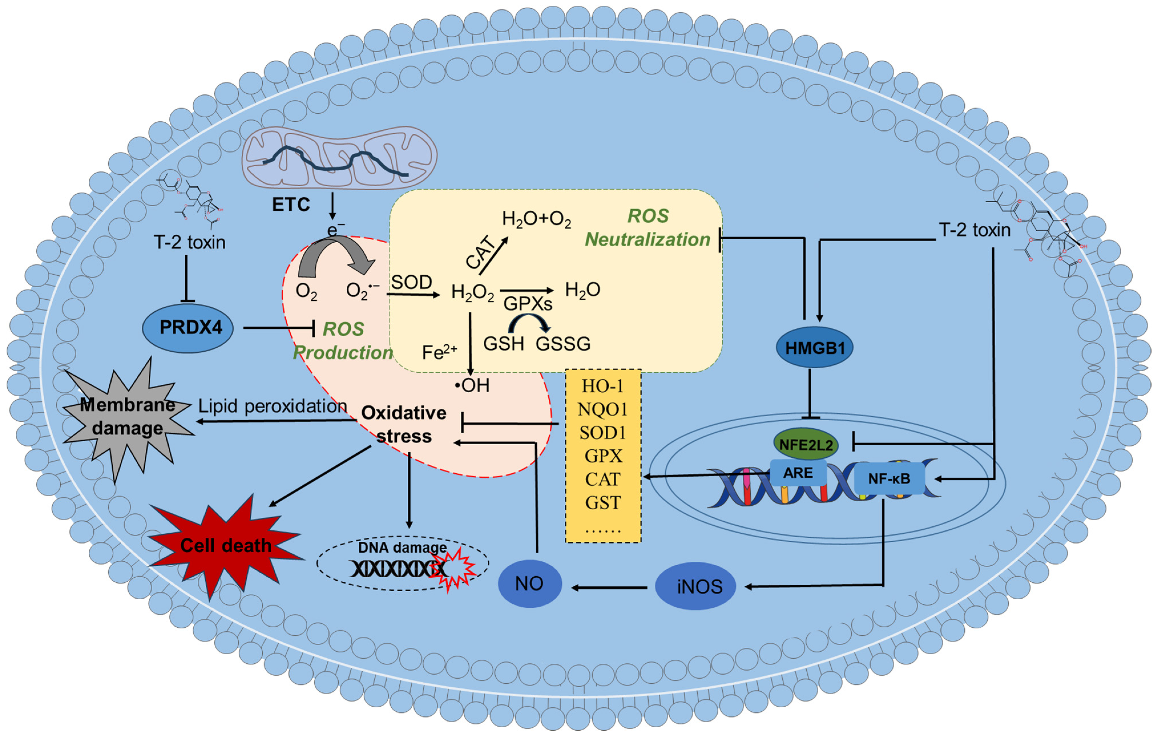
 indicates inhibition.
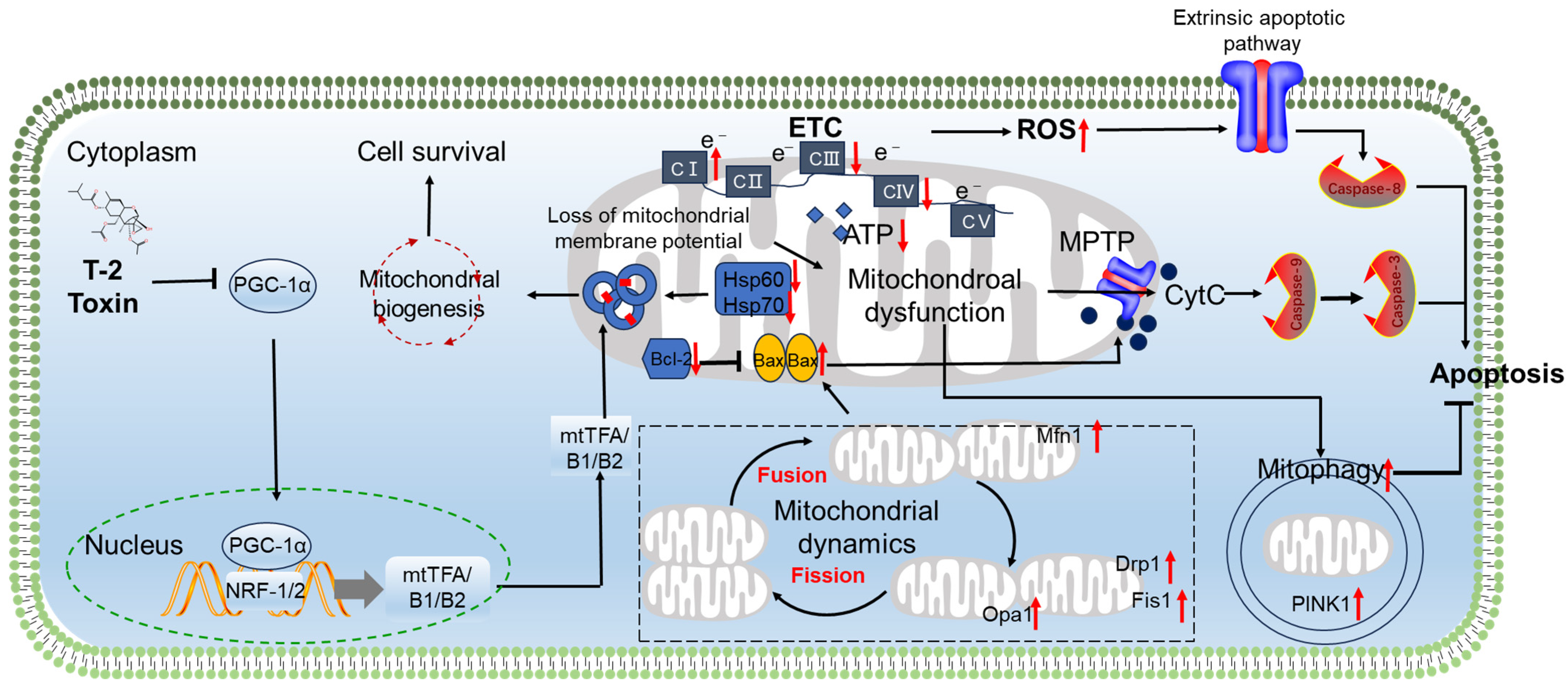
indicates inhibition.
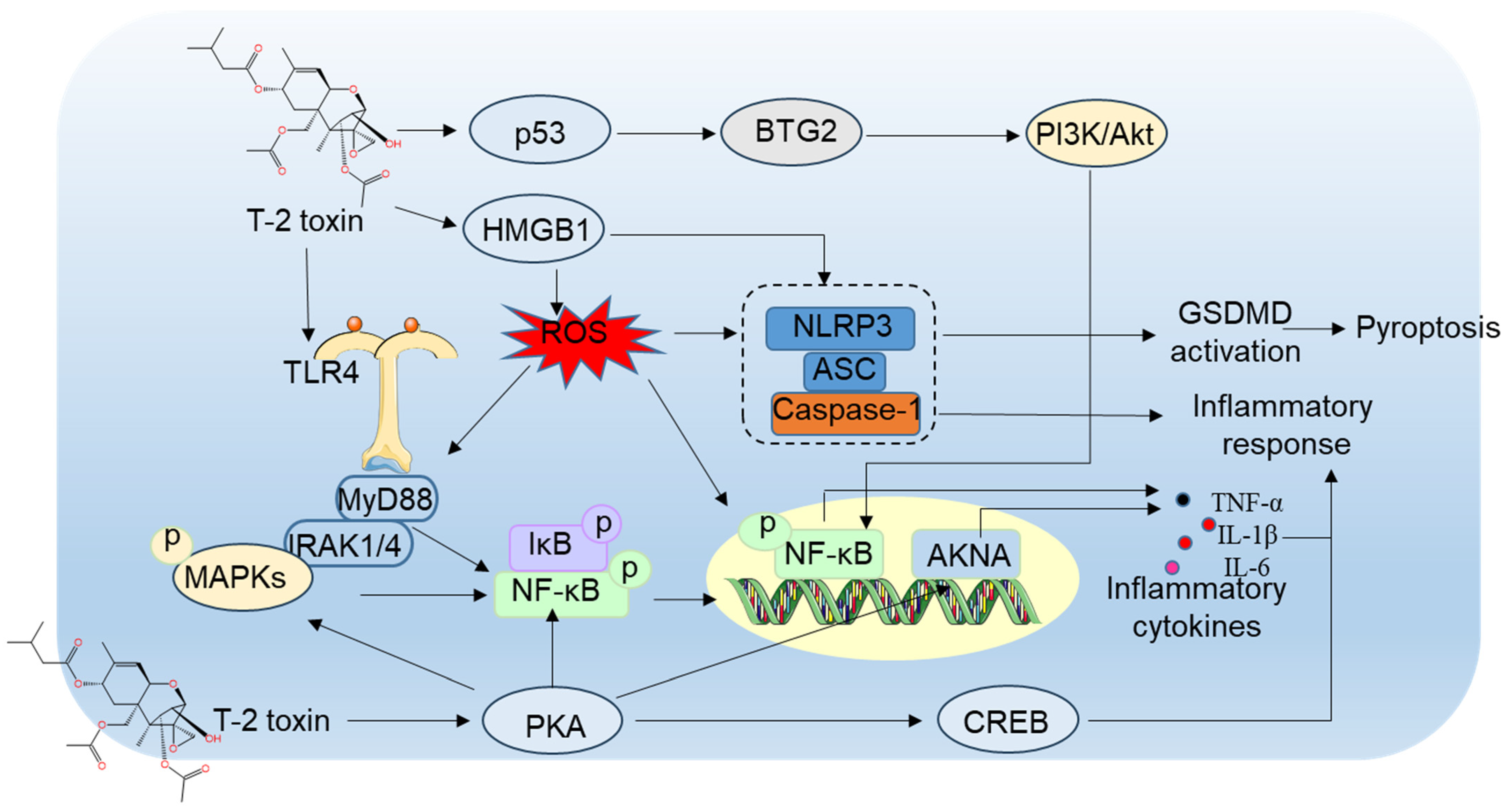
indicates inhibition.


| Model | Treatment Time and Dosage | Toxic Effects and Potential Mechanisms | References | |
|---|---|---|---|---|
| In vitro cell model | Mouse N2a cells | Cells were treated with T-2 toxin at the dose range of 5–80 ng/mL for 6–24 h | T-2 toxin can dose-dependently induce cytotoxicity and apoptosis. It involves the upregulation of oxidative stress and mitochondrial dysfunction. It also upregulated the expression of p53, Bax, and caspase-8 mRNAs and proteins, and downregulated the expression of NFE2L2 and HO-1 mRNAs and proteins. | [12] |
| Rat PC12 cells | Cells were treated with T-2 toxin at 1–12 ng/mL for 24 h | T-2 toxin can dose-dependently induce cytotoxicity and apoptosis in PC12 cells. It also induced the production of ROS and a decrease in antioxidant enzymes’ activities, causing oxidative stress. Additionally, T-2 toxin treatment promoted NF-κB and HMGB1-mediated inflammation response. | [70] | |
| Human IMR-32 cells | Cells were treated with T-2 toxin at 10–100 ng/mL for 8–48 h | T-2 toxin treatment markedly induced oxidative stress, mitochondrial dysfunction, cell apoptosis, caspase activation, and cell cycle arrest. T-2 toxin treatment also upregulated the expression of p-ERK, p-JNK, p-p38, Ras, Raf, and c-Fos proteins. Targeting the inhibition of caspase, ERK, p38, and Raf can effectively inhibit the cell apoptosis caused by T-2 toxin treatment. | [71] | |
| Human astrocytes | Cells were treated with T-2 toxin at 1 nM–200 μM for 6–48 h | T-2 toxin treatment can dose-dependently induce cytotoxicity, apoptosis, and necrosis in human astrocytes. | [36] | |
| Mouse BV2 cells | Cells were treated with T-2 toxin at 1.25–5 ng/mL for 24 h | T-2 toxin treatment significantly induced ROS production, causing oxidative stress and mitochondrial dysfunction. Additionally, T-2 toxin significantly decreased the expression of NFE2L2 and HO-1 proteins, activated cell autophagy, and upregulated autophagy flux. Inhibition of autophagy promoted T-2 toxin-caused cytotoxicity and cell apoptosis. | [72] | |
| Mouse HT22 cells | HT22 cells were treated with T-2 toxin at 0.5–4 ng/mL for 1–24 h | T-2 toxin treatment dose-dependently induced a decrease in cell viability and cell apoptosis. It can also induce cell pyroptosis via triggering NLRP3-caspase-1 inflammasome and gasdermin D (GSDMD) pathways. | [67] | |
| In vivo anima model | Male albino mice | T-2 toxin was single intravenously injected into mice at the doses of 2, 4, and 6 μg/kg body weight | T-2 toxin treatment caused gliosis in the cerebrum hippocampus tissues and acute inflammatory infiltrates at the focal areas, indicating encephalitis. T-2 toxin also damaged glial cells in the brain tissues and significantly downregulated the expression of aquaporin-4 mRNA. | [66] |
| Male C57/BL6 mice | Mice were orally administered with T-2 toxin at doses of 0.5, 1, and 2 mg/kg body weight for 28 days | T-2 toxin can induce arrangement disorder in hippocampal cells and the abnormal staining of neurons. Additionally, it also induced neuronal apoptosis and NLRP3-caspase-1 inflammasome and GSDMD-mediated cell pyroptosis in hippocampal tissues. | [67] | |
| Male Kunming mice | Mice were orally administered with T-2 toxin at 4 mg/kg body weight for 14 days | Neuronal loss, cellular swelling, pericellular space widening, massive bleeding, and cognitive dysfunction were observed in T-2 toxin-treated mice. Additionally, apoptosis, inflammation, oxidative stress, and abnormal neurotransmitter levels in T-2 toxin-treated brain tissues were detected. | [74] | |
| Specific pathogen-free female Wistar rats | T-2 toxin treatment via oral administration at a signal dose of 2 mg/kg body weight | The main behavioral changes are manifested as psychological fear and poor mental state. Additionally, marked pathological damage was detected in the brains of the T-2 toxin-treated rats. Marked mitochondrial damage, autophagy activation, and apoptosis were also detected in the brain tissues. | [49] | |
| Swiss albino female mice | T-2 toxin treatment at 5.94 mg/kg body weight via the dermal route or at 1.54 mg/kg body weight via the subcutaneous route; mice were sacrificed at 1st, 3rd, and 7th days after exposure | A decrease in drinking water and weight loss was observed. T-2 toxin treatment through dermal or subcutaneous injection can induce ROS generation, GSH depletion, lipid peroxidation and the alteration of phase II detoxifying enzymes. | [61] | |
| Male C57BL6 mice | T-2 toxin treatment at 0.5–5 mg/kg body weight via oral administration | The appetite of mice is significantly suppressed after T-2 toxin exposure. It also increased the expression of IL-1β, TNF-α, and IL-6 mRNAs in the brain tissue and the expression of c-Fos protein in the brainstem tissues of mice after unilateral vagotomy. | [64] | |
| Pregnant Wistar rats | Rats were treated with a single oral dose of T-2 toxin at 2 mg/kg body weight | T-2 toxin induced cell apoptosis in fetal brains. T-2 toxin exposure also induced the expression of genes enriched in oxidative stress, mitogen-activated protein kinase (MAPK) (such as MEKK1 and c-jun), and other apoptosis-related genes (such as caspase-2 and insulin-like growth factor-binding protein-3-related genes). | [75] | |
| Female B6C3F1 mice | Mice were orally treated with T-2 toxin at 1 mg/kg body weight; mice were sacrificed at 0, 0.5, 2, 6 and 24 h after exposure to T-2 toxin | The behavior in food intake was markedly inhibited. T-2 toxin increased levels of plasma 5-HT and SP and resulted in anorexia in mice. | [76] | |
| Male Wistar rats | Rats were orally administrated with T-2 toxin at the doses of 0.2, 0.4 and 0.8 mg/kg body weight for 4 weeks | Decreased spatial orientation and learning efficiency and impaired memory function were observed. T-2 toxin exposure triggered marked hippocampal pathological damage. It also induced oxidative damage and cell apoptosis in hippocampal tissues. | [77] | |
| Male C57BL/6 J mice | Mice were orally administrated with 1.5 mg/kg T-2 toxin daily for 14 d | T-2 toxin induced a significant increment in immobile time in the tail suspension test and a decline in sucrose preference in the sucrose preference test. T-2 toxin treatment also significantly reduced the level of dopamine and elevated the expression of dopamine transporter protein in the reward center nucleus accumbens of the brain. | [65] | |
| Models | Antioxidants/Natural Products/Small Molecular Inhibitors | Treatments | The Protective Effects | Reference | |
|---|---|---|---|---|---|
| In vitro | N2a neuronal cells | NAC (an antioxidant) | Cells were treated with NAC at 5 mM or cotreated with T-2 toxin at 20 ng/mL for 24 h. | NAC supplementation significantly improved T-2 toxin exposure-induced GSH deletion, downregulated the activities of caspases-9 and -3, and finally attenuated T-2 toxin-induced cytotoxicity. | [12] |
| Mouse microglia BV2 cells | NAC (an antioxidant) | Cells were pre-treated with NAC at 2.5 mM for 2 h, then co-treated with or without T-2 toxin at 2.5 ng/mL for 24 h. | NAC supplementation significantly improved T-2 toxin exposure-induced ROS production and cytotoxicity. | [72] | |
| Mouse hippocampal neuron cell line (HT22) | VX-765 (a caspase-1 inhibitor) | Cells were pretreated with dimethyl fumarate at 10 μM, then co-treated with or without T-2 toxin at 3 ng/mL for additional 24 h. | VX-765 pretreatment significantly attenuated T-2 toxin exposure-induced decreases in cell viability and pyroptosis, which was evident by the decreased expression of cleaved caspase-1, HMGB1, IL-1β, IL-18, GSDMD-NT proteins. | [67] | |
| Mouse microglia BV2 cells | PDTC (a NF-κB inhibitor), SP600125 (a JNK inhibitor), and PD98059 (an ERK inhibitor) | Cells were pre-treated with PDTC, SP600125, PD98059 for 2 h, then co-treated with T-2 toxin at 5 ng/mL for 12 h. | These inhibitors can all inhibit the T-2 toxin-induced expression of TNF-α, COX-2, IL-6, and IL-1β mRNAs in BV-2 cells, following to reduce microglial activation. | [37] | |
| Mouse microglia BV2 cells | LY294002 (a PI3K inhibitor) | Cells were pre-treated with LY294002 at 10 μM for 2 h, then co-treated with T-2 toxin at 5 ng/mL for 12 h. | LY294002 supplementation markedly inhibited the T-2 toxin exposure-induced activation of NF-κB and the expression of downstream inflammatory factors, such as TNF-α, IL-6, and IL-1β. | [84] | |
| Human neuroblastoma SH-SY5Y cells | YC-1 (a HIF-1α inhibitor) | Cells were pre-treated with YC-1 at 10 μM for 24 h, then co-treated with T-2 toxin at 6 nM for 2, 6, 12 or 24 h. | YC-1 treatment markedly inhibited T-2 toxin-induced cellular senescence by reducing the expression of p53, p21, p16, CCL-2, and IL-8 proteins. | [125] | |
| Mouse hippocampal neuron cell line (HT22) | Dimethyl fumarate (a NFE2L2 activator) | Cells were pretreated with dimethyl fumarate at 25 μM, then co-treated with or without T-2 toxin at 3 ng/mL for additional 24 h. | Dimethyl fumarate supplementation significantly attenuated the T-2 toxin exposure-induced release of LDH and the decreases in cell viability. It also reduced neuronal cell pyroptosis via inhibiting the expression of cleaved IL-1β, IL-18, and GSDMD-NT proteins. | [67] | |
| In vivo | Male Kunming mice | Vitamin E (an antioxidant) | Mice were pretreated with vitamin E via the oral administration at 100 mg/kg body weight per day for 14 days, then mice were exposed with T-2 toxin by a single intraperitoneal injection of 4 mg/kg. | Vitamin E supplementation markedly attenuated T-2 toxin-induced oxidative stress and neuronal cell apoptosis by reducing the levels of ROS and MDA and increasing the GSH levels and GPX activities in the brain tissues of mice. It also decreased T-2 toxin-induced neuroinflammatory response. | [74] |
| Mice | AHN 1–055 hydrochloride (a dopamine uptake inhibitor) | Mice were orally administration with T-2 toxin at 1.5 mg/kg body weight per day for 14 days. AHN 1–055 hydrochloride was infused into nucleus accumbens (NAc) of mice at 0.1 μg via cannulas before each behavioral test. | Pharmacological inhibition of DAT in NAc reverses the T-2 toxin-triggered depression-like behaviors and the reduced DA level in NAc in mice. | [65] | |
| Female Kunming mouse | Daucosterol (a natural product) | In animal model, mice were intraperitoneally injected with daucosterol at 30 mg/kg body, at 4 h before T-2 toxin treatment (at 1.57 mg/kg; subcutaneous injection). | Daucosterol supplementation markedly attenuated T-2 toxin-induced dysfunction of blood–brain barrier by promoting the transcription activation of PGC-1α and increasing the expression of claudin-5 (CLDN5), occludin (OCLN), and zonula occludens-1 (ZO-1) proteins in mouse brain tissues or human brain microvascular endothelial cells. It also reduced T-2 toxin exposure-induced neuroinflammatory response and neuronal cell apoptosis in the brain tissues of mice. | [85] | |
| Male C57BL/6 J mice | Resveratrol (a natural product) | Mice were given an intraperitoneal injection of 4 mg/kg T-2 toxin, then were given an intraperitoneal injection of 100 mg/kg resveratrol. | Resveratrol supplementation markedly ameliorated T-2 toxin exposure-induced spatial learning and memory impairments via upregulating the expression of postsynaptic density protein 95, synaptophysin I, and brain-derived neurotrophic factor (BDNF) proteins in the brain tissues. It can also restore intestinal flora disorders caused by T-2 toxin exposure and reduce the risk of inflammatory responses in the hippocampus, intestine, and the whole body. | [128] | |
| Mice | Betulinic acid (a natural product) | Mice were pretreated with betulinic acid via the oral administration at 0.25, 0.5, or 1 mg/kg body weight per day for 14 days, then were exposed with T-2 toxin through a single intraperitoneal injection of 4 mg/kg. | Betulinic acid supplementation markedly alleviated T-2 toxin exposure-caused cognitive impairment. It also markedly decreased T-2 toxin-induced oxidative stress and neuronal cell apoptosis by inhibiting the production of MDA and ROS and increasing the activities of GPX and GSH levels in brain tissues. Additionally, betulinic acid can reduce T-2 toxin-induced neuroinflammation and upregulate the levels of dopamine in the brain tissues. | [74] | |
| Mice | Minocycline (a semi-synthetic tetracycline antibiotic) | Mice were intraperitoneally injected with minocycline at 50 mg/kg body weight or co-treated intraperitoneally with or without T-2 toxin at 4 mg/kg body weight. | Minocycline supplementation markedly attenuated T-2 toxin exposure-caused spatial learning and memory and locomotor activity impairment via inhibiting microglial activation in the brain tissues of mice. | [37] | |
Disclaimer/Publisher’s Note: The statements, opinions and data contained in all publications are solely those of the individual author(s) and contributor(s) and not of MDPI and/or the editor(s). MDPI and/or the editor(s) disclaim responsibility for any injury to people or property resulting from any ideas, methods, instructions or products referred to in the content. |
© 2026 by the authors. Licensee MDPI, Basel, Switzerland. This article is an open access article distributed under the terms and conditions of the Creative Commons Attribution (CC BY) license.
Share and Cite
Xu, C.; Oliveri Conti, G.; Tang, S.; Shen, J.; Dai, C. T-2 Toxin Neurotoxicity: Molecular Mechanisms and Emerging Chemoprotective Strategies. Antioxidants 2026, 15, 278. https://doi.org/10.3390/antiox15030278
Xu C, Oliveri Conti G, Tang S, Shen J, Dai C. T-2 Toxin Neurotoxicity: Molecular Mechanisms and Emerging Chemoprotective Strategies. Antioxidants. 2026; 15(3):278. https://doi.org/10.3390/antiox15030278
Chicago/Turabian StyleXu, Chunyan, Gea Oliveri Conti, Shusheng Tang, Jianzhong Shen, and Chongshan Dai. 2026. "T-2 Toxin Neurotoxicity: Molecular Mechanisms and Emerging Chemoprotective Strategies" Antioxidants 15, no. 3: 278. https://doi.org/10.3390/antiox15030278
APA StyleXu, C., Oliveri Conti, G., Tang, S., Shen, J., & Dai, C. (2026). T-2 Toxin Neurotoxicity: Molecular Mechanisms and Emerging Chemoprotective Strategies. Antioxidants, 15(3), 278. https://doi.org/10.3390/antiox15030278