Review

Applications of Antioxidant Nanoparticles in Immune-Mediated Inflammatory Diseases

1
Department of Radiology, The Children’s Hospital, Zhejiang University School of Medicine, National Clinical Research Center for Child Health, Hangzhou 310058, China
2
Department of Plastic Surgery, Zhongshan Hospital Affiliated to Xiamen University, School of Medicine, Xiamen University, Xiamen 361005, China
3
Department of Dermatology, The Children’s Hospital, Zhejiang University School of Medicine, National Clinical Research Center for Child Health, Hangzhou 310058, China
*
Authors to whom correspondence should be addressed.
These authors have contributed equally to this work.
Antioxidants 2025, 14(9), 1128; https://doi.org/10.3390/antiox14091128
Submission received: 20 June 2025 / Revised: 5 September 2025 / Accepted: 10 September 2025 / Published: 18 September 2025
(This article belongs to the Special Issue Applications of Antioxidant Nanoparticles, 2nd Edition)

Abstract

Immune-mediated inflammatory diseases (IMIDs) encompass a wide range of disorders, including autoimmune, acute, and chronic inflammatory conditions, which are often characterized by immune dysregulation and excessive oxidative stress. Oxidative stress plays a pivotal role in the initiation and progression of these diseases by promoting tissue damage and sustaining inflammation. However, conventional antioxidant therapies are limited by poor bioavailability, inadequate targeting, and short-lived efficacy. In recent years, nano-antioxidants have emerged as a promising therapeutic approach due to their enhanced stability, targeted delivery capabilities, and multifunctional therapeutic effects. This review provides a comprehensive overview of recent advances in the application of nano-antioxidants in the treatment of IMIDs. Their therapeutic roles are categorized into three major groups: autoimmune diseases, acute inflammatory diseases, and chronic inflammatory diseases. In autoimmune disorders such as alopecia areata and multiple sclerosis, nano-antioxidants have demonstrated the ability to reduce oxidative damage, modulate immune responses, and alleviate clinical symptoms. In acute inflammatory conditions, including acute kidney injury and acute liver injury, these nanomaterials exert protective effects by scavenging ROS, mitigating tissue injury, and restoring organ function. In chronic inflammatory diseases such as inflammatory bowel disease and ulcerative colitis, nano-antioxidants contribute to maintaining mucosal integrity, suppressing chronic inflammation, and improving therapeutic outcomes through localized delivery and sustained release. In summary, nano-antioxidants represent a novel and promising therapeutic strategy for the management of IMIDs. Their unique physicochemical properties offer significant advantages over traditional treatments. Further research is needed to optimize their delivery platforms, evaluate long-term safety, and facilitate clinical translation.

1. Introduction

Alopecia areata (AA), multiple sclerosis (MS), and other conditions—including inflammatory bowel disease (IBD), acute liver injury (ALI), acute kidney injury (AKI), and vitiligo—result from immune system dysregulation. Collectively termed immune-mediated inflammatory diseases (IMIDs), these disorders comprise autoimmune pathologies and span both chronic and acute inflammatory processes [1]. Among the diverse pathogenic mechanisms involved, oxidative stress has emerged as a pivotal contributor to both disease onset and progression [2,3]. Oxidative stress, resulting from an imbalance between reactive oxygen/nitrogen species and intrinsic antioxidant systems, contributes to tissue dysfunction and cellular injury [4]. Accumulating evidence links oxidative stress to immune activation and tissue damage, while also implicating it in the amplification of inflammatory responses, thereby establishing a self-perpetuating pathogenic cycle.
Antioxidants, due to their capacity to counteract oxidative stress, have garnered considerable interest as therapeutic candidates for IMIDs. Conventional small-molecule antioxidants—such as vitamin C, vitamin E, and glutathione—exhibit limited clinical efficacy, primarily owing to low bioavailability, poor tissue specificity, and rapid systemic clearance [5,6]. Recent advances in nanotechnology have enabled new approaches for enhancing antioxidant therapy. Nano-antioxidants, constructed from nanomaterials with high surface-to-volume ratios, favorable biocompatibility, and modifiable surface chemistry, demonstrate improved physicochemical stability, enhanced biological targeting, and elevated therapeutic efficiency [7]. Notably, several nanomaterials possess intrinsic enzyme-mimetic properties. For instance, iron oxide- and manganese-based nanoparticles emulate the catalytic activities of endogenous antioxidant enzymes, including superoxide dismutase (SOD) and catalase (CAT), thereby facilitating efficient elimination of ROS and RNS [8,9]. Curcumin, a natural polyphenol derived from Curcuma longa, exhibits potent antioxidant and anti-inflammatory activities. However, its clinical application remains constrained by poor aqueous solubility, rapid metabolic degradation, and limited oral bioavailability. To overcome these barriers, various nanocarrier-based delivery platforms have been developed—such as liposomes, polymeric nanoparticles, solid lipid nanoparticles, and metal–organic frameworks [10]. These formulations significantly enhance curcumin’s systemic stability and targeting efficiency, while simultaneously potentiating its antioxidative and immunomodulatory effects. Preclinical studies indicate that curcumin nanoformulations attenuate inflammation, mitigate tissue damage, and improve disease outcomes in autoimmune models such as MS, primarily through ROS scavenging [11]. Beyond curcumin, alternative nano-antioxidant systems exhibit therapeutic potential across multiple IMIDs. Gold nanoparticles (GNPs), owing to their superior biocompatibility and tunable physicochemical properties, have been widely investigated in MS and other immune-related conditions [12]. Experimental findings suggest that GNPs reduce oxidative damage and modulate inflammatory signaling, thereby limiting immune-mediated tissue injury [13,14]. In the context of acute inflammatory pathologies, nano-antioxidants also demonstrate promising efficacy. A recent study reported an ROS-responsive nanoplatform integrating mitochondrial-targeted cerium oxide nanoparticles with atorvastatin. This system enabled site-specific drug release in a septic AKI model, suppressed oxidative stress and inflammation, reduced tubular epithelial apoptosis, and preserved renal architecture [15]. In diseases like IBD, characterized by persistent inflammation, oxidative stress is deeply implicated in driving the pathological cascade leading to tissue damage. Nano-antioxidants have been shown to alleviate intestinal inflammation and reinforce epithelial barrier function by modulating ROS levels and regulating signaling cascades, including NF-κB and Nrf2. Furthermore, co-administration with probiotics enhances therapeutic effects through modulation of gut microbiota homeostasis [16]. Despite substantial progress, several obstacles impede the clinical translation of nano-antioxidants, including uncertainties regarding long-term biocompatibility, metabolic fate, and immunogenicity. Future efforts should emphasize the rational design of nanotherapeutic systems featuring controlled release, disease-specific targeting, and optimal safety profiles to accelerate their deployment in the management of IMIDs.
We summarize the current applications and mechanisms of nano-antioxidants in IMIDs in Table 1, Table 2 and Table 3. This review provides a comprehensive overview of recent advances in the application of nano-antioxidants in immune-mediated inflammatory diseases, with a particular focus on their roles in autoimmune, acute inflammatory, and chronic inflammatory conditions. The underlying mechanisms, therapeutic efficacy, and safety profiles of nano-antioxidants are critically examined, and future directions for their development and clinical translation are discussed.

2. Methods

Relevant studies discussing nano-antioxidants in IMIDs were identified through an extensive literature survey utilizing five core academic databases, including PubMed, Embase, Scopus, Web of Science, and Google Scholar, focusing on publications from January 2020 to June 2025. The literature search included publications available up to June 2025. Keywords and search phrases were carefully selected, including terms such as “nano-antioxidants”, “autoimmune diseases”, “acute inflammation”, “chronic inflammatory diseases”, “alopecia areata”, “multiple sclerosis”, “ulcerative colitis”, “periodontitis”, and “inflammatory bowel disease”. The search strategy focused on identifying studies that explored the application, mechanism, or therapeutic efficacy of nano-antioxidants in immune-related inflammatory conditions. Studies meeting the following criteria were included: (1) investigations that evaluated nano-antioxidant therapies related to autoimmune, acute, or chronic inflammatory diseases; (2) research involving nanoformulations designed to improve antioxidant delivery or bioactivity; (3) preclinical or clinical studies with reproducible methodologies and consistent outcome measures; and (4) experimental studies—either in vitro or in vivo—examining the antioxidant capacity or immune-modulating effects of nanomaterials. Exclusion criteria were as follows: (1) studies with inconclusive evidence regarding the therapeutic benefit of nano-antioxidants; (2) publications lacking rigorous experimental protocols or clearly defined results; (3) non-randomized or observational studies without proper controls; (4) articles not published in English; and (5) narrative reviews, opinion pieces, or book chapters without primary research data.

3. Nano-Antioxidants for Autoimmune Diseases

3.1. Alopecia Areata

Characterized by patchy, non-scarring hair loss primarily on the scalp and other hairy areas, AA represents a T cell-mediated autoimmune inflammatory disorder [44]. Though the etiological framework of AA has not been fully delineated, the impairment of immune privilege within hair follicles continues to dominate current pathogenic models [45]. In addition, genetic predisposition, hypersensitivity, dysbiosis of skin and gut microbiota, and psychological stress are also recognized as important contributing factors to disease onset. Epidemiological data from the United States estimate the prevalence of AA to be approximately 0.1–0.2% [46]; however, this may be an underestimation due to underreporting and limited sample representativeness. AA significantly impairs patients’ quality of life and remains a therapeutic challenge in clinical practice. In recent years, oxidative stress—defined as an imbalance between pro-oxidant and antioxidant defenses—has been increasingly implicated in the disruption of immune privilege, contributing significantly to the pathogenesis of AA [47].
AA patients exhibit elevated systemic oxidative stress, as evidenced by increased serum levels of advanced glycation end-products (AGEs) and advanced oxidation protein products, alongside decreased antioxidant indices such as ferric reducing antioxidant power (FRAP), paraoxonase-1, and lecithin–cholesterol acyltransferase. Notably, FRAP levels correlated positively with the extent of hair loss and negatively with C-reactive protein, indicating a close association between oxidative stress, inflammation, and disease severity [48]. Similarly, in a cohort of 40 patients with alopecia areata (AA), Sachdeva et al. [49] reported significantly elevated serum malondialdehyde (MDA) levels, indicative of heightened oxidative stress. Concurrently, both total antioxidant status (TAS) and SOD activity were markedly reduced compared to healthy controls, with the degree of reduction showing a clear correlation with disease severity. Moreover, impaired autophagy has been implicated in AA, as evidenced by reduced expression of autophagy-related proteins ATG5 and LC3B and increased expression of p62 in hair matrix tissues of AA patients. Concurrently, oxidative stress biomarkers—including MDA, AGEs, and ischemia-modified albumin (IMA)—are significantly elevated in serum and lesional skin, suggesting potential diagnostic and monitoring value [50]. Taskin et al. [51] further demonstrated significantly increased serum levels of nitric oxide (NO·), peroxynitrite (ONOO), and nitric oxide synthase (NOS) activity in AA patients, indicating that nitrosative stress also contributes to AA pathophysiology. As ONOO represents a convergence point between oxidative and nitrosative stress, it may exacerbate oxidative injury. These findings highlight NOS-related parameters as potential biomarkers. Accumulated evidence from observational studies over the past two decades (2000–2024) indicates that patients with AA consistently exhibit elevated oxidative stress markers (e.g., OSI, MDA) and reduced antioxidant enzyme activity, including SOD and glutathione peroxidase, suggesting a compromised antioxidant defense system. Collectively, these findings underscore the central role of oxidative stress in AA pathogenesis and provide a theoretical foundation for antioxidant-based therapeutic strategies [52].
Meanwhile, localized drug delivery approaches—particularly microneedle patches—have attracted increasing attention. A hydrogel-based microneedle immunomodulatory system has been developed to deliver regulatory T cell (Treg)-associated factors, such as CCL22 and interleukin-2 (IL-2), to effectively recruit and expand Tregs within lesional areas, thereby re-establishing local immune tolerance. IL-2 functions as a crucial survival and proliferation factor for regulatory T cells (Tregs) and has been widely utilized to selectively expand Tregs in both experimental and clinical settings. In AA mouse models, this system significantly enhanced Treg infiltration, suppressed inflammatory signaling pathways, and promoted hair regrowth [53]. Additionally, soluble microneedle array patches (S-MAPs) composed of hyaluronic acid have been employed for the transdermal delivery of the Janus kinase (JAK) inhibitor baricitinib in a painless and noninvasive manner, thereby avoiding systemic adverse effects. In AA models, this strategy achieved significantly higher hair regrowth rates (82% vs. 31%) compared to oral administration [54]. Although baricitinib is not a direct antioxidant, it exerts indirect antioxidant effects by downregulating pro-inflammatory cytokines such as IL-6, IFN-γ, and TNF-α via JAK/STAT pathway inhibition, thereby reducing oxidative enzyme activity and ROS production, contributing to a combined anti-inflammatory and antioxidant effect [55]. In the realm of nanomaterials, Xiao et al. [17] examined the effects of molybdenum nanoparticles (Mo NPs) on AA progression through the modulation of oxidative stress. As a novel nano-antioxidant, Mo NPs possess a multivalent electronic structure and can promote hair regrowth by eliminating ROS, alleviating oxidative stress, and activating hair follicle stem cells. Characterization studies revealed that Mo NPs had an average diameter of approximately 50 nm (TEM, Figure 1A) and exhibited characteristic Mo 3d multivalence peaks (XPS, Figure 1B). In vivo, mice treated with Mo NPs exhibited visible hair regrowth as early as day 7 (Figure 1C), and hair shaft length was significantly greater than in controls (Figure 1D,E), with synergistic effects observed when combined with minoxidil. Mechanistically, Mo NPs significantly downregulated the expression of the oxidative stress markers iNOS and COX2 in hair follicle tissue (Figure 1F–H), further supporting their hair-promoting effect via antioxidative pathways. In conclusion, Mo NPs demonstrate excellent antioxidant activity and can act synergistically with conventional therapies such as minoxidil, offering a novel direction and translational potential for antioxidant-based treatment of AA. Future studies should explore their long-term efficacy and safety to facilitate clinical application.

3.2. Multiple Sclerosis

MS is characterized by immune-mediated damage targeting the central nervous system (CNS), leading to the formation of multifocal lesions in the brain and spinal cord, primarily involving demyelination and neuroinflammation. These lesions are predominantly located in the white matter and can be detected by magnetic resonance imaging. However, due to technical limitations, the identification of gray matter and cortical lesions remains challenging, posing a major difficulty in both diagnosis and treatment [56]. MS is frequently accompanied by psychiatric symptoms such as anxiety and depression, which significantly compromise patients’ quality of life and adherence to treatment. Reports indicate that between 2015 and 2022, the age- and sex-standardized incidence of MS showed a slight declining trend, ranging from 17.46 per 100,000 population in 2016 (95% CI: 17.12–17.80) to 15.65 per 100,000 in 2019 (95% CI: 15.34–15.97) [57]. The pathogenesis of MS is closely associated with immune-mediated inflammation and oxidative stress, the latter of which induces neuronal damage through ROS-related signaling pathways [58]. Fluctuations in the redox environment can trigger cell type-specific stress responses, disrupt metabolic homeostasis, and exacerbate disease progression. Therefore, therapeutic strategies targeting oxidative stress, particularly low-toxicity and multi-targeted nano-antioxidants, have emerged as a research hotspot in MS therapy.
To improve its chemical stability and enhance absorption, curcumin—a natural antioxidant—has been integrated into a range of nanocarrier technologies [59]. For example, dendritic nanocarriers (DNCs) based on curcumin have been shown to dose-dependently promote the differentiation of neural stem cells (NSCs) and oligodendrocyte precursor cells (OPCs) into oligodendrocytes, thereby enhancing remyelination capacity in vivo. DNCs also improve the survival and differentiation efficiency of transplanted NSCs, synergistically facilitating myelin repair [18]. In addition, a high-density lipoprotein-mimicking peptide-phospholipid scaffold (HPPS) has been utilized to deliver curcumin (Cur-HPPS) in a targeted manner. This system exerts anti-inflammatory and antioxidant effects by inhibiting the NF-κB pathway and reducing the expression of key mediators involved in immune cell recruitment, offering a novel direction for the precision treatment of MS [60]. Another study developed a system of iron oxide nanoparticles loaded with berberine (BBN-IONPs), which demonstrated superior neuroprotective and antioxidant effects in MS animal models compared to free berberine. This nanoplatform improved CPZ-induced cognitive impairment, elevated glutathione levels and total antioxidant capacity in brain tissues, reduced lipid peroxidation and inflammatory mediators, and modulated the expression of the key molecules MBP and MMP-9 and other key effector molecules [19].
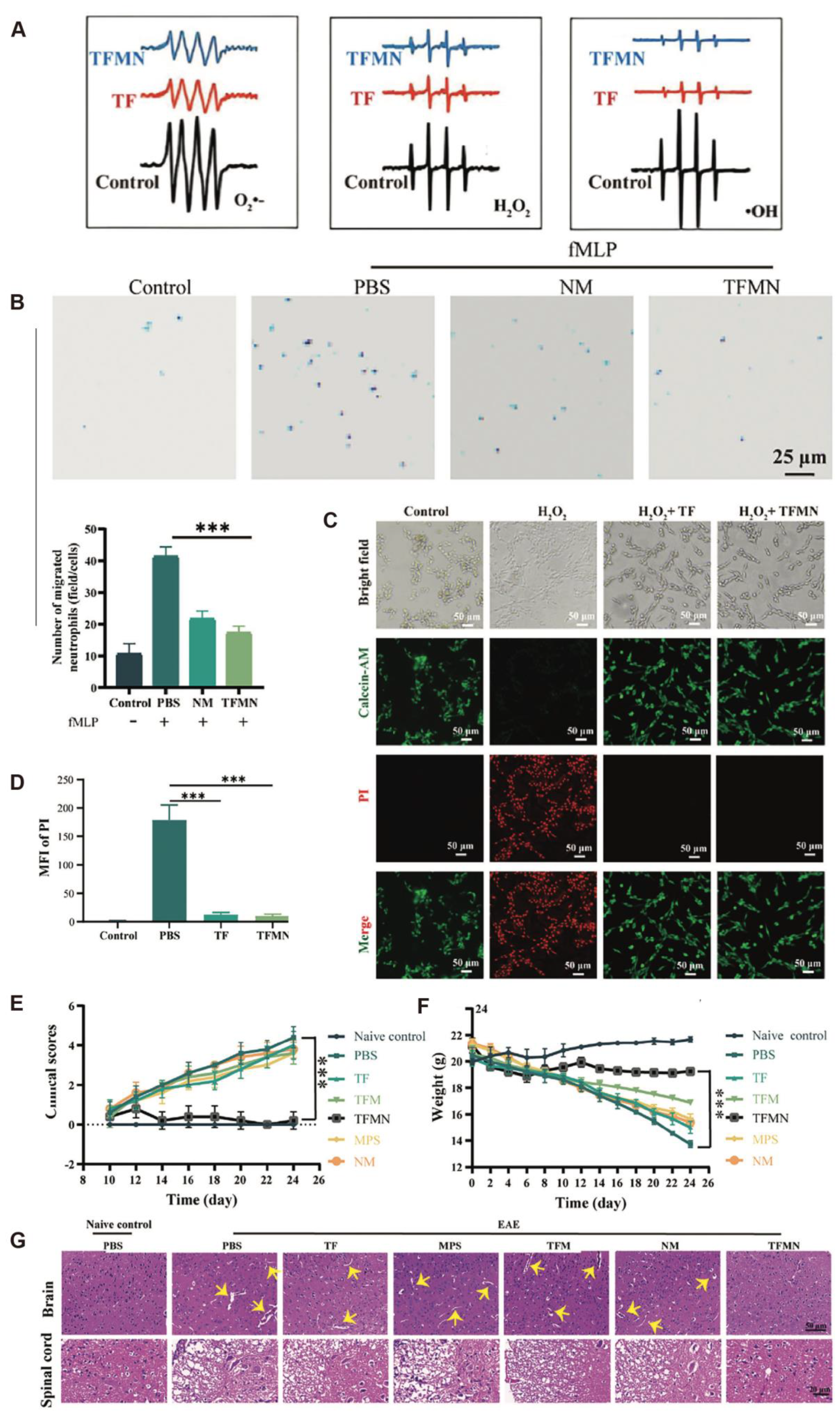
In recent years, antioxidant strategies based on nanotechnology have shown broad application prospects in autoimmune diseases of the CNS. Low-toxicity magnesium hydride nanoparticles (MgH2) have also shown promising efficacy in MS mouse models, significantly alleviating anxiety- and depression-like behaviors and attenuating inflammatory demyelinating lesions [20]. The underlying mechanism may involve the promotion of M2-type microglial polarization, the inhibition of M1-type polarization, and the mitigation of oxidative stress and mitochondrial damage, suggesting their potential in treating MS-related neuropsychiatric disorders. Sanadgol et al. [61] showed that metformin facilitates the maturation of oligodendrocyte progenitor cells via the activation of the AMPK/mTOR signaling pathway while enhancing Nrf2-mediated antioxidant responses, thereby facilitating remyelination. Abdelalim et al. [21] developed a double-layered nanoemulsion system using lactoferrin and chitosan to encapsulate clobetasol propionate (CP), improving brain-targeted delivery and reducing systemic toxicity. The prepared nanoparticles exhibited stable physicochemical properties (mean particle size: ~220 nm; zeta potential: +30.23 mV) and an antioxidant capacity equivalent to 9.8 μM of vitamin C. Rezaeimanesh et al. [22] were the first to evaluate the effects of crocin–selenium nanoparticles (Cor@SeNs) on cognitive function and oxidative stress status in patients with MS. In a triple-blind, randomized controlled trial, 60 MS patients treated with Cor@SeNs for 12 weeks showed a marked enhancement in total antioxidant capacity (TAC; p = 0.01) and marked improvements in several cognitive assessment scores, including CVLT-II, CVLT-II-delay, and SDMT (all p < 0.01), indicating its potential in ameliorating MS-associated cognitive impairment. Yang et al. [23] developed a biomimetic nanosystem (TFMN) camouflaged with neutrophil membranes, which demonstrated multiple therapeutic effects in MS models. TFMN demonstrated potent scavenging activity against key reactive oxygen species, including O2, H2O2, and OH (Figure 2A), thereby reducing ROS-mediated neuronal injury, and significantly inhibited the migration of inflammatory cells across the blood–brain barrier (Figure 2B). In a PC12 cell oxidative injury model, TFMN significantly enhanced cell viability (Figure 2C–E), and in EAE mouse models, it lowered clinical scores, maintained stable body weight (Figure 2E,F), and mitigated inflammatory cell infiltration and demyelination in the brain and spinal cord (Figure 2G), indicating excellent safety and therapeutic potential. In summary, antioxidant strategies—particularly those based on multi-targeted nanomaterials—exhibit substantial advantages in the treatment of MS. These approaches not only alleviate inflammation and oxidative damage but also show potential in improving cognitive and psychiatric comorbidities, providing new perspectives and experimental foundations for the future development of individualized and precision therapies for MS.

4. Nano-Antioxidants for Acute Inflammatory Diseases

4.1. Acute Kidney Injury

The kidneys play a critical role in maintaining systemic homeostasis due to their abundant blood perfusion and high oxygen consumption, enabling efficient clearance of metabolic waste. However, these same characteristics also make the kidneys more susceptible to exposure to toxic substances and hypoxic conditions [62]. In AKI, renal function declines rapidly within a limited period, often leading to systemic complications that carry high morbidity and mortality. In the 2017 study population, the age-standardized incidence rates of AKI varied across regions, ranging from 12.9 to 14.9 cases per 1000 person-years [63]. Currently, no specific clinical therapies are available for AKI, highlighting the urgent need to develop effective therapeutic strategies. Accumulating evidence indicates that the excessive accumulation of reactive oxygen and nitrogen species (RONS) at injury sites is a key driver of AKI progression. However, due to the structural complexity, cellular heterogeneity, and specialized physiology of the kidneys, conventional antioxidant and anti-inflammatory small-molecule drugs exhibit limited efficacy, primarily due to poor targeting capability and adverse side effects [64].
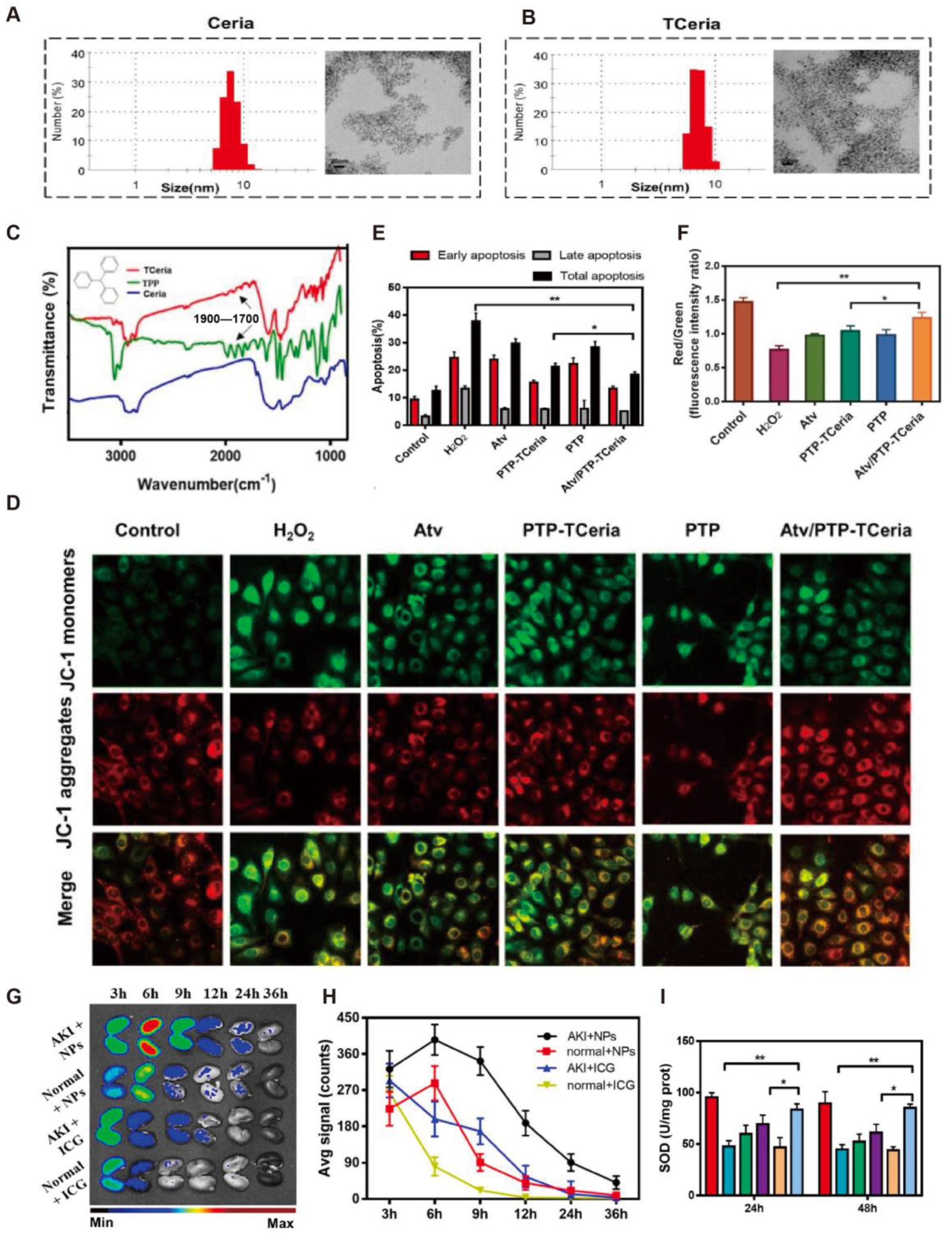
To address these limitations, various nanomedicine-based antioxidant strategies have emerged. Gu et al. [24] synthesized a cerium–luteolin coordination nanocomposite (CeLutNCs), which effectively scavenges ROS, inhibits apoptosis, downregulates inflammatory cytokines, modulates macrophage activity, and suppresses NF-κB signaling. In both AKI models, CeLutNCs significantly improved organ function and reduced oxidative stress and inflammation, demonstrating strong therapeutic potential for ROS-related diseases. Zheng et al. [25] developed ultrasmall rhodium nanoparticles modified with L-serine (Rh-Ser, 2–4 nm), which mimic multiple antioxidant enzymes to efficiently eliminate RONS. Additionally, Rh-Ser generated oxygen at inflamed sites, alleviating hypoxia and suppressing inflammation, and increased the survival rate of AKI mice from 0% to 80%, outperforming traditional treatments. Yan et al. [26] designed a tetrahedral framework nucleic acid (tFNA)-based nanodrug delivery system (TTC), which specifically targets renal tubular cells and mitochondria, reduces oxidative stress, and suppresses apoptosis. In vitro and in vivo ischemia–reperfusion (I/R) models showed that TTC significantly ameliorated renal injury and restored renal function, indicating promising antioxidant-mediated therapeutic effects. Pan et al. [27] developed a pH-stable antioxidant nanoparticle system (EGA NPs), based on epigallocatechin gallate (EGCG) and 5-aminosalicylic acid (5-ASA). Through a “dissociation-reassembly” mechanism under acidic conditions, these particles self-assemble into stable nanoparticles with robust antioxidant and anti-inflammatory properties, suitable for both oral and intravenous administration. EGA NPs not only effectively scavenge ROS and mitigate oxidative stress but also serve as a drug delivery vehicle for anti-inflammatory agents such as curcumin, significantly enhancing their therapeutic efficacy in vivo. Jia et al. [28] proposed a supramolecular nanoplatform (Ser-HPEC) for AKI therapy, possessing ROS-responsive antioxidant capabilities. Leveraging a Kim-1-mediated targeting mechanism via L-serine, Ser-HPEC selectively accumulates at I/R-injured renal sites and, under high ROS conditions, releases two antioxidant molecules—ethyl caffeate and 4-hydroxybenzyl alcohol. This results in efficient ROS scavenging, attenuation of oxidative stress and acute inflammation, and preservation of renal structure and function. Furthermore, a cerium-based nanozyme system with mitochondrial targeting capacity was successfully constructed. Transmission electron microscopy (TEM) images revealed that unmodified Ceria nanoparticles (Ceria NPs) [15] had a uniform spherical morphology with an average diameter of 7.74 ± 1.35 nm, while triphenylphosphine (TPP)-modified TCeria NPs exhibited a slightly larger diameter of 8.16 ± 1.98 nm, retaining good shape and dispersity (Figure 3A,B). Fourier-transform infrared spectroscopy (FT-IR) confirmed successful TPP modification, with characteristic absorption peaks at 1700–1900 cm−1 corresponding to the aromatic structure of TPP (Figure 3C). Mitochondrial membrane potential (Δψm) was assessed using JC-1 dye. In H2O2-treated cells, a significant reduction in the red/green fluorescence ratio indicated mitochondrial depolarization, whereas treatment with Atv/PTP-TCeria NPs restored Δψm to near-normal levels (Figure 3D). Flow cytometry showed that this formulation significantly reduced H2O2-induced apoptosis (below 20%), outperforming both free drugs and empty carriers (Figure 3E). Quantitative fluorescence analysis further validated its mitochondrial protective effect (Figure 3F). In vivo imaging using near-infrared fluorescent dye (ICG)-labeled nanoparticles revealed sustained accumulation of Atv/PTP-TCeria NPs in the kidneys of AKI mice for over 36 h, indicating excellent passive targeting capability (Figure 3G). Quantitative analysis confirmed significantly higher fluorescence intensity in AKI kidneys compared to healthy controls, consistent with enhanced permeability and retention (EPR) effects (Figure 3H). Additionally, treatment with Atv/PTP-TCeria NPs significantly increased renal SOD activity, restoring redox homeostasis and demonstrating superior ROS-scavenging ability compared to free drugs and control groups (Figure 3I). In summary, the constructed Atv/PTP-TCeria NPs system possesses multiple advantages, including precise size control, effective mitochondrial targeting, ROS responsiveness, and synergistic drug release. These features enable it to significantly ameliorate AKI-associated pathological processes at both cellular and animal levels, offering a promising nanotherapeutic strategy for the treatment of AKI.

4.2. Acute Liver Injury

ALI is a clinically common and severe liver disorder that can be triggered by a wide range of etiologies, including drug overdose, viral infections, toxic exposures, pregnancy, autoimmune diseases, inherited metabolic disorders such as Wilson’s disease, or vascular abnormalities such as Budd–Chiari syndrome. ALI is characterized by extensive hepatocellular necrosis and rapid deterioration of liver function. In certain cases, it may rapidly progress to acute liver failure (ALF), a life-threatening condition with a mortality rate of up to 80% [65]. Oxidative stress, particularly the excessive accumulation of ROS in hepatic tissue, is considered a key pathogenic mechanism in the onset and progression of ALI. Although broad-spectrum antioxidants such as N-acetylcysteine (NAC) have demonstrated partial therapeutic benefits, their limited targeting capacity, low clearance efficiency, and poor bioavailability severely hinder clinical outcomes. Therefore, there is an urgent need to develop more efficient, liver-targeted ROS-scavenging strategies to provide novel therapeutic approaches for ALI management [66].
Given the central role of oxidative stress in ALI pathogenesis, the development of nanomedicine-based antioxidant platforms with high ROS-scavenging efficiency and hepatic specificity has emerged as a promising research direction. Several studies have focused on integrating natural antioxidant molecules with functional nanomaterials to enhance therapeutic efficacy. Luo et al. [29] fabricated phlorotannin-based nanoparticles (PT NPs) from seaweed-derived polyphenols via a glycine-assisted Mannich reaction to improve their stability and bioavailability. These nanoparticles exhibited robust ROS-scavenging activity in H2O2-induced HepG2 cell models and effectively alleviated oxidative. Similarly, Jang et al. [30] developed a catechin-based thermosensitive nanoformulation (CCN150), which significantly improved the solubility and stability of catechin while retaining its intracellular ROS-scavenging activity. CCN150 selectively accumulated at inflammation sites in vivo, attenuated systemic inflammation driven by ROS, and demonstrated excellent biocompatibility. In the context of acetaminophen (APAP)-induced liver injury, Song et al. [67] designed a carbon monoxide-releasing molecule, SMA/CORM2, which enhanced hepatic CO levels and suppressed oxidative stress. Mechanistically, SMA/CORM2 downregulated HIF-1α expression, promoted M2 polarization of macrophages, and activated the PI3K/Akt/mTOR pathway, thereby mitigating liver damage and facilitating tissue regeneration. Additionally, Mn3O4 nanozymes showed comparable efficacy to NAC at lower doses by simultaneously modulating the Nrf2 antioxidant axis and suppressing NF-κB-mediated inflammatory responses, offering synergistic antioxidative and anti-inflammatory effects [68].
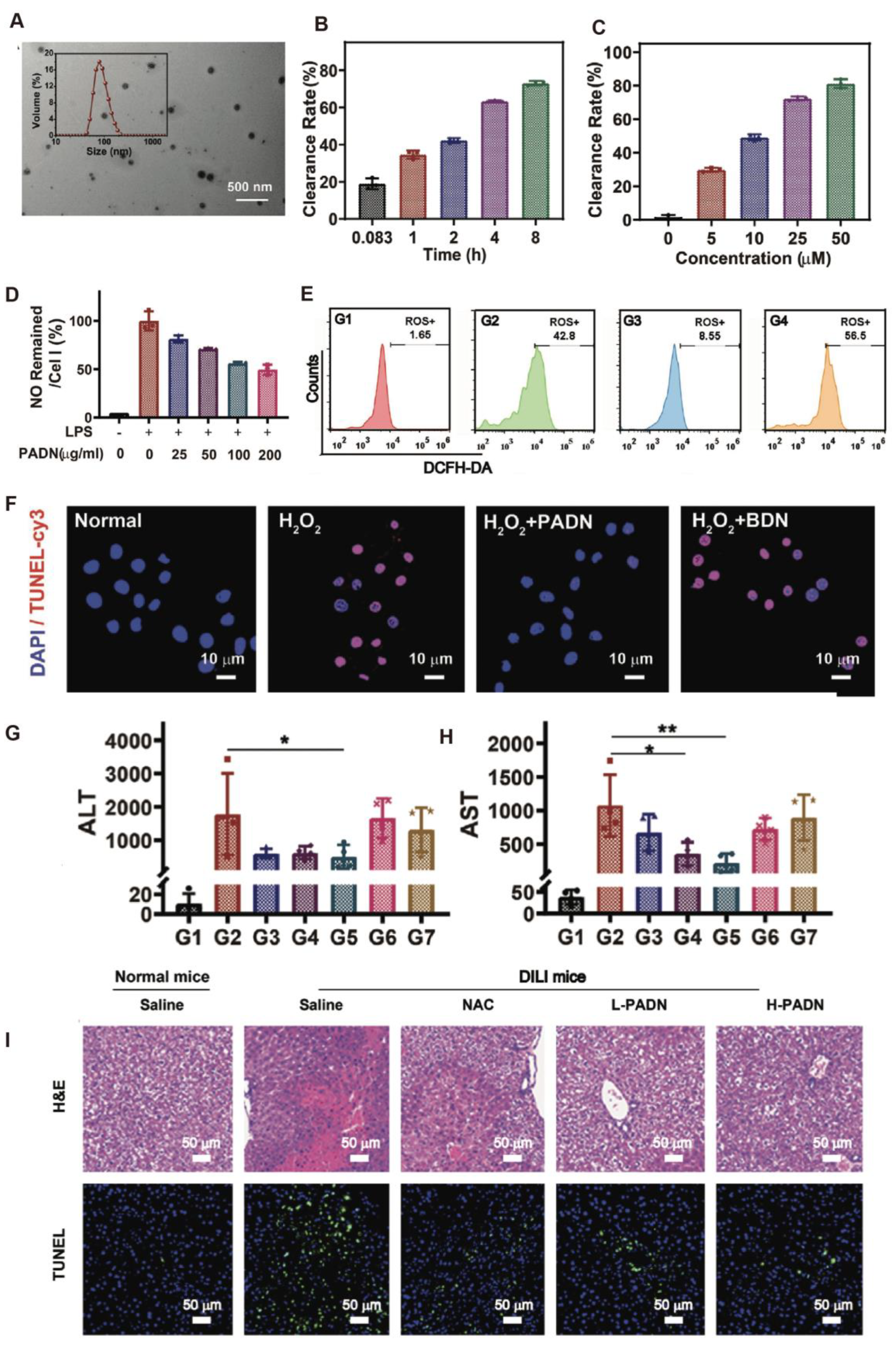
Curcumin, a natural compound widely used in traditional Chinese medicine, has shown hepatoprotective potential in APAP-induced ALI through mechanisms involving ROS scavenging, inhibition of lipid peroxidation, restoration of glutathione levels, and anti-apoptotic effects. The incorporation of curcumin into nanocarriers can significantly enhance its bioavailability and therapeutic performance. Wang et al. [31] further developed BSA-SNO nanoparticles capable of stably releasing nitric oxide (NO). These nanoparticles alleviated oxidative stress, improved hepatic microcirculation, and increased survival rates in ischemia–reperfusion and endotoxin-induced ALI models. Hepatoprotection was achieved via Nrf2/HO-1 pathway activation and suppression of NF-κB-driven inflammation and mitochondrial apoptosis. A saponin–gallic acid conjugate-based nano-delivery system also demonstrated promising anti-inflammatory effects in CCl4-induced ALI. By employing Zn/Ga@Gd-LDH as a pH-responsive carrier, the system enabled controlled drug release in the acidic microenvironment, suppressed p-p65 phosphorylation in the NF-κB pathway, and reduced CRP levels, exhibiting both in vitro and in vivo anti-inflammatory efficacy [69]. Yuan et al. [66] developed SeMC nanoparticles derived from the self-assembly of L-selenomethionine (SeMC), modified with glycyrrhizic acid for hepatic targeting. These nanoparticles significantly reduced lipid peroxidation, tissue vacuolation, and aminotransferase elevation in both APAP- and CCl4-induced ALI models while enhancing endogenous antioxidant enzyme activity, showing favorable biocompatibility and liver-protective potential. Moreover, Zhao et al. [32] proposed a responsive biomimetic antioxidant nanoplatform (PADN) composed of PEGylated L-DOPA precursors protected by boronic acid groups. Upon exposure to oxidative stress, PADN undergoes deprotection to release PEG–L-DOPA, which subsequently undergoes a melanogenesis-like transformation into a high-molecular-weight melanin-like polymer with broad-spectrum ROS-scavenging capability. The self-assembled PADN exhibited a particle size of approximately 106.6 nm (Figure 4A) and maintained excellent colloidal stability for over three months under physiological conditions. It efficiently eliminated multiple ROS types, including H2O2, in a time- and dose-dependent manner (Figure 4B,C). At the cellular level, PADN significantly suppressed NO production and intracellular ROS accumulation in LPS-activated RAW264.7 macrophages (Figure 4D,E) and reduced TUNEL-positive hepatocytes in an H2O2-induced apoptosis model (Figure 4F). In vivo, PADN (50 mg/kg) effectively decreased serum ALT and AST levels in APAP-induced ALI mice, outperforming NAC in liver function recovery (Figure 4G–H). Histological analyses revealed reduced necrosis, inflammatory infiltration, and central venous congestion in PADN-treated mice, with the liver architecture approaching normal (Figure 4I). Additionally, PADN possessed photoacoustic imaging capabilities, enabling real-time visualization of lesion sites. Collectively, PADN exemplifies an oxidative microenvironment-responsive nanoplatform capable of integrating efficient ROS scavenging, inflammation suppression, and hepatocyte protection. Its stable structure and biocompatibility make it a strong candidate for clinical translation of antioxidant nanotherapies in ALI treatment.

5. Nano-Antioxidants for Chronic Inflammatory Diseases

5.1. Periodontitis

The global health impact of periodontitis, a chronic inflammatory condition affecting millions, has shown a consistent upward trend over recent years. Data from 2011 to 2020 indicate that approximately 62% of dentate individuals worldwide are affected, with over half of the cases classified as moderate to severe and nearly one-quarter as severe [70]. Compared with epidemiological data from 1990 to 2010, the prevalence of severe periodontitis increased from 10.8% to 23.6% [71]. By 2019, the global number of individuals affected had reached 1.1 billion [72].
Beyond being a prevalent oral disease, periodontitis is closely associated with a range of systemic conditions. While microbial plaque serves as the primary etiological factor, the progression of the disease—especially in its severe or refractory forms—is predominantly driven by the host immune response. Neutrophil-mediated mechanisms, notably the overproduction of ROS, play a pivotal role in tissue degradation. Oxidative stress, induced by intrinsic factors (e.g., metabolic disorders) and external triggers (e.g., smoking), creates a pro-inflammatory microenvironment that exacerbates periodontal damage. Thus, antioxidant-based therapies, such as those incorporating resveratrol, are being explored as promising strategies for periodontal intervention [73]. Li et al. [74] developed a novel bioactive glass-based microneedle system loaded with antioxidant agents for the treatment of diabetes-associated periodontitis. This system comprises Zn/V co-doped bioactive glass nanoparticles, gallic acid, and oxidized methacrylated hyaluronic acid, offering both antioxidative and antibacterial properties. By penetrating the mucosal barrier, the microneedles effectively deliver active agents into gingival tissues, enhancing retention and bioavailability. In diabetic periodontitis rat models, the system significantly promoted alveolar bone regeneration and alleviated local inflammation via suppression of the JAK-STAT and NF-κB signaling pathways. Yang et al. [33] constructed a mesoporous bioactive glass-based nanocarrier system for quercetin delivery, addressing its poor water solubility and limited bioavailability. This sustained-release platform promoted both osteogenesis and angiogenesis in experimental periodontitis-related bone defects. Additionally, it modulated the local immune microenvironment by regulating macrophage polarization. Mechanistically, quercetin acted through the miR-21a-5p/PDCD4/NF-κB axis to restore bone immune homeostasis. Similarly, Wang et al. [75] incorporated quercetin into octahedral nanoceria structures to enhance its antioxidative and immunomodulatory effects. The resulting nanocomposite effectively scavenged ROS, inhibited pro-inflammatory M1 macrophage polarization, and promoted the reparative M2 phenotype, enabling a smooth transition from inflammation to regeneration. This shift was evidenced by an increased M2/M1 polarization ratio, along with a rebalanced inflammatory profile in vivo, underscoring its therapeutic potential for managing periodontal inflammation.
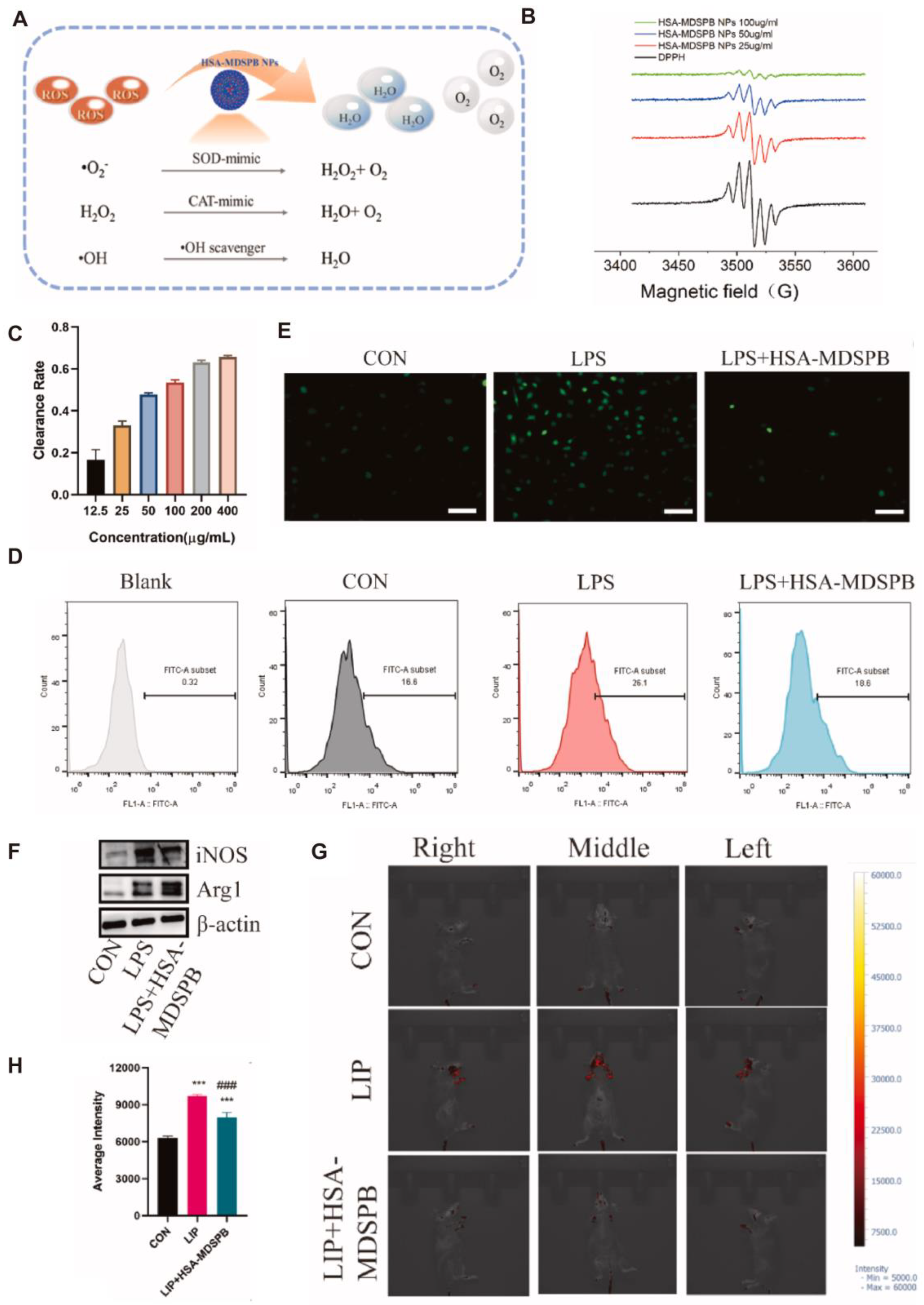
To address oxidative stress-induced tissue injury in chronic periodontitis, Xin et al. [34] developed melatonin-derived carbon dots (MT-CDs) with excellent water solubility, biocompatibility, and ROS-scavenging capacity. MT-CDs stabilized mitochondrial function, mitigated oxidative cellular damage, and suppressed pro-inflammatory cytokine production. In a mouse model of periodontitis, MT-CDs reduced alveolar bone resorption and osteoclast activity. Cao et al. [35] designed a dual-polymer functionalized melanin-silver nanocomposite (P/D-MNP-Ag) with sequential therapeutic capabilities. One polymer becomes more cationic in acidic conditions to enhance membrane penetration, while the phosphate-rich polymer chelates Ca2+, promoting adhesion to hydroxyapatite surfaces. Initially, the system enables Ag+-mediated photothermal antibacterial activity; subsequently, as silver ions deplete, melanin’s intrinsic antioxidative properties are restored, allowing ROS clearance in inflamed regions. Animal studies confirmed its ability to disrupt biofilms, attenuate inflammation, and reduce alveolar bone loss. Moreover, Cao et al. [36] developed negatively charged human serum albumin-crosslinked, manganese-doped Prussian blue nanoparticles (HSA-MDSPB NPs) capable of broadly scavenging ROS species—including superoxide anions (O2), hydroxyl radicals (OH), hydrogen peroxide (H2O2), and singlet oxygen (1O2). As shown in Figure 5A–C, the nanoparticles exhibited strong radical-neutralizing capacity in DPPH and ABTS assays, achieving over 60% scavenging efficiency at 200 μg/mL. In vitro, they significantly reduced LPS-induced oxidative damage in periodontal cells: fluorescence analysis (Figure 5D,E) demonstrated decreased ROS levels in mouse gingival fibroblasts and bone marrow-derived macrophages (BMDMs). Additionally, HSA-MDSPB NPs facilitated macrophage repolarization, favoring an anti-inflammatory M2 profile over the pro-inflammatory M1 state (Figure 5F), thus promoting tissue repair. In vivo, using a ligature-induced periodontitis model (Figure 5G,H), these nanoparticles markedly reduced ROS accumulation, downregulated pro-inflammatory cytokines (e.g., IL-1β, TNF-α), and mitigated alveolar bone loss. In summary, by integrating ROS scavenging, immunomodulation, and osteogenesis promotion, HSA-MDSPB NPs—and related nanoplatforms—present a promising nanotherapeutic strategy for managing periodontitis, particularly under oxidative stress-driven pathological conditions.

5.2. Ulcerative Colitis

IBD, a chronic immune-driven condition targeting the gastrointestinal tract, primarily includes Crohn’s disease (CD) and ulcerative colitis (UC). In 2021, the global age-standardized incidence rate among adolescents and young adults reached a notable level (4.08 per 100,000 population) [76]. Although the precise etiology of IBD remains unclear, dysregulation of intestinal immune responses is widely acknowledged as a central mechanism. The dysregulated production of ROS and RNS under oxidative stress conditions significantly accelerates IBD development [3]. These reactive species can compromise intestinal epithelial integrity and activate pro-inflammatory signaling pathways, thereby promoting the progression of inflammation.
UC, one of the most prevalent forms of IBD, is characterized by a complex pathogenesis, multifactorial etiology, and recurrent clinical course. Current treatment strategies face significant limitations, including suboptimal efficacy, adverse effects, and high relapse rates. Thus, the development of novel therapeutic approaches with high efficiency, targeted delivery, and minimal toxicity has become a research priority.
Recent advances highlight the therapeutic promise of nano-antioxidants in UC, attributed to their targeted delivery, high loading efficiency, and diverse bioactivities. For instance, Ye et al. [77] developed selenium nanoparticles modified with Eucommia ulmoides polysaccharides (EUP-SeNPs), which significantly alleviated DSS-induced UC in mice by restoring colonic histology, enhancing mucosal barrier function, reducing pro-inflammatory cytokines, and modulating gut microbiota. Moreover, EUP-SeNPs exerted anti-inflammatory effects via suppression of the TLR4/NF-κB signaling pathway and improved antioxidant capacity in colon tissues. In further studies, EUP-SeNPs were dually functionalized with somatostatin (SST) and manno-oligosaccharides (MOSs) for epithelial–macrophage dual-targeting and encapsulated within alginate-based microspheres (SA/SST/MOS@EUP-SeNPs) to improve oral stability and intestinal localization [37]. This system enabled targeted accumulation at inflamed sites, enhanced antioxidative and anti-inflammatory efficacy, promoted mucosal barrier repair, and effectively relieved UC symptoms. To address the limitations of traditional enema therapy—such as short retention time and systemic toxicity—thiolated anionic nanoliposomes were constructed to deliver the natural antioxidant gallic acid. This system exhibited enhanced mucosal adhesion at inflamed colonic sites, thereby prolonging drug residence time and enhancing local therapeutic effects. In vivo experiments demonstrated that this nanocarrier significantly inhibited NF-κB, HIF-1α, and MMP-9 expression, reduced neutrophil infiltration, and promoted MUC2 expression and mucosal repair [38]. Another strategy utilized ROS-responsive nanocarriers based on diselenide-modified carboxymethyl cellulose (CMC) to deliver curcumin. This platform displayed excellent biocompatibility and ROS-triggered release. Through electrostatic interaction with cationic proteins in inflamed tissues, the system achieved targeted delivery and effectively scavenged ROS, restored mucosal integrity, and regulated gut microbiota composition to mitigate UC pathology [39].
To overcome the low bioavailability of the resveratrol analog pterostilbene (PSB), researchers developed folate-modified, ROS-responsive nanoparticles (PSB@NP-FA) with an average size of ~231 nm. In inflammatory environments, PSB was released in a controlled manner, allowing efficient uptake by macrophages and epithelial cells. This formulation modulated dendritic cell function, promoted M2 polarization of macrophages, and regulated T cell infiltration. When co-administered with dexamethasone, it showed synergistic therapeutic benefits [40]. In the field of nanozymes, researchers constructed a polyethylene glycol-modified 2D Mo3Se4 nanosheet nanozyme (PMNFs), which mimicked multiple enzymatic activities, including peroxidase, glutathione peroxidase, superoxide dismutase, and catalase. In DSS-induced UC models, PMNFs efficiently scavenged ROS and improved colonic tissue damage. Mechanistically, they activated the Nrf2–Keap1 antioxidant pathway, inhibited TLR4/NF-κB-mediated inflammation, and protected tight junction proteins and MUC2 expression, thereby preserving intestinal barrier function [41]. Wei et al. [42] designed a pH-responsive, hyaluronic acid-modified nanoplatform (HA-L-Arg-CO2@NPs) for pterostilbene delivery. Upon encountering lysosomal acidic environments, the nanocarriers released CO2 to generate a “nanobomb” effect, promoting cytosolic drug delivery and site-specific accumulation. This system exhibited favorable colonic targeting and biocompatibility, significantly downregulated inflammatory mediators, restored barrier permeability, and effectively alleviated UC symptoms. Inspired by traditional Chinese medicine, Wu et al. [43] developed a novel functional carbon dot (MML-CD) synthesized via hydrothermal treatment of magnetite and Medicated Leaven. These ultrasmall nanoparticles (~3.2 nm) possess Fe-rich surfaces, excellent gastrointestinal stability, potent ROS-scavenging capacity, and high biocompatibility. In a UC mouse model, MML-CDs demonstrated promising therapeutic efficacy. They maintained physicochemical stability in various solutions, simulated digestive environments (Figure 6A), and displayed dose-dependent antioxidant activity against DPPH and ABTS+ (Figure 6B,C). MML-CDs significantly improved symptoms such as bloody diarrhea, restored colon length, and alleviated tissue damage and inflammation (Figure 6D). Furthermore, they promoted hemostasis, reduced bleeding time and volume and prolonged APTT and PT, indicating regulation of coagulation responses (Figure 6E–I). Collectively, these findings suggest that MML-CDs offer a novel oral nano-antioxidant strategy for UC treatment through multi-modal mechanisms including antioxidation, anti-inflammation, and hemostatic enhancement. In summary, nano-antioxidant strategies employing targeted delivery, functional modification, and responsive release show significant promise in UC therapy. These systems enable precise accumulation at inflamed sites, reinforce antioxidative and anti-inflammatory defenses, and modulate mucosal immunity and barrier function—offering new opportunities for effective clinical translation.

5.3. Crohn’s Disease

CD is one of the major subtypes of IBD, and its global incidence has been steadily rising since 2000, with a prevalence of up to 1 in 200 individuals in Western countries. A prospective population-based cohort study conducted in Hungary between 2007 and 2018 reported an average annual incidence of CD at 9.9 per 100,000 population (95% CI: 9.0–10.9) [78]. CD can affect any part of the gastrointestinal tract and is characterized by segmental, chronic, relapsing, and transmural inflammation. Clinical manifestations include abdominal pain, diarrhea, bowel obstruction, and perianal lesions. While the exact cause of CD is not fully understood, substantial evidence points to the critical roles of genetic susceptibility, oxidative stress, immune dysregulation, and environmental stimuli in driving its onset and development [79]. Among these factors, oxidative stress plays a key role in IBD, especially in UC [80]. Multiple meta-analyses have confirmed the presence of significant oxidative damage in UC patients, particularly during active disease phases. This is characterized by elevated levels of lipid peroxidation products (e.g., MDA, 8-iso-prostaglandin F2α), and protein oxidation markers (e.g., advanced oxidation protein products) [81].
In this context, antioxidant-based therapeutic strategies have emerged as promising interventions for the prevention and treatment of CD. Crohn’s disease develops through multifactorial interactions among genetic, immunological, microbial, and environmental influences, wherein oxidative stress caused by disrupted redox balance plays a central pathogenic role. Both endogenous and exogenous antioxidants can neutralize ROS and attenuate oxidative damage, thereby modulating intestinal inflammation and serving as important adjuvant approaches to mitigate disease progression and improve clinical outcomes [82]. Chen et al. [83] systematically investigated the associations between genetically predicted levels of antioxidants, minerals, and vitamins and the risk of Crohn’s disease (CD) and ulcerative colitis (UC). Although not directly addressing immune metabolism, their findings revealed that specific micronutrient levels may influence disease susceptibility through oxidative stress and mitochondrial pathways. Furthermore, mechanistic studies highlighted the role of AFG3L2 in preserving mitochondrial integrity and enhancing antioxidant signaling via the PPARA/GPX4 axis, suggesting potential links between metabolic stress responses and intestinal inflammation [84].
Nanocarrier-based antioxidant therapies have emerged as a promising avenue for Crohn’s disease treatment in recent years [85]. Ibrahim et al. [86] developed a multi-strain probiotic formulation encapsulated in a nanoparticle delivery system (BBLNPs), comprising Bifidobacterium breve, Bacillus coagulans, and Lactobacillus plantarum. This formulation significantly improved inflammation in mouse model. BBLNPs effectively reduced oxidative stress markers (e.g., MDA, ROS, H2O2) and enhanced the activity of antioxidant enzymes (SOD, CAT, GSH-Px), thereby exhibiting synergistic regulation of oxidative stress, inflammation, autophagy, and metabolic pathways and showing substantial therapeutic potential. Mohanbhai et al. [87] proposed a colon-targeted drug delivery system encapsulating melatonin within chitosan nanoparticles, further coated with Eudragit-S-100 to achieve colonic release upon oral administration. In an LPS-induced macrophage model, this system demonstrated strong nitric oxide-scavenging ability. In a CD mouse model, it alleviated tissue inflammation, protected mucosal structure, reduced goblet cell loss, and decreased immune cell infiltration, indicating both antioxidant and anti-inflammatory efficacy. Zhu et al. [88] designed a selenium-enriched Prussian blue nanozyme (Se-HMPB) with multi-enzyme mimetic activities, capable of efficiently scavenging ROS at inflammatory sites and exhibiting pronounced antioxidant effects. The selenium component endowed the nanozyme with glutathione peroxidase-like activity, enabling inhibition of ferroptosis and reduction in lipid peroxidation in intestinal epithelial cells. Furthermore, Se-HMPB promoted intestinal barrier repair and regulated T cell differentiation, thereby strengthening immune barrier function and demonstrating favorable therapeutic efficacy in CD models. Shah et al. [89] synthesized a novel nitric oxide (NO)-releasing nano-antioxidant, S-nitrosoglutathione–alginate (SNA), constructed via conjugation and nitrosation of glutathione with alginate. This nanoformulation exhibited high NO loading capacity and sustained-release properties, offering a new antioxidant therapeutic option with potential for oral delivery in CD treatment.
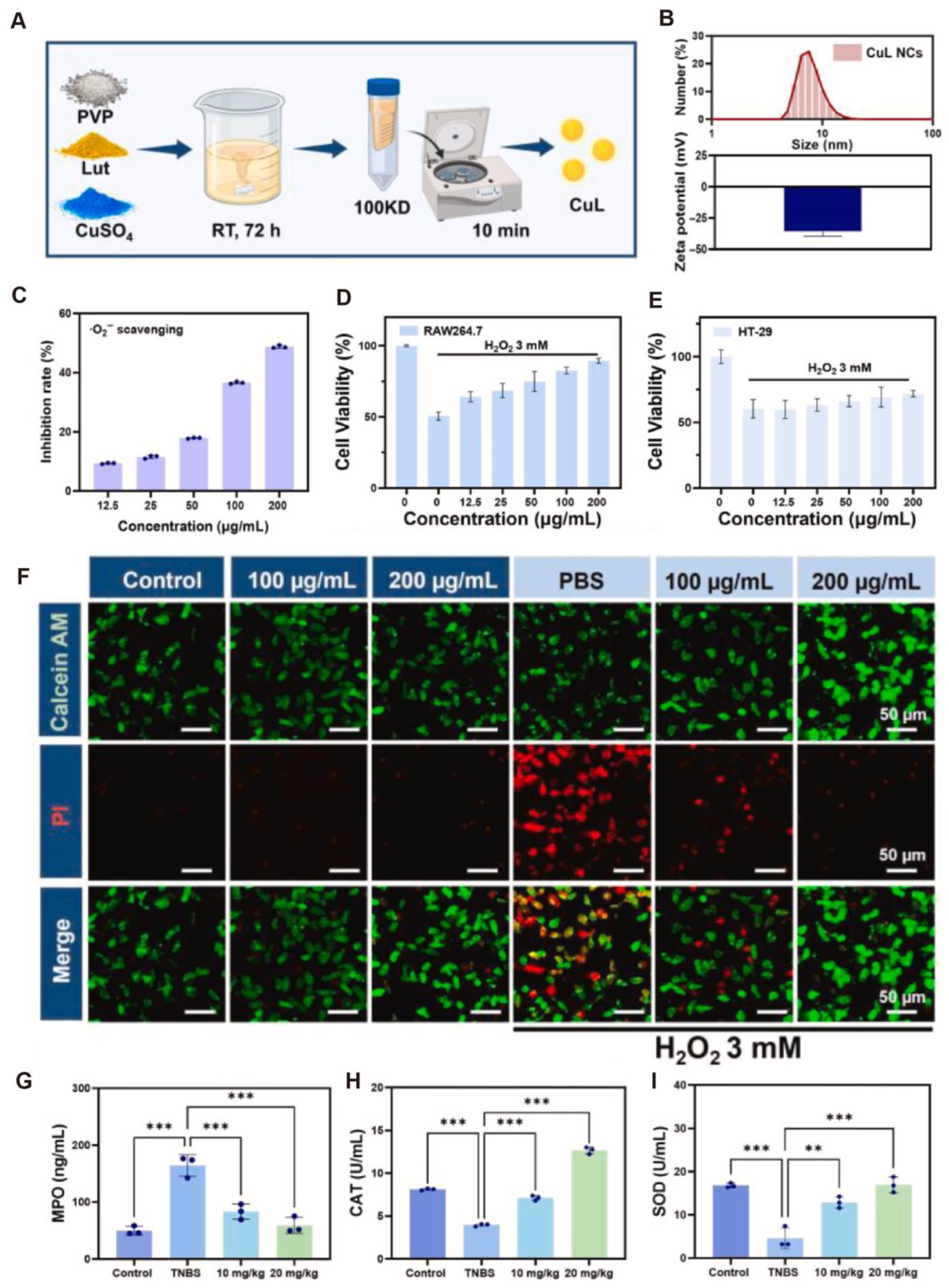
Additionally, Fu et al. [90] developed a copper–flavonoid nanocomposite (CuL NCs) with multifunctional properties. The composite was synthesized through metal–polyphenol coordination self-assembly between luteolin and Cu2+ ions, forming uniformly sized nanoparticles (~7.58 ± 0.90 nm) with excellent dispersibility and colloidal stability in aqueous solution (Figure 7A,B). CuL NCs also exhibited strong free radical-scavenging capacity, particularly against superoxide anions (O2•−), in a dose-dependent manner, outperforming individual components and their physical mixture (Figure 7C). In H2O2-induced oxidative stress cell models, CuL NCs significantly improved the viability of cells (RAW264.7; HT-29), fully restoring cell viability at 200 μg/mL and demonstrating robust cytoprotective effects (Figure 7D–F). In a TNBS-induced CD mouse model, CuL NCs markedly reduced MDA levels and MPO activity in colonic tissues while restoring CAT enzyme activity (Figure 7G–I), indicating effective mitigation of CD-associated oxidative injury. In summary, CuL NCs possess both SOD-like and CAT-like activities, efficiently scavenging ROS and ameliorating the oxidative microenvironment. Through a tri-modal mechanism encompassing antioxidant, anti-inflammatory, and barrier-protective actions, they exert multidimensional therapeutic effects in CD. These findings provide a strong theoretical foundation and emerging evidence for the application of nano-antioxidants in CD and other chronic inflammatory bowel diseases.

6. Others

In addition to their well-documented applications in autoimmune, acute, and chronic inflammatory diseases, antioxidants have also demonstrated significant therapeutic potential in a variety of dermatological conditions. As previously reviewed, antioxidants have shown promise in managing skin disorders such as atopic dermatitis and psoriasis [91]. Beyond their role in alopecia areata, we also explore their relevance in androgenetic alopecia, vitiligo, and rheumatoid arthritis.
Recent research has increasingly recognized oxidative stress as a key contributor to various forms of hair loss, especially androgenetic alopecia (AGA). Although conventional agents such as minoxidil can promote hair growth, they often exhibit slow onset, limited efficacy, and insufficient ability to mitigate oxidative stress-induced follicular damage. In this context, Xiao et al. [17] proposed the use of nanomolybdenum, a transition metal-based antioxidant material, as a novel therapeutic approach. Due to its excellent electron transfer capabilities, nanomolybdenum effectively modulates local redox homeostasis, promotes follicular regeneration, and downregulates the expression of iNOS, COX-2, and androgen receptors, thereby accelerating the hair regeneration process. Moreover, its combination with minoxidil produced a synergistic effect, suggesting a potential therapeutic approach for nanomaterial-based antioxidant therapy in alopecia. Zhang et al. [92] further emphasized the role of oxidative stress in AGA by employing machine learning techniques to identify an efficient SOD-mimicking nanomaterial—manganese thiophosphate (MnPS3). This material exhibited a half-maximal inhibitory concentration (IC50) of just 3.61 μg/mL, outperforming most reported SOD-like nanozymes. They also developed MnPS3-loaded microneedle patches capable of penetrating the skin and targeting hair follicles, thereby effectively scavenging local ROS. Yang et al. [93] developed a multifunctional microneedle patch delivery system based on a curcumin–zinc metal–organic framework (ZnMOF-MN), which enables efficient transdermal delivery of Zn2+ and curcumin. This system exerts antioxidant, anti-apoptotic, and anti-androgenic effects, significantly enhancing dermal papilla cell activity and promoting hair follicle regeneration. Yang et al. [94] developed a quercetin–polydopamine-integrated nanoplatform (PDA@QLipo) designed to modulate the perfollicular microenvironment and stimulate follicle regeneration. This nanosystem not only exhibited potent ROS-scavenging activity but also promoted neovascularization, collectively contributing to a regenerative microenvironment favorable for hair growth. The pathogenesis of vitiligo involves oxidative stress as a major driving force behind melanocyte injury and programmed cell death. ROS (e.g., O2 and H2O2) generated during melanin synthesis render melanocytes particularly vulnerable to oxidative injury [95]. A disrupted antioxidant system is evident in the affected skin of vitiligo patients, marked by elevated antioxidant enzyme activity (e.g., SOD) and impaired hydrogen peroxide detoxification. This redox imbalance may perpetuate melanocyte degeneration in depigmented lesions. To address this, Li et al. [96] developed a microneedle-delivered antioxidant and immunomodulatory nanodrug (PDA-JAKi) for targeted vitiligo therapy. This system consists of polydopamine nanoparticles loaded with the JAK inhibitor tofacitinib. The nanodrug alleviated ROS-induced melanocyte apoptosis and suppressed the release of self-antigens and subsequent immune activation. In vivo studies demonstrated its ability to restore pigmentation, reduce ROS accumulation, and improve tissue integrity, underscoring its dual antioxidant and immunomodulatory efficacy. In rheumatoid arthritis (RA), Han et al. [97] synthesized ultrasmall iron–quercetin coordination nanoclusters (Fe-Qur NCNs) to mitigate oxidative and inflammatory responses associated with RA. These nanoparticles retained the ROS-scavenging properties of quercetin while improving its water solubility and biocompatibility. In addition, excessive ROS were effectively neutralized by Fe-Qur NCNs, inhibited pro-inflammatory macrophage polarization, and suppressed NF-κB pathway activation, thereby reducing apoptosis and inflammatory cytokine release. In vivo, the nanodrug alleviated joint swelling and bone erosion, inhibited immune cell infiltration, promoted anti-inflammatory macrophage phenotypes, and attenuated osteoclast activity in RA mouse models. These results suggest that metal–natural product coordination nanoplatforms represent a promising strategy for managing RA and other oxidative stress-related diseases. In summary, oxidative stress contributes critically to the development of diverse cutaneous and immune-mediated inflammatory conditions. Nanomaterial-based antioxidant strategies, owing to their excellent targeting ability and biocompatibility, not only offer standalone therapeutic benefits but also demonstrate synergistic effects when combined with conventional treatments, highlighting their significant potential for clinical translation and further research.

7. Discussion

The treatment of IMIDs remains a significant challenge in both clinical and translational medicine, primarily due to their heterogeneous pathogenesis, chronic progression, and frequent systemic involvement [98]. IMIDs encompass a wide spectrum of disease types, including autoimmune disorders such as AA and MS, acute inflammatory injuries like AKI and ALI, and chronic inflammatory conditions such as IBD and periodontitis. A unifying pathological feature among these diseases is the excessive production of ROS, which not only directly damages tissues but also exacerbates immune dysregulation, thereby perpetuating a vicious cycle of inflammation [99,100]. Consequently, targeting oxidative stress has emerged as a theoretically grounded and promising therapeutic strategy.
However, the clinical efficacy of conventional antioxidant therapies remains limited, largely due to inherent shortcomings such as poor pharmacokinetics, inadequate tissue targeting, rapid metabolic degradation, and low intracellular retention. To address these limitations, recent research has increasingly focused on the development and application of nano-antioxidants. These nanomaterials or nano-delivery systems are engineered to exert direct or indirect antioxidative effects and demonstrate superior therapeutic performance. Nano-antioxidant platforms include metal oxide nanoparticles (e.g., zinc oxide, manganese dioxide), carbon-based nanomaterials (e.g., graphene oxide, fullerenes), polymeric nanoparticles, and lipid-based or exosome-mimetic delivery vehicles [101,102]. Compared to traditional antioxidants, these nanostructures offer several advantages, including enhanced stability in physiological environments, improved cellular uptake, targeted delivery to sites of inflammation or oxidative stress, and the ability to achieve sustained, localized drug release to maximize therapeutic efficacy. Furthermore, many nano-antioxidants possess intrinsic ROS-scavenging capabilities without the need for additional drug loading, thereby minimizing systemic exposure and reducing potential side effects [103]. Although nanoparticles or metallic nanoparticles (mNPs) are known to induce oxidative stress through excessive generation of ROS, recent advancements in nanoparticle engineering have demonstrated their potential to mitigate oxidative damage. Green-synthesized and surface-modified mNPs—such as plant-derived Cu and Pt nanoparticles—can scavenge free radicals via enzyme-mimetic activities resembling catalase or superoxide dismutase. These nano-antioxidants downregulate key pro-oxidant signaling pathways (e.g., NF-κB, JAK/STAT) and enhance cellular antioxidant defense mechanism [104]. Additionally, PEGylation and conjugation with natural antioxidants (e.g., flavonoids, polyphenols) offer effective strategies to reduce immune recognition and ROS-induced toxicity. Thus, depending on their composition and surface chemistry, mNPs can function either as pro-oxidant or antioxidant agents, providing a versatile platform for therapeutic modulation of oxidative stress [105].

8. Conclusions

An increasing body of evidence from in vitro studies, animal models, and early translational investigations suggests that nano-antioxidants can effectively alleviate inflammation, preserve tissue integrity, and modulate immune responses. For instance, in autoimmune diseases like AA, nano-antioxidants have been shown to mitigate inflammation by scavenging superoxide anions and hydrogen peroxide, offering a novel therapeutic avenue [17]. In MS models, polymeric nanoparticles loaded with antioxidants can traverse the blood–brain barrier and precisely deliver therapeutic agents to neuroinflammatory sites, thereby reducing demyelination and preserving neurological function [106]. In acute inflammatory conditions such as AKI, nano-antioxidants help alleviate renal oxidative stress, maintain mitochondrial function, and potentially enhance treatment outcomes [107]. Similarly, in chronic inflammatory diseases like IBD and ulcerative colitis, these agents promote mucosal healing, modulate gut microbiota composition, and reduce inflammatory cell infiltration in the colon [108].
Despite these encouraging preclinical findings, the clinical translation of nano-antioxidants remains in its infancy and faces several critical challenges. First, the biocompatibility and long-term safety of various nanomaterials are not yet fully understood, particularly under conditions of repeated or prolonged use. Second, the heterogeneity of IMIDs necessitates precision medicine approaches, as disease-specific redox pathways, immune activation patterns, and tissue microenvironments may significantly influence the efficacy and biodistribution of nano-antioxidants. Third, practical issues such as scalable manufacturing, regulatory approval, and cost-effectiveness must be systematically addressed. Moreover, nano-antioxidants must demonstrate therapeutic efficacy that is superior to—or at least non-inferior to—current standard treatments in rigorously designed randomized controlled clinical trials before widespread adoption can be recommended. Future research should prioritize the development of “smart” nano-antioxidant systems capable of responding to disease-specific microenvironmental cues (e.g., pH, ROS levels, enzymatic activity) to achieve precise and controlled drug release. Combining nano-antioxidant therapy with other modalities, such as immunomodulators, gene editing technologies (e.g., CRISPR/Cas9), or cell-based therapies, may further enhance therapeutic outcomes through synergistic effects. Additionally, personalized nanomedicine strategies—tailoring antioxidant interventions based on an individual’s disease phenotype, genetic markers, and microbiome characteristics—hold promise for improving treatment precision and minimizing adverse effects. In summary, nano-antioxidants represent a highly promising and multifunctional therapeutic platform for the management of diverse IMIDs. By overcoming the limitations of traditional antioxidants and leveraging the unique properties of nanomaterials, this strategy offers novel avenues for the targeted and sustained regulation of redox balance and immune function. Although substantial progress has been made, multidisciplinary collaboration and robust clinical validation are essential to fully realize their clinical potential. If successfully translated, nano-antioxidant therapies may substantially transform the therapeutic landscape of IMIDs and bring new hope to affected patients.

Author Contributions

Y.X. and Y.-Q.R.: study conception and design. H.-W.S. and B.-C.Y.: organized the article’s table section. Y.X. and H.-W.S.: organized the figures. Y.X. and H.-W.S.: copyright permission. H.-W.S. and B.-C.Y.: drafting the manuscripts. Y.X. and Y.-Q.R.: critical revision of the manuscript. All authors have read and agreed to the published version of the manuscript.

Funding

This work was supported by the Zhejiang Provincial Natural Science Foundation of China under grant LQN25H110001, the Key Research and Development Plan of Zhejiang Province (2025C02080), ZJU-GENSCI CHILDREN’S HEALTH RESEARCH & DEVELOPMENT CENTER (ZJU-GENSCI2024YB004), Zhejiang Provincial Natural Science Foundation of China (LTGD23H110001), National Key R&D Program of China (Grant No.2022YFC2705300), and 2023 Xiamen Municipal Natural Science Foundation Project (3502Z202374016).

Institutional Review Board Statement

Not applicable.

Informed Consent Statement

Not applicable.

Data Availability Statement

No new data were created in this study. Permission for re-use of previously published figures has been obtained.

Acknowledgments

We thank Su-Zhuang Hong, Qi Chen, Ji-Peng Liu and Ke-Yi Ren for assistance with figure arrangement and Zheng-Ying Zhang, Yu-Xiang Yang, Can Lai and Xiao-Hui Ma for support in copyright permissions and organizing the article’s table section.

Conflicts of Interest

The authors declare no conflicts of interest.

References

  1. Luo, H. Global burden and cross-country inequalities in six major immune-mediated inflammatory diseases from 1990 to 2021: A systemic analysis of the Global Burden of Disease Study 2021. Autoimmun. Rev. 2024, 23, 103639. [Google Scholar] [CrossRef]
  2. Dobrica, E.C.; Cozma, M.A.; Gaman, M.A.; Voiculescu, V.M.; Gaman, A.M. The Involvement of Oxidative Stress in Psoriasis: A Systematic Review. Antioxidants 2022, 11, 282. [Google Scholar] [CrossRef] [PubMed]
  3. Muro, P.; Zhang, L.; Li, S.; Zhao, Z.; Jin, T.; Mao, F.; Mao, Z. The emerging role of oxidative stress in inflammatory bowel disease. Front. Endocrinol. 2024, 15, 1390351. [Google Scholar] [CrossRef]
  4. Abot, A.; Fried, S.; Cani, P.D.; Knauf, C. Reactive Oxygen Species/Reactive Nitrogen Species as Messengers in the Gut: Impact on Physiology and Metabolic Disorders. Antioxid. Redox Signal. 2022, 37, 394–415. [Google Scholar] [CrossRef]
  5. Lapenna, D. Glutathione and glutathione-dependent enzymes: From biochemistry to gerontology and successful aging. Ageing Res. Rev. 2023, 92, 102066. [Google Scholar] [CrossRef] [PubMed]
  6. Zhang, J.; Ren, X.; Nie, Z.; You, Y.; Zhu, Y.; Chen, H.; Yu, H.; Mo, G.P.; Su, L.; Peng, Z.; et al. Dual-responsive renal injury cells targeting nanoparticles for vitamin E delivery to treat ischemia reperfusion-induced acute kidney injury. J. Nanobiotechnol. 2024, 22, 626. [Google Scholar] [CrossRef]
  7. Akanchise, T.; Angelova, A. Potential of Nano-Antioxidants and Nanomedicine for Recovery from Neurological Disorders Linked to Long COVID Syndrome. Antioxidants 2023, 12, 393. [Google Scholar] [CrossRef] [PubMed]
  8. Bardestani, A.; Ebrahimpour, S.; Esmaeili, A.; Esmaeili, A. Quercetin attenuates neurotoxicity induced by iron oxide nanoparticles. J. Nanobiotechnol. 2021, 19, 327. [Google Scholar] [CrossRef] [PubMed]
  9. Chen, X.; Zhang, L.; Zeng, H.; Meng, W.; Liu, G.; Zhang, W.; Zhao, P.; Zhang, Q.; Chen, M.; Chen, J. Manganese-Based Immunomodulatory Nanocomposite with Catalase-Like Activity and Microwave-Enhanced ROS Elimination Ability for Efficient Rheumatoid Arthritis Therapy. Small 2023, 19, e2304610. [Google Scholar] [CrossRef]
  10. Laurindo, L.F.; de Carvalho, G.M.; de Oliveira Zanuso, B.; Figueira, M.E.; Direito, R.; de Alvares Goulart, R.; Buglio, D.S.; Barbalho, S.M. Curcumin-Based Nanomedicines in the Treatment of Inflammatory and Immunomodulated Diseases: An Evidence-Based Comprehensive Review. Pharmaceutics 2023, 15, 229. [Google Scholar] [CrossRef]
  11. Indriyani, N.; Nur’aeny, N. The Therapeutic Effects of Curcumin on Oral Disease: A Systematic Review. Clin. Pharmacol. 2025, 17, 13–24. [Google Scholar] [CrossRef]
  12. Mohammadi, S.; Farjam, H.; Hosseini, S.; Larijani, K. Inhibitory effects of saffron compounds on multiple sclerosis: Molecular docking and dynamic, enhancing properties with gold nanoparticles and evaluating antioxidant, antibacterial, cytotoxicity. Heliyon 2025, 11, e42693. [Google Scholar] [CrossRef] [PubMed]
  13. Bemidinezhad, A.; Mirzavi, F.; Gholamhosseinian, H.; Gheybi, F.; Soukhtanloo, M. Gold-containing liposomes and glucose-coated gold nanoparticles enhances the radiosensitivity of B16F0 melanoma cells via increasing apoptosis and ROS production. Life Sci. 2023, 318, 121495. [Google Scholar] [CrossRef]
  14. Liu, X.; Ou, X.; Zhang, T.; Li, X.; Qiao, Q.; Jia, L.; Xu, Z.; Zhang, F.; Tian, T.; Lan, H.; et al. In situ neutrophil apoptosis and macrophage efferocytosis mediated by Glycyrrhiza protein nanoparticles for acute inflammation therapy. J. Control. Release 2024, 369, 215–230. [Google Scholar] [CrossRef] [PubMed]
  15. Yu, H.; Jin, F.; Liu, D.; Shu, G.; Wang, X.; Qi, J.; Sun, M.; Yang, P.; Jiang, S.; Ying, X.; et al. ROS-responsive nano-drug delivery system combining mitochondria-targeting ceria nanoparticles with atorvastatin for acute kidney injury. Theranostics 2020, 10, 2342–2357. [Google Scholar] [CrossRef]
  16. Hu, Q.; Li, J.; Wang, T.; Xu, X.; Duan, Y.; Jin, Y. Polyphenolic Nanoparticle-Modified Probiotics for Microenvironment Remodeling and Targeted Therapy of Inflammatory Bowel Disease. ACS Nano 2024, 18, 12917–12932. [Google Scholar] [CrossRef]
  17. Xiao, Q.; Lu, Y.; Yao, W.; Gong, C.; Jia, C.; Gao, J.; Guo, J.; Qiu, T.; Jiang, Y.; Huang, M.; et al. Molybdenum nanoparticles as a potential topical medication for alopecia treatment through antioxidant pathways that differ from minoxidil. J. Trace Elem. Med. Biol. 2024, 82, 127368. [Google Scholar] [CrossRef]
  18. Motavaf, M.; Sadeghizadeh, M.; Babashah, S.; Zare, L.; Javan, M. Dendrosomal nanocurcumin promotes remyelination through induction of oligodendrogenesis in experimental demyelination animal model. J. Tissue Eng. Regen. Med. 2020, 14, 1449–1464. [Google Scholar] [CrossRef]
  19. Ibrahim Fouad, G.; Mabrouk, M.; El-Sayed, S.A.M.; Abdelhameed, M.F.; Rizk, M.Z.; Beherei, H.H. Berberine-loaded iron oxide nanoparticles alleviate cuprizone-induced astrocytic reactivity in a rat model of multiple sclerosis. Biometals 2025, 38, 203–229. [Google Scholar] [CrossRef]
  20. Li, Z.; Chen, K.; Shao, Q.; Lu, H.; Zhang, X.; Pu, Y.; Sun, X.; He, H.; Cao, L. Nanoparticulate MgH(2) ameliorates anxiety/depression-like behaviors in a mouse model of multiple sclerosis by regulating microglial polarization and oxidative stress. J. Neuroinflamm. 2023, 20, 16. [Google Scholar] [CrossRef] [PubMed]
  21. Abdelalim, L.R.; Elnaggar, Y.S.R.; Abdallah, O.Y. Lactoferrin, chitosan double-coated oleosomes loaded with clobetasol propionate for remyelination in multiple sclerosis: Physicochemical characterization and in-vivo assessment in a cuprizone-induced demyelination model. Int. J. Biol. Macromol. 2024, 277 Pt 1, 134144. [Google Scholar] [CrossRef] [PubMed]
  22. Rezaeimanesh, N.; Rafiee, P.; Saeedi, R.; Khosravian, P.; Sahraian, M.A.; Eskandarieh, S.; Moghadasi, A.N.; Jahromi, S.R. The effect of crocin-selenium nanoparticles on the cognition and oxidative stress markers of multiple sclerosis patients: A randomized triple-blinded placebo-controlled clinical trial. Biometals 2024, 37, 305–319. [Google Scholar] [CrossRef] [PubMed]
  23. Yang, Y.; Zhao, Y.; Liu, H.; Wu, X.; Guo, M.; Xie, L.; Wang, G.; Shi, J.; Yu, W.; Dong, G. Inflammation-Targeted Biomimetic Nano-Decoys via Inhibiting the Infiltration of Immune Cells and Effectively Delivering Glucocorticoids for Enhanced Multiple Sclerosis Treatment. Adv. Healthc. Mater. 2025, 14, e2402965. [Google Scholar] [CrossRef] [PubMed]
  24. Gu, J.; Zhang, P.; Li, H.; Wang, Y.; Huang, Y.; Fan, L.; Ma, X.; Qian, X.; Xi, J. Cerium–Luteolin Nanocomplexes in Managing Inflammation-Related Diseases by Antioxidant and Immunoregulation. ACS Nano 2024, 18, 6229–6242. [Google Scholar] [CrossRef]
  25. Zheng, Y.; Yi, H.; Zhan, Z.; Xue, S.S.; Tang, G.; Yu, X.; Zhang, D.Y. Reactive oxygen/nitrogen species scavenging and inflammatory regulation by renal-targeted bio-inspired rhodium nanozymes for acute kidney injury theranostics. J. Colloid. Interface Sci. 2024, 662, 413–425. [Google Scholar] [CrossRef]
  26. Yan, R.; Cui, W.; Ma, W.; Li, J.; Liu, Z.; Lin, Y. Typhaneoside-Tetrahedral Framework Nucleic Acids System: Mitochondrial Recovery and Antioxidation for Acute Kidney Injury treatment. ACS Nano 2023, 17, 8767–8781. [Google Scholar] [CrossRef]
  27. Pan, Q.; Xie, L.; Cai, P.; Wu, D.; Zhu, H.; Xu, L.; Liu, R.; Luo, K.; He, B.; Pu, Y. Acid-Resistant Nano-antioxidants Based on Epigallocatechin Gallate Alleviate Acute Intestinal and Kidney Inflammation. ACS Appl. Mater. Interfaces 2024, 16, 46090–46101. [Google Scholar] [CrossRef]
  28. Jia, F.; Yu, B.; Li, J.; Cai, F.; Fu, G.; Jin, Q.; Ji, J. Supramolecular Nano-Assembly of Caffeate-Strengthened Phenylboronic Ester with Multistep ROS Scavenging Ability for Targeted Therapy of Acute Kidney Injury. Adv. Healthc. Mater. 2023, 12, e2301615. [Google Scholar] [CrossRef]
  29. Luo, F.; Zhu, B.; Wu, D.; Xu, Y.; Chen, T.; Li, Y.; Hu, J. Construction of Phlorotannin-Based Nanoparticles for Alleviating Acute Liver Injury. ACS Appl. Mater. Interfaces 2023, 15, 47338–47349. [Google Scholar] [CrossRef]
  30. Jang, D.; Choi, H.; Lee, J.; Chun, Y.; Heo, Y.H.; Lee, L.P.; Ahn, D.J.; Shin, I.S.; Kim, D.H.; Seo, Y.H.; et al. Inflamed Tissue-Targeting Polyphenol-Condensed Antioxidant Nanoparticles with Therapeutic Potential. Adv. Healthc. Mater. 2025, 14, e2500495. [Google Scholar] [CrossRef]
  31. Wang, Y.; Liu, X.; Li, K.; Wang, X.; Zhang, X.; Qian, D.; Meng, X.; Yu, L.; Yan, X.; He, Z. Self-Sulfhydrated, Nitro-Fixed Albumin Nanoparticles as a Potent Therapeutic Agent for the Treatment of Acute Liver Injury. ACS Nano 2024, 18, 20772–20791. [Google Scholar] [CrossRef] [PubMed]
  32. Zhao, C.; Li, Z.; Chen, J.; Su, L.; Wang, J.; Chen, D.S.; Ye, J.; Liao, N.; Yang, H.; Song, J.; et al. Site-Specific Biomimicry of Antioxidative Melanin Formation and Its Application for Acute Liver Injury Therapy and Imaging. Adv. Mater. 2021, 33, e2102391. [Google Scholar] [CrossRef] [PubMed]
  33. Yang, S.Y.; Hu, Y.; Zhao, R.; Zhou, Y.N.; Zhuang, Y.; Zhu, Y.; Ge, X.L.; Lu, T.W.; Lin, K.L.; Xu, Y.J. Quercetin-loaded mesoporous nano-delivery system remodels osteoimmune microenvironment to regenerate alveolar bone in periodontitis via the miR-21a-5p/PDCD4/NF-kappaB pathway. J. Nanobiotechnol. 2024, 22, 94. [Google Scholar] [CrossRef]
  34. Xin, X.; Liu, J.; Liu, X.; Xin, Y.; Hou, Y.; Xiang, X.; Deng, Y.; Yang, B.; Yu, W. Melatonin-Derived Carbon Dots with Free Radical Scavenging Property for Effective Periodontitis Treatment via the Nrf2/HO-1 Pathway. ACS Nano 2024, 18, 8307–8324. [Google Scholar] [CrossRef]
  35. Cao, B.; Zhang, J.; Ma, Y.; Wang, Y.; Li, Y.; Wang, R.; Cao, D.; Yang, Y.; Zhang, R. Dual-Polymer Functionalized Melanin-AgNPs Nanocomposite with Hydroxyapatite Binding Ability to Penetrate and Retain in Biofilm Sequentially Treating Periodontitis. Small 2024, 20, e2400771. [Google Scholar] [CrossRef]
  36. Cao, B.; Da, X.; Wu, W.; Xie, J.; Li, X.; Wang, X.; Xu, H.; Gao, J.; Yang, H.; Su, J. Multifunctional human serum albumin-crosslinked and self-assembling nanoparticles for therapy of periodontitis by anti-oxidation, anti-inflammation and osteogenesis. Mater. Today Bio. 2024, 28, 101163. [Google Scholar] [CrossRef]
  37. Ye, R.; Guo, J.; Yang, Z.; Wang, Z.; Chen, Y.; Huang, J.; Dong, Y. Somatostatin and Mannooligosaccharide Modified Selenium Nanoparticles with Dual-Targeting for Ulcerative Colitis Treatment. ACS Nano 2025, 19, 14914–14930. [Google Scholar] [CrossRef]
  38. Kanika; Ahmad, A.; Kumar, A.; Rahul; Mishra, R.K.; Ali, N.; Navik, U.; Parvez, S.; Khan, R. Leveraging thiol-functionalized biomucoadhesive hybrid nanoliposome for local therapy of ulcerative colitis. Biomaterials 2025, 312, 122747. [Google Scholar] [CrossRef]
  39. Cai, W.Q.; Liang, W.; Li, D.; Dai, W.; Li, Z.; Wei, X.; Cheng, L.; Zhang, B.B.; Yang, Q. Reactive Oxygen Species-Responsive Polymer Drug Delivery System Targeted Oxidative Stressed Colon Cells to Ameliorate Colitis. ACS Nano 2025, 19, 17287–17308. [Google Scholar] [CrossRef] [PubMed]
  40. Yan, X.; Meng, L.; Zhang, X.; Deng, Z.; Gao, B.; Zhang, Y.; Yang, M.; Ma, Y.; Zhang, Y.; Tu, K.; et al. Reactive oxygen species-responsive nanocarrier ameliorates murine colitis by intervening colonic innate and adaptive immune responses. Mol. Ther. 2023, 31, 1383–1401. [Google Scholar] [CrossRef]
  41. Guo, H.; Guo, H.; Xie, Y.; Chen, Y.; Lu, C.; Yang, Z.; Zhu, Y.; Ouyang, Y.; Zhang, Y.; Wang, X. Mo3Se4 nanoparticle with ROS scavenging and multi-enzyme activity for the treatment of DSS-induced colitis in mice. Redox Biol. 2022, 56, 102441. [Google Scholar] [CrossRef] [PubMed]
  42. Wei, W.; Zhang, Y.; Li, R.; Cao, Y.; Yan, X.; Ma, Y.; Zhang, Y.; Yang, M.; Zhang, M. Oral Delivery of Pterostilbene by L-Arginine-Mediated “Nano-Bomb” Carrier for the Treatment of Ulcerative Colitis. Int. J. Nanomed. 2022, 17, 603–616. [Google Scholar] [CrossRef]
  43. Wu, T.; Bai, X.; Zhang, Y.; Dai, E.; Ma, J.; Yu, C.; He, C.; Li, Q.; Yang, Y.; Kong, H.; et al. Natural medicines-derived carbon dots as novel oral antioxidant administration strategy for ulcerative colitis therapy. J. Nanobiotechnol. 2024, 22, 511. [Google Scholar] [CrossRef] [PubMed]
  44. Dai, Z.; Wang, E.H.C.; Petukhova, L.; Chang, Y.; Lee, E.Y.; Christiano, A.M. Blockade of IL-7 signaling suppresses inflammatory responses and reverses alopecia areata in C3H/HeJ mice. Sci. Adv. 2021, 7, eabd1866. [Google Scholar] [CrossRef]
  45. Zhou, C.; Li, X.; Wang, C.; Zhang, J. Alopecia Areata: An Update on Etiopathogenesis, Diagnosis, and Management. Clin. Rev. Allergy Immunol. 2021, 61, 403–423. [Google Scholar] [CrossRef]
  46. McMichael, A.J.; Roberson, M.L. Characterizing Epidemiology and Burden of Disease in Alopecia Areata-Making It Count. JAMA Dermatol. 2023, 159, 369–370. [Google Scholar] [CrossRef]
  47. Ma, Y.Q.; Sun, Z.; Li, Y.M.; Xu, H. Oxidative stress and alopecia areata. Front. Med. 2023, 10, 1181572. [Google Scholar] [CrossRef]
  48. Shakoei, S.; Mirmiranpoor, H.; Nakhjavani, M.; Nasimi, M.; Bakhshi, G.; Azizpour, A. Oxidative stress and antioxidant markers in patients with alopecia areata: A comparative cross-sectional study. Indian J. Dermatol. Venereol. Leprol. 2023, 89, 411–415. [Google Scholar] [CrossRef]
  49. Sachdeva, S.; Khurana, A.; Goyal, P.; Sardana, K. Does oxidative stress correlate with disease activity and severity in alopecia areata? An analytical study. J. Cosmet. Dermatol. 2022, 21, 1629–1634. [Google Scholar] [CrossRef] [PubMed]
  50. Peterle, L.; Sanfilippo, S.; Borgia, F.; Cicero, N.; Gangemi, S. Alopecia Areata: A Review of the Role of Oxidative Stress, Possible Biomarkers, and Potential Novel Therapeutic Approaches. Antioxidants 2023, 12, 135. [Google Scholar] [CrossRef]
  51. Taskin, S.; Celik, H.; Cakirca, G.; Manav, V.; Taskin, A. Nitric oxide synthase activity: A novel potential biomarker for predicting Alopecia areata. J. Cosmet. Dermatol. 2022, 21, 7075–7080. [Google Scholar] [CrossRef]
  52. Perlmutter, J.; Akouris, P.P.; Fremont, S.; Yang, B.; Toth, E.; Eze, M.; Wiseman, M. The Role of Reactive Oxygen Species in the Pathogenesis of Alopecia Areata: A Systematic Review and Meta-Analysis. Skin. Pharmacol. Physiol. 2025, 38, 59–67. [Google Scholar] [CrossRef]
  53. Younis, N.; Puigmal, N.; El Kurdi, A.; Badaoui, A.; Zhang, D.; Morales-Garay, C.; Saad, A.; Cruz, D.; Al Rahy, N.; Daccache, A.; et al. Microneedle-Mediated Delivery of Immunomodulators Restores Immune Privilege in Hair Follicles and Reverses Immune-Mediated Alopecia. Adv. Mater. 2024, 36, e2312088. [Google Scholar] [CrossRef]
  54. Jeong, D.I.; Lee, S.Y.; Kim, S.; Jo, S.-M.; Yang, H.; You, J.; Hwang, C.; Huh, J.W.; Choi, Y.E.; Jo, W.; et al. Baricitinib-loaded separable microneedle array patch (S-MAP) based on hyaluronic acid for alopecia areata therapy. Carbohydr. Polym. 2025, 364, 123789. [Google Scholar] [CrossRef]
  55. Hindam, M.O.; Ahmed, L.A.; El Sayed, N.S.; Khattab, M.; Sallam, N.A. Repositioning of baricitinib for management of memory impairment in ovariectomized/D-galactose treated rats: A potential role of JAK2/STAT3-PI3K/AKT/mTOR signaling pathway. Life Sci. 2024, 351, 122838. [Google Scholar] [CrossRef]
  56. McGinley, M.P.; Goldschmidt, C.H.; Rae-Grant, A.D. Diagnosis and Treatment of Multiple Sclerosis. JAMA 2021, 325, 765–779. [Google Scholar] [CrossRef]
  57. Graf, J.; Akmatov, M.K.; Meuth, S.G.; Tremlett, H.; Holstiege, J. Updated Multiple Sclerosis Incidence, 2015–2022. JAMA Neurol 2024, 81, 1100–1102. [Google Scholar] [CrossRef] [PubMed]
  58. Dash, U.C.; Bhol, N.K.; Swain, S.K.; Samal, R.R.; Nayak, P.K.; Raina, V.; Panda, S.K.; Kerry, R.G.; Duttaroy, A.K.; Jena, A.B. Oxidative stress and inflammation in the pathogenesis of neurological disorders: Mechanisms and implications. Acta Pharm. Sin. B 2025, 15, 15–34. [Google Scholar] [CrossRef]
  59. Kasprzak-Drozd, K.; Nizinski, P.; Hawryl, A.; Gancarz, M.; Hawryl, D.; Oliwa, W.; Palka, M.; Markowska, J.; Oniszczuk, A. Potential of Curcumin in the Management of Skin Diseases. Int. J. Mol. Sci. 2024, 25, 3617. [Google Scholar] [CrossRef] [PubMed]
  60. Lu, L.; Qi, S.; Chen, Y.; Luo, H.; Huang, S.; Yu, X.; Luo, Q.; Zhang, Z. Targeted immunomodulation of inflammatory monocytes across the blood-brain barrier by curcumin-loaded nanoparticles delays the progression of experimental autoimmune encephalomyelitis. Biomaterials 2020, 245, 119987. [Google Scholar] [CrossRef] [PubMed]
  61. Sanadgol, N.; Barati, M.; Houshmand, F.; Hassani, S.; Clarner, T.; Shahlaei, M.; Golab, F. Metformin accelerates myelin recovery and ameliorates behavioral deficits in the animal model of multiple sclerosis via adjustment of AMPK/Nrf2/mTOR signaling and maintenance of endogenous oligodendrogenesis during brain self-repairing period. Pharmacol. Rep. 2020, 72, 641–658. [Google Scholar] [CrossRef] [PubMed]
  62. Qin, W.; Huang, J.; Zhang, M.; Xu, M.; He, J.; Liu, Q. Nanotechnology-Based Drug Delivery Systems for Treating Acute Kidney Injury. ACS Biomater. Sci. Eng. 2024, 10, 6078–6096. [Google Scholar] [CrossRef]
  63. Jensen, S.K.; Rasmussen, T.B.; Jacobsen, B.H.; Heide-Jørgensen, U.; Sawhney, S.; Gammelager, H.; Birn, H.; Johnsen, S.P.; Christiansen, C.F. Regional variation in incidence and prognosis of acute kidney injury. Nephrol. Dial. Transpl. 2024, 39, 1171–1180. [Google Scholar] [CrossRef]
  64. Levey, A.S.; Eckardt, K.-U.; Dorman, N.M.; Christiansen, S.L.; Hoorn, E.J.; Ingelfinger, J.R.; Inker, L.A.; Levin, A.; Mehrotra, R.; Palevsky, P.M.; et al. Nomenclature for kidney function and disease: Report of a Kidney Disease: Improving Global Outcomes (KDIGO) Consensus Conference. Kidney Int. 2020, 97, 1117–1129. [Google Scholar] [CrossRef] [PubMed]
  65. He, K.; Zhou, D.; Pu, Z.; Chen, S.; Shen, Y.; Zhao, S.; Qian, X.; Hu, Q.; Wu, X.; Xie, Z.; et al. Cellular Senescence in Acute Liver Injury: What Happens to the Young Liver? Aging Dis. 2024, 16, 1347–1362. [Google Scholar] [CrossRef]
  66. Yuan, X.; Zhou, Y.; Sun, J.; Wang, S.; Hu, X.; Li, J.; Huang, J.; Chen, N. Preventing acute liver injury via hepatocyte-targeting nano-antioxidants. Cell Prolif. 2023, 56, e13494. [Google Scholar] [CrossRef]
  67. Song, B.; Zhang, C.; Hu, W.; Guo, C.; Xia, Z.; Hu, W.; Qin, M.; Jiang, W.; Lv, J.; Xu, D.; et al. Nano-designed carbon monoxide donor SMA/CORM2 exhibits protective effect against acetaminophen induced liver injury through macrophage reprograming and promoting liver regeneration. J. Control. Release 2021, 331, 350–363. [Google Scholar] [CrossRef]
  68. Liu, M.; Wu, H.; Li, Q.; Liu, H.; Chen, C.; Yin, F.; Wang, H.; Zha, Z.; Wang, F. Mn3O4 nanozymes prevent acetaminophen-induced acute liver injury by attenuating oxidative stress and countering inflammation. J. Colloid Interface Sci. 2024, 654 Pt A, 83–95. [Google Scholar] [CrossRef]
  69. Aslam, M.; Fatima, B.; Batool, R.; Imran, M.; Hussain, D.; Najam-Ul-Haq, M.; Mehmood, R. Saponin Gallate-Loaded Gd-Doped Zinc-Gallium Layered Double Hydroxides (Zn/Ga@Gd-LDH) Nanocarrier for Attenuating NF-kappaB-Mediated Inflammation. ACS Appl. Mater. Interfaces 2025, 17, 26263–26281. [Google Scholar] [CrossRef]
  70. Trindade, D.; Carvalho, R.; Machado, V.; Chambrone, L.; Mendes, J.J.; Botelho, J. Prevalence of periodontitis in dentate people between 2011 and 2020: A systematic review and meta-analysis of epidemiological studies. J. Clin. Periodontol. 2023, 50, 604–626. [Google Scholar] [CrossRef] [PubMed]
  71. Kassebaum, N.J.; Bernabe, E.; Dahiya, M.; Bhandari, B.; Murray, C.J.; Marcenes, W. Global burden of severe periodontitis in 1990–2010: A systematic review and meta-regression. J. Dent. Res. 2014, 93, 1045–1053. [Google Scholar] [CrossRef] [PubMed]
  72. Chen, M.X.; Zhong, Y.J.; Dong, Q.Q.; Wong, H.M.; Wen, Y.F. Global, regional, and national burden of severe periodontitis, 1990–2019: An analysis of the Global Burden of Disease Study 2019. J. Clin. Periodontol. 2021, 48, 1165–1188. [Google Scholar] [CrossRef]
  73. Sczepanik, F.S.C.; Grossi, M.L.; Casati, M.; Goldberg, M.; Glogauer, M.; Fine, N.; Tenenbaum, H.C. Periodontitis is an inflammatory disease of oxidative stress: We should treat it that way. Periodontol. 2000 2020, 84, 45–68. [Google Scholar] [CrossRef]
  74. Li, L.; Qin, W.; Ye, T.; Wang, C.; Qin, Z.; Ma, Y.; Mu, Z.; Jiao, K.; Tay, F.R.; Niu, W.; et al. Bioactive Zn-V-Si-Ca Glass Nanoparticle Hydrogel Microneedles with Antimicrobial and Antioxidant Properties for Bone Regeneration in Diabetic Periodontitis. ACS Nano 2025, 19, 7981–7995. [Google Scholar] [CrossRef]
  75. Wang, Y.; Li, C.; Wan, Y.; Qi, M.; Chen, Q.; Sun, Y.; Sun, X.; Fang, J.; Fu, L.; Xu, L.; et al. Quercetin-Loaded Ceria Nanocomposite Potentiate Dual-Directional Immunoregulation via Macrophage Polarization against Periodontal Inflammation. Small 2021, 17, 2101505. [Google Scholar] [CrossRef]
  76. Zhao, M.; Zhai, H.; Li, H.; Wei, F.; Ma, H.; Liu, Y.; Li, W.; Wei, P. Age-standardized incidence, prevalence, and mortality rates of autoimmune diseases in adolescents and young adults (15–39 years): An analysis based on the global burden of disease study 2021. BMC Public Health 2024, 24, 1800. [Google Scholar] [CrossRef]
  77. Ye, R.; Guo, Q.; Huang, J.; Wang, Z.; Chen, Y.; Dong, Y. Eucommia ulmoides polysaccharide modified nano-selenium effectively alleviated DSS-induced colitis through enhancing intestinal mucosal barrier function and antioxidant capacity. J. Nanobiotechnol. 2023, 21, 222. [Google Scholar] [CrossRef]
  78. Gonczi, L.; Lakatos, L.; Kurti, Z.; Golovics, P.A.; Pandur, T.; David, G.; Erdelyi, Z.; Szita, I.; Lakatos, P.L. Incidence, Prevalence, Disease Course, and Treatment Strategy of Crohn’s Disease Patients from the Veszprem Cohort, Western Hungary: A Population-based Inception Cohort Study Between 2007 and 2018. J. Crohns Colitis 2023, 17, 240–248. [Google Scholar] [CrossRef] [PubMed]
  79. Roda, G.; Chien Ng, S.; Kotze, P.G.; Argollo, M.; Panaccione, R.; Spinelli, A.; Kaser, A.; Peyrin-Biroulet, L.; Danese, S. Crohn’s disease. Nat. Rev. Dis. Primers 2020, 6, 22. [Google Scholar] [CrossRef] [PubMed]
  80. Xu, S.; Li, X.; Zhang, S.; Qi, C.; Zhang, Z.; Ma, R.; Xiang, L.; Chen, L.; Zhu, Y.; Tang, C.; et al. Oxidative stress gene expression, DNA methylation, and gut microbiota interaction trigger Crohn’s disease: A multi-omics Mendelian randomization study. BMC Med. 2023, 21, 179. [Google Scholar] [CrossRef]
  81. Tratensek, A.; Locatelli, I.; Grabnar, I.; Drobne, D.; Vovk, T. Oxidative stress-related biomarkers as promising indicators of inflammatory bowel disease activity: A systematic review and meta-analysis. Redox Biol. 2024, 77, 103380. [Google Scholar] [CrossRef]
  82. Jarmakiewicz-Czaja, S.; Ferenc, K.; Filip, R. Antioxidants as Protection against Reactive Oxidative Stress in Inflammatory Bowel Disease. Metabolites 2023, 13, 573. [Google Scholar] [CrossRef] [PubMed]
  83. Chen, J.; Ruan, X.; Yuan, S.; Deng, M.; Zhang, H.; Sun, J.; Yu, L.; Satsangi, J.; Larsson, S.C.; Therdoratou, E.; et al. Antioxidants, minerals and vitamins in relation to Crohn’s disease and ulcerative colitis: A Mendelian randomization study. Aliment. Pharmacol. Ther. 2023, 57, 399–408. [Google Scholar] [CrossRef]
  84. Chen, W.; Xu, Z.; Jiang, J.; Feng, W.; Li, B. Mitochondrial Protease AFG3L2 Inhibits Ferroptosis of Intestinal Epithelial Cells through PPARA/GPX4 Signaling Pathway to Improve Experimental Enteritis. Am. J. Pathol. 2025, 195, 1443–1456. [Google Scholar] [CrossRef]
  85. Zhong, H.; Luo, X.; Abdullah; Liu, X.; Hussain, M.; Guan, R. Nano-targeted delivery system: A promising strategy of anthocyanin encapsulation for treating intestinal inflammation. Crit. Rev. Food Sci. Nutr. 2025, online ahead of print. [Google Scholar] [CrossRef]
  86. Ibrahim, D.; Khater, S.I.; Sherkawy, H.S.; Elgamal, A.; Hasan, A.A.; Muhammed, A.A.; Farag, M.F.M.; Eissa, S.A.; Ismail, T.A.; Eissa, H.M.; et al. Protective Role of Nano-encapsulated Bifidobacterium breve, Bacilllus coagulans, and Lactobacillus plantarum in Colitis Model: Insights Toward Propagation of Short-Chain Fatty Acids and Reduction of Exaggerated Inflammatory and Oxidative Response. Probiotics Antimicrob. Proteins 2025, online ahead of print. [Google Scholar] [CrossRef]
  87. Mohanbhai, S.J.; Sardoiwala, M.N.; Gupta, S.; Shrimali, N.; Choudhury, S.R.; Sharma, S.S.; Guchhait, P.; Karmakar, S. Colon targeted chitosan-melatonin nanotherapy for preclinical Inflammatory Bowel Disease. Biomater. Adv. 2022, 136, 212796. [Google Scholar] [CrossRef]
  88. Zhu, D.; Wu, H.; Jiang, K.; Xu, Y.; Miao, Z.; Wang, H.; Ma, Y. Zero-Valence Selenium-Enriched Prussian Blue Nanozymes Reconstruct Intestinal Barrier against Inflammatory Bowel Disease via Inhibiting Ferroptosis and T Cells Differentiation. Adv. Healthc. Mater. 2023, 12, e2203160. [Google Scholar] [CrossRef]
  89. Shah, S.U.; Socha, M.; Fries, I.; Gibaud, S. Synthesis of S-nitrosoglutathione-alginate for prolonged delivery of nitric oxide in intestines. Drug Deliv. 2016, 23, 2927–2935. [Google Scholar] [CrossRef] [PubMed]
  90. Fu, W.; Huang, Z.; Li, W.; Xu, L.; Yang, M.; Ma, Y.; Liu, H.; Qian, H.; Wang, W. Copper-luteolin nanocomplexes for Mediating multifaceted regulation of oxidative stress, intestinal barrier, and gut microbiota in inflammatory bowel disease. Bioact. Mater. 2025, 46, 118–133. [Google Scholar] [CrossRef] [PubMed]
  91. Xue, Y.; Wang, T.; Liu, J.P.; Chen, Q.; Dai, X.L.; Su, M.; Cheng, Y.H.; Chu, C.C.; Ren, Y.Q. Recent Trends in the Development and Application of Nano-Antioxidants for Skin-Related Disease. Antioxidants 2024, 14, 27. [Google Scholar] [CrossRef]
  92. Zhang, C.; Yu, Y.; Shi, S.; Liang, M.; Yang, D.; Sui, N.; Yu, W.W.; Wang, L.; Zhu, Z. Machine Learning Guided Discovery of Superoxide Dismutase Nanozymes for Androgenetic Alopecia. Nano Lett. 2022, 22, 8592–8600. [Google Scholar] [CrossRef]
  93. Yang, Y.; Wang, P.; Gong, Y.; Yu, Z.; Gan, Y.; Li, P.; Liu, W.; Wang, X. Curcumin-zinc framework encapsulated microneedle patch for promoting hair growth. Theranostics 2023, 13, 3675–3688. [Google Scholar] [CrossRef]
  94. Yang, W.; Lv, Y.; Wang, B.; Luo, S.; Le, Y.; Tang, M.; Zhao, R.; Li, Y.; Kong, X. Polydopamine Synergizes with Quercetin Nanosystem to Reshape the Perifollicular Microenvironment for Accelerating Hair Regrowth in Androgenetic Alopecia. Nano Lett. 2024, 24, 6174–6182. [Google Scholar] [CrossRef] [PubMed]
  95. Speeckaert, R.; Bulat, V.; Speeckaert, M.M.; van Geel, N. The Impact of Antioxidants on Vitiligo and Melasma: A Scoping Review and Meta-Analysis. Antioxidants 2023, 12, 2082. [Google Scholar] [CrossRef]
  96. Li, C.; Wang, W.; Shao, J.; Zhou, S.; Ji, X.; Xi, Y.; Xu, Q.; Huang, Y.; Wang, J.; Wan, Y.; et al. Biomimetic polydopamine loaded with janus kinase inhibitor for synergistic vitiligo therapy via hydrogel microneedles. J. Nanobiotechnol. 2025, 23, 63. [Google Scholar] [CrossRef] [PubMed]
  97. Han, Z.; Gao, X.; Wang, Y.; Cheng, S.; Zhong, X.; Xu, Y.; Zhou, X.; Zhang, Z.; Liu, Z.; Cheng, L. Ultrasmall iron-quercetin metal natural product nanocomplex with antioxidant and macrophage regulation in rheumatoid arthritis. Acta Pharm. Sin. B 2023, 13, 1726–1739. [Google Scholar] [CrossRef]
  98. McInnes, I.B.; Gravallese, E.M. Immune-mediated inflammatory disease therapeutics: Past, present and future. Nat. Rev. Immunol. 2021, 21, 680–686. [Google Scholar] [CrossRef]
  99. Jing, W.; Liu, C.; Su, C.; Liu, L.; Chen, P.; Li, X.; Zhang, X.; Yuan, B.; Wang, H.; Du, X. Role of reactive oxygen species and mitochondrial damage in rheumatoid arthritis and targeted drugs. Front. Immunol. 2023, 14, 1107670. [Google Scholar] [CrossRef] [PubMed]
  100. Bao, M.; Wang, K.; Li, J.; Li, Y.; Zhu, H.; Lu, M.; Zhang, Y.; Fan, Q.; Han, L.; Wang, K.; et al. ROS Scavenging and inflammation-directed polydopamine nanoparticles regulate gut immunity and flora therapy in inflammatory bowel disease. Acta Biomater. 2023, 161, 250–264. [Google Scholar] [CrossRef]
  101. Asif, N.; Amir, M.; Fatma, T. Recent advances in the synthesis, characterization and biomedical applications of zinc oxide nanoparticles. Bioprocess. Biosyst. Eng. 2023, 46, 1377–1398. [Google Scholar] [CrossRef] [PubMed]
  102. Jomova, K.; Alomar, S.Y.; Alwasel, S.H.; Nepovimova, E.; Kuca, K.; Valko, M. Several lines of antioxidant defense against oxidative stress: Antioxidant enzymes, nanomaterials with multiple enzyme-mimicking activities, and low-molecular-weight antioxidants. Arch. Toxicol. 2024, 98, 1323–1367. [Google Scholar] [CrossRef]
  103. Kim, Y.G.; Lee, Y.; Lee, N.; Soh, M.; Kim, D.; Hyeon, T. Ceria-Based Therapeutic Antioxidants for Biomedical Applications. Adv. Mater. 2024, 36, e2210819. [Google Scholar] [CrossRef]
  104. Awasthi, N.; Liongue, C.; Ward, A.C. STAT proteins: A kaleidoscope of canonical and non-canonical functions in immunity and cancer. J. Hematol. Oncol. 2021, 14, 198. [Google Scholar] [CrossRef]
  105. Summer, M.; Ashraf, R.; Ali, S.; Bach, H.; Noor, S.; Noor, Q.; Riaz, S.; Khan, R.R.M. Inflammatory response of nanoparticles: Mechanisms, consequences, and strategies for mitigation. Chemosphere 2024, 363, 142826. [Google Scholar] [CrossRef] [PubMed]
  106. Mohajeri, M.; Sadeghizadeh, M.; Najafi, F.; Javan, M. Polymerized nano-curcumin attenuates neurological symptoms in EAE model of multiple sclerosis through down regulation of inflammatory and oxidative processes and enhancing neuroprotection and myelin repair. Neuropharmacology 2015, 99, 156–167. [Google Scholar] [CrossRef]
  107. Hou, J.; Wang, H.; Ge, Z.; Zuo, T.; Chen, Q.; Liu, X.; Mou, S.; Fan, C.; Xie, Y.; Wang, L. Treating Acute Kidney Injury with Antioxidative Black Phosphorus Nanosheets. Nano Lett. 2020, 20, 1447–1454. [Google Scholar] [CrossRef] [PubMed]
  108. Cao, L.; Duan, D.; Peng, J.; Li, R.; Cao, Q.; Li, X.; Guo, Y.; Li, J.; Liu, K.; Li, Y.; et al. Oral enzyme-responsive nanoprobes for targeted theranostics of inflammatory bowel disease. J. Nanobiotechnol. 2024, 22, 484. [Google Scholar] [CrossRef]
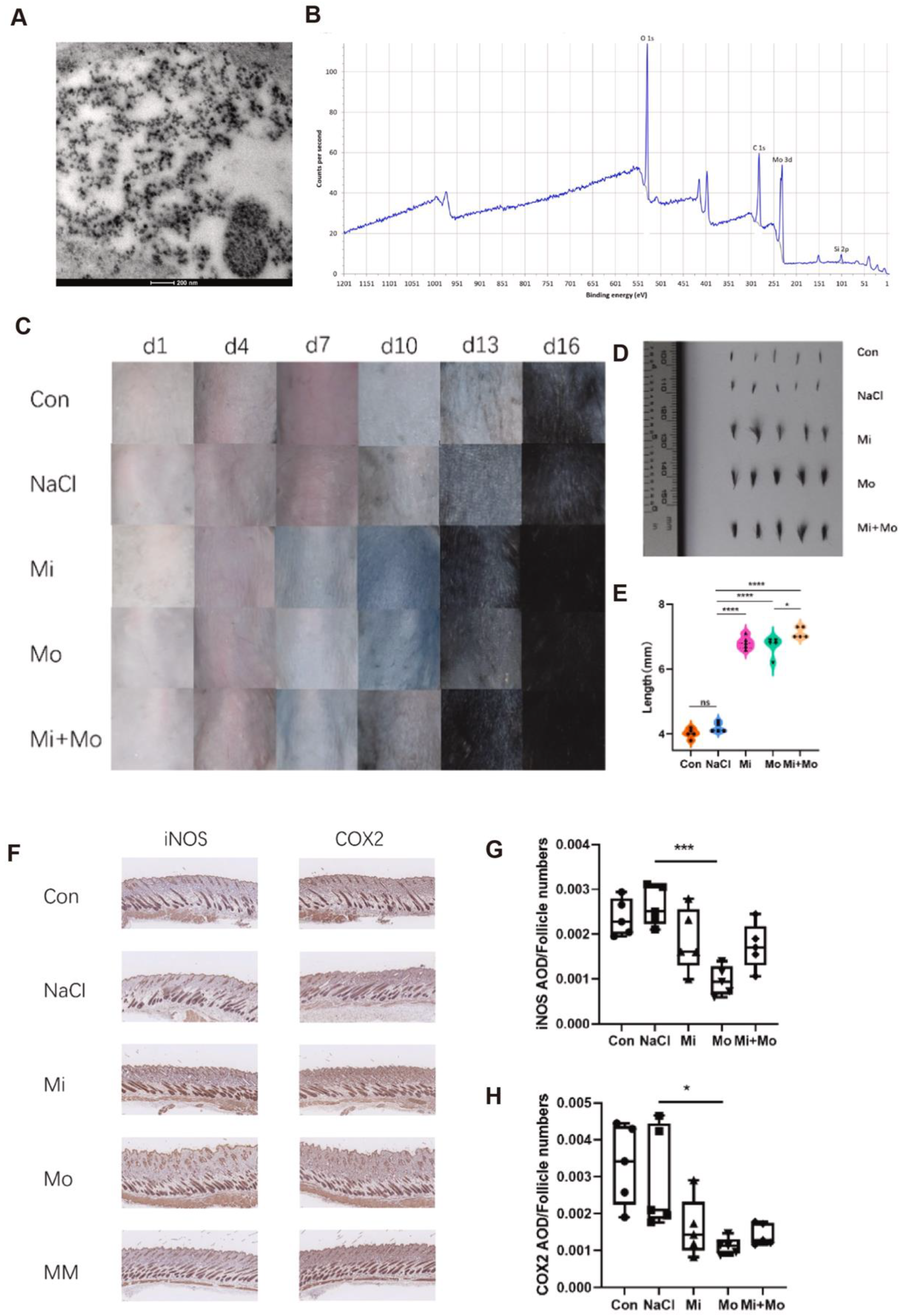
Figure 1. (A). TEM image showing Mo NPs distributed on mouse skin (scale bar: 200 nm). (B). XPS spectrum of Mo NPs, with O, C, and Si peaks as references. (C). Representative dorsal skin images on days 1, 4, 7, 10, 13, and 16 across treatment groups. (D). Hair length assessment on day 16 via representative photos. (E). Comparison of hair length among five groups on day 16. (F). Immunohistochemical staining of iNOS and COX2 in skin tissues (10×, scale bar: 100 μm) from Con, NaCl, Mi, Mo, and MM groups. (G,H). Quantitative AOD analysis of iNOS and COX2 per follicle. Mean ± SD values were obtained from five mice in each group (n = 5). * p < 0.05; *** p < 0.001 indicate statistical significance. Copyright 2024 Elsevier GmbH.
Antioxidants 14 01128 g001
Figure 2. (A). Electron paramagnetic resonance (EPR) spectra of O2•−, H2O2, and OH in various treatment groups, with 5,5-dimethyl-1-pyrroline N-oxide (DMPO) employed as the spin-trapping reagent. (B). Representative images and quantification of neutrophil migration (n = 3). Scale bar: 25 μm. (C,D). Live/dead cell staining and corresponding quantification of propidium iodide (PI) fluorescence intensity in PC12 cells under different treatments. (E). Average clinical scores recorded for mice in each experimental group (n = 6). (F). Changes in body weight of EAE mice subjected to distinct treatment protocols (n = 6). (G). H&E-stained sections of brain and spinal cord tissues from EAE mice. Scale bars: 50 μm (brain) and 20 μm (spinal cord). Yellow arrows highlight areas of demyelination. Statistical significance assessed by one-way ANOVA followed by Tukey’s post hoc test, *** p < 0.001. Copyright 2024 Wiley-VCHGmbH.
Antioxidants 14 01128 g002
Figure 3. (A). Particle size distribution and transmission electron microscopy (TEM) images of Ceria NPs (scale bar: 20 nm). (B). Corresponding size distribution and TEM images of TCeria NPs (scale bar: 20 nm). (C). Fourier-transform infrared (FT-IR) spectra of TPP, Ceria NPs, and TCeria NPs. (D). Mitochondrial membrane potential (Δψm) assessed via confocal microscopy across treatment groups (scale bar: 20 μm). (E,F). Semi-quantitative analysis of Δψm alterations by flow cytometry and image-based quantification, respectively. Data are presented as mean ± SD (n = 3 per group). (G). Temporal changes in renal fluorescence intensity in AKI mice treated with ICG-loaded PTP-TCeria NPs versus free ICG. (H). Quantitative analysis of fluorescence intensity from panel. (I). Renal SOD levels measured at 24 and 48 h post-treatment. * p < 0.05, ** p < 0.01. CC BY 4.0.
Antioxidants 14 01128 g003
Figure 4. (A). TEM image and size distribution of PADN. (B). Time-dependent H2O2 scavenging activity of PADN. (C). Concentration-dependent H2O2 scavenging activity of PADN after 4 h incubation. (D). NO levels in RAW 264.7 cells (LPS-stimulated) treated with varying concentrations of PADN. (E). Analysis of intracellular ROS in cells (RAW 264.7) under different treatments by flow cytometry (G1: normal + saline; G2: DILI + saline; G3: DILI + NAC; G4: DILI + L-PADN; G5: DILI + H-PADN; G6: DILI + L-BDN; G7: DILI + H-BDN). (F). CLSM images of L02 cells stained with DAPI (blue) and TUNEL-Cy3 (red) under different treatments. (G,H). Serum ALT (G) and AST (H) levels in DILI mice receiving different pretreatments. (I). H&E and TUNEL staining of liver sections from each group. t-test, n = 4; * p < 0.05, ** p < 0.01. Copyright 2021 Wiley-VCH GmbH.
Antioxidants 14 01128 g004
Figure 5. (A). Schematic diagram illustrating the antioxidative mechanism of HSA-MDSPB nanoparticles. (B,C). Assessment of the overall antioxidant activity of HSA-MDSPB NPs using DPPH and ABTS assays. (D). Fluorescence intensity analysis of MGF under different treatment conditions using DCFH-DA staining. (E). Representative fluorescence images of BMDMs stained with DCFH-DA (green) following various treatments (scale bar = 100 μm). (F). Western blot analysis showing protein expression in BMDMs under different treatment conditions. (G,H). In vivo imaging and quantitative evaluation of ROS levels in periodontal tissues on day 7 using the L012 probe. Statistical significance: p < 0.05, p < 0.001 (***, ###). CC BY 4.0.
Antioxidants 14 01128 g005
Figure 6. (A) DLS results of MML-CDs in different media (water, PBS, DMEM). (B,C) Free radical scavenging by MML-CDs (n = 6): (B) DPPH·; (C) ABTS+. (D) Colon morphology in each treatment group. (E) Colon length measurements (n = 5) (top: 40×, scale bar: 1 cm; bottom: 400×, scale bar: 100 μm). (F) H&E-stained colon sections. (GI) Oxidative stress markers after oral MML-CDs in UC mice: (G) MPO, (H) NO, and (I) GSH (n = 8). Data were analyzed using the Kruskal–Wallis test or one-way ANOVA with Tukey’s correction. (### p < 0.001 vs. control; * p < 0.05, ** p < 0.01, *** p < 0.001 vs. DSS group). CC BY 4.0.
Antioxidants 14 01128 g006
Figure 7. (A). Schematic illustration of the synthesis of CuL NCs. (B). Characterization of CuL NCs by dynamic light scattering (DLS) and zeta potential analysis. (C). Evaluation of concentration-dependent superoxide anion (O2•−)-scavenging activity. (D,E). Assessment of cell viability in RAW264.7 and HT-29 cells under H2O2-induced oxidative stress after CuL NCs treatment. (F). Fluorescence imaging of cell viability following H2O2 exposure with or without CuL NC pretreatment. (G). Measurement of MDA levels in colonic tissues. (H). Detection of myeloperoxidase (MPO) activity in colonic tissues. (I). Analysis of CAT activity in colonic tissues. ** p < 0.01, *** p < 0.001. CC BY 4.0.
Antioxidants 14 01128 g007
Table 1. A brief summary of autoimmune disease treatment based on nano-antioxidant strategies.
DiseasesActive IngredientNanomaterialSide EffectBenefitRef.
Alopecia areata (AA)MinoxidilMolybdenum nanoparticles (Mo NPs)Mo-NPs may cause liver and kidney toxicity, oxidative imbalance, and potential inflammatory responsesOffering a novel direction and translational potential for antioxidant-based treatment of AA[17]
Multiple Sclerosis (MS)CurcuminDendritic nanocarriers (DNCs)High doses (20 µM) reduce neural stem cell viability and increase oxidative stress, shifting differentiation toward astrocytes instead of oligodendrocytesEnhancing remyelination capacity in vivo[18]
BBN-IONPsIron oxide nanoparticlesMay not adequately reflect long-term effects, with the absence of human trials restricting therapeutic relevanceElevated glutathione levels and total antioxidant capacity in brain tissues, reduced lipid peroxidation and inflammatory mediators[19]
MgH2Low-toxicity magnesium hydride nanoparticles (MgH2)Uncertainties remain regarding mechanism, brain region sensitivity, and variable microglial polarization effectsSignificantly alleviating anxiety- and depression-like behaviors and attenuating inflammatory demyelinating lesions[20]
Clobetasol propionate (CP)Double-layered nanoemulsion Serious side effects include osteoporosis, glaucoma, and adrenal suppressionImproving brain-targeted delivery and reducing systemic toxicity[21]
Selenium and crocinCrocin–selenium nanoparticles (Cor@SeNs) Headache in two patients; no severe adverse events observed, generally well toleratedAmeliorating MS-associated cognitive impairment[22]
Methylprednisolone, neutrophil membranesBiomimetic nanosystem (TFMN)Negligible cytotoxicity up to 200 μg/mL; no severe adverse effects observed in vivo, but long-term safety unknownIndicating excellent safety and therapeutic potential[23]
Abbreviation: BBN-IONPs, BBN loaded within iron oxide nanoparticles.
Table 2. A brief summary of acute inflammatory disease treatment based on nano-antioxidant strategies.
DiseasesActive IngredientNanomaterialSide EffectBenefitRef.
Acute kidney injuryCerium and luteolin Cerium–luteolin coordination nanocomposite (CeLutNCs)Excellent biocompatibility with no significant toxicity or adverse effects in vitro and in vivoSignificantly improved organ function and reduced oxidative stress and inflammation[24]
L-serineL-serine (Rh-Ser, 2–4 nm)Rh-Ser showed no obvious adverse reactions or systemic toxicity in the experimentMimicked multiple antioxidant enzymes to efficiently eliminate RONS[25]
Tetrahedral framework nucleic acid (tFNA)Nanodrug delivery system (TTC)tFNAs showed good biocompatibility with no nephrotoxicity or significant side effects in vitro and in vivoSignificantly ameliorated renal injury and restored renal function, indicating promising antioxidant-mediated therapeutic effects[26]
Epigallocatechin gallate (EGCG) and 5-aminosalicylic acid (5-ASA)pH-stable antioxidant nanoparticle system (EGA NPs)EGCG shows poor stability in acidic environments, limiting oral bioavailabilityEffectively scavenged ROS and mitigated oxidative stress[27]
ethyl caffeate and 4-hydroxybenzyl alcoholSupramolecular nanoplatform (Ser-HPEC)No significant systemic side effects in vivo in the experimentPreserved renal structure and function[28]
Acute liver injuryPolyphenolPhlorotannin-based nanoparticles (PT NPs) Traditional polyphenols face bioavailability and immunogenicity issuesEffectively alleviated oxidative and inflammatory damage[29]
CatechinCCN150CCN150 showed negligible acute toxicity, but potential long-term risks likeReduced systemic inflammation driven by ROS [30]
NOBSA-SNO NPsNo significant toxicity observed; potential risks include immune response, NO over-release, or thiol oxidation/cross-linkingAlleviated oxidative stress, improved hepatic microcirculation[31]
phenylboronic-acid-protected L-DOPA precursor (PAD)Responsive biomimetic antioxidant nanoplatform (PADN)Potential risks include batch variability, incomplete H2O2 scavenging, and reactive group-related off-target effectsIntegrated efficient ROS scavenging, inflammation suppression, and hepatocyte protection[32]
Abbreviation: CCN150, catechin-condensed nanotherapeutics; NO, nitric oxide; BSA, serum albumin; BSA-SNO NPs, self-sulfhydrated, nitro-fixed albumin nanoparticles.
Table 3. A brief summary of chronic inflammatory disease healing based on nano-antioxidant strategies.
DiseasesActive IngredientNanomaterialSide EffectBenefitRef.
PeriodontitisquercetinA mesoporous bioactive glass-based nanocarrier systemNo significant toxicity observed; potential risks include initial burst release and uncertain long-term local effects.Promoted both osteogenesis and angiogenesis in experimental periodontitis-related bone defects[33]
melatonin (MT)melatonin-derived carbon dots (MT-CDs)No significant side effects were observed during the experimentReduced alveolar bone resorption and osteoclast activity[34]
melaninP/D-MNP-AgNo significant organ inflammation observed in the experimentDisrupted biofilms, attenuated inflammation, and reduced alveolar bone loss[35]
HAS, Mn2+, [Fe(CN)6]4−HSA-MDSPB NPsNo significant side effects were observed during the experimentBroadly scavenged ROS species[36]
Ulcerative colitis (UC)somatostatin (SST) and manno-oligosaccharide (MOS)EUP-SeNPsNo obvious toxicity observed, potential risks include selenium accumulationImproved oral stability and intestinal localization[37]
gallic acidthiolated anionic nanoliposomesNo apparent toxicity observedProlonged drug residence time and enhanced local therapeutic effects [38]
curcuminCMCNo obvious toxicity or systemic side effects were observed in this experimentEffectively scavenged ROS, restored mucosal integrity, and regulated gut microbiota composition to mitigate UC pathology[39]
PSBPSB@NP-FAMinimized systemic toxicity compared to free DEXModulated dendritic cell function, promoted M2 polarization of macrophages, and regulated T cell infiltration[40]
TMCs and PEGPMNFsNo significant side effectsIncreased junction protein and MUC2 expression, thereby preserving intestinal barrier function[41]
pterostilbeneHA-L-Arg-CO2@NPsNo significant side effectsDownregulated inflammatory mediators, restored barrier permeability, and effectively alleviated UC symptoms[42]
carbon dots MML-CDsNo significant side effects were observed during the experimentDemonstrated promising therapeutic efficacy[43]
Abbreviation: P, phosphate group; D, 2-(Dimethylamino)ethyl methacrylate; MNP-Ag, melanin-Ag; HSA-MDSPB, human serum albumin-crosslinked, manganese-doped, self-assembling Prussian blue; CMC, carboxymethyl cellulose; PSB, pterostilbene; NP, nanoparticle; FA, folic acid; TMCs, transition metal chalcogenides; PEG, polyethylene glycol; PMNFs, PEG-modified Mo3Se4 nano flakes; HA-L-Arg-CO2@NPs, hyaluronic acid (HA)-modified L-arginine CO2 nanoparticles; MML-CDs, carbon dots derived from magnetite and medicated leaven.
Disclaimer/Publisher’s Note: The statements, opinions and data contained in all publications are solely those of the individual author(s) and contributor(s) and not of MDPI and/or the editor(s). MDPI and/or the editor(s) disclaim responsibility for any injury to people or property resulting from any ideas, methods, instructions or products referred to in the content.