Article

Enhancing Antioxidants Performance of Ceria Nanoparticles in Biological Environment via Surface Engineering with o-Quinone Functionalities

by
Pierluigi Lasala
1,
Tiziana Latronico
2,
Umberto Mattia
1,
Rosa Maria Matteucci
3,4,
Antonella Milella
1,
Matteo Grattieri
1,4,5,
Grazia Maria Liuzzi
2,
Giuseppe Petrosillo
6,
Annamaria Panniello
4,
Nicoletta Depalo
4,5,
Maria Lucia Curri
1,4,5 and
Elisabetta Fanizza
1,4,5,*
1
Chemistry Department, University of Bari, Via Orabona 4, 70126 Bari, Italy
2
Department of Bioscience, Biotechnology and Environment, University of Bari, Via Orabona 4, 70126 Bari, Italy
3
Department of Electrical and Information Engineering, Polytechnic University of Bari, Via Orabona 4, 70125 Bari, Italy
4
CNR—IPCF Institute for Chemical-Physical Process, Via Orabona 4, 70126 Bari, Italy
5
National Interuniversity Consortium of Materials Science and Technology, INSTM, Bari Research Unit, 70126 Bari, Italy
6
CNR—IBIOM Institute of Biomembranes, Bioenergetics and Molecular Biotechnologies, Via Giovanni Amendola, 122/O, 70126 Bari, Italy
*
Author to whom correspondence should be addressed.
Antioxidants 2025, 14(8), 916; https://doi.org/10.3390/antiox14080916
Submission received: 28 June 2025 / Revised: 23 July 2025 / Accepted: 23 July 2025 / Published: 25 July 2025
(This article belongs to the Section Natural and Synthetic Antioxidants)

Abstract

The development of ceria (CeO2−x)-based nanoantioxidants requires fine-tuning of structural and surface properties for enhancing antioxidant behavior in biological environments. In this contest, here ultrasmall water-dispersible CeO2−x nanoparticles (NPs), characterized by a high Ce3+/Ce4+ ratio, were synthesized in a non-polar solvent and phase-transfer to an aqueous environment through ligand-exchange reactions using citric acid (CeO2−x@Cit) and post-treatment with dopamine hydrochloride (CeO2−x@Dopa). The concept behind this work is to enhance via surface engineering the intrinsic antioxidant properties of CeO2−x NPs. For this purpose, thanks to electron transfer reactions between dopamine and CeO2−x, the CeO2−x@Dopa was obtained, characterized by increased surface Ce3+ sites and surface functionalized with polydopamine bearing o-quinone structures as demonstrated by complementary spectroscopic (UV–vis, FT-IR, and XPS) characterizations. To test the antioxidant properties of CeO2−x NPs, the scavenging activity before and after dopamine treatment against artificial radical 1,1-diphenyl-2-picrylhydrazyl (DPPH·) and the ability to reduce the reactive oxygen species in Diencephalic Immortalized Type Neural Cell line 1 were evaluated. CeO2−x@Dopa demonstrated less efficiency in DPPH· scavenging (%radical scavenging activity 13% versus 42% for CeO2−x@Cit before dopamine treatment at 33 μM DPPH· and 0.13 mg/mL loading of NPs), while it markedly reduced intracellular ROS levels (ROS production 35% compared to 66% of CeO2−x@Cit before dopamine treatment with respect to control—p < 0.001 and p < 0.01, respectively). While steric hindrance from the dopamine-derived polymer layer limited direct electron transfer from CeO2−x NP surface to DPPH·, within cells the presence of o-quinone groups contributed with CeO2−x NPs to break the autoxidation chain of organic substrates, enhancing the antioxidant activity. The functionalization of NPs with o-quinone structures represents a valuable approach to increase the inherent antioxidant properties of CeO2−x NPs, enhancing their effectiveness in biological systems by promoting additional redox pathways.

Graphical Abstract

1. Introduction

Nanoceria is commonly referred to CeO2−x [1] to reflect the significant presence of Ce3+ sites alongside Ce4+, compared to its bulk counterpart. In small NPs, oxygen loss from the crystal lattice—due to surface strain—leads to the formation of oxygen vacancies accompanied by the concurrent reduction of Ce4+ to Ce3+ [2,3,4,5]. The coexistence of the two valence states in CeO2−x NPs, and the low redox potential of the Ce4+/Ce3+ couple, in nanosized particles that make easy the shift between Ce3+ and Ce4+ determine the intrinsic antioxidant properties within cells and tissues [6]. Concomitantly, it is important to note that CeO2−x NPs can also act as pro-oxidants, particularly under acidic conditions [7], and recent studies highlighted the ability to produce oxidative stress under acidic conditions. However, under physiologic pH antioxidant activity is generally favored [8].
The antioxidant behavior and their widely recognized role as nanozymes are due to CeO2−x NPs ability to scavenge reactive oxygen species (ROS), such as superoxide (O2) and hydrogen peroxide (H2O2), which, in general, are physiologically degraded by the redox enzymes superoxide dismutase (SOD) and catalase (CAT) [9,10,11].
In biological environments, the NPs SOD-like activity (Figre1A) can attenuate the production of ·O2, which is transformed into O2 and H2O2. Concomitantly, the CAT-like activity (Figure 1A) can remove the H2O2 disproportionated into H2O and O2. Theoretical studies suggest that, under physiological pH, the direct reduction of ·O2 to oxygen is not feasible and only the conversion to H2O2 occurs. Similarly, the CAT-like activity only proceeds through H2O2 adsorption onto the NP surface, forming a cerium-oxo-peroxo complex that subsequently dissociates to release O2. Therefore, CeO2−x NPs can reversibly regenerate their oxidation state, shifting between Ce3+ and Ce4+, by interacting sequentially with ·O2 and H2O2 species.
Interestingly, CeO2−x NPs exhibit antioxidant properties beyond these enzyme-like mechanisms. Recent findings indicate that they also catalyze additional redox reactions, including chain breaking of organic substrate autoxidation (e.g., lipid peroxidation) and quenching of hydroperoxyl radical HOO· [6,12], both strongly contributing to the propagation of oxidative stress and damage of biomolecules [13]. In the autoxidation pathway (Figure 1B), alkyl radicals, R·, generated by the reaction of bioorganic compounds with radical initiators, readily react with O2 and convert into alkylperoxyl ROO· radicals. A cyclic reaction involving RH species contributes to the propagation of the organic substrate autooxidation [12]. Amorati et al. [6,12,14] demonstrated that CeO2−x NPs can break the autoxidation of organic substrate, competing with RH (Figure 1D, right side), with Ce(III)-OH sites giving a proton and electron to ROO·, forming ROOH while oxidizing to Ce(IV)[14]. Furthermore, CeO2−x NPs can also effectively quench HOO· [14], the protonated form of ·O2, which is produced either under acidic conditions (pKa = 4.8) (Figure 1D) or generated by the autoxidation of various organic substrates (Figure 1C).
While acidic pH conditions are uncommon under physiological conditions, specific membrane domains may sustain the formation of HOO·. CeO2−x NPs can quench HOO· by taking advantage of its reducing and oxidizing power, wherein HOO· radicals donate an electron and a proton to Ce(IV) surface sites, generating Ce(III)-OH and releasing O2 (Figure 1E left side). This Ce(III)-OH can subsequently regenerate Ce(IV) by further reacting with ROO· or HOO·. These diverse antioxidant mechanisms are closely related to the Ce3+/Ce4+ ratio within the NPs. Several literature studies have highlighted the superior ability of Ce3+ to engage in redox reactions compared to Ce4+, facilitating more effective neutralization of ROS [15] and, thus, enhancement of the antioxidant activities [4]. The Ce3+ content is highly dependent on NP size and surface chemistry. As the size of the CeO2−x NPs decreases, NPs undergo an increase in Ce3+/Ce4+ ratio [16,17], which may promote improved antioxidant properties [18]. Furthermore, rich surface chemistry, including surface hydroxylation and surface functionalization, can significantly affect redox behavior and hence catalytic activities of CeO2−x NPs. Modifications of ceria surface states due to molecule adsorption or anchoring can increase or decrease the antioxidant properties of CeO2−x NPs depending on the electronic properties and density of the functionalization [19]. Polymers or co-polymer coatings [20,21], such as polyethylene glycol or polyacrylic acid, have been explored to impart good colloidal stability, which enhances surface availability and hence catalytic reaction, while inhibiting protein adsorption in biological media. However, the nature and the thickness of the polymer coating can variably impact the enzyme-like activity of CeO2−x NPs. While some reports show minimal effects on CAT- and SOD-like functions, others note suppression of CAT and oxidase mimetic activity, with a concurrent increase in peroxidase-like activity. Lee et al. [22] demonstrated that thicker polymer coatings can diminish intracellular and in vitro radical scavenging due to reduced accessibility of redox active sites. Surface functionalization with polymers featuring reactive groups, such as catechols, has also been investigated. Hu et al. [16] functionalized CeO2−x NPs with catechol-grafted poly(ethylene)glycol. The interaction of CeO2−x NPs with catechol functionalities, studied in the last decade using biologically relevant catechol-containing molecules [23], such as dopamine [24], highlights that these molecules can form strong metal–ligand complexes by transferring electrons from the catechol group (oxidized to o-quinone) to Ce4+, reducing it to Ce3+, and thereby enhancing the antioxidant capacity of the NPs via increased Ce3+ content. However, the specific role of surface o-quinone functionalities in enhancing redox activity, and hence antioxidant behavior of the hybrid structure, remains underexplored. Recent studies by Amorati et al. [25] have demonstrated that o-quinone exhibits significant reactivity towards HOO·, effectively interrupting the autoxidation chain through concurrent scavenging of both HOO· and ROO. radicals.
Based on this insight, the aim of this study was to develop water-dispersible ultrasmall NPs CeO2−x NPs with enhanced antioxidant properties through targeted surface engineering. Specifically, we employed a two- step strategy: (i) synthesis of ultrasmall hydrophobic ceria NPs via thermal decomposition of a cerium precursor in an organic solvent in the presence of oleyl amine, followed by (ii) surface modification through ligand exchange with citric acid (CeO2−x@Cit) to render them water dispersible, and subsequent post-functionalization using dopamine hydrochloride (CeO2−x@Dopa). The post-synthetic treatment was designed to promote Ce3+ surface enrichment and to introduce o-quinone functionalities on the NP surface and at the CeO2−x NPs, via a metal–ligand electron transfer reaction with catechol or polydopamine moieties, which promote catalytic redox reactions that concomitantly quench HOO· radicals, ultimately preventing the autoxidation chain propagation. Through a combination of spectroscopic and thermogravimetric analyses, the NPs surface properties have been depicted, and by testing CeO2−x before and after dopamine treatment in scavenging DPPH· and reacting with H2O2 as well as in a biological environment using Diencephalic Immortalized Type Neural Cell line 1 (DITNC1), superior intracellular ROS scavenging activity of CeO2−x@Dopa has been observed.

2. Materials and Methods

2.1. Materials

Cerium(III) nitrate hexahydrate (Ce(NO3)3·6H2O), oleylamine (OAm), oleic acid (ODE), toluene, methanol, isopropanol, acetone, hexane, chloroform, cyclohexane, citric acid, dopamine hydrochloride, ciclopentyl methyl ether (CME), 1,1-Diphenyl-2-picrylhydrazyl (DPPH), ammonium hydroxide (NH4OH), 2′,7′-Dichlorodihydrofluorescein diacetate (DCFH-DA, from Calbiochem), 3-(4,5-dimethylthiazol-2-yl)-2.5-diphenyltetrazolium bromide (MTT), ultrapure Milli-Q water (18 MΩ cm–1). All reagents were purchased from Sigma-Aldrich (St. Louis, MO, USA), if not differently stated. The DI-TNC1 cell line (ATCC CRL-2005) was acquired and authenticated from the ATCC (www.lgcstandards-atcc.org, accessed on 21 November 2024). All chemicals and solvents were of analytical grade and used as received without further purification.

2.2. Synthesis of Oleyl Amine-Capped CeO2−x NPs (CeO2−x@OAm)

CeO2−x NPs synthesis was carried out using a Schlenk line under nitrogen atmosphere and temperature conditions, following a procedure reported in literature [26,27,28] with minor modification. Briefly, 0.434 g (1 mmol) of Ce(NO3)3·6H2O was poured in a three-neck flask, and 2 mL (6 mmol) of OAm and 6 mL of ODE were added. The reaction mixture was first put under vacuum for 30 min at 80 °C under continuous stirring, obtaining a brown-colored solution, then nitrogen flowed into the flask and the temperature was increased up to 300 °C. The dark brown solution was left at this temperature for 60 min prior to being cooled down to room temperature. Once 160 °C was reached during the cooling ramp, 2 mL of toluene were injected into the reaction flask. NPs were finally collected, and unreacted reagents were removed by four cycles of addition of a large excess (10 times the reaction mixture volume) of isopropanol: acetone (1:1 volumetric ratio), followed by centrifugation at 1000 x g for 10 min. After discharging the supernatant, a few drops of hexane were added, followed by the addition of isopropanol/acetone and a centrifugation step to collect a black precipitate, which was finally dispersed in 5 mL of chloroform. An aliquot of the NPs solution was put under nitrogen flux to evaporate the solvent, and the remaining pellet was weighed to determine the NPs concentration, which resulted in about 56 mg/mL.

2.3. Phase Transfer of Organic-Capped CeO2−x Nanoparticles in Aqueous Medium by Ligand-Exchange Reaction with Citrate (CeO2−x@Cit)

In order to replace OAm with citrate, a ligand-exchange procedure was developed by slightly modifying the Monnier et al. protocol [29]. Briefly, 5 mg of citric acid was dissolved in 1 mL of CME, then 100 μL of the CeO2−x@OAm NPs was added to the mixture and left to stir at 100 °C for 18 h. The NPs were then collected by centrifugation by adding methanol (16,000× g for 10 min). Two repeated cycles of dispersion in acetone and a centrifugation step at 16,000× g for 15 min were then carried out. The recovered pellet was redispersed in MilliQ H2O, where a few drops of NH4OH solution were added, resulting in a stable colloidal solution, which was stored at 4 °C for further use. An aliquot of the sample, labelled CeO2−x@Cit, was freeze-dried, resulting in a concentration of 44 mg/mL.

2.4. Functionalization of CeO2−x@Cit NPs with Dopamine (CeO2−x@Dopa)

A sample labelled CeO2−x@Dopa was prepared by adding dopamine hydrochloride to CeO2−x@Cit. In particular, 80 μL of 1.5 mM dopamine hydrochloride aqueous solution was added to 1 mL of 10 mg/mL of CeO2−x@Cit sample. Upon addition of dopamine hydrochloride, the NPs suspension turns brown. The solution was left stirring overnight at room temperature and finally collected after several cycles of purification by centrifugation and pellet dispersion in MilliQ water. An aliquot of the sample was freeze-dried, resulting in a concentration of 11 mg/mL.

2.5. Spectrophotometric Characterization of CeO2−x Nanoparticles Treated with H2O2

The reactivity of CeO2−x@Cit or CeO2−x@Dopa towards H2O2 was spectrophotometrically evaluated. In a typical experiment, 1.5 mL of aqueous solution of NPs, either CeO2−x@Cit or CeO2−x@Dopa, at a concentration of 0.1 mg/mL was prepared, and 30 µL of H2O2 50 mM was added ([H2O2] = 1 mM) and stirred for 30 min at room temperature. UV–vis absorption spectra of the colloidal solutions before and after H2O2 addition were recorded in the 200–800 nm wavelength range.

2.6. Spectrophotometric Analysis of 1,1-diphenyl-2-picrylhydrazil (DPPH·) Radical Scavenging by CeO2−x Nanoparticles

The DPPH· radical scavenging activity (RSA) was evaluated by monitoring the absorbance decrease at 517 nm of a DPPH· solution at 0.033 mM in EtOH after it was left in the dark for 30 min with and without CeO2−x NPs samples at different concentrations. For this purpose, different volumes of CeO2−x@Cit NPs suspensions (nearly 10 mg/mL) were added to 500 μL of the 0.1 mM DPPH solution diluted to 1.5 mL in EtOH, resulting in NP concentrations ranging from 0.13 mg/mL to 0.8 mg/mL. A control experiment was performed by simply diluting 500 μL of the DPPH· solution 0.1 mM to 1.5 mL (AbsDPPH_t0). Each mixture was left to react under dark and vigorous stirring at room temperature for 30 min; then the supernatant was recovered by centrifugation, and the UV–vis absorption spectrum was recorded. In particular, the absorption intensity at 517 was monitored for each solution (AbsDPPH_NPs) and the % RSA measured based on the following equation:
%   R S A = ( A b s D P P H · _ t 0 A b s D P P H · _ N P s ) A b s D P P H · _ t 0 × 100
A control experiment was carried out using citric acid alone (0.03 mg/mL) in DPPH· assay to assess the contribution of the ligand to the DPPH· scavenging activity of CeO2@Cit NPs. The concentration of the citric acid solution was set following the thermogravimetric analysis on CeO2@Cit NPs, from which the weight percentage attributed to citric acid was estimated. For CeO2@Dopa, a control experiment was performed using polydopamine purposely synthesized by polymerization of dopamine hydrochloride in aqueous solution under alkaline conditions by NaOH (pH = 10). After 48 h, a brown pellet was recovered and used for DPPH· assay at a concentration of 0.01 mg/mL and 0.02 mg/mL.

2.7. Evaluation of Cell Viability

The effect of CeO2−x@Cit NPs on DITNC1 cell viability was assessed using the MTT assay [3-(4,5-dimethylthiazol-2-yl)-2,5-diphenyltetrazolium bromide] [30]. Briefly, DITNC1 seeded in 96-well plates were treated with CeO2−x@Cit (CeO2−x@Dopa) NPs at concentrations ranging from 5 to 100 μg/mL. After 24 h of incubation at 37 °C, 5%, CO2 cells were washed with PBS and incubated for 2 h at 37 °C and 5% CO2 with 0.5 mg/mL of MTT. At the end of the incubation period, the culture medium was discarded, and the formazan crystals formed within the cells were dissolved using absolute ethanol. The concentration of the formazan product was measured by optical absorbance at 545 nm, with 690 nm as the reference wavelength. Cell viability was calculated as a percentage in comparison to the negative control (ctrl), represented by untreated cells, which was set at 100%.

2.8. Intracellular Reactive Oxygen Species Detection

The detection of ROS in DITNC 1 was performed as previously described [31]. The cells treated only with DCFH-DA without and with rotenone were used as negative (CTRL) and positive (ROT) controls, respectively. To test the ROS production in the presence of CeO2−x NPs, briefly, cells were pre-treated for 90 min at 37 °C, 5%, CO2 with CeO2−x@Cit or CeO2−x@Dopa NP preparations at concentrations of 10 μg/mL. To assess the intrinsic pro-oxidant or antioxidant effect of CeO2−x@Cit and CeO2−x@Dopa NP, in a separate set of experiments, cells were pre-treated with the NPs samples as described above and were subsequently loaded with 4 μM DCFH-DA, without rotenone or with 10 μM rotenone. After incubation for 50 min, the supernatants were discarded, and the fluorescence intensity of cells was analyzed via spectrofluorimetric analysis at 525 nm under excitation at 485 nm in a microplate reader. Results were normalized to cell viability, and ROS production was expressed as the relative percentage of photoluminescence (PL) intensity compared to the negative or positive control.
Parametric one-way analysis of variance (ANOVA), followed by Dunnett’s multiple comparison post hoc test, was used to compare ROS levels and cell viability under different experimental conditions. Data were obtained from at least three independent experiments, with each data point representing the mean of triplicate measurements within a single experiment. Data were analyzed by GraphPad Prism 5.0 (GraphPad Software, Inc., San Diego, CA, USA).

2.9. Characterization Techinques

TEM characterization was performed using a JEOL JEM 1011 (JEOL, Akishima, Tokyo, Japan) transmission electron microscope operating at 100 kV and equipped with a high-resolution CCD camera. Two microliters of the NP suspension were dropped onto a carbon-coated copper grid, and the solvent was left to evaporate. Statistical analysis of NP sizes in the samples and their respective distributions was conducted using the image analysis software ImageJ 1.54g. For each sample, the average NP size and the relative percentage standard deviation (σ%) were calculated.
The FT-IR characterization was carried out by using a 670 FT-IR spectrometer (Varian, Palo Alto, CA, USA) equipped with a diamond ATR accessory of 2 mm and a deuteratedtryglicine sulfate (DTGS) detector. One microliter of each sample was put on the internal reflection element, and the solvent was allowed to evaporate. Spectra were recorded in the range 4000–400 cm−1 acquiring 16 scans with a nominal resolution of 1 cm−1.
Thermogravimetric analysis (TGA) was carried out using a Pyris 1-Perkin Elmer instrument under a nitrogen flow of 40 mL/min at the heating rate of 20 °C/min in a temperature range from 50 °C to 700 °C. Thermograms were collected using powder of dried NP samples [32].
UV–vis absorption spectra were recorded using a Cary 5000 (Agilent, Santa Clara, USA) UV/vis/NIR spectrophotometer. The samples were purposely diluting, and spectra in the wavelength range 200–800 nm were recorded.
XPS analyses were performed with a PHI 5000 Versa Probe II spectrometer (Physical Electronics) equipped with a monochromatic Al Kα X-ray source (1486.6 eV), operated at 15 kV and 24.8 W, with a spot size of 100 µm. Survey (0–1200 eV) and high-resolution spectra (C1s, O1s, and Ce3d) were recorded in FAT mode at pass energies of 187.85 and 29.35 eV, respectively. Surface charging was compensated by means of a dual-beam charge neutralization system. All spectra were collected at an angle of 45° with respect to the sample surface. The hydrocarbon component of the C1s spectrum was used as an internal standard for charging correction, and it was fixed at 284.8 eV. Spectra were processed with MultiPak software v. 9.5.0.8 (Physical Electronics). Atomic concentrations were determined from the high-resolution spectra after subtracting a Shirley-type background, using the Scofield sensitivity factors set in the MultiPak software.
The Ce 3d core level splits into two states by spin–orbit interaction, with Ce 3d5/2 and Ce 3d3/2 assigned as bands of v and u type, respectively. The Ce 3d fitted spectra, deconvoluted in ten Gaussian–Lorentzian peaks, were assigned separately to the two ionic cerium species, and the area of each species was calculated as follows:
A ( C e 3 + ) = A v 0 + A v + A u 0 + A ( u )
A ( C e 4 + ) = A v + A v + A v + A u + A u + A ( u )
The relative concentration of Ce3+ and Ce4+ ions was calculated as follows:
C e 3 + = A ( C e 3 + ) A C e 3 + + A ( C e 4 + ) 100
C e 4 + = A ( C e 4 + ) A C e 3 + + A ( C e 4 + ) 100

3. Results and Discussion

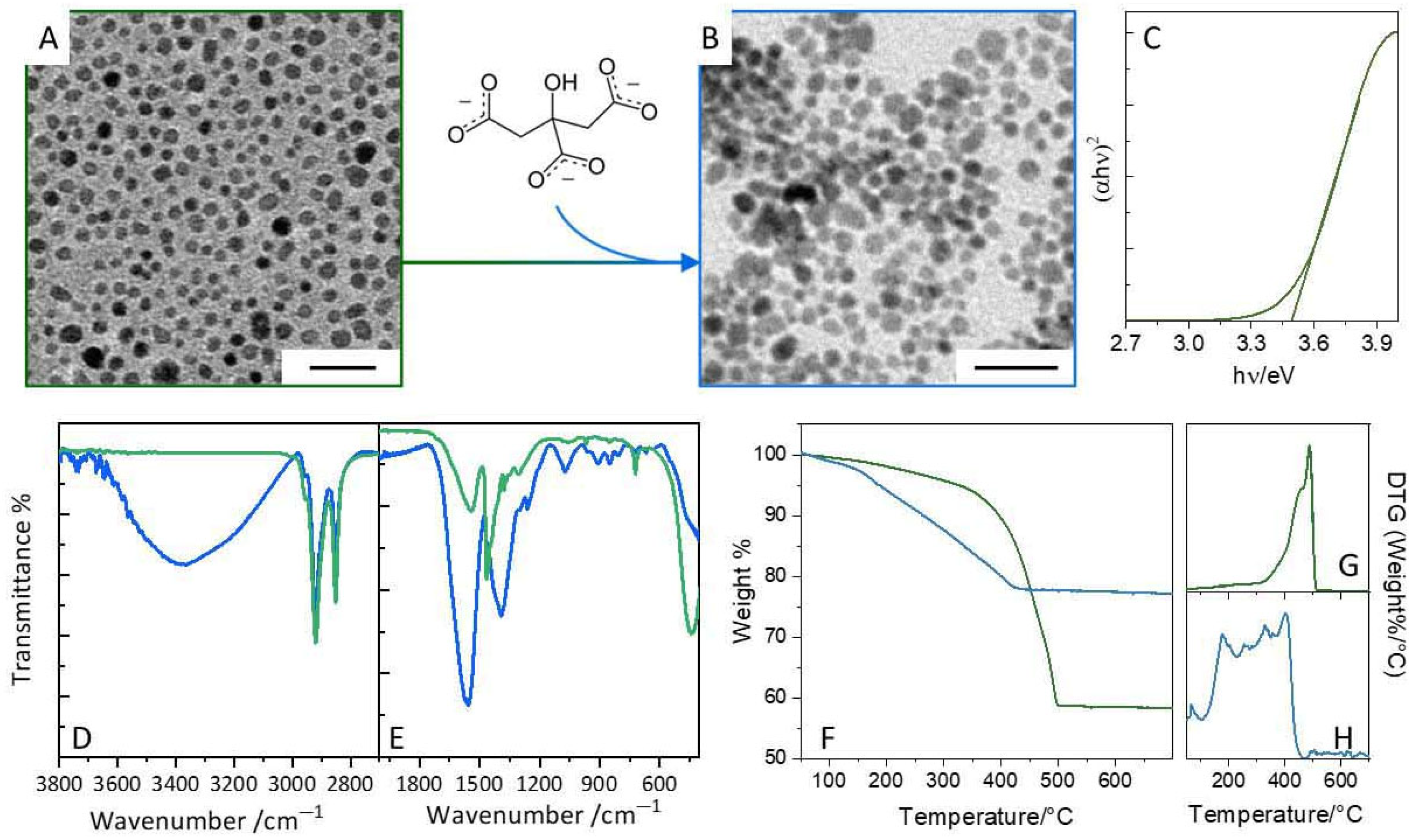
3.1. Synthesis and Aqueous Phase Transfer of CeO2−x@OAm Nanoparticles via Citrate Ligand Exchange

CeO2−x NPs have been synthesized by thermal decomposition of Ce(NO3)3·6H2O in high-boiling ODE in the presence of OAm as a coordinating agent [26]. This method yields ultra-small, monodispersed, and colloidally stable NPs. OAm is expected to bind to the NP surface via amino groups, controlling NP growth, while the exposed long alkyl chain provides steric stabilization, ensuring long-term stability and long-term dispersion in non-polar media. TEM micrograph (Figure 2A) reveals spherical CeO2−x@OAm NPs, with an average diameter of ~3 nm (σ% = 21%). Optical characterization shows an energy gap of 3.5 eV, measured by the Tauc plot (Figure 2C). Such a value is significantly larger than that reported for bulk CeO2 (3.2 eV) consistent with NPs in with the quantum confinement regime (Bohr radius ~ 3.5 nm). The ultrasmall particle attained through this synthetic procedure is favorable for enhancing antioxidant activity [33]. However, despite the advantages of the synthetic approach in providing ultrasmall and minimally aggregated NPs, their dispersibility in organic solvents demands the development of an effective phase transfer procedure to render them water dispersible and thus suitable for biological systems [34].
Citric acid is an effective ligand to achieve water-dispersible CeO2−x NPs. At physiologic pH, citrate molecules coordinate the CeO2−x NPs surface via multidentate interactions, exposing outside the negatively charged carboxylate groups that confer good colloidal stability in aqueous medium via Coulombic repulsion [35]. To successfully replace the pristine OAm capping layer via ligand exchange, it is essential to choose a solvent or solvent mixture capable of solubilizing both the hydrophobic-capped NPs and the hydrophilic citrate ligands. While Monnier et al. [29] previously used a mixture of chlorobenzene and DMSO for this purpose on Fe2O3 NPs, in this work chlorobenzene has been replaced for the first time by CME as a more environmentally sustainable alternative. The high boiling point of CME allows the ligand-exchange reaction to be carried out at high temperature (100 °C), which promotes efficient desorption of OAm and anchoring of citrates onto the NP surface. TEM analysis of CeO2−x@Cit (Figure 2B) confirms that NPs preserve their original shape and size (Figure S1). Compared to CeO2−x@OAm, the citrate-coated NPs appear more closely packed, consistent with the presence of a thinner, less sterically hindered surface layer. Ligand exchange and surface modification have been further validated through FT-IR spectroscopy in ATR mode (Figure 2D–E, see Figure S2) and thermogravimetric analysis (Figure 2F–H) comparing samples before (green line) and after (blue line) the ligand exchange. The FT-IR spectrum of CeO2−x@OAm shows intense peaks at 2920 cm−1 and 2850 cm−1 corresponding to the antisymmetric and symmetric C-H stretching vibrations of the -CH2- groups, respectively, along with a weaker signal at 2959 cm−1, characteristic of the asymmetric stretching mode of C-H in the methyl group [36,37,38]. A broad band at 1542 cm−1 corresponds to stretching modes of oxidized –NH2 groups (e.g., nitrile and imine), while a weak shoulder at 1645 cm−1 may arise from oleyl chain C=C stretching or the NH2 rocking vibration of unbound OAm. Additional features at 1465 cm−1 (CH2 scissoring), 1378 cm−1 (CH3 bending), and 1059 cm−1 (C-N stretching) further confirm the presence of OAm.
After ligand exchange with citrate, the FT-IR spectrum of CeO2−x@Cit NPs displays a dominant broad O–H stretching band between 3200 and 3500 cm−1, indicative of hydroxyl groups or adsorbed water, confirming successful surface functionalization with citrate [39,40]. The presence of citrate is also confirmed by the two broad bands at 1560 cm−1 and 1390 cm−1 [41], which are assigned to the symmetrical and asymmetrical vibrations of the carboxylate groups. Additional peaks at 1255 cm−1 and 1069 cm−1 correspond to the C-O stretching modes of citrate. Both NPs samples show a Ce-O-C bond at 1060 cm−1 and Ce-O bending modes in the 600–400 cm−1 region [42,43,44,45]. Characteristic signals of cerium oxide are also present in the 800–880 cm−1 range [46].
To further evaluate the nature and amount of ligands on the CeO2−x NP surface, thermogravimetric analysis (TGA, Figure 2F) has been performed, from which first derivative curves (Figure 2G–H) have been plotted. For, CeO2−x@OAm NPs a progressive weight loss is observed between 100 and 330 °C, corresponding to the evaporation of free or physically adsorbed OAm. Further weight losses occur in a wide temperature range between 330–500 °C, which are ascribed to OAm bound to the NP surface (330–400 °C) [47] and to the conversion of residual Ce(OH)3, on the NP surface, into CeO2 at 450 °C and 488 °C [48]. In the case of CeO2−x@Cit NPs, the first weight loss occurs between 50° and 100 °C, associated with the evaporation of weakly bound water. A second loss, between 100° and 220 °C, is attributed to the release of more strongly bound intramolecular water. These results are consistent with FT-IR findings, which also indicate the presence of water molecules on the NP surface. The weight loss between 220 and 340 °C can be ascribed to surface-bound citrate molecules, covering a broad temperature range due to hydrogen interactions that influence the thermal behavior of the passivating layer. Additional weight losses at higher temperatures can be ascribed to the conversion of surface cerium hydroxylate groups. Based on thermogravimetric data, the citrate content in the NP sample is estimated to be approximately 10% by weight.
XPS investigation has been carried out on CeO2−x@Cit NPs and high-resolution spectra of Ce3d, O1s, C1s, and N1s are reported in Figure 3. For materials with mixed +3/+4 cerium valence states, the XPS high-resolution spectrum of Ce3d is complex and comprised of ten peaks. As described by Deshpande et al. [18], based on Burrough’s convention [49], four peaks are associated with Ce3+, (v0, v′, u0, and u′), and six peaks with Ce4+ (v, v″, v‴, u, u″, and u‴) [50,51,52,53,54]. The content of 48% and 52% determined for Ce3+ and Ce4+ (Figure 2G), respectively, highlights a higher Ce3+ content than that usually reported for bulk ceria (30% and 70% for Ce3+ and Ce4+, respectively) [55], confirming that the decrease in CeO2−x particle size is accompanied by reduction of Ce4+ to Ce3+ to counteract the oxygen loss from the surface.
The O1s high-resolution spectrum (Figure 3B) shows two contributions at 529.3 eV and 530.6 eV, which can be attributed to lattice oxygen in ceria NPs and to the presence of the OH groups on the nanoceria surface, respectively. To get further information on the passivating shell at the NP surface, high-resolution C1s spectrum is acquired and curve-fitted with four contributions (Figure 3C, Table S1) peaked at 284.8, 286.1, 288.3, and 289.0 eV, which are characteristic of C−C or C−H, C−OH and/or C-OC, coordinated carboxylates or carbonyl, and free carboxyl moieties, respectively. Since no signal of nitrogen has been detected in the XPS survey spectrum, complete removal of OAm from the NPs surface and its successful replacement by citrates is confirmed.

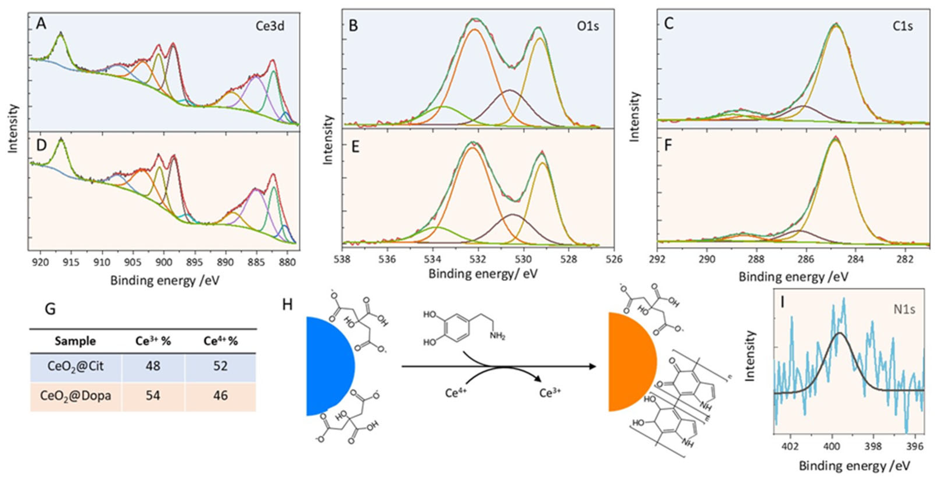
3.2. Ligand-Exchange with Dopamine and Characterization of CeO2−x@Dopa

CeO2−x@Dopa NPs have been prepared starting from CeO2−x@Cit NPs by treating them with a dilute dopamine hydrochloride solution. Dopamine is reported to spontaneously form a metal complex at the CeO2−x surface, transferring electrons to Ce4+ sites, which are then reduced to Ce3+, simultaneously oxidizing the catechol structure into quinone structures [16,24,56,57]. Under ambient conditions, dopamine polymerization can also naturally occur. The surface chemistry and the cerium valence state following dopamine treatment have been rationalized by means of XPS characterization. Quantitative analysis of the Ce3d high-resolution spectrum (Figure 3D,G) reveals a slightly larger Ce3+ content (54%) in CeO2−x@Dopa NPs than CeO2−x@Cit NPs (46%), confirming the reducing effect induced by treatment with dopamine. High-resolution C1s and N1s spectra (Figure 3F and 3I, respectively; Table S1) have been examined to understand the chemical nature of the surface-bound species. In the C1s spectrum, a decrease in the peak at 286.1 eV, assigned to C-OH and an increase in the component at nearly 288.3 eV, characteristic of carbonyl moieties (Table S1), support the formation of oxidized o-quinones structures bound to the NP surface. Additionally, the decrease in the contribution at nearly 289.0 eV, attributed to COOH, suggests that residual citric acid molecules are mostly in their deprotonated form. Even though π−π* shakeup peaks at nearly 291 eV, specific of the aromatic groups, are not visible, being usually poorly intense, the evidence of nitrogen in the N1s (Figure 3I) high-resolution spectrum, which is not present in the CeO2−x@Cit sample, confirms the success of the ligand exchange procedure. The binding energy value of the N1s peak at 399.6 eV suggests the formation of nitrogen-heterocyclic structures such as those expected in dimeric [58] or polymeric structures of dopamine [59]. The occurrence of polymerization is also confirmed by TGA (see Figure S3), that which a prominent weight loss at 440 °C, a temperature higher than those reported for dopamine and dopamine derivatives.
This in-depth characterization opens the venue to a different representation of the surface functionalization of CeO2−x NPs treated with dopamine than compared to previous studies [60,61]. Besides the confirmation of an increase in the Ce3+/Ce4+ ratio, even though not to a large extent, and the formation of o-quinone structures, polymerization of dopamine with nitrogen involved in heterocyclic structures is also proposed [16].
Based on this comprehensive characterization, after dopamine treatment the CeO2−x@Dopa NPs feature an inorganic core bearing a size and surface cerium valence ratio similar to CeO2−x@Cit, and specific o-quinone groups and a polymerized coating. To test the antioxidant activity of CeO2−x@Dopa NPs, a comparative approach is used by performing chemical assays and in vitro experiments in a biological environment, using CeO2−x@Cit as a control sample.

3.3. Catalytic and Radical Scavenging Activity of CeO2−x@Cit and CeO2−x@Dopa

To preliminarily evaluate the catalytic and radical scavenging activity of the two CeO2−x NPs samples, two in vitro non-enzymatic chemical assays have been performed. These included monitoring the spectral change of the CeO2−x NPs solutions upon treatment with H2O2 and in the colorimetric DPPH· assay. Even though these non-enzymatic chemical tests do not fully replicate the complex behavior of NPs in a biological environment, they allow a preliminary evaluation of the catalytic activity displayed by each NPs sample.

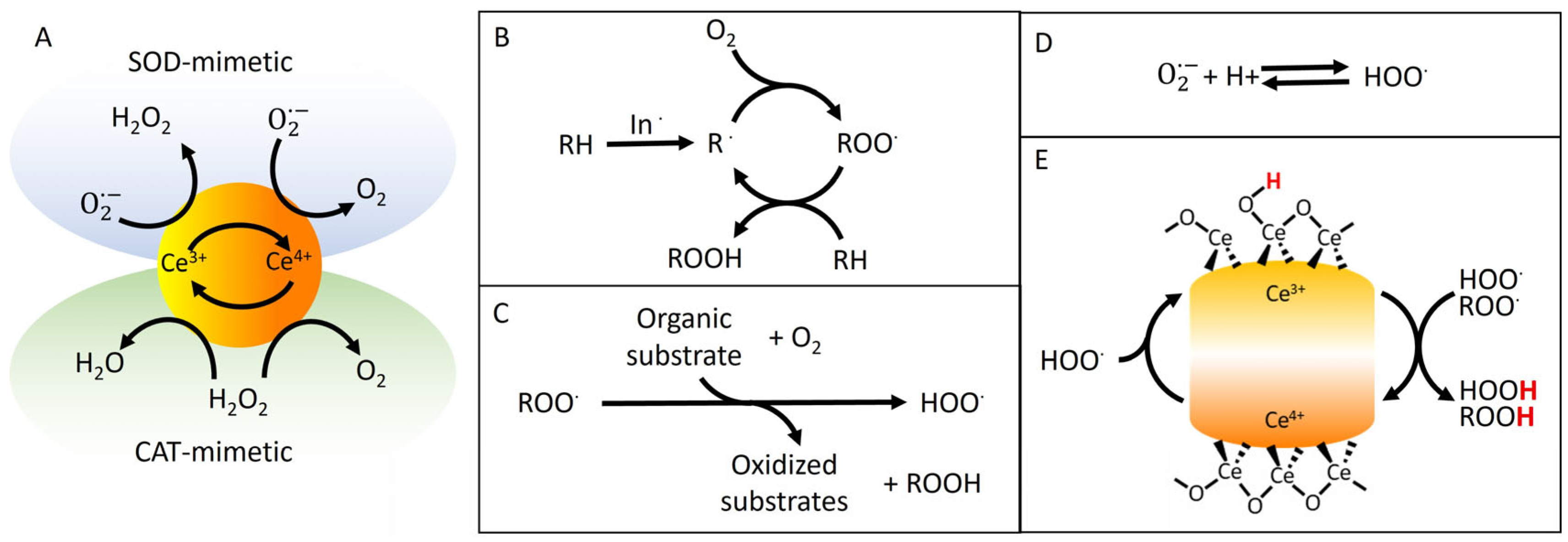
3.3.1. Catalytic Activity of CeO2−x Nanoparticles Towards H2O2

H2O2 is an ROS endogenously produced under oxidative stress conditions, and naturally enzymatically removed by CAT activity or in situ decomposition into ·OH species following the Fenton reaction. CeO2−x NPs can mimic the enzymatic reaction and scavenge H2O2. First, H2O2 reacts at the surface of CeO2−x NPs, forming cerium-peroxo and hydroperoxo complexes (Ce-O22− and/or Ce-OOH complexes), further transformed into O2 or ·OH via reaction mediated by the easy shift between cerium valence states. Formation of these complexes has been reported to be accompanied by a visible color change of the colloidal solution of CeO2−x NPs from yellow to brown and a significant increase in the absorption intensity between 300 and 600 nm [22,62]. This spectral response is commonly exploited to qualitatively assess the interaction of H2O2 with the CeO2−x NP surface and to compare the catalytic activity across different CeO2−x NP samples [21]. Finocchiaro et al. [62] demonstrated that adsorption of H2O2 is size-dependent, with smaller CeO2−x NPs (e.g., 4.5 nm) exhibiting enhanced absorption compared to the larger ones (7.8 nm, 23 nm, and 28 nm) upon addition of H2O2 at increasing concentrations. The higher reactivity shown by the smaller NPs is ascribed to the high fraction of Ce3+ relative to the larger ones. Surface coating also plays a critical role: hydrophobic or bulky ligands can hinder H2O2 diffusion towards CeO2−x NPs active surface sites, thereby limiting the formation of reactive cerium-peroxo or hydroperoxo intermediates [21,62].
To evaluate this, UV–vis absorption spectra of untreated and H2O2-treated CeO2−x@Cit and CeO2−x@Dopa NPs have been recorded (Figure 4A). Both untreated samples show the characteristic profile of CeO2−x NPs with an absorption shoulder at 300 nm attributed to Ce 5d–O 2p valence states. CeO2−x@Cit sample displays an additional band at 265 nm, attributed to surface-bound hydrated species, which disappear after dopamine treatment. Furthermore, CeO2−x@Dopa shows an increased intensity of the absorption tail in the 300–500 nm range, consistent with the characteristic spectral signature of o-quinone groups, in line with the XPS findings [63,64]. Upon treatment with H2O2 both samples display increased absorption below 270 nm, due to free H2O2, and enhanced absorption above 300 nm [65].
By subtracting the absorbance of the suspensions devoid of H2O2 (Figure 4B) from the absorbance of the H2O2-treated samples, an absorption peak centered at 390 nm is observed, which can be attributed to cerium-peroxide and cerium-hydroperoxide species generated due to adsorption of H2O2 at the particle surface [15,21,62].
Notably, CeO2−x@Cit NPs exhibit a stronger absorption peak than CeO2−x@Dopa, suggesting higher H2O2 interaction and, hence, greater intermediate formation. Since both samples share similar size and Ce3+/Ce4+ ratios, as assessed through chemical and morphological characterization, the different reactivity towards H2O2 can be attributed to surface chemistry. CeO2−x@Cit NPs, capped with a small hydrophilic citrate coating, permits easier H2O2 diffusion and surface interaction. In contrast, the dense hydrophobic polymeric coating on CeO2−x@Dopa NPs hinders H2O2, limiting the formation of peroxo or hydroperoxo species at the NP surface and hence reactivity [62].

3.3.2. Assessment of DPPH· Scavenging Activity

The colorimetric DPPH· assay is a well-established, widely employed protocol to evaluate the antioxidant capacity of natural and synthetic molecules or nanoantioxidant materials, based on monitoring the bleaching of the absorption band of the stable radical DPPH·, centered at nearly 516 nm, due to its conversion into DPPH2 by electron and proton transfer [66]. CeO2−x NPs have demonstrated the ability to scavenge DPPH [67]. Compared to the redox potential of the couple Ce4+(aq)/Ce3+(aq) (1.71 V versus SHE), CeO2−x NPs exhibit a significantly lower redox potential of nearly 0.2 V (versus SHE at pH 7) [67,68], which makes them capable of reducing the DPPH·/DPPH2 couple (0.77 V versus SHE). Electrons are supplied by the cerium states switching between Ce+4 and Ce+3 and protons by metal oxide surface hydroxyl groups, ligands, or solvent. However, the surface coating on CeO2−x NPs can influence this process, either hampering electron transfer through steric hindrance [21] or enhancing the reduction of DPPH· by providing protons or due to intrinsic antioxidant properties [48].
As shown in Figure 4C, exposure to both CeO2−x@Cit and CeO2−x@Dopa NPs results in a noticeable color change of the DPPH· solution from violet to pale yellow (Figure 4C(a–c)) alongside a decrease in absorbance at 516 nm after 30 min. At a concentration of 130 μg/mL, CeO2−x@Cit displays markedly higher radical scavenging activity (42%) than CeO2−x@Dopa (13%) at 33 μM DPPH·. Control experiments have been carried out using citrate alone to determine whether the radical scavenging activity could be partially attributed to surface-bound ligands. A citrate solution at a concentration equivalent to that estimated to be on CeO2−x@Cit NPs based on TGA analysis has been tested in the DPPH· assay. This citrate solution showed minimal reduction in DPPH· absorption after 30 min, suggesting a negligible DPPH· scavenging activity of citrate at the tested concentration, being not relevant in the CeO2−x@Cit scavenging behavior. In the case of CeO2−x@Dopa NPs, control experiments with independently synthesized polydopamine present a significant challenge, as it is difficult to replicate the specific functionalities of the polymer coating formed in the presence of CeO2−x. This is due to electron transfer between dopamine or its polymerized derivatives and CeO2−x surface, which alters the chemical structure of the resulting PDA layer. When polydopamine is, synthesized separately in alkaline aqueous solution without CeO2−x@Cit NPs, it predominantly features catechol groups (Figure S2). These catechol-rich structures are known to exhibit different reactivity compared to quinone-containing systems [6,25]. Tested in the DPPH· assay, the synthesized polydopamine, despite its catechol-dominated composition, shows negligible DPPH· scavenging activity, at the concentration equivalent to that estimated on the CeO2−x@Dopa surface. A modest increase in scavenging (up to ~7%) is observed only when the PDA concentration is doubled (Figure S4). These results, consistent with previous reports [25], suggest that the negligible DPPH· scavenging activity of CeO2−x@Dopa can be attributed to their sample surface functionalization [25,69], characterized by the presence of o-quinone, and possibly to the hampered accessibility of DPPH· to the ceria core, hindered by the sterically protective PDA coating [70].
Based on these observations, CeO2−x@Dopa appear less reactive toward H2O2 and in radical scavenging DPPH· compared to CeO2−x@Cit. This reduced activity is likely due to the polymeric coating, which may limit radical species diffusion to the NP surface—an essential step preliminary to electron transfer. It is worth noting that the DPPH· assay, despite its usefulness for quick screening, does not accurately reflect the complex interactions occurring with biologically relevant radicals. It also does not capture antioxidant effects, such as inhibition of autoxidation of organic substrates or other cellular processes. Therefore, to fully evaluate the potential offered by these nanoantioxidants, additional in vitro experiments within cells are required.

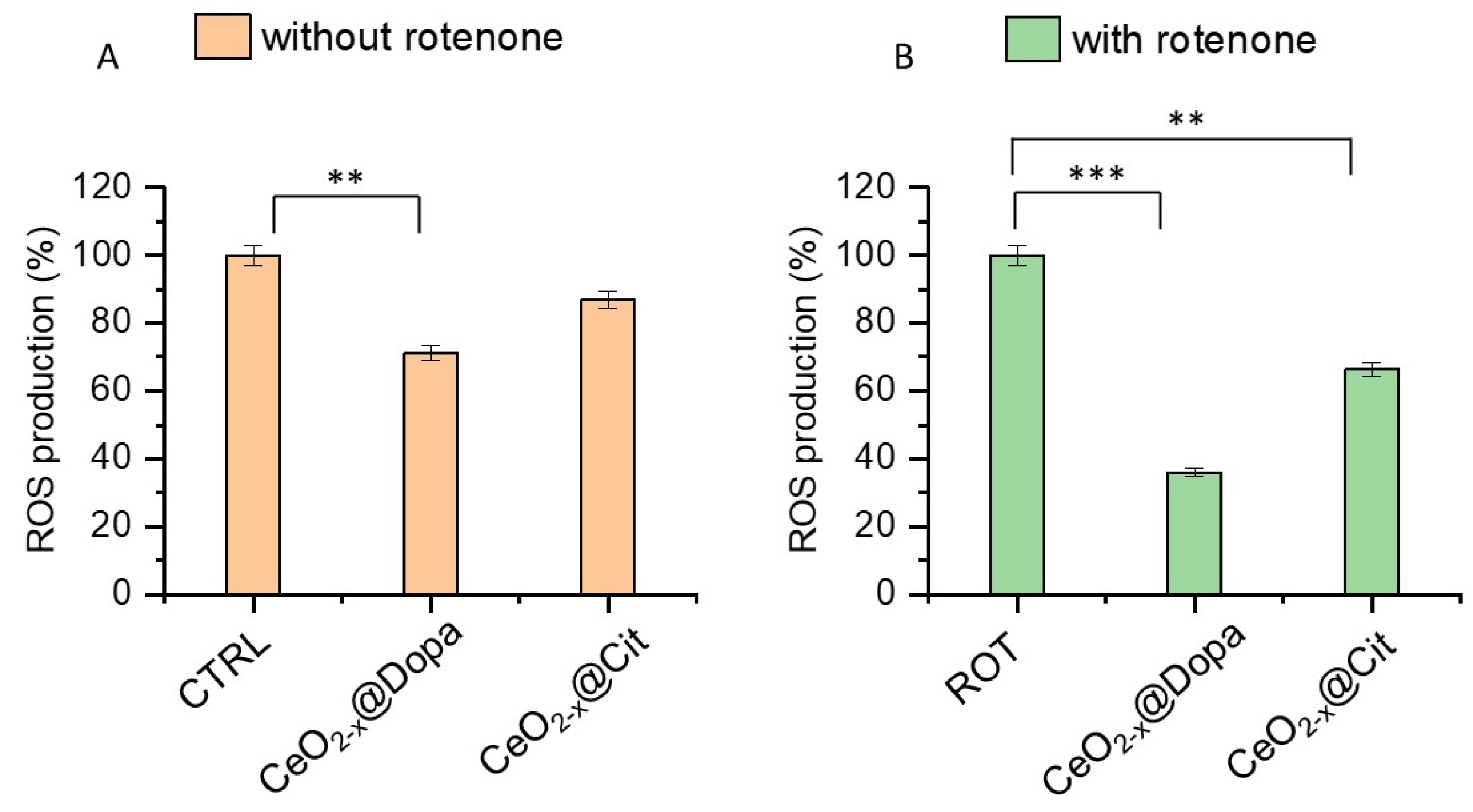
3.3.3. Antioxidant Effect of CeO2−x@Cit and CeO2−x@Dopa on DITNC1 Cells

Astrocytes play a crucial role in maintaining redox homeostasis in the brain, making them particularly relevant for assessing the neuroprotective potential of antioxidant compounds [71]. In this study, the DITNC1 cell line, represented by immortalized astrocytes derived from rat brain tissue, has been selected to evaluate the antioxidant power effects of CeO2−x NPs since it represents a valuable model for investigating oxidative stress in the central nervous system. Cell cultures have been treated with CeO2−x NPs at a concentration of 10 μg/mL that falls within the non-cytotoxic range (Figure S5). The production of ROS under both basal and oxidative stress conditions, induced by the addition of rotenone, was monitored using the DCF-DA assay. Even though DCFH-DA lacks selectivity in ROS detection, it has been reported as an easily and widely available procedure, relatively inexpensive, and easy to load into cells.
Rotenone is a potent inhibitor of mitochondrial Complex I, a critical enzyme involved in the electron transport chain, and has been widely used to study mitochondrial-induced oxidative stress [72]. Indeed, the inhibition of Complex I causes an accumulation of electrons within the mitochondrial matrix, which can react with O2 to produce ROS, particularly ·O2. Because of the increased ROS levels, cell components, including lipids, proteins, and DNA, are damaged, contributing to mitochondrial dysfunction and cellular oxidative stress.
The incubation of DITNC1 with each CeO2−x NPs preparation, under basal conditions, resulted in a statistically significant decrease in ROS production for both CeO2−x@Cit (10%) and CeO2−x@Dopa (28%) compared to the corresponding control (Figure 5A). Treatment with rotenone resulted in a 70% increase in ROS production in DITNC1 cells compared to the negative control, confirming the role of this compound as an inducer of oxidative stress. The ROS scavenging activity shown by CeO2−x NPs enhanced under oxidative stressed conditions (Figure 5B) induced by the addition of rotenone. In this case the decrease in ROS production, in comparison to ROT control, was 34% and 65% for CeO2−x@Cit and CeO2−x@Dopa, respectively. A greater ability to scavenge ROS is experienced by the cells incubated with CeO2−x@Dopa, both without and with addition of rotenone, rather than CeO2−x@Cit.
Discussion. The evaluation of ROS production within DITNC1 cells without and with the addition of rotenone confirms the intrinsic antioxidant activity of CeO2−x NPs. Even though according to literature studies [4,15,73], the enhanced antioxidant activity shown by CeO2−x NPs treated with dopamine might be attributed to the higher Ce3+/Ce4+ ratio, quantification of cerium valence ratio by XPS has highlighted only a slightly different Ce3+/Ce4+ ratio between CeO2−x@Cit and CeO2−x@Dopa, whose extent could not support the high efficacy in ROS scavenging shown by CeO2−x@Dopa.
Thanks to an extensive characterization of the NPs, which offered a clear picture of the surface functional groups, and thanks to fundamental knowledge built on solid literature studies [6,25,58], we proposed that the reason behind the better performance in limiting ROS production in biological environments demonstrated by CeO2−x@Dopa compared to CeO2−x@Cit, should not be only attributed to the intrinsic properties of CeO2−x NPs but should also be sought in the reactivity associated with the functional groups at the surface of the NPs. In particular, the presence of a surface coating bearing o-quinone structures must be considered. Although several studies described the improvement in antioxidant activity offered by citrates in citrate-functionalized NPs [74,75], our results indicate that o-quinone functionalization is more effective in boosting the catalytic activity of CeO2−x NPs in biological environments.
As illustrated in Scheme 1 and demonstrated by Amorati et al. for polydopamine [25,58,69], o-quinones can scavenge HOO· radicals by oxidizing them to O2 while being reduced to o-semiquinones. These intermediates can then interrupt the autoxidation chain of organic substrates by donating electrons and protons to ROO·, thereby regenerating the o-quinone form. This redox cycling adds a complementary mechanism to the intrinsic activity of CeO2−x NPs, enhancing chain-breaking antioxidant capacity and suppressing the propagation of ROS production induced by autoxidation of organic substrates.
The improved ROS scavenging activity observed for CeO2−x@Dopa arises from these complex redox mechanisms that are not captured by the DPPH· assay. In the latter, the antioxidant activity appears underestimated due to the limited diffusion of the DPPH· radical toward the NPs surface, which is hindered by the surface coating, and the lack of reactivity between o-quinone and DPPH·.
The comparative experimental approach employed, using CeO2−x@Cit NPs as a control to evaluate the redox behavior of our CeO2−x@Dopa NPs, and the detailed characterization of both samples, focusing on their chemical states and functional groups, along with well-established literature knowledge of the antioxidant properties of o-quinone functionalities and CeO2−x NPs, provided a robust structure–function correlation. The comparative framework offered meaningful insight into the enhanced antioxidant performance of CeO2−x@Dopa. Our findings highlight the potential of engineered nanostructures to interfere with oxidative stress pathways and suppress oxidative chain reactions more effectively. Indeed, this work, being clearly a material-oriented study, does not present an exhaustive mechanistic investigation of the antioxidant behavior, even though solid background supports the discussion of the achieved outcomes and conclusion.

4. Conclusions

This study has confirmed the pivotal role of CeO2−x NPs in quenching ROS and its effective enhancement by NP surface engineering. Water-dispersible CeO2−x@Dopa NPs have been synthesized using a colloidal approach involving thermal decomposition of a precursor in hot coordination solvent, yielding ultrasmall NPs approximately 3 nm in diameter, followed by phase transfer by ligand-exchange reaction with citric acid and post-synthetic treatment with dopamine hydrochloride. A more sustainable ligand-exchange procedure has been exploited, using CME with DMSO as the reaction medium. Comprehensive characterization has revealed two key findings: (i) CeO2−x NPs showed an increased Ce3+/Ce4+ ratio compared to bulk CeO2 (Ce3+/Ce4+ 30%/70%), attributed to their nanoscale; (ii) post-synthetic treatment with dopamine resulted in a slight increase in Ce3+ and a surface coating characterized by a polymeric structure bearing o-quinone structures. Evaluation of the radical scavenging activity—both via chemical non-enzymatic assays and in vitro experiments within DITNC1 cells—has highlighted the crucial role played by surface chemistry in the material’s antioxidant behavior. Despite the diffusion of DPPH· and H2O2 on the CeO2−x@Dopa surface being partially hindered by the polymeric coating, the overall ROS scavenging activity in the biological environment has increased. Unlike earlier studies that attributed antioxidant enhancement solely to a higher Ce3+/Ce4+ ratio, our comparative analysis between CeO2−x@Cit and CeO2−x@Dopa suggested that the increased ROS scavenging ability can be traced back to the presence in the latter sample of the o-quinone surface functionalities. These groups, whose presence in the polymer coating of CeO2−x@Dopa has been demonstrated by complementary UV–vis absorption, FT-IR, and XPS characterization, can contribute, by quenching HOO· radicals, to disrupting the autoxidation chain of organic substrate, a key process in the propagation of oxidative stress. This finding underscores the strategic value of dopamine treatment, which synergistically amplifies the NPs antioxidant capacity of CeO2−x, offering an innovative approach for applications requiring high antioxidant activity in complex biological settings.
The mechanistic insights presented in this study can open the venue for future in-depth investigations. As demonstrated by Amorati et al., the use of well-defined chemical systems—such as those able to generate hydroperoxyl radical in situ and promote the autoxidation of organic substrates—offers a valuable strategy for evaluating the antioxidant activity of CeO2−x@Dopa compared to CeO2−x@Cit, particularly through kinetic monitoring of oxygen consumption. In parallel, fluorescence microscopy investigation of properly treated cells could provide high spatial resolution and sensitivity, enabling real-time visualization of ROS production and identification of intracellular compartments involved. Furthermore, for any potential biomedical application, it will be essential to assess NPs colloidal stability in physiological media as well as their interaction with biological systems, including protein corona formation, biodistribution, and clearance, prior to advancing towards in vivo studies and clinical translation.

Supplementary Materials

The following supporting information can be downloaded at: https://www.mdpi.com/article/10.3390/antiox14080916/s1, Figure S1: size distribution obtained by TEM of CeO2−x NPs before and after ligand-exchange with citric acid; Table S1: Sketches of the two water-dispersible sample of CeO2−x@Cit and CeO2−x@Dopa and peak area of the different contributions of the XPS high resolution C1s spectrum; Figure S2: FT-IR of CeO2−x@OAm, CeO2−x@Cit, CeO2−x@Dopa and polydopamine; Figure S3: TGA of CeO2−x@Dopa; Figure S4: Test of the scavenging activity of DPPH· with polydopamine at different concentrations; Figure S5: cell viability test of CeO2−x@Cit and CeO2−x@Dopa.

Author Contributions

P.L.: investigation, validation, visualization, writing—original draft preparation; T.L.: investigation, validation, writing—review and editing, writing—original draft preparation, visualization; U.M.: investigation, visualization, validation; R.M.M.: investigation, validation; A.M.: investigation, validation, visualization, writing—original draft preparation, writing—review and editing; M.G.: resources, writing—review and editing; G.M.L.: resources, writing—review and editing; G.P.: investigation, methodology, validation, writing—original draft preparation, writing—review and editing; A.P.: writing—review and editing, N.D.: writing—review and editing resources; M.L.C.: resources, visualization, writing—review and editing; E.F.: conceptualization, resources, writing—original draft preparation, supervision, visualization, writing—review and editing. All authors have read and agreed to the published version of the manuscript.

Funding

This research received no external funding.

Data Availability Statement

Dataset available on request from the authors.

Acknowledgments

The authors want to thank for supporting the work the European Union through Next Generation EU, Mission 4 Component 1, and MUR within PRIN call 2022 PNRR, Project title: Photocatalytically regenerable hierarchically porous adsorbents for efficient water treatment PHOTOPAD, P2022FP2W4, 2023-2025CUP B53D23027540001.

Conflicts of Interest

The authors declare no conflicts of interest.

References

  1. Damatov, D.; Laga, S.M.; Mader, E.A.; Peng, J.; Agarwal, R.G.; Mayer, J.M. Redox Reactivity of Colloidal Nanoceria and Use of Optical Spectra as an In Situ Monitor of Ce Oxidation States. Inorg. Chem. 2018, 57, 14401–14408. [Google Scholar] [CrossRef]
  2. Eriksson, P.; Tal, A.A.; Skallberg, A.; Brommesson, C.; Hu, Z.; Boyd, R.D.; Olovsson, W.; Fairley, N.; Abrikosov, I.A.; Zhang, X.; et al. Cerium oxide nanoparticles with antioxidant capabilities and gadolinium integration for MRI contrast enhancement. Sci. Rep. 2018, 8, 6999. [Google Scholar] [CrossRef] [PubMed]
  3. Das, S.; Dowding, J.M.; Klump, K.E.; McGinnis, J.F.; Self, W.; Seal, S. Cerium oxide nanoparticles: Applications and prospects in nanomedicine. Nanomedicine 2013, 8, 1483–1508. [Google Scholar] [CrossRef] [PubMed]
  4. Xue, Y.; Yang, F.; Wu, L.; Xia, D.; Liu, Y. CeO2 nanoparticles to promote wound healing: A systematic review. Adv. Healthc. Mater. 2024, 13, 2302858. [Google Scholar] [CrossRef] [PubMed]
  5. Nelson, B.C.; Johnson, M.E.; Walker, M.L.; Riley, K.R.; Sims, C.M. Antioxidant Cerium Oxide Nanoparticles in Biology and Medicine. Antioxidants 2016, 5, 15. [Google Scholar] [CrossRef]
  6. Amorati, R.; Guo, Y.; Budhlall, B.M.; Barry, C.F.; Cao, D.; Challa, S.S.R.K. Tandem Hydroperoxyl–Alkylperoxyl Radical Quenching by an Engineered Nanoporous Cerium Oxide Nanoparticle Macrostructure (NCeONP): Toward Efficient Solid-State Autoxidation Inhibitors. ACS Omega 2023, 8, 40174–40183. [Google Scholar] [CrossRef]
  7. Grulke, E.; Reed, K.; Beck, M.; Huang, X.; Cormack, A.; Seal, S. Nanoceria: Factors affecting its pro- and anti-oxidant properties. Environ. Sci. Nano 2014, 1, 429–444. [Google Scholar] [CrossRef]
  8. Datta, A.; Mishra, S.; Manna, K.; Saha, K.D.; Mukherjee, S.; Roy, S. Pro-oxidant therapeutic activities of cerium oxide nanoparticles in colorectal carcinoma cells. ACS Omega 2020, 5, 9714–9723. [Google Scholar] [CrossRef]
  9. Wang, Z.; Shen, X.; Gao, X.; Zhao, Y. Simultaneous enzyme mimicking and chemical reduction mechanisms for nanoceria as a bio-antioxidant: A catalytic model bridging computations and experiments for nanozymes. Nanoscale 2019, 11, 13289–13299. [Google Scholar] [CrossRef]
  10. Othman, A.; Gowda, A.; Andreescu, D.; Hassan, M.H.; Babu, S.; Seo, J.; Andreescu, S. Two decades of ceria nanoparticles research: Structure, properties and emerging applications. Mater. Horiz. 2024, 11, 3213–3266. [Google Scholar] [CrossRef]
  11. Baldim, V.; Bedioui, F.; Mignet, N.; Margaill, I.; Berret, J.-F. The enzyme-like catalytic activity of cerium oxide nanoparticles and its dependency on Ce 3+ surface area concentration. Nanoscale 2018, 10, 6971–6980. [Google Scholar] [CrossRef] [PubMed]
  12. Baschieri, A.; Amorati, R. Methods to Determine Chain-Breaking Antioxidant Activity of Nanomaterials beyond DPPH•. A Review. Antioxidants 2021, 10, 1551. [Google Scholar] [CrossRef] [PubMed]
  13. Valgimigli, L. Lipid Peroxidation and Antioxidant Protection. Biomolecules 2023, 13, 1291. [Google Scholar] [CrossRef] [PubMed]
  14. Baschieri, A.; Zongxin, J.; and Amorati, R. Hydroperoxyl radical (HOO•) as a reducing agent: Unexpected synergy with antioxidants. A review. Free Radic. Res. 2023, 57, 115–129. [Google Scholar] [CrossRef]
  15. Damatov, D.; Mayer, J.M. (Hydro)peroxide ligands on colloidal cerium oxide nanoparticles. Chem. Commun. 2016, 52, 10281–10284. [Google Scholar] [CrossRef]
  16. Hu, Y.; Zhang, Q.; Garcia-Rojas, D.; Ling, V.; Masterson, C.M.; Bi, Y.; Xiao, Z.; Guo, X.; Villanova, J.; Dunn, J.; et al. Increasing the antioxidant capacity of ceria nanoparticles with catechol-grafted poly(ethylene glycol). J. Mater. Chem. B 2022, 10, 10042–10053. [Google Scholar] [CrossRef]
  17. Chen, L.; Fleming, P.; Morris, V.; Holmes, J.D.; Morris, M.A. Size-Related Lattice Parameter Changes and Surface Defects in Ceria Nanocrystals. J. Phys. Chem. C 2010, 114, 12909–12919. [Google Scholar] [CrossRef]
  18. Deshpande, S.; Patil, S.; Kuchibhatla, S.V.; Seal, S. Size dependency variation in lattice parameter and valency states in nanocrystalline cerium oxide. Appl. Phys. Lett. 2005, 87, 133113. [Google Scholar] [CrossRef]
  19. Lord, M.S.; Berret, J.F.; Singh, S.; Vinu, A.; Karakoti, A.S. Redox Active Cerium Oxide Nanoparticles: Current Status and Burning Issues. Small 2021, 17, 2102342. [Google Scholar] [CrossRef]
  20. Goujon, G.; Baldim, V.; Roques, C.; Bia, N.; Seguin, J.; Palmier, B.; Graillot, A.; Loubat, C.; Mignet, N.; Margaill, I. Antioxidant activity and toxicity study of cerium oxide nanoparticles stabilized with innovative functional copolymers. Adv. Healthc. Mater. 2021, 10, 2100059. [Google Scholar] [CrossRef]
  21. Baldim, V.; Yadav, N.; Bia, N.; Graillot, A.; Loubat, C.; Singh, S.; Karakoti, A.S.; Berret, J.-F. Polymer-Coated Cerium Oxide Nanoparticles as Oxidoreductase-like Catalysts. ACS Appl. Mater. Interfaces 2020, 12, 42056–42066. [Google Scholar] [CrossRef]
  22. Lee, S.S.; Song, W.; Cho, M.; Puppala, H.L.; Nguyen, P.; Zhu, H.; Segatori, L.; Colvin, V.L. Antioxidant Properties of Cerium Oxide Nanocrystals as a Function of Nanocrystal Diameter and Surface Coating. ACS Nano 2013, 7, 9693–9703. [Google Scholar] [CrossRef] [PubMed]
  23. Bülbül, G.; Hayat, A.; Liu, X.; Andreescu, S. Reactivity of nanoceria particles exposed to biologically relevant catechol-containing molecules. RSC Adv. 2016, 6, 60007–60014. [Google Scholar] [CrossRef]
  24. Hayat, A.; Andreescu, D.; Bulbul, G.; Andreescu, S. Redox reactivity of cerium oxide nanoparticles against dopamine. J. Colloid Interface Sci. 2014, 418, 240–245. [Google Scholar] [CrossRef] [PubMed]
  25. Mollica, F.; Lucernati, R.; Amorati, R. Expanding the spectrum of polydopamine antioxidant activity by nitroxide conjugation. J. Mater. Chem. B 2021, 9, 9980–9988. [Google Scholar] [CrossRef]
  26. Lee, S.S.; Zhu, H.; Contreras, E.Q.; Prakash, A.; Puppala, H.L.; Colvin, V.L. High Temperature Decomposition of Cerium Precursors To Form Ceria Nanocrystal Libraries for Biological Applications. Chem. Mater. 2012, 24, 424–432. [Google Scholar] [CrossRef]
  27. Berestok, T.; Guardia, P.; Blanco, J.; Nafria, R.; Torruella, P.; López-Conesa, L.; Estradé, S.; Ibáñez, M.; de Roo, J.; Luo, Z.; et al. Tuning Branching in Ceria Nanocrystals. Chem. Mater. 2017, 29, 4418–4424. [Google Scholar] [CrossRef]
  28. Yu, T.; Park, Y.I.; Kang, M.C.; Joo, J.; Park, J.K.; Won, H.Y.; Kim, J.J.; Hyeon, T. Large-scale synthesis of water dispersible ceria nanocrystals by a simple sol–gel process and their use as a chemical mechanical planarization slurry. Small 2008, 6, 855–858. [Google Scholar] [CrossRef]
  29. Monnier, C.A.; Lattuada, M.; Burnand, D.; Crippa, F.; Martinez-Garcia, J.C.; Hirt, A.M.; Rothen-Rutishauser, B.; Bonmarin, M.; Petri-Fink, A. A lock-in-based method to examine the thermal signatures of magnetic nanoparticles in the liquid, solid and aggregated states. Nanoscale 2016, 8, 13321–13332. [Google Scholar] [CrossRef]
  30. Latronico, T.; Rizzi, F.; Panniello, A.; Laquintana, V.; Arduino, I.; Denora, N.; Fanizza, E.; Milella, S.; Mastroianni, C.M.; Striccoli, M. Luminescent PLGA nanoparticles for delivery of darunavir to the brain and inhibition of matrix metalloproteinase-9, a relevant therapeutic target of HIV-associated neurological disorders. ACS Chem. Neurosci. 2021, 12, 4286–4301. [Google Scholar] [CrossRef]
  31. Petraglia, T.; Latronico, T.; Pepe, A.; Crescenzi, A.; Liuzzi, G.M.; Rossano, R. Increased Antioxidant Performance of Lignin by Biodegradation Obtained from an Extract of the Mushroom Pleurotus eryngii. Molecules 2024, 29, 5575. [Google Scholar] [CrossRef]
  32. Giancaspro, M.; Grisorio, R.; Alò, G.; Margiotta, N.; Panniello, A.; Suranna, G.P.; Depalo, N.; Striccoli, M.; Curri, M.L.; Fanizza, E. Molecular insights into the growth and time evolution of surface states of CsPbBr3 nanoparticles synthesized using a scalable room temperature approach. Mater. Chem. Front. 2023, 7, 2637–2650. [Google Scholar] [CrossRef]
  33. Kim, J.; Hong, G.; Mazaleuskaya, L.; Hsu, J.C.; Rosario-Berrios, D.N.; Grosser, T.; Cho-Park, P.F.; Cormode, D.P. Ultrasmall Antioxidant Cerium Oxide Nanoparticles for Regulation of Acute Inflammation. ACS Appl. Mater. Interfaces 2021, 13, 60852–60864. [Google Scholar] [CrossRef] [PubMed]
  34. Parak, W.; Gerion, D.; Pellegrino, T.; Zanchet, D.; Micheel, C.; Williams, S.; Boudreau, R.; Gros, M.; Larabell, C.; Alivisatos, A. Biological Applications of Colloidal Nanocrystals. Nanotechnology 2003, 14, R15. [Google Scholar] [CrossRef]
  35. Hancock, M.L.; Yokel, R.A.; Beck, M.J.; Calahan, J.L.; Jarrells, T.W.; Munson, E.J.; Olaniyan, G.A.; Grulke, E.A. The characterization of purified citrate-coated cerium oxide nanoparticles prepared via hydrothermal synthesis. Appl. Surf. Sci. 2021, 535, 147681. [Google Scholar] [CrossRef]
  36. Thistlethwaite, P.; Hook, M. Diffuse Reflectance Fourier Transform Infrared Study of the Adsorption of Oleate/Oleic Acid onto Titania. Langmuir 2000, 16, 4993–4998. [Google Scholar] [CrossRef]
  37. Fanizza, E.; Depalo, N.; Clary, L.; Agostiano, A.; Striccoli, M.; Curri, M.L. A combined size sorting strategy for monodisperse plasmonic nanostructures. Nanoscale 2013, 5, 3272–3282. [Google Scholar] [CrossRef]
  38. Altomare, M.; Fanizza, E.; Corricelli, M.; Comparelli, R.; Striccoli, M.; Curri, M.L. Patterned assembly of luminescent nanocrystals: Role of the molecular chemistry at the interface. J. Nanoparticle Res. 2014, 16, 1–14. [Google Scholar] [CrossRef]
  39. Phoka, S.; Laokul, P.; Swatsitang, E.; Promarak, V.; Seraphin, S.; Maensiri, S. Synthesis, structural and optical properties of CeO2 nanoparticles synthesized by a simple polyvinyl pyrrolidone (PVP) solution route. Mater. Chem. Phys. 2009, 115, 423–428. [Google Scholar] [CrossRef]
  40. Park, J.-W.; Shumaker-Parry, J.S. Structural study of citrate layers on gold nanoparticles: Role of intermolecular interactions in stabilizing nanoparticles. J. Am. Chem. Soc. 2014, 136, 1907–1921. [Google Scholar] [CrossRef]
  41. Nara, M.; Torii, H.; Tasumi, M. Correlation between the Vibrational Frequencies of the Carboxylate Group and the Types of Its Coordination to a Metal Ion: An ab Initio Molecular Orbital Study. J. Phys. Chem. 1996, 100, 19812–19817. [Google Scholar] [CrossRef]
  42. Andreescu, D.; Matijević, E.; Goia, D.V. Formation of uniform colloidal ceria in polyol. Colloids Surf. Physicochem. Eng. Asp. 2006, 291, 93–100. [Google Scholar] [CrossRef]
  43. Culica, M.E.; Chibac-Scutaru, A.L.; Melinte, V.; Coseri, S. Cellulose Acetate Incorporating Organically Functionalized CeO2 NPs: Efficient Materials for UV Filtering Applications. Materials 2020, 13, 2955. [Google Scholar] [CrossRef]
  44. Zhang, Z.; Yu, L.; Liu, W.; Song, Z. Surface modification of ceria nanoparticles and their chemical mechanical polishing behavior on glass substrate. Appl. Surf. Sci. 2010, 256, 3856–3861. [Google Scholar] [CrossRef]
  45. Baranchikov, A.E.; Sozarukova, M.M.; Mikheev, I.V.; Egorova, A.A.; Proskurnina, E.V.; Poimenova, I.A.; Krasnova, S.A.; Filippova, A.D.; Ivanov, V.K. Biocompatible ligands modulate nanozyme activity of CeO2 nanoparticles. New J. Chem. 2023, 47, 20388–20404. [Google Scholar] [CrossRef]
  46. Ioannou, M.E.; Pouroutzidou, G.K.; Chatzimentor, I.; Tsamesidis, I.; Florini, N.; Tsiaoussis, I.; Lymperaki, E.; Komninou, P.; Kontonasaki, E. Synthesis and Characterization of Cerium Oxide Nanoparticles: Effect of Cerium Precursor to Gelatin Ratio. Appl. Sci. 2023, 13, 2676. [Google Scholar] [CrossRef]
  47. Mourdikoudis, S.; Menelaou, M.; Fiuza-Maneiro, N.; Zheng, G.; Wei, S.; Pérez-Juste, J.; Polavarapu, L.; Sofer, Z. Oleic acid/oleylamine ligand pair: A versatile combination in the synthesis of colloidal nanoparticles. Nanoscale Horiz. 2022, 7, 941–1015. [Google Scholar] [CrossRef]
  48. Pinna, A.; Cali, E.; Kerherve, G.; Galleri, G.; Maggini, M.; Innocenzi, P.; Malfatti, L. Fulleropyrrolidine-functionalized ceria nanoparticles as a tethered dual nanosystem with improved antioxidant properties. Nanoscale Adv. 2020, 2, 2387–2396. [Google Scholar] [CrossRef]
  49. Seal, S.; Jeyaranjan, A.; Neal, C.J.; Kumar, U.; Sakthivel, T.S.; Sayle, D.C. Engineered defects in cerium oxides: Tuning chemical reactivity for biomedical, environmental, & energy applications. Nanoscale 2020, 12, 6879–6899. [Google Scholar] [CrossRef]
  50. Allahgholi, A.; Flege, J.I.; Thieß, S.; Drube, W.; Falta, J. Oxidation-State Analysis of Ceria by X-ray Photoelectron Spectroscopy. ChemPhysChem 2015, 16, 1083–1091. [Google Scholar] [CrossRef] [PubMed]
  51. Sims, C.M.; Maier, R.A.; Johnston-Peck, A.C.; Gorham, J.M.; Hackley, V.A.; Nelson, B.C. Approaches for the quantitative analysis of oxidation state in cerium oxide nanomaterials. Nanotechnology 2019, 30, 085703. [Google Scholar] [CrossRef]
  52. Zhang, F.; Wang, P.; Koberstein, J.; Khalid, S.; Chan, S.-W. Cerium oxidation state in ceria nanoparticles studied with X-ray photoelectron spectroscopy and absorption near edge spectroscopy. Surf. Sci. 2004, 563, 74–82. [Google Scholar] [CrossRef]
  53. Morgan, D.J. Photoelectron spectroscopy of ceria: Reduction, quantification and the myth of the vacancy peak in XPS analysis. Surf. Interface Anal. 2023, 55, 845–850. [Google Scholar] [CrossRef]
  54. Ma, W.; Mashimo, T.; Tamura, S.; Tokuda, M.; Yoda, S.; Tsushida, M.; Koinuma, M.; Kubota, A.; Isobe, H.; Yoshiasa, A. Cerium oxide (CeO2−x) nanoparticles with high Ce3+ proportion synthesized by pulsed plasma in liquid. Ceram. Int. 2020, 46, 26502–26510. [Google Scholar] [CrossRef]
  55. Mullins, D.R. The surface chemistry of cerium oxide. Surf. Sci. Rep. 2015, 70, 42–85. [Google Scholar] [CrossRef]
  56. Yadav, S.V.; Rathod, V.K. Oxidase-like activity of magnetically separable nano ceria for catechol detection. SN Appl. Sci. 2019, 1, 1071. [Google Scholar] [CrossRef]
  57. Kim, H.; Yook, S.H.; Kim, H.Y.; Choi, Y.; Lim, Y.; Hwang, Y.; Kim, J.; Lee, K.Y.; Jang, S.S.; Park, J.; et al. Tailor-Made Charged Catechol-Based Polymeric Ligands to Build Robust Fuel Cells Containing Antioxidative Nanoparticles. Adv. Electron. Mater. 2022, 8, 2200171. [Google Scholar] [CrossRef]
  58. Guo, Y.; Baschieri, A.; Mollica, F.; Valgimigli, L.; Cedrowski, J.; Litwinienko, G.; Amorati, R. Hydrogen Atom Transfer from HOO. to ortho-Quinones Explains the Antioxidant Activity of Polydopamine. Angew. Chem. Int. Ed. 2021, 60, 15220–15224. [Google Scholar] [CrossRef]
  59. Liebscher, J.; Mrówczyński, R.; Scheidt, H.A.; Filip, C.; Hădade, N.D.; Turcu, R.; Bende, A.; Beck, S. Structure of Polydopamine: A Never-Ending Story? Langmuir 2013, 29, 10539–10548. [Google Scholar] [CrossRef]
  60. Andreescu, D.; Bulbul, G.; Özel, R.E.; Hayat, A.; Sardesai, N.; Andreescu, S. Applications and implications of nanoceria reactivity: Measurement tools and environmental impact. Environ. Sci. Nano 2014, 1, 445–458. [Google Scholar] [CrossRef]
  61. Lazić, V.; Živković, L.S.; Sredojević, D.; Fernandes, M.M.; Lanceros-Mendez, S.; Ahrenkiel, S.P.; Nedeljković, J.M. Tuning Properties of Cerium Dioxide Nanoparticles by Surface Modification with Catecholate-type of Ligands. Langmuir 2020, 36, 9738–9746. [Google Scholar] [CrossRef]
  62. Finocchiaro, G.; Ju, X.; Mezghrani, B.; Berret, J.-F. Cerium Oxide Catalyzed Disproportionation of Hydrogen Peroxide: A Closer Look at the Reaction Intermediate. Chem. A Eur. J. 2024, 30, e202304012. [Google Scholar] [CrossRef]
  63. Goia, S.; Richings, G.W.; Turner, M.A.P.; Woolley, J.M.; Tully, J.J.; Cobb, S.J.; Burriss, A.; Robinson, B.R.; Macpherson, J.V.; Stavros, V.G. Ultrafast Spectroelectrochemistry of the Catechol/o-Quinone Redox Couple in Aqueous Buffer Solution. ChemPhotoChem 2024, 8, e202300325. [Google Scholar] [CrossRef]
  64. Boutaybi, M.E.; Titi, A.; Alzahrani, A.Y.; Bahari, Z.; Tillard, M.; Hammouti, B.; Touzani, R. Aerial Oxidation of Phenol/Catechol in the Presence of Catalytic Amounts of [(Cl) 2Mn (RCOOET)], RCOOET= Ethyl-5-Methyl-1-(((6-Methyl-3-Nitropyridin-2-yl) Amino) Methyl)-1 H-Pyrazole-3-Carboxylate. Catalysts 2022, 12, 1642. [Google Scholar] [CrossRef]
  65. Seminko, V.; Maksimchuk, P.; Grygorova, G.; Okrushko, E.; Avrunin, O.; Semenets, V.; Malyukin, Y. Mechanism and Dynamics of Fast Redox Cycling in Cerium Oxide Nanoparticles at High Oxidant Concentration. J. Phys. Chem. C 2021, 125, 4743–4749. [Google Scholar] [CrossRef]
  66. Munteanu, I.G.; Apetrei, C. Analytical Methods Used in Determining Antioxidant Activity: A Review. Int. J. Mol. Sci. 2021, 22, 3380. [Google Scholar] [CrossRef]
  67. Popov, A.L.; Shcherbakov, A.B.; Zholobak, N.; Baranchikov, A.Y.; Ivanov, V.K. Cerium dioxide nanoparticles as third-generation enzymes (nanozymes). Nanosyst. Phys. Chem. Math. 2017, 8, 760–781. [Google Scholar] [CrossRef]
  68. Nagarale, R.K.; Hoss, U.; Heller, A. Mixed-Valence Metal Oxide Nanoparticles as Electrochemical Half-Cells: Substituting the Ag/AgCl of Reference Electrodes by CeO2–x Nanoparticles. J. Am. Chem. Soc. 2012, 134, 20783–20787. [Google Scholar] [CrossRef]
  69. Liu, H.; Qu, X.; Tan, H.; Song, J.; Lei, M.; Kim, E.; Payne, G.F.; Liu, C. Role of polydopamine’s redox-activity on its pro-oxidant, radical-scavenging, and antimicrobial activities. Acta Biomater. 2019, 88, 181–196. [Google Scholar] [CrossRef]
  70. Echegaray, N.; Pateiro, M.; Munekata, P.E.S.; Lorenzo, J.M.; Chabani, Z.; Farag, M.A.; Domínguez, R. Measurement of Antioxidant Capacity of Meat and Meat Products: Methods and Applications. Molecules 2021, 26, 3880. [Google Scholar] [CrossRef]
  71. Baxter, P.S.; Hardingham, G.E. Adaptive regulation of the brain’s antioxidant defences by neurons and astrocytes. Free Radic. Biol. Med. 2016, 100, 147–152. [Google Scholar] [CrossRef] [PubMed]
  72. Li, N.; Ragheb, K.; Lawler, G.; Sturgis, J.; Rajwa, B.; Melendez, J.A.; Robinson, J.P. Mitochondrial complex I inhibitor rotenone induces apoptosis through enhancing mitochondrial reactive oxygen species production. J. Biol. Chem. 2003, 278, 8516–8525. [Google Scholar] [CrossRef]
  73. Damle, M.A.; Jakhade, A.P.; Chikate, R.C. Modulating Pro- and Antioxidant Activities of Nanoengineered Cerium Dioxide Nanoparticles against Escherichia coli. ACS Omega 2019, 4, 3761–3771. [Google Scholar] [CrossRef]
  74. Moglianetti, M.; Pedone, D.; Udayan, G.; Retta, S.F.; Debellis, D.; Marotta, R.; Turco, A.; Rella, S.; Malitesta, C.; Bonacucina, G. Intracellular antioxidant activity of biocompatible citrate-capped palladium nanozymes. Nanomaterials 2020, 10, 99. [Google Scholar] [CrossRef]
  75. Cao, G.; Chen, Y.; Jiang, C.; Xu, J.; Xue, W.; Zhang, H.; Tian, Y. Synthesis of Citrate-Capped Sn Nanoparticles with Excellent Oxidation Resistance for High-Performance Electrically Conductive Adhesives. ACS Appl. Electron. Mater. 2023, 5, 1164–1173. [Google Scholar] [CrossRef]
Figure 1. (A) Sketch of superoxide dismutase and catalase mimetic activity of CeO2−x NPs. (B) Scheme of the autoxidation chain of organic substrates, contributing to the release of hydroperoxyl radical (HOO·, (C)) also produced by protonation of superoxide anion radical (D). Role of CeO2−x NPs in chain breaking autoxidation processes (E).
Antioxidants 14 00916 g001
Figure 2. TEM micrographs (A,B), Tauc plot as determined by applying Kubelka–Munk function (C); FT-IR in attenuated total reflection mode (D,E), thermogravimetric (F) and first derivative (GH) curves of CeO2−x@OAm (green line) and CeO2−x@Cit (blue line).
Antioxidants 14 00916 g002
Figure 3. High-resolution XPS spectra Ce3d (A,D), O1s (B,E), and C1s (C,F), N1s (I) of CeO2−x@Cit sample (AC) and CeO2−x@Dopa sample (DF,I). Table of the composition of Ce3+ and Ce4+ (G), as determined from weight ratio of the integral area of Ce3+ and Ce4+ components. (H) Representative scheme of t CeO2−x@Cit NPs functionalization with dopamine with a sketch of the surface chemistry as estimated by chemical characterization. Color code: blue for CeO2−x@Cit sample and orange for CeO2−x@Dopa sample.
Antioxidants 14 00916 g003
Figure 4. (A) UV–vis absorption spectra of CeO2−x@Cit (blue line) and CeO2−x@Dopa (orange lines) samples before (solid line) and after (dashed line) addition of H2O2. (B) Absorption peak obtained after subtracting the absorbance from dispersion devoid of H2O2 for CeO2−x@Cit (blue line) and CeO2−x@Dopa (orange line) samples. (C) Test of the scavenging activity of DPPH·: Vis absorption spectra of DPPH· before (black trace) and after addition of citric acid (0.01 mg/mL, dashed green trace), CeO2−x@Cit (0.13 mg/mL blue line) and CeO2−x@Dopa (0.13 mg/mL orange line) samples. Inset, pictures of the DPPH· solutions after 30 minutes in the dark without (a)and with CeO2−x@Dopa (b) and with CeO2−x@Cit (c). (D) Bar graph of the percentage of DPPH· radical scavenging activity (RSA%) of CeO2−x@Cit at increasing dose (blue bars) and CeO2−x@Dopa (0.13 mg/mL orange bar).
Antioxidants 14 00916 g004
Figure 5. ROS production in DITNC1 cells treated with CeO2−x@Cit or CeO2−x@Dopa. The presence of ROS assessed by measuring the changes of the fluorescent signal of DCFH-DA. ROS production expressed as percentage (%) of photoluminescence (PL) intensity in comparison to negative (CTRL) (A) or positive control (ROT) (B) represented by unstimulated and untreated cells in serum-free medium (CTRL) and rotenone treated cells (ROT), respectively. Bar graphs in panel A and panel B referred to cells treated with or without rotenone, respectively, also in the presence of CeO2−x@Cit or CeO2−x@Dopa. The bar value represents the mean value ± Standard deviation of three independent experiments performed on different cell populations. Statistically significant decrease in comparison to CTRL or ROT indicated by asterisks (one-way ANOVA followed by Dunnet’s post hoc test was used to compare ROS levels and cell viability under different experimental conditions ** p < 0.01, *** p < 0.001. Data were obtained from at least three independent experiments, with each data point representing the mean of triplicate measurements within a single experiment.).
Antioxidants 14 00916 g005
Scheme 1. Representative scheme of the hydroperoxyl radical scavenging activity and organic substrate autoxidation chain breaking promoted by CeO2−x NPs functionalized with o-quinone structures generated after treatment with dopamine.
Antioxidants 14 00916 sch001
Disclaimer/Publisher’s Note: The statements, opinions and data contained in all publications are solely those of the individual author(s) and contributor(s) and not of MDPI and/or the editor(s). MDPI and/or the editor(s) disclaim responsibility for any injury to people or property resulting from any ideas, methods, instructions or products referred to in the content.

