Article

Dietary Carnosic Acid Supplementation Improves the Growth Performance, the Antioxidant Status, and Diversity of Intestinal Microbiota in Broilers

Institute of Animal Science, Guangdong Academy of Agricultural Sciences, State Key Laboratory of Swine and Poultry Breeding Industry, Key Laboratory of Animal Nutrition and Feed Science in South China, Ministry of Agriculture and Rural Affairs, Guangdong Provincial Key Laboratory of Animal Breeding and Nutrition, Guangzhou 510640, China
*
Authors to whom correspondence should be addressed.
These authors contributed equally to this work.
Antioxidants 2025, 14(8), 1026; https://doi.org/10.3390/antiox14081026
Submission received: 26 June 2025 / Revised: 12 August 2025 / Accepted: 18 August 2025 / Published: 21 August 2025
(This article belongs to the Section Health Outcomes of Antioxidants and Oxidative Stress)

Abstract

Carnosic acid (CA), a natural phenolic terpenoid compound, is widely distributed in plants such as sage and rosemary, and exhibits a strong antioxidant capacity. The aim of this study was to investigate the effects of different levels of CA on growth performance, antioxidant capacity, and intestinal health of yellow-feathered broilers, and then to determine the optimal dose of CA to promote sustainable broiler production. A total of 384 1-day-old yellow-feathered broilers were randomly allocated into six treatment groups with eight replicates per group and eight birds per replicate pen. The control group (CON) was fed a basal diet and the CA treated groups (CA5, CA10, CA20, CA40, and CA80) were fed diets given different doses of CA (5, 10, 20, 40, and 80 mg/kg), respectively, for 53 days (1~21 d and 22~53 d). The results showed that, in the later stages of the experiment, supplementation with 10, 20, and 40 mg/kg of CA increased (p < 0.05) the final body weight and average daily gain. Morphometric analyses of the jejunum showed that supplementation of CA increased (p < 0.05) the ratio of villus height to crypt depth (V/C). Antioxidant indices revealed that CA significantly reduced MDA levels in plasma, liver, and jejunum, while enhancing activities of GSH-Px, T-SOD, and T-AOC (p < 0.05). Moreover, CA upregulated hepatic Nrf2, HO-1, GSH-Px, and GSR expression via downregulated Keap1. The analysis of intestinal microbiota showed that CA increased (p < 0.05) microbial α diversity (Ace, Chao, and Sobs indices) and increased (p < 0.05) beneficial bacteria, such as Streptococcus, Enterococcus, and Phascolarctobacterium. In conclusion, CA improves growth performance, antioxidant capacity, intestinal health, and gut microbial diversity in broilers. Under the conditions of this experiment, quadratic regressions for different variables showed that the optimal range for supplemental CA in chicken’s diet was 19.11~76.85 mg/kg. Combined with experimental observation and regression analysis, the optimal level of supplementation was 40 mg/kg.

1. Introduction

Chicken has become an important part of the global meat market, ranking second only to pork in consumption [1]. Poultry production faces serious challenges after the total ban on antimicrobials in feed, such as reduced growth performance, impaired meat quality, and intestinal damage [2], which prompts the search for bioactive alternatives to antibiotics to maintain the health production in poultry. Currently, more and more natural compounds with antioxidant activity have shown beneficial effects in poultry production as promising antibiotic substitutes [3,4,5].
As natural and nutritionally bioactive substances, plant extracts play an important role in enhancing growth performance [6], alleviating oxidative stress [7], and improving intestinal health of poultry [8]. Among them, phenolic antioxidants demonstrate strong free radical-scavenging activity by donating hydrogen ion and electron via phenolic hydroxyl groups or chelating metal ions [9]. Phenolic compounds, with their remarkable antioxidant properties, can regulate the Keap1/Nrf2 signaling pathway through multiple targets, thereby exerting cytoprotective effects [10]. Carnosic acid (C20H28O4, CA), a phenolic terpenoid compound, is widely present in plants such as Salvia and rosemary (Rosmarinus officinalis L.), and exhibits multiple benefits in livestock and poultry nutrition, including the following: (1) enhancing antioxidant capacity by regulating the endogenous antioxidant system, including upregulating antioxidant-related genes and the activity of antioxidant enzymes [11]; (2) reducing the secretion of inflammatory factors and inhibiting the proliferation of harmful bacteria [12]; (3) improving meat quality by regulating fat metabolism [13]. Previous research has indicated that CA can improve the growth performance of broiler chickens, reduce diarrhea rate and mortality rate [14,15], enhance the reproductive performance and egg quality of breeder chickens [16], and alleviate cold/heat stress damage [17,18], yet findings on its efficacy and optimal dosage remain inconsistent. In this regard, further research is needed to evaluate the effects of CA on broilers, including its impacts on growth performance, meat quality, and antioxidant status, as well as to determine the most effective supplementation level in diets, further providing a basis for its potential application in broiler production.

2. Materials and Methods

2.1. Animal Experimental Design

A total of 384 1-day-old fast-growing, yellow-feathered broilers were randomly allocated into 6 treatments, with 8 replicates per treatment and 8 birds per replicate pen. Birds in the control group (CON) were fed a basal diet, and the other groups (CA5, CA10, CA20, CA40, and CA80) were fed diets supplemented with 5, 10, 20, 40, and 80 mg/kg of CA (85%, Shanghai Yuanye Bio-Technology Co., Ltd., Shanghai, China), respectively.

2.2. Diets and Chicken Husbandry

The diets were formulated as recommended by Chinese Nutrient Requirements of Yellow Chickens (NY/T3645-2020, Ministry of Agriculture PRC, 2020) [19]. The composition and nutrient content of diets are shown in Table 1. The experiment was carried out in the testing farm of Institute of Animal Science, Guangdong Academy of Agricultural Sciences. Birds were raised in floor pens with wood shavings litter for 53 d. Water and diets were provided ad libitum throughout.

2.3. Measurement of Growth Performance

Birds were weighed at d l, d 21, and d 53 on a pen basis, the average daily gain (ADG), average daily feed intake (ADFI), and feed to gain ratio (F/G) were calculated.

2.4. Sample Collection

At d 21 and d 53, after 12 h of feed-withdrawal, one bird close to average body weight (BW) per replicate was chosen. Blood (5 mL) was collected from the wing veins and drawn into vacutainers containing anticoagulant (BD™ Vacutainer, Franklin Lakes, NJ, USA). The blood samples were then centrifuged at 3500× g for 10 min at 4 °C to separate the plasma, which was subsequently stored at −80 °C until analysis. Then, the birds were euthanized and dissected for samples. For birds at 21 d, the duodenum, jejunum, ileum, liver, spleen, thymus, and bursa of Fabricius were collected. For birds at 53 d, the duodenum, jejunum, ileum, liver, spleen, cecum, and pectoral muscle were collected and the mid-jejunal segments were rinsed with sterile saline and then partially fixed by immersion in 4% paraformaldehyde for further morphological observation.

2.5. Calculation of Relative Weights of Immune Organs

The liver, spleen, thymus, and bursa of Fabricius of birds at 21 d and the liver and spleen of birds at 53 d were blotted and weighed. The relative weights (weight of organ/BW × 100%) were calculated.

2.6. Determination of Meat Quality

Pectoralis major muscles were blotted, and objective indicators of meat quality, including instrumental color as L*(lightness), a*(redness), and b*(yellowness) value, shear force, drip loss, and pH value, were determined at 45 min and 24 h post mortem, with the methods described previously by Wang et al. [20]. The rate of pectoral muscle was calculated as the weight of right pectoralis major muscle/BW × 100%. The intramuscular fat (IMF) content of the left pectoralis muscle was determined using the Soxhlet extraction method [21] and calculated using the following formula: IMF content = fat content/dry weight of muscle × 100%.

2.7. Determination of Antioxidant Variables

Samples of the liver, duodenum, jejunum, and ileum were homogenized with saline and centrifuged (12,000 rpm, 10 min, 4 °C) for clarified homogenates. The activities of glutathione peroxidase (GSH-Px), total superoxide dismutase (T-SOD), total antioxidant capacity (T-AOC), and catalase (CAT), and the concentration of MDA in the plasma, liver, duodenum, jejunum, and ileum were assayed with colorimetric kits (A001-3-2, A005-1-2, A015-1, A003-1-1, A007-1-1, Nanjing Jiancheng Institute of Bioengineering, Nanjing, PRC) and a Microplate Reader (Bio-Rad, Hercules, CA, USA). Results of the intestines were normalized to protein concentration, quantified by the BCA Protein Assay Kit (NCI3225 CH, Thermo Fisher Scientific, Waltham, MA, USA).

2.8. Intestinal Morphological Observation

The jejunum fixed with paraformaldehyde was dehydrated and embedded in paraffin (5 μm thick). After hematoxylin–eosin (H&E) staining, morphological images were scanned with a white light scanner (Hamamatsu Photonics Co., Ltd., Hamamatsu City, Japan); then the field of view was intercepted with NDP.view2.9.22 software. The villus height and crypt depth were measured using Image J software (version 2.1.4.7, Media Cybernetics, Bethesda, MD, USA), and the ratio of villus height to crypt depth (V/C) was calculated.

2.9. Real-Time PCR Analysis

The relative expressions of GST, SOD-1, SOD-2, GSR, Keap1, HO-1, Nrf2, and GSH-Px were determined by Real-time PCR. Total RNA was extracted from hepatic tissue using TRIzol™ reagent (Takara Bio, Tokyo, Japan), and was reverse-transcribed with the PrimeScript II 1st Strand cDNA Synthesis Kit (Takara, Tokyo, Japan). Real-time PCR was performed on the CFX 96 Real-time PCR Detection System (Bio-Rad, Hercules, CA, USA). The primer sequences used are shown in Table 2. The relative mRNA expression was calculated according to the 2−ΔΔCt method, and data for each target transcript were normalized to the mRNA level of the β-actin gene.

2.10. 16S rRNA Gene Sequencing

Cecal contents were used to extract DNA using FastDNA™ Spin Kit for Feces (116570200, MP Biomedicals, Irvine, CA, USA). The V3 to V4 hypervariable region of the 16 S rRNA gene was amplified using 338 F (5′-ACTCCTACGGGAGGCAGCAG-3′) and 806 R (5′-GGACTACHVGGGTWTCTAAT-3′) primers. Purification of PCR products was performed with AxyPrep DNA Gel Extraction Kit (AP-GX-250 G, Axygen Scientific, Inc., Union City, CA, USA). After quantification of the purified pooled amplicon, NEXTFLEX® Rapid DNA-Seq Kit (NOVA-5188-403, Bioo Scientific, Austin, TX, USA) was used to construct the library, which was then sequenced with Illumina NovaSeq PE250 system. Fastp 0.23.1 was applied to process and assess the quality of the raw 16 S sequence data, and Flash 1.2.7 was used to concatenate paired-end reads. UPARSE 7.1 was employed to quality-filter and cluster into operational taxonomic units (OTUs) at 97% similarity. RDP classifier 2.2 was used to annotate OTUs picking against the Silva 16 S rRNA database (v138). Then, OTU clustering, Venn diagrams, alpha diversity analyses, and community bar plots were obtained. The relative abundance of Kyoto Encyclopedia of Genes and Genomes (KEGG) Ortholog (KO) was obtained by Phylogenetic Investigation of Communities by Reconstruction of Unobserved State (PICRUSt2) against KO information through the OTUs greengenes ID.

2.11. Statistical Analysis

The experimental data were analyzed by one-way analysis of variance (ANOVA) in SPSS software (version 27.0), and when p < 0.05, multiple comparisons between treatments were performed using Duncan’s multiple range tests. The results were presented as the mean ± standard error (SEM). Differences were considered to be statistically significant at p < 0.05, and not statistically significant at p ≥ 0.05. Where appropriate, polynomial regressions were fitted to test for linear and quadratic effects in response to CA supplementation [22]. When a significant quadratic component was demonstrated (p < 0.05), regression analysis was used to estimate supplemental CA optimization.

3. Results

3.1. Growth Performance

The effects of supplementation with CA on the growth performance of broilers are shown in Table 3. From 1 to 21 days, supplementation with CA had no significant effects on the growth performance of broilers (p > 0.05). The final BW (at d 53) and ADG (from 22 to 53 and 1 to 53 days of age) were increased quadratically (p < 0.05). Compared with birds in the CON group, supplementation with 10, 20, and 40 mg/kg of CA significantly increased these three above variables. Furthermore, 80 mg/kg of CA decreased the survival rate of birds from 22 to 53 or 1 to 53 days of age (p < 0.05).

3.2. Relative Weights of Immune Organs

As shown in Table 4, supplementation with CA had no influence on relative weights of the liver, spleen, thymus, and bursa of Fabricius of broilers at 21 days of age. For birds at 53 d, there were both linear (p < 0.05) and quadratic (p < 0.05) effects of the supplemental CA on the relative weight of the liver. Broilers supplemented with 80 mg/kg of CA decreased (p < 0.05) the relative weight of liver, compared with that in the CON group.

3.3. Meat Quality

As shown in Table 5, dietary supplementation with CA had no significant effect on the objective indicators of meat quality. Dietary supplementation with CA reduced the IMF content in the pectoral muscle quadratically (p < 0.05) and, compared with birds in the CON group, 40 mg/kg of CA reduced the IMF content of broilers (p < 0.05).

3.4. Jejunal Morphology

The morphological structure of the jejunum by H&E staining is shown in Figure 1A. The intestinal morphology of each group was intact without obvious damage. Statistical results (Figure 1B) showed that dietary supplementation with CA had both linear and quadratic effects on the jejunal V/C of broilers (p < 0.05), and supplementation with 5, 20, 40, and 80 mg/kg of CA increased the V/C ratio compared with the control group (p < 0.05).

3.5. Antioxidant Capacity

As shown in Table 6, for the plasma, supplemental CA showed both linear and quadratic effects (p < 0.05) on activities of GSH-Px, T-SOD, and T-AOC. Compared with birds in the CON group, supplementation with CA increased (p < 0.05) the activities of GSH-Px (CA80) and T-AOC (CA10 and CA80). For the liver, compared with the CON group, supplemental CA significantly decreased the MDA level (CA80), and increased the CAT activity (CA5 and CA10). For the duodenum, supplemental CA showed both linear and quadratic effects (p < 0.05) on GSH-Px and T-SOD activities. Specifically, compared with birds in the CON group, supplementation with CA increased (p < 0.05) the activities of GSH-Px (CA10, CA20, CA40, and CA80), T-AOC and T-SOD (CA5, CA10, CA40, and CA80). For the ileum, supplemental CA showed both linear and quadratic effects (p < 0.05) on GSH-Px and T-AOC activities; compared with the CON group, birds in CA5, CA10, CA20, and CA40 showed higher GSH-Px activity.
As shown in Table 7, for 53-d broilers, there were quadratic effects of supplemental CA on MDA level in the plasma, liver, and jejunum (p < 0.05). In detail, compared with birds in the CON group, supplementation with CA decreased (p < 0.05) MDA levels in the plasma (CA20, CA40, and CA80), the liver (all CA groups), and the jejunum (CA5 and CA80). Also, CA increased (p < 0.05) the activity of T-SOD (CA40 and CA80) in the liver, as well as the activity of GSH-Px (CA5, CA10, and CA20) and CAT (CA20 and CA80) in the ileum.

3.6. The Relative Expression of Antioxidative Genes

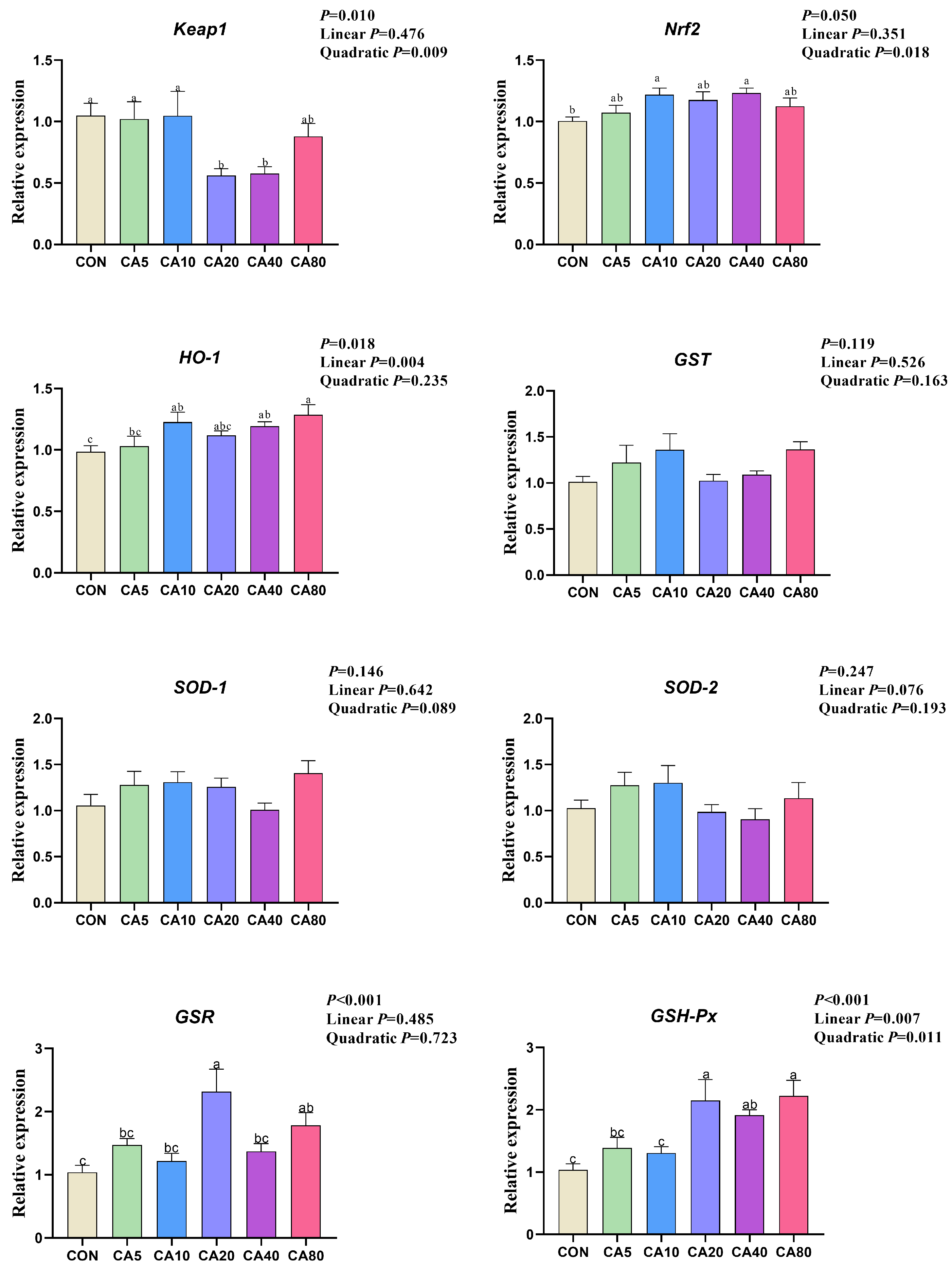
As shown in Figure 2, for 53-d broilers, supplemental CA affected the relative expression of Keap1 (quadratic), Nrf2 (quadratic), and HO-1 (linear) in the liver. Compared with birds in the CON group, supplementation with CA significantly decreased the relative expression of Keap1 (CA20 and CA40), and increased the relative expression of Nrf2 (CA10 and CA40) and HO-1 (CA10, CA40 and CA80) in the liver. There were also significant effects of the supplemental CA on the relative expression of GSH-Px (linear and quadratic) and GSR in the liver. In detail, compared with birds in the CON group, supplementation with CA significantly increased the relative expression of GSR (CA20 and CA80) and GSH-Px (CA20, CA40, and CA80).

3.7. Regression Analysis of Optimal Supplemental Level of CA Based on Quadratic Regression

The optimal levels of supplemental CA on yellow-feathered broilers from the quadratic regressions (the maximum response from a quadratic model) are shown in Table 8. The optimal levels of CA supplementation were 40.53, 40.14, and 40.52 mg/kg for BW at 53 d, ADG (from 22 to 53 d), and ADG (from 1 to 53 d), respectively.
For 21-d broilers, the optimal supplemental levels of CA were 24.17 mg/kg for hepatic T-AOC, 74.61 mg/kg for duodenal T-SOD activity, 60.05 mg/kg for duodenal GSH-Px activity, and 30.81 mg/kg for ileal GSH-Px activity. For 53-d broilers, the optima was 19.11 mg/kg for relative weight of the liver, and 43.04 mg/kg for IMF in the pectoral muscle. Also, the optima was 57.28 mg/kg for the jejunal V/C, 55.23, 50.58, and 56.49 mg/kg for MDA levels in the plasma and liver, 71.47 mg/kg for T-SOD activity, and 45.02, 44.35, 76.85, and 61.58 mg/kg for relative expression of Keap1, Nrf2, HO-1 and GSH-Px in the liver.

3.8. Intestinal Microbiota

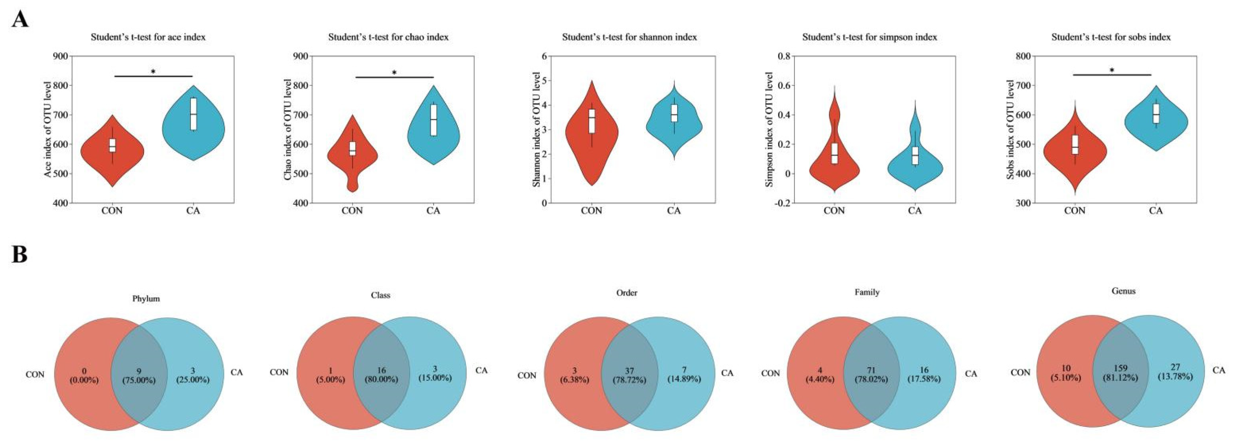
According to the above results, the cecal content of birds in the CON and CA40 groups were used for microbiota analysis. As shown in Figure 3A, the birds with CA supplementation featured a higher α-diversity characterized by increased Ace, Chao, and Sobs indices (p < 0.05). Venn diagrams (Figure 3B) indicated that the number of OTUs in the CA supplementation group was higher than that in the control group at the phylum, class, order, family, and genus levels. In addition to the nine phyla shared with the control group, three unique phyla (Synergistota, Deferribacterota, and Patescibacteria) were detected in the CA supplementation group.
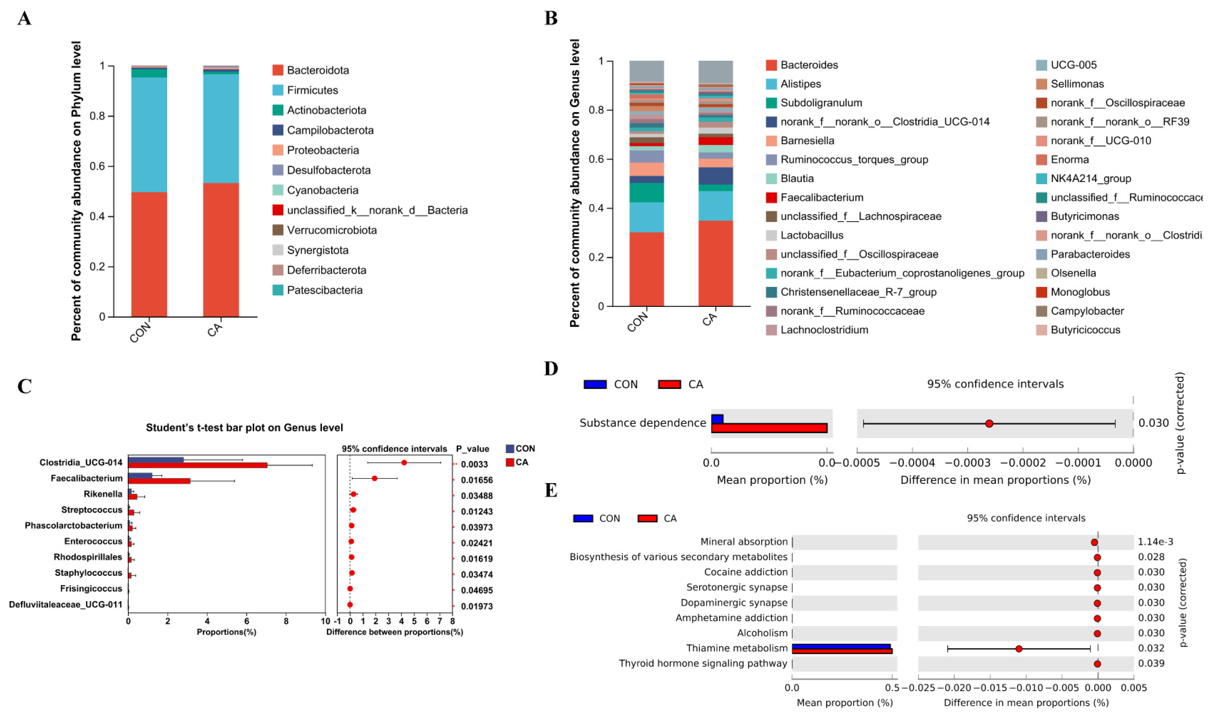
As shown in Figure 4A, microbiota profiling revealed that the phyla Bacteroidota and Firmicutes dominated the bird cecal contents. Among them, mainly genus Bacteroides and Alistipes accounted for about 40% (Figure 4B). Clostridia_UCG-014, Faecalibacterium, Rikenella, Streptococcus, Phascolarctobacterium, Enterococcus, and Bacteria were significantly (p < 0.05) enriched in the CA group (Figure 4C). As shown in Figure 4D,E, PICRUSt2 showed that CA supplementation enriched (p < 0.05) the substance dependence signaling pathway at the Level 2 KEGG pathway, and mineral absorption, biosynthesis of various secondary metabolites-part 3, cocaine addiction, serotonergic synapse, dopaminergic synapse, amphetamine addiction, alcoholism, thiamine metabolism, and thyroid hormone signaling pathway at the Level 3 KEGG pathway.

4. Discussion

There are few studies on the effect of CA on broiler chicken growth performance; however, there are relevant studies demonstrating the growth-promoting potential of phenolic plant extracts in poultry production. For example, the supplementation with polyphenol-rich grape seed to the ration increased the final BW and ADG of Cobb-500 broilers and improved the feed conversion ratio [23]. In addition, dietary supplementation with 400 mg/kg resveratrol increased the ADG of white broilers, reduced the feed-to-weight ratio, and improved the growth performance; while high doses of resveratrol (1000 mg/kg) inhibited the growth and reduced the survival rate of broilers [24]. Furthermore, our previous research found that supplementation with CA increased the ADG of LPS-challenged broilers [14]. Similar results were shown in the current research, in which supplementation with 10, 20, or 40 mg/kg of CA increased the final BW and ADG of broilers at the later phase, and the survival rate was significantly reduced when the CA supplementation level reached 80 mg/kg. Moreover, 80 mg/kg of CA significantly reduced the relative weight of the liver, indicating that a high concentration of CA might damage liver cells and their functions, lead to abnormal liver metabolism, and thereby affect the production performance of broiler chickens. The above together suggested that, while appropriate doses of plant polyphenols might enhance growth performance, excessive amounts could have adverse effects.
Although CA supplementation showed no significant effects on pectoral muscle yield and meat quality, the CA40 treatment exhibited a tendency to reduce IMF content, which mainly refers to the lipids located in skeletal muscle tissue, and is an important indicator for evaluating meat quality [25]. This suggested that CA might improve meat quality to a certain extent, although the effect was limited.
Intestinal villi increased the contact surface area for promoting nutrient absorption, while a shallower crypt depth indicated the higher maturation rate of intestinal epithelial cells and the greater absorptive capacity [26,27]. In the current research, supplementation with CA increased the V/C of the jejunum. For LPS-challenged broilers in our previous research, supplementation with CA increased the villus height to crypt depth ratio and elevated the relative expression of Claudin-1/-2 and ZO-1/-2/-3 in intestine, strengthening the tight junction in intestinal epithelial cells of the duodenum [14]. Polyphenols have demonstrated positive effects on intestinal morphology and mucosal development. For instance, magnolol increased the duodenal and the ileal villus height [28], as well as the duodenal wall thickness [29]. Therefore, CA might potentially enhance nutrient absorption and growth performance by increasing the intestinal V/C and improving intestinal health.
The presence of numerous stressors (feed, environment, etc.) in the intensive broiler farming model could cause peroxidation in livestock [30,31], causing damage to the intestines of the broiler chickens, which consequently reduced the feed conversion efficiency of the broiler chickens and affected their production performance [32,33]. Adding moderate amounts of antioxidant supplements to feed could serve as a safe and effective nutritional regulation strategy to enhance the capacity of the antioxidant system [34]. The content of lipid peroxides (such as MDA) reflected the level of oxidative stress, and their excessive accumulation could cause further damage to cells and tissues [35,36]. By activating the enzymatic antioxidant system and by enhancing the activities of enzymes such as GSH-Px, T-AOC, T-SOD and CAT, the clearance of reactive oxygen species (ROS) could be accelerated, the chain reaction of lipid peroxidation could be blocked, and the content of MDA could be reduced [37,38,39]. The antioxidant properties of CA have been confirmed in multiple in vitro studies [40,41]. Poultry research has further supported these findings, with 40 mg/kg of CA supplementation reducing serum MDA levels in LPS-stressed AA broilers [11], and rosemary leaf powder (main ingredient was CA) increasing activities of jejunal SOD and CAT in laying hens [16]. Previous findings were consistent with the results of the current study, in which CA reduced the MDA levels in the plasma and liver of broiler chickens, while increasing the activities of hepatic T-SOD, as well as ileal GSH-Px and CAT, demonstrating a clear dose-dependent response. Supplementation with 20 and 40 mg/kg of CA showed most antioxidant effects, whereas a higher dose of 80 mg/kg of CA showed reduced efficacy, indicating an upper limit for beneficial antioxidant effects.
As a pivotal transcription factor governing redox homeostasis in cellular oxidative stress responses, Nrf2 upregulated the expression and biosynthesis of antioxidant enzymes, thereby efficiently scavenging free radicals to reduce oxidative damage and improve intestinal function [42,43]. HO-1, GSR, and GSH-Px acted as downstream targets of Nrf2 to directly enhance antioxidant capacity through distinct mechanisms: HO-1 mediated the heme metabolism to generate antioxidant byproducts (e.g., bilirubin) [44], GSR maintained reduced GSH levels by regenerating GSH from GSSG [45], and GSH-Px catalyzed free radical scavenging using GSH as a co-substrate [46]. In vitro studies have demonstrated that CA (1 μM, 12 h) promoted GSH synthesis by decreasing SOD and increasing γ-glutamylcysteine ligase (γ-GCL) activity in SH-SY5 Y cells [47,48]. In vivo evidence has showed that supplementation with 30 or 60 mg/kg (BW) of CA improved both non-enzymatic (GSH) and enzymatic (GSH-Px, SOD, and CAT) antioxidant system and decreased MDA levels in mouse eye and brain tissues [49]. Treatment with CA reduced oxidative damage in mice with traumatic brain injury by activating the Nrf2–ARE signaling pathway [50]. In the current research, supplementation with 40 mg/kg of CA increased the expression of antioxidant-related genes (e.g., Nrf2, HO-1, GSR, GSH-Px), while also decreasing the Keap1 expression of the broilers, indicating the activated Nrf2–Keap1 signaling axis enhances the cellular antioxidant defenses of broilers. As an exogenous antioxidant, CA protected the intestinal health and improved the growth performance in broilers by downregulating the expression of Keap1 and activating Nrf2, primarily by activating antioxidant enzyme genes to enhance antioxidant enzyme activity, and thus protecting the intestinal health of broilers and improving their growth performance.
The intestinal microbiota was an important factor affecting poultry health and production, and its homeostasis played a significant role in intestinal function. In this study, supplementation with CA increased the α-diversity of the cecal microbiota, as evidenced by the increased Ace, Chao, and Sobs indices, indicating a more complex and diverse microbial ecosystem. The elevation in microbial diversity was associated with improved intestinal health and resistance against pathogen invasion [51]. This observation was further supported by a Venn diagram analysis, revealing that the CA group contained more OTUs at different taxonomic levels, which indicated that CA supplementation promoted a richer and more diverse microbiota composition. Notably, three bacterial phyla were uniquely present in the CA group, while absent in the CON group, demonstrating the distinct microbial profile induced by CA. These results indicated that CA might facilitate either the introduction or proliferation of beneficial microbes, thereby contributing to the homeostasis and health of the gut environment [52].
Microbiota analyses revealed that Bacteroidota and Firmicutes were the dominant phyla in the cecum contents of broilers, consistent with previous findings [53]. Notably, the abundances of beneficial genera (e.g., Clostridia_UCG-014, Faecalibacterium, Rikenella, Phascolarctobacterium) were significantly increased in the CA group. These genera had been associated with the production of short-chain fatty acids (SCFAs), particularly butyrate and propionate [54,55], suggesting that CA might enhance intestinal barrier function, mitigate inflammation, and improve antioxidant capacity through the beneficial modulation of SCFA production [56,57]. Streptococcus and Enterococcus were often considered commensal bacteria, and their increase further suggested that the microbial community in birds receiving CA was in balance to compete with pathogenic bacteria and maintain intestinal homeostasis [58]. Functional analyses using PICRUSt2 showed that CA supplementation significantly enriched various KEGG pathways, including those related to substance dependence signaling and metabolic processes. Enrichment of the pathways for mineral absorption, biosynthesis of various secondary metabolites, and thiamine metabolism was shown to promote nutrient metabolism and absorption, thereby further maintaining broiler gut health and growth performance [59]. Enrichment of the pathways related to substance dependence—including cocaine addiction, serotonergic synapse, and dopaminergic synapse—might be associated with the gut–brain axis, whereby gut microbiota influence brain function and behavior [60,61]. Emerging evidence suggested that the microbiota-mediated regulation of the gut–brain axis contributed to the alleviation of oxidative stress [62,63]. However, the specific role of antioxidant CA on the microbiota–gut–brain axis required further investigation.

5. Conclusions

Supplementation with CA enhanced the antioxidant capacity of broilers, increased the diversity and abundance of gut microbiota, and thus protected the intestinal health and improved the growth performance in broilers. The enhancing effect on antioxidant capacity is speculated to be related to the regulation of the Keap1/Nrf2 signaling pathway. However, this mechanism of action still requires further clarification of the causal relationship in subsequent experiments.
Under the conditions of this experiment, quadratic regressions for different variables showed that the optimal range for supplemental CA in the chicken’s diet was 19.11~76.85 mg/kg. Combined with experimental observation and regression analysis, the optimal level of supplementation was 40 mg/kg.

Author Contributions

Conceptualization, S.Z. and Y.W.; methodology, S.Z., Q.W., and Y.W.; validation, Q.W., J.D., and G.L.; formal analysis, Q.W. and J.D.; investigation, Q.W., K.N., and J.P.; resources, S.J. and Y.W.; data curation, S.Z. and Y.W.; writing—review and editing, S.Z. and Q.W.; visualization, S.Z. and L.X.; supervision, Q.W., S.J., and Y.W.; project administration; S.Z., Q.W., J.P., and Y.W.; funding acquisition, S.J. and Y.W. All authors have read and agreed to the published version of the manuscript.

Funding

This research was funded by China Agriculture Research System of MOF and MARA (CARS-41-G06), the National Key R&D Project (2021 YFD1300404), Guangzhou Municipal Applied Basic Research Program (2023 A04 J0794), and Special Fund for Scientific Innovation Strategy—Construction of High-Level Academy of Agriculture Science (R2023 PY-QY012, R2021 YJ-YB3012, 2022 GZ01).

Institutional Review Board Statement

This experiment was approved by the Animal Protection Committee of the Institute of Animal Science, Guangdong Academy of Agricultural Sciences, Guangzhou, China, with the approval number of GAASISA-2022-016.

Informed Consent Statement

Not applicable.

Data Availability Statement

The original contributions presented in this study are included in the article. Further inquiries can be directed to the corresponding authors.

Conflicts of Interest

The authors declare no conflicts of interest.

References

  1. Da Silva, B.D.; Conte-Junior, C.A. Perspectives on cultured meat in countries with economies dependent on animal production: A review of potential challenges and opportunities. Trends Food Sci. Technol. 2024, 149, 104551. [Google Scholar] [CrossRef]
  2. Shao, Y.; Wang, Y.; Yuan, Y.; Xie, Y. A systematic review on antibiotics misuse in livestock and aquaculture and regulation implications in China. Sci. Total Environ. 2021, 798, 149205. [Google Scholar] [CrossRef]
  3. Zhang, K.; Li, X.; Zhao, J.; Wang, Y.; Hao, X.; Liu, K.; Liu, H. Protective effects of chlorogenic acid on the meat quality of oxidatively stressed broilers revealed by integrated metabolomics and antioxidant analysis. Food Funct. 2022, 13, 2238–2252. [Google Scholar] [CrossRef]
  4. Wei, R.; Lin, Q.; Adu, M.; Huang, H.; Yan, Z.; Shao, F.; Zhong, G.; Zhang, Z.; Sang, Z.; Cao, L.; et al. The sources, properties, extraction, biosynthesis, pharmacology, and application of lycopene. Food Funct. 2023, 14, 9974–9998. [Google Scholar] [CrossRef]
  5. Wang, C.; Chen, D.; Wu, S.; Zhou, W.; Chen, X.; Zhang, Q.; Wang, L. Dietary supplementation with Neolamarckia cadamba leaf extract improves broiler meat quality by enhancing antioxidant capacity and regulating metabolites. Anim. Nutr. 2024, 17, 358–372. [Google Scholar] [CrossRef] [PubMed]
  6. Tu, M.; Cai, G.; Ma, L.; Yan, L.; Wang, T.; Shi, Z.; Wang, C.; Chen, Z. Effects of different levels of Lycium barbarum flavonoids on growth performance, immunity, intestinal barrier and antioxidant capacity of meat ducks. Antioxidants 2025, 14, 67. [Google Scholar] [CrossRef] [PubMed]
  7. Silva-Guillen, Y.V.; Arellano, C.; Wiegert, J.; Boyd, R.D.; Martinez, G.E.; Van Heugten, E. Supplementation of vitamin E or a botanical extract as antioxidants to improve growth performance and health of growing pigs housed under thermoneutral or heat-stressed conditions. J. Anim. Sci. Biotechnol. 2024, 15, 27. [Google Scholar] [CrossRef]
  8. Lv, Y.; Zeng, N.; Feng, Y.; Zhang, S.; Zhou, X.; Gao, C. Eugenol accelerates intestinal stem cell regeneration to protect the intestinal barrier integrity through inhibiting JAK2/STAT3 signaling pathway in Salmonella enteritidis-challenged broiler chicks. J. Anim. Sci. Biotechnol. 2025, 16, 40. [Google Scholar] [CrossRef] [PubMed]
  9. Platzer, M.; Kiese, S.; Tybussek, T.; Herfellner, T.; Schneider, F.; Schweiggert-Weisz, U.; Eisner, P. Radical scavenging mechanisms of phenolic compounds: A Quantitative Structure-Property Relationship (QSPR) study. Front. Nutr. 2022, 9, 882458. [Google Scholar] [CrossRef]
  10. Kalvala, A.K.; Kumar, R.; Sherkhane, B.; Gundu, C.; Arruri, V.K.; Kumar, A. Bardoxolone methyl ameliorates hyperglycemia induced mitochondrial dysfunction by activating the Keap1-Nrf2-ARE pathway in experimental diabetic neuropathy. Mol. Neurobiol. 2020, 57, 3616–3631. [Google Scholar] [CrossRef]
  11. Lee, D.K.; Jang, H.D. Carnosic acid attenuates an early increase in ROS levels during adipocyte differentiation by suppressing translation of Nox4 and inducing translation of antioxidant enzymes. Int. J. Mol. Sci. 2021, 22, 6096. [Google Scholar] [CrossRef]
  12. Wan, S.; Li, X.; Liu, S.; Tang, S. The function of carnosic acid in lipopolysaccharides-induced hepatic and intestinal inflammation in poultry. Poult. Sci. 2024, 103, 103415. [Google Scholar] [CrossRef]
  13. Zhang, Q.; Gong, J.; Xiang, H.; Hu, R.; Yang, X.; Lv, J.; Zhang, W.; Liu, M.; Deng, X.; Yuan, X.; et al. Effects of main active components of rosemary on growth performance, meat quality and lipid metabolism in finishing pigs. Anim. Nutr. 2023, 15, 341–349. [Google Scholar] [CrossRef]
  14. Liu, C.; Huang, H.; Chen, Y.; Zhou, Y.; Meng, T.; Tan, B.; He, W.; Fu, X.; Xiao, D. Dietary supplementation with mulberry leaf flavonoids and carnosic acid complex enhances the growth performance and antioxidant capacity via regulating the p38 MAPK/Nrf2 pathway. Front. Nutr. 2024, 11, 1428577. [Google Scholar] [CrossRef] [PubMed]
  15. Wang, Q.; Li, J.; Li, G.; Zang, Y.; Fan, Q.; Ye, J.; Wang, Y.; Jiang, S. Protective effects of carnosic acid on growth performance, intestinal barrier, and cecal microbiota in yellow-feathered broilers under lipopolysaccharide challenge. Poultry Sci. 2025, 104, 104688. [Google Scholar] [CrossRef]
  16. Zhang, L.; Ge, J.; Gao, F.; Yang, M.; Li, H.; Xia, F.; Bai, H.; Piao, X.; Sun, Z.; Shi, L. Rosemary leaf powder improves egg quality, antioxidant status, gut barrier function, and cecal microbiota and metabolites of late-phase laying hens. Anim. Nutr. 2024, 17, 325–334. [Google Scholar] [CrossRef]
  17. Bahadoran, S.; Teymouri, Y.; Hassanpour, H.; Mohebbi, A.; Akbari, M.R. Effect of sage (Salvia officinalis L.) extract in antioxidant status and intestinal morphology of pulmonary hypertensive chickens. Vet. Med. Sci. 2023, 9, 2176–2184. [Google Scholar] [CrossRef] [PubMed]
  18. Peng, Y.; Chen, C.; Luo, D.; Yu, W.; Li, S.; Xiao, Y.; Yuan, B.; Liang, S.; Yao, X.; Kim, N.H.; et al. Carnosic acid improves porcine early embryonic development by inhibiting the accumulation of reactive oxygen species. J. Reprod. Dev. 2020, 66, 555–562. [Google Scholar] [CrossRef] [PubMed]
  19. Ministry of Agriculture of the People’s Republic of China (PRC). Nutrient Requirements of Yellow Chicken; China Agricultural Press: Beijing, China, 2020. [Google Scholar]
  20. Wang, Y.; Wang, Y.; Wang, B.; Mei, X.; Jiang, S.; Li, W. Protocatechuic acid improved growth performance, meat quality, and intestinal health of Chinese yellow-feathered broilers. Poult. Sci. 2019, 98, 3138–3149. [Google Scholar] [CrossRef]
  21. Zerehdaran, S.; Vereijken, A.L.; Van Arendonk, J.A.; Van der Waaijt, E.H. Estimation of genetic parameters for fat deposition and carcass traits in broilers. Poult. Sci. 2004, 83, 521–525. [Google Scholar] [CrossRef] [PubMed]
  22. Wang, Y.; Gou, Z.; Lin, X.; Fan, Q.; Ye, J.; Jiang, S. Optimal Level of Supplemental manganese for yellow-feathered broilers during the growth phase. Animals 2021, 11, 1389. [Google Scholar] [CrossRef] [PubMed]
  23. Abu Hafsa, S.H.; Ibrahim, S.A. Effect of dietary polyphenol-rich grape seed on growth performance, antioxidant capacity and ileal microflora in broiler chicks. J. Anim. Physiol. Anim. Nutr. 2018, 102, 268–275. [Google Scholar] [CrossRef] [PubMed]
  24. Ding, K.; Lu, M.; Guo, Y.; Liang, S.; Mou, R.; He, Y.; Tang, L. Resveratrol relieves chronic heat stress-induced liver oxidative damage in broilers by activating the Nrf2-Keap1 signaling pathway. Ecotoxicol. Environ. Saf. 2023, 249, 114411. [Google Scholar] [CrossRef]
  25. Wang, Z.; Tian, W.; Guo, Y.; Wang, D.; Zhang, Y.; Zhi, Y.; Li, D.; Li, W.; Li, Z.; Jiang, R.; et al. Dynamic alternations of three-dimensional chromatin architecture contribute to phenotypic characteristics of breast muscle in chicken. Commun. Biol. 2024, 7, 910. [Google Scholar] [CrossRef]
  26. Ducatelle, R.; Goossens, E.; De Meyer, F.; Eeckhaut, V.; Antonissen, G.; Haesebrouck, F.; Van Immerseel, F. Biomarkers for monitoring intestinal health in poultry:present status and future perspectives. Vet. Res. 2018, 49, 43. [Google Scholar] [CrossRef]
  27. Grego-Bessa, J.; Bloomekatz, J.; Castel, P.; Omelchenko, T.; Baselga, J.; Anderson, K.V. The tumor suppressor PTEN and the PDK1 kinase regulate formation of the columnar neural epithelium. eLife 2016, 5, e12034. [Google Scholar] [CrossRef]
  28. Chen, F.; Zhang, H.; Du, E.; Jin, F.; Zheng, C.; Fan, Q.; Zhao, N.; Guo, W.; Zhang, W.; Huang, S.; et al. Effects of magnolol on egg production, egg quality, antioxidant capacity, and intestinal health of laying hens in the late phase of the laying cycle. Poult. Sci. 2021, 100, 835–843. [Google Scholar] [CrossRef]
  29. Chen, F.; Zhang, H.; Zhao, N.; Du, E.; Jin, F.; Fan, Q.; Guo, W.; Huang, S.; Wei, J. Effects of magnolol and honokiol blend on performance, egg quality, hepatic lipid metabolism, and intestinal morphology of hens at late laying cycle. Animal 2022, 16, 100532. [Google Scholar] [CrossRef]
  30. Aryal, B.; Kwakye, J.; Ariyo, O.W.; Ghareeb, A.F.A.; Milfort, M.C.; Fuller, A.L.; Khatiwada, S.; Rekaya, R.; Aggrey, S.E. Major Oxidative and Antioxidant Mechanisms During Heat Stress-Induced Oxidative Stress in Chickens. Antioxidants 2025, 14, 471. [Google Scholar] [CrossRef] [PubMed]
  31. Oke, O.E.; Akosile, O.A.; Oni, A.I.; Opowoye, I.O.; Ishola, C.A.; Adebiyi, J.O.; Odeyemi, A.J.; Adjei-Mensah, B.; Uyanga, V.A.; Abioja, M.O. Oxidative stress in poultry production. Poult. Sci. 2024, 103, 104003. [Google Scholar] [CrossRef]
  32. Wang, W.; Zhu, J.; Cao, Q.; Zhang, C.; Dong, Z.; Feng, D.; Ye, H.; Zuo, J. Dietary catalase supplementation alleviates deoxynivalenol-induced oxidative stress and gut microbiota dysbiosis in broiler chickens. Toxins 2022, 14, 830. [Google Scholar] [CrossRef] [PubMed]
  33. Li, Y.; Wang, K.; Li, C. Oxidative stress in poultry and the therapeutic role of herbal medicine in intestinal health. Antioxidants 2024, 13, 1375. [Google Scholar] [CrossRef] [PubMed]
  34. Kouvedaki, I.; Pappas, A.C.; Surai, P.F.; Zoidis, E. Nutrigenomics of natural antioxidants in broilers. Antioxidants 2024, 13, 270. [Google Scholar] [CrossRef]
  35. He, R.; Liu, B.; Xiong, R.; Geng, B.; Meng, H.; Lin, W.; Hao, B.; Zhang, L.; Wang, W.; Jiang, W.; et al. Itaconate inhibits ferroptosis of macrophage via Nrf2 pathways against sepsis-induced acute lung injury. Cell Death Discov. 2022, 8, 43. [Google Scholar] [CrossRef] [PubMed]
  36. Tschuck, J.; Theilacker, L.; Rothenaigner, I.; Weiss, S.A.I.; Akdogan, B.; Van Thanh, L.; Mueller, C.; Graf, R.; Brandner, S.; Puetz, C.; et al. Farnesoid X receptor activation by bile acids suppresses lipid peroxidation and ferroptosis. Nat. Commun. 2023, 14, 6908. [Google Scholar] [CrossRef]
  37. Luo, J.; Wu, X.; Chen, D.; Yu, B.; He, J. Dietary ferulic acid supplementation enhances antioxidant capacity and alleviates hepatocyte pyroptosis in diquat challenged piglets. J. Anim. Sci. Biotechnol. 2024, 15, 134. [Google Scholar] [CrossRef]
  38. Yuan, H.; Xu, Y.; Luo, Y.; Zhang, J.; Zhu, X.; Xiao, J. Ganoderic acid D prevents oxidative stress-induced senescence by targeting 14-3-3ε to activate CaM/CaMKII/NRF2 signaling pathway in mesenchymal stem cells. Aging Cell 2022, 21, e13686. [Google Scholar] [CrossRef]
  39. Zhan, S.; Wu, L.; Lv, Y.; Huang, W.; Ge, C.; Hu, Z.; Shen, X.; Lin, G.; Yu, D.; Liu, B. Lactobacillus reuteri alleviates diquat induced hepatic impairment and mitochondrial dysfunction via activation of the Nrf2 antioxidant system and suppression of NF-κB inflammatory response. Poult. Sci. 2025, 104, 104997. [Google Scholar] [CrossRef]
  40. Jordán, M.J.; Lax, V.; Rota, M.C.; Lorán, S.; Sotomayor, J.A. Relevance of carnosic acid, carnosol, and rosmarinic acid concentrations in the in vitro antioxidant and antimicrobial activities of Rosmarinus officinalis (L.) methanolic extracts. J. Anim. Sci. Biotechnol. 2012, 60, 9603–9608. [Google Scholar] [CrossRef]
  41. Karagianni, K.; Pettas, S.; Kanata, E.; Lioulia, E.; Thune, K.; Schmitz, M.; Tsamesidis, I.; Lymperaki, E.; Xanthopoulos, K.; Sklaviadis, T.; et al. Carnosic acid and carnosol display antioxidant and anti-prion properties in in vitro and cell-free models of prion diseases. Antioxidants 2022, 11, 726. [Google Scholar] [CrossRef]
  42. Geertsema, S.; Bourgonje, A.R.; Fagundes, R.R.; Gacesa, R.; Weersma, R.K.; Van Goor, H.; Mann, G.E.; Dijkstra, G.; Faber, K.N. The NRF2/Keap1 pathway as a therapeutic target in inflammatory bowel disease. Trends Mol. Med. 2023, 29, 830–842. [Google Scholar] [CrossRef] [PubMed]
  43. Kozieł, M.J.; Kowalska, K.; Piastowska-Ciesielska, A.W. Nrf2: A main responsive element in cells to mycotoxin-induced toxicity. Arch. Toxicol. 2021, 95, 1521–1533. [Google Scholar] [CrossRef]
  44. Huang, L.; Wang, X.; Zheng, Y.; Lang, D.; Wang, J.; Yan, S.; Chen, Y. EGCG-NPs inhibition HO-1-mediated reprogram iron metabolism against ferroptosis after subarachnoid hemorrhage. Redox Biol. 2024, 70, 103075. [Google Scholar] [CrossRef]
  45. Biondini, M.; Lehuédé, C.; Tabariès, S.; Annis, M.G.; Pacis, A.; Ma, E.H.; Tam, C.; Hsu, B.E.; Audet-Delage, Y.; Abu-Thuraia, A.; et al. Metastatic breast cancer cells are metabolically reprogrammed to maintain redox homeostasis during metastasis. Redox Biol. 2024, 75, 103276. [Google Scholar] [CrossRef] [PubMed]
  46. Wang, Y.; Jiu, R.; Li, Z.; Wang, Q.; Lei, X.; Chen, J.; Liu, H.; Liu, J. Tyrosine-modified tilapia skin antioxidant peptides and their hydroxyl radical quenching activities. J. Mater. Chem. B. 2025, 13, 2400–2408. [Google Scholar] [CrossRef] [PubMed]
  47. De Oliveira, M.R.; Da Costa Ferreira, G.; Peres, A.; Dal Bosco, S.M. Carnosic acid suppresses the H2O2-induced mitochondria-related bioenergetics disturbances and redox impairment in Sh-Sy5 y cells: Role for nrf2. Mol. Neurobiol. 2018, 55, 968–979. [Google Scholar] [CrossRef]
  48. De Souza, I.C.C.; Gobbo, R.C.B.; De Almeida, F.J.S.; Luckachaki, M.D.; De Oliveira, M.R. Carnosic acid depends on glutathione to promote mitochondrial protection in methylglyoxal-exposed SH-SY5 Y cells. Metab. Brain Dis. 2021, 36, 471–481. [Google Scholar] [CrossRef]
  49. AlKahtane, A.A.; Ghanem, E.; Bungau, S.G.; Alarifi, S.; Ali, D.; AlBasher, G.; Alkahtani, S.; Aleya, L.; Abdel-Daim, M.M. Carnosic acid alleviates chlorpyrifos-induced oxidative stress and inflammation in mice cerebral and ocular tissues. Environ. Sci. Pollut. Res. 2020, 27, 11663–11670. [Google Scholar] [CrossRef]
  50. Miller, D.M.; Singh, I.N.; Wang, J.; Hall, E.D. Nrf2-ARE activator carnosic acid decreases mitochondrial dysfunction, oxidative damage and neuronal cytoskeletal degradation following traumatic brain injury in mice. Exp. Neurol. 2015, 264, 103–110. [Google Scholar] [CrossRef]
  51. Glendinning, L.; Wu, Z.; Vervelde, L.; Watson, M.; Balic, A. Infectious bronchitis virus vaccination, but not the presence of XCR1, is correlated with large differences in chicken caecal microbiota. Microb. Genom. 2024, 10, 001289. [Google Scholar] [CrossRef]
  52. Wang, Y.; Li, A.; Zhang, L.; Waqas, M.; Mehmood, K.; Iqbal, M.; Can, M.; Li, Z.; Lian, Y.; Sizhu, S.; et al. Probiotic potential of Lactobacillus on the intestinal microflora against Escherichia coli induced mice model through high-throughput sequencing. Microb. Pathog. 2019, 137, 103760. [Google Scholar] [CrossRef]
  53. Zhang, J.; Han, H.; Zhang, L.; Wang, T. Dietary bisdemethoxycurcumin supplementation attenuates lipopolysaccharide-induced damages on intestinal redox potential and redox status of broilers. Poult. Sci. 2021, 100, 101061. [Google Scholar] [CrossRef] [PubMed]
  54. Chang, Y.Q.; Moon, S.K.; Wang, Y.Q.; Xie, L.M.; Cho, H.S.; Kim, S.K. Supplemental effects of different production methods of pine needle additives on growth performance, intestinal environment, meat quality and serum of broiler chickens. Anim. Biosci. 2024, 37, 1263–1276. [Google Scholar] [CrossRef]
  55. Wu, L.; Niu, Y.; Ren, B.; Wang, S.; Song, Y.; Wang, X.; Zhao, K.; Yue, Z.; Li, Y.; Gao, J. Naringenin promotes gastrointestinal motility in mice by impacting the SCF/c-Kit pathway and gut microbiota. Foods 2024, 13, 2520. [Google Scholar] [CrossRef]
  56. Arifuzzaman, M.; Won, T.H.; Li, T.T.; Yano, H.; Digumarthi, S.; Heras, A.F.; Zhang, W.; Parkhurst, C.N.; Kashyap, S.; Jin, W.B.; et al. Inulin fibre promotes microbiota-derived bile acids and type 2 inflammation. Nature 2022, 611, 578–584. [Google Scholar] [CrossRef]
  57. Wang, R.; Yang, X.; Liu, J.; Zhong, F.; Zhang, C.; Chen, Y.; Sun, T.; Ji, C.; Ma, D. Gut microbiota regulates acute myeloid leukaemia via alteration of intestinal barrier function mediated by butyrate. Nat. Commun. 2022, 13, 2522. [Google Scholar] [CrossRef]
  58. Lei, L.; Yang, Y.; Yang, Y.; Wu, S.; Ma, X.; Mao, M.; Hu, T. Mechanisms by which small rnas affect bacterial activity. J. Dent. Res. 2019, 98, 1315–1323. [Google Scholar] [CrossRef] [PubMed]
  59. Zimatkina, T.I.; Chernikevich, I.P.; Zimatkin, S.M.; Deitrich, R.A. Thiamine status in liver and brain of rats genetically selected for different sensitivity to hypnotic effect of alcohol. Alcohol Clin. Exp. Res. 2000, 24, 1620–1624. [Google Scholar] [CrossRef] [PubMed]
  60. Behura, S.K.; Kelleher, A.M.; Spencer, T.E. Evidence for functional interactions between the placenta and brain in pregnant mice. FASEB J. 2019, 33, 4261–4272. [Google Scholar] [CrossRef]
  61. Lu, J.; Cheng, Y.; Wang, X.; Woodson, K.; Kemper, C.; Disney, E.; Wang, J. Alcohol intake enhances glutamatergic transmission from D2 receptor-expressing afferents onto D1 receptor-expressing medium spiny neurons in the dorsomedial striatum. Neuropsychopharmacology 2019, 44, 1123–1131. [Google Scholar] [CrossRef]
  62. Banc, R.; Rusu, M.E.; Filip, L.; Popa, D.S. The impact of ellagitannins and their metabolites through gut microbiome on the gut health and brain wellness within the gut-brain axis. Foods 2023, 12, 270. [Google Scholar] [CrossRef] [PubMed]
  63. Pan, S.; Wei, H.; Yuan, S.; Kong, Y.; Yang, H.; Zhang, Y.; Cui, X.; Chen, W.; Liu, J.; Zhang, Y. Probiotic pediococcus pentosaceus ameliorates MPTP-induced oxidative stress via regulating the gut microbiota-gut-brain axis. Front. Cell. Infect. Microbiol. 2022, 12, 1022879. [Google Scholar] [CrossRef] [PubMed]
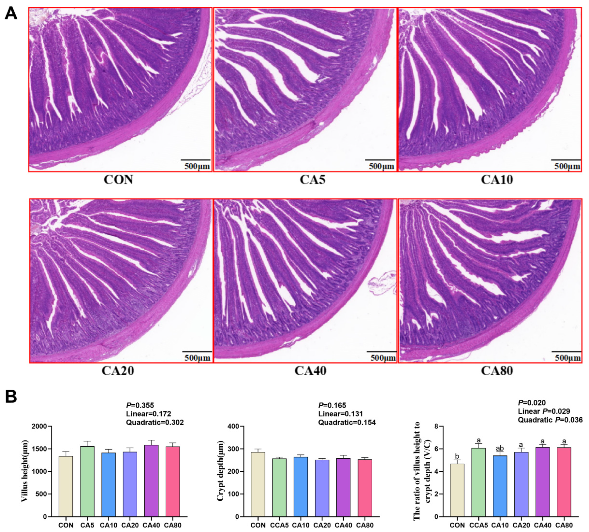
Figure 1. Effects of carnosic acid on jejunal morphology of 53-day-old broilers. (A) Hematoxylin–eosin (H&E) stained jejunum. Scale bar at 500 μm. (B) Villus height, crypt depth, and the ratio of villus height to crypt depth (V/C). ab Means with common superscripts within a row do not differ significantly (p ≥ 0.05). CON: basal diet; CA5/10/20/40/80: basal diet supplemented with 5, 10, 20, 40, and 80 mg/kg of carnosic acid.
Antioxidants 14 01026 g001
Figure 2. Effects of carnosic acid on relative expression of antioxidant genes in the liver of 53-day-old broilers. a–c Means with common superscripts do not differ significantly (p ≥ 0.05). CON: basal diet; CA5/10/20/40/80: basal diet supplemented with 5, 10, 20, 40, and 80 mg/kg of CA; SOD-1: superoxide dismutase 1; SOD-2: superoxide dismutase 2; GST: glutathione s-transferase; GSR: glutathione reductase; GSH-Px: glutathione peroxidase; Keap1: kelch-like ECH-associated protein-1; Nrf2: nuclear factor erythroid 2-related factor 2; HO-1: heme oxygenase-1.
Antioxidants 14 01026 g002
Figure 3. Effect of carnosic acid on α diversity and microbial composition of the cecum. (A) Alpha-diversity (Shannon, Chao, Ace, Sobs, Simpson). (B) Number of OTUs at the phylum, class, order, family, and genus level. Dates are shown as mean ± SEM (n = 8), * p < 0.05. CON: basal diet; CA: basal diet + 40 mg/kg of carnosic acid.
Antioxidants 14 01026 g003
Figure 4. Effect of carnosic acid on differential microorganisms of the cecum. The microbial composition in cecal contents at the phylum (A) and genus (B) levels. Differences in microbes in the cecal between the CON and CA groups at the genus (C) levels. PICRUSt2 combined with the KEGG database to predict the function of bacterial microbiota, showing the results of the KEGG pathway in Level 2 (D) and Level 3 (E). CON: basal diet; CA: basal diet + 40 mg/kg carnosic acid. * p < 0.05. ** p < 0.01.
Antioxidants 14 01026 g004
Table 1. Composition and nutrient content of diets (air dry-basis).
Items1 to 21 Days of Age22 to 53 Days of Age
Ingredient, %
Corn56.5063.40
Soybean meal35.9030.40
Soybean oil2.601.50
L-Lys HCl (78.8%)0.200.20
DL-Met (99.0%)0.300.30
Talcum powder1.001.00
Dicalcium phosphate2.201.90
NaCl0.300.30
Premix 11.001.00
Total100.00100.00
Nutrient levels 2
Nitrogen-corrected metabolizable energy, MJ/kg11.9212.34
Crude protein, %20.9417.81
Calcium, %1.010.92
Total phosphorus, %0.720.67
Non-phytate phosphorus, %0.470.41
Lysine, %1.291.15
Methionine, %0.520.48
Methionine + Cysteine, %0.930.85
1 The premix provided the following per kilogram of diets during 1 to 21 days of age: VA 15,000 IU, VD3 3300 IU, VE 20 IU, VK3 6.00 mg, VB1 1.80 mg, VB2 9.00 mg, VB6 3.50 mg, VB12 0.01 mg, choline chloride 500.00 mg, niacin 60.00 mg, pantothenic acid 16.00 mg, folic acid 0.55 mg, biotin 0.15 mg, Fe 80.00 mg, Cu 8.00 mg, Mn 80.00 mg, Zn 60.00 mg, I 0.35 mg, and Se 0.30 mg. The premix provided the following per kilogram of diets during 22 to 53 days of age: VA 15,000 IU, VD3 3300 IU, VE 20 IU, VK3 6.00 mg, VB1 3.00 mg, VB2 9.00 mg, VB6 6.00 mg, VB12 0.03 mg, choline chloride 1000.00 mg, niacin 60.00 mg, pantothenic acid 18.00 mg, folic acid 0.75 mg, biotin 0.10 mg, Fe 80.00 mg, Cu 12.00 mg, Mn 100.00 mg, Zn 75.00 mg, I 0.35 mg, and Se 0.15 mg. 2 Values were calculated according to Nutrient Requirements of Yellow Chickens [20], and values of crude protein, calcium and total phosphorus were determined by analysis.
Table 2. Primer sequences for Real-time PCR.
GenePrimer Sequence (5′ to 3′)GenBank ID
β-actinF: GAGAAATTGTGCGTGACATCA
R: CCTGAACCTCTCATTGCCA
NM_001101.5
GSTF: GATGAACGTCGTCCAACCAG
R: TCATGTCCGTGGTCCTTCAA
NM_001001777.2
SOD1F: GGTGCTCACTTTAATCCTG
R: CTACTTCTGCCACTCCTCC
NM_205064.2
SOD2F: TGGTGTTCAAGGATCAGGCT
R: CCCAGCAATGGAATGAGACC
NM_204211.2
GSRF: ATACCCGGCGTCAGGTTTAA
R: CCTGTCGCAATGAGGATGTG
XM_040671422.2
Keap1F: CAACTTCGCCGAGCAGA
R: CGTGGAACACCTCCGACT
XM_040671565.2
HO-1F: CTTCGCACAAGGAGTGTTAAC
R: CATCCTGCTTGTCCTCTCAC
NM_205344.2
Nrf2F: ATCACCTCTTCTGCACCGAA
R: GCTTTCTCCCGCTCTTTCTG
XM_046921122.1
GSH-PxF: AAGTGCCAGGTGAACGGGAAGG
R: AGGGCTGTAGCGGCGGAAAG
NM_001277854.3
F: forward primer; R: reverse primer; GST: glutathione s-transferase; SOD1: superoxide dismutase 1; SOD2: superoxide dismutase 2; GSR: glutathione reductase; Keap1: kelch-like ECH-associated protein-1; Nrf2: nuclear factor erythroid 2-related factor 2; HO-1: heme oxygenase-1; GSH-Px: glutathione peroxidase.
Table 3. Effects of carnosic acid on growth performance of broilers.
ItemsCONCA5CA10CA20CA40CA80SEMp-ValueLinearQuadratic
1 to 21 days of age
Initial BW, g40.9040.8340.8340.8740.9240.880.030.2660.3400.552
Final BW, g468.67462.33461.50464.50470.00465.004.240.8160.7890.901
ADG, g21.3921.0721.0321.1821.4521.210.210.8140.7890.905
ADFI, g36.1735.5435.9335.6735.5335.260.340.6610.1410.329
F/G1.681.701.691.691.661.660.010.1220.0170.057
Survival rate, %100.00100.0098.81100.00100.0097.620.710.1860.0430.064
22 to 53 days of age
Final BW, g2013.06 b2056.04 ab2077.70 a2085.06 a2074.50 a2040.36 ab15.210.0190.9800.012
ADG, g48.18 b49.77 a50.37 a50.52 a49.98 a49.15 ab0.520.0440.9140.049
ADFI, g119.19120.05121.92122.23121.16118.691.470.6180.5010.256
F/G2.462.422.422.412.482.390.020.1220.4270.292
Survival rate, %100.00 a98.61 a98.61 a100.00 a98.61 a90.15 b1.68<0.001<0.001<0.001
1 to 53 days of age
ADG, g35.86 b36.64 ab37.03 a37.17 a36.97 a36.35 ab0.370.0190.9790.011
ADFI, g90.0990.9092.4491.9991.5890.121.380.6400.0880.007
F/G2.512.482.502.482.532.450.030.1710.2160.107
Survival rate, %100.00 a98.61 a95.83 a100.00 a98.61 a88.89 b1.88<0.001<0.001<0.001
ab Means with common superscripts within a row do not differ significantly (p ≥ 0.05). CON: basal diet; CA5/10/20/40/80: basal diet supplemented with 5, 10, 20, 40, and 80 mg/kg of carnosic acid; BW: body weight; ADG: average daily gain; ADFI: average daily feed intake; F/G: feed to gain ratio.
Table 4. Effects of carnosic acid on relative weights of immune organs of broilers.
ItemsCONCA5CA10CA20CA40CA80SEMp-ValueLinearQuadratic
21 days of age
Liver/BW3.273.252.943.153.283.350.120.7280.1120.245
Spleen/BW0.170.170.140.160.160.170.010.6700.7030.390
Thymus/BW0.480.380.420.410.440.430.030.3300.9360.537
Bursa of Fabricius/BW0.270.250.230.240.250.250.020.3070.2870.568
53 days of age
Liver/BW2.29 ab2.24 bc2.37 a2.26 b2.28 ab2.14 c0.020.0100.0020.003
Spleen/BW0.220.230.210.220.220.200.010.3800.0590.160
a–c Means with common superscripts within a row do not differ significantly (p ≥ 0.05). CON: basal diet; CA5/10/20/40/80: basal diet supplemented with 5, 10, 20, 40, and 80 mg/kg of carnosic acid; BW: body weight.
Table 5. Effects of carnosic acid on meat quality and IMF content of 53-day-old broilers.
ItemsCONCA5CA10CA20CA40CA80SEMp-ValueLinearQuadratic
Pectoral muscle rate, %12.6712.4212.2312.5812.6312.850.140.1950.0590.103
Shear force24h, N22.5524.3224.7323.7024.2226.420.880.8940.1760.207
Drip loss24h, %1.831.631.691.701.551.570.040.3960.2070.176
a*45min13.9413.5413.8714.0013.3013.480.100.2680.2980.556
b*45min9.759.959.979.829.699.500.110.8510.2450.508
L*45min49.9550.2950.3149.8150.5749.760.180.7710.7750.822
pH45min6.136.086.055.956.196.160.030.1230.2640.527
a*24h13.9213.5813.1913.1813.1713.370.230.9280.5500.621
b*24h10.1010.1110.2610.4610.0210.210.130.9520.9280.995
L*24h55.0353.3154.4654.7254.2854.220.290.6580.7840.683
pH24h5.655.685.655.665.675.640.010.8550.1760.207
Intramuscular fat, %8.51 a8.19 a8.66 a8.13 ab3.78 b8.62 a0.820.0010.3400.001
ab Means with common superscripts within a row do not differ significantly (p ≥ 0.05). CON: basal diet; CA5/10/20/40/80: basal diet supplemented with 5, 10, 20, 40, and 80 mg/kg of carnosic acid; a* = redness; b* = yellowness; L* = lightness.
Table 6. Effects of carnosic acid on antioxidant capacity of 21-day-old broilers.
ItemCONCA5CA10CA20CA40CA80SEMp-ValueLinearQuadratic
Plasma
MDA, nmol/mL2.492.342.392.502.702.590.110.2530.4990.752
GSH-Px, U/mL1491.86 bc1548.84 b1439.53 c1405.81 c1469.77 bc1768.6 a19.02<0.001<0.001<0.001
T-SOD, U/mL164.19 a163.66 a164.73 a164.53 a164.06 a162.05 b0.19<0.0010.014<0.001
T-AOC, U/mL1.34 c1.37 c2.14 ab1.22 c1.62 bc2.41 a0.270.0040.0040.011
Liver
MDA, nmol/mg prot0.29 a0.22 ab0.24 a0.21 ab0.21 ab0.13 b0.010.0380.0020.130
GSH-Px, U/mg prot75.4782.8984.1977.6676.7274.491.490.3130.1720.390
T-SOD, U/mg prot23.2424.8924.8824.3824.0823.560.420.4800.4240.717
T-AOC, U/mg prot0.13 ab0.13 ab0.14 a0.13 ab0.12 bc0.11 c0.020.0020.0010.004
CAT, U/mg prot74.72 b82.92 a82.22 a76.29 ab76.43 ab75.11 b1.010.0480.0700.155
Duodenum
MDA, nmol/mg prot0.890.700.710.730.960.990.090.0690.0230.075
GSH-Px, U/mg prot12.90 c19.74 bc21.54 ab31.35 a23.61 ab30.51 a1.52<0.0010.0020.002
T-SOD, U/mg prot29.20 c38.94 a37.42 ab33.52 bc39.07 a40.34 a0.78<0.0010.0010.003
T-AOC, U/mg prot2.61 c3.83 a3.29 ab2.84 bc3.38 ab3.36 ab0.08<0.0010.4140.678
CAT, U/mg prot1.15 1.18 1.32 1.44 0.76 1.11 0.19 0.1880.2310.430
Jejunum
T-SOD, U/mg prot22.99 ab21.93 ab21.97 ab22.92 ab21.54 b23.59 a0.200.0470.1850.084
GSH-Px, U/mg prot119.54111.93137.00119.48108.26131.583.370.0930.7310.394
T-AOC, U/mg prot0.240.220.230.240.210.250.010.1640.2810.318
Ileum
MDA, nmol/mg prot0.110.130.140.170.120.120.020.2890.6020.424
T-SOD, U/mg prot8.527.867.838.418.247.770.280.2460.3070.466
GSH-Px, U/mg prot14.22 b28.51 a23.49 a24.15 a25.24 a9.80 b1.18<0.0010.002<0.001
T-AOC, U/mg prot0.15 abc0.16 a0.15 ab0.15 abc0.13 bc0.13 c1.170.009<0.0010.003
CAT2.622.742.793.302.362.771.000.1760.8780.727
a–c Means with common superscripts within a row do not differ significantly (p ≥ 0.05). CON: basal diet; CA5/10/20/40/80: basal diet supplemented with 5, 10, 20, 40, and 80 mg/kg of carnosic acid; MDA: malondialdehyde; GSH-Px: glutathione peroxidase; T-SOD: total superoxide dismutase; T-AOC: total antioxidant capacity; CAT: catalase.
Table 7. Effects of carnosic acid on antioxidant capacity of 53-day-old broilers.
ItemCONCA5CA10CA20CA40CA80SEMp-ValueLinearQuadratic
Plasma
MDA, nmol/mL9.40 a9.31 a8.17 a4.10 b3.71 b4.26 b1.760.0270.0070.003
GSH-Px, U/mL1376.601379.491355.691360.741386.701329.0140.420.9400.3190.486
T-SOD, U/mL151.32152.92149.89147.19129.51130.527.700.0850.0020.006
T-AOC, U/mL6.186.236.546.515.846.340.360.7980.7660.900
Liver
MDA, nmol/mg prot1.36 a0.82 b0.80 b0.75 bc0.55 c0.70 bc0.11<0.0010.001<0.001
GSH-Px, U/mg prot1436.441551.88 1626.89 1389.36 1712.59 1392.63124.570.3490.6750.391
T-SOD, U/mg prot577.78 b576.59 b584.04 b579.68 ab607.88 a609.67 a8.060.001 <0.001<0.001
T-AOC, U/mg prot2.462.462.682.202.562.880.170.0920.0640.110
CAT, U/mg prot4.72 ab 4.80 a 4.80 a 4.72 ab 4.56 b 4.58 b 0.06 0.0070.0010.052
Duodenum
MDA, nmol/mg prot0.66 0.49 0.72 0.52 0.56 0.64 0.08 0.326 0.9950.760
GSH-Px, U/mg prot2302.82 3340.94 2676.97 2824.52 4463.37 4510.80 758.35 0.182 0.0190.051
T-SOD, U/mg prot825.25 825.74 806.00 770.15 755.95 762.21 27.94 0.273 0.0420.051
T-AOC, U/mg prot21.4318.4318.5019.0619.6719.910.390.260 0.0660.086
CAT, U/mg prot1.15 1.18 1.32 1.44 0.76 1.11 0.19 0.183 0.2710.469
Jejunum
MDA, nmol/mg prot1.36 a 0.57 b 0.61 ab 0.70 ab 0.64 a 0.31 b 0.29 0.0480.0390.066
GSH-Px, U/mg prot35.69 36.62 43.24 43.05 33.87 36.71 4.56 0.638 0.6530.896
T-SOD, U/mg prot20.46 20.61 20.32 20.55 19.92 20.84 0.52 0.894 0.8930.680
T-AOC, U/mg prot0.250.25 0.280.290.27 0.27 0.02 0.4880.1800.279
CAT, U/mg prot3.76 3.72 3.56 4.05 3.05 3.80 0.47 0.785 07270.800
Ileum
MDA, nmol/mg prot0.961.091.070.980.911.040.070.5640.6190.651
GSH-Px, U/mg prot21.50 c45.26 a47.88 a41.02 ab27.09 c32.35 bc4.62<0.0010.2980.471
T-SOD, U/mg prot15.4515.5615.3215.3116.1215.140.300.4640.8960.243
T-AOC, U/mg prot0.240.250.250.250.270.240.010.5810.8250.070
CAT, U/mg prot2.39 c2.57 bc3.11 bc3.58 ab2.80 bc3.84 a0.300.0090.0250.062
a–c Means with common superscripts within a row do not differ significantly (p ≥ 0.05). CON: basal diet; CA5/10/20/40/80: basal diet supplemented with 5, 10, 20, 40, and 80 mg/kg of CA; MDA: malondialdehyde; GSH-Px: glutathione peroxidase; T-SOD: total superoxide dismutase; T-AOC: total antioxidant capacity; CAT: catalase.
Table 8. Regression analysis of the optimal supplemental level of carnosic acid for broilers based on quadratic regression.
Itemp-ValueR2Regression EquationCA Supplemental Level (mg/kg) 1
Final BW (53 d), g0.0120.243y = −0.0356117 CA2 + 2.88631 CA + 2034.32940.53
ADG (22 to 53 d), g0.0490.171y = −0.0010260 CA2 + 0.08236 CA + 49.21140.14
ADG (1 to 53 d), g0.0110.244y = −0.006492 CA2 + 0.05261 CA + 36.24440.52
21 d
Hepatic T-AOC, U/mg prot0.0040.160y = −0.0000020 CA2 + 0.00009 CA + 0.13024.17
Duodenal T-SOD, U/mg prot0.0030.188y = −0.0012707 CA2 + 0.18963 CA + 33.01874.61
Duodenal GSH-Px, U/mg prot0.0020.208y = −0.0042395 CA2 + 0.50918 CA + 15.97760.05
Ileal GSH-Px, U/mg prot<0.0010.407y = −0.0067097 CA2 + 0.41349 CA + 19.53530.81
53 d
Relative weight of liver, %0.0030.165y = −0.0000429 CA2 + 0.00164 CA + 2.28119.11
Intramuscular fat, %0.0010.432y = −0.0022318 CA2 + 0.19212 CA + 9.43943.04
Jejunal V/C0.0360.143y = −0.0003450 CA2 + 0.03950 CA + 5.16957.28
Plasma MDA, nmol/mL0.0030.234y = 0.0023638 CA2 − 0.26109 CA +10.04755.23
Hepatic MDA, nmol/mg prot<0.0010.483y = 0.0002654 CA2 − 0.02685 CA + 1.16150.58
Hepatic T-SOD, U/mg prot<0.0010.329y = −0.0075841 CA2 + 1.08413 CA + 566.67771.47
Hepatic Keap1, relative expression0.0090.293y = 0.0003332 CA2 − 0.0300 CA + 1.19445.02
Hepatic Nrf2, relative expression0.0180.215y = −0.0001084 CA2 + 0.00962 CA + 1.04444.35
Hepatic HO-1, relative expression0.0120.235y = −0.00004167 CA2 + 0.00640 CA + 1.03476.85
Hepatic GSH-Px, relative expression0.0110.345y = −0.0003055 CA2 + 0.037626 CA + 1.13361.58
1 CA was supplemental level of carnosic acid (mg/kg), and y was the dependent variable. BW: body weight; ADG: average daily gain; T-AOC: total antioxidant capacity; T-SOD: total superoxide dismutase; GSH-Px: glutathione peroxidase; MDA: malondialdehyde; V/C: the ratio of villus height to crypt depth; Keap1: kelch-like ECH-associated protein-1; Nrf2: nuclear factor erythroid 2-related factor 2, HO-1: heme oxygenase-1; GSR: glutathione reductase.
Disclaimer/Publisher’s Note: The statements, opinions and data contained in all publications are solely those of the individual author(s) and contributor(s) and not of MDPI and/or the editor(s). MDPI and/or the editor(s) disclaim responsibility for any injury to people or property resulting from any ideas, methods, instructions or products referred to in the content.