Article

Guarana, Selenium, and L-Carnitine Supplementation Improves the Oxidative Profile but Fails to Reduce Tissue Damage in Rats with Osteoarthritis

by
Aline Zuanazzi Pasinato
1,
José Eduardo Vargas
2,*,
Julia Spanhol da Silva
3,
Joana Grandó Moretto
4,
Cibele Ferreira Teixeira
5,
Verônica Farina Azzolin
6,
Ivana Beatrice Mânica da Cruz
4,
Camile da Rosa Trevisan
2,
Emanuele Cristina Zub
2,
Renato Puga
7,
Verónica Inés Vargas
8,
Grethel León-Mejía
9,† and
Rômulo Pillon Barcelos
1,3,4,†
1
Institute of Biological Sciences, University of Passo Fundo (UPF), Passo Fundo 99042-800, Rio Grande do Sul, Brazil
2
Laboratory of Inflammatory and Neoplastic Cells, Department of Cell Biology, Federal University of Paraná (UFPR), Curitiba 80060-000, Paraná, Brazil
3
Graduate Program in Toxicological Biochemistry, Center for Natural and Exact Sciences (CCNE), Federal University of Santa Maria (UFSM), Santa Maria 97105-900, Rio Grande do Sul, Brazil
4
Graduate Program in Bioexperimentation, University of Passo Fundo (UPF), Passo Fundo 99042-800, Rio Grande do Sul, Brazil
5
Graduate Program in Pharmacology, Federal University of Santa Maria (UFSM), Santa Maria 97105-900, Rio Grande do Sul, Brazil
6
Foundation of the State University of Amazonas (FUNATI), State University of Amazonas, Manaus 69050-010, Amazonas, Brazil
7
Israelita Albert Einstein Hospital, São Paulo 05652-900, São Paulo, Brazil
8
Progress and Health Foundation, 41092 Sevilla, Andalucia, Spain
9
Facultad de Ciencias Básicas, Centro de Investigaciones en Ciencias de la Vida (CICV), Universidad Simón Bolívar, Barranquilla 080002, Colombia
*
Author to whom correspondence should be addressed.
These authors shared senior authorship.
Antioxidants 2025, 14(7), 881; https://doi.org/10.3390/antiox14070881
Submission received: 18 May 2025 / Revised: 28 May 2025 / Accepted: 2 June 2025 / Published: 18 July 2025
(This article belongs to the Special Issue The OxInflammation Process and Tissue Repair)

Abstract

Osteoarthritis (OA) is a progressive joint disease that is commonly managed with palliative drugs, many of which are associated with undesirable side effects. This study investigated the therapeutic potential of a novel supplementation with guarana, selenium, and L-carnitine (GSC) in a rat model of chemically induced OA. Forty male Wistar rats (8–9 weeks old) received intra-articular sodium monoiodoacetate (Mia) to induce OA, and were subsequently treated with GSC. Inflammatory and oxidative stress parameters were analyzed at the end of the experiment. GSC supplementation enhanced endogenous antioxidant defenses, suggesting systemic antioxidant activity. However, no histological improvement was observed. In silico analyses indicated that Mia-induced OA may involve a complex molecular environment that GSC, at the tested dose, failed to modulate at the site of injury. Despite the limited local effects, these findings support the systemic benefits of GSC and highlight the potential of natural compound-based strategies in OA management. Given the adverse effects of conventional pharmacotherapy, the development of alternative, naturally derived treatments remains a promising avenue for future research.

Graphical Abstract

1. Introduction

Osteoarthritis (OA) is one of the most common musculoskeletal diseases in the world and one of the main causes of disability among the elderly [1], and it may involve the hip, knee, and hand [2]. Risk factors such as age, gender, hormonal profile, bone density, obesity, and biomechanical joint changes, combined with molecular damage and an inability to effectively manage physical forces, lead to the development of this disease [3]. At the molecular level, OA is associated with higher levels of reactive oxygen species (ROS) in patients [4,5,6], inducing DNA damage mediated by interleukin-1 (IL-1) [7]. Lipid peroxidation products, such as oxidized low-density lipoprotein and nitrated compounds, are present in OA cartilage and biological fluids, linking ROS to cartilage degradation [8]. Conversely, antioxidant enzymes are reduced in OA, reinforcing oxidative stress as a key factor in pathogenesis [7,9,10]
Several natural compounds have shown promise as putative therapeutics for OA, a degenerative joint disease characterized by cartilage breakdown and inflammation. Among them, curcumin, derived from Curcuma longa, has demonstrated anti-inflammatory and antioxidant properties by modulating NF-κB and MAPK signaling pathways [11]. Resveratrol, a polyphenol found in grapes and red wine, exerts chondroprotective effects through the activation of SIRT1 and AMPK, thereby reducing oxidative stress and apoptosis in chondrocytes [12]. Similarly, epigallocatechin-3-gallate, the main catechin in green tea, suppresses IL-1β-induced catabolic activity and matrix degradation by inhibiting NF-κB activation [13,14]. Boswellic acids, obtained from Boswellia serrata, act via 5-lipoxygenase inhibition, and have been associated with pain relief and improved joint function in clinical trials [15]. In addition, ginger-derived compounds such as gingerols and shogaols modulate cyclooxygenase and lipoxygenase pathways, contributing to their anti-inflammatory efficacy [16]. Finally, avocado/soybean unsaponifiables have been reported to inhibit pro-inflammatory cytokine production and matrix-degrading enzymes, with evidence of symptom improvement and potential structure-modifying effects [17]. These compounds represent a complementary approach to conventional OA management, warranting further investigation in well-designed clinical studies [18].
In this context, the GSC formulation emerges as a promising nutritional supplement candidate for OA, due to its unique composition and synergistic effects. GSC is composed of guarana seeds (Paullinia cupana), which possess well-documented antioxidant, anti-inflammatory, and immunomodulatory properties [19,20,21]; Brazil nuts (Bertholletia excelsa), which are a natural source of selenium, an essential micronutrient involved in oxidative stress regulation [22] and immune function [23]; and red meal, which contains L-carnitine and exhibits antioxidant activity [24]. Previous studies have demonstrated that the combination of these three components in the GSC formulation exerts a more potent effect in terms of reducing oxidative stress and inflammation compared to each compound alone [25]. Moreover, safety assessments analyzed by our group confirm that this formulation is safe for human consumption [26]. In addition, a previous pilot study also conducted by our group with 28 patients showed positive results for multiple sclerosis [25]. Taken together, these findings support the inclusion of GSC among the growing list of natural products with potential benefits in the management of OA. Since, to date, no study has demonstrated the benefits of the joint use of the aforementioned components, this work aimed to identify the possible effects of GSC supplementation in the treatment of induced OA in rats in a multisystemic context and at the local site of injury.

2. Materials and Methods

2.1. Animal Model

Forty male Wistar rats (8 to 9 weeks of age) were received and housed in the Central Vivarium of the University of Passo Fundo (UPF), Brazil. The animals were kept under a controlled temperature (22 ± 2 °C) and a light/dark cycle, and had free access to water and food. The experimental protocol was approved by the Ethics Committee on the Use of Animals of the University of Passo Fundo (CEUA-UPF) under registration n˚ 26/2018.

2.2. GSC Supplement

The nutritional supplement used in this study consisted of 100 mg of guarana dry extract (containing 7.79% caffeine and 1.02% tannins), 50 μg of selenium in chelated form (selenium bis-glycinate 0.5%), and 400 mg of L-carnitine (as L-carnitine L-tartrate), totaling 500 mg per capsule. All compounds were acquired in 2023 from certified suppliers in São Paulo, Brazil: guarana extract from SM Pharmaceutical Enterprises Ltd., selenium from Fagron Brazil, and L-carnitine from Via Pharma. According to each manufacturer’s technical specifications and certificates of analysis, the compounds were subjected to rigorous quality control procedures, confirming the absence of heavy metals (lead, copper, antimony, cadmium, arsenic, and mercury) and microbial contamination.
The supplement was formulated and encapsulated in gelatin-based capsules by a licensed compounding pharmacy, under Good Manufacturing Practices, at the request of the research team. The capsules were then transferred to the laboratory and opened under aseptic conditions. For in vivo administration, the capsule contents were diluted in distilled water and administered via oral gavage at a dose of 44.27 mg/kg of body weight, with the animals averaging 280 g prior to treatment.

2.3. Experimental Design and Induction of OA with Mia

A chemical model that mimics OA in rodents can be obtained through a single intra-articular injection of Mia, a glycolysis inhibitor, which can induce progressive cartilage degeneration caused by inhibition of chondrocyte metabolism [27]. Studies demonstrate that a single injection of Mia into the knee joint is capable of inducing peripheral changes, including acute local inflammation, followed by cartilage erosion and joint disorganization, as well as increased expression of pro-inflammatory cytokines [28,29,30]. In this sense, OA induction was performed in the right posterior knee with a single intra-articular injection of Mia (2 mg/50 μL, in 0.9% NaCl) [31]. The injection was made through the suprapatellar ligament into the synovial cavity of the animals, under inhalation anesthesia with isoflurane. Both control and GSC-treated animals received injections via the same route: control animals received 50 μL of 0.9% NaCl, while treated animals received GSC. For the experiment, the rats were randomly divided into 4 groups (n = 10), designated as follows: control (control); treatment with GSC (GSC); injury with Mia (Mia); and both injury with Mia and treatment with GSC (Mia+GSC). Seven days after Mia or NaCl injection, gavage treatment was started with the GSC nutritional supplement for 30 days (1 dose/day, 5 doses/week). After 48 h had passed since the last administration, the animals were anesthetized by inhalation with isoflurane and had their trunks decapitated [32]. The liver, kidney, and brain were excised for biochemical analysis of oxidative and inflammatory parameters.

2.4. Biochemical Analysis of Oxidative Parameters

2.4.1. Production of Thiobarbituric Acid Reactive Species (TBARS)

The levels of TBARS, a biomarker of lipid peroxidation, were measured by reacting the samples with thiobarbituric acid and quantifying malondialdehyde concentration, as described by Jentzsch et al. (1996) [33]. The reaction mixture consisted of 200 μL of homogenate, 500 μL of acetic acid buffer, 500 μL of 0.8% thiobarbituric acid (TBA), 200 μL of 8.1% sodium dodecyl sulfate (SDS), and 100 μL of H2O. The mixture was incubated at 95 °C for 2 h, and then read in a spectrophotometer at 532 nm. The results were expressed in nmol MDA/mg of protein.

2.4.2. Production of ROS

The production of ROS by cells was determined by measuring the levels of 2′-7′- dichlorofluorescein (DCF) [34]. For the test, 50 μL of homogenate was added to a medium comprising Tris-HCl (10 mM) and 2′-7′-dichlorofluorescein diacetate (DCFH-DA) (1 mM), and incubated for 1 h in the dark. Fluorescence was evaluated (excitation at 488 nm, emission at 525 nm and aperture at 1.5 nm) and the results corrected for protein levels (adapted from Saccol et al., 2020 [35]). The results were expressed in μmol DCF/mg of protein.

2.4.3. Catalase (CAT) Activity

CAT activity was determined according to the method of Nelson and Kiesow (1972) [36]. This method consists of adding 20 μL of homogenate to a mixture of 2.000 μL of 50 mM, pH 7 potassium phosphate buffer (TFK) and 100 μL of 0.3 M hydrogen peroxide (H2O2). Absorbance was recorded for 1 min (every 15 s) at 240 nm (adapted from Saccol et al., 2020 [35]). The results were expressed in μmol/mg of protein/min.

2.4.4. Acetylcholinesterase (AChE)

AChE activity was estimated using the Ellman method, with acetylthiocholine iodide (ATC) as the substrate. The protocol involved adding 880 μL of 0.1 M TFK buffer to 25 μL of tissue sample, followed by incubation in a water bath at 30 °C for 2 min. Then, 50 μL of ATC 9 mM and 50 μL of DTNB 6 mM were added in order to take readings at 412 nm at the times points of 0, 60, and 120 s [37,38]. The results were expressed in mmol ATC/min/mg of protein.

2.4.5. Determination of Total Thiols (T-SH)

To determine the levels of total thiols (T-SH), the method of Ellman (1959) [39] was used, with some modifications. A 200 μL volume of liver or kidney homogenate, 750 μL of 1 M, pH 7.4 potassium phosphate buffer (TFK), and 50 μL of 10 mM 5.5′-dithio-bis (2-nitrobenzoic acid) (DTNB) were added. The reaction product was measured at 412 nm. The results were expressed in μmol T-SH/mg of protein.

2.4.6. Total Antioxidant Capacity (TAC)

The TAC was determined by the phosphomolybdenum method, based on the spectrophotometric determination of the reduction of Mo + 4 to Mo + 5, with subsequent formation of Mo + 5 phosphate, which shows maximum absorption at 695 nm [40]. Homogenized tissue samples (10 mg/mL), dissolved in distilled water or 1% DMSO, were combined, in an Eppendorf, with 1 mL of reagent solution (sulfuric acid 600 mM, sodium phosphate 28 mM and ammonium molybdate 4 mM). The closed tubes were incubated at 95 °C for 90 min. After cooling to room temperature, the absorbance at 695 nm was determined. The results were expressed in mmol/g of protein.

2.5. Histopathological Evaluation

The right posterior knees of the rats were removed, identified, fixed in 10% formaldehyde, and decalcified in 10% nitric acid. Histological sections were processed and stained for histopathology with Masson’s Trichrome, then analyzed and photographed under a microscope Zeiss Axio Lab.A1 (Carl Zeiss Microscopy GmbH, Jena, Germany) using a digital image-capture camera (AxioCam ERc5s). The Osteoarthritis Research Society International (OARSI) score was used to assess the severity of OA, analyzing the degree of cartilage histopathology (Adaptado Pritzker et al., 2006 [41]). The slides were analyzed randomly and blindly, and the degrees of histopathology are presented in Table 1.

2.6. Statistical Analysis

Statistical analysis was performed using the GraphPad Prism 6® software, with bidirectional analysis of variance (two-way ANOVA), followed by the Sidak post-test. Data were expressed as the mean ± the standard error of the mean (S.E.M.), with statistical significance established at a p-value < 0.05.

2.7. In Silico Analysis

Gene Ontology (GO) terms related to “oxidative stress” and “response to oxidative stress” were retrieved from the Rat Genome Database, https://rgd.mcw.edu/GO/ (accessed on 16 June 2024). The associated genes were integrated with bioactive compounds from guarana seeds (Paullinia cupana)—theobromine, caffeine, catechin, epicatechin, theophylline, and proanthocyanidins [42,43]—along with carnitine and selenium, to construct a chemical–protein–protein interaction (CP-PPI) network using STITCH 5.0 [44], with a confidence score ≥0.7.
The resulting network was analyzed for centrality using the CentiScaPe 2.2 plugin in Cytoscape [45]. Degree centrality, indicating the number of direct connections per node, and betweenness centrality, representing the frequency with which a node lies on the shortest path between other nodes, were both calculated. Mean values were used to identify key nodes: those with scores above the network average were defined as hubs (high degree) and bottlenecks (high betweenness). Nodes with values above the mean for both parameters were further categorized as H-B nodes.
Differentially expressed genes (DEGs) were identified using data from the Gene Expression Omnibus (GEO) dataset GSE103416. This dataset comprises knee cartilage samples collected from male Wistar rats subjected to Mia-induced OA at 0, 2, 14, and 28 days after treatment. Mia (3 mg) was administered intra-articularly, and treated animals were compared to intact controls. A two-sample t-test was applied to each gene to assess differential gene expression (DEG) between groups. To evaluate statistical significance while controlling for multiple testing, a permutation-based approach with 1000 random permutations of sample labels was employed to generate empirical p-values. These p-values were subsequently adjusted using the Bonferroni correction method. In parallel, fold-change values were calculated for each gene to quantify the magnitude and direction of expression changes. Genes with Bonferroni-adjusted p-values below 0.01 were considered significantly differentially expressed. In addition, the Venn diagram tool was used to identify common genes between DEGs induced by OA at 0, 2, 14, and 28 days after treatment and H-B genes from the CP-PPI network. This bioinformatics tool is available at: http://bioinformatics.psb.ugent.be/webtools/Venn/ (accessed on 3 September 2024).

3. Results

3.1. Production of TBARS
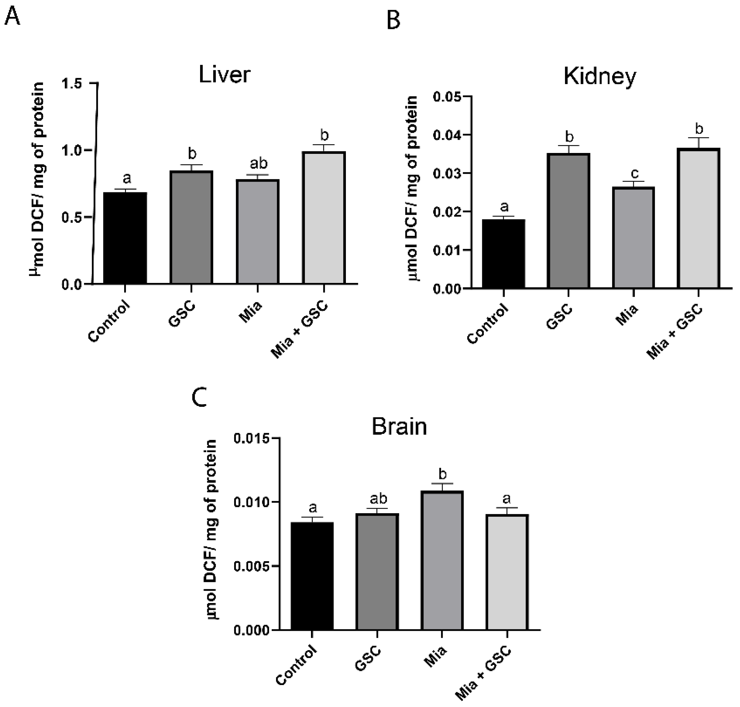
Lipid peroxidation levels were significantly increased in hepatic (Figure 1A, p < 0.0001), renal (Figure 1B, p = 0.0158), and cerebral (Figure 1C, p = 0.0204) tissues in the Mia group compared to the control. However, treatment with GSC (Mia+GSC group) restored TBARS levels to values comparable to those of the control group in all three tissues (Figure 1A–C).

3.2. ROS Production

ROS production in the liver (Figure 2A) was increased in both the GSC (p = 0.0286) and Mia+GSC (p < 0.0001) groups compared to the control group. A similar effect was observed in the kidneys (Figure 2B); however, the Mia group also showed an increase in ROS production compared to the control (p = 0.0184). In the brain (Figure 2C), ROS levels were elevated in the Mia group compared to the control (p = 0.0040), but GSC treatment prevented this increase.

3.3. CAT Activity

CAT activity in the liver (Figure 3A) was significantly reduced in the GSC (p = 0.0019) and Mia+GSC (p = 0.0002) groups compared to the control group. In the kidneys (Figure 3B), a decrease in enzyme activity was observed in the Mia+GSC group relative to the control (p = 0.0345). In the brain (Figure 3C), no significant difference was found between the control and Mia+GSC groups.

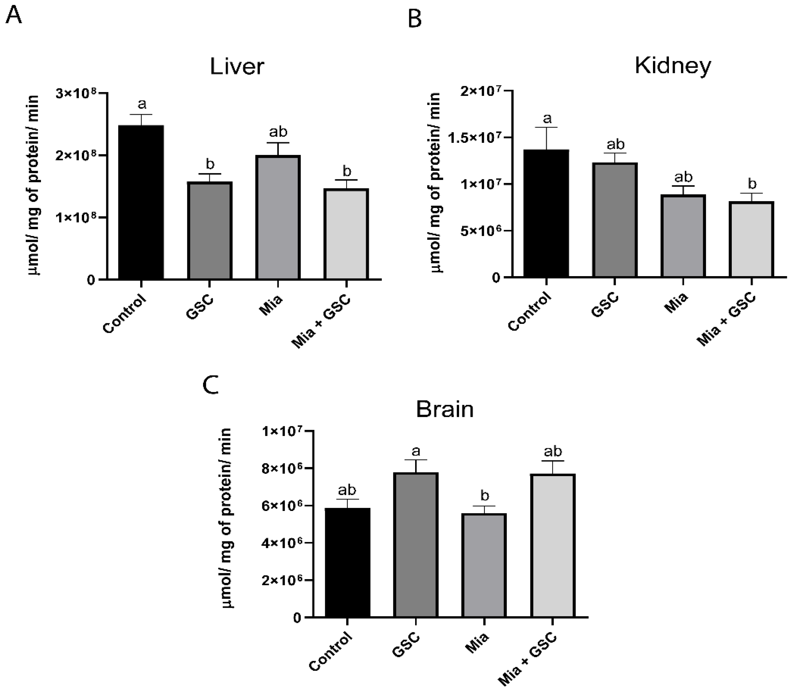
3.4. AChE Quantification

AChE enzyme activity in the liver (Figure 4A) was significantly increased in the Mia group compared to in the control, GSC, and Mia+GSC groups (p = 0.0015, p = 0.0003, p = 0.0183, respectively). GSC treatment was able to reverse this increase, restoring AChE activity to levels similar to those of the control group. No significant changes in enzyme activity were observed in renal tissue (Figure 4B). In the brain (Figure 4C), the Mia+GSC group exhibited a reduction in AChE activity compared to the Mia group (p = 0.0058), reaching levels comparable to those of the control group.

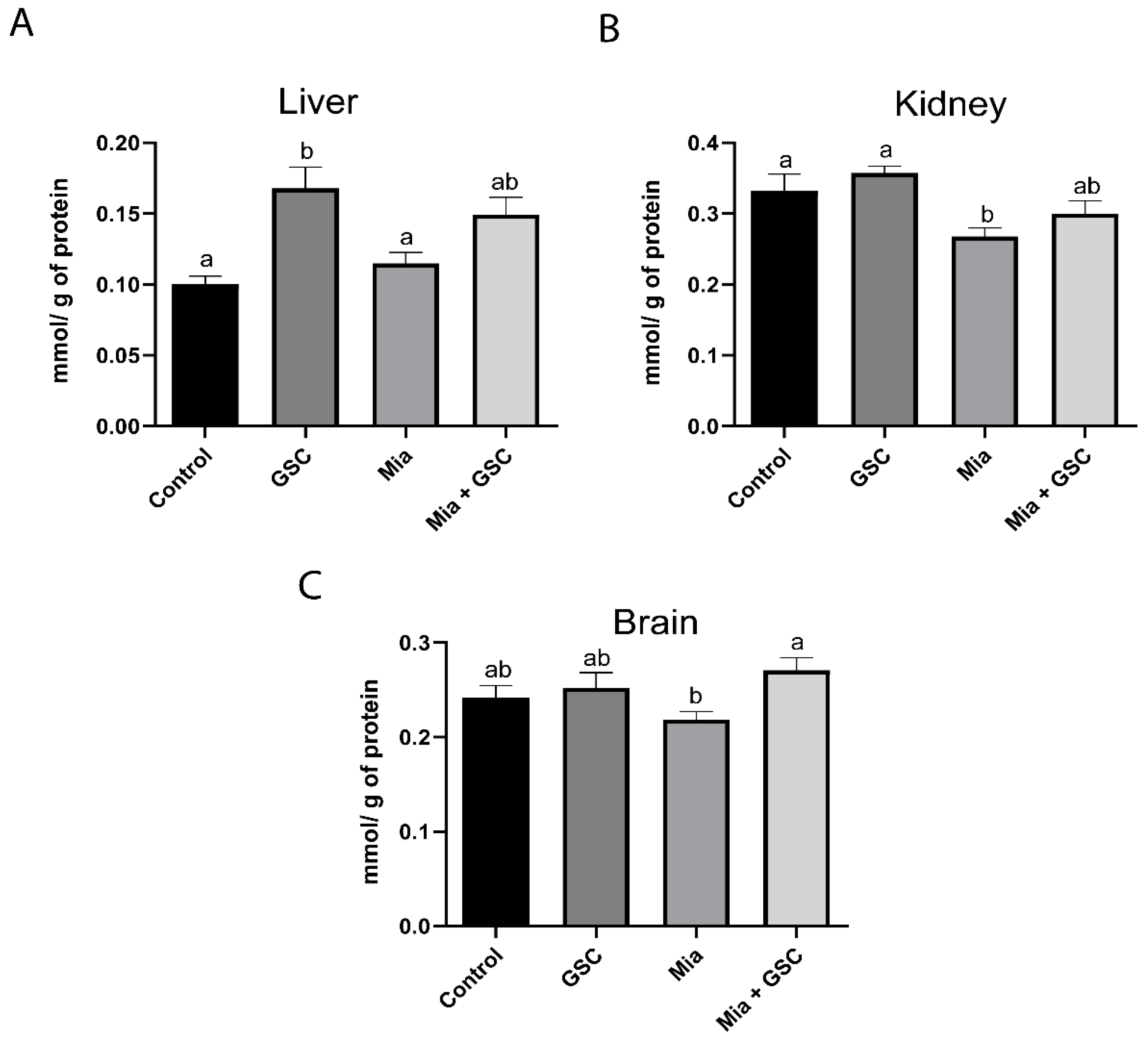
3.5. Determination of Total T-SH

Total thiol levels were not significantly different in hepatic (Figure 5A) and renal (Figure 5B) tissues. However, in the brain (Figure 5C), the GSC group showed an increase in total thiol levels compared to the control group (p = 0.0244), while the levels of the Mia+GSC group remained similar to those of the control.

3.6. Measure of TAC

Analysis of liver TAC (Figure 6A) revealed a significant increase in the GSC group compared to in the control group (p = 0.009). Additionally, GSC treatment normalized TAC levels in the Mia+GSC group, making them comparable to those of the control. In the kidneys (Figure 6B), CT values were reduced in the Mia group relative to the control (p = 0.0317), while the Mia+GSC group showed values similar to those of the control. In the brain (Figure 6C), CT was increased in the Mia+GSC group compared to the Mia group (p = 0.0339), with levels also matching those of the control group.

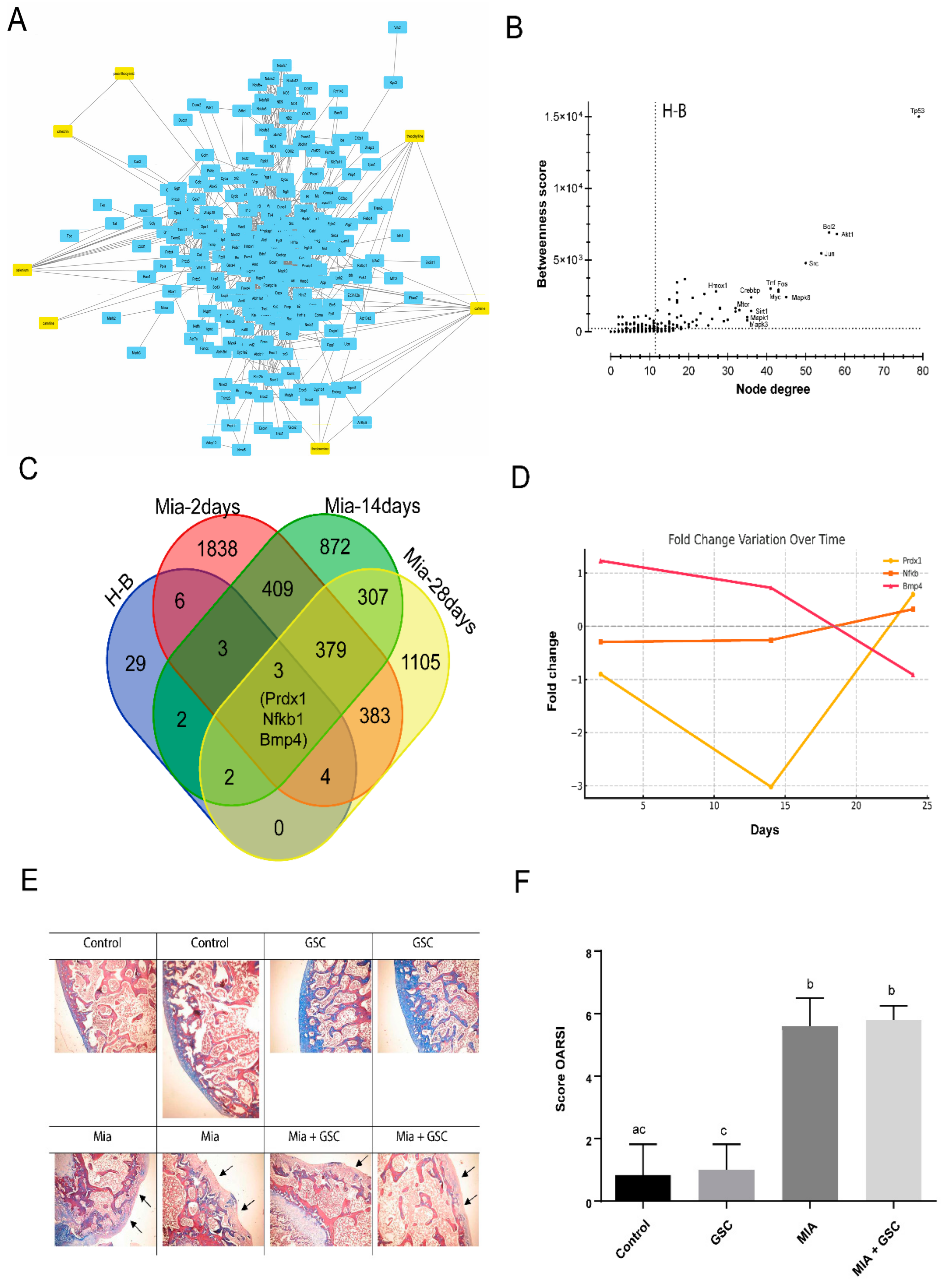
3.7. Systems Biology Analysis and Histological Evaluation

To investigate potential interactions between the GSC mix formulation and pathways associated with oxidative stress, a chemical–protein–protein interaction (CP-PPI) network consisting of 274 nodes and 1404 edges was predicted (Figure 7A). Centrality analysis was performed, identifying 49 H-B nodes (Figure 7B). Interestingly, Venn diagram analysis revealed an uneven distribution of differentially expressed genes (DEGs) among these H-B nodes during Mia-induced osteoarthritis at 0, 2, 14, and 28 days post treatment (Figure 7C). Only three genes—Prdx1, Nfkb1, and Bmp4—were found to be differentially expressed under the analyzed conditions. However, the expression levels of these genes varied depending on the timing following Mia administration (Figure 7D). Histopathological analysis (Figure 7E) demonstrated cartilage denudation and subchondral bone degeneration in Mia-treated animals, which corresponded to significantly higher OARSI scores in these groups (Figure 7F), with no observable alterations following treatment with the GSC mix.

4. Discussion

The effects of caffeine [46], selenium [47], and L-carnitine [48] have been extensively characterized in previous studies. In the present study, we aimed to investigate the antioxidant mechanisms and potential therapeutic effects of the complete guarana extract combined with selenium and L-carnitine in the context of OA, rather than examining the effects of these constituents individually. This approach was reflected in the actual experimental conditions, as the animals were administered the whole extract, whose absorption, pharmacokinetics, and systemic distribution differ substantially from those of isolated compounds. Our results confirmed the antioxidant effects of the supplementation, particularly illustrated by a reduction in TBARS levels in OA animals given the GSC supplementation. This effect can be attributed to caffeine’s capacity to inhibit lipid peroxidation and decrease ROS production [49]. In contrast, the non-supplemented OA groups exhibited elevated TBARS levels, likely as a result of inflammation-induced ROS production, which contributes significantly to cartilage and bone degradation [50], as well as neutrophil degranulation and the release of harmful enzymes [51]. The established link between inflammation and oxidative stress is particularly critical, considering that the brain, with its high oxygen consumption and limited antioxidant defenses, is especially vulnerable to ROS-induced damage [52]. Notably, our results indicated a reduction in brain ROS levels in OA animals supplemented with GSC, supporting the neuroprotective effects associated with caffeine and L-carnitine [53,54]. However, the observed increase in ROS levels in the liver and kidney tissues of supplemented animals might reflect the complex dose-dependent effects of caffeine, which can manifest both beneficial and adverse outcomes [49].
To modulate oxidative damage, the body relies on antioxidant defense mechanisms, among which enzymatic systems like CAT play a vital role. Our findings revealed tissue-specific variations in CAT activity: GSC supplementation resulted in increased CAT activity in brain tissue, indicating a protective role, but it was associated with reduced CAT levels in other tissues. Such variability underscores the complex regulatory mechanisms controlling antioxidant enzyme activity across different tissues [55]. In the context of inflammatory states, acetylcholine—a neurotransmitter essential for neuromuscular communication and immune modulation—undergoes rapid degradation mediated by AChE. Elevated AChE activity has been linked to inflammatory conditions [56]. In our study, OA animals exhibited increased AChE levels; conversely, GSC supplementation led to a reduction in AChE activity, reinforcing the protective effects of selenium on immune function and cellular homeostasis [57]. Moreover, thiols serve as effective biomarkers of oxidative stress, providing insights into the balance between free radical production and antioxidant defenses [58]. While our results showed no significant alterations in total TS-H levels among injured groups, the increase in TS-H levels in GSC-supplemented animals supports the beneficial effects of its components in enhancing immune function and mitigating oxidative stress [59]. Total antioxidant capacity (TAC) is another key indicator of systemic antioxidant defense efficiency [60]. Previous studies have established a positive correlation between selenium supplementation and increased TAC, alongside decreased malondialdehyde levels [58]. Consistent with these findings, our study reported elevated TAC in GSC-supplemented groups, further confirming the antioxidant properties attributed to caffeine [49,61] and underscoring the potential protective effect of the combined use of guarana, selenium, and L-carnitine in the context of OA. It is important to note that many natural compounds, such as harpagoside [62] and avocado/soybean unsaponifiables [63], primarily exhibit local anti-inflammatory or chondroprotective effects, with limited systemic antioxidant activity.
Despite this systemic benefit, the findings revealed that GSC supplementation failed to ameliorate local tissue damage resulting from OA. This implies that reducing oxidative stress alone is inadequate to counteract the structural deterioration associated with the disease. The in silico analyses indicated a complex molecular environment in Mia-induced OA that was not sufficiently modulated by GSC under the tested dose and timeframe. Although there was documented improvement in oxidative parameters in supplemented animals, histopathological analysis indicated no significant improvement in cartilage preservation or OARSI scores in GSC-treated animals. Our results also highlighted that only three central genes—Prdx1, Nfkb1, and Bmp4—were differentially expressed during Mia-induced osteoarthritis, demonstrating a limited overlap in DEGs related to Mia-induced OA. This suggests dose-dependent variability in pathway-level responses to Mia-induced OA. However, the centrality of Prdx1 and Nfkb1 underscores their relevance in OA-related oxidative stress, positioning them as potential biomarkers or therapeutic targets.
These findings can also be used to guide future optimization of the GSC formulation, dosing strategies, and timing of interventions, emphasizing that the lack of measurable protective effects does not equate to ineffectiveness, but rather reflects the complexity of OA pathophysiology and the necessity for refined therapeutic approaches that target early oxidative responses. One possible explanation for the lack of histological improvement is that, while oxidative stress is known to contribute to OA pathogenesis, other factors—such as persistent biomechanical stress, chronic inflammation, and catabolic enzyme activity—may play more significant roles in cartilage degradation. GSC’s inability to counter these mechanisms highlights the multifaceted nature of OA. Furthermore, the intricate interplay between oxidative stress and inflammatory pathways suggests that although antioxidants like those in GSC may reduce oxidative damage, they may not directly inhibit the inflammatory mediators responsible for tissue destruction. Given the adverse effects of conventional pharmacotherapy, the development of alternative, naturally derived treatments remains a promising avenue for future research in OA management.

5. Conclusions

In summary, our results indicate that supplementation with guarana, selenium, and L-carnitine effectively improves oxidative parameters in OA; however, it does not prevent joint tissue damage. This highlights the complexity of OA pathogenesis and suggests that targeting oxidative stress alone is insufficient to achieve structural protection of the joints.
It is important to acknowledge certain limitations of our study. Only a single dose of each compound was tested, and the treatment duration was relatively short, which may have limited the possibility of detecting structural benefits in cartilage within this progressive disease model. Moreover, although the study focused primarily on oxidative stress, future investigations should also explore inflammatory pathways and cartilage matrix degradation. Such research is essential to fully elucidate the mechanisms underlying OA and to evaluate combined therapeutic approaches that address oxidative stress, inflammation, and biomechanical factors contributing to disease progression.

Author Contributions

Conceptualization, R.P.B. and A.Z.P.; data curation, J.E.V., J.S.d.S., C.F.T. and J.G.M.; formal analysis, A.Z.P., R.P., J.E.V. and J.S.d.S.; funding acquisition, R.P.B. and G.L.-M.; investigation, V.I.V., V.F.A., C.d.R.T. and E.C.Z.; methodology, A.Z.P., R.P., J.E.V. and J.S.d.S.; project administration, R.P.B and I.B.M.d.C.; resources, R.P.B. and G.L.-M.; supervision, R.P.B.; validation, A.Z.P., R.P., J.E.V. and J.S.d.S.; visualization, J.E.V. and A.Z.P.; writing—original draft, A.Z.P., J.E.V. and J.G.M.; writing—review and editing, V.I.V., J.E.V. and R.P.B. All authors have read and agreed to the published version of the manuscript.

Funding

This study was supported by CAPES/PROEX 23038.005848/2018-31.

Informed Consent Statement

All animal procedures were approved by the Ethics Committee on the Use of Animals of the University of Passo Fundo (CEUA-UPF) under registration no. 26/2018 and conducted in accordance with national and institutional guidelines for the care and use of laboratory animals.

Data Availability Statement

The data presented in this study are available upon request from the corresponding author.

Conflicts of Interest

The authors declare no conflicts of interest. The funders had no role in the design of the study; in the collection, analyses, or interpretation of data; in the writing of the manuscript; or in the decision to publish the results.

References

  1. GBD 2021 Osteoarthritis Collaborators. Global, regional, and national burden of osteoarthritis, 1990-2020 and projections to 2050: A systematic analysis for the Global Burden of Disease Study 2021. Lancet Rheumatol. 2023, 5, e508–e522. [Google Scholar] [CrossRef] [PubMed]
  2. Mandl, L.A. Osteoarthritis year in review 2018: Clinical. Osteoarthr. Cartil. 2019, 27, 359–364. [Google Scholar] [CrossRef]
  3. Ho, J.; Mak, C.C.H.; Sharma, V.; To, K.; Khan, W. Mendelian Randomization Studies of Lifestyle-Related Risk Factors for Osteoarthritis: A PRISMA Review and Meta-Analysis. Int. J. Mol. Sci. 2022, 23, 11906. [Google Scholar] [CrossRef] [PubMed]
  4. Altay, M.A.; Erturk, C.; Bilge, A.; Yapti, M.; Levent, A.; Aksoy, N. Evaluation of prolidase activity and oxidative status in patients with knee osteoarthritis: Relationships with radiographic severity and clinical parameters. Rheumatol. Int. 2015, 35, 1725–1731. [Google Scholar] [CrossRef] [PubMed]
  5. Erturk, C.; Altay, M.A.; Selek, S.; Kocyigit, A. Paraoxonase-1 activity and oxidative status in patients with knee osteoarthritis and their relationship with radiological and clinical parameters. Scand. J. Clin. Lab. Investig. 2012, 72, 433–439. [Google Scholar] [CrossRef]
  6. Altindag, O.; Erel, O.; Aksoy, N.; Selek, S.; Celik, H.; Karaoglanoglu, M. Increased oxidative stress and its relation with collagen metabolism in knee osteoarthritis. Rheumatol. Int. 2007, 27, 339–344. [Google Scholar] [CrossRef]
  7. Davies, C.M.; Guilak, F.; Weinberg, J.B.; Fermor, B. Reactive nitrogen and oxygen species in interleukin-1-mediated DNA damage associated with osteoarthritis. Osteoarthr. Cartil. 2008, 16, 624–630. [Google Scholar] [CrossRef]
  8. Li, D.; Xie, G.; Wang, W. Reactive oxygen species: The 2-edged sword of osteoarthritis. Am. J. Med. Sci. 2012, 344, 486–490. [Google Scholar] [CrossRef]
  9. Lepetsos, P.; Papavassiliou, A.G. ROS/oxidative stress signaling in osteoarthritis. Biochim. Biophys. Acta 2016, 1862, 576–591. [Google Scholar] [CrossRef]
  10. Scott, J.L.; Gabrielides, C.; Davidson, R.K.; Swingler, T.E.; Clark, I.M.; Wallis, G.A.; Boot-Handford, R.P.; Kirkwood, T.B.; Taylor, R.W.; Young, D.A. Superoxide dismutase downregulation in osteoarthritis progression and end-stage disease. Ann. Rheum. Dis. 2010, 69, 1502–1510. [Google Scholar] [CrossRef]
  11. Peng, Y.; Ao, M.; Dong, B.; Jiang, Y.; Yu, L.; Chen, Z.; Hu, C.; Xu, R. Anti-Inflammatory Effects of Curcumin in the Inflammatory Diseases: Status, Limitations and Countermeasures. Drug Des. Dev. Ther. 2021, 15, 4503–4525. [Google Scholar] [CrossRef] [PubMed]
  12. Yang, S.; Sun, M.; Zhang, X. Protective Effect of Resveratrol on Knee Osteoarthritis and its Molecular Mechanisms: A Recent Review in Preclinical and Clinical Trials. Front. Pharmacol. 2022, 13, 921003. [Google Scholar] [CrossRef] [PubMed]
  13. Akhtar, N.; Haqqi, T.M. Epigallocatechin-3-gallate suppresses the global interleukin-1beta-induced inflammatory response in human chondrocytes. Arthritis Res. Ther. 2011, 13, R93. [Google Scholar] [CrossRef] [PubMed]
  14. Yang, D.; Cao, G.; Ba, X.; Jiang, H. Epigallocatechin-3-O-gallate promotes extracellular matrix and inhibits inflammation in IL-1beta stimulated chondrocytes by the PTEN/miRNA-29b pathway. Pharm. Biol. 2022, 60, 589–599. [Google Scholar] [CrossRef]
  15. Majeed, M.; Majeed, S.; Narayanan, N.K.; Nagabhushanam, K. A pilot, randomized, double-blind, placebo-controlled trial to assess the safety and efficacy of a novel Boswellia serrata extract in the management of osteoarthritis of the knee. Phytother. Res. PTR 2019, 33, 1457–1468. [Google Scholar] [CrossRef]
  16. Szymczak, J.; Grygiel-Gorniak, B.; Cielecka-Piontek, J. Zingiber Officinale Roscoe: The Antiarthritic Potential of a Popular Spice-Preclinical and Clinical Evidence. Nutrients 2024, 16, 741. [Google Scholar] [CrossRef]
  17. Salehi, B.; Rescigno, A.; Dettori, T.; Calina, D.; Docea, A.O.; Singh, L.; Cebeci, F.; Ozcelik, B.; Bhia, M.; Dowlati Beirami, A.; et al. Avocado-Soybean Unsaponifiables: A Panoply of Potentialities to Be Exploited. Biomolecules 2020, 10, 130. [Google Scholar] [CrossRef]
  18. Fang, S.; Zhang, B.; Xiang, W.; Zheng, L.; Wang, X.; Li, S.; Zhang, T.; Feng, D.; Gong, Y.; Wu, J.; et al. Natural products in osteoarthritis treatment: Bridging basic research to clinical applications. Chin. Med. 2024, 19, 25. [Google Scholar] [CrossRef]
  19. Azzolin, V.F.; Azzolin, V.F.; da Silva Maia, R.; Mastella, M.H.; Sasso, J.S.; Barbisan, F.; Bitencourt, G.R.; de Azevedo Mello, P.; Ribeiro, E.M.A.; Ribeiro, E.E.; et al. Safety and efficacy indicators of guarana and Brazil nut extract carried in nanoparticles of coenzyme Q10: Evidence from human blood cells and red earthworm experimental model. Food Chem. Toxicol. Int. J. Publ. Br. Ind. Biol. Res. Assoc. 2024, 191, 114828. [Google Scholar] [CrossRef]
  20. Machado, K.N.; Paula Barbosa, A.; de Freitas, A.A.; Alvarenga, L.F.; Padua, R.M.; Gomes Faraco, A.A.; Braga, F.C.; Vianna-Soares, C.D.; Castilho, R.O. TNF-alpha inhibition, antioxidant effects and chemical analysis of extracts and fraction from Brazilian guarana seed powder. Food Chem. 2021, 355, 129563. [Google Scholar] [CrossRef]
  21. Manica-Cattani, M.F.; Hoefel, A.L.; Azzolin, V.F.; Montano, M.A.E.; da Cruz Jung, I.E.; Ribeiro, E.E.; Azzolin, V.F.; da Cruz, I.B.M. Amazonian fruits with potential effects on COVID-19 by inflammaging modulation: A narrative review. J. Food Biochem. 2022, 46, e14472. [Google Scholar] [CrossRef] [PubMed]
  22. Macan, T.P.; Magenis, M.L.; Damiani, A.P.; Monteiro, I.O.; Silveira, G.B.; Zaccaron, R.P.; Silveira, P.C.L.; Teixeira, J.P.F.; Gajski, G.; Andrade, V.M. Brazil nut consumption reduces DNA damage in overweight type 2 diabetes mellitus patients. Mutat. Res. Genet. Toxicol. Environ. Mutagen. 2024, 895, 503739. [Google Scholar] [CrossRef] [PubMed]
  23. Filippini, T.; Fairweather-Tait, S.; Vinceti, M. Selenium and immune function: A systematic review and meta-analysis of experimental human studies. Am. J. Clin. Nutr. 2023, 117, 93–110. [Google Scholar] [CrossRef] [PubMed]
  24. Rastgoo, S.; Fateh, S.T.; Nikbaf-Shandiz, M.; Rasaei, N.; Aali, Y.; Zamani, M.; Shiraseb, F.; Asbaghi, O. The effects of L-carnitine supplementation on inflammatory and anti-inflammatory markers in adults: A systematic review and dose-response meta-analysis. Inflammopharmacology 2023, 31, 2173–2199. [Google Scholar] [CrossRef]
  25. Teixeira, C.F.; Azzolin, V.F.; Rodrigues Dos Passos, G.; Turra, B.O.; Alves, A.O.; Bressanim, A.C.M.; Canton, L.E.L.; Vieira Dos Santos, A.C.; Mastella, M.H.; Barbisan, F.; et al. A coffee enriched with guarana, selenium, and l-carnitine (GSC) has nutrigenomic effects on oxi-inflammatory markers of relapsing-remitting multiple sclerosis patients: A pilot study. Mult. Scler. Relat. Disord. 2023, 71, 104515. [Google Scholar] [CrossRef]
  26. Teixeira, C.F.; da Cruz, I.B.M.; Ribeiro, E.E.; Pillar, D.M.; Turra, B.O.; Praia, R.S.; Barbisan, F.; Alves, A.O.; Sato, D.K.; Assmann, C.E.; et al. Safety indicators of a novel multi supplement based on guarana, selenium, and L-carnitine: Evidence from human and red earthworm immune cells. Food Chem. Toxicol. Int. J. Publ. Br. Ind. Biol. Res. Assoc. 2021, 150, 112066. [Google Scholar] [CrossRef]
  27. Vonsy, J.L.; Ghandehari, J.; Dickenson, A.H. Differential analgesic effects of morphine and gabapentin on behavioural measures of pain and disability in a model of osteoarthritis pain in rats. Eur. J. Pain 2009, 13, 786–793. [Google Scholar] [CrossRef]
  28. Fernihough, J.; Gentry, C.; Malcangio, M.; Fox, A.; Rediske, J.; Pellas, T.; Kidd, B.; Bevan, S.; Winter, J. Pain related behaviour in two models of osteoarthritis in the rat knee. Pain 2004, 112, 83–93. [Google Scholar] [CrossRef]
  29. Guzman, R.E.; Evans, M.G.; Bove, S.; Morenko, B.; Kilgore, K. Mono-iodoacetate-induced histologic changes in subchondral bone and articular cartilage of rat femorotibial joints: An animal model of osteoarthritis. Toxicol. Pathol. 2003, 31, 619–624. [Google Scholar] [CrossRef]
  30. Orita, S.; Ishikawa, T.; Miyagi, M.; Ochiai, N.; Inoue, G.; Eguchi, Y.; Kamoda, H.; Arai, G.; Toyone, T.; Aoki, Y.; et al. Pain-related sensory innervation in monoiodoacetate-induced osteoarthritis in rat knees that gradually develops neuronal injury in addition to inflammatory pain. BMC Musculoskelet. Disord. 2011, 12, 134. [Google Scholar] [CrossRef]
  31. Chandran, P.; Pai, M.; Blomme, E.A.; Hsieh, G.C.; Decker, M.W.; Honore, P. Pharmacological modulation of movement-evoked pain in a rat model of osteoarthritis. Eur. J. Pharmacol. 2009, 613, 39–45. [Google Scholar] [CrossRef] [PubMed]
  32. van Rijn, C.M.; Krijnen, H.; Menting-Hermeling, S.; Coenen, A.M. Decapitation in rats: Latency to unconsciousness and the ‘wave of death’. PLoS ONE 2011, 6, e16514. [Google Scholar] [CrossRef] [PubMed]
  33. Jentzsch, A.M.; Bachmann, H.; Furst, P.; Biesalski, H.K. Improved analysis of malondialdehyde in human body fluids. Free Radic. Biol. Med. 1996, 20, 251–256. [Google Scholar] [CrossRef] [PubMed]
  34. Myhre, O.; Andersen, J.M.; Aarnes, H.; Fonnum, F. Evaluation of the probes 2′,7′-dichlorofluorescin diacetate, luminol, and lucigenin as indicators of reactive species formation. Biochem. Pharmacol. 2003, 65, 1575–1582. [Google Scholar] [CrossRef]
  35. Saccol, R.; da Silveira, K.L.; Manzoni, A.G.; Abdalla, F.H.; de Oliveira, J.S.; Dornelles, G.L.; Barbisan, F.; Passos, D.F.; Casali, E.A.; de Andrade, C.M.; et al. Antioxidant, hepatoprotective, genoprotective, and cytoprotective effects of quercetin in a murine model of arthritis. J. Cell. Biochem. 2020, 121, 2792–2801. [Google Scholar] [CrossRef]
  36. Nelson, D.P.; Kiesow, L.A. Enthalpy of decomposition of hydrogen peroxide by catalase at 25 °C (with molar extinction coefficients of H2O2 solutions in the UV). Anal. Biochem. 1972, 49, 474–478. [Google Scholar] [CrossRef]
  37. Ellman, G.L.; Courtney, K.D.; Andres, V., Jr.; Feather-Stone, R.M. A new and rapid colorimetric determination of acetylcholinesterase activity. Biochem. Pharmacol. 1961, 7, 88–95. [Google Scholar] [CrossRef]
  38. Worek, F.; Mast, U.; Kiderlen, D.; Diepold, C.; Eyer, P. Improved determination of acetylcholinesterase activity in human whole blood. Clin. Chim. Acta 1999, 288, 73–90. [Google Scholar] [CrossRef]
  39. Ellman, G.L. Reprint of: Tissue Sulfhydryl Groups. Arch. Biochem. Biophys. 2022, 726, 109245. [Google Scholar] [CrossRef]
  40. Prieto, P.; Pineda, M.; Aguilar, M. Spectrophotometric quantitation of antioxidant capacity through the formation of a phosphomolybdenum complex: Specific application to the determination of vitamin E. Anal. Biochem. 1999, 269, 337–341. [Google Scholar] [CrossRef]
  41. Pritzker, K.P.; Gay, S.; Jimenez, S.A.; Ostergaard, K.; Pelletier, J.P.; Revell, P.A.; Salter, D.; van den Berg, W.B. Osteoarthritis cartilage histopathology: Grading and staging. Osteoarthr. Cartil. 2006, 14, 13–29. [Google Scholar] [CrossRef] [PubMed]
  42. Bittencourt Lda, S.; Zeidan-Chulia, F.; Yatsu, F.K.; Schnorr, C.E.; Moresco, K.S.; Kolling, E.A.; Gelain, D.P.; Bassani, V.L.; Moreira, J.C. Guarana (Paullinia cupana Mart.) prevents beta-amyloid aggregation, generation of advanced glycation-end products (AGEs), and acrolein-induced cytotoxicity on human neuronal-like cells. Phytother. Res. PTR 2014, 28, 1615–1624. [Google Scholar] [CrossRef] [PubMed]
  43. da Silva, G.S.; Canuto, K.M.; Ribeiro, P.R.V.; de Brito, E.S.; Nascimento, M.M.; Zocolo, G.J.; Coutinho, J.P.; de Jesus, R.M. Chemical profiling of guarana seeds (Paullinia cupana) from different geographical origins using UPLC-QTOF-MS combined with chemometrics. Food Res. Int. 2017, 102, 700–709. [Google Scholar] [CrossRef] [PubMed]
  44. Szklarczyk, D.; Santos, A.; von Mering, C.; Jensen, L.J.; Bork, P.; Kuhn, M. STITCH 5: Augmenting protein-chemical interaction networks with tissue and affinity data. Nucleic Acids Res. 2016, 44, D380–D384. [Google Scholar] [CrossRef]
  45. Scardoni, G.; Tosadori, G.; Faizan, M.; Spoto, F.; Fabbri, F.; Laudanna, C. Biological network analysis with CentiScaPe: Centralities and experimental dataset integration. F1000Research 2014, 3, 139. [Google Scholar] [CrossRef]
  46. Guillan-Fresco, M.; Franco-Trepat, E.; Alonso-Perez, A.; Jorge-Mora, A.; Lopez-Fagundez, M.; Pazos-Perez, A.; Gualillo, O.; Gomez, R. Caffeine, a Risk Factor for Osteoarthritis and Longitudinal Bone Growth Inhibition. J. Clin. Med. 2020, 9, 1163. [Google Scholar] [CrossRef]
  47. Cheng, H.L.; Yen, C.C.; Huang, L.W.; Hu, Y.C.; Huang, T.C.; Hsieh, B.S.; Chang, K.L. Selenium Lessens Osteoarthritis by Protecting Articular Chondrocytes from Oxidative Damage through Nrf2 and NF-kappaB Pathways. Int. J. Mol. Sci. 2024, 25, 2511. [Google Scholar] [CrossRef]
  48. Kou, H.; Li, B.; Wang, Z.; Ma, J. Effect of l-Carnitine Supplementation on Osteoarthritis: A Systematic Review. Mol. Nutr. Food Res. 2024, 68, e2300614. [Google Scholar] [CrossRef]
  49. Barcelos, R.P.; Lima, F.D.; Carvalho, N.R.; Bresciani, G.; Royes, L.F. Caffeine effects on systemic metabolism, oxidative-inflammatory pathways, and exercise performance. Nutr. Res. 2020, 80, 1–17. [Google Scholar] [CrossRef]
  50. Jeon, S.; Min Kim, T.; Kwon, G.; Park, J.; Park, S.Y.; Lee, S.H.; Jin, E.J. Targeting ROS in osteoclasts within the OA environment: A novel therapeutic strategy for osteoarthritis management. J. Tissue Eng. 2024, 15, 20417314241279935. [Google Scholar] [CrossRef]
  51. Wright, H.L.; Lyon, M.; Chapman, E.A.; Moots, R.J.; Edwards, S.W. Rheumatoid Arthritis Synovial Fluid Neutrophils Drive Inflammation Through Production of Chemokines, Reactive Oxygen Species, and Neutrophil Extracellular Traps. Front. Immunol. 2020, 11, 584116. [Google Scholar] [CrossRef] [PubMed]
  52. Jimenez-Blasco, D.; Almeida, A.; Bolanos, J.P. Brightness and shadows of mitochondrial ROS in the brain. Neurobiol. Dis. 2023, 184, 106199. [Google Scholar] [CrossRef] [PubMed]
  53. Devasagayam, T.P.; Kamat, J.P.; Mohan, H.; Kesavan, P.C. Caffeine as an antioxidant: Inhibition of lipid peroxidation induced by reactive oxygen species. Biochim. Biophys. Acta 1996, 1282, 63–70. [Google Scholar] [CrossRef] [PubMed]
  54. Li, J.L.; Wang, Q.Y.; Luan, H.Y.; Kang, Z.C.; Wang, C.B. Effects of L-carnitine against oxidative stress in human hepatocytes: Involvement of peroxisome proliferator-activated receptor alpha. J. Biomed. Sci. 2012, 19, 32. [Google Scholar] [CrossRef]
  55. Scaglione, C.N.; Xu, Q.; Ramanujan, V.K. Direct measurement of catalase activity in living cells and tissue biopsies. Biochem. Biophys. Res. Commun. 2016, 470, 192–196. [Google Scholar] [CrossRef]
  56. Benfante, R.; Di Lascio, S.; Cardani, S.; Fornasari, D. Acetylcholinesterase inhibitors targeting the cholinergic anti-inflammatory pathway: A new therapeutic perspective in aging-related disorders. Aging Clin. Exp. Res. 2021, 33, 823–834. [Google Scholar] [CrossRef]
  57. Rayman, M.P. Selenoproteins and human health: Insights from epidemiological data. Biochim. Biophys. Acta 2009, 1790, 1533–1540. [Google Scholar] [CrossRef]
  58. Malek Mahdavi, A.; Mahdavi, R.; Kolahi, S.; Zemestani, M.; Vatankhah, A.M. L-Carnitine supplementation improved clinical status without changing oxidative stress and lipid profile in women with knee osteoarthritis. Nutr. Res. 2015, 35, 707–715. [Google Scholar] [CrossRef]
  59. Ricordi, C.; Garcia-Contreras, M.; Farnetti, S. Diet and Inflammation: Possible Effects on Immunity, Chronic Diseases, and Life Span. J. Am. Coll. Nutr. 2015, 34 (Suppl. 1), 10–13. [Google Scholar] [CrossRef]
  60. Lozano-Paniagua, D.; Parron, T.; Alarcon, R.; Requena, M.; Gil, F.; Lopez-Guarnido, O.; Lacasana, M.; Hernandez, A.F. Biomarkers of oxidative stress in blood of workers exposed to non-cholinesterase inhibiting pesticides. Ecotoxicol. Environ. Saf. 2018, 162, 121–128. [Google Scholar] [CrossRef]
  61. Osz, B.E.; Jitca, G.; Stefanescu, R.E.; Puscas, A.; Tero-Vescan, A.; Vari, C.E. Caffeine and Its Antioxidant Properties-It Is All about Dose and Source. Int. J. Mol. Sci. 2022, 23, 13074. [Google Scholar] [CrossRef] [PubMed]
  62. Haseeb, A.; Ansari, M.Y.; Haqqi, T.M. Harpagoside suppresses IL-6 expression in primary human osteoarthritis chondrocytes. J. Orthop. Res. 2017, 35, 311–320. [Google Scholar] [CrossRef] [PubMed]
  63. Lambert, C.; Bellemere, G.; Boyer, G.; Ponelle, F.; Bauer, T.; Legeny, M.C.; Baudouin, C.; Henrotin, Y. Composition Analysis and Pharmacological Activity of Avocado/Soybean Unsaponifiable Products Used in the Treatment of Osteoarthritis. Front. Pharmacol. 2021, 12, 781389. [Google Scholar] [CrossRef] [PubMed]
Figure 1. The effect of GSC supplementation on the production of TBARS in the liver (A), kidney (B), and brain (C) of animals with osteoarthritis. The animals were divided into four groups (n = 10): control, GSC, Mia, and Mia+GSC. The data are expressed as the mean ± S.E.M. Means with different letters indicate significant differences (p < 0.05).
Antioxidants 14 00881 g001
Figure 2. The effect of GSC supplementation on ROS production in the liver (A), kidney (B), and brain (C) of animals with osteoarthritis. The animals were divided into four groups (n = 10): control, GSC, Mia, and Mia+GSC. The data are expressed as the mean ± S.E.M. Means with different letters indicate significant differences (p < 0.05).
Antioxidants 14 00881 g002
Figure 3. The effect of GSC supplementation on CAT activity in the liver (A), kidney (B), and brain (C) of animals with osteoarthritis. The animals were divided into four groups (n = 10): control, GSC, Mia, and Mia+GSC. The data are expressed as the mean ± S.E.M. Means with different letters indicate significant differences (p < 0.05).
Antioxidants 14 00881 g003
Figure 4. The effect of GSC supplementation on the activity of the enzyme AChE in the liver (A), kidney (B), and brain (C) of animals with osteoarthritis. The animals were divided into four groups (n = 10): control, GSC, Mia, and Mia+GSC. The data are expressed as the mean ± S.E.M. Means with different letters indicate significant differences (p < 0.05).
Antioxidants 14 00881 g004
Figure 5. The effect of GSC supplementation on total T-SH levels in the liver (A), kidney (B), and brain (C) of animals with osteoarthritis. The animals were divided into four groups (n = 10): control, GSC, Mia, and Mia+GSC. The data are expressed as the mean ± S.E.M. Means with different letters indicate significant differences (p < 0.05).
Antioxidants 14 00881 g005
Figure 6. The effect of GSC supplementation on TAC in the liver (A), kidney (B), and brain (C) of animals with osteoarthritis. The animals were divided into four groups (n = 10): control, GSC, Mia, and Mia+GSC. The data are expressed as the mean ± S.E.M. Means with different letters indicate significant differences (p < 0.05).
Antioxidants 14 00881 g006
Figure 7. The interaction network, gene expression analysis related to oxidative stress, and histological evaluation in Mia-induced osteoarthritis and GSC treatment. (A) The CP-PPI network constructed using STITCH 5.0, integrating bioactive compounds from the GSC mix with oxidative stress-related genes. Compounds are highlighted in yellow. (B) Centrality analysis performed using the CentiScaPe 2.2 plugin in Cytoscape. The H-B nodes exhibit both degree and betweenness centrality scores above the network average. (C) A Venn diagram showing the distribution of differentially expressed genes (DEGs) among H-B nodes at 0, 2, 14, and 28 days after Mia administration. (D) Fold-change values of the three DEGs (Prdx1, Nfkb1, and Bmp4) at the indicated time points following Mia treatment. (E) Representative histological sections of knee cartilage showing cartilage denudation and subchondral bone degeneration in Mia-treated animals, with or without GSC treatment. Animals were divided into four groups (n = 10): control, GSC, Mia, and Mia+GSC. Blue staining indicates the presence of collagen, while black arrows denote areas of collagen loss. The data are expressed as the mean ± S.E.M. Groups with different letters are significantly different (p < 0.05). (F) Histopathological scoring using the OARSI grading system.
Antioxidants 14 00881 g007
Table 1. Histological grading of cartilage degeneration.
DegreeCharacteristic
0Surface and cartilage with intact morphology
1Intact surface
2Discontinuity surface
3Vertical cracks
4Erosion
5Denudation
6Deformation
Disclaimer/Publisher’s Note: The statements, opinions and data contained in all publications are solely those of the individual author(s) and contributor(s) and not of MDPI and/or the editor(s). MDPI and/or the editor(s) disclaim responsibility for any injury to people or property resulting from any ideas, methods, instructions or products referred to in the content.

