Article

The Antioxidant Properties of Extracts of Cuscuta spp. Depend on the Parasite and the Host Species

by
Vanina Lozanova
1,
Denitsa Teofanova
1,
Bilyana Chakarova
2,
Krasimir Rusanov
2,3,
Kalina Pachedjieva
1,
Anita Tosheva
1,
Tzvetelina Zagorcheva
2,4 and
Lyuben Zagorchev
1,*
1
Faculty of Biology, Sofia University “St. Kliment Ohridski”, 8 Dragan Tsankov blvd., 1164 Sofia, Bulgaria
2
AgroBioInstitute, Agricultural Academy, 8 Dragan Tsankov blvd., 1164 Sofia, Bulgaria
3
Centre of Competence “Sustainable Utilization of Bio-Resources and Waste of Medicinal and Aromatic Plants for Innovative Bioactive Products” (BIORESOURCES BG), 1000 Sofia, Bulgaria
4
Research and Development and Innovation Consortium, Sofia Tech Park JSC, 111, Tsarigradsko Shose blvd., 1784 Sofia, Bulgaria
*
Author to whom correspondence should be addressed.
Antioxidants 2025, 14(7), 761; https://doi.org/10.3390/antiox14070761
Submission received: 28 April 2025 / Revised: 11 June 2025 / Accepted: 17 June 2025 / Published: 20 June 2025
(This article belongs to the Section Natural and Synthetic Antioxidants)

Abstract

Dodders (Cuscuta spp.) are prominent parasitic plants widely known and exploited in traditional medicine. They are rich in polyphenolics, which determine their strong antioxidant potential. However, comparatively few of the nearly 200 known species have been characterized for their medicinal potential. In the present study, we aimed to explore the antioxidant potential of four of the most widely distributed Cuscuta species in Bulgaria—C. campestris, C. monogyna, C. epithymum, and C. europaea. They differed significantly in polyphenolic content and accordingly differed in their antioxidant properties, although this correlation is not always straightforward, as shown in C. europaeae. Furthermore, we evaluated the host plant species’ influence on the polyphenolic content, antioxidant properties, and flavonoid profile of C. campestris, finding a significant enhancement when the parasite was grown on aromatic plants—rosemary and thyme—compared to a model host—Arabidopsis thaliana. Seven major flavonoids and phenolic acids—chlorogenic acid, kaempferol-3,7-O-diglucoside, quercetin-3-O-galactoside, kaempferol-3-O-galactoside, quercetin-3-O-glucoside, astragalin, and isorhamnetin-7-glucoside—were annotated after HPLC-MS analysis and found to be affected by the host species. In conclusion, it was found that extracts from different Cuscuta species differ in their antioxidant potential, which the host plants might further modify.

1. Introduction

Parasitic flowering plants are a widely variable group of nearly 4500 species, adapted to be partially (hemiparasites) or fully (holoparasites) dependent on their hosts for water, minerals, and organic nutrients [1]. Although traditionally regarded as harmful, very few represent serious agricultural pests, most notably several species of the Striga, Orobanche/Phelipanche, and Cuscuta genera [2]. Besides this negative impact, most of them are intriguing organisms with peculiar adaptations related to their semi- or fully non-photosynthetic lifestyle. They also play an important role in natural communities, regulating biodiversity by preferentially affecting particular plant groups, thus benefiting others [3,4]. Interestingly, most, if not all, are also employed in traditional medicine and are known to contain a large variety of bioactive compounds. Virtually all groups are known to possess medicinal properties, including members of the prominent families of broomrapes, Orobanchaceae [5]; mistletoes in the order Santalales, family Santalaceae [6]; and Loranthaceae [7], as well as some even more peculiar species like the “Maltese mushroom”, Cynomorium coccineum L. [8], and “the world’s largest flower”, Rafflesia spp. [9].
In addition to the abovementioned, members of the genus Cuscuta (family Convolvulaceae), or dodders, are also known for their use in traditional medicine. They are a diverse, widespread genus of about 200 species of stem (e.g., infecting the above-ground tissues of the host) and holoparasitic (e.g., depending fully on the host) plants [10]. Some, like C. campestris Yunck., are widespread agricultural pests [2], while others are rare or even threatened with extinction and need conservation efforts [11]. In Chinese traditional medicine, this genus is particularly appreciated for its medicinal properties and wide range of bioactivity. The most exploited species, C. chinensis Lam., locally known as Tu-Si-Zi, is sold as dried seeds and used as an extract as a tonic, improving sexual function and exhibiting positive effects on the immune system [12]. Ahmad [13] summarized a wide range of antibacterial, antifungal, antioxidant, anticancer, anti-inflammatory, hepatoprotective, and numerous other effects of about a dozen of Cuscuta spp., determined by hundreds of different bioactive compounds, including alkaloids, glycosides, steroids, terpenoids, carotenoids, organic acids, and (most notably) flavonoids. The antioxidant activity was found to be strongly correlated with the flavonoid content, especially quercetin and kaempferol [14]. While seeds are traditionally used, vegetative parts (e.g., stems) are equally rich in bioactive compounds [12].
An important question is whether all Cuscuta spp. are similar in the accumulation of bioactive compounds. Significant differences in flavonoids have been found between species, and they have even been proposed as chemotaxonomical markers [12,13]. In addition to the genetically determined species differences, the metabolic constituents in parasitic plants, and Cuscuta spp. in particular, could be affected by the host plant’s identity. Flores-Sanchez [15] concluded that at least some of the specialized metabolites in Cuscuta are transferred from the host through the haustoria (the parasitic organ, essentially a link between the vascular elements of the host and the parasite) and further modified in the parasite.
Recent reports suggest around nine or ten naturally occurring Cuscuta species in Bulgaria [16,17], of which at least some are listed as medicinal in the literature [18,19], with C. europaea L. being at the top of these lists. However, few studies have been conducted in this country [20] or globally [21]. Another prominent medicinal Cuscuta species that is not listed as such in Bulgaria but has been established in the country is C. epithymum L. [22]. As suggested by the recent literature, every Cuscuta species is a potential source of bioactive compounds. With the present study, we aimed to further explore the biology of these otherwise harmful and neglected parasitic plants in Bulgaria by focusing on their antioxidant properties and phenolic content as a pilot study on the potential for their sustainable utilization.

2. Materials and Methods

2.1. Plant Material

A total of 27 samples of C. campestris, 5 samples of C. epithymum, and 2 samples of C. europaea, which were previously described [23] and stored at −80 °C, were used in the present study. In addition, three samples of C. monogyna Vahl. collected in 2022 were also included in the analyses.
For laboratory-grown plant samples, seeds of C. campestris from an in-house sustained line (SU-BG-CA-2024.3) were treated according to a widely accepted method of sulfuric acid scarification. Germinated seedlings were used for the infection of fully grown Arabidopsis thaliana plants—Columbia ecotype, Col-0—and commercially available thyme (Thymus vulgaris L.) and rosemary (Salvia rosmarinus Spenn. (syn. Rosmarinus officinalis L.)) plants, all in triplicate. Parasite–host pairs were grown in glasshouse conditions for three weeks, which was sufficient to collect about 0.5–1 g of Cuscuta fresh mass from each host individual.

2.2. Extraction and Polyphenol Determination

The extraction of polyphenolic compounds was carried out by grinding 100 mg of vegetative mass in liquid nitrogen and adding 1 mL of 100% methanol (HPLC grade). After thorough homogenization, the material was centrifuged at 12,000× g for 10 min at 4 °C, and the resulting supernatant was separated for further analyses. Total polyphenols were measured by the Folin-Ciocalteu method [24], as employed by Thitilertdecha and Rakariyatham [25] with slight modifications. Briefly, 20 µL of the plant extract was mixed with 1.58 mL of dH2O, 0.1 mL of Folin reagent, and 0.3 mL of 1.8 M Na2CO3. Following incubation at 40 °C for 30 min in a heat block, absorbance at 765 nm was measured on a UV–Vis spectrophotometer (Jenway 6305), and polyphenol concentrations were calculated as gallic acid equivalents using a molar absorption coefficient of 1.075. Statistical significance was tested by Student’s t-test in GraphPad Prism version 8.0.0 (Boston, MA, USA).

2.3. ABTS Radical Scavenging Assay

The antioxidant activity of plant extracts was measured by the 2,2′-azino-bis(3-ethylbenzothiazoline-6-sulfonic acid (ABTS) radical scavenging assay according to Re [26]. Briefly, a solution of 7 mM ABTS and 2.45 mM K2S2O8 was left for 16 h at room temperature in the dark to allow for ABTS•+ formation. The resulting colored solution was diluted in dH2O until absorption of 0.7–0.8 (λ = 734 nm) was reached. In total, 0.9 mL of the ABTS•+ solution was mixed with 0.1 mL of plant extract in different dilutions, and absorption was measured at λ = 734 nm. Antioxidant activity was expressed as the percent inhibition of the ABTS radical using Equation (1), where Ab is the absorption of the blank (0.1 mL 100% methanol instead of plant extract), and As is the sample absorption.
%   i n h i b i t i o n = A b A s A b · 100

2.4. HPLC-MS Analysis

HPLC-MS analysis was performed using a Shimadzu Nexera 40 HPLC system, equipped with a PDA and LC-MS-2050 mass detector (Shimadzu Corporation, Kyoto, Japan). Separation was performed using a Shimadzu Shim-pack Velox C18 (1.8 μm; 2.1 × 50 mm) chromatographic column at 28 °C temperature. The mobile phase consisted of 0.1% formic acid in water (A) and 0.1% formic acid in acetonitrile (B), exploiting the following gradient at a 0.4 mL min−1 flow rate: 30 min, 5 to 46% B; 2 min, 100% B, 10 min, 100% B (isocratic), 3 min, 5% B. The initial analysis employed ESI-MS detection in negative ion mode ([M-H]) between 100 and 1000 mz and simultaneous detection at 280 nm using the PDA detector. Chromatograms were visualized, and peak areas were integrated with the LabSolutions 5.123 software. The Mass Profiler Professional 15.1 software (Agilent Technologies, Santa Clara, CA, USA) was used for the statistical analysis of identified compounds based on the obtained peak areas from UV chromatograms. ANOVA with a significance level of α = 0.05 was used to determine the compounds with statistically significant differences between the studied Cuscuta plants. Hierarchical clustering was performed to display the relationship between different samples based on compound relative abundance. Individual compounds were annotated based on comparisons of chromatograms, retention time, and the molecular ion mass, acquired in a preliminary study of Cuscuta campestris [27].

3. Results

3.1. Total Polyphenolics and Antioxidant Activity

The number of phenolic compounds contained in the plant extracts is likely to be relevant to their antioxidant potential [28]. Therefore, the total concentration of polyphenolics was estimated, and mean values for each species are presented as gallic acid (GA) equivalents (Figure 1A). The highest concentration was found in C. epithymum, reaching up to 7.2 mg g−1 fresh weight, followed closely by C. campestris and C. monogyna with approximately three times lower concentrations. The lowest polyphenolic content was found in C. europaea. The samples with the highest polyphenolic concentration within each Cuscuta species were chosen for antioxidant activity measurements.
Antioxidant activity, expressed as the inhibition of ABTS radical formation, is presented in Figure 1B. The experimental data distinguished extracts of C. epithymum and C. europaea, which reached 98% inhibition at only 4-fold dilution. In comparison, the calculated values for C. campestris and C. monogyna at this concentration were 60% and 84%, respectively. However, all samples except C. campestris showed 100% inhibition of the initial, non-diluted extract, demonstrating their antioxidant potential in a concentration-dependent manner. The best antioxidant capacity was established for C. epithymum, as it retained high levels of inhibition (67%) even at a 32-fold dilution.

3.2. Flavonoid Content of Cuscuta Campestris in Relation to the Host Species

Cuscuta campestris was chosen to determine the influence of the host species on the antioxidant activity despite the comparatively low polyphenolic content and antioxidant activity. This choice was governed by the wide distribution of the species, its broad host species range [29], the facility of growth, and the availability of an in-house sustained line. After the preliminary screening of different hosts and based on previous results [30], A. thaliana and two aromatic plants—rosemary and thyme—were selected for this study, ensuring the similar growth of the parasite. The phenolic concentrations were measured simultaneously in the host and the parasite (Figure 2A). The extracts of Th. vulgaris and C. campestris, grown on this host, showed the highest values of 16.2 and 14.5 mg g−1 GA equivalents. Comparable results were obtained for S. rosmarinus, where the polyphenolic concentration of the parasite was even higher than in the host. The extract of A. thaliana was distinguished by the lowest measured values, while the C. campestris grown on it had an approximately 8-fold higher concentration, highlighting the ability of the parasite to synthesize its own secondary polyphenolics. In conformity with the polyphenolic concentrations, the extracts of C. campestris differed in their ABTS scavenging activity (Figure 2B), with similar results on hosts Th. vulgaris and S. rosmarinus and much lower activity on host A. thaliana.
The flavonoid composition of C. campestris, grown on different hosts, was investigated through HPLC-MS. The resulting chromatograms from the PDA detector are shown in Figure 3 and Supplementary Figure S1. Arrows and the corresponding numbers indicate the compounds detected in these Cuscuta samples based on retention time and MS data (Table 1). A total of seven compounds were annotated. Analysis using ANOVA showed that six of the seven compounds varied statistically significantly (p < 0.05) among the three Cuscuta samples on different hosts, including chlorogenic acid, kaempferol-3,7-O-diglucoside, hyperoside, isoquercitrin, astragalin, and isorhamnetin-7-glucoside.
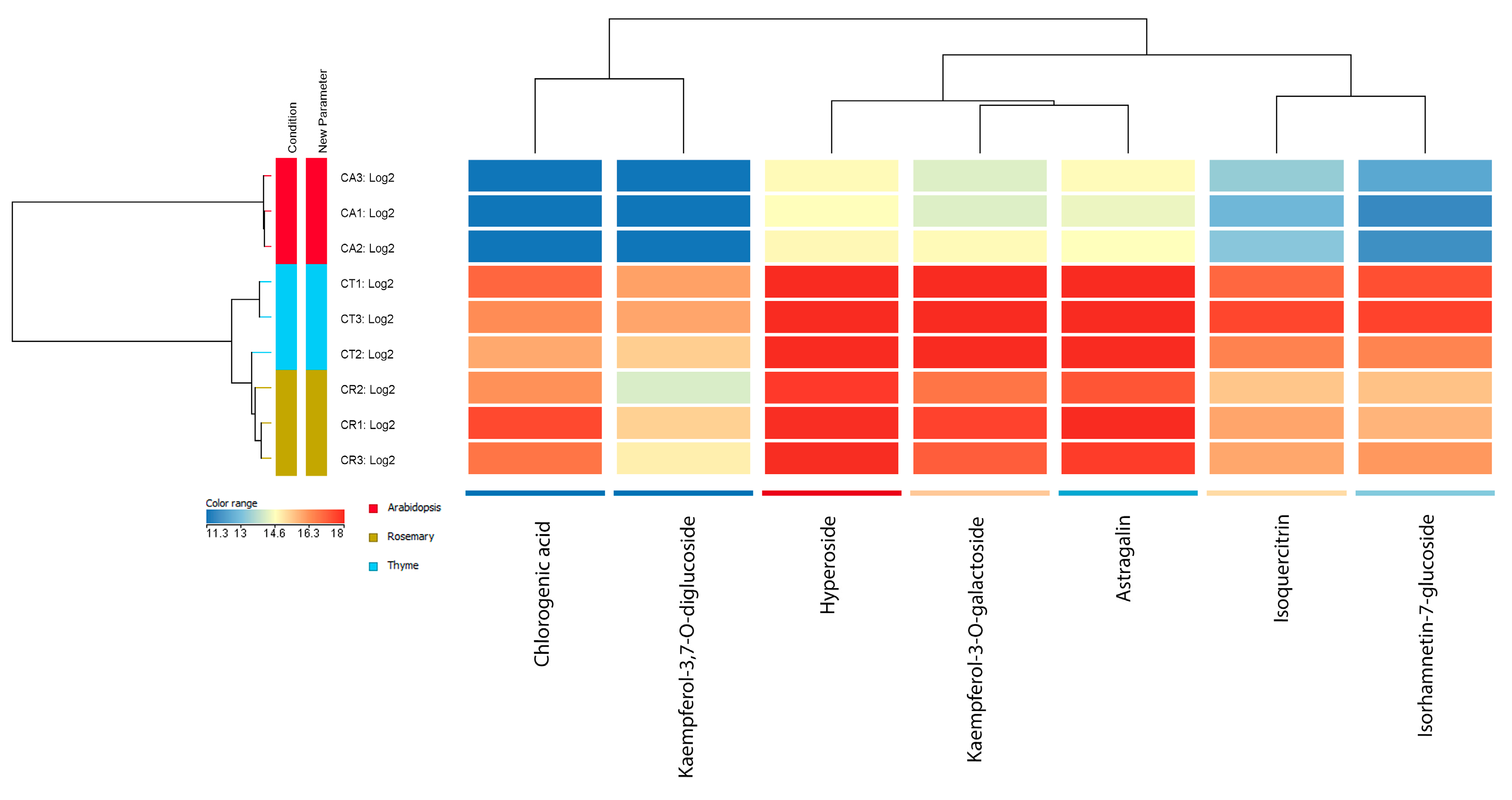
The cluster analysis showed a separation based on the host species (Figure 4). According to clustering by samples, there was greater similarity between Cuscuta plants grown on Th. vulgaris and S. rosmarinus, while those on Arabidopsis were characterized by the greatest differences.
Higher flavonoid compound content was found in Cuscuta using thyme and rosemary as hosts. In addition, chlorogenic acid and kaempferol-3,7-O-diglucoside were absent from the composition of Cuscuta samples grown on Arabidopsis. A comparison between thyme and rosemary host samples showed that, on average, more flavonoid compounds (kaempferol-3,7-O-diglucoside, hyperoside, kaempferol-3-O-galactoside, astragalin, isoquercitrin, and isorhamnetin-7-glucoside) were present in the samples grown on thyme, whereas, in the presence of rosemary as host, higher average content for the hydroxycaffeic acid derivative chlorogenic acid was observed (Figure 4). These results indicate the differences in flavonoid composition even when comparing laboratory-grown C. campestris plants infecting hosts of the same family (Lamiaceae). This suggests the existence of much greater diversity and variation in the flavonoid profile of wild populations of Cuscuta. Host effects in wild populations are much more difficult to study because of the large number of hosts and the effects of multiple environmental factors that cannot be controlled.

4. Discussion

The relationship between antioxidant activity and polyphenolic concentration in plant extracts has been widely documented in the literature. Jafari et al. [31] specifically investigated this correlation in methanolic extracts of the aerial parts of three Cuscuta species. Two of these, C. campestris and C. monogyna, were included in the present study. Their analysis, using HPLC, revealed significant variations in phenolic retention, detecting approximately 16 metabolites, including gallic acid, catechin, caffeic acid, quercetin, coumarin, carvacrol, and vanillin. Among the species studied, C. monogyna exhibited the highest concentration of phenolic compounds (49.59 mg g−1 GA equivalents), followed by C. campestris (~40 mg g−1). This study also assessed the antioxidant activity of the extracts, with C. monogyna again demonstrating the highest antioxidant potential [31]. Our experimental results align with this, most notably in C. epithymum, which exhibited both the highest total polyphenolic content and the greatest antioxidant potential. A notable discrepancy was observed in C. europaea, which demonstrated strong antioxidant potential despite its low polyphenolic content. Although most studies relate the antioxidant potential in Cuscuta extracts to polyphenols, and flavonoids in particular [14,31], other phytochemicals, like carotenoids [32], may also contribute to this [33].
Given the parasitic nature of Cuscuta species and their nonspecific relationships with host plants, researchers have increasingly focused on characterizing plant–plant metabolic exchanges. Kumar et al. conducted a metabolic profiling study on C. campestris grown on three different host plants, revealing significant variations in both primary and secondary metabolite levels. The highest polyphenolic concentrations were detected in the haustoria—the specialized structures facilitating host contact—originating from the parasite’s stem [34]. Similarly, analysis of Cuscuta–host interactions through metabolomic and transcriptomic analyses indicated that 72 h after infection, phenolic acids were among the most abundant metabolite groups exchanged between the host and parasite [35].
Thyme (Thymus vulgaris) and rosemary (Salvia rosmarinus) are well-known medicinal plants with strong antioxidant, antibacterial, and anti-inflammatory properties, widely used in traditional medicine. Their health benefits are primarily attributed to their high polyphenolic content. Thyme contains flavonoids such as apigenin and luteolin in various glycoside forms, while rosemary is rich in flavonoids, including flavones (e.g., diosmin), flavonols (e.g., kaempferol, quercetin, and their glycosides), and rosemary-specific compounds like rosmarinic acid [36,37]. In contrast to the diverse polyphenolic and volatile compound profiles of thyme and rosemary, the model plant Arabidopsis thaliana has a simpler phenolic composition, primarily consisting of quercetin, kaempferol, and their glycosides [38]. In alignment with this, the extracts of C. campestris, parasitizing the two aromatic plants, showed much higher polyphenolic content and respective antioxidant activity. These results indicate that the parasitic plant not only has its own polyphenolic synthesis, as shown in the Arabidopsis host, but also obtains polyphenolics from aromatic plants as hosts. So far, it is hard to say how efficient the secondary metabolism in these parasites is. Further, flavonoids obtained from the host plant may undergo additional modifications. For example, glycosylation and oxidation have been observed in C. campestris and C. kotchiana, leading to the formation of metabolites distinct from those in the host plant [15].
Further studies have explored how host plant species influence the bioactive compound profile of C. campestris. When grown on herbaceous hosts, it exhibited higher concentrations of bioactive compounds such as flavonoids, phytosterols, and polyphenols. Among the tested hosts, thyme supported the highest levels of bergenin and total phenols, and its associated Cuscuta extract displayed some of the strongest antioxidant activities [39]. A similar study on C. reflexa showed host-dependent variations in rutin, quercetin, catechin, gallic acid, and vanillic acid concentrations [40]. Apparently, in laboratory conditions, as in our results, the diversity and content of flavonoids are much lower than in naturally grown populations. However, it was still demonstrated that the flavonoid profile of these parasitic plants was strongly affected by the host species, and even within the same family (Lamiaceae in this case), different hosts led to differences in the parasite.
Overall, members of the genus Cuscuta are generalists, meaning they have a broad host range from a taxonomical perspective [29], but they also infect multiple hosts simultaneously [41]. This not only ensures adequate nutrient flow but also the transfer of molecules with diverse functions. As previously established [42], the haustoria enable intense macromolecular exchange. By forming an extensive network on infected plants, a single dodder individual may benefit from receiving key signals of potential threats like herbivores [43] and may acquire defensive molecules. This is clearly a strong adaptive advantage. Considering flavonoids, our previous results showed a species-specific profile, somehow affected by the hosts [27]. In the present study, it was found that in C. campestris, the role of the host mainly involves the relative abundance of individual compounds and the overall quantity of polyphenolics, rather than dramatic differences in the profile. However, when grown on a single host as in this case, the diversity of flavonoids seems to be much lower than in samples collected in the wild from multiple hosts [27]. Especially for dodders, the accumulation of these compounds may be of crucial importance due to their antioxidant and photoprotective role in the plant kingdom [44]. Along with the unique carotenoid cycle [32], they would be crucial because of the direct exposure to light of this parasite.

5. Conclusions

The present study demonstrated that parasitic plants of the genus Cuscuta possess high polyphenolic content and antioxidant potential. These features are species-dependent but also strongly depend on the host species, as some host plant species further enhance the polyphenolic content of the parasite. While growing equally well on the flavonoid-poor model host Arabidopsis thaliana and the aromatic hosts—thyme and rosemary—quantitatively, the number of polyphenols was much higher in the parasite when parasitizing the later hosts. This is also evidence that these parasites do not rely on their hosts only for water and nutrients but also on protective molecules, such as flavonoids, which are important in antioxidant defense and photoprotection. Their ability to simultaneously parasitize individuals from different taxonomic groups may be an important feature for acquiring compounds with diverse structures and functions, enabling metabolic plasticity and better adaptability to environmental factors. However, a more thorough study, encompassing additional secondary metabolites, along with more accurate quantitative analyses, would further help us understand the chemical basis of the antioxidative potential of Cuscuta extracts. Another important question not addressed here is the effect of Cuscuta parasitism on the flavonoid (and, overall, secondary metabolite) content in the host plants, which would further contribute to understanding the phenomenon of plant-to-plant parasitism.

Supplementary Materials

The following supporting information can be downloaded at https://www.mdpi.com/article/10.3390/antiox14070761/s1.

Author Contributions

Conceptualization, D.T., K.R. and L.Z.; methodology, K.R. and T.Z.; software, K.R. and T.Z.; formal analysis, V.L., B.C., K.R. and T.Z.; investigation, V.L., B.C., K.P., A.T. and T.Z.; writing—original draft preparation, D.T. and L.Z.; writing—review and editing, K.R., K.P. and A.T.; visualization, B.C. and K.R.; supervision, L.Z.; funding acquisition, L.Z. All authors have read and agreed to the published version of the manuscript.

Funding

This study was financed by the European Union—NextGenerationEU through the National Recovery and Resilience Plan of the Republic of Bulgaria, project No BG-RRP-2.004-0008, and the Center of Competence “Sustainable Utilization of Bio-resources and Waste of Medicinal and Aromatic Plants for Innovative Bioactive Products” (BIORESOURCES BG) project BG16RFPR002-1.014-0001, funded by the program “Research, Innovation and Digitization for Smart Transformation” 2021–2027, co-funded by the EU.

Institutional Review Board Statement

Not applicable.

Informed Consent Statement

Not applicable.

Data Availability Statement

The data are contained within the article or Supplementary Materials.

Acknowledgments

We are thankful to Rosen Tsonev, Ivan Traykov, and George Kunev for providing us with some of the samples.

Conflicts of Interest

The authors declare no conflicts of interest.

Abbreviations

The following abbreviations are used in this manuscript: ABTS—2,2′-azino-bis(3-ethylbenzothiazoline-6-sulfonic acid; ANOVA—analysis of variance; dH2O—distilled water; ESI—electrospray ionization; GA—gallic acid; HPLC—high-performance liquid chromatography; MS—mass spectrometry; PDA—photodiode array; SEM—standard error of the mean.

References

  1. Těšitel, J. Functional biology of parasitic plants: A review. Plant Ecol. Evol. 2016, 149, 5–20. [Google Scholar] [CrossRef]
  2. Parker, C. Parasitic weeds: A world challenge. Weed Sci. 2012, 60, 269–276. [Google Scholar] [CrossRef]
  3. Press, M.C.; Phoenix, G.K. Impacts of parasitic plants on natural communities. New Phytol. 2005, 166, 737–751. [Google Scholar] [CrossRef] [PubMed]
  4. Těšitel, J.; Li, A.-R.; Knotková, K.; McLellan, R.; Bandaranayake, P.C.; Watson, D.M. The bright side of parasitic plants: What are they good for? Plant Physiol. 2021, 185, 1309–1324. [Google Scholar] [CrossRef] [PubMed]
  5. Shi, R.; Zhang, C.; Gong, X.; Yang, M.; Ji, M.; Jiang, L.; Leonti, M.; Yao, R.; Li, M. The genus Orobanche as food and medicine: An ethnopharmacological review. J. Ethnopharmacol. 2020, 263, 113154. [Google Scholar] [CrossRef]
  6. Kleszken, E.; Timar, A.V.; Memete, A.R.; Miere, F.; Vicas, S.I. On overview of bioactive compounds, biological and pharmacological effects of mistletoe (Viscum Album L). Pharmacophore 2022, 13, 10–26. [Google Scholar] [CrossRef]
  7. Adesina, S.K.; Illoh, H.; Johnny, I.I.; Jacobs, I.E. African mistletoes (Loranthaceae); ethnopharmacology, chemistry and medicinal values: An update. Afr. J. Tradit. Complement. Altern. Med. 2013, 10, 161–170. [Google Scholar] [CrossRef]
  8. Cui, Z.; Guo, Z.; Miao, J.; Wang, Z.; Li, Q.; Chai, X.; Li, M. The genus Cynomorium in China: An ethnopharmacological and phytochemical review. J. Ethnopharmacol. 2013, 147, 1–15. [Google Scholar] [CrossRef]
  9. Puttipan, R.; Okonogi, S. Antioxidant activity of Rafflesia kerrii flower extract. Drug Discov. Ther. 2014, 8, 18–24. [Google Scholar] [CrossRef]
  10. Jhu, M.-Y.; Sinha, N.R. Cuscuta species: Model organisms for haustorium development in stem holoparasitic plants. Front. Plant Sci. 2022, 13, 1086384. [Google Scholar] [CrossRef]
  11. Costea, M.; Stefanović, S. Cuscuta jepsonii (Convolvulaceae): An invasive weed or an extinct endemic? Am. J. Bot. 2009, 96, 1744–1750. [Google Scholar] [CrossRef]
  12. Ye, M.; Yan, Y.; Guo, D.a. Characterization of phenolic compounds in the Chinese herbal drug Tu-Si-Zi by liquid chromatography coupled to electrospray ionization mass spectrometry. Rapid Commun. Mass. Spectrom. Int. J. Devoted Rapid Dissem. Up Minute Res. Mass. Spectrom. 2005, 19, 1469–1484. [Google Scholar] [CrossRef] [PubMed]
  13. Ahmad, A.; Tandon, S.; Xuan, T.D.; Nooreen, Z. A Review on Phytoconstituents and Biological activities of Cuscuta species. Biomed. Pharmacother. 2017, 92, 772–795. [Google Scholar] [CrossRef] [PubMed]
  14. Yen, F.-L.; Wu, T.-H.; Lin, L.-T.; Cham, T.-M.; Lin, C.-C. Concordance between antioxidant activities and flavonol contents in different extracts and fractions of Cuscuta chinensis. Food Chem. 2008, 108, 455–462. [Google Scholar] [CrossRef] [PubMed]
  15. Flores-Sánchez, I.J.; Garza-Ortiz, A. Is there a secondary/specialized metabolism in the genus Cuscuta and which is the role of the host plant? Phytochem. Rev. 2019, 18, 1299–1335. [Google Scholar] [CrossRef]
  16. Assyov, B.; Petrova, A. Conspectus of the Vascular Plants in Bulgaria; Bulgarian Biodiversity Foundation: Sofia, Bulgaria, 2012; pp. 1–489. [Google Scholar]
  17. Stoyanov, K.; Raycheva, T.; Cheschmedzhiev, I. Key to the Native and Foreign Vascular Plants in Bulgaria; Agricultural University Plovdiv Academic Press: Plovdiv, Bulgaria, 2021. [Google Scholar]
  18. Dimitrova, V.; Tashev, A. Medicinal Plants of Bulgaria. Curr. Perspect. Med. Aromat. Plants 2019, 2, 29–39. [Google Scholar] [CrossRef]
  19. Stojanov, N.; Kitanov, B. Wild Useful Plants in Bulgaria; Bulgarian Academy of Sciences: Sofia, Bulgaria, 1960. [Google Scholar]
  20. Zhelev, Z.D.; Stanilova, S.A.; Carpenter, B.G. Isolation, Partial Characterization, and Complement-Inhibiting Activity of a New Glycoprotein from Cuscuta europea. Biochem. Biophys. Res. Commun. 1994, 202, 186–194. [Google Scholar] [CrossRef]
  21. Perveen, S.; Bukhari, I.H.; Kousar, S.; Rehman, J. Antimicrobial, antioxidant and minerals evaluation of Cuscuta europea and Cuscuta reflexa collected from different hosts and exploring their role as functional attribute. Int. Res. J. Pharm. Appl. Sci. 2013, 3, 43–49. [Google Scholar]
  22. Chabra, A.; Monadi, T.; Azadbakht, M.; Haerizadeh, S.I. Ethnopharmacology of Cuscuta epithymum: A comprehensive review on ethnobotany, phytochemistry, pharmacology and toxicity. J. Ethnopharmacol. 2019, 231, 555–569. [Google Scholar] [CrossRef]
  23. Teofanova, D.; Lozanova, Y.; Lambovska, K.; Pachedjieva, K.; Tosheva, A.; Odjakova, M.; Zagorchev, L. Cuscuta spp. populations as potential reservoirs and vectors of four plant viruses. Phytoparasitica 2022, 50, 555–566. [Google Scholar] [CrossRef]
  24. Singleton, V.L.; Orthofer, R.; Lamuela-Raventós, R.M. Analysis of total phenols and other oxidation substrates and antioxidants by means of folin-ciocalteu reagent. In Methods in Enzymology; Elsevier: Amsterdam, The Netherlands, 1999; Volume 299, pp. 152–178. [Google Scholar]
  25. Thitilertdecha, N.; Rakariyatham, N. Phenolic content and free radical scavenging activities in rambutan during fruit maturation. Sci. Hortic. 2011, 129, 247–252. [Google Scholar] [CrossRef]
  26. Re, R.; Pellegrini, N.; Proteggente, A.; Pannala, A.; Yang, M.; Rice-Evans, C. Antioxidant activity applying an improved ABTS radical cation decolorization assay. Free Radic. Biol. Med. 1999, 26, 1231–1237. [Google Scholar] [CrossRef] [PubMed]
  27. Chakarova, B.; Zagorchev, L.; Pachedjieva, K.; Tosheva, A.; Zagorcheva, T.; Rusanov, K.; Teofanova, D. Analysis of Variations in the Flavonoid Profiles of Cuscuta campestris and Cuscuta epithymum in Bulgaria as a Potential Chemotaxonomical Marker. Plants 2025, 14, 1220. [Google Scholar] [CrossRef]
  28. Shahidi, F.; Ambigaipalan, P. Phenolics and polyphenolics in foods, beverages and spices: Antioxidant activity and health effects—A review. J. Funct. Foods 2015, 18, 820–897. [Google Scholar]
  29. Barath, K.; Csiky, J. Host range and host choice of Cuscuta species in Hungary. Acta Bot. Croat. 2012, 71, 215–227. [Google Scholar] [CrossRef]
  30. Zagorchev, L.I.; Petrova, V.P.; Albanova, I.; Georgieva, K.P.; Sarić-Krsmanović, M.; Muscolo, A.; Teofanova, D.R. Salinity modulates crop plants suitability as hosts for Cuscuta campestris parasitism. J. Saudi Soc. Agric. Sci. 2022, 21, 324–330. [Google Scholar] [CrossRef]
  31. Jafari, E.; Bahmanzadegan, A.; Ghanbarian, G.; Rowshan, V. Antioxidant activity and total phenolic content from aerial parts of three Cuscuta species. Anal. Chem. Lett. 2015, 5, 377–384. [Google Scholar] [CrossRef]
  32. Snyder, A.M.; Clark, B.M.; Bungard, R.A. Light-dependent conversion of carotenoids in the parasitic angiosperm Cuscuta reflexa L. Plant Cell Environ. 2005, 28, 1326–1333. [Google Scholar] [CrossRef]
  33. Young, A.J.; Lowe, G.L. Carotenoids—Antioxidant properties. Antioxidants 2018, 7, 28. [Google Scholar] [CrossRef]
  34. Kumar, K.; Amir, R. The effect of a host on the primary metabolic profiling of Cuscuta campestris’ main organs, haustoria, stem and flower. Plants 2021, 10, 2098. [Google Scholar] [CrossRef]
  35. Guo, C.; Qin, L.; Ma, Y.; Qin, J. Integrated metabolomic and transcriptomic analyses of the parasitic plant Cuscuta japonica Choisy on host and non-host plants. BMC Plant Biol. 2022, 22, 393. [Google Scholar] [CrossRef] [PubMed]
  36. Mena, P.; Cirlini, M.; Tassotti, M.; Herrlinger, K.A.; Dall’Asta, C.; Del Rio, D. Phytochemical profiling of flavonoids, phenolic acids, terpenoids, and volatile fraction of a rosemary (Rosmarinus officinalis L.) extract. Molecules 2016, 21, 1576. [Google Scholar] [CrossRef] [PubMed]
  37. Hammoudi Halat, D.; Krayem, M.; Khaled, S.; Younes, S. A focused insight into thyme: Biological, chemical, and therapeutic properties of an indigenous Mediterranean herb. Nutrients 2022, 14, 2104. [Google Scholar] [CrossRef] [PubMed]
  38. Saito, K.; Yonekura-Sakakibara, K.; Nakabayashi, R.; Higashi, Y.; Yamazaki, M.; Tohge, T.; Fernie, A.R. The flavonoid biosynthetic pathway in Arabidopsis: Structural and genetic diversity. Plant Physiol. Biochem. 2013, 72, 21–34. [Google Scholar] [CrossRef]
  39. Ramezan, D.; Farrokhzad, Y.; Zargar, M.; Stybayev, G.; Kipshakbayeva, G.; Baitelenova, A. An Insight into Cuscuta campestris as a Medicinal Plant: Phytochemical Variation of Cuscuta campestris with Various Host Plants. Agriculture 2023, 13, 770. [Google Scholar] [CrossRef]
  40. Tanruean, K.; Poolprasert, P.; Kumla, J.; Suwannarach, N.; Lumyong, S. Bioactive compounds content and their biological properties of acetone extract of Cuscuta reflexa Roxb. grown on various host plants. Nat. Prod. Res. 2019, 33, 544–547. [Google Scholar] [CrossRef]
  41. Kaiser, B.; Vogg, G.; Fürst, U.B.; Albert, M. Parasitic plants of the genus Cuscuta and their interaction with susceptible and resistant host plants. Front. Plant Sci. 2015, 6, 45. [Google Scholar] [CrossRef]
  42. Kim, G.; Westwood, J.H. Macromolecule exchange in Cuscuta–host plant interactions. Curr. Opin. Plant Biol. 2015, 26, 20–25. [Google Scholar] [CrossRef]
  43. Hettenhausen, C.; Li, J.; Zhuang, H.; Sun, H.; Xu, Y.; Qi, J.; Zhang, J.; Lei, Y.; Qin, Y.; Sun, G. Stem parasitic plant Cuscuta australis (dodder) transfers herbivory-induced signals among plants. Proc. Natl. Acad. Sci. USA 2017, 114, E6703–E6709. [Google Scholar] [CrossRef]
  44. Agati, G.; Brunetti, C.; Di Ferdinando, M.; Ferrini, F.; Pollastri, S.; Tattini, M. Functional roles of flavonoids in photoprotection: New evidence, lessons from the past. Plant Physiol. Biochem. 2013, 72, 35–45. [Google Scholar] [CrossRef]
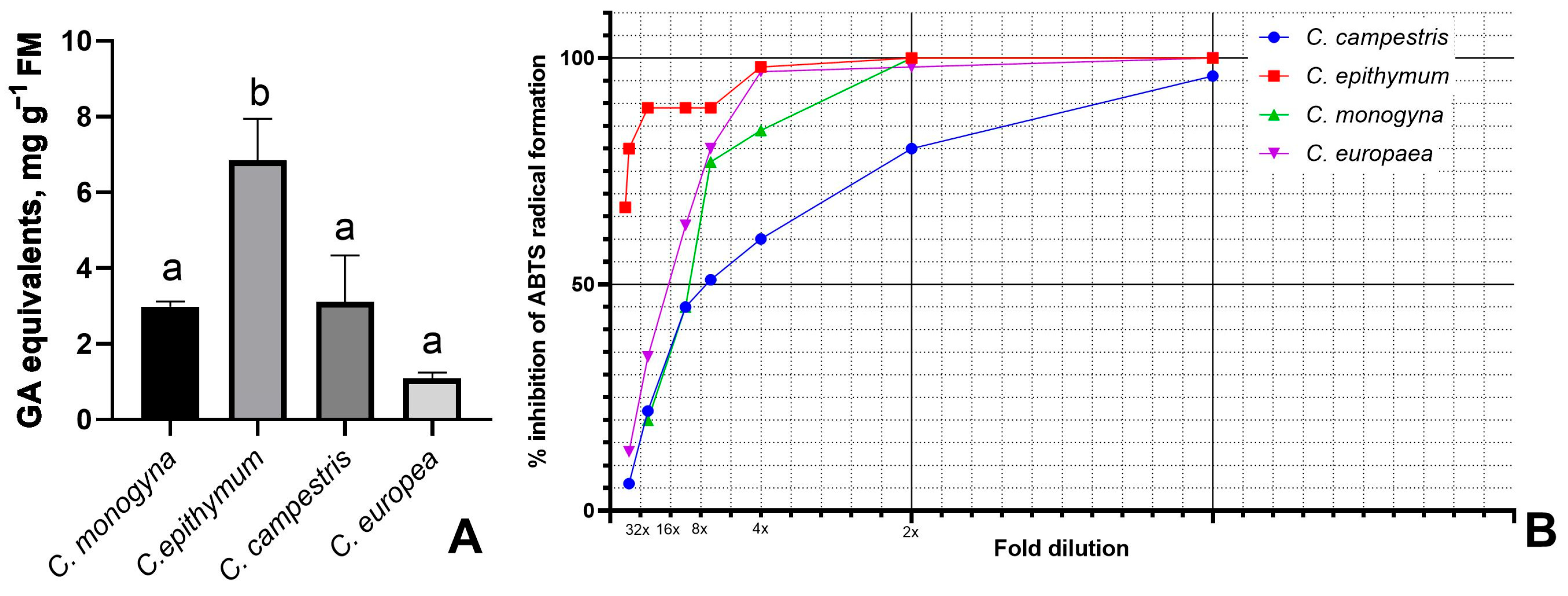
Figure 1. (A) Total polyphenolics in four Cuscuta species: mean values of different numbers of samples from wild populations ± SEM. Different letters indicate significant differences at p ≤ 0.05. (B) ABTS radical scavenging activity of 100% (v/v) methanolic extracts at different dilutions.
Antioxidants 14 00761 g001
Figure 2. (A) Total polyphenolics of Cuscuta campestris and its host species, mean values ± SEM (n = 3). Different letters indicate significant differences at p ≤ 0.05. (B) ABTS radical scavenging activity of 100% (v/v) methanolic extracts at different dilutions.
Antioxidants 14 00761 g002
Figure 3. HPLC-PDA chromatograms of C. campestris grown on Arabidopsis thaliana (A), Thymus vulgaris (B), and Salvia rosmarinus (C). Numbers correspond to compounds in Table 1.
Antioxidants 14 00761 g003
Figure 4. Hierarchical clustering of C. campestris samples grown on different hosts and the 7 flavonoid compounds. The dendrogram on the left shows the separation of samples into three main groups according to the host. The dendrogram at the top shows the relationship of the 7 compounds that varied significantly between samples based on their content in the samples.
Antioxidants 14 00761 g004
Table 1. Compounds, annotated in Cuscuta campestris by HPLC-MS. tR—retention time. [M-H] m/z—mass-to-charge ratio of the molecular ion in negative ion mode.
PeaktR (min)Identity[M-H]
m/z
13.15Chlorogenic acid353.0858
24.85Kaempferol-3,7-O-diglucoside609.1439
38.59Quercetin-3-O-galactoside
(Hyperoside)
463.0853
48.94Kaempferol-3-O-galactoside447.0902
510.17Quercetin-3-O-glucoside (Isoquercitrin)463.0852
610.36Astragalin447.0902
710.66Isorhamnetin-7-glucoside477.1005
Disclaimer/Publisher’s Note: The statements, opinions and data contained in all publications are solely those of the individual author(s) and contributor(s) and not of MDPI and/or the editor(s). MDPI and/or the editor(s) disclaim responsibility for any injury to people or property resulting from any ideas, methods, instructions or products referred to in the content.