Article

Phytochemical Profile and Antioxidant Activity of the Tuber and Peel of Pachyrhizus erosus

Institute of Biochemistry and Molecular Biology, Hunan Provincial Key Laboratory for Special Pathogens Prevention and Control, Hengyang Medical College, University of South China, Hengyang 421001, China
*
Author to whom correspondence should be addressed.
Antioxidants 2025, 14(4), 416; https://doi.org/10.3390/antiox14040416
Submission received: 11 February 2025 / Revised: 20 March 2025 / Accepted: 26 March 2025 / Published: 31 March 2025

Abstract

:
This study systematically investigated the antioxidant activities and phytochemical profiles of petroleum ether (PE), ethyl acetate (EtOAc), n-butanol (n-BuOH) and aqueous fractions of edible (tubers) and non-edible portions (peels) of Pachyrhizus erosus. The results showed that both the tubers and peels from P. erosus were rich in polyphenols and flavonoids, whereas the EtOAc fraction of peels had the highest polyphenol content, and the PE fraction of peels had the highest total flavonoid content. ABTS, DPPH, and FRAP assays revealed that both the EtOAc fraction of tubers and peels from P. erosus showed significant antioxidant activity, whereas the EtOAc fraction of peels possessed better antioxidant activity than that of tubers. UPLC-Orbitrap-MS/MS analysis indicated that thirty compounds were identified from the EtOAc fractions of peels and tubers, including twenty-one flavonoids, six phenolics, two coumarins, and one lignan, some of which have previously been revealed to display significant antioxidant and anti-inflammatory effects via the Nrf2-Keap1 and NF-κB signaling pathways. These findings provide robust scientific evidence for the health-promoting properties and pharmaceutical potential of P. erosus, and its non-edible portion (peels) has great potential for use as a natural antioxidant in the food, cosmetic, and pharmaceutical industries.

1. Introduction

Oxidative stress is caused by excessive free radicals, which play a crucial role in the pathogenesis of various chronic diseases, such as cardiovascular diseases, atherosclerosis, metabolic syndrome, diabetes, neurodegenerative disorders, and cancer [1]. Many antioxidants commonly exist in fruits and vegetables, which can effectively neutralize free radicals, inhibit oxidative enzymes, and chelate metals, thereby preventing oxidative stress-related damage [2].
In recent years, increasingly more attention has been paid to the health-promoting effects of natural antioxidants. Numerous epidemiological studies and clinical trials have demonstrated that diets rich in natural antioxidants can protect the human body from oxidative stress and reduce the incidence of chronic diseases (e.g., cardiovascular diseases, cancer) [3]. Therefore, enhancing the intake of foods containing high natural antioxidants is vital for maintaining overall health and reducing the risk of chronic diseases.
Pachyrhizus erosus, a perennial plant from the Fabaceae family, is also known as bang kuang (China), jicama (Mexico), Kuzu-imo (Japan), Sankalu (India), pupur dingin (Indonesia), and yam bean (the United States) [4]. Native to Mexico and Central America, P. erosus is now widely cultivated in tropical and sub-tropical regions globally, including China, the United States, Brazil, Philippines, Indonesia, Thailand, India, and some African countries. The tubers of P. erosus are rich in various nutrients, proteins, carbohydrates, polysaccharides (inulin), vitamins, and minerals, and are low in calories and abundant in dietary fiber, making them one of the top ten health foods globally [5]. In addition, the tubers of P. erosus have also been traditionally utilized for medical applications due to their diverse pharmacological activities [6].
Previous phytochemical studies have reported about 50 compounds that have been isolated and identified mainly from the seeds, tubers, and leaves of P. erosus, including flavonoids, triterpenes, phenolics, saponins, organic acids, and fatty acids, which contribute to the diverse biological activities of P. erosus, such as antioxidant, anti-aging, anti-cancer, anti-diabetes, immunomodulatory, antiviral, antiosteoporosis, skin whitening, gastroprotective, and anti-obesity effects [6].
Early studies demonstrated that the tubers of P. erosus exhibited good antioxidant properties. For instance, Choi revealed that the ethyl acetate extract from the tubers from P. erosus exhibited the highest antioxidant activity in both DPPH and ABTS radical scavenging assays [5]; Bhanja found that the inulin in the tubers of P. erosus showed good antioxidant and in vitro probiotic effects, indicating its role in promoting gut health [7]; Laovachirasuwan indicated that the dichloromethane extract from the tubers from P. erosus exhibited significant anti-melanogenesis and antioxidant effects, positioning it as a promising source of natural antioxidants and anti-melanogenesis agents for use in cosmetic products [8]. Additionally, regarding the antioxidant activity of peels from P. erosus, only Siregar reported that the 70% ethanol extract of peels presented strong DPPH scavenging activity [9]. Despite several studies reporting on the antioxidant activities of tubers and peels from P. erosus, systematic evaluations of their antioxidant effects on a chemical basis have not been fully elucidated.
Thus, this study focused on comparing the total polyphenol content (TPC), total flavonoid content (TFC), and antioxidant activities (DPPH, ABTS, and FRAP assays) of petroleum ether (PE), ethyl acetate (EtOAc), n-butanol (n-BuOH), and aqueous fractions of tubers and peels from P. erosus, as well as on analyzing the chemical compositions of their strongly active fraction using UPLC-Orbitrap-MS/MS. The goal of this study was to elucidate the chemical basis of the antioxidant activity of P. erosus and further explore the potential utilization of the peels of P. erosus which are often discarded. This research will provide a theoretical basis for the application of the peels and tubers of P. erosus as a natural antioxidant in the food, cosmetic, and pharmaceutical industries.

2. Materials and Methods

2.1. Chemicals and Reagents

Folin–Ciocalteu reagent was acquired from Macklin (Shanghai, China). 2,2-diphenyl-1-picrylhydrazyl (DPPH), 2,4,6-tris-(2-pyridyl)-s-triazine (TPTZ), 2,2′-azino-bis (3-ethylbenzothiazoline-6-sulfonic acid) (ABTS), rutin, and gallic acid were obtained from Aladdin Biotechnology Co., Ltd. (Shanghai, China). LC- and MS-grade methanol, acetonitrile, formic acid, and isopropanol were purchased from ANPEL (Shanghai, China). Analytical grade ethanol, methanol, petroleum ether, ethyl acetate, and n-butanol were purchased from Sinopharm Chemical Reagent Co., Ltd. (Beijing, China). Ultrapure water was prepared using a Milli-Q water purification system (Millipore, Bedford, MA, USA).

2.2. Plant Materials and Extract Preparation

P. erosus was collected from Loudi City, Hunan Province. After washing, the peels and tubers of P. erosus were manually separated. The dried peels and tubers (500 g) were then subjected to extraction with 95% ethanol at room temperature, respectively. Subsequently, the samples were rotary evaporated and concentrated to afford the ethanol extracts. The resulting ethanol extracts were suspended in H2O and successively partitioned with petroleum ether, ethyl acetate, and n-butanol to afford a petroleum ether fraction (PE), ethyl acetate fraction (EtOAc), n-butanol fraction (n-BuOH), and aqueous fraction (Aqueous) after condensation to dry material in vacuo. The yields of the four fractions (PE, EtOAc, n-BuOH, and Aqueous) from P. erosus peels were 3.158 g, 3.993 g, 5.521 g, and 47.761 g, respectively. Conversely, the yields of the four fractions (PE, EtOAc, n-BuOH, and Aqueous) from P. erosus tubers were 1.101 g, 0.923 g, 14.892 g, and 102.057 g, respectively.

2.3. Antioxidant Activity Assays

2.3.1. ABTS Radical Scavenging Activity

The ABTS radical scavenging activity was determined using a previously described method [10] with slight modifications. First, we mixed 5 mL ABTS (7.0 mmol/L) with 88 µL K2S2O8 (140 mmol/L) and let the mixture stand in the dark at room temperature for 12–16 h to prepare ABTS working solution. The ABTS solution was diluted with 10 mmol/L phosphate-buffered saline (PBS) to adjust the absorption value to 0.70 ± 0.02 at 734 nm. Then, 200 µL of the ABTS working solution was mixed with 10 µL of the sample in a 96-well microplate. Then, the mixture was reacted for 6 min. The absorbance of the mixture was measured at 405 nm. The ABTS radical scavenging activity was calculated using the following formula: ABTS radical scavenging rate (%) = [1 − (A1 − A2)/A0] × 100%, where A0 is the absorbance of ABTS and DMSO; A1 is the absorbance with the sample reaction solution; and A2 is the absorbance of sample and PBS.

2.3.2. DPPH Radical Scavenging Activity

The DPPH radical scavenging activity was performed using the previous method [11] with slight modifications. Briefly, 20 µL of the sample was mixed with 180 µL of 0.1 mM DPPH methanol solution. After incubation in the dark at room temperature for 30 min, the absorbance was measured at 515 nm using a microplate reader. The DPPH radical scavenging activity was calculated using the following formula: DPPH radical scavenging rate (%) = [1 − (A1 − A2)/A0] × 100%, where A0 is the absorbance of the DPPH without samples, A1 is the absorbance of samples with DPPH, and A2 is the absorbance of samples without DPPH.

2.3.3. Ferric Reducing Antioxidant Power (FRAP) Assay

The FRAP assay was assessed according to the previously method [12] with some modifications. FRAP working solution was prepared with 0.3 M acetate buffer (pH 3.6), and 10 mM 2,4,6-tris-(2-pyridyl)-s-triazine (TPTZ) in 40 mM HCl solution, and 20 mM FeCl3 solution in a 10:1:1 (v/v/v) ratio. 20 µL of sample and 180 µL of FRAP working solution were mixed in a 96-well plate and incubated at 37 °C for 30 min in the dark. The absorbance of the mixture was measured at 593 nm. Ferrous sulfate (FeSO4) solutions (0.6–2.1 mM) were used to construct the standard curve in the same way. The FRAP value of the samples were calculated based on the linear standard curve and expressed as mmol FeSO4 equivalents per gram of sample (mmol/g FeSO4 equivalents).

2.4. Determination of Total Polyphenol and Flavonoid Content

2.4.1. Total Polyphenol Content

The total polyphenol content was determined using the Folin–Ciocalteu method [13] with some modifications. Briefly, 0.1 mL of extract (1 mg/mL) and 0.1 mL of Folin–Ciocalteu reagent (1 N) were mixed and reacted with 2.8 mL of distilled water in a 10 mL brown volumetric flask for 8 min. Then, 2 mL of 7.5% Na2CO3 solution was added and incubated in a water bath at 37 °C for 1 h. The absorbance of the mixture was measured at 765 nm. Gallic acid (0–0.3 mg/mL) was used as the standard to establish the calibration curve. The total polyphenol content of the extracts was expressed as mg of gallic acid equivalents (GAE)/g extract.

2.4.2. Total Flavonoid Content

The total flavonoid content was determined using the aluminum chloride colorimetric method [14] with some modifications. Briefly, 1.0 mL of extract (1 mg/mL) and 0.3 mL of 5% NaNO2 solution were mixed and reacted with 4.0 mL of distilled water in a 10 mL brown volumetric flask for 5 min. Then, 0.3 mL of 10% AlCl3 solution was added and retained in the dark for 6 min. Afterward, 2.0 mL of 1.0 M NaOH solution was added, and then the final volume was made up to 10 mL with distilled water. The absorbance of the mixture was measured at 510 nm. Rutin (0–0.5 mg/mL) was used as the standard to establish the calibration curve. The total flavonoid content of the extracts was expressed as mg of rutin equivalents (RE)/g extract.

2.5. UPLC-Orbitrap-MS/MS Analysis

The ethyl acetate fractions of tubers and peels from P. erosus (10 mg) was dissolved in 10 mL methanol, respectively, then filtered through a 0.22 μm membrane and stored at −20 °C.
The qualitative determination of chemical composition of the ethyl acetate fractions of tubers and peels from P. erosus was conducted using the UPLC-Orbitrap-MS system (UPLC, Vanquish; MS, HFX). Separation was performed on a Waters HSS T3 column (100 mm × 2.1 mm, 1.8 μm) (Waters Corporation, Milford, MA, USA). The column temperature was 40 °C. The mobile phase A was Milli-Q water with 0.1% formic acid and B was acetonitrile with 0.1% formic acid. The gradient program was performed as follows: 0 min, A/B (100:0, v/v); 1 min, A/B (100:0, v/v); 12 min, A/B (5:95, v/v); 13 min, A/B (5:95, v/v); 13.1 min, A/B (100:0, v/v); 17 min, A/B (100:0, v/v). The injection volume was set to 2.0 μL, and the flow rate was set at 0.3 mL/min.
HRMS data were recorded on a Q Exactive HFX Hybrid Quadrupole Orbitrap mass spectrometer equipped with a heated ESI source (Thermo Fisher Scientific, Waltham, MA, USA) utilizing the Full-ms-ddMS2 acquisition methods. The ESI source parameters were set as follows: sheath gas pressure, 40 arb; aux gas pressure, 10 arb; spray voltage, +3000 V/−2800 V; temperature, 350 °C; and ion transport tube temperature, 320 °C. The scanning range of the primary mass spectrometry (m/z range) was 70–1050 Da, with a primary resolution of 70,000 and secondary resolution of 17,500. Thermo scientific Xcalibur 4.1 software was applied to acquire and deal with the original data.

2.6. Statistical Analysis

Experimental data are expressed as the mean ± standard deviation (SD) of three independent experiments. Statistical analysis of experimental data by t-test or one-way analysis of variance (ANOVA) was performed using GraphPad Prism 9.5.1 software (San Diego, CA, USA), with p ≤ 0.05 considered significant.

3. Results

3.1. Antioxidant Activity

Oxidative stress is associated with many chronic diseases and aging [15]. Antioxidants can react differently with various free radicals or oxidizing agents that are crucial for protecting biological systems from oxidative stress. Generally, one method is not sufficient for predicting the antioxidant activity of plant extracts; thus, it is necessary to use multiple methods, such as DPPH, ABTS radical scavenging and ferric reducing antioxidant power (FRAP) to comprehensively evaluate the antioxidant activity [16,17].

3.1.1. ABTS Radical Scavenging Activity

The ABTS radical scavenging activities of PE, EtOAc, n-BuOH, and aqueous fractions of tubers and peels from P. erosus are shown in Figure 1A,B. As depicted, the inhibition of the ABTS radical scavenging activity of each fraction exhibited a concentration-dependent trend. Among the fractions from the tubers of P. erosus, the EtOAc fraction possessed the greatest ABTS radical scavenging capacity with an IC50 value of 1.126 ± 0.089 mg/mL. Similarly, among the fractions from the peels of P. erosus, the EtOAc fraction also presented the greatest ABTS radical scavenging capacity with an IC50 value of 0.1749 ± 0.017 mg/mL. Comparing the tuber and peel extracts, both peels and tubers showed some ABTS radical scavenging capacity. Furthermore, the ABTS radical scavenging activities of PE, EtOAc, n-BuOH, and the aqueous fractions of peels were noticeably higher than that of the tubers. This trend was especially evident in the EtOAc fraction at a dose of 0.625 mg/mL, which demonstrated that the EtOAc fractions from the peels of P. erosus displayed the most outstanding antioxidant activity.

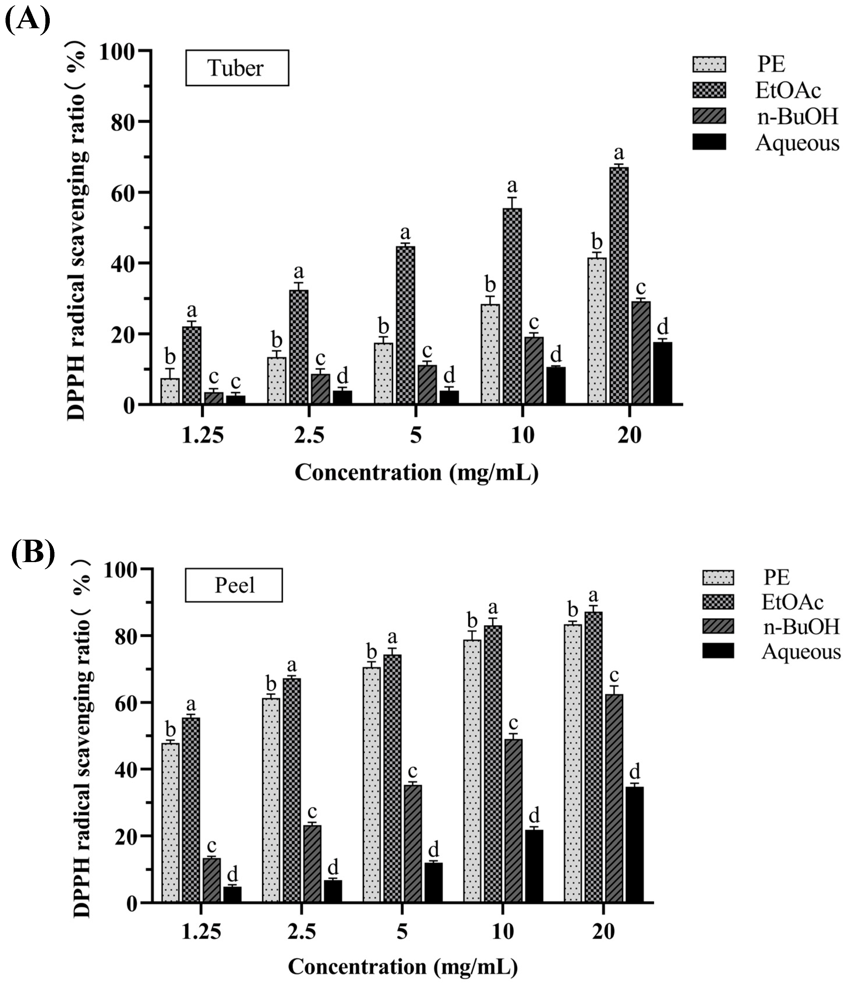
3.1.2. DPPH Radical Scavenging Activity

The DPPH radical scavenging assay is a common method used for evaluating the antioxidant capacity of plant extracts by assessing the reduction in DPPH radicals [18]. Figure 2A,B shows the DPPH radical scavenging activities of PE, EtOAc, n-BuOH, and aqueous fractions of tubers and peels from P. erosus at different concentrations (1.25, 2.5, 5, 10, 20 mg/mL). For all the extracts, the inhibition of the DPPH activity was concentration dependent. Among the fractions of tubers, the EtOAc fraction exhibited the greatest DPPH radical scavenging capacity with an IC50 value of 6.443 ± 0.595 mg/mL. Similarly, the EtOAc fraction of peels also presented the greatest DPPH radical scavenging capacity with an IC50 value of 0.772 ± 0.078 mg/mL. Comparing the tuber and peel extracts, both peels and tubers showed good scavenging effects on DPPH radicals. Particularly in the EtOAc fraction, the antioxidant activity of the peels was significantly higher than that of the tubers.

3.1.3. FRAP Assay

The FRAP (ferric-reducing antioxidant power) assay is a widely used colorimetric method for evaluating the antioxidant effects of various plant extracts [19]. Specifically, this method measures the ability of antioxidants to reduce the colorless Fe (III)-TPTZ complex to the blue-colored Fe (II)-TPTZ complex. The intensity of the blue color, measured spectrophotometrically at 593 nm, correlates with the antioxidant capacity of the substance. The FRAP values of PE, EtOAc, n-BuOH, and aqueous fractions of tubers and peels from P. erosus are shown in Table 1. FeSO4 was used to establish a standard curve and its linear equation was y = 0.4196x + 0.0023, R2 = 0.9997. Among these fractions, the EtOAc fraction of peels showed the higher FRAP value (12.56 ± 1.720 mM Fe (II)/g extract) than that of the EtOAc fraction of tubers (4.113 ± 1.158 mM Fe (II)/g extract), indicating that the peels exhibited the strongest Fe3+-reducing power.

3.2. Determination of Total Polyphenol and Flavonoid Content

Total polyphenol compounds and flavonoids are widely distributed in plants, which are significantly positively correlated with antioxidant activity. For example, plants (sweet chestnut and sweet rowan) contain higher levels of these compounds, which are associated with stronger antioxidant activity [20,21]. Figure 3A,B shows the total polyphenol and flavonoid contents of the PE, EtOAc, n-BuOH, and aqueous fractions of tubers and peels from P. erosus. The results indicated that the EtOAc fraction of peels had the highest polyphenol content (156.2 ± 3.282 mg GAE/g extract), while the PE fraction of peels had the highest total flavonoid content (151.7 ± 4.510 mg RE/g extract). Notably, within the same fraction, the total polyphenol content in the peels was significantly higher than that in the tubers (p < 0.05). In contrast, the n-BuOH fraction of tuber had higher total flavonoid content than that of peel (p < 0.05). The peels of P. erosus contain higher amounts of total polyphenol sand total flavonoids, which could be attributed to its prolonged exposure to environmental factors and direct contact with soil, necessitating more polyphenols and flavonoids to counteract environmental stresses and pest attacks. Besides, these high content polyphenol and flavonoids may play an important role in antioxidant activity of peels from P. erosus (Supplementary Materials, Figure S32).

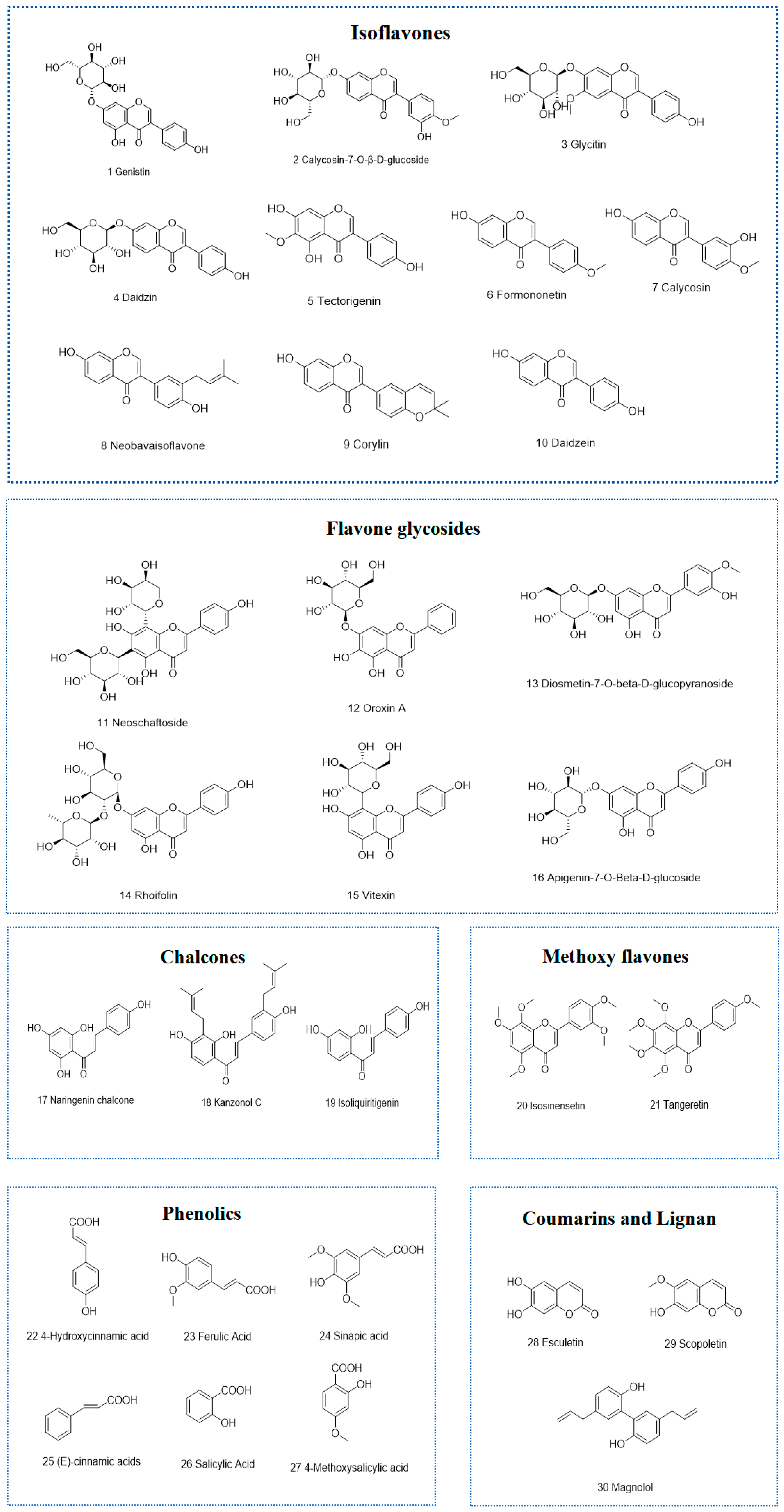
3.3. Identification of Chemical Constituents of the Strong Active Ethyl Acetate Fractions of Tubers and Peels from P. erosus

To date, several studies have investigated the antioxidant activities of tubers and peels from P. erosus, but systematic evaluations on the chemical basis of their antioxidant effect have not yet been fully carried out. In this study, using antioxidant activity screening, it was found that the EtOAc fractions of peels and tubers from P. erosus showed the best antioxidant activity. The chemical compositions of the EtOAc fractions of peels and tubers were characterized by UPLC-Orbitrap-MS/MS.
Based on retention time, molecular formula, detected negative or positive ionization modes, mass error (<3 ppm), mass-to-charge ratio, and the MS/MS fragment ions, thirty compounds were identified from the strongly active EtOAc fractions of peels and tubers (Figure 4 and Table 2), including twenty-one flavonoid compounds (ten isoflavones, six flavonoid glycosides, three chalcones, two methoxy flavones), six phenolic compounds, two coumarins, and one lignan. The total ion chromatograms (TIC) in both positive and negative ionization modes and MS/MS2 spectrum of compounds are shown in the Supplementary Materials (Figures S1–S31 and Table S1).

4. Discussion

Increasing evidence suggests that oxidative stress and inflammation are key factors in many chronic diseases [22]. Natural antioxidants, such as flavonoids, phenolic compounds, and coumarins, are commonly used in daily life, which can alleviate oxidative stress and inflammatory responses, thus improving the prognosis of these chronic diseases. P. erosus belongs to the Fabaceae family, whose tubers are abundant in various nutrients and dietary fiber, and is considered one of the top ten health foods globally [5]. Previous studies demonstrated that the tubers and peels of P. erosus possessed antioxidant activities. However, to date, the chemical basis of their antioxidant effect has still not been fully elucidated. Therefore, this study focused on evaluating the total polyphenol and total flavonoid content, antioxidant activities, and phytochemical profile of PE, EtOAc, n-BuOH, and aqueous fractions of tubers and peels from P. erosu, which aimed to elucidate the chemical basis of the antioxidant activity of P. erosus and further explore the potential utilization of the peels of P. erosus which are often discarded.
The results indicated that the EtOAc fraction of peels had the highest polyphenol content, while the PE fraction of peels had the highest total flavonoid content. Though ABTS, DPPH, and FRAP assays, it was found that both the EtOAc fraction of tubers and peels from P. erosus showed significant antioxidant activity, whereas the EtOAc fraction of peels possessed better antioxidant activity than that of tubers. These findings are consistent with those documented by Choi et al., who established that the EtOAc fraction of the tuber from P. erosus exhibits notable DPPH and ABTS radical scavenging activities [5]. Additionally, Siregar demonstrated that the peel extracts from P. erosus displayed stronger DPPH radical scavenging activity than that of tuber extracts [9]. This discovery further supports our present results, which highlights the superior antioxidant activity of the peel from P. erosus.
To further elucidate the chemical basis of antioxidant activities, UPLC-Orbitrap-MS/MS was employed to analyze the chemical compositions of the EtOAc fractions obtained from both peels and tubers. A total of thirty compounds were identified, including twenty-one flavonoid compounds (ten isoflavones, six flavonoid glycosides, three chalcones, and two methoxy flavones), six phenolic compounds, two coumarins, and ome lignan. In particular, except for five compounds (genistin, daidzin, daidzein, formononetin, and salicylic acid) that were previously reported in the tubers of P. erosus [5,6], the other remaining compounds were found in the tubers of P. erosus for the first time. As far as we know, this is the first time that the phytochemical profiles of peels from P. erosus have been analyzed and studied. Thirty compounds were identified in the peels, which were mainly flavonoids and phenolics. The identification of these compounds not only contributes to a more detailed understanding of the phytochemical profile of P. erosus, but also offers insights into the observed significant antioxidant activity.
Among these compounds, many have been reported to exhibit significant pharmacological activities, including cardioprotective, anti-cancer, antidiabetic, antiepileptic, neuroprotective, gastric ulcer preventive, antidepressant, antiallergic, anti-osteoporosis, antimicrobial, anti-thrombosis, and anticonvulsant effects [23,24,25,26,27,28,29,30,31,32,33,34,35,36,37,38,39,40,41,42,43,44,45,46,47,48,49,50,51,52,53] (Table 3).
Nuclear factor E2-related factor 2 (Nrf2) is a widely present transcription factor, which binds to the antioxidant response element (ARE), and activates a series of antioxidant genes, thereby regulating the expression of various antioxidant enzymes, such as heme oxygenase-1 (HO-1), NAD(P)H quinone dehydrogenase 1 (NQO1), superoxide dismutase (SOD), catalase (CAT), and glutathione peroxidase (GSH-Px). Under homeostasis conditions, Nrf2 is retained in the cytoplasm by binding to its inhibitory protein Keap1. The E3 ubiquitin ligase complex formed by Keap1 and Cullin 3 (CUL3) mediates the ubiquitination and degradation of Nrf2, thus inhibiting its activity. However, under the conditions of oxidative stress, Nrf2 is released from Keap1 and translocated to the nucleus, which activates massive gene expression of antioxidant enzyme to exert an antioxidant effect. This major antioxidant regulatory mechanism has been validated in several of the metabolites we screened. For example, esculetin (28) can activate the ERK signaling pathway and promote Nrf2 phosphorylation and nuclear translocation, thereby upregulating NQO1 expression and significantly alleviating H2O2-induced oxidative damage in C2C12 cells [54]. In a myocardial ischemia–reperfusion injury model, isoliquiritigenin (19) reduces oxidative stress and ferroptosis by promoting Nrf2 nuclear translocation and activating the expression of HO-1, SLC7A11, and GPX4 [55]. Tangeretin (21) maintains Nrf2 activation by inhibiting CUL3-mediated ubiquitination, further emphasizing the central role of Nrf2 in antioxidant defense [56]. Additionally, neobavaisoflavone (8) protects cells from oxidative stress-induced damage via the CRNDE-mediated Nrf2/HO-1 signaling pathway [57].
Furthermore, a growing body of literature has reported that numerous plant metabolites can improve oxidative stress-induced damage by enhancing the activity of antioxidant enzymes (SOD, HO-1, CAT, and GSH-Px), while simultaneously reducing the levels of malondialdehyde (MDA) and other inflammation-related biomarkers. For instance, some metabolites, such as daidzin (4) [58], calycosin (7) [59], magnolol (30) [60], formononetin (6) [61], vitexin (15) [62], and isosinensetin (20) [39], have been demonstrated to upregulate cellular antioxidant enzyme activities and the expression of various antioxidant enzymes, thereby alleviating oxidative stress-induced damage.
The NF-κB signaling pathway also plays a crucial role in the oxidative stress response. As a major intracellular pro-inflammatory transcription factor, NF-κB mediates inflammatory responses by regulating the expression of a series of pro-inflammatory genes. In the canonical pathway, the NF-κB heterodimer p65/p50 binds to the inhibitory protein IκB, maintaining an inactive state. Upon inflammatory stimuli or oxidative stress, the IκB kinase (IKK) complex activates, phosphorylates and degrades IκB, releasing the NF-κB complex (p65/p50), which is translocated to the nucleus to initiate the expression of inflammatory genes. Studies have shown that rhoifolin (14) significantly alleviates ethanol-induced liver damage in mice by inhibiting the TLR4/NF-κB signaling pathway [63]. Additionally, glycitin (3) protects lung tissue from LPS-induced inflammation via inhibiting the TLR4-mediated NF-κB and MAPKs signaling pathways [64].
Collectively, the secondary metabolites that were contained in the ethyl acetate fraction of tubers and peels from P. erosus exhibited significant antioxidant and anti-inflammatory activities by modulating the Nrf2-Keap1 and NF-κB signaling pathways (Figure 5). These results provide scientific evidence for the application of P. erosus extracts in health promotion and drug development. Future studies should explore more natural products with potential applications by integrating the regulatory mechanisms of the Nrf2-Keap1 and NF-κB pathways.

5. Conclusions

This study is the first to systematically study the antioxidant activities and phytochemical profiles of petroleum ether, ethyl acetate, n-butanol, and aqueous fractions of the edible (tubers) and non-edible portions (peels) of P. erosus. Specifically, the ethyl acetate fraction was identified as the most active fraction in both the peels and tubers, and their chemical constituents were analyzed and identified. Thirty potential metabolites were screened, including twenty-one flavonoid compounds (ten isoflavones, six flavonoid glycosides, three chalcones, two methoxy flavones), six phenolic compounds, two coumarins, and one lignan. Some of these metabolites (flavonoids and phenolic compounds) existed in both the tubers and peels, and have been revealed to exert significant antioxidant effects through the Nrf2-Keap1 and NF-κB signaling pathways. Notably, the peels of P. erosus, often considered agricultural waste, showed better antioxidant activity than the tubers in our study. These findings not only provide a reference for the further development and utilization of P. erosus, but also suggest a new direction for the utilization of the peels of P. erosus, indicating its potential as a natural antioxidant in the food, cosmetic, and pharmaceutical industries.

Supplementary Materials

The following supporting information can be downloaded at: https://www.mdpi.com/article/10.3390/antiox14040416/s1, Figure S1: Base peak chromatogram of the EtOAc fraction of peels and tubers from P. erosus; Figures S2–S31: MS/MS2 spectrum of compounds 1–30; Figure S32: PCA scores plot showing the relationship among TFC, TPC, and antioxidant activity (DPPH, ABTS, FRAP). Table S1: 30 compounds screened from the highly active EtOAc fraction of P. erosus peel and tuber according to the scoring.

Author Contributions

Conceptualization, Y.L.; methodology, H.S.; software, J.X.; validation, F.Z.; formal analysis, Y.H.; data curation, X.W.; writing—original draft preparation, J.X.; writing—review and editing, Y.L. and S.T.; project administration, J.X. and S.H.; funding acquisition, Y.L. All authors have read and agreed to the published version of the manuscript.

Funding

This work was financially supported by the Science Fund of Hunan Provincial Education Department (Nos. 24A0286 and 23B0395), the Natural Science Foundation of Hunan Province (No. 2018JJ3433), the Health Research Project of Hunan Provincial Health Commission (No. D202302047808), the Undergraduate Training Programs for Innovation and Entrepreneurship of Hunan Province (No. D202305162258543382), and the Undergraduate Training Programs for In-novation and Entrepreneurship of University of South China (No. 2022X10555110).

Institutional Review Board Statement

Not applicable.

Informed Consent Statement

Not applicable.

Data Availability Statement

Data will be made available on request from the corresponding author.

Conflicts of Interest

The authors declare no conflicts of interest.

References

  1. Shang, Z.X.; Li, M.Q.; Zhang, W.W.; Cai, S.B.; Hu, X.S.; Yi, J.J. Analysis of phenolic compounds in pickled chayote and their effects on antioxidant activities and cell protection. Food Res. Int. 2022, 157, 111325. [Google Scholar] [PubMed]
  2. Lobo, V.; Patil, A.; Phatak, A.; Chandra, N. Free radicals, antioxidants and functional foods: Impact on human health. Pharmacogn. Rev. 2010, 4, 118–126. [Google Scholar] [PubMed]
  3. Wang, X.; Ouyang, Y.Y.; Liu, J.; Zhu, M.M.; Zhao, G.; Bao, W.; Hu, F.B. Fruit and vegetable consumption and mortality from all causes, cardiovascular disease, and cancer: Systematic review and dose-response meta-analysis of prospective cohort studies. BMJ 2014, 349, g4490. [Google Scholar] [PubMed]
  4. Kishore, A.; Patil, R.J.; Singh, A.; Pati, K. Jicama (Pachyrhizus spp.) a nonconventional starch: A review on isolation, composition, structure, properties, modifications and its application. Int. J. Biol. Macromol. 2024, 258, 129095. [Google Scholar]
  5. Choi, M.H.; Yang, S.H.; Lee, Y.J.; Sohn, J.H.; Lee, K.S.; Shin, H.J. Anti-obesity effect of daidzein derived from Pachyrhizus erosus (L.) Urb. extract via PPAR pathway in MDI-induced 3T3-L1 cell line. Cosmetics 2023, 10, 164. [Google Scholar] [CrossRef]
  6. Jaiswal, V.; Chauhan, S.; Lee, H.J. The bioactivity and phytochemicals of Pachyrhizus erosus (L.) Urb.: A multifunctional underutilized crop plant. Antioxidants 2021, 11, 58. [Google Scholar] [CrossRef]
  7. Bhanja, A.; Paikra, S.K.; Sutar, P.P.; Mishra, M. Characterization and identification of inulin from Pachyrhizus erosus and evaluation of its antioxidant and in-vitro prebiotic efficacy. J. Food Sci. Technol. 2023, 60, 328–339. [Google Scholar]
  8. Laovachirasuwan, P.; Phadungkit, M. Total phenolic and flavonoid contents, anti-tyrosinase and antioxidant activities of Pachyrhizus erosus extracts. Pharmacogn. J. 2023, 15, 839–842. [Google Scholar]
  9. Siregar, I.D.; Kusuma, H.S.W.; Widowati, W.; Marpaung, H.H.; Ferdinand, S.; Fachrial, E.; Lister, I.N.E. Antioxidant and antityrosinase activities of ethanolic Pachyrhizuserosus peel and tuber extract. MKB. 2019, 51, 75–81. [Google Scholar]
  10. He, Z.Y.; Tao, Y.D.; Zeng, M.M.; Zhang, S.; Tao, G.J.; Qin, F.; Chen, J. High pressure homogenization processing, thermal treatment and milk matrix affect in vitro bioaccessibility of phenolics in apple, grape and orange juice to different extents. Food Chem. 2016, 200, 107–116. [Google Scholar]
  11. Brand-Williams, W.; Cuvelier, M.E.; Berset, C. Use of a free radical method to evaluate antioxidant activity. LWT-Food Sci. Technol. 1995, 28, 25–30. [Google Scholar] [CrossRef]
  12. Benzie, I.F.F.; Strain, J.J. The ferric reducing ability of plasma (FRAP) as a measure of “antioxidant power”: The FRAP assay. Anal. Biochem. 1996, 239, 70–76. [Google Scholar] [CrossRef] [PubMed]
  13. Xu, J.G.; Tian, C.R.; Hu, Q.P.; Luo, J.Y.; Wang, X.D.; Tian, X.D. Dynamic changes in phenolic compounds and antioxidant activity in oats (Avena nuda L.) during steeping and germination. J. Agric. Food Chem. 2009, 57, 10392–10398. [Google Scholar] [CrossRef] [PubMed]
  14. Baba, S.A.; Malik, S.A. Determination of total phenolic and flavonoid content, antimicrobial and antioxidant activity of a root extract of Arisaema jacquemontii Blume. J. Taibah Univ. Sci. 2015, 9, 449–454. [Google Scholar]
  15. Ou, B.; Hampsch-Woodill, M.; Prior, R.L. Development and validation of an improved oxygen radical absorbance capacity assay using fluorescein as the fluorescent probe. J. Agric. Food Chem. 2001, 49, 4619–4626. [Google Scholar]
  16. Prior, R.L.; Wu, X.L.; Schaich, K. Standardized methods for the determination of antioxidant capacity and phenolics in foods and dietary supplements. J. Agric. Food Chem. 2005, 53, 4290–4302. [Google Scholar] [CrossRef]
  17. Shahidi, F.; Zhong, Y. Measurement of antioxidant activity. J. Funct. Foods 2015, 18, 757–781. [Google Scholar]
  18. Gulcin, İ.; Alwasel, S.H. DPPH radical scavenging assay. Processes 2023, 11, 2248. [Google Scholar] [CrossRef]
  19. Wojtunik-Kulesza, K.A. Approach to optimization of FRAP methodology for studies based on selected monoterpenes. Molecules 2020, 25, 5267. [Google Scholar]
  20. Martínez, S.; Fuentes, C.; Carballo, J. Antioxidant activity, total phenolic content and total flavonoid content in sweet chestnut (Castanea sativa Mill.) cultivars grown in Northwest Spain under different environmental conditions. Foods 2022, 11, 3519. [Google Scholar] [CrossRef]
  21. Orsavová, J.; Juríková, T.; Bednaříková, R.; Mlček, J. Total phenolic and total flavonoid content, individual phenolic compounds and antioxidant activity in sweet rowanberry cultivars. Antioxidants 2023, 12, 913. [Google Scholar] [CrossRef] [PubMed]
  22. Davinelli, S.; Saso, L.; D’Angeli, F.; Calabrese, V.; Intrieri, M.; Scapagnini, G. Astaxanthin as a modulator of Nrf2, NF-κB, and their crosstalk: Molecular mechanisms and possible clinical applications. Molecules 2022, 27, 502. [Google Scholar] [CrossRef] [PubMed]
  23. Shang, X.Y.; Li, J.L.; Zhu, Y.; Zhou, T.Y.; Wang, H.F.; Zhou, Y.; Su, T.; Zeng, B.B.; Tao, Z.; Chen, Y.; et al. Chemical constituents, pharmacology and safety of isoflavones in Puerariae Lobatae Radix. Pharmacol. Discov. 2024, 4, 21. [Google Scholar]
  24. Han, L.L.; Song, J.Q.; Yan, C.Q.; Wang, C.Q.; Wang, L.W.; Li, W.; Du, Y.; Li, Q.S.; Liang, T.G. Inhibitory activity and mechanism of calycosin and calycosin-7-O-β-D-glucoside on α-glucosidase: Spectroscopic and molecular docking analyses. Process Biochem. 2022, 118, 227–235. [Google Scholar] [CrossRef]
  25. Zang, Y.Q.; Igarashi, K.; Yu, C.Q. Anti-obese and anti-diabetic effects of a mixture of daidzin and glycitin on C57BL/6J mice fed with a high-fat diet. Biosci. Biotechnol. Biochem. 2015, 79, 117–123. [Google Scholar]
  26. Li, Y.H.; Hu, S.Y.; Chen, Y.Q.; Zhang, X.; Gao, H.C.; Tian, J.; Chen, J. Calycosin inhibits triple-negative breast cancer progression through down-regulation of the novel estrogen receptor-α splice variant ER-α30-mediated PI3K/AKT signaling pathway. Phytomedicine 2023, 118, 154924. [Google Scholar]
  27. Guo, C.; Tong, L.; Xi, M.M.; Yang, H.F.; Dong, H.L.; Wen, A.D. Neuroprotective effect of calycosin on cerebral ischemia and reperfusion injury in rats. J. Ethnopharmacol. 2012, 144, 768–774. [Google Scholar]
  28. Hao, F.X.; Zeng, M.N.; Cao, B.; Liang, X.W.; Ye, K.L.; Jiao, X.M.; Feng, W.S.; Zheng, X.K. Neobavaisoflavone ameliorates memory deficits and brain damage in Aβ25-35-induced mice by regulating SIRT1. CNS Neurosci. Ther. 2024, 30, e70068. [Google Scholar] [CrossRef]
  29. Chen, C.Y.; Chen, C.C.; Shieh, T.M.; Hsueh, C.; Wang, S.H.; Leu, Y.L.; Lian, J.H.; Wang, T.H. Corylin suppresses hepatocellular carcinoma progression via the inhibition of epithelial-mesenchymal transition, mediated by long noncoding RNA GAS5. Int. J. Mol. Sci. 2018, 19, 380. [Google Scholar] [CrossRef]
  30. He, J.; Du, L.S.; Bao, M.M.; Zhang, B.; Qian, H.X.; Zhou, Q.S.; Cao, Z.F. Oroxin A inhibits breast cancer cell growth by inducing robust endoplasmic reticulum stress and senescence. AntiCancer Drugs 2016, 27, 204–215. [Google Scholar]
  31. Sun, W.L.; Zhang, B.W.; Yu, X.X.; Zhuang, C.L.; Li, X.; Sun, J.; Xing, Y.; Xiu, Z.; Dong, Y.S. Oroxin A from oroxylum indicum prevents the progression from prediabetes to diabetes in streptozotocin and high-fat diet induced mice. Phytomedicine 2018, 38, 24–34. [Google Scholar] [PubMed]
  32. Kılıç, C.S.; Kışla, M.M.; Amasya, G.; Şengel-Türk, C.T.; Alagöz, Z.A.; Özkan, A.M.G.; Ates, I.; Gümüşok, S.; Herrera-Bravo, J.; Sharifi-Rad, J.; et al. Rhoifolin: A promising flavonoid with cytotoxic and anticancer properties–molecular mechanisms and therapeutic potential. Excli J. 2025, 24, 289–320. [Google Scholar] [PubMed]
  33. Choi, H.J.; Eun, J.S.; Kim, B.G.; Kim, S.Y.; Jeon, H.; Soh, Y. Vitexin, an HIF-1α inhibitor, has anti-metastatic potential in PC12 cells. Mol. Cells 2006, 22, 291–299. [Google Scholar] [PubMed]
  34. Can, Ö.D.; Demir Özkay, Ü.; Üçel, U.İ. Anti-depressant-like effect of vitexin in BALB/c mice and evidence for the involvement of monoaminergic mechanisms. Eur. J. Pharmacol. 2013, 699, 250–257. [Google Scholar]
  35. Zhang, S.; Jiang, Z.F.; Pan, Q.; Song, C.Y.; Zhang, W.H. Anti-cancer effect of naringenin chalcone is mediated via the induction of autophagy, apoptosis and activation of PI3K/Akt signalling pathway. Bangladesh J. Pharmacol. 2016, 11, 684–690. [Google Scholar]
  36. Escribano-Ferrer, E.; Queralt Regué, J.; Garcia-Sala, X.; Boix Montañés, A.; Lamuela-Raventos, R.M. In vivo anti-inflammatory and antiallergic activity of pure naringenin, naringenin chalcone, and quercetin in mice. J. Nat. Prod. 2019, 82, 177–182. [Google Scholar]
  37. Liu, C.; Liu, X.Y.; Ma, Q.; Su, F.Y.; Cai, E.B. Design, synthesis, and antitumor activity of isoliquiritigenin amino acid ester derivatives. Molecules 2024, 29, 2641. [Google Scholar] [CrossRef]
  38. Gaur, R.; Yadav, K.S.; Verma, R.K.; Yadav, N.P.; Bhakuni, R.S. In vivo anti-diabetic activity of derivatives of isoliquiritigenin and liquiritigenin. Phytomedicine 2014, 21, 415–422. [Google Scholar]
  39. Qin, Y.W.; Song, D.Z.; Liao, S.J.; Chen, J.C.; Xu, M.L.; Su, Y.G.; Lian, H.Y.; Peng, H.; Wei, L.H.; Chen, K.; et al. Isosinensetin alleviates estrogen deficiency-induced osteoporosis via suppressing ROS-mediated NF-κB/MAPK signaling pathways. Biomed. Pharmacother. 2023, 160, 114347. [Google Scholar]
  40. Ting, Y.W.; Chiou, Y.S.; Pan, M.H.; Ho, C.T.; Huang, Q.R. In vitro and in vivo anti-cancer activity of tangeretin against colorectal cancer was enhanced by emulsion-based delivery system. J. Funct. Foods 2015, 15, 264–273. [Google Scholar]
  41. Yao, X.L.; Zhu, X.R.; Pan, S.Y.; Fang, Y.P.; Jiang, F.Y.; Phillips, G.O.; Xu, X.Y. Antimicrobial activity of nobiletin and tangeretin against pseudomonas. Food Chem. 2012, 132, 1883–1890. [Google Scholar] [CrossRef]
  42. Park, S.; Kim, Y.A.; Lee, J.; Seo, H.; Nam, S.J.; Jo, D.G.; Hyun, D.H. 4-Hydroxycinnamic acid attenuates neuronal cell death by inducing expression of plasma membrane redox enzymes and improving mitochondrial functions. Food Sci. Hum. Wellness 2023, 12, 1287–1299. [Google Scholar]
  43. Sova, M. Antioxidant and antimicrobial activities of cinnamic acid derivatives. Mini. Rev. Med. Chem. 2012, 12, 749–767. [Google Scholar] [PubMed]
  44. Shi, C.; Zhang, X.R.; Sun, Y.; Yang, M.C.; Song, K.K.; Zheng, Z.W.; Chen, Y.F.; Liu, X.; Jia, Z.Y.; Dong, R.; et al. Antimicrobial activity of ferulic acid against Cronobacter Sakazakii and possible mechanism of action. Foodborne Pathog. Dis. 2016, 13, 196–204. [Google Scholar]
  45. Kim, M.S.; Shin, W.C.; Kang, D.K.; Sohn, H.Y. Anti-thrombosis activity of sinapic acid isolated from the lees of bokbunja wine. J. Microbiol. Biotechnol. 2016, 26, 61–65. [Google Scholar]
  46. Taştemur, Ş.; Hacısüleyman, L.; Karataş, Ö.; Yulak, F.; Ataseven, H. Anticancer activity of sinapic acid by inducing apoptosis in HT-29 human colon cancer cell line. Can. J. Physiol. Pharmacol. 2023, 101, 361–368. [Google Scholar]
  47. Silambarasan, T.; Manivannan, J.; Krishna Priya, M.; Suganya, N.; Chatterjee, S.; Raja, B. Sinapic acid prevents hypertension and cardiovascular remodeling in pharmacological model of nitric oxide inhibited rats. PLoS ONE 2014, 9, e115682. [Google Scholar]
  48. Ghasemi, M.; Amini-Khoei, H.; Bijad, E.; Rafieian-Kopaei, M.; Sureda, A.; Lorigooini, Z. Sinapinic acid as a potential therapeutic agent for epilepsy through targeting NMDA receptors and nitrite level. Sci. Rep. 2024, 14, 24941–24951. [Google Scholar] [CrossRef]
  49. Da Rocha Neto, A.C.; Maraschin, M.; Di Piero, R.M. Antifungal activity of salicylic acid against Penicillium expansum and its possible mechanisms of action. Int. J. Food Microbiol. 2015, 215, 64–70. [Google Scholar]
  50. Cai, T.; Cai, B. Pharmacological activities of esculin and esculetin: A review. Medicine 2023, 102, e35306. [Google Scholar]
  51. Firmansyah, A.; Winingsih, W.; Manobi, J.D.Y. Review of scopoletin: Isolation, analysis process, and pharmacological activity. Biointerface Res. Appl. Chem. 2021, 11, 12006–12019. [Google Scholar]
  52. Cho, S.Y.; Lee, J.H.; Bae, K.H.; Kim, Y.S.; Jeong, C.S. Anti-gastritic effects of magnolol and honokiol from the stem bark of Magnolia obovata. Biomol. Ther. 2008, 16, 270–276. [Google Scholar]
  53. Chen, Y.H.; Lu, M.H.; Guo, D.S.; Zhai, Y.Y.; Miao, D.; Yue, J.Y.; Yuan, C.H.; Zhao, M.M.; An, D.R. Antifungal effect of magnolol and honokiol from Magnolia officinalis on alternaria alternata causing tobacco brown spot. Molecules 2019, 24, 2140. [Google Scholar] [CrossRef] [PubMed]
  54. Han, M.H.; Park, C.; Lee, D.S.; Hong, S.H.; Choi, I.W.; Kim, G.Y.; Choi, S.H.; Shim, J.H.; Chae, J.I.; Yoo, Y.H.; et al. Cytoprotective effects of esculetin against oxidative stress are associated with the upregulation of Nrf2-mediated NQO1 expression via the activation of the ERK pathway. Int. J. Mol. Med. 2017, 39, 380–386. [Google Scholar] [CrossRef]
  55. Yao, D.S.; Bao, L.X.; Wang, S.C.; Tan, M.; Xu, Y.Y.; Wu, T.X.; Zhang, Z.G.; Gong, K.Z. Isoliquiritigenin alleviates myocardial ischemia-reperfusion injury by regulating the Nrf2/HO-1/SLC7a11/GPX4 axis in mice. Free Radic. Biol. Med. 2024, 221, 1–12. [Google Scholar] [CrossRef]
  56. Wang, Y.; Jin, R.; Chen, J.B.; Cao, J.P.; Xiao, J.; Li, X.; Sun, C.D. Tangeretin maintains antioxidant activity by reducing CUL3 mediated NRF2 ubiquitination. Food Chem. 2021, 365, 130470. [Google Scholar]
  57. Zhu, Z.L.; Wang, X.Y.; Wang, Z.D.; Zhao, Z.; Zhou, P.H.; Gao, X.B. Neobavaisoflavone protects osteoblasts from dexamethasone-induced oxidative stress by upregulating the CRNDE-mediated Nrf2/HO-1 signaling pathway. Drug Dev. Res. 2021, 82, 1044–1054. [Google Scholar]
  58. Kazmi, Z.; Zeeshan, S.; Khan, A.; Malik, S.; Shehzad, A.; Seo, E.K.; Khan, S. Anti-epileptic activity of daidzin in PTZ-induced mice model by targeting oxidative stress and BDNF/VEGF signaling. Neurotoxicology 2020, 79, 150–163. [Google Scholar]
  59. Li, M.D.; Huan, Y.Q.; Jiang, T.Q.; He, Y.X.; Gao, Z.X. Rehabilitation training enhanced the therapeutic effect of calycosin on neurological function recovery of rats following spinal cord injury. J. Chem. Neuroanat. 2024, 136, 102384. [Google Scholar]
  60. Peng, W.S.; Zhou, N.X.; Song, Z.H.; Zhang, H.H.; He, X. Magnolol as a protective antioxidant alleviates rotenone-induced oxidative stress and liver damage through MAPK/mTOR/Nrf2 in broilers. Metabolites 2023, 13, 84. [Google Scholar] [CrossRef]
  61. Jain, P.G.; Nayse, P.G.; Patil, D.J.; Shinde, S.D.; Surana, S.J. The possible antioxidant capabilities of formononetin in guarding against streptozotocin-induced diabetic nephropathy in rats. Futur. J. Pharm. Sci. 2020, 6, 53. [Google Scholar] [CrossRef]
  62. Li, S.; Liang, T.; Zhang, Y.; Huang, K.; Yang, S.Y.; Lv, H.Y.; Chen, Y.; Zhang, C.H.; Guan, X. Vitexin alleviates high-fat diet induced brain oxidative stress and inflammation via anti-oxidant, anti-inflammatory and gut microbiota modulating properties. Free Radic. Biol. Med. 2021, 171, 332–344. [Google Scholar] [CrossRef] [PubMed]
  63. Mai, B.Y.; Han, L.; Zhong, J.R.; Shu, J.Q.; Cao, Z.L.; Fang, J.Q.; Zhang, X.Y.; Gao, Z.L.; Xiao, F.X. Rhoifolin alleviates alcoholic liver disease in vivo and in vitro via inhibition of the TLR4/ NF-κB signaling pathway. Front. Pharmacol. 2022, 13, 878898. [Google Scholar] [CrossRef] [PubMed]
  64. Chen, Y.; Guo, S.; Jiang, K.F.; Wang, Y.; Yang, M.; Guo, M.Y. Glycitin alleviates lipopolysaccharide-induced acute lung injury via inhibiting NF-κB and MAPKs pathway activation in mice. Int. Immunopharmacol. 2019, 75, 105749. [Google Scholar] [CrossRef]
Figure 1. The ABTS radical scavenging activities of tuber (A) and peel (B) extracts from P. erosus. Results are expressed as mean ± SD (n = 3). Different letters as superscript were significantly different (p < 0.05).
Antioxidants 14 00416 g001
Figure 2. The DPPH radical scavenging activities of tuber (A) and peel (B) extracts from P. erosus. Results are expressed as mean ± SD (n = 3). Results with different letters as superscript were significantly different (p < 0.05).
Antioxidants 14 00416 g002
Figure 3. Total polyphenol content (TPC) and total flavonoid content (TFC) of tuber and peel extracts from P. erosus. (A) Total polyphenol content (TPC) expressed as mg GAE/g extract. (B) Total flavonoid content (TFC) expressed as mg RE/g extract. Results are presented as mean ± SD (n = 3). Significant differences between tuber and peel at the same extraction solvent are indicated by asterisks (*** p < 0.001, ns: not significant).
Antioxidants 14 00416 g003
Figure 4. Compounds in the EtOAc fraction of peels and tubers from P. erosus.
Antioxidants 14 00416 g004
Figure 5. Secondary metabolites contained in tubers and peels of P. erosus that exhibit significant antioxidant and anti-inflammatory activities via modulating the Nrf2-Keap1 and NF-κB signaling pathways.
Antioxidants 14 00416 g005
Table 1. The FRAP values of tuber and peel extracts from P. erosus.
FRAP
(mM Fe (II)/g Extract)
PEEtOAcn-BuOHAqueousStandard Curve
Peel7.023 ± 0.559112.56 ± 1.7202.519 ± 0.41090.6443 ± 0.09023Y = 0.4196x + 0.0023, R2 = 0.9997
Tuber3.242 ± 0.81314.113 ± 1.1581.200 ± 0.27840.3636 ± 0.1828Y = 0.4196x + 0.0023, R2 = 0.9997
Data are expressed as mean ± SD (n = 3).
Table 2. Identification of chemical constituents of the EtOAc fraction of peels and tubers from P. erosus by UPLC-Orbitrap-MS/MS.
No.Compound NametR (min)FormulaModeMass Error (ppm)m/z
Isoflavones
1Genistin5.000733333C21H20O10pos−1.436209521433.1123027
2Calycosin-7-O-β-D-glucoside5.482933333C22H22O10pos−1.394990775447.127951
3Glycitin5.653616667C22H22O10pos−1.516097102447.127897
4Daidzin5.684666667C21H20O9neg−0.120060599415.1034058
5Tectorigenin6.809166667C16H12O6neg−0.613314336299.0559276
6Formononetin8.593933333C16H12O4neg−0.38949262267.066178
7Calycosin7.305166667C16H12O5pos−2.282324897285.0751016
8Neobavaisoflavone9.39485C20H18O4pos−2.253131166323.1270597
9Corylin10.12973333C20H16O4neg−0.747793006319.0973432
10Daidzein15.10716667C15H10O4neg−0.162841595253.050591
Flavone glycosides
11Neoschaftoside5.23475C26H28O14neg−0.008722792563.1406242
12Oroxin A5.246433333C21H20O10neg−0.900136026431.0979815
13Diosmetin-7-O-Beta-D-glucopyranoside5.26775C22H22O11neg0.220291863461.1090369
14Rhoifolin5.73185C27H30O14neg0.192143435577.1563903
15Vitexin5.9234C21H20O10pos−1.766953107433.1121598
16Apigenin-7-O-Beta-D-glucoside6.586366667C21H20O10neg−0.461695408431.0981709
Chalcones
17Naringenin chalcone7.83725C15H12O5pos−2.643945598273.0750306
18Kanzonol C8.081333333C25H28O4neg−6.316384301391.1890057
19Isoliquiritigenin8.369116667C15H12O4neg0.041119868255.066293
Methoxy flavones
20Isosinensetin8.305783333C20H20O7pos−2.241569305373.1273453
21Tangeretin9.87585C20H20O7pos−2.043774215373.1274189
Phenolics
224-Hydroxycinnamic acid5.785916667C9H8O3neg−0.108469542163.0400499
23Ferulic acid6.00555C10H10O4pos−1.140024779195.064964
24Sinapinic acid6.014833333C11H12O5neg−0.173561152223.0611582
25(E)-Cinnamic acid6.185683333C9H8O2neg*−0.734750116193.0505236
26Salicylic acid6.251733333C7H6O3neg−0.687050513137.0243228
274-Methoxysalicylic acid7.014166667C8H8O4neg−0.327807242167.0349272
Coumarins
28Esculetin5.108033333C9H6O4neg−0.58725632177.0192277
29Scopoletin6.094333333C10H8O4neg0.026476372191.0349874
Lignan
30Magnolol10.99731667C18H18O2neg−0.277636992265.1233295
neg*: neg (M + FA − H), FA = HCOOH.
Table 3. Secondary metabolites contained in tubers and peels of P. erosus that exhibit significant pharmacological activities.
Compound NameActivityReferences
Genistin (1)Cardioprotective; Anti-diabetes[23]
Calycosin-7-O-β-D-glucoside (2)Anti-diabetes[24]
Glycitin (3)Anti-obese; Anti-diabetes[25]
Daidzin (4)Anti-epileptic[23]
Tectorigenin (5)Neuroprotective effect; Anti-diabetic cardiomyopathy[23]
Formononetin (6)Anti-gastric ulcers; Anti-cancer[23]
Calycosin (7)Anti-cancer; Neuroprotective effect[26,27]
Neobavaisoflavone (8)Neuroprotective effect[28]
Corylin (9)Anti-cancer[29]
Daidzein (10)Anti-obesity; Neuroprotective effect[5,23]
Oroxin A (12)Anti-cancer; Anti-diabetes[30,31]
Rhoifolin (14)Anti-cancer[32]
Vitexin (15)Anti-cancer; Anti-depressant[33,34]
Naringenin chalcone (17)Anti-cancer; Anti-allergic[35,36]
Isoliquiritigenin (19)Anti-cancer; Anti-diabetes[37,38]
Isosinensetin (20)Anti-Osteoporosis [39]
Tangeretin (21)Anti-cancer; Anti-microbia[40,41]
4-Hydroxycinnamic acid (22)Neuroprotective effect[42]
Ferulic acid (23)Antimicrobial[43,44]
Sinapinic acid (24)Anti-thrombosis; Anti-cancer; Cardioprotective; Anti-convulsant[45,46,47,48]
Salicylic acid (26)Anti-fungal[49]
Esculetin (28)Anti-tumor; Anti-diabetes[50]
Scopoletin (29)Anti-bacterial; Anti-fungal[51]
Magnolol (30)Anti-gastritic; Anti-fungal[52,53]
Disclaimer/Publisher’s Note: The statements, opinions and data contained in all publications are solely those of the individual author(s) and contributor(s) and not of MDPI and/or the editor(s). MDPI and/or the editor(s) disclaim responsibility for any injury to people or property resulting from any ideas, methods, instructions or products referred to in the content.

