Review

Advancements in Biosensors for Lipid Peroxidation and Antioxidant Protection in Food: A Critical Review

1
Department of Chemistry, Faculty of Mathematics and Natural Sciences, University of Pristina, Str. Mother Teresa, 10000 Prishtina, Kosovo
2
NanoAlb, Albanian NanoScience and Nanotechnology Unit, Academy of Sciences of Albania, Shëtitorja Murat Toptani, 1000 Tiranë, Albania
3
Dipartimento di Scienze e Tecnologie Agro-Alimentari, Alma Mater Studiorum-Università di Bologna, Viale G. Fanin 40, 40127 Bologna, Italy
4
Dipartimento di Chimica “Giacomo Ciamician”, Alma Mater Studiorum-Università di Bologna, Via Gobetti 83, 40129 Bologna, Italy
*
Authors to whom correspondence should be addressed.
Antioxidants 2024, 13(12), 1484; https://doi.org/10.3390/antiox13121484
Submission received: 31 October 2024 / Revised: 26 November 2024 / Accepted: 29 November 2024 / Published: 5 December 2024
(This article belongs to the Section Natural and Synthetic Antioxidants)

Abstract

:
This review highlights the progress made in recent years on biosensors aimed at detecting relevant analytes/markers of food peroxidation. Starting from the basic definition of biosensors and the chemical features of peroxidation, here we describe the different approaches that can be used to obtain information about the progress of peroxidation and the efficacy of antioxidants. Aptamers, metal–organic frameworks, nanomaterials, and supported enzymes, in conjunction with electrochemical methods, can provide fast and cost-effective detection of analytes related to peroxidation, like peroxides, aldehydes, and metals. The determination of (poly)phenols concentrations by biosensors, which can be easily obtained by using immobilized enzymes (like laccase), provides an indirect measure of peroxidation. The rationale for developing new biosensors, with a special focus on food applications, is also discussed.

Graphical Abstract

1. Introduction

The autoxidation of lipids and proteins under O2, reaction, also known as peroxidation, is of significant concern for the food industry as it results in food spoilage, off-flavors (including rancid), and a decline in nutritional value with the loss of nutrients [1]. Antioxidants are of great importance to slow down peroxidation processes, thus allowing increases in food shelf-life and safety and a reduction in food waste. Therefore, the development of quick and affordable methods for tracking the progression of peroxidation and/or the consumption of antioxidants is essential for the advancement of this field. Conventionally, the extent of peroxidation is determined through labor-intensive methods requiring laboratory facilities and/or specialized equipment, such as the determination of peroxides, conjugated dienes, and volatile aldehydes [2]. To further advance research about peroxidation and antioxidants, new techniques that combine user-friendliness, affordability, and chemical significance are needed. In this sense, biosensors are probably the most promising prospect to achieve these goals. As will be described in this review, miniaturized devices can be produced at a relatively reduced cost and could potentially be employed on-field, without specialized equipment or operators. The possibility to attain multi-parameter sensing on a sample of reduced volume would also provide a comprehensive insight into peroxidation, thus potentially allowing a more effective prediction of shelf-life and the effect of antioxidants.
However, because of the chemical complexity and the slow kinetics of peroxidation, the development of suitable biosensors will face problems of selectivity and sensitivity. In this review, we aim at describing the state of the art of biosensor-based methods to measure the progression of peroxidation and the efficacy of the antioxidants. Moreover, we provide a rationale for developing new biosensors, with a special focus on food applications.

2. Lipid Peroxidation and Antioxidants

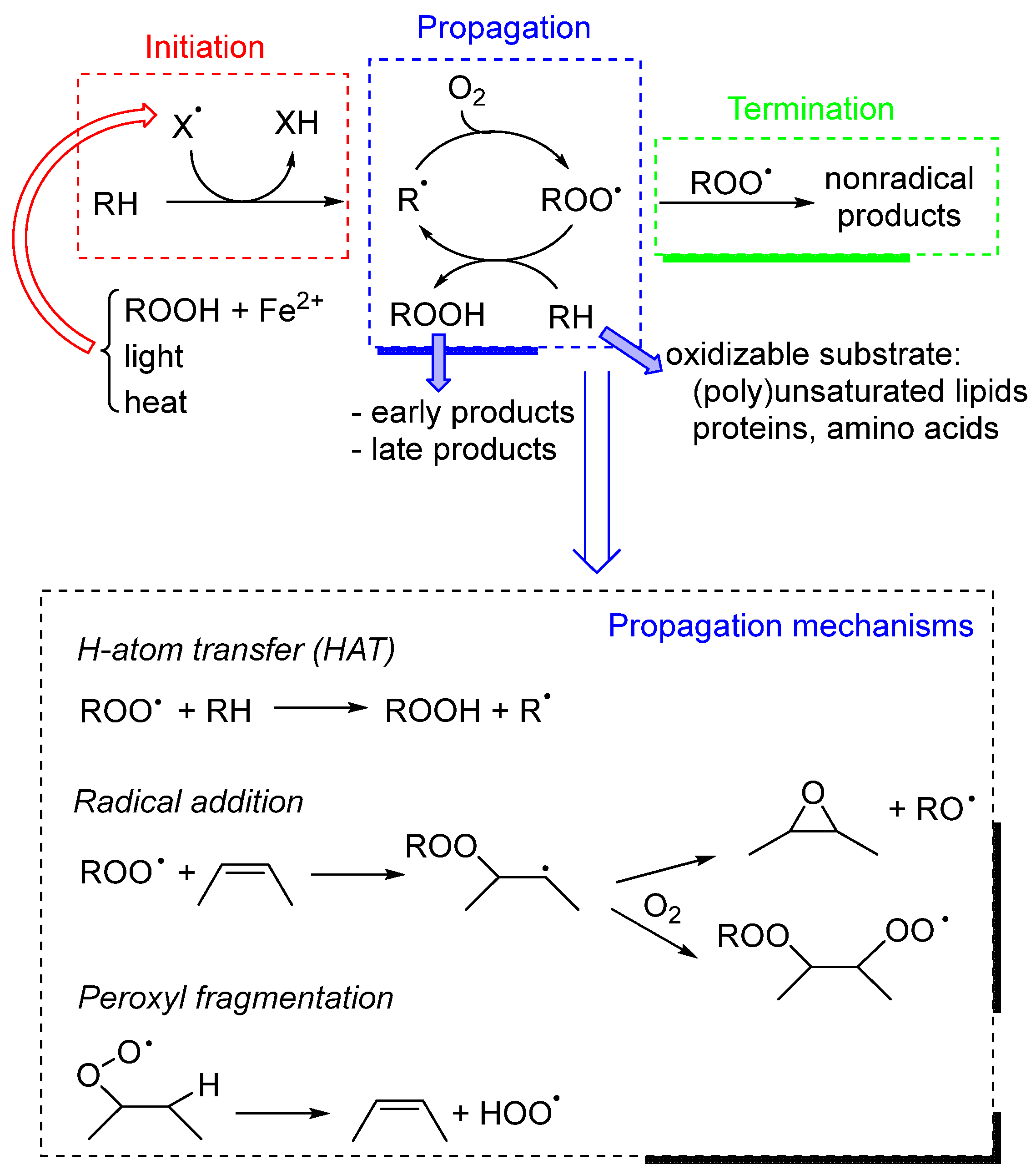
2.1. Chemical Mechanism of Lipid Peroxidation

Lipid-containing foods and emulsions (like mayonnaise) undergo oxidative degradation, which is primarily caused by lipid peroxidation [3,4]. Despite the multiplicity of situations in which peroxidation is observed, the mechanism is similar, consisting of a radical chain reaction carried on by oxygen-centered peroxyl radicals, ROO [5]. Because of the very fast reaction of carbon-centered (alkyl) radicals R with (triplet) O2, virtually all alkyl radicals are transformed into ROO even at low O2 concentrations, making ROO the most relevant radical to be considered during lipid peroxidation [6]. Lipid peroxidation can be divided into three broad mechanistic steps, shown in Scheme 1.
  • Initiation consists of the formation of radicals in the systems. In food, this process can be due to various mechanisms: direct reaction of O2 with bisallylic C-H groups (usually accelerated by heat) [7], photosensitization [8], and metal-catalyzed decomposition of hydroperoxides [9,10]. It should be remembered that, because of the radical chain nature of peroxidation, several substrate molecules are destroyed by one radical, and even a very small initiation rate is able to produce a dramatic effect over long periods.
  • Propagation is based on the reaction of the (alkyl)peroxyl radical ROO with the substrate, with the subsequent formation of a hydroperoxide (ROOH) and a carbon-centered radical that reacts with O2 forming a new ROO. In lipids, propagation mainly involves polyunsaturated fatty acids (PUFAs), because the bisallylic position is (at room temperature) hundreds of times more reactive toward ROO than allylic or aliphatic ones [5]. However, the propagation mechanism is in many cases more complex than the one described here; for instance, it may involve ROO fragmentation to produce HOO or RO, together with closed-shell oxygenated early products like epoxides and carbonyls [11].
  • Termination is the step in which radicals disappear from the system by reacting with each other (i.e., by bimolecular decay). Typically, two secondary ROO radicals (like in the case of lipids) generate a carbonyl and an alcohol. While radical–radical reactions are typically fast, the low concentration of radicals nonetheless makes termination a statistically rare event, which may be further slowed down by lipid compartmentalization strategies (i.e., emulsified lipids) [5].

2.2. Products

The products of peroxidation can be divided into early products, which are those directly produced during radical chain reactions, and late products, which derive from the degradation or transformation of early products. However, the distinction is not sharp because some products can be formed by both mechanisms.

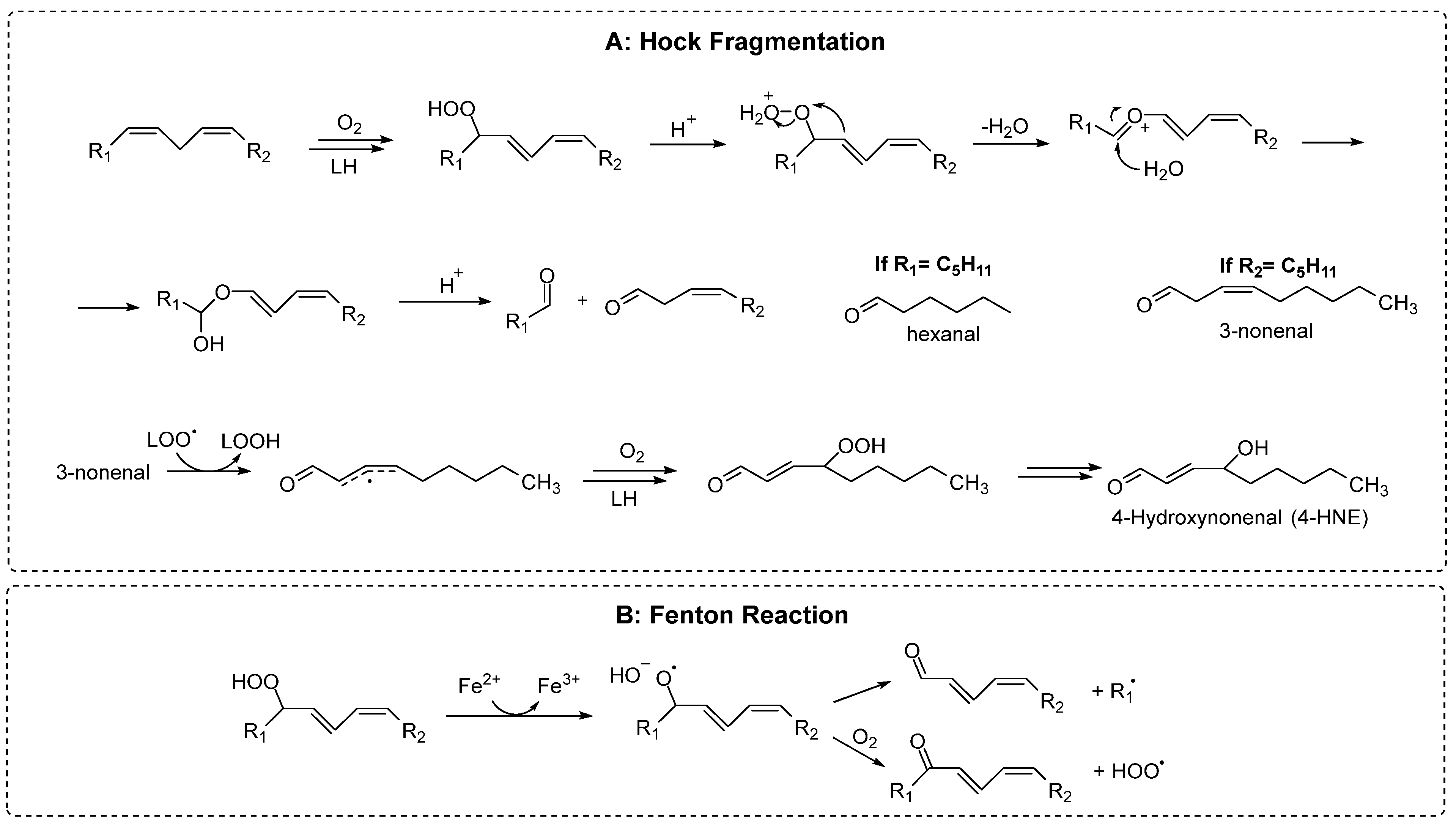
2.2.1. Early Products

Early products are represented mainly by hydroperoxides, especially in the case of PUFA. In the case of linoleate residues, for example, the complete product distribution has been clarified as reported in Scheme 2, showing that the relative position of the double bonds and their cis/trans isomers depend on how fast the peroxyl radicals are quenched by H-atom donors [12]. Therefore, during the peroxidation of triacylglycerols, transtrans hydroperoxides are typically formed in much larger amounts than cistrans hydroperoxides. However, their relative concentration is reversed in the presence of antioxidants [12]. Polyunsaturated lipids form hydroperoxides having conjugated double bonds, which can be easily detected spectrophotometrically [13].
The product distribution becomes more complex with an increasing number of double bonds in the fatty acids, due to the occurrence of fast cyclization reactions that may involve both peroxyl and alkyl radicals [12]. In the case of substrates having only one unsaturation, like oleic acid and cholesterol, the H-atom abstraction by ROO that would yield hydroperoxides is not thermodynamically favored; thus ROO addition to the double bond also takes place, in a competitive manner. Radical addition on non-conjugated double bonds typically generates an epoxide and a secondary alkoxyl radical, RO (see Scheme 1) [12], which can rearrange to an alpha-hydroxyl alkyl radical that, upon reaction with O2, finally yields a ketone and HOO [14].

2.2.2. Late Products
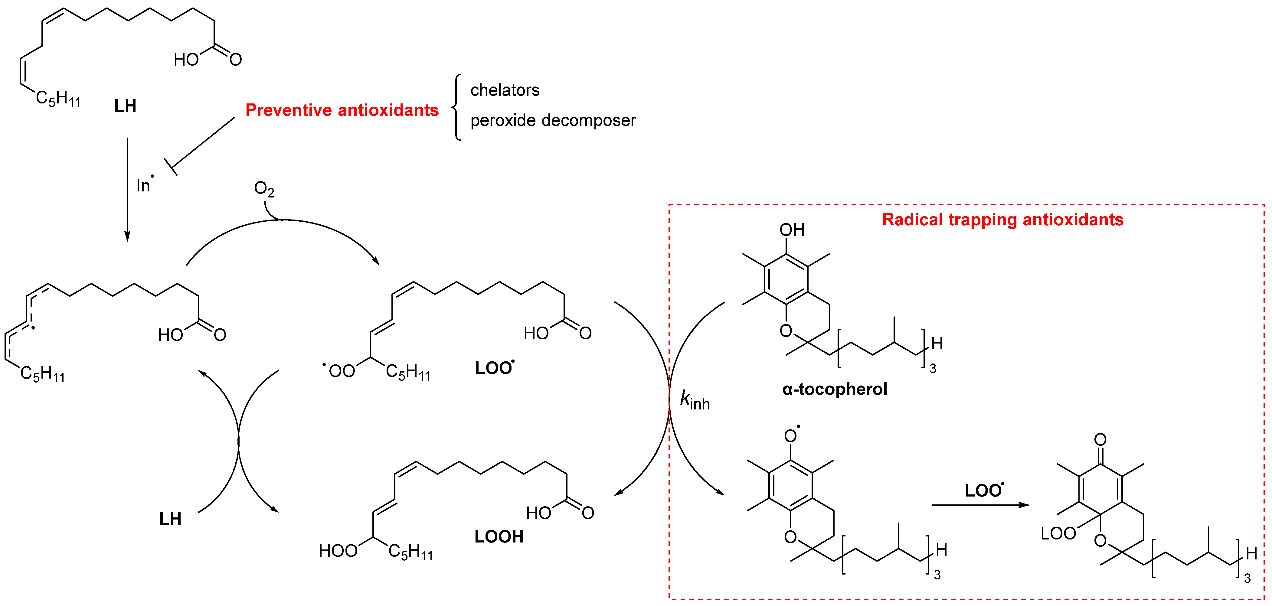
The formation of late products is kindled by the homolytic or heterolytic reaction of unstable hydroperoxides. For this reason, the hydroperoxide level increases only at the beginning of lipid peroxidation, followed by a decrease as they convert into more stable secondary products that tend to accumulate [15]. In fact, it is common practice to evaluate the oxidative stability of lipid materials by measuring the content of both hydroperoxides and late products (typically volatile and non-volatile aldehydes) [16].
Although the reaction network leading to secondary products is very complex (for a comprehensive review, see Refs. [5,6,12]), two main pathways of hydroperoxide decay can be recognized: Hock fragmentation and radical homolysis. The latter is catalyzed by reduced transition metals (like Fe2+) and provides alkoxyl radicals, which then may rearrange into alpha-hydroxy alkyls, or fragment into alkyl radicals and aldehydes (see Scheme 3). This reaction is of paramount importance in the initiation of peroxidation reaction, because traces of metals and hydroperoxides are always present in lipid materials [17].
Hock fragmentation is instead catalyzed by acids and heat, and through a non-radical pathway transforms a hydroperoxide into two aldehydes [5]. It was recently shown that this mechanism is fundamental for forming oxylipins of cholesterol [18]. The nature of the aldehydes formed during lipid peroxidation critically depends upon the kind of fatty acid being oxidized. As expected, omega-3 lipids provide aldehydes with a shorter chain (and higher volatility) than omega-6 ones [19]. Hydroperoxides can also react with alkenes forming epoxides, which in turn can be attacked by water or acids forming diols or diesters, respectively [20]. In addition to carbonyls and epoxides, other minor products that derive from the advanced oxidation of aldehydes are also formed, like furans, aldol condensation products [19], and short-chain acids like acetic and formic acid [15], which are the main water-soluble volatiles that are detected by the Rancimat assay [21].
Late products may be divided into two categories: volatile and non-volatile. The former are the ones that give lipid peroxidation its rancid, off-flavor. As they derive from the fragmentation of the fatty acid carbon chain, non-volatile products are most often represented by the fragment of the carboxylic acid, while volatile products are from the distal part [3].

3. Antioxidants

In the context of lipid peroxidation, antioxidants are natural compounds or synthetic additives able to slow down the formation of oxidation products. They can be divided into preventive and chain-breaking antioxidants, depending on whether they antagonize the initiation or propagation step, respectively [22]. Preventive antioxidants are represented, for example, by metal chelators (like phytic acid) [23] which inactivate or remove iron ions from the water/lipid interface [24]. Chain-breaking (or radical-trapping) antioxidants act by directly trapping ROO [25] or by catalytic HOO/ROO quenching (see Scheme 4) [26].
Most small-molecule natural antioxidants belong to the chain-breaking class, including tocopherols, phenolic acids, flavonoids, etc., as well as synthetic additives like tert-butylhydroquinone (TBHQ), butylated hydroxytoluene (BHT), and propyl gallate (PG) [25]. All these antioxidants are consumed during their activity, and for this reason, they provide inhibition for a limited time, which depends on their concentration [7].
Most natural lipids contain antioxidants. Seed oils, for example, have relatively large amounts of tocopherols or tocotrienols, while olive oil also contains polyphenols like oleuropein and hydroxytyrosol [27,28]. As chain-breaking antioxidants are consumed during peroxidation, the determination of their level can represent an indirect way to measure the extent of radical attack [29,30].
Figure 1 reports the variation in the concentrations of the oxidizable lipids of t,c and t,t hydroperoxides and of α-tocopherol (α-TOH) or hydroxytyrosol acetate during the peroxidation of sunflower oil at 70 °C [15]. These results show that at the beginning of the reaction, there is a slow consumption of the bisallylic groups, because of the presence of the antioxidants. However, under these conditions, hydroxytyrosol acetate was demonstrated to be a much better antioxidant than α-TOH (see Figure 1B,C), because it resulted in a longer inhibition period and a stronger reduction in hydroperoxide production. In Figure 1D,E, it is shown that α-TOH is consumed faster than hydroxytyrosol acetate, suggesting that the disappearance of antioxidants represents an index to describe the oxidative stability of the system.

4. Biosensors for Lipid Peroxidation Analysis

4.1. What Are Biosensors

According to the IUPAC, biosensors are defined as “a device that uses specific biochemical reactions mediated by isolated enzymes, immunosystems, tissues, organelles, or whole cells to detect a chemical compound” [31]. The sensors are constituted by a recognition part, which enables the production of the analytical signal through the interaction with the analyte or group of analytes to which it is specific or selective. This interaction between the receptor and the analyte/s produces a change in a physical parameter, which can be electrochemical, optical, thermal, electronic, gravimetric, or acoustic, and transmitted through the transducer to the electrical circuit (amplifier), thus giving a signal that is proportional to the concentration of the analyte [32]. The selectivity of the sensor depends on the receptor chosen for its construction. If the recognition part is a biological receptor, they are biosensors. Based on the type of bioreceptor, they are classified into enzymatic biosensors, immunosensors, aptasensors, cell sensors, DNA sensors, etc., as summarized in Figure 2.

4.1.1. Enzymatic Biosensors

The ability of enzymes to act as specific biocatalysts has largely been utilized for the development of biosensors that employ a variety of transducers, including thermal, optical, and electrochemical ones. The enzyme glucose oxidase, which is present in the biosensor for glucose, catalyzes the conversion of glucose to gluconic acid and, when exposed to oxygen, oxidizes to produce hydrogen peroxide. During such an enzymatic reaction, all the changes that occur can be used to produce an analytical signal proportional to the glucose concentration. For instance, the production of gluconic acid leads to a change in pH that is proportional to the amount of glucose present in the solution, so by fixing the enzyme on the surface of the pH electrode, a biosensor for glucose can be built.
The oxygen used for the reoxidation of the enzyme also enables the determination of glucose, because for every molecule of glucose that is oxidized, one molecule of oxygen is consumed. Therefore, due to the equivalence with the amount of oxygen, the quantitative determination of glucose can be carried out by means of an oxygen electrode [33]. Moreover, the determination of hydrogen peroxide as a product of enzyme reoxidation makes it possible to determine glucose, because the amount of peroxide generated on the surface of the electrode is equivalent to the amount of glucose [34].
The use of enzymes as bioreceptors represents a good opportunity to build very specific biosensors, and many research groups over the years have investigated the construction of enzymatic biosensors with a focus on their application in different types of samples, such as biological fluids, food, water, environment, etc. [35,36,37,38]. The challenge of developing specific biosensors to be applied to real complex samples has been faced by using modifiers in transducers that enable operation at lower potentials; this approach facilitates electron transfer and thus reduces the possibility of oxidation/reducing interferences at lower operating potentials [39,40,41,42]. Research on electrochemical biosensors has gone even further, by trying to make a direct connection between the active center of the enzyme with the electrode through nanowires and nanoparticles, thus enabling the direct transfer of electrons between the electrode and the enzyme; this results in an electrical signal with higher sensitivity [43,44,45].
The new nanomaterials that imitate the catalytic activity of enzymes are called nanozymes, which have many advantages over enzymes, such as high stability, high resistance to harsh environments, low cost, the possibility of mass production, and a longer lifespan [46]. By exploiting this concept, nanostructures built on the surface of the sensor able to mimic the catalytic effect of enzymes have been developed [47,48,49].

4.1.2. Aptasensors

Aptamers are synthetic oligonucleotides that exhibit the ability to bind a particular target molecule, functioning akin to “chemical antibodies” and facilitating the creation of specific biosensors. Aptamers are obtained through the Systematic Evolution of Ligands by Exponential Enrichment (SELEX) process [46,50] where, in the presence of a target molecule, the ligands library of oligonucleotides with high affinity to the analyte are cyclically partitioned and reproduced, thus increasing the number of aptamers with selectivity to the analyte. These aptamers can be used with various transducers, providing analytical signals with high specificity and application in food control [51,52,53], health [54,55,56], environment, etc. [57,58,59,60].

4.1.3. Immunosensors

The use of antigen–antibody interaction (lock and key) for the detection of low concentrations with high selectivity by using antibodies as bioreceptors has aroused great research interest in the field of biosensors [61]. The fixation of antibodies on the surface of the biosensor produces an analytical signal in the presence of the antigen that is proportional to its concentration. Depending on the detection method, immunosensors can be labeled with electroactive species [62] or can be label-free [63]. Labeled antibodies are used to increase sensitivity and specificity for applications in complex sample matrices. To amplify the signal, immunosensors incorporating a second antibody, linked to an electroactive compound, have been developed. After binding to the antigen attached to the first antibody, it enables the generation of an electrochemical signal. Enzyme-labeled antibodies amplify the signal in the presence of the enzyme substrate, which can be electrochemically detected. The modification of second antibodies with metal nanoparticles is a good possibility for signal amplification in electrochemical sensors; this indeed enables the determination of the metal, which is proportional to the concentration of the antigen to which the second antibody is bound [62,64,65,66].

4.1.4. Molecularly Imprinted Polymers (MIP)

They are also known as “plastic receptors”, as they are built through a polymerization reaction in the presence of the analyte. After the polymerization process, the analyte is removed from these polymers with solvent, leaving vacant spaces and creating a suitable structure for a selective binding site for the analyte. This type of receptor has found great application in the construction of sensors, enabling the generation of the analytical signal in the presence of the analyte with minimal interference. In conjunction with an electrochemical interface, the use of MIP has shown an application in complex samples, thus enabling the production of sensors with low cost and high stability compared to biosensors, which require more specific storage conditions and have higher costs. MIP sensors have been built to detect toxins in food [67], biomedical analysis [68], food allergens [69], food adulterants [70], etc. However, the signal produced by the connection between the polymer and the analyte often does not reach a high analytical sensitivity. Attempts have been made to incorporate various nanomaterials [68,69,71,72] that facilitate the signal transmission process in the production of the electrical signal, thus making possible the determination of analytes at low concentration levels in quite complex samples [70,73].

4.2. Antioxidant Analysis

Over the past decade, the utilization of antioxidants in the food industry has significantly increased. They are frequently used to ensure the quality of food products and prevent their oxidation [1,74]. The most widely used natural antioxidants in food are ascorbate (vitamin C), tocopherols (vitamin E), and polyphenols (including flavonoids and phenolic acids) [75].
Among synthetic antioxidants, the most commonly employed in food formulations are butylated hydroxyanisole (BHA), BHT, PG, and TBHQ [76,77].
As discussed in Section 3, as antioxidants are consumed during peroxidation, antioxidant levels can also provide a measure of the overall oxidation stability of the material. Conventional techniques like gas chromatography (GC) [78,79] and liquid chromatography (LC) [78,80] are costly, time-consuming, and involve intricate sample preparations. Colorimetry methods, on the other hand, although much simpler than GC and LC, are also time-consuming and require larger amounts of samples and reagents [81], in addition to their frequent lack of specificity which can lead to quantification problems in complex food matrices. To overcome these limitations, various sensors and biosensors have emerged as valid tools for assessing antioxidant levels in food. These devices effectively identify and measure specific analytes within food matrices, offering high sensitivity, specificity, fast analysis performance, and minimal sample pre-treatment requirements, coupled with the convenience of miniaturization [82,83,84,85,86,87,88].

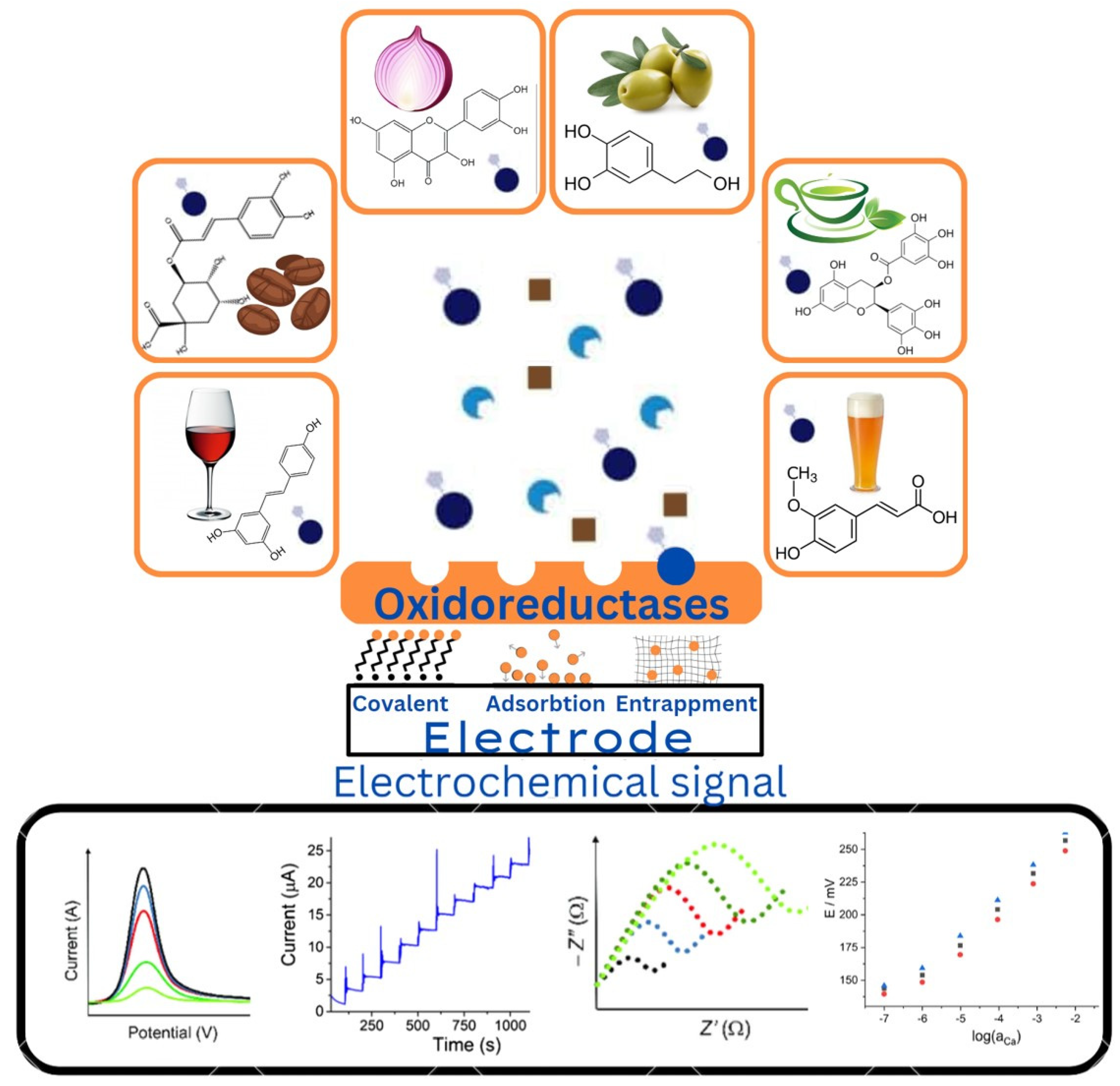
4.2.1. Electrochemical Biosensors for Polyphenol Detection

The evaluation of the quantity of phenols in a sample (food products, beverages, plant extracts, or environmental samples) can be performed by measuring the concentration of specific phenolic compounds or by determining the total phenolic content (TPC). Given that about 9000 plant phenolic compounds have been identified to date, this technique faces a challenging task [89]. Alternatively, the TPC approach is less time-consuming, involving the semiquantitative evaluation of complicated multicomponent samples (plant-based materials, processed foods, or environmental matrices). In a thorough examination of biosensors tailored for studying rosmarinic acid, David et al. highlighted persistent challenges within this field [90]. One critical challenge lies in enhancing selectivity towards closely related compounds, such as other hydroxycinnamic acids. To face this challenge, it is necessary to utilize chemically modified electrodes, including techniques like molecularly imprinted polymers. These modifications have the potential to enhance selectivity while simultaneously lowering detection limits. By doing so, they have the potential to improve the accuracy and effectiveness of electroanalysis for rosmarinic acid [91].

4.2.2. Enzyme-Based Biosensors

In biosensors, the most used enzymes for the detection of antioxidants in food belong to the oxidoreductase, hydrolase, or lyase categories (Figure 3). These enzymes can identify particular substrates and produce quantifiable signals, which enable biosensors to precisely determine the amount of antioxidants present in dietary samples. The relationship between how much inhibitor is present and how much it slows down or reduces the enzyme’s activity enables both quantitative and qualitative assessments of analytes [82,83,84,85,86,87,88].
Oxidoreductase enzymes facilitate the transfer of electrons from the phenolic substrate to an electron acceptor, usually molecular oxygen. Once they are oxidized by the enzyme, phenolic compounds are converted into quinone derivatives; the latter undergo reduction at the electrode surface of the biosensor back into the initial phenolic compounds, leading to the generation of electrochemical signals. The electrochemical signal generated during this process is proportional to the concentration of phenolic compounds present in the sample [85,86,87]. The application of biosensors based on phenol oxidase for the identification of antioxidants in food is a viable strategy with several benefits. Enzymes including tyrosinase and laccase belong to this group [85,87].

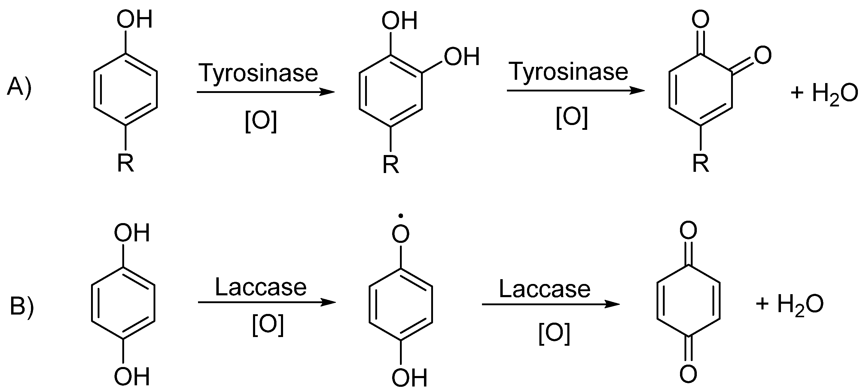
4.2.3. Tyrosinase-Based Biosensors

Tyrosinase is a copper-containing enzyme found in plants, animals, and fungi that catalyzes the hydroxylation of mono-phenols to ortho-diphenols via the monophenolase reaction and the oxidization of ortho-diphenols to ortho-quinone through the diphenolase reaction (see Scheme 5A). L-tyrosine participates as a substrate in both reactions. In tyrosinase-based biosensors, the reversible electrochemical reduction of ortho-quinone is measured. The obtained signals reflect the concentration of polyphenols in the solution.
For detecting phenolic antioxidants in beer, a tyrosinase-based electrochemical biosensor showed a good correlation with the Folin–Ciocalteu (FC) method, even though the values obtained from the FC assay were significantly higher [91]. This indicates the selective nature of the biosensing platform as the application of tyrosinase ensures that the electrochemical signal is acquired only from the phenolic substrates of the enzyme, thus eliminating any interference and limiting phenol overestimation.
A nanostructured surface tyrosinase biosensor named poly-(3,4-ethylenedioxythiophene)-tyrosinase/sonogel-carbon (PEDOT-Tyr/SNGC) was used for evaluating the polyphenol content of beers and wines. Using caffeic acid as the reference polyphenol, the biosensor exhibited a high sensitivity with a limit of detection of 4.3 µM and a correlation coefficient (R2) of 0.9987. The advantages of using this biosensor lie on its simplicity and rapidity, with enzyme and polymer deposition occurring simultaneously in one step for a few minutes while delivering an instantaneous low-cost response [92].

4.2.4. Laccase-Based Biosensors

Similar to tyrosinase, laccase is an oxidoreductase enzyme that catalyzes the oxidation of a wide range of phenolic and non-phenolic compounds. Laccase catalyzes the oxidation of substrates by transferring electrons from the substrate to molecular oxygen, resulting in the formation of water as a byproduct. During this process, laccase generates reactive oxygen species, such as hydroxyl radicals, which can further oxidize substrates (Scheme 5B) [87,93].
Boubezari et al. created a new biosensor using laccase encapsulated within a chitosan–galactomannan composite to identify the phenolic chemicals in olive oil [94]. Pyrocatechol was successfully identified by the biosensor, and the measurement of ortho-quinone at a potential of −0.447 V served as the basis for the detection. In the concentration range from 10−16 M to 10−4 M, the biosensor demonstrated a record-low LOD (limit of detection), and its voltammetric response stayed constant for more than two weeks. Moreover, the chitosan–galactomannan composite did not lose its laccase activity [94].
Salamanca-Neto et al. [95] presented a novel approach to measure the chlorogenic acid content in brewed coffee beverages, by using a biosensing device fabricated with platinum nanoparticles and laccase. The authors found that, under optimized experimental conditions, the biosensing device showed a linear response for chlorogenic acid in the range of 0.56–7.3 µmol L−1 with high accuracy and sensitivity. They also found that the biosensing device had a storage stability of 30 days and a lifetime of 150 measurements, indicating its potential for practical use in analyzing the chlorogenic acid content in brewed coffee beverages. The detection and quantification limits of the biosensor were found to be 0.18 and 0.59 µM, respectively, which are comparable to those of traditional analytical methods, such as HPLC [95].
Mohtar et al. [37] presented a novel biosensor utilizing laccase for the detection of polyphenols in propolis. The biosensing scheme is based on a nanocomposite film of laccase immobilized on gold nanoparticles electrodeposited on a screen-printed carbon electrode modified with polypyrrole (Ppy) through in situ electropolymerization (Ppy/Lac/AuNPs/SPCE). Polyphenols contained in ethanolic extracts of propolis were first oxidized by laccase, then reduced by Ppy/Lac/AuNPs/SPCE providing an amperometric response. The results showed a linear response in the concentration range from 1 to 250 μM expressed as caffeic acid, with a limit of detection of 0.83 μM. The biosensor exhibited a rapid analysis time (15 min), good selectivity, stability, and reproducibility [37].
To identify hydroquinone and other phenolic chemicals in wine and blueberry syrup, another laccase-based biosensor was immobilized onto a screen-printed carbon electrode enhanced by gold nanoparticles and graphene nanoplatelets (LACC/AuNP/GNPl/SPCE). A low detection limit of 1.5 µM was obtained, with a linear HQ concentration range from 4 to 130 µM. The biosensor demonstrated good repeatability and reproducibility; however, additional validation is required to determine its selectivity for phenolic chemicals in comparison to other interfering molecules [96].
In a recent study by Mediavilla et al. [97], the total polyphenol content was determined in rice leaf extracts, wine, and juice, by using a direct fluorescence assay. A bioconjugate, generated by the laccase enzyme (from Trametes versicolor) and carbon nanodots, was biofunctionalized and used as a fluorescent label for the new biosensor. The biosensor detected phenolic chemicals (such as gallic acid, GA) by the enzymatic reaction of the bioconjugate and GA, generating quinones that led to quenched fluorescence. The quantification and detection limits were 25 μM and 7.4 μM, respectively [97].
Additionally, Gomes et al. [98] conducted a study wherein they immobilized laccase from B. rhodina MAMB-05 on carboxymethyl-botryosphaeran to measure the presence of quercetin in drugs, drinks, and biological materials. In the range of 4.98–50.0 × 10⁻⁸ mol L−1, the biosensor displayed a linear response for quercetin, with a predicted detection limit of 2.6 × 10⁻⁸ mol L−1 [98].
To identify a wider range of phenolic chemicals, enzymatic biosensors containing both tyrosinase and laccase enzymes have been developed [99,100]. To detect catechol in wines, Apetrei et al. [99] looked into the self-encapsulation of a polyphenol oxidase enzyme extract (synthesized by T. pubescens) within a conducting polypyrrole material. The three biosensor types that were compared were Chem-Ppy-TP (chemically synthesized Ppy with enzyme extract), Bio-Ppy (biosynthesized Ppy-based biosensor), and Bio-Ppy-TP (Bio-Ppy with additional enzyme extract). All three biosensors effectively identified catechols in fruit beverages (such as pomegranate, blackberry, and blueberry) at low concentrations (1–60 μM) [99].

4.2.5. Metal–Organic Framework

The use of metal–organic frameworks (MOFs) as catalysts [101] in the construction of sensors has also aroused great interest. This is primarily because of the porous nature of the crystalline material and the large active surface area [102]. The use of MOFs in the construction of electrochemical sensors is possible by mixing them with other materials that enable electrochemical signal transduction. The determination of rutine is a typical example based on MOFs, using cerium (Ce) as an ion with catalytic properties and poly(3,4-ethylenedioxythiophene (PEDOT) as a good conductor to build a Ce-PEDOT nanocomposite with catalytic properties [103].
Carbon nanofibers, synthesized starting from cubic nanoparticles of glycosyl-MOF-5 and a suspension of polyacrylonitrile through electrospinning, have provided a good sensitivity in the electrochemical determination of quercetin using a glassy carbon electrode [104]. Another ultrasensitive sensor for the determination of baicalin was built from graphitized carbon nitride, single-walled carbon nanotubes, and reduced graphene oxide by the electrodeposition of cyclodextrin-MOF and was successfully applied to real samples (human serum and bear bile scutellaria eye drops) [105]. The use of mesoporous carbon mixed with a zirconium fumarate metal oxide framework increased the conductivity, specific surface area, and pore volume and showed an electrocatalytic effect in the determination of gallic acid and luteolin in green tea and urine samples [106]. Using MOF as a nanocomposite in the construction of electrochemical sensors reduced the oxidation overpotential and increased the sensitivity for flavonoids and phenolic acids, such as kaempferol [107,108], catechin and sunset yellow [109,110], caffeic acid [111,112], catechol [113] and phenolic acids [114], gallic acid [115], coumaric acid [116], etc.
Electrochemical sensors with only MOF have also been built for the determination of ascorbic acid and L-tryptophan [117,118]. However, the application of MOF in the development of the sensors also has shortcomings, such as low selectivity for specific compounds, costly preparation, and the use of non-green chemicals. In any case, the preparation of sensors with MOFs is a good opportunity for the preparation of Point-Of-Care (POC) sensors, since they shorten the analysis time and enable on-time quality control for several widely consumed food products.
The use of enzymes as receptors of biosensors shows their limitations due to their low durability and stability, expensive mass production, and the limited environments where they can be employed; all these aspects have led researchers to build new materials that imitate their catalytic activity.

4.2.6. Nanozymes

Nanozymes are nanomaterials that have enabled the replacement of enzymes as bioreceptors in the construction of biosensors, especially peroxidases, catalase, and superoxide dismutase (Table 1).
Nanozymes built with phosphorus-doped carbon spheres having a nanoporous structure were used in the voltammetric determination of baicalin and luteolin [119]. The electrochemical determination of phenolic compounds has been successfully carried out by constructing a nanocomposite with enzyme-like properties from manganese oxide (IV) and quantum graphene nanodots, which has also proven its performance in the colorimetric determination of catechol and dopamine [120]. The increase in the selectivity of nanozymes is achieved with the construction of single-atom nanozymes (SANs), managing to be anchored in the metal oxide framework in porous carbon; this enables the simultaneous electrochemical determination of catechol and hydroquinone [121].

4.2.7. Molecular Imprinted Polymers (MIPs)

A MIP made of methacrylic acid and ethylene glycol dimethacrylate as a cross-linking agent, in the presence of α-tocopherol as a template, was able to bind α-tocopherol in edible oils with a better affinity than TBHQ and Trolox (the hydrosoluble α-tocopherol analog). The binding extent was measured by Raman spectrometry [122]. Despite these interesting results, no further attempts have been made to assess the capability of this method in food samples undergoing oxidation.

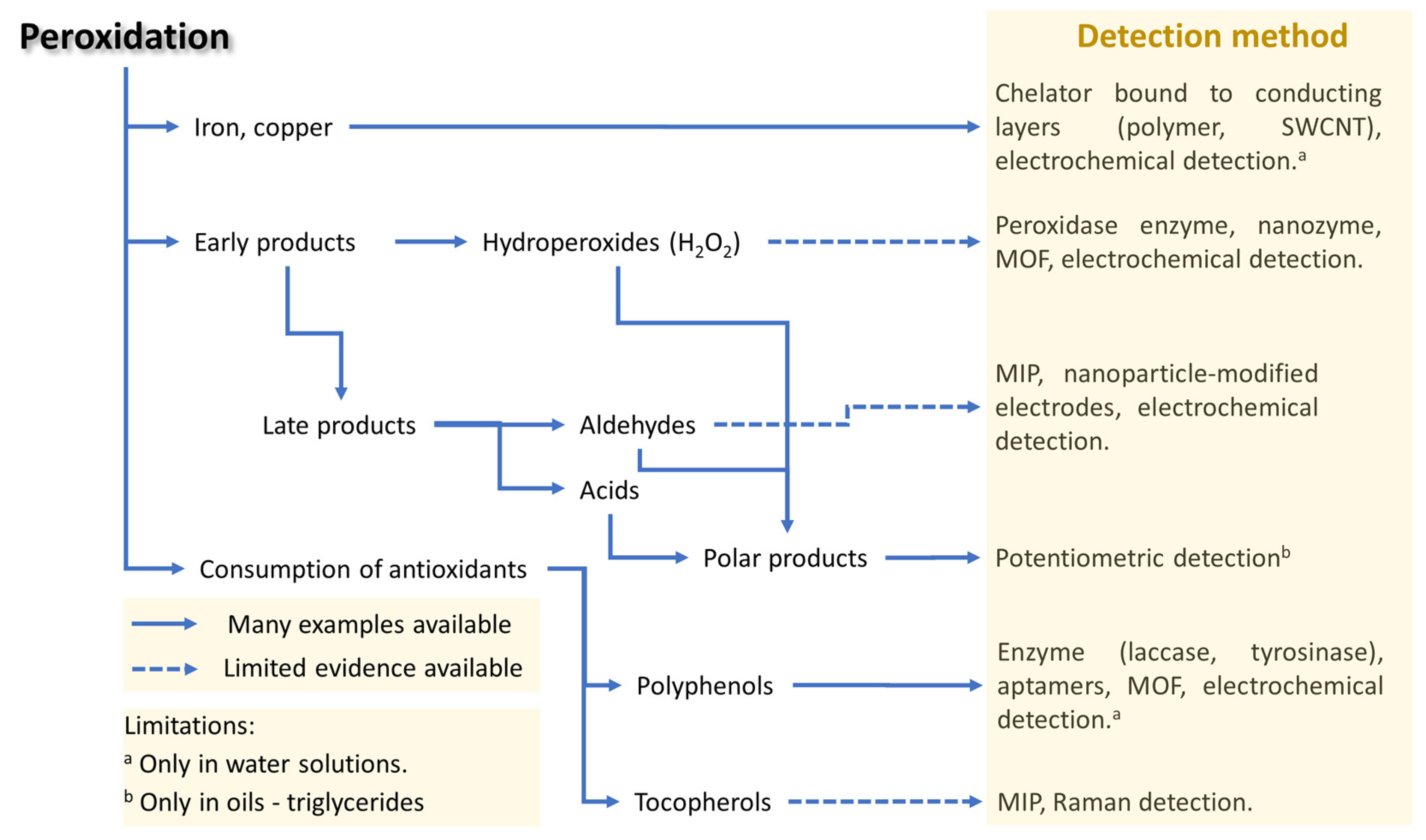
4.3. Peroxidation Analysis

4.3.1. Hydroperoxides

Since hydroperoxides are the primary early peroxidation products, determining their presence is crucial to understanding how food oxidizes. Regretfully, research has mostly focused on hydrogen peroxide, with little attention paid to determining alkyl hydroperoxides (the primary products of peroxidation), while H2O2 is merely a side product (see Scheme 1). The only example of alkyl hydroperoxide detection is represented by an electrode featuring horseradish peroxidase absorbed on polypyrrole with amperometric detection. This method was able to quantify hydroperoxides like tert-butylhydroperoxide in a mixed aqueous–organic system [123]. Many biosensors have been developed for the determination of hydrogen peroxide, enabling the monitoring of food quality. Using nanomaterials for the construction of biosensors increases sensitivity by providing an easier connection and electron transfer between the receptor and the transducer.
For instance, cress peroxidase entrapped on ferrimagnetic nanoparticles with amperometric detection [124], organic–inorganic hybrids made of peroxidase–copper phosphate [125], and AgNPs coupled with ferrous disulfide as peroxidase mimics [126] can be considered attempts to build, through nanomaterials, chemical receptors for H2O2 that mimic the function of enzymes (also called nanozymes).
The use of single-atom catalysts has aroused great interest in various types of electrochemical energy conversion/storage systems, including oxygen reduction, oxygen evolution reaction, hydrogen evolution reaction, and CO2 reduction [127]. MOFs represent a promising approach to building sensors for measuring hydrogen peroxide, possibly replacing precious metals and enzymes [128]. Different modification elements have been used for hydrogen peroxide detection, such as cobalt-MOFs [129], trimetallic-MOF nanosheets [130], magnetic covalent organic frameworks [131], Au@Pt nanoflower MOFs [128], metal–organic Langmuir–Blodgett films as electrocatalysts [132], and metal nanoparticle/MOF nanozymes [133] (see Table 2).

4.3.2. Aldehydes

Specific detection of peroxidation-derived aldehydes by using biosensors has not yet been developed. However, 5-hydroxymethylfurfural (HMF) can be considered an example of how to detect aldehydes, despite not being a direct product of peroxidation. HMF originates from the reaction of reducing sugars and proteins during heating through the Maillard reaction, and it is known for its potential adverse effects on human health. The determination of HMF is an important parameter used as an indicator of the quality of food products and the way they have been processed and stored. The construction of a sensor with high specificity for the determination of HMF is a challenge that has been addressed in different ways (see Table 3). It has also been proposed to determine the “furanic index” through which it is claimed to determine food freshness, all furanic derivatives thus being determined as products of the Maillard reaction [134] despite the fact that furans can also be generated by oxidation.
Different types of MIPs have been employed in electrochemical sensors for the determination of HMF and its derivatives, such as a low sensor for 2-furaldehyde detection in beverages using in situ polymerization [135] and electropolymerized molecularly imprinted polymers combined with salting-out assisted liquid–liquid extraction (SALLE)) for the detection of HMF [136]. Electrochemical sensors have been developed for the determination of HMF in honey, coffee, and date molasses, using electrode modifiers such as nickel oxide nanoparticles [137], Cu-Ni bimetallic microparticles on copper electrodes [138], NiO/carbon black [139], Ag nanoparticles incorporated onto carboxymethyl cellulose [140], silver microdendrites [141], and the electrosynthesis of Co-based layered double hydroxides [142]. Graphene quantum dots incorporated in a NiAl2O4 nanocomposite based on MIP showed increased sensitivity for HMF and were successfully applied in HMF determination in coffee samples [143].

4.3.3. Volatile Aldehydes (VA) and Protein Carbonyls (PC)

Different sensors have been built for the determination of volatile aldehydes using different methods and techniques to improve selectivity [144]. Sensors for nonanal determination incorporate Au nanoparticles to increase the MIP conductivity, transferring the resistance change as signal for the analyte [145]. Hian et al. [146] enhanced the selectivity for gas sensing using MOF, with zinc oxide and zeolitic imidazole framework as a molecular sieve for the detection of formaldehyde. The 3D structure of a Co3O4-derived MOF was also successfully applied to determine the formation of formaldehyde, due to its higher porosity and presence of active sites [147]. The use of imprinted polymers as receptors and their modification with nanomaterials (carbon nanomaterials) that change their electroactivity in the presence of volatile aldehydes is a good possibility for building resistive sensors that can determine this group of biomarkers.
The determination of protein carbonyls is very important as a biomarker of oxidation in protein-containing food. One of the methods used to determine protein carbonyls is the detection of the dinitrophenol hydrazone (DNP)-carbonyl product from the reaction of dinitrophenylhydrazine (DNPH) with the carbonyl groups in proteins. Protein carbonyls can be determined by using high-specificity antibodies against DNP or by measuring absorbance. Entrapment of DNPH at the electrode surface using Nafion was successfully applied to the electrochemical determination of protein carbonyls [148].

4.3.4. Metals

Iron or copper ions are often involved in the peroxidation of organic materials and food as catalysts for radical production through the Fenton reaction. Therefore, their presence in excess represents an important initiating factor that greatly increases the peroxidation of the material. The Fe or Cu ion recognition is similar to that of heavy metals (cadmium (Cd), lead (Pb), arsenic (As), mercury (Hg), and zinc (Zn)), which are contaminants that can be found in food. Due to their negative effects on the human organism, their fast determination at low concentration levels is of paramount importance. The development of electrochemical sensors for the simultaneous determination of heavy metals has aroused great interest in many research groups.
The most used techniques are the voltammetric techniques, which enable the simultaneous determination of heavy metals; increased sensitivity and lower detection limits (LOD) have been achieved by using different modifiers, as summarized in Table 4. The presence of lead and cadmium was determined in rice and water using carbon nanomaterials (like MWCNT) as modifiers of the electrode surface, obtaining an LOD of 2.7 nM and 0.9 nM for Pb and Cd, respectively [149]. Electrochemically reduced graphene oxide on bismuth film electrodes has successfully been applied for the determination of Pb and Cd in milk samples [150].
The electrochemical sensor for Cu at low concentrations has been built based on the electrocatalytic effect of Cu in the hydrogen evolution reaction, which gives a signal that is proportional to the accumulated amount of Cu on the surface of the pencil graphite electrode modified with a strong ligand chelator (p-aminobenzyl-C-functionalized cyclam) for Cu2+ [151]. This method has lowered the detection limit for Cu to 1.1 nM.
The simultaneous determination of heavy metal ions based on the construction of graphene aerogel nanocomposites increases the conductivity and also facilitates the transfer of electrons through the metal oxide framework (MOF-UiO-66-NH2) [152], which serves as accumulation points for heavy metals due to the interaction of hydrophilic groups with metal ions. Through this operating principle of the sensor, it has been possible to simultaneously determine Cd2+, Pb2+, Cu2+, and Hg2+ with very high sensitivity and selectivity in water samples.
Regarding iron, an ion-selective electrode based on polyaniline, a conducting polymer, and chelating agents allowed for the potentiometric detection of Fe2+ or Fe3+ ions in aqueous solution containing albumin [164]. Ferric ions could be monitored in wine by a biosensor based on a field effect transistor, where a network of carbon nanotubes act as the conductor channel and transferrin as the Fe3+ binding receptor [123].

4.3.5. Polar Compounds

Deep frying is one of the most popular food-cooking processes, due to the sensory traits that it confers to food. However, repeated deep frying promotes different chemical reactions, including lipid hydrolysis, oxidation, and polymerization [165].
The degradation products released during deep frying cause changes in the dielectric constant of the frying oil, which is related to their level of total polar compounds (TPCs), the only legally recognized parameter to evaluate the overall frying oil degradation. A high content of TPCs in the frying oil increases the dielectric moment, which can be used to monitor the oil quality [166].
Due to the toxicity of some of the produced species, it is very important to determine their concentration level in the frying oil, because the latter penetrates fried foods, thus favoring their accumulation therein which could cause negative health effects. Different rapid tests are commercially available today to monitor frying oil, which are simple, quick, cheap, and do not require specialized personnel to perform them. Their operation principle is based on measurements of viscosity, dielectric constant, and fatty acid content [167].
Interdigitated electrodes, as capacitive sensors, have been used for the assessment of frying oil quality, considering the relation between electrical capacitance and the content of oil-degradation products [168,169,170,171]. Clearly, this method can be applied only to low-polarity samples, like edible frying oils where the buildup of polar molecules can effectively increase the dielectric constant.

5. Overview of Biosensors Applications

Figure 4 reports the main molecules involved in the various steps of peroxidation, encompassing the initiation species (metals), oxidation products (classified depending on the formation stage), and antioxidants.
The analytes are connected by arrows to the corresponding biosensor technology, wherein solid and dotted lines represent, respectively, convincing or somewhat weak evidence in this specific sector, mainly ascribable to the low number of examples reported in the literature. From this figure, it can be observed that metals, polyphenols, and polar compounds can potentially be detected in complex systems like food using the existing technology, whereas organic hydroperoxides, aldehydes, and tocopherols still require further studies.

Examples of Applications in Food: Milk and Plant-Based Milk Alternatives

Oxidative stability is a major concern for extending shelf-life and the long-time conservation of milk and plant-based milk alternatives (obtained from soybean, almond, rice, oats, hazelnut, coconut, etc.) [172]. This issue is more severe for preparations with higher concentrations of PUFA, which can be added on purpose (such as omega-3 fatty acid fortification) or occur naturally [173].
The precise processes causing the oxidation of the various products can vary due to their differing compositions. For instance, the naturally occurring enzyme lactoperoxidase can cause protein oxidation in milk while also helping to regulate bacterial growth [174]. Similarly, when milk is exposed to light, the native component riboflavin acts as a photosensitizer, thus promoting oxidation [175]. Conversely, the elevated PUFA concentration in some plant-based milk substitutes, like soybean beverages, promotes major lipid peroxidation when particular stimuli (like traces of iron) are present [175]; moreover, oxidation can specifically lead to a decrease in the polyphenolic content of plant-based milk alternatives. Another relevant aspect to consider is the interaction of lipids and proteins, which can favor the co-oxidation of the macromolecules [176]; this phenomenon has been observed in milk [177].
The oxidation biomarkers are universal and indicate the formation of radicals, primary oxidation products (H2O2 and hydroperoxides), and secondary oxidation products (aldehydes, protein carbonyls), regardless of the cause and specific oxidative pathway. The oxidation markers that may be considered for milk and plant-based milk alternatives are peroxides [173], malondialdehyde [172], volatile aldehydes (hexanal, 2-hexenal, heptanal, 2-heptenal, octanal, 2-octenal, nonanal, etc.) [173,175], protein carbonyls [174,175], polyphenols [172], and dimethyl sulfide [175].
In order to be useful for monitoring the stability of complex food matrices, a biosensor for peroxidation often has to overcome problems caused by a lack of selectivity and/or low sensitivity. For example, in milk or plant-based milk alternatives, there are reducing sugars that may interfere with sensing of aldehydes formed by peroxidation. Similarly, peroxides may be confounded by H2O2 produced by lactoperoxidase. Moreover, the detection of primary and secondary products of peroxidation should be effective for low analyte concentrations (a few parts per million), because at larger concentrations rancid off-flavors start to develop. To assess consumer acceptance of dairy products based on appearance and flavor, the sensory analysis remains the primary method. In fact, trained sensory panels can detect off-flavors like rancidity in milk and milk powders. The International Dairy Federation has reported that the sensory threshold for detecting a rancid flavor in milk is typically between 1.5 and 2.0 mEq of FFA/100 g fat. Values exceeding approximately 1.5 mmol of FFA L−1 are considered to reach an unacceptable rancidity threshold. Specific thresholds for individual fatty acids associated with rancidity have also been identified. For caproic acid (C10:0) and lauric acid (C12:0), the detection thresholds are approximately 7 ppm and 8 ppm, respectively [178,179,180].
The combined used of biosensors and sensory analysis to identify and quantify specific VOCs associated with lipid oxidation can provide deeper insights into the oxidation process and help in maximizing the shelf-life of food products without compromising their quality [179].

6. Concluding Remarks

The current high interest in biosensors is due to the fact that they provide rapid identification of substances, making them invaluable in urgent situations where quick results are essential. In food quality control, fast detection ensures prompt decisions regarding product safety and shelf-life. Their relatively inexpensive production makes biosensors a cost-effective solution for various applications, promoting widespread adoption in the food industry. Additionally, their miniaturization enables integration into portable devices, facilitating on-site testing and reducing reliance on centralized laboratories.
However, applications to measure lipid peroxidation and antioxidant protection in food require significant work to overcome a number of challenges, the most important of which are selectivity, sensitivity, and the ability to perform the measurement in complex lipid-containing matrices.
The selectivity problem is based on the similitude of the molecules specifically released by lipid peroxidation to other unrelated molecules. For example, peroxidation releases lipid aldehydes and short-chain fatty acids, but the aldehyde functional group can be present in reducing sugars, while fatty acids can also be produced by bacteria. Therefore, biosensors should be able to detect oxidation products having a specific structure, and simply being able to interact with a target functional group may not be sufficient to reach the desired outcome.
Because of human olfaction’s extreme ability to detect peroxidation-related chemicals associated with food spoilage, sensitivity is a major issue in food applications. To be useful in this field, biosensors must be capable of dealing with analyte concentrations in the ppm range.
Working with lipid-containing matrices or bulk lipids appears to be an unresolved issue. For instance, phenolic antioxidants are typically detected using water-based solvents that support enzyme activity and electrochemical methods, whereas lipid-soluble phenols are rarely considered. Another example is that water-soluble hydroperoxides, such as H2O2, can be easily detected in aqueous media (see Section Examples of Applications in Food: Milk and Plant-Based Milk Alternatives), whereas lipid hydroperoxides, which are relevant to food rancidity, are difficult to detect using current biosensor technology.
Despite the challenges, the development of biosensors for lipid peroxidation and antioxidant effect measurement in food is a very promising research field with enormous benefits for food security, safety, and waste reduction, with a relevant potential impact on the improvement of food analysis sustainability.

Funding

R.A. acknowledges funding by the European Union—NextGenerationEU—under the National Recovery and Resilience Plan (PNRR), Mission 4 Education and Research, Component 2: From research to business—Investment 1.1 Notice Prin 2022—DD N. 104 del 2/2/2022, from PRIN20227XZKBY—Superoxide responsive redox-active systems and nano smart materials to target ferroptosis—FEROX—CUP J53D23008550006. M.T.R-E. acknowledges funding by the Alma Mater Studiorum—Università di Bologna (Basic Research Funding RFO-2024). L.B: acknowledges funding by the Academy of Science of Albania—NanoALB Unit (ignite projects) HMF-Sens project.

Acknowledgments

Z. J. and Y. H. acknowledge a fellowship from the China Scholarship Council.

Conflicts of Interest

The authors declare no conflicts of interest.

References

  1. Carocho, M.; Morales, P.; Ferreira, I.C.F.R. Antioxidants: Reviewing the Chemistry, Food Applications, Legislation and Role as Preservatives. Trends Food Sci. Technol. 2018, 71, 107–120. [Google Scholar] [CrossRef]
  2. Amorati, R.; Valgimigli, L. Advantages and Limitations of Common Testing Methods for Antioxidants. Free Radic. Res. 2015, 49, 633–649. [Google Scholar] [CrossRef] [PubMed]
  3. Ghelichi, S.; Hajfathalian, M.; Yesiltas, B.; Sørensen, A.M.; García-Moreno, P.J.; Jacobsen, C. Oxidation and Oxidative Stability in Emulsions. Compr. Rev. Food Sci. Food Saf. 2023, 22, 1864–1901. [Google Scholar] [CrossRef] [PubMed]
  4. Suhag, R.; Ferrentino, G.; Morozova, K.; Zatelli, D.; Scampicchio, M.; Amorati, R. Antioxidant Efficiency and Oxidizability of Mayonnaise by Oximetry and Isothermal Calorimetry. Food Chem. 2024, 433, 137274. [Google Scholar] [CrossRef] [PubMed]
  5. Valgimigli, L. Lipid Peroxidation and Antioxidant Protection. Biomolecules 2023, 13, 1291. [Google Scholar] [CrossRef] [PubMed]
  6. Helberg, J.; Pratt, D.A. Autoxidation vs. Antioxidants—The Fight for Forever. Chem. Soc. Rev. 2021, 50, 7343–7358. [Google Scholar] [CrossRef]
  7. Mollica, F.; Bonoldi, L.; Amorati, R. Kinetic Analysis of High-Temperature Sunflower Oil Peroxidation Inhibited by the Major Families of Phenolic Antioxidants Unveils the Extraordinary Activity of 1,4-Hydroquinones. Antioxidants 2022, 11, 2142. [Google Scholar] [CrossRef]
  8. Lopes, C.R.B.; Courrol, L.C. Evaluation of Steady-State and Time-Resolved Fluorescence Spectroscopy as a Method for Assessing the Impact of Photo-Oxidation on Refined Soybean Oils. Foods 2023, 12, 1862. [Google Scholar] [CrossRef]
  9. Mallais, M.; Hanson, C.S.; Giray, M.; Pratt, D.A. General Approach to Identify, Assess, and Characterize Inhibitors of Lipid Peroxidation and Associated Cell Death. ACS Chem. Biol. 2023, 18, 561–571. [Google Scholar] [CrossRef]
  10. Scurti, S.; Caretti, D.; Mollica, F.; Di Antonio, E.; Amorati, R. Chain-Breaking Antioxidant and Peroxyl Radical Trapping Activity of Phenol-Coated Magnetic Iron Oxide Nanoparticles. Antioxidants 2022, 11, 1163. [Google Scholar] [CrossRef]
  11. Baschieri, A.; Jin, Z.; Amorati, R. Hydroperoxyl Radical (HOO) as a Reducing Agent: Unexpected Synergy with Antioxidants. A Review. Free Radic. Res. 2023, 57, 115–129. [Google Scholar] [CrossRef] [PubMed]
  12. Zielinski, Z.A.M.; Pratt, D.A. Lipid Peroxidation: Kinetics, Mechanisms, and Products. J. Org. Chem. 2017, 82, 2817–2825. [Google Scholar] [CrossRef] [PubMed]
  13. Losada-Barreiro, S.; Paiva-Martins, F.; Bravo-Díaz, C. Analysis of the Efficiency of Antioxidants in Inhibiting Lipid Oxidation in Terms of Characteristic Kinetic Parameters. Antioxidants 2024, 13, 593. [Google Scholar] [CrossRef]
  14. Saraev, D.D.; Wu, Z.; Kim, H.-Y.H.; Porter, N.A.; Pratt, D.A. Intramolecular H-Atom Transfers in Alkoxyl Radical Intermediates Underlie the Apparent Oxidation of Lipid Hydroperoxides by Fe(II). ACS Chem. Biol. 2023, 18, 2073–2081. [Google Scholar] [CrossRef]
  15. Caño-Ochoa, S.D.; Ruiz-Aracama, A.; Guillén, M.D. Individual and Joint Effect of Alpha-Tocopherol and Hydroxytyrosol Acetate on the Oxidation of Sunflower Oil Submitted to Oxidative Conditions: A Study by Proton Nuclear Magnetic Resonance. Antioxidants 2022, 11, 1156. [Google Scholar] [CrossRef]
  16. Pignoli, G.; Bou, R.; Rodriguez-Estrada, M.T.; Decker, E.A. Suitability of Saturated Aldehydes as Lipid Oxidation Markers in Washed Turkey Meat. Meat Sci. 2009, 83, 412–416. [Google Scholar] [CrossRef]
  17. Ten Klooster, S.; Takeuchi, M.; Schroën, K.; Tuinier, R.; Joosten, R.; Friedrich, H.; Berton-Carabin, C. Tiny, yet Impactful: Detection and Oxidative Stability of Very Small Oil Droplets in Surfactant-Stabilized Emulsions. J. Colloid Interface Sci. 2023, 652, 1994–2004. [Google Scholar] [CrossRef]
  18. Zielinski, Z.A.M.; Pratt, D.A. H-Atom Abstraction vs Addition: Accounting for the Diverse Product Distribution in the Autoxidation of Cholesterol and Its Esters. J. Am. Chem. Soc. 2019, 141, 3037–3051. [Google Scholar] [CrossRef] [PubMed]
  19. Grebenteuch, S.; Kroh, L.W.; Drusch, S.; Rohn, S. Formation of Secondary and Tertiary Volatile Compounds Resulting from the Lipid Oxidation of Rapeseed Oil. Foods 2021, 10, 2417. [Google Scholar] [CrossRef]
  20. Barani, M.; Bonetti, R.; Parker, W.O. Thermal Oxidation of Model Molecules to Reveal Vegetable Oil Polymerization Studied by NMR Spectroscopy and Self-Diffusion. J. Am. Oil Chem. Soc. 2023, 100, 551–560. [Google Scholar] [CrossRef]
  21. Flitsch, S.; Neu, P.M.; Schober, S.; Kienzl, N.; Ullmann, J.; Mittelbach, M. Quantitation of Aging Products Formed in Biodiesel during the Rancimat Accelerated Oxidation Test. Energy Fuels 2014, 28, 5849–5856. [Google Scholar] [CrossRef]
  22. Mavridi-Printezi, A.; Menichetti, A.; Mordini, D.; Amorati, R.; Montalti, M. Recent Applications of Melanin-like Nanoparticles as Antioxidant Agents. Antioxidants 2023, 12, 863. [Google Scholar] [CrossRef]
  23. Pei, Y.; Deng, Q.; McClements, D.J.; Li, J.; Li, B. Impact of Phytic Acid on the Physical and Oxidative Stability of Protein-Stabilized Oil-in-Water Emulsions. Food Biophys. 2020, 15, 433–441. [Google Scholar] [CrossRef]
  24. Daoud, S.; Bou-Maroun, E.; Waschatko, G.; Cayot, P. Lipid Oxidation in Oil-in-Water Emulsions: Iron Complexation by Buffer Ions and Transfer on the Interface as a Possible Mechanism. Food Chem. 2021, 342, 128273. [Google Scholar] [CrossRef] [PubMed]
  25. Gutiérrez-del-Río, I.; López-Ibáñez, S.; Magadán-Corpas, P.; Fernández-Calleja, L.; Pérez-Valero, Á.; Tuñón-Granda, M.; Miguélez, E.M.; Villar, C.J.; Lombó, F. Terpenoids and Polyphenols as Natural Antioxidant Agents in Food Preservation. Antioxidants 2021, 10, 1264. [Google Scholar] [CrossRef]
  26. Baschieri, A.; Amorati, R. Methods to Determine Chain-Breaking Antioxidant Activity of Nanomaterials beyond DPPH•. A Review. Antioxidants 2021, 10, 1551. [Google Scholar] [CrossRef]
  27. Lozano-Castellón, J.; Rinaldi De Alvarenga, J.F.; Vallverdú-Queralt, A.; Lamuela-Raventós, R.M. Cooking with Extra-Virgin Olive Oil: A Mixture of Food Components to Prevent Oxidation and Degradation. Trends Food Sci. Technol. 2022, 123, 28–36. [Google Scholar] [CrossRef]
  28. Shah, R.; Farmer, L.A.; Zilka, O.; Van Kessel, A.T.M.; Pratt, D.A. Beyond DPPH: Use of Fluorescence-Enabled Inhibited Autoxidation to Predict Oxidative Cell Death Rescue. Cell Chem. Biol. 2019, 26, 1594–1607.e7. [Google Scholar] [CrossRef]
  29. Li, J.; Yuan, F.; Teng, J.; Li, F.; Zhou, P.; Bi, Y. Effects of Tea Polyphenols and Tertiary Butylhydroquinone on Quality of Palm Oils and Losses of Endogenous Vitamin E during Batch Frying and Oxidative Stability of Fried Instant Noodles. Food Chem. X 2023, 20, 101049. [Google Scholar] [CrossRef]
  30. Bayram, I.; Parra-Escudero, C.; Decker, E.A.; Lu, J. Mathematical Modeling of Alpha-Tocopherol Early Degradation Kinetics to Predict the Shelf-Life of Bulk Oils. J. Agric. Food Chem. 2024, 72, 4939–4946. [Google Scholar] [CrossRef]
  31. Nagel, B.; Dellweg, H.; Gierasch, L.M. Glossary for Chemists of Terms Used in Biotechnology (IUPAC Recommendations 1992). Pure Appl. Chem. 1992, 64, 143–168. [Google Scholar] [CrossRef]
  32. Lobnik, A.; Turel, M.; Urek, Š.K.; Košak, A. Nanostructured Materials Use in Sensors: Their Benefits and Drawbacks. In Carbon and Oxide Nanostructures; Advanced Structured Materials; Springer: Berlin/Heidelberg, Germany, 2010; Volume 5, pp. 307–354. ISBN 978-3-642-14672-5. [Google Scholar]
  33. Clark, L.C.; Lyons, C. Electrode Systems for Continuous Monitoring in Cardiovascular Surgery. Ann. N. Y. Acad. Sci. 1962, 102, 29–45. [Google Scholar] [CrossRef] [PubMed]
  34. Guilbault, G.G.; Lubrano, G.J. An Enzyme Electrode for the Amperometric Determination of Glucose. Anal. Chim. Acta 1973, 64, 439–455. [Google Scholar] [CrossRef]
  35. Kalinke, C.; De Oliveira, P.R.; Marcolino-Júnior, L.H.; Bergamini, M.F. Nanostructures of Prussian Blue Supported on Activated Biochar for the Development of a Glucose Biosensor. Talanta 2024, 274, 126042. [Google Scholar] [CrossRef]
  36. Li, Z.; Cao, L.; Sui, J.; Wang, L.; Lin, H.; Wang, K. Bimetallic Fe/Ni Metal Organic Framework-Based Hypoxanthine Biosensor for Early Monitoring of Freshness Changes of Aquatic Products. Food Chem. 2024, 447, 138902. [Google Scholar] [CrossRef]
  37. Mohtar, L.G.; Aranda, P.; Messina, G.A.; Nazareno, M.A.; Pereira, S.V.; Raba, J.; Bertolino, F.A. Amperometric Biosensor Based on Laccase Immobilized onto a Nanostructured Screen-Printed Electrode for Determination of Polyphenols in Propolis. Microchem. J. 2019, 144, 13–18. [Google Scholar] [CrossRef]
  38. Zhang, J.; Lei, J.; Liu, Z.; Chu, Z.; Jin, W. Nanomaterial-Based Electrochemical Enzymatic Biosensors for Recognizing Phenolic Compounds in Aqueous Effluents. Environ. Res. 2022, 214, 113858. [Google Scholar] [CrossRef]
  39. Bi, R.; Ma, X.; Miao, K.; Ma, P.; Wang, Q. Enzymatic Biosensor Based on Dendritic Gold Nanostructure and Enzyme Precipitation Coating for Glucose Sensing and Detection. Enzyme Microb. Technol. 2023, 162, 110132. [Google Scholar] [CrossRef] [PubMed]
  40. Chavez-Urbiola, I.R.; Reséndiz-Jaramillo, A.Y.; Willars-Rodriguez, F.J.; Martinez-Saucedo, G.; Arriaga, L.G.; Alcantar-Peña, J.; Escalona-Villalpando, R.A.; Ledesma-García, J. Glucose Biosensor Based on a Flexible Au/ZnO Film to Enhance the Glucose Oxidase Catalytic Response. J. Electroanal. Chem. 2022, 926, 116941. [Google Scholar] [CrossRef]
  41. Estrada-Osorio, D.V.; Escalona-Villalpando, R.A.; Gutiérrez, A.; Arriaga, L.G.; Ledesma-García, J. Poly-L-Lysine-Modified with Ferrocene to Obtain a Redox Polymer for Mediated Glucose Biosensor Application. Bioelectrochemistry 2022, 146, 108147. [Google Scholar] [CrossRef]
  42. Wang, Y.; Zhao, J.; Yang, T.; Zhang, Y.; Tao, D.; Hasebe, Y.; Zhang, Z. Electrochemical Evaluation of Sulfide Mineral Modified Glassy Carbon Electrode as Novel Mediated Glucose Biosensor. J. Electroanal. Chem. 2021, 894, 115357. [Google Scholar] [CrossRef]
  43. Jung, J.; Lim, S. ZnO Nanowire-Based Glucose Biosensors with Different Coupling Agents. Appl. Surf. Sci. 2013, 265, 24–29. [Google Scholar] [CrossRef]
  44. Wang, K.; Liu, Q.; Guan, Q.-M.; Wu, J.; Li, H.-N.; Yan, J.-J. Enhanced Direct Electrochemistry of Glucose Oxidase and Biosensing for Glucose via Synergy Effect of Graphene and CdS Nanocrystals. Biosens. Bioelectron. 2011, 26, 2252–2257. [Google Scholar] [CrossRef] [PubMed]
  45. Wang, Y.; Liu, L.; Li, M.; Xu, S.; Gao, F. Multifunctional Carbon Nanotubes for Direct Electrochemistry of Glucose Oxidase and Glucose Bioassay. Biosens. Bioelectron. 2011, 30, 107–111. [Google Scholar] [CrossRef]
  46. Liang, M.; Yan, X. Nanozymes: From New Concepts, Mechanisms, and Standards to Applications. Acc. Chem. Res. 2019, 52, 2190–2200. [Google Scholar] [CrossRef]
  47. Li, P.; Kalambate, P.K.; Harris, K.D.; Jemere, A.B.; Tang, X. (Shirley) Robust and Flexible Electrochemical Lactate Sensors for Sweat Analysis Based on Nanozyme-Enhanced Electrode. Biosens. Bioelectron. X 2024, 17, 100455. [Google Scholar] [CrossRef]
  48. Liang, Y.; Liu, Y.; Zhao, P.; Chen, Y.; Lei, J.; Hou, J.; Hou, C.; Huo, D. An Electrochemical Sensor Based on FeCo Bimetallic Single-Atom Nanozyme for Sensitive Detection of H2O2. Anal. Chim. Acta 2023, 1281, 341867. [Google Scholar] [CrossRef]
  49. Sun, J.; Wang, Z.; Guan, J. Single-Atom Nanozyme-Based Electrochemical Sensors for Health and Food Safety Monitoring. Food Chem. 2023, 425, 136518. [Google Scholar] [CrossRef]
  50. Wu, X.; Chen, J.; Wu, M.; Zhao, J.X. Aptamers: Active Targeting Ligands for Cancer Diagnosis and Therapy. Theranostics 2015, 5, 322–344. [Google Scholar] [CrossRef]
  51. Jin, Y.; Zhang, Y.; Xu, H.; Lu, X.; Yuan, Y.; Zhang, W. A Ratiometric Electrochemical Aptasensor for Sensitive Detection of Kanamycin in Food Based on Entropy-Driven Strand Displacement Reaction. Food Control 2024, 161, 110390. [Google Scholar] [CrossRef]
  52. Khoshbin, Z.; Moeenfard, M.; Abnous, K.; Taghdisi, S.M. A Label-Free Aptasensor for Colorimetric Detection of Food Toxin: Mediation of Catalytically Active Gold Nanozymes and Smartphone Imaging Strategy. Food Chem. 2024, 433, 137355. [Google Scholar] [CrossRef] [PubMed]
  53. Mishra, A.; Roy, S.; Israel Shaikh, N.; Malave, P.; Mishra, A.; Anish Alam, M.; Ghorpade, Y.; Rahil Hasan, M.; Nizam, A. Recent Advances in Multiplex Aptasensor Detection Techniques for Food-Borne Pathogens: A Comprehensive Review of Novel Approaches. Biosens. Bioelectron. X 2024, 16, 100417. [Google Scholar] [CrossRef]
  54. Figueroa-Miranda, G.; Chen, S.; Neis, M.; Zhou, L.; Zhang, Y.; Lo, Y.; Tanner, J.A.; Kreidenweiss, A.; Offenhäusser, A.; Mayer, D. Multi-Target Electrochemical Malaria Aptasensor on Flexible Multielectrode Arrays for Detection in Malaria Parasite Blood Samples. Sens. Actuators B Chem. 2021, 349, 130812. [Google Scholar] [CrossRef]
  55. Hamdi, F.; Roushani, M.; Nasibipour, M.; Hoseini, S.J. Aptasensor Based on High Surface Area Covalent Organic Framework for Simple and Ultrasensitive Detection of Sarcosine in the Diagnosis of Prostate Cancer. Anal. Chim. Acta 2024, 1291, 342235. [Google Scholar] [CrossRef]
  56. Tseng, C.-C.; Lu, S.-Y.; Chen, S.-J.; Wang, J.-M.; Fu, L.-M.; Wu, Y.-H. Microfluidic Aptasensor POC Device for Determination of Whole Blood Potassium. Anal. Chim. Acta 2022, 1203, 339722. [Google Scholar] [CrossRef]
  57. Lee, K.; Ha, S.M.; Gurudatt, N.G.; Heo, W.; Hyun, K.-A.; Kim, J.; Jung, H.-I. Machine Learning-Powered Electrochemical Aptasensor for Simultaneous Monitoring of Di(2-Ethylhexyl) Phthalate and Bisphenol A in Variable pH Environments. J. Hazard. Mater. 2024, 462, 132775. [Google Scholar] [CrossRef]
  58. Tian, C.; Zhao, L.; Zhu, J.; Zhang, S. Ultrasensitive Detection of Trace Hg2+ by SERS Aptasensor Based on Dual Recycling Amplification in Water Environment. J. Hazard. Mater. 2021, 416, 126251. [Google Scholar] [CrossRef]
  59. Wang, X.; Xu, M.; Kuang, Y.; Liu, X.; Yuan, J. A Novel Ratiometric Electrochemical Aptasensor Based on M-Shaped Functional DNA Complexes for Simultaneous Detection of Trace Lead and Mercury Ions in Series Aquatic Edible Vegetables. J. Hazard. Mater. 2024, 465, 133169. [Google Scholar] [CrossRef] [PubMed]
  60. Zhang, Z.; Luan, Y.; Ru, S.; Teng, H.; Li, Y.; Liu, M.; Wang, J. A Novel Electrochemical Aptasensor for Ultrasensitive Detection of Herbicide Prometryn Based on Its Highly Specific Aptamer and Ag@Au Nanoflowers. Talanta 2023, 265, 124838. [Google Scholar] [CrossRef]
  61. Köhler, G.; Milstein, C. Continuous Cultures of Fused Cells Secreting Antibody of Predefined Specificity. Nature 1975, 256, 495–497. [Google Scholar] [CrossRef]
  62. Feng, Y.; Liu, Y.; Li, Y.; Lv, J.; Chen, H. Electrochemical Detection of Lactobacillus Rhamnosus in Fermented Food Using Magnetic Immunosensor Based on Au-Fe3O4. Int. J. Electrochem. Sci. 2022, 17, 220329. [Google Scholar] [CrossRef]
  63. Chomthong, K.; Kunpatee, K.; Pimpitak, U.; Puthong, S.; Komolpis, K.; Wonsawat, W.; Nuanualsuwan, S.; Yakoh, A.; Khongchareonporn, N.; Ruecha, N.; et al. Label-Free Simultaneous Detection of Quinolone Antibiotic Residues Using an Origami Paper–Based Electrochemical Immunosensor. Sens. Actuators B Chem. 2024, 410, 135667. [Google Scholar] [CrossRef]
  64. Hu, J.; Wen, P.; Wang, Y.; Yang, J.; Xiao, Z.; Xu, Z.; Shen, Y.; Wang, H.; Hammock, B.D. Fabrication of a Label-Free Electrochemical Immunosensor by Functionalized Nanofiber Membrane for the Ultrasensitive Detection of Quinalphos. Food Control 2024, 162, 110423. [Google Scholar] [CrossRef]
  65. Subjakova, V.; Oravczova, V.; Tatarko, M.; Hianik, T. Advances in Electrochemical Aptasensors and Immunosensors for Detection of Bacterial Pathogens in Food. Electrochim. Acta 2021, 389, 138724. [Google Scholar] [CrossRef]
  66. Wu, C.-C.; Lin, C.-C.; Yin, H.-Y.; Tsai, W.-C.; Yang, P.-F.; Liu, H.-J.; Wen, H.-W. Rapid Detection of Mango Allergen in Processed Foods Using an Immunomagnetic Nanoparticle-Based Electrochemical Immunosensor. Microchem. J. 2024, 198, 110070. [Google Scholar] [CrossRef]
  67. Ayerdurai, V.; Cieplak, M.; Kutner, W. Molecularly Imprinted Polymer-Based Electrochemical Sensors for Food Contaminants Determination. TrAC Trends Anal. Chem. 2023, 158, 116830. [Google Scholar] [CrossRef]
  68. Ni, X.; Tang, X.; Wang, D.; Zhang, J.; Zhao, L.; Gao, J.; He, H.; Dramou, P. Research Progress of Sensors Based on Molecularly Imprinted Polymers in Analytical and Biomedical Analysis. J. Pharm. Biomed. Anal. 2023, 235, 115659. [Google Scholar] [CrossRef]
  69. Sundhoro, M.; Agnihotra, S.R.; Amberger, B.; Augustus, K.; Khan, N.D.; Barnes, A.; BelBruno, J.; Mendecki, L. An Electrochemical Molecularly Imprinted Polymer Sensor for Rapid and Selective Food Allergen Detection. Food Chem. 2021, 344, 128648. [Google Scholar] [CrossRef]
  70. Meng, F.; Duan, M.; Wu, W.; Shao, S.; Qin, Y.; Zhang, M. Enzymatic Construction Au NPs-rGO Based MIP Electrochemical Sensor for Adulteration Detection of Bovine-Derived Allergen in Camel Milk. Food Chem. 2024, 436, 137638. [Google Scholar] [CrossRef]
  71. Lu, Z.; Wei, K.; Ma, H.; Xiong, Q.; Li, Y.; Sun, M.; Wang, X.; Wang, Y.; Wu, C.; Su, G.; et al. Nanoarchitectonics of on–off Ratiometric Signal Amplified Electrochemical Sensor for Chlorpromazine with Molecularly Imprinted Polymer Based on Ni-MOF/Fe-MOF-5 Hybrid Au Nanoparticles. Sep. Purif. Technol. 2023, 327, 124858. [Google Scholar] [CrossRef]
  72. Sun, R.; Han, S.; Zong, W.; Chu, H.; Zhang, X.; Jiang, H. Ultrasensitive Detection of Chlortetracycline in Animal-Origin Food Using Molecularly Imprinted Electrochemical Sensor Based on SnS2/ZnCo-MOF and AuNPs. Food Chem. 2024, 452, 139537. [Google Scholar] [CrossRef] [PubMed]
  73. Krishnan, H.; Gopinath, S.C.B.; Arshad, M.K.M.; Zulhaimi, H.I.; Anbu, P.; Subramaniam, S. Molecularly Imprinted Polymer Enhances Affinity and Stability over Conventional Aptasensor for Blood Clotting Biomarker Detection on Regimented Carbon Nanohorn and Gold Nanourchin Hybrid Layers. Sens. Actuators B Chem. 2022, 363, 131842. [Google Scholar] [CrossRef]
  74. Atta, E.M.; Mohamed, N.H.; Abdelgawad, A.A.M. Antioxidants: An Overview on the Natural and Synthetic Types. Eur. Chem. Bull. 2017, 6, 365. [Google Scholar] [CrossRef]
  75. Poljsak, B.; Kovač, V.; Milisav, I. Antioxidants, Food Processing and Health. Antioxidants 2021, 10, 433. [Google Scholar] [CrossRef] [PubMed]
  76. Arab, Z.; Jafarian, S.; Karimi-Maleh, H.; Roozbeh Nasiraie, L.; Ahmadi, M. Monitoring of Butylated Hydroxyanisole in Food and Wastewater Samples Using Electroanalytical Two-Fold Amplified Sensor. Sustainability 2022, 14, 2169. [Google Scholar] [CrossRef]
  77. López-Pedrouso, M.; Lorenzo, J.M.; Franco, D. Advances in Natural Antioxidants for Food Improvement. Antioxidants 2022, 11, 1825. [Google Scholar] [CrossRef]
  78. Cannavacciuolo, C.; Pagliari, S.; Giustra, C.M.; Carabetta, S.; Guidi Nissim, W.; Russo, M.; Branduardi, P.; Labra, M.; Campone, L. LC-MS and GC-MS Data Fusion Metabolomics Profiling Coupled with Multivariate Analysis for the Discrimination of Different Parts of Faustrime Fruit and Evaluation of Their Antioxidant Activity. Antioxidants 2023, 12, 565. [Google Scholar] [CrossRef]
  79. Hasan, M.R.; Haque, M.M.; Hoque, M.A.; Sultana, S.; Rahman, M.M.; Ali Shaikh, M.A.; Sarker, M.K.U. Antioxidant Activity Study and GC-MS Profiling of Camellia Sinensis Linn. Heliyon 2024, 10, e23514. [Google Scholar] [CrossRef]
  80. Du, J.; Zhong, B.; Subbiah, V.; Barrow, C.; Dunshea, F.; Suleria, H. LC-ESI-QTOF-MS/MS Profiling and Antioxidant Activity of Phenolics from Custard Apple Fruit and By-Products. Separations 2021, 8, 62. [Google Scholar] [CrossRef]
  81. Hamasaki, T.; Kashiwagi, T.; Komatsu, T.; Kabayama, S.; Nakamichi, N.; Teruya, K.; Shirahata, S. A New Colorimetric Method for Determining Antioxidant Levels Using 3,5-Dibromo-4-Nitrosobenzene Sulfonate (DBNBS). MethodsX 2022, 9, 101797. [Google Scholar] [CrossRef]
  82. Alam, M.W.; Najeeb, J.; Naeem, S.; Usman, S.M.; Nahvi, I.; Alismail, F.; Abuzir, A.; Farhan, M.; Nawaz, A. Electrochemical Methodologies for Investigating the Antioxidant Potential of Plant and Fruit Extracts: A Review. Antioxidants 2022, 11, 1205. [Google Scholar] [CrossRef] [PubMed]
  83. David, I.G.; Iorgulescu, E.E.; Popa, D.E.; Buleandra, M.; Cheregi, M.C.; Noor, H. Curcumin Electrochemistry—Antioxidant Activity Assessment, Voltammetric Behavior and Quantitative Determination, Applications as Electrode Modifier. Antioxidants 2023, 12, 1908. [Google Scholar] [CrossRef] [PubMed]
  84. Forzato, C.; Vida, V.; Berti, F. Biosensors and Sensing Systems for Rapid Analysis of Phenolic Compounds from Plants: A Comprehensive Review. Biosensors 2020, 10, 105. [Google Scholar] [CrossRef]
  85. Lzaod, S.; Dutta, T. Recent Advances in the Development of Oxidoreductase-Based Biosensors for Detection of Phenolic Antioxidants in Food and Beverages. ACS Omega 2022, 7, 47434–47448. [Google Scholar] [CrossRef]
  86. Raymundo-Pereira, P.A.; Silva, T.A.; Caetano, F.R.; Ribovski, L.; Zapp, E.; Brondani, D.; Bergamini, M.F.; Marcolino, L.H.; Banks, C.E.; Oliveira, O.N.; et al. Polyphenol Oxidase-Based Electrochemical Biosensors: A Review. Anal. Chim. Acta 2020, 1139, 198–221. [Google Scholar] [CrossRef] [PubMed]
  87. Tarasov, A.; Stozhko, N.; Bukharinova, M.; Khamzina, E. Biosensors Based on Phenol Oxidases (Laccase, Tyrosinase, and Their Mixture) for Estimating the Total Phenolic Index in Food-Related Samples. Life 2023, 13, 291. [Google Scholar] [CrossRef]
  88. Wijayanti, S.D.; Tsvik, L.; Haltrich, D. Recent Advances in Electrochemical Enzyme-Based Biosensors for Food and Beverage Analysis. Foods 2023, 12, 3355. [Google Scholar] [CrossRef]
  89. Satchanska, G. Antibacterial Activity of Plant Polyphenols. In Secondary Metabolites—Trends and Reviews; Vijayakumar, R., Selvapuram Sudalaimuthu Raja, S., Eds.; IntechOpen: London, UK, 2022; ISBN 978-1-80355-207-1. [Google Scholar]
  90. Cerrato-Alvarez, M.; Bernalte, E.; Bernalte-García, M.J.; Pinilla-Gil, E. Fast and Direct Amperometric Analysis of Polyphenols in Beers Using Tyrosinase-Modified Screen-Printed Gold Nanoparticles Biosensors. Talanta 2019, 193, 93–99. [Google Scholar] [CrossRef]
  91. David, I.G.; Popa, D.E.; Buleandră, M.; Cheregi, M.C. Electrochemical Methods and (Bio) Sensors for Rosmarinic Acid Investigation. Chemosensors 2020, 8, 74. [Google Scholar] [CrossRef]
  92. García-Guzmán, J.; López-Iglesias, D.; Cubillana-Aguilera, L.; Lete, C.; Lupu, S.; Palacios-Santander, J.; Bellido-Milla, D. Assessment of the Polyphenol Indices and Antioxidant Capacity for Beers and Wines Using a Tyrosinase-Based Biosensor Prepared by Sinusoidal Current Method. Sensors 2018, 19, 66. [Google Scholar] [CrossRef]
  93. Kadam, A.A.; Saratale, G.D.; Ghodake, G.S.; Saratale, R.G.; Shahzad, A.; Magotra, V.K.; Kumar, M.; Palem, R.R.; Sung, J.-S. Recent Advances in the Development of Laccase-Based Biosensors via Nano-Immobilization Techniques. Chemosensors 2022, 10, 58. [Google Scholar] [CrossRef]
  94. Boubezari, I.; Bessueille, F.; Bonhomme, A.; Raimondi, G.; Zazoua, A.; Errachid, A.; Jaffrezic-Renault, N. Laccase-Based Biosensor Encapsulated in a Galactomannan-Chitosan Composite for the Evaluation of Phenolic Compounds. Biosensors 2020, 10, 70. [Google Scholar] [CrossRef] [PubMed]
  95. Salamanca-Neto, C.A.R.; Marcheafave, G.G.; Scremin, J.; Barbosa, E.C.M.; Camargo, P.H.C.; Dekker, R.F.H.; Scarminio, I.S.; Barbosa-Dekker, A.M.; Sartori, E.R. Chemometric-Assisted Construction of a Biosensing Device to Measure Chlorogenic Acid Content in Brewed Coffee Beverages to Discriminate Quality. Food Chem. 2020, 315, 126306. [Google Scholar] [CrossRef]
  96. Zrinski, I.; Pungjunun, K.; Martinez, S.; Zavašnik, J.; Stanković, D.; Kalcher, K.; Mehmeti, E. Evaluation of Phenolic Antioxidant Capacity in Beverages Based on Laccase Immobilized on Screen-Printed Carbon Electrode Modified with Graphene Nanoplatelets and Gold Nanoparticles. Microchem. J. 2020, 152, 104282. [Google Scholar] [CrossRef]
  97. Mediavilla, M.; Revenga-Parra, M.; Gutiérrez-Sánchez, C.; Hernández-Apaolaza, L.; Pariente, F.; Lorenzo, E. Fluorescent Enzymatic Assay for Direct Total Polyphenol Determination in Food-Related Samples. Talanta 2022, 247, 123576. [Google Scholar] [CrossRef] [PubMed]
  98. Gomes, A.; Mattos, G.J.; Coldibeli, B.; Dekker, R.F.H.; Barbosa Dekker, A.M.; Sartori, E.R. Covalent Attachment of Laccase to Carboxymethyl-Botryosphaeran in Aqueous Solution for the Construction of a Voltammetric Biosensor to Quantify Quercetin. Bioelectrochemistry 2020, 135, 107543. [Google Scholar] [CrossRef]
  99. Apetrei, R.-M.; Cârâc, G.; Bahrim, G.; Camurlu, P. Utilization of Enzyme Extract Self-Encapsulated within Polypyrrole in Sensitive Detection of Catechol. Enzyme Microb. Technol. 2019, 128, 34–39. [Google Scholar] [CrossRef]
  100. Batista, É.A.; Silva, G.N.M.; Sgobbi, L.F.; Machado, F.B.; Macedo, I.Y.; Moreno, E.K.; Neto, J.R.; Scalize, P.S.; Gil, E.S. Enzymatic Electroanalytical Biosensor Based on Maramiellus Colocasiae Fungus for Detection of Phytomarkers in Infusions and Green Tea Kombucha. Biosensors 2021, 11, 91. [Google Scholar] [CrossRef]
  101. Yoon, M.; Srirambalaji, R.; Kim, K. Homochiral Metal–Organic Frameworks for Asymmetric Heterogeneous Catalysis. Chem. Rev. 2012, 112, 1196–1231. [Google Scholar] [CrossRef]
  102. Li, C.; Shen, J.; Wu, K.; Yang, N. Metal Centers and Organic Ligands Determine Electrochemistry of Metal–Organic Frameworks. Small 2022, 18, 2106607. [Google Scholar] [CrossRef]
  103. Wang, Y.; Chen, J.; Wang, C.; Zhang, L.; Yang, Y.; Chen, C.; Xie, Y.; Zhao, P.; Fei, J. An Electrochemical Sensor Based on Ce-MOF-Derived Ce-Doped Poly(3,4-Ethylenedioxythiophene) Composite for Efficient Determination of Rutin in Food. Talanta 2023, 263, 124678. [Google Scholar] [CrossRef] [PubMed]
  104. Liu, Y.; Fan, J.; He, F.; Li, X.; Tang, T.; Cheng, H.; Li, L.; Hu, G. Glycosyl/MOF-5-Based Carbon Nanofibers for Highly Sensitive Detection of Anti-Bacterial Drug Quercetin. Surf. Interfaces 2021, 27, 101488. [Google Scholar] [CrossRef]
  105. Zhao, P.; Huang, L.; Wang, H.; Wang, C.; Chen, J.; Yang, P.; Ni, M.; Chen, C.; Li, C.; Xie, Y.; et al. An Ultrasensitive High-Performance Baicalin Sensor Based on C3N4-SWCNTs/Reduced Graphene Oxide/Cyclodextrin Metal-Organic Framework Nanocomposite. Sens. Actuators B Chem. 2022, 350, 130853. [Google Scholar] [CrossRef] [PubMed]
  106. Liu, H.; Hassan, M.; Bo, X.; Guo, L. Fumarate-Based Metal-Organic Framework/Mesoporous Carbon as a Novel Electrochemical Sensor for the Detection of Gallic Acid and Luteolin. J. Electroanal. Chem. 2019, 849, 113378. [Google Scholar] [CrossRef]
  107. Tan, Q.; Chen, C.; Lin, C.; Zhang, J.; Liu, S.; Zhang, J. Highly Sensitive Detection of Kaempferol Using Electrochemical Sensors Based on 3D-Ordered Mesh Interconnect C60-GO, Ni-MOF, and β-Cyclodextrin. Microchem. J. 2024, 197, 109866. [Google Scholar] [CrossRef]
  108. Zhang, L.; Li, J.; Wang, C.; Tang, J.; Chen, X.; Li, Y.; Shi, J.; Zhao, P.; Xie, Y.; Fei, J. A Novel Kaempferol Electrochemical Sensor Based on Glass Carbon Electrode Modified by Poly (3, 4-Ethylenedioxythiophene) Decorated with Green Synthesized MIL-100(Fe)-Multi- Walled Carbon Nanotubes Composites. Colloids Surf. Physicochem. Eng. Asp. 2022, 649, 129484. [Google Scholar] [CrossRef]
  109. Hu, Y.; Wang, C.; Zhao, P.; Zhang, L.; Fei, J.; Xie, Y. A Novel Catechin Electrochemical Sensor Based on a Two-Dimensional MOFs Material Derivative Zn Doped Carbon Nanosheets and Multi-Walled Carbon Nanotubes Composite Film. Talanta 2022, 246, 123520. [Google Scholar] [CrossRef]
  110. Li, J.; Zhu, D.; Huang, H.; Xie, S.; Xu, J.; Yue, R.; Duan, X. High-Efficient Electrochemical Sensing Platform Based on MOF-Doped Au/PEDOT Composites toward Simultaneous Detection of Catechin and Sunset Yellow in Tea Beverage. Electrochim. Acta 2023, 462, 142732. [Google Scholar] [CrossRef]
  111. Sivasankar, K.; Devasenathipathy, R.; Wang, S.-F.; Kohila Rani, K.; Raja, D.S.; Lin, C.-H. Synthesis of Hierarchical Mesoporous Graphite Oxide/Al2O3 from MIL-100(Al) for the Electrochemical Determination of Caffeic Acid in Red Wine Samples. J. Taiwan Inst. Chem. Eng. 2018, 84, 188–195. [Google Scholar] [CrossRef]
  112. Venkadesh, A.; Mathiyarasu, J.; Radhakrishnan, S. MOF Mediated Synthesis of Porous Copper Oxide and Their Electrochemical Sensing of Caffeic Acid in Caffeinated Drinks. Inorg. Chem. Commun. 2021, 128, 108573. [Google Scholar] [CrossRef]
  113. Mollarasouli, F.; Kurbanoglu, S.; Asadpour-Zeynali, K.; Ozkan, S.A. Preparation of Porous Cu Metal Organic Framework/ZnTe Nanorods/Au Nanoparticles Hybrid Platform for Nonenzymatic Determination of Catechol. J. Electroanal. Chem. 2020, 856, 113672. [Google Scholar] [CrossRef]
  114. Yan, Y.; Bo, X.; Guo, L. MOF-818 Metal-Organic Framework-Reduced Graphene Oxide/Multiwalled Carbon Nanotubes Composite for Electrochemical Sensitive Detection of Phenolic Acids. Talanta 2020, 218, 121123. [Google Scholar] [CrossRef] [PubMed]
  115. Wang, H.; Feng, X.; Bo, X.; Zhou, M.; Guo, L. Nickel-Based Metal-Organic Framework/Crosslinked Tubular Poly(3,4-ethylenedioxythiophene) Composite as an Electrocatalyst for the Detection of Gallic Acid and Tinidazole. ChemElectroChem 2020, 7, 4031–4037. [Google Scholar] [CrossRef]
  116. Şenocak, A. Fast, Simple and Sensitive Determination of Coumaric Acid in Fruit Juice Samples by Magnetite Nanoparticles-zeolitic Imidazolate Framework Material. Electroanalysis 2020, 32, 2330–2339. [Google Scholar] [CrossRef]
  117. Shen, T.; Liu, T.; Mo, H.; Yuan, Z.; Cui, F.; Jin, Y.; Chen, X. Cu-Based Metal–Organic Framework HKUST-1 as Effective Catalyst for Highly Sensitive Determination of Ascorbic Acid. RSC Adv. 2020, 10, 22881–22890. [Google Scholar] [CrossRef]
  118. Wu, X.-Q.; Ma, J.-G.; Li, H.; Chen, D.-M.; Gu, W.; Yang, G.-M.; Cheng, P. Metal–Organic Framework Biosensor with High Stability and Selectivity in a Bio-Mimic Environment. Chem. Commun. 2015, 51, 9161–9164. [Google Scholar] [CrossRef]
  119. Yi, Y.; Wu, S.; Luo, H.; He, L.; Yang, Y.; Xue, T.; Xu, J.; Wen, Y.; Wang, P. Soft Template Assisted Hydrothermal Synthesis of Phosphorus Doped Porous Carbon Spheres with Tunable Microstructure as Electrochemical Nanozyme Sensor for Distinguishable Detection of Two Flavonoids Coupled with Derivative Voltammetry. J. Electroanal. Chem. 2021, 897, 115563. [Google Scholar] [CrossRef]
  120. Facure, M.H.M.; Andre, R.S.; Cardoso, R.M.; Mercante, L.A.; Correa, D.S. Electrochemical and Optical Dual-Mode Detection of Phenolic Compounds Using MnO2/GQD Nanozyme. Electrochim. Acta 2023, 441, 141777. [Google Scholar] [CrossRef]
  121. Niu, Y.; Kang, K.; Wang, B.; Wang, L.; Li, C.; Gao, X.; Zhao, Z.; Ji, X. Ultrasensitive Electrochemical Sensing of Catechol and Hydroquinone via Single-Atom Nanozyme Anchored on MOF-Derived Porous Carbon. Talanta 2024, 268, 125349. [Google Scholar] [CrossRef]
  122. Feng, S.; Gao, F.; Chen, Z.; Grant, E.; Kitts, D.D.; Wang, S.; Lu, X. Determination of α-Tocopherol in Vegetable Oils Using a Molecularly Imprinted Polymers–Surface-Enhanced Raman Spectroscopic Biosensor. J. Agric. Food Chem. 2013, 61, 10467–10475. [Google Scholar] [CrossRef]
  123. Cámara-Martos, F.; Da Costa, J.; Justino, C.I.L.; Cardoso, S.; Duarte, A.C.; Rocha-Santos, T. Disposable Biosensor for Detection of Iron (III) in Wines. Talanta 2016, 154, 80–84. [Google Scholar] [CrossRef] [PubMed]
  124. Caglar, B.; İçer, F.; Özdokur, K.V.; Caglar, S.; Özdemir, A.O.; Guner, E.K.; Beşer, B.M.; Altay, A.; Çırak, Ç.; Doğan, B.; et al. A Novel Amperometric H2O2 Biosensor Constructed by Cress Peroxidase Entrapped on BiFeO3 Nanoparticles. Mater. Chem. Phys. 2021, 262, 124287. [Google Scholar] [CrossRef]
  125. Bhapkar, S.; Choudhari, U.; Jadhav, U.; Jagtap, S. Evaluation of Soybean Peroxidase—Copper Phosphate Mediated Organic-Inorganic Hybrid for Hydrogen Peroxide Biosensor Application. Sens. Int. 2023, 4, 100242. [Google Scholar] [CrossRef]
  126. Li, D.; Tian, R.; Kang, S.; Chu, X.-Q.; Ge, D.; Chen, X. Fabrication of Ag Nanoparticles Coupled with Ferrous Disulfide Biocatalyst as a Peroxidase Mimic for Sensitive Electrochemical and Colorimetric Dual-Mode Biosensing of H2O2. Food Chem. 2022, 393, 133386. [Google Scholar] [CrossRef]
  127. Kim, I.H.; Lim, J.; Kim, S.O. Discovery of Single-Atom Catalyst: Customized Heteroelement Dopants on Graphene. Acc. Mater. Res. 2021, 2, 394–406. [Google Scholar] [CrossRef]
  128. Wang, S.; Zhang, T.; Zhu, X.; Zu, S.; Xie, Z.; Lu, X.; Zhang, M.; Song, L.; Jin, Y. Metal–Organic Frameworks for Electrocatalytic Sensing of Hydrogen Peroxide. Molecules 2022, 27, 4571. [Google Scholar] [CrossRef]
  129. Portorreal-Bottier, A.; Gutiérrez-Tarriño, S.; Calvente, J.J.; Andreu, R.; Roldán, E.; Oña-Burgos, P.; Olloqui-Sariego, J.L. Enzyme-like Activity of Cobalt-MOF Nanosheets for Hydrogen Peroxide Electrochemical Sensing. Sens. Actuators B Chem. 2022, 368, 132129. [Google Scholar] [CrossRef]
  130. Ma, Y.; Wei, P.; Chen, M.; Shi, X.; Lu, X.; Zhang, X.; Sun, D. Trimetallic Metal–Organic Framework Nanosheets as Nanozymes for the Electrochemical Sensing of H2O2. J. Electroanal. Chem. 2023, 940, 117490. [Google Scholar] [CrossRef]
  131. Tian, L.; Zhao, B.; Zhang, J.; Luo, X.; Wu, F. Magnetic Covalent Organic Framework Nanospheres with Enhanced Peroxidase-like Activity for Colorimetric Detection of H2O2 and Glucose. Colloids Surf. Physicochem. Eng. Asp. 2023, 666, 131309. [Google Scholar] [CrossRef]
  132. Zhang, T.; Zheng, B.; Li, L.; Song, J.; Song, L.; Zhang, M. Fewer-Layer Conductive Metal-Organic Langmuir-Blodgett Films as Electrocatalysts Enable an Ultralow Detection Limit of H2O2. Appl. Surf. Sci. 2021, 539, 148255. [Google Scholar] [CrossRef]
  133. Chen, S.; Xie, Y.; Guo, X.; Sun, D. Self-Supporting Electrochemical Sensors for Monitoring of Cell-Released H2O2 Based on Metal Nanoparticle/MOF Nanozymes. Microchem. J. 2022, 181, 107715. [Google Scholar] [CrossRef]
  134. Salis, S.; Spano, N.; Ciulu, M.; Floris, I.; Pilo, M.I.; Sanna, G. Electrochemical Determination of the “Furanic Index” in Honey. Molecules 2021, 26, 4115. [Google Scholar] [CrossRef] [PubMed]
  135. Pesavento, M.; Merli, D.; Biesuz, R.; Alberti, G.; Marchetti, S.; Milanese, C. A MIP-Based Low-Cost Electrochemical Sensor for 2-Furaldehyde Detection in Beverages. Anal. Chim. Acta 2021, 1142, 201–210. [Google Scholar] [CrossRef]
  136. Francisco, K.C.A.; Lobato, A.; Tasić, N.; Cardoso, A.A.; Gonçalves, L.M. Determination of 5-Hydroxymethylfurfural Using an Electropolymerized Molecularly Imprinted Polymer in Combination with Salle. Talanta 2022, 250, 123723. [Google Scholar] [CrossRef]
  137. Khonyoung, S.; Upan, J.; Mool-am-kha, P.; Lerdsri, J.; Jakmunee, J.; Reanpang, P. A Rapid and Reliable Electrochemical Determination of 5-Hydroxymethylfurfural in Honey Exploiting Nickel Oxide Nanoparticles Modified Electrode. Talanta 2024, 268, 125373. [Google Scholar] [CrossRef] [PubMed]
  138. Ye, Y.; Zhang, H.; Kahaljan, G.; Wang, M.; Mohet, A.; He, S.; Cao, X.; Zheng, H. Electro-Oxidation and Determination 5-Hydroxymethylfurfural in Food on Co-Electrodeposited Cu-Ni Bimetallic Microparticles Modified Copper Electrode. Food Chem. 2022, 367, 130659. [Google Scholar] [CrossRef]
  139. Laolue, P.; Lerdsri, J. Development of Square Wave Voltammetry Method Using Working Electrodes Modified with Nickel Oxide and Carbon Black for Determination of 5-Hydroxymethylfurfural in Honey. J. Food Compos. Anal. 2023, 124, 105699. [Google Scholar] [CrossRef]
  140. Alqahtani, N.K.; Alnemr, T.M.; Shulaybi, F.A.; Mohamed, H.A.; Gouda, M. Carboxymethyl-Cellulose-Containing Ag Nanoparticles as an Electrochemical Working Electrode for Fast Hydroxymethyl-Furfural Sensing in Date Molasses. Polymers 2022, 15, 79. [Google Scholar] [CrossRef]
  141. Upan, J.; Lerdsri, J.; Soongsong, J.; Mool-am-kha, P.; Sridara, T.; Reanpang, P.; Jakmunee, J. A Novel and Portable Electrochemical Sensor for 5-Hydroxymethylfurfural Detection Using Silver Microdendrite Electrodeposited Paper-Based Electrode. The Analyst 2022, 147, 2170–2179. [Google Scholar] [CrossRef]
  142. Musella, E.; Gualandi, I.; Scavetta, E.; Rivalta, A.; Venuti, E.; Christian, M.; Morandi, V.; Mullaliu, A.; Giorgetti, M.; Tonelli, D. Newly Developed Electrochemical Synthesis of Co-Based Layered Double Hydroxides: Toward Noble Metal-Free Electro-Catalysis. J. Mater. Chem. A 2019, 7, 11241–11249. [Google Scholar] [CrossRef]
  143. Turan, H.E.; Medetalibeyoglu, H.; Polat, İ.; Yola, B.B.; Atar, N.; Yola, M.L. Graphene Quantum Dots Incorporated NiAl2O4 Nanocomposite Based Molecularly Imprinted Electrochemical Sensor for 5-Hydroxymethyl Furfural Detection in Coffee Samples. Anal. Methods 2023, 15, 1932–1938. [Google Scholar] [CrossRef] [PubMed]
  144. Kau, N.; Jindal, G.; Kaur, R.; Rana, S. Progress in Development of Metal Organic Frameworks for Electrochemical Sensing of Volatile Organic Compounds. Results Chem. 2022, 4, 100678. [Google Scholar] [CrossRef]
  145. Jahangiri-Manesh, A.; Mousazadeh, M.; Nikkhah, M.; Abbasian, S.; Moshaii, A.; Masroor, M.J.; Norouzi, P. Molecularly Imprinted Polymer-Based Chemiresistive Sensor for Detection of Nonanal as a Cancer Related Biomarker. Microchem. J. 2022, 173, 106988. [Google Scholar] [CrossRef]
  146. Tian, H.; Fan, H.; Li, M.; Ma, L. Zeolitic Imidazolate Framework Coated ZnO Nanorods as Molecular Sieving to Improve Selectivity of Formaldehyde Gas Sensor. ACS Sens. 2016, 1, 243–250. [Google Scholar] [CrossRef]
  147. Zhou, W.; Wu, Y.-P.; Zhao, J.; Dong, W.-W.; Qiao, X.-Q.; Hou, D.-F.; Bu, X.; Li, D.-S. Efficient Gas-Sensing for Formaldehyde with 3D Hierarchical Co3O4 Derived from Co5-Based MOF Microcrystals. Inorg. Chem. 2017, 56, 14111–14117. [Google Scholar] [CrossRef] [PubMed]
  148. Enache, T.A.; Matei, E.; Diculescu, V.C. Electrochemical Sensor for Carbonyl Groups in Oxidized Proteins. Anal. Chem. 2019, 91, 1920–1927. [Google Scholar] [CrossRef]
  149. Selvan, K.S.; Narayanan, S.S. Synthesis, Structural Characterization and Electrochemical Studies Switching of MWCNT/Novel Tetradentate Ligand Forming Metal Complexes on PIGE Modified Electrode by Using SWASV. Mater. Sci. Eng. C 2019, 98, 657–665. [Google Scholar] [CrossRef]
  150. Ping, J.; Wang, Y.; Wu, J.; Ying, Y. Development of an Electrochemically Reduced Graphene Oxide Modified Disposable Bismuth Film Electrode and Its Application for Stripping Analysis of Heavy Metals in Milk. Food Chem. 2014, 151, 65–71. [Google Scholar] [CrossRef]
  151. Challier, L.; Forget, A.; Bazin, C.; Tanniou, S.; Doare, J.L.; Davy, R.; Bernard, H.; Tripier, R.; Laes-Huon, A.; Poul, N.L. An Ultrasensitive and Highly Selective Nanomolar Electrochemical Sensor Based on an Electrocatalytic Peak Shift Analysis Approach for Copper Trace Detection in Water. Electrochim. Acta 2022, 434, 141298. [Google Scholar] [CrossRef]
  152. Lu, M.; Deng, Y.; Luo, Y.; Lv, J.; Li, T.; Xu, J.; Chen, S.-W.; Wang, J. Graphene Aerogel–Metal–Organic Framework-Based Electrochemical Method for Simultaneous Detection of Multiple Heavy-Metal Ions. Anal. Chem. 2019, 91, 888–895. [Google Scholar] [CrossRef]
  153. Pan, D.; Wang, Y.; Chen, Z.; Lou, T.; Qin, W. Nanomaterial/Ionophore-Based Electrode for Anodic Stripping Voltammetric Determination of Lead: An Electrochemical Sensing Platform toward Heavy Metals. Anal. Chem. 2009, 81, 5088–5094. [Google Scholar] [CrossRef] [PubMed]
  154. Alizadeh, T.; Amjadi, S. Preparation of Nano-Sized Pb2+ Imprinted Polymer and Its Application as the Chemical Interface of an Electrochemical Sensor for Toxic Lead Determination in Different Real Samples. J. Hazard. Mater. 2011, 190, 451–459. [Google Scholar] [CrossRef]
  155. He, X.; Su, Z.; Xie, Q.; Chen, C.; Fu, Y.; Chen, L.; Liu, Y.; Ma, M.; Deng, L.; Qin, D.; et al. Differential Pulse Anodic Stripping Voltammetric Determination of Cd and Pb at a Bismuth Glassy Carbon Electrode Modified with Nafion, Poly(2,5-Dimercapto-1,3,4-Thiadiazole) and Multiwalled Carbon Nanotubes. Microchim. Acta 2011, 173, 95–102. [Google Scholar] [CrossRef]
  156. Ye, W.; Li, Y.; Wang, J.; Li, B.; Cui, Y.; Yang, Y.; Qian, G. Electrochemical Detection of Trace Heavy Metal Ions Using a Ln-MOF Modified Glass Carbon Electrode. J. Solid State Chem. 2020, 281, 121032. [Google Scholar] [CrossRef]
  157. Wen, S.-H.; Wang, Y.; Yuan, Y.-H.; Liang, R.-P.; Qiu, J.-D. Electrochemical Sensor for Arsenite Detection Using Graphene Oxide Assisted Generation of Prussian Blue Nanoparticles as Enhanced Signal Label. Anal. Chim. Acta 2018, 1002, 82–89. [Google Scholar] [CrossRef]
  158. Li, S.-S.; Zhou, W.-Y.; Jiang, M.; Guo, Z.; Liu, J.-H.; Zhang, L.; Huang, X.-J. Surface Fe(II)/Fe(III) Cycle Promoted Ultra-Highly Sensitive Electrochemical Sensing of Arsenic(III) with Dumbbell-Like Au/Fe3O4 Nanoparticles. Anal. Chem. 2018, 90, 4569–4577. [Google Scholar] [CrossRef]
  159. Wan, H.; Sun, Q.; Li, H.; Sun, F.; Hu, N.; Wang, P. Screen-Printed Gold Electrode with Gold Nanoparticles Modification for Simultaneous Electrochemical Determination of Lead and Copper. Sens. Actuators B Chem. 2015, 209, 336–342. [Google Scholar] [CrossRef]
  160. Hou, X.; Cheng, Q.; Wang, H. Self-Cleaning Paper-Based Microfluidic Biosensor Employing DNAzyme and Semiconducting Single-Walled Carbon Nanotube for Copper Ion Detection. Bioelectrochemistry 2024, 155, 108602. [Google Scholar] [CrossRef]
  161. Soares, P.I.; Lima, T.M.; Do Nascimento, L.A.; Coelho, R.M.; Franco, D.L.; Pereira, A.C.; Ferreira, L.F. Co-detection of Copper and Lead in Artisanal Sugarcane Spirit Using Caffeic Acid-modified Graphite Electrodes. Electroanalysis 2023, 35, e202200302. [Google Scholar] [CrossRef]
  162. Berrabah, S.E.; Benchettara, A.; Smaili, F.; Tabti, S.; Benchettara, A. Electrodeposition of Zinc Hydroxide on Carbon Graphite Electrode for Electrochemical Determination of Trace Copper in Water Samples Using Square Wave Anodic Stripping Voltammetry. Mater. Chem. Phys. 2022, 278, 125670. [Google Scholar] [CrossRef]
  163. Qin, J.; Li, W.; Cai, K.; Wang, D.; Peng, C.; Luo, L.; Song, S.; Mei, Y.; Wang, Y. Simultaneous Electrochemical Detection of Zinc and Copper in Fruit Juice Using Hg/CMWCNTs@ZIF-8 Modified Glassy Carbon Electrode. Microporous Mesoporous Mater. 2023, 360, 112721. [Google Scholar] [CrossRef]
  164. Ismail, R.; Šeděnková, I.; Černochová, Z.; Romanenko, I.; Pop-Georgievski, O.; Hrubý, M.; Tomšík, E. Potentiometric Performance of Ion-Selective Electrodes Based on Polyaniline and Chelating Agents: Detection of Fe2+ or Fe3+ Ions. Biosensors 2022, 12, 446. [Google Scholar] [CrossRef] [PubMed]
  165. Valdés, A.F.; Garcia, A.B. A Study of the Evolution of the P and Structural Characteristics of Olive and Sunflower Oils after Heating at Frying Temperatures. Food Chem. 2006, 98, 214–219. [Google Scholar] [CrossRef]
  166. Šegatin, N.; Pajk Žontar, T.; Poklar Ulrih, N. Dielectric Properties and Dipole Moment of Edible Oils Subjected to ‘Frying’ Thermal Treatment. Foods 2020, 9, 900. [Google Scholar] [CrossRef]
  167. Fatima, S.; Kumar, V.; Bhadauria, G.; Verma, H. Quality Indicators Based Rapid Test Kits for Detection of Frying Oil Quality: A Review. Food Chem. Adv. 2023, 2, 100305. [Google Scholar] [CrossRef]
  168. Khaled, A.Y.; Aziz, S.A.; Rokhani, F.Z. Capacitive Sensor Probe to Assess Frying Oil Degradation. Inf. Process. Agric. 2015, 2, 142–148. [Google Scholar] [CrossRef]
  169. Liu, M.; Qin, X.; Chen, Z.; Tang, L.; Borom, B.; Cao, N.; Barnes, D.; Cheng, K.; Chen, J.; Wang, T.; et al. Frying Oil Evaluation by a Portable Sensor Based on Dielectric Constant Measurement. Sensors 2019, 19, 5375. [Google Scholar] [CrossRef] [PubMed]
  170. Rubalya Valantina, S. Measurement of Dielectric Constant: A Recent Trend in Quality Analysis of Vegetable Oil—A Review. Trends Food Sci. Technol. 2021, 113, 1–11. [Google Scholar] [CrossRef]
  171. Stevan, S.; Paiter, L.; Galvão, J.; Roque, D.; Chaves, E. Sensor and Methodology for Dielectric Analysis of Vegetal Oils Submitted to Thermal Stress. Sensors 2015, 15, 26457–26477. [Google Scholar] [CrossRef]
  172. Moretto, L.; Tonolo, F.; Folda, A.; Scalcon, V.; Bindoli, A.; Bellamio, M.; Feller, E.; Rigobello, M.P. Comparative Analysis of the Antioxidant Capacity and Lipid and Protein Oxidation of Soy and Oats Beverages. Food Prod. Process. Nutr. 2021, 3, 1. [Google Scholar] [CrossRef]
  173. Qiu, X.; Jacobsen, C.; Sørensen, A.-D.M. The Effect of Rosemary (Rosmarinus officinalis L.) Extract on the Oxidative Stability of Lipids in Cow and Soy Milk Enriched with Fish Oil. Food Chem. 2018, 263, 119–126. [Google Scholar] [CrossRef] [PubMed]
  174. Østdal, H.; Bjerrum, M.J.; Pedersen, J.A.; Andersen, H.J. Lactoperoxidase-Induced Protein Oxidation in Milk. J. Agric. Food Chem. 2000, 48, 3939–3944. [Google Scholar] [CrossRef] [PubMed]
  175. Brothersen, C.; McMahon, D.J.; Legako, J.; Martini, S. Comparison of Milk Oxidation by Exposure to LED and Fluorescent Light. J. Dairy Sci. 2016, 99, 2537–2544. [Google Scholar] [CrossRef] [PubMed]
  176. Estévez, M.; Xiong, Y.L. Protein Oxidation in Foods: Mechanisms, Consequences, and Antioxidant Solutions. Foods 2021, 10, 2346. [Google Scholar] [CrossRef]
  177. Wüst, J.; Pischetsrieder, M. Methionine Sulfoxide Profiling of Milk Proteins to Assess the Influence of Lipids on Protein Oxidation in Milk. Food Funct. 2016, 7, 2526–2536. [Google Scholar] [CrossRef]
  178. Deeth, H.C. Lipoprotein Lipase and Lipolysis in Milk. Int. Dairy J. 2006, 16, 555–562. [Google Scholar] [CrossRef]
  179. Clarke, H.J.; McCarthy, W.P.; O’Sullivan, M.G.; Kerry, J.P.; Kilcawley, K.N. Oxidative Quality of Dairy Powders: Influencing Factors and Analysis. Foods 2021, 10, 2315. [Google Scholar] [CrossRef]
  180. Lim, S.H.; Chin, N.L.; Sulaiman, A.; Tay, C.H.; Wong, T.H. Sensory Analysis for Cow Milk Product Development Using High Pressure Processing (HPP) in the Dairy Industry. Foods 2022, 11, 1233. [Google Scholar] [CrossRef]
Scheme 1. Mechanism of peroxidation.
Antioxidants 13 01484 sch001
Scheme 2. Simplified mechanism of the formation of early oxidation products from linoleate residues. A and B represent the two ends of the fatty acid, and XH is a generic H-atom donor. From Ref. [6].
Antioxidants 13 01484 sch002
Scheme 3. Examples of mechanisms leading to the formation of late products of lipid peroxidation.
Antioxidants 13 01484 sch003
Scheme 4. Mechanism of action of antioxidants.
Antioxidants 13 01484 sch004
Figure 1. Concentration of (A) bisallylic groups; (B) c,t hydroperoxides; (C) t,t hydroperoxides; (D,E) antioxidants during the autoxidation of sunflower oil at 70 °C under different conditions: without added antioxidants (S), with 5 mM α-TOH (S5t), with 10 mM α-TOH (S10T), or with 10 mM hydroxytyrosol acetate (S10H). Figure adapted from Ref. [15].
Antioxidants 13 01484 g001
Figure 2. General scheme of biosensor operation principle.
Antioxidants 13 01484 g002
Figure 3. Oxidoreductase used in biosensors for the quantification of natural phenolic compounds.
Antioxidants 13 01484 g003
Scheme 5. Mechanism of enzymatic oxidation of phenols and polyphenols by tyrosinase (A) or laccase (B).
Antioxidants 13 01484 sch005
Figure 4. Summary of the application of biosensors to address peroxidation. SWCNT = single-walled carbon nanotubes; MOFs = metal–organic frameworks; MIP = molecularly imprinted polymers.
Antioxidants 13 01484 g004
Table 1. Metal–organic framework (MOF) nanocomposites used in electrochemical sensors.
Sample MatrixNanomaterialTarget AnalyteDetection MethodLODRef.
Buckwheat tea and orangeCe-PEDOT nanocompositeRutinECD14.7 nM[103]
Plant extractglycosyl-MOF-5 and polyacrylonitrileQuercetinECD83.3 nM[104]
Human serumC3N4-SWCNT/rGO/cyclodextrin MOFBaicalinECD0.46 nM[105]
Green tea and urineMesoporous carbon and Zr-MOF-801Gallic acid and luteolinECD0.15 μM[106]
PharmaceuticalsNanocomposite of MIL-100(Fe)-MWCNTs/PEDOTKaempferolECD13.2 nM[108]
BroccoliNanocomposite β-CD/C60-GO/Ni-MOFKaempferolECD58 nM[107]
Tea beverageMOF-doped Au/PEDOTCatechin/sunset yellowECD, DPV10 nM/150 nM[110]
Model system (water solution)Zn CNs/MWCNTs-COOH nanocompositeCatechinECD10 nM[109]
Caffeinated drinks and wineCuO -Cu-MOFCaffeic acidECD40 nM[112]
WineMesoporous graphite oxide/Al2O3, MIT-100(Al)Caffeic acidECD4 nM[111]
Pharmaceuticals, wastewater, well water, tap water, tea samples, and biological fluidsZnTe NRs and Cu MOFCatecholECD16 nM[113]
Human serum and urineBimetallic MOF-818 and RGO/MWCNTsCaffeic acid, chlorogenic acid, and gallic acidECD, DPV5.2 nM
5.7 nM
180 nM
[114]
Human serum and urineNi-MOF/T-PEDOTGallic acid and tinidazoleECD [115]
Orange juicesMagnetic nanoparticle Fe3O4-ZIF-4Coumaric acidECD180 nM[116]
Healthcare tabletsCu HKUST-1Ascorbic acidECD3 μM[117]
Abbreviations used in the table: Ce-PEDOT—Ce-doped poly(3,4-ethylenedioxythiophene); ECD—electrochemical detection; MOF-5—functionalized Metal Organic Framework-5; C3N4-SWCNTs—graphitized carbon-nitride–single-walled carbon nanotubes; Zr-MOF-801—zirconium fumarate metal–organic framework; MIT—mesoporous iron trimesate; MWCNT—multi-walled carbon nanotubes; PEDOT—poly (3,4-ethylenedioxythiophene); β-CD—beta cyclodextrine; C60-GO—fullerene graphene oxide; Ni-MOF—nickel metal–organic framework; MOF—metal–organic framework; Au-PEDOT—gold poly (3,4-ethylenedioxythiophene); DPV—differential pulse voltammetry; Zn-CN—zinc-doped carbon nanosheets; ZnTe-NRs—ZnTe nanorods; rGO/MWCNT—reduced graphene oxide/multi-walled carbon nanotubes; Ni-MOF—nickel-based metal–organic framework; T-PEDOT—tubular poly(3,4-ethylenedioxythiophene; ZIF-4—zeolitic imidalozate framework.
Table 2. Electrochemical sensors based on nanozyme/MOF sensors for hydrogen peroxide detection.
Sample MatrixNanomaterialDetection MethodLODRef.
Milk samplesBiFeO3 and cress peroxidaseAD70 nM[124]
Milk samplesSoybean peroxidase/OIHCV 190 nM[125]
Real serum, milk, and orange juiceAg NPs/FeS2/ITO nanozymeECD 600 nM[126]
2D cobalt-MOF nanosheets (nanozyme)ECD [129]
Serum samples2D NiCoM nanosheetsECD2.1 μM[130]
2D MOF [Co3(HOB)2]n filmsECD3.08 nM[132]
AgNPs/2D Zn-MOFsECD1.7 μM[133]
Abbreviations used in the table: AD—amperometric detection; OIH—organic–inorganic hybrid; SBP—soybean peroxidase; CV—cyclic voltammetry; AgNPs—silver nanoparticles; ECD—electrochemical detection; ITO—indium tin oxide; 2D—two-dimensional; HOB—1,2,3,4,5,6-benzenehexaol; NiCoM—trimetallic M (Cu, Fe, Zn).
Table 3. Electrochemical sensors for HMF determination based on nanomaterials’ electrode surface modification.
Sample MatrixNanomaterialDetection MethodLODRef.
BeveragesMIPSWV50 μM[135]
CoffeeElectropolymerized
MIP
DPV0.37 mg L−1[136]
Honey samplesNiONPs-modified SPCESWV0.24 ppm[137]
FoodCu-Ni bimetallic microparticlesChAD3.5 μM [138]
HoneyNiO-CB (nickel oxide and carbon black)SWV5.4 mg kg−1[139]
MolassesAg@CMC/GCECV and LSV[140]
HoneySilver microdendrite/SPCEECD1.0 ppm[141]
Model system (water solution)Co-based layered double hydroxidesChAD0.1 mM[142]
CoffeeGraphene quantum dots NiAl2O4—MIPECD, SWV0.30 ng L−1[143]
Abbreviation used in the table: SWV—square-wave voltammetry; ECD—electrochemical detection; DPV—differential pulse voltammetry; ChAD—chronoamperometric detection; CV—cyclic voltammetry; LSV—linear scan voltammetry; MIP—molecularly imprinted polymer; SPCE—screen-printed carbon paste electrode; NiONPs—nickel oxide nanoparticles; NiOCB—nickel oxide and carbon black; Ag@CMC—Ag incorporated into carboxymethyl cellulose; GCE—glassy carbon electrode; GrQD—graphene quantum dots; μP—microparticles; μD—microdendrite; SWV—square-wave voltammetry.
Table 4. Developed electrochemical sensors for the detection of heavy metals in different samples.
Sample MatrixNanomaterialTarget AnalyteDetection MethodLODRef.
Rice and tap waterMWCNT/carboxamide ligand PIGEPb(II) and Cd(II)ECD, SWV2.7 nM and 0.92 nM[149]
MilkeRGO/BiPb(II) and Cd(IISWASV0.8 μg L−1
0.5 μg L−1
[150]
WaterGE-cyclamCu(II)AdASV1.1 nM[151]
River water, soil, and vegetable (spinach)Graphene aerogel (GA)-MOFCd2+, Pb2+, Cu2+, and Hg2+ DPSV20 nM Cd2+, 1.5 nM Pb2+, 7 nM Cu2+, 2 nM Hg2+[152]
Water samplesNanomaterial/ionophorePb2+ASV1.0 nM[153]
Water samplesNano-sized Pb2+ MIPPb2+SV0.6 nM[154]
Water samplesPDMcT-MWCNTs
(poly(2,5-dimercapto-1,3,4-thiadiazole))
Pb2+ and Cd2+ASDPV0.05 μg L−1
0.03 μg L−1
[155]
Water samplesLn-MOF/GCEPb2+ and Cd2+SWASV1.10 nM
1.66 nM
[156]
Water samplesGO on ssDNA-PBNP
Prussian blue nanoparticles
AsECD 0.058 ppb[157]
Water samplesAu/Fe3O4 nanoparticlesAsECD, SWASV0.0215 ppb[158]
Water samplesSPGE/AuNPPb2+ and Cu2+SWASV2.2 ppb
1.6 ppb
[159]
Livestock feed and manureDNAzyme-SWCNTCu2+Resistance0.65 nM[160]
Artisanal sugarcane spiritGE/poly(CA)
caffeic acid (CA)-modified graphite electrodes
Pb2+ and Cu2+SWASV3.01 μg/L Pb(II), and 4.50 μg/L Cu(II)[161]
Water samplesZn(OH)2@CGECu2+SWASV0.9 nM[162]
Fruit juice beveragesHg/CMWCNTs@ZIF-8
-GCE
Zn2+ and Cu2+Voltammetry5.23 × 10−3 and 6.52 × 10⁻³ mg L−1[163]
Abbreviation used in the table: MWCNT—multi-walled carbon nanotubes; SWASV—square-wave anodic stripping voltammetry; PIGE—paraffin-impregnated graphite electrodes; eRGO—electrochemically reduced graphene oxide; GA—graphene aerogel; PDMcT—poly(2,5-dimercapto-1,3,4-thiadiazole); GCE—glassy carbon electrode; GO—graphene oxide; PBNP—Prussian blue nanoparticles; SPGE—screen-printed gold electrode; SWCNT—single-walled carbon nanotubes; GE-cyclam—graphite electrode—(1,4,8,11-tetraazamacrocycle); CA—caffeic acid; CGE—carbon graphite electrode; CMWCNT@ZIF-8—carboxylated multi-walled carbon nanotube–zeolitic imidazolate framework; AdASV—adsorptive anodic stripping voltammetry; ASV—anodic stripping voltammetry; SV—stripping voltammetry.
Disclaimer/Publisher’s Note: The statements, opinions and data contained in all publications are solely those of the individual author(s) and contributor(s) and not of MDPI and/or the editor(s). MDPI and/or the editor(s) disclaim responsibility for any injury to people or property resulting from any ideas, methods, instructions or products referred to in the content.

