Article

Effects of α-Lipoic Acid Supplementation on Growth Performance, Liver Histology, Antioxidant and Related Genes Expression of Hybrid Grouper (Epinephelus fuscoguttatus ♀ × E. lanceolatus ♂)

1
Laboratory of Aquatic Nutrition and Feed, College of Fisheries, Guangdong Ocean University, Zhanjiang 524088, China
2
Guangdong Engineering Technology Research Center of Aquatic Animals Precision Nutrition and High Efficiency Feed, Zhanjiang 524088, China
3
Key Laboratory of Aquatic, Livestock and Poultry Feed Science and Technology in South China, Ministry of Agriculture, Zhanjiang 524000, China
*
Author to whom correspondence should be addressed.
Antioxidants 2024, 13(1), 88; https://doi.org/10.3390/antiox13010088
Submission received: 29 November 2023 / Revised: 4 January 2024 / Accepted: 9 January 2024 / Published: 10 January 2024
(This article belongs to the Special Issue Antioxidants Benefits in Aquaculture 2.0)

Abstract

:
This study aimed to assess the impact of α-lipoic acid on the growth performance, antioxidant capacity and immunity in hybrid groupers (♀ Epinephelus fuscoguttatus × ♂ E. lanceolatus) fed with a high-lipid diet. Groupers (8.97 ± 0.01 g) were fed six different diets, with α-lipoic acid content in diets being 0, 400, 800, 1200, 1600, and 2000 mg/kg, named S1, S2, S3, S4, S5, and S6, respectively. The results show that the addition of 2000 mg/kg α-lipoic acid in the diet inhibited the growth, weight gain rate (WGR), and specific growth rate (SGR), which were significantly lower than other groups. In serum, catalase (CAT) and superoxide dismutase (SOD) were significantly higher in the S5 group than in the S1 group. In the liver, CAT, SOD and total antioxidative capacity (T-AOC) levels were significantly increased in α-lipoic acid supplemented groups. α-lipoic acid significantly upregulated liver antioxidant genes sod and cat, anti-inflammatory factor interleukin 10 (il10) and transforming growth factor β (tgfβ) mRNA levels. Conclusion: the addition of 2000 mg/kg of α-lipoic acid inhibits the growth of hybrid groupers. In addition, 400–800 mg/kg α-lipoic acid contents improve the antioxidant capacity of groupers and have a protective effect against high-lipid-diet-induced liver oxidative damage.

1. Introduction

The world’s population has been increasing in recent years, leading to an increased demand for aquatic products [1]. To meet this demand, high-density intensive aquaculture has been widely promoted [2]. In intensive aquaculture, there is a growing preference for a high-lipid diet [3]. Lipids, along with other essential nutrients such as protein, carbohydrates and trace elements, are key components of aquatic animal feed [3]. Lipids are crucial components of fish tissue cells, and they can also be broken down to provide energy for the body’s self-repair [4]. Simultaneously, lipids serve as carriers for fat-soluble vitamins, promoting their absorption and positively impacting the body [5,6]. Against the backdrop of declining fishery resources nationwide [7], resulting in a consistent year-on-year decrease in fishmeal production and a rise in prices, the increase in raw material prices inevitably leads to a hike in feed prices. These consequences are severe: escalating farming costs for aquaculture practitioners, raising the aquaculture industry’s threshold and causing widespread unemployment, adversely affecting the global economy’s development. As researchers, we are constantly exploring methods to reduce aquaculture costs without compromising the health of marine organisms. Current research in this area is focused on fishmeal substitution, such as using soybean meal instead of fishmeal [8]. However, a high-lipid diet can reduce overall feed costs [9]. As an energy feedstuff, lipids can promote fish growth and conserve protein, making a high-lipid diet favored by the aquaculture industry [3,10]. The base feed for this experiment is a high-lipid diet, aligning with the contemporary theme of aquatic feed industry development.
Despite the advantages of a high-lipid diet in promoting growth and increasing protein efficiency, their disadvantages should not be overlooked [11]. Suo et al. demonstrated that a high-lipid diet (with a lipid level of 16.42%) could indeed promote growth and development [10]. However, they also observed a reduction in the lipid metabolic capacity of the liver, an increase in the fat content of fish muscle and the presence of fatty liver indicators in liver oil-red sections [10]. Therefore, there is an urgent need to find a suitable additive to address the shortcomings of high-lipid diets, which is currently a prominent research topic.
α-lipoic acid is an antioxidant and plays a role in the formation of various types of dehydrogenases involved in energy metabolism [12]. Its ability to regenerate allows it to provide continuous protection to organisms [13]. Adding α-lipoic acid to the diets of Nile tilapia (Oreochromis niloticus) and crucian carp (Carassius auratus) can enhance their weight gain rate and specific growth rate [14,15]. However, excessive amounts may adversely affect palatability [14]. Meanwhile, the antioxidant properties of α-lipoic acid have been demonstrated in various species, including wrinkled disc abalone [16,17].
The hybrid grouper (Epinephelus fuscoguttatus ♀ × E. lanceolatus ♂), a cross between the brown-spotted grouper and the saddle-banded grouper, is a carnivorous fish known for its unique meaty flavor [18]. It is popular among consumers and favored by farmers due to its superior growth rate, disease resistance and higher return on farm investment [19]. Positive feedback from both farmers and consumers has contributed to the production of pearl gentian grouper in China’s aquaculture, reaching up to 205,816 tonnes in 2022, making it one of the top three fish species in marine fish farming production [20]. Therefore, this study combines a high-lipid diet with an antioxidant (alpha-lipoic acid) to assess the advantages and disadvantages of the feed in terms of growth performance, liver antioxidation and intestinal health. This aims to provide reference data for the high-density aquaculture of hybrid groupers and enrich the nutritional experimental database for groupers.

2. Materials and Methods

2.1. Experiment Diets

According to the nutritional requirements of hybrid groupers, using fish meal, wheat gluten and casein as the main protein sources, and fish oil, corn oil and lecithin as the main oil sources, six iso-protein and iso-lipid diets with α-lipoic acid at the levels of 0, 0.04, 0.08, 0.12, 0.16 and 0.20% were made, named S1, S2, S3, S4, S5 and S6, respectively. We obtained the feed ingredients from Zhanjiang Yuehai Feed Co. (Zhanjiang, China). The ingredients were initially crushed and then sieved manually through a 60-mesh sieve. Following the feed formula (Table 1), precise calculations were performed, and the necessary components were carefully weighed and blended. We utilized a step-by-step expansion method for mixing the raw materials, starting with the lesser proportion of raw materials and eventually incorporating them using a V-mixer [21]. Subsequently, fish oil, corn oil and lecithin were introduced, thoroughly mixed, and filtered through a 40-mesh sieve. The oiled ingredients were then combined with water during mixing (at a ratio of 30% per kilogram of diet) and extruded in a twin-screw extruder (F–26, South China University of Technology, Guangzhou, Guangdong, China) before being air-dried naturally. Finally, the air-dried feed was packaged in plastic sealing bags and stored in a freezer at −20 °C.

2.2. Fish and Feeding Trial

The fish were procured from a farm in Zhanjiang (Guangdong Province, China) and then transported to the experimental aquaculture base of Guangdong Ocean University on Donghai Island (Zhanjiang, China). Groupers were temporarily housed in concrete ponds (5 m × 4 m × 1.8 m) to acclimate to their new environment and were fed commercial feed (50% protein level, 10% lipid level, Haida Aquatic Diet Co., Ltd., Zhanjiang, China) two times daily for one week. By the onset of the trial, the tanks underwent sterilization, and 540 fish, each weighing 8.97 ± 0.01 g, were randomly distributed within the tanks as soon as the culture facilities were prepared. The experimental setup included 6 groups, with 3 replicates in each group, totaling 18 tanks. Feeding took place at 8:00 a.m. and 4:00 p.m. Satiation feeding was implemented, and daily feed intake was meticulously recorded. After one hour of feeding, check each tank, remove residual feed in time, dry and record the weight. Any feces that may have accumulated at the tank bottoms were promptly removed at 2 h after feeding. Furthermore, 70% of the water in each tank was replaced daily. The daily water temperature, salinity and dissolved oxygen levels were measured, maintaining the following parameters: 27–31 °C, 26–28, and >7 mg/L, respectively, using a PTF-001B multi-parameter water quality detector (WBD Biotechnology Co., Ltd., Shanghai, China).

2.3. Sample Collection

After eight weeks of feeding experiments, we proceeded with sample collection. Feeding was halted the day before sampling. Fish were carefully removed from the tanks, weighed, and counted to assess growth performance, among other indexes. The experimental fish were then transferred to a sampling workstation located on ice to maintain optimal conditions. Blood samples were collected from six fish by gently inserting a 2.5 mL syringe into the caudal fin and transferring the blood to 1.5 mL centrifuge tubes. Four fish were delicately dissected to isolate the visceral mass, intestines and livers. A portion of the liver was transferred to a 2 mL cryotube for enzyme activity testing, while another portion was cut into small samples resembling soybeans and placed in RNA later for preservation. Finally, three fish underwent similar dissection procedures; a minor incision was made with a scalpel, and the liver samples were removed and preserved in formaldehyde solution for liver histology.

2.4. Methods of Analyses

2.4.1. Growth Performance Formula

Weight gain rate (WGR, %) = 100 × [final body weight (FBW) − initial body weight (IBW)]/IBW; Specific growth rate (SGR, %/d) = 100 × [ln (FBW) − ln (IBW)]/days of the experiment. Survival rate (SR, %) = 100% × (total number of fish at termination/total number of fish stocked) Feed conversion ratio (FCR) = total dry feed intake/total weight gain.
The diets were analyzed according to the method of the Association of Official Analytical Chemists (AOAC) [22]. The moisture content was measured by drying at 105 °C to a constant weight, and ash content was determined by combustion at 550 °C for 6 h. The crude protein content was determined by the Kjeldahl method. The crude lipid content was determined by the Soxhlet extraction method [9].

2.4.2. Measurement of Enzyme Activities

The activities of catalase (CAT), superoxide dismutase (SOD), glutathione peroxidase (GSH-Px), alkaline phosphatase (AKP), acid phosphatase (ACP), lysozyme (LYS), aspartate transaminase (AST), alanine transaminase (ALT) and the content of malondialdehyde (MDA), reactive oxygen species (ROS), and immunoglobulin M (IgM) were analyzed using commercial ELISA kits (Shanghai Enzyme-linked Biotechnology Co., Ltd., Shanghai, China) Total antioxidant capacity (T-AOC) in the serum and liver checked by the kit (DPPH method, Shanghai Enzyme-linked Biotechnology Co., Ltd., Shanghai, China). Liver samples were weighed and homogenized on ice with saline (1:9) (EasyWell series JY98-IIIN model cell crusher) after homogenization (TGL16 M Benchtop High-Speed Freezing Centrifuge by Shanghai Lu Xiang Yi Centrifuge Instruments Co., Shanghai, China.) at 3500 rpm for 15 min, extract the supernatant. All biochemical parameters were determined by Rayto, RT-6100 enzyme-linked immunosorbent assay. All index measures were carried out in strict accordance with the kit instructions, following a previously described method by Liu et al. [21].

2.4.3. Hepatic Histological Structures

Specific production steps of sectioning included ethanol dehydration, embedding, sectioning, dewaxing and staining (85% ethanol for 5 min; 95% ethanol for 5 min; finally, stain sections with Eosin dye for min), rinsing with pure water and sealing with neutral resin. Liver sections were observed and measured by an inverted fluorescence microscope (Nikon Eclipse Ti-E).

2.4.4. Analysis of Antioxidant and Immune-Related Gene Expression in Liver

One milliliter of Tranzol UP (TransGen Biotech, Beijing, China) was added to the samples, and total RNA was extracted according to the manufacturer’s protocol, and the quantity and quality of the isolated RNA was determined by a NanoDrop 2000 spectrophotometer (Gene Company Limited, Guangzhou, China) and 1% agarose gel electrophoresis at 260 nm and 280 nm, respectively. The first strand of cDNA was extracted using Evo M-MLV Kit AG11728 (Changsha, Hunan, China) and synthesized according to the manufacturer’s instructions. cDNA was stored at −20 °C for real-time quantitative polymerase chain reaction (RT-qPCR). RT-qPCR assays were performed using SYBR® Green Pro Taq HS qPCR (AG11702) and Roche Fluorescence quantification machines (Light Cycler 480II, Rotkreuz, Switzerland). The PCR conditions were set using a thermal programmer at 95 °C for 30 s, 40 cycles of 95 °C for 5 s and 60 °C for 34 s. Each sample was run in triplicate. RT-qPCR primers (Table 2) were designed based on published grouper sequences, and relative expression was calculated using the 2−ΔΔCt method [23].

2.5. Statistical Analysis

Results are presented as “means ± standard error (SEM)”. Before performing a one-way analysis of variance (ANOVA), all data were tested for normality distribution (Kolmogorov–Smirnov test) and homogeneity of variances (Levene’s test), followed by Duncan’s multiple range tests. A p-value < 0.05 was considered significant. All statistical analyses were performed using SPSS version 20.0 ((SPSS Inc., Michigan Avenue, Chicago, IL, USA)) for Windows.

3. Results

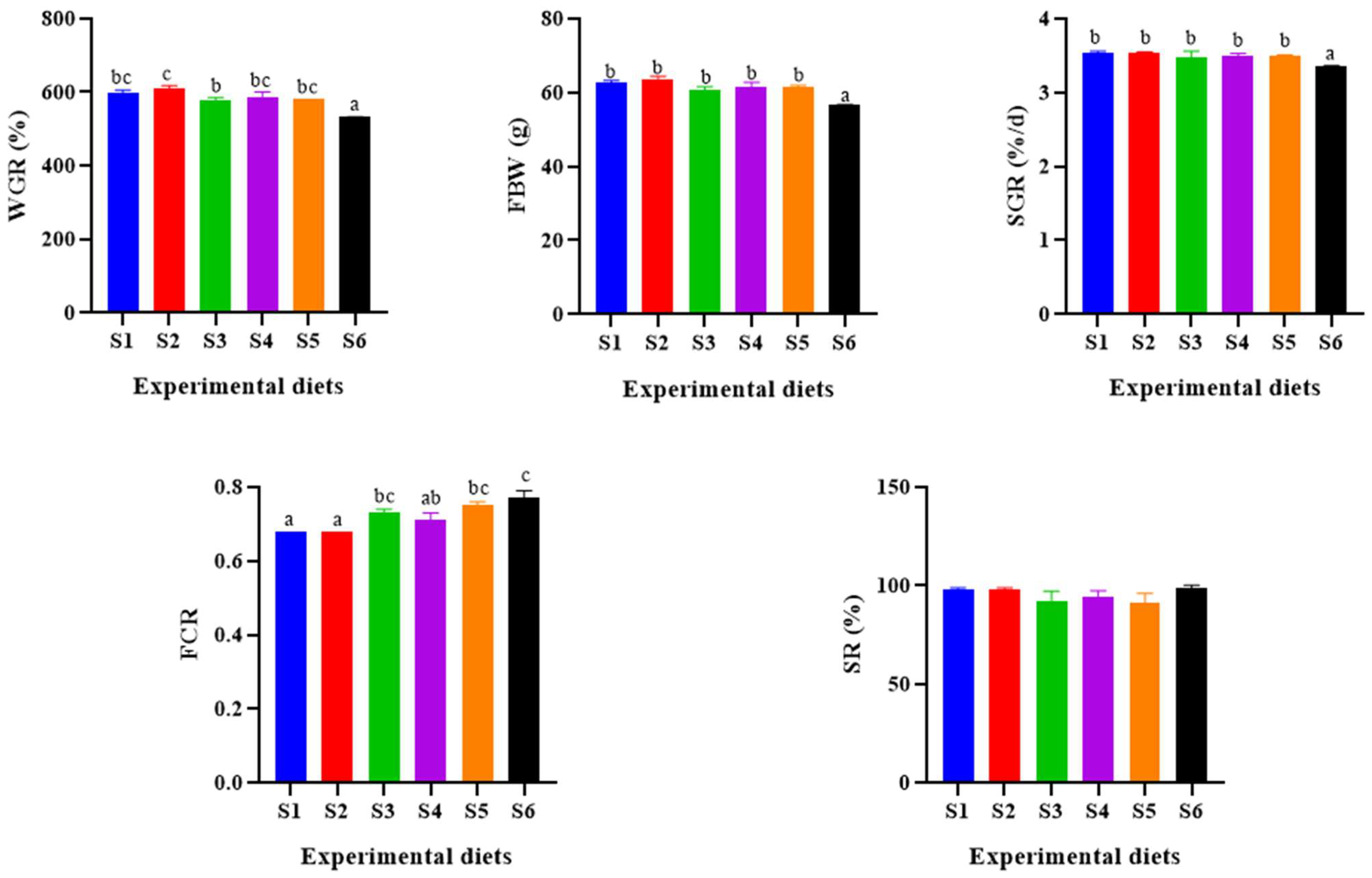
3.1. Growth Performance

The growth performance data are presented in Figure 1, providing insights into the differences among various groups. Notably, the FBW and WGR of the S6 group were significantly lower compared to the other groups (p < 0.05), while the S2 group exhibited the highest FBW, WGR and SGR values. However, these values did not significantly differ from those of the S1 group (p > 0.05). Furthermore, the FCR values for the S3, S5 and S6 groups were notably higher than those for the S1 and S2 groups, with the S6 group having the highest FCR value (p < 0.05). It is worth mentioning that the dietary levels of α-lipoic acid had no significant impact on SR (p > 0.05).

3.2. Serum Antioxidant Indexes

As depicted in Table 3, α-lipoic acid significantly enhanced the activities of antioxidant enzymes, including serum CAT, SOD, and GSH-Px in hybrid grouper. These enhancements were most pronounced in the S5 group, with all three enzymes reaching their maximum levels (p < 0.05). Notably, no significant change was observed in T-AOC.

3.3. Liver Antioxidant and Immune Indexes

As shown in Table 4, α-lipoic acid resulted in a significant reduction of ROS and MDA levels in the liver of hybrid groupers. In addition, liver CAT, SOD enzymes activities and T-AOC content showed a significant increase with increasing α-lipoic acid levels, reaching their peak values in the S5 group (p < 0.05). Furthermore, the enzyme activities of GSH-Px and AKP were significantly higher in all groups except the S3 group than in the S1 group (p < 0.05). Conversely, liver AST, ALT, ACP enzyme activities and IgM content remained largely unchanged despite variations in α-lipoic acid concentration (p > 0.05).

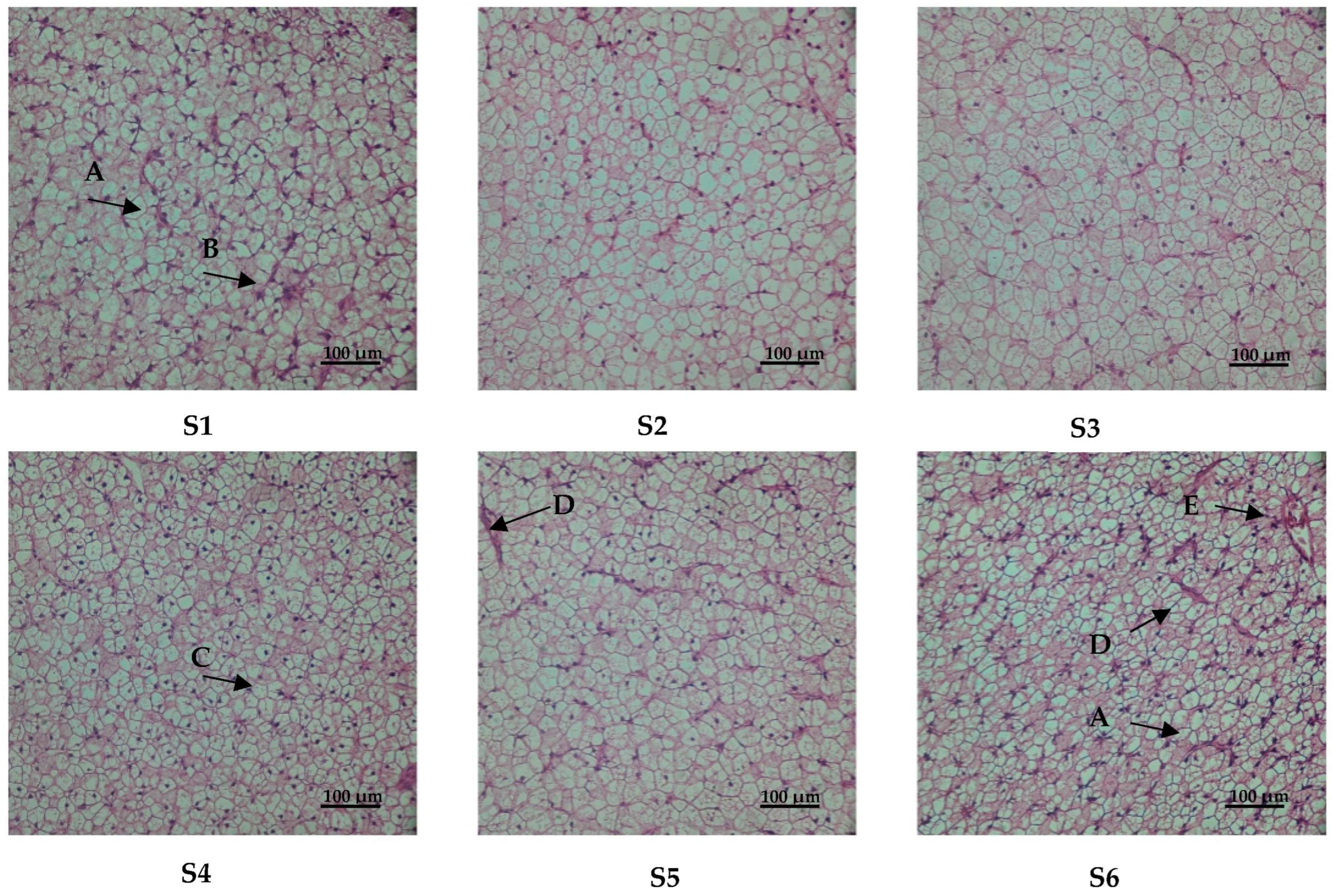
3.4. Liver Histology

The results of HE staining of the liver are shown in Figure 2. The hepatocytes of the S2 and S3 groups had complete hepatic lobules and clear cell outlines. In group S1, we observed karyopyknosis and fat vacuolization. In group S4, hepatocellular vacuolation was observed. Hepatocytes in the S5 group show punctate necrosis. In group S6, there was karyopyknosis, punctate necrosis and inflammation.

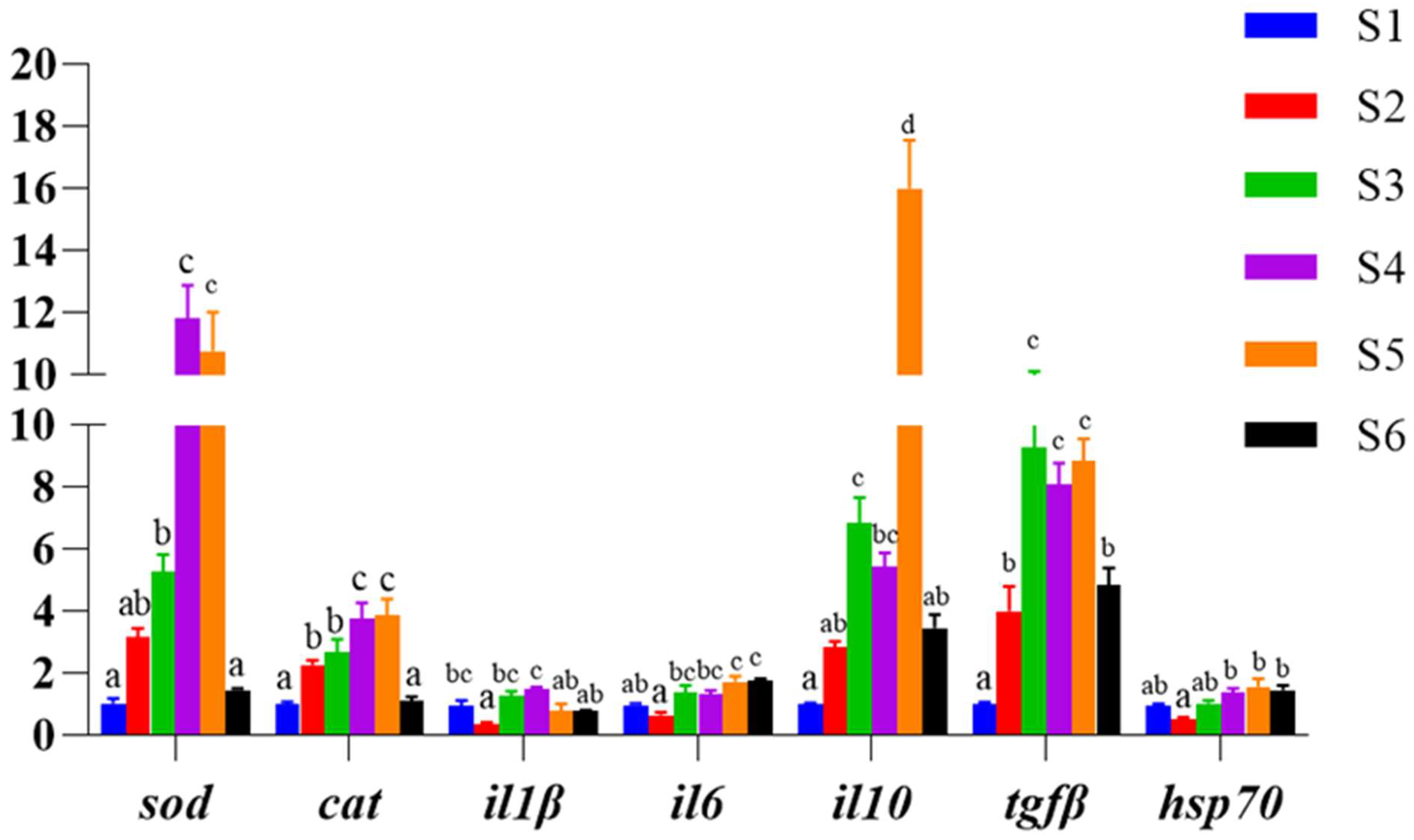
3.5. Antioxidant and Immune-Related Gene Expression in Liver

As shown in Figure 3, the expression of antioxidant genes superoxide dismutase (sod) and catalase (cat) (p < 0.05) was significantly upregulated in the liver except for the S6 group, and the expression of pro-inflammatory cytokine interleukin 1β (il1β) gene was significantly inhibited in the S2 group (p < 0.05), while the expression levels of interleukin 6 (il6) gene mRNA were significantly upregulated in the S5 and S6 groups (p < 0.05). The expression of the anti-inflammatory factors interleukin 10 (il10) and transforming growth factor β (tgfβ) showed a significant upward and then downward trend, and the expression level of il10 in all groups was significantly higher than that in the control group S1, except for the S2 and S6 groups, while the expression of tgfβ in all groups was significantly higher than that in the S1 group (p < 0.05). The expression level of the heat shock protein 70 (hsp70) was the lowest in the S2 group, but there was no significant difference between the treatment groups and the S1 group (p > 0.05).

4. Discussion

In our study, the parameters FBW (final body weight), WGR (weight gain rate) and SGR (specific growth rate) exhibited an increasing trend followed by a decrease with escalating levels of dietary α-lipoic acid supplementation. WGR reached their peak in group S2, showing no significant difference from the control group. However, FBW, WGR and SGR were significantly reduced at 2000 mg/kg alpha lipoic acid levels in the S6 group compared to the control group. Additionally, FCR (feed conversion ratio) in the S3, S5 and S6 groups significantly increased with higher α-lipoic acid levels. This phenomenon could be attributed to the reduced appetite and feeding behavior in fish due to the high concentration of α-lipoic acid, resulting in a significant decline in their overall growth performance. These findings align with previous studies, such as the research on Trachinotus marginatus, where α-lipoic acid inclusion at 316.4–524 mg/kg significantly improved WGR. However, at higher levels (890 and 1367 mg/kg), WGR diminished significantly, and FCR increased markedly [24]. Similarly, adding 1200 mg/kg α-lipoic acid to the diet of grass carp (Ctenopharyngodon idella) resulted in reduced feed intake and decreased FBW and WG [25]. Furthermore, in Haliotis discus hannai, WGR was enhanced when α-lipoic acid was included in the feed, peaking at 800 mg/kg, but declined considerably at 1600 and 3200 mg/kg [16]. In summary, the addition of α-lipoic acid to the diet positively influences animal growth within specific concentration ranges. However, excessive levels of α-lipoic acid can suppress appetite and food intake, ultimately hampering overall growth performance. These observations emphasize the importance of carefully regulating α-lipoic acid supplementation to optimize growth outcomes in fish.
The antioxidant defense mechanism of the body is a set of antioxidant enzymes, including T-AOC, CAT, SOD and GSH-Px, which protect the body from damage caused by reactive oxygen species [26,27]. CAT also plays an important role in the antioxidant defense of the body by converting H2O2 to O2 and water, thus protecting cells from damage caused by hydrogen peroxide [28,29]. In addition, glutathione peroxidase (GSH-Px), an important peroxidolytic enzyme widely present in the organism, reduces toxic peroxides to non-toxic hydroxyl compounds, thus protecting cell structure and function from peroxide interference and damage [29,30]. Excessive fat intake and deposition can aggravate the degree of oxidative damage in the organism [4,31]. The oxidative and antioxidant defense systems in animals are in a dynamic equilibrium under normal physiological conditions, and when the organism is stressed, the intracellular mitochondrial morphology is altered, leading to abnormal function and the production of ROS [32,33]. High levels of ROS tend to attack important intracellular biomolecules such as lipids, proteins and nucleic acids, triggering oxidative stress in the organism [34,35]. Malondialdehyde (MDA) is a product of the peroxidation of polyunsaturated fatty acids, and its level is a measure of the degree of oxidative stress [27,36,37]. In the present experiment, CAT, SOD and GSH-Px enzyme activities in serum and liver, as well as T-AOC in the livers of groupers, were significantly increased with increasing α-lipoic acid concentration, while ROS and MDA levels were significantly decreased. This suggests that α-lipoic acid as an additive can attenuate oxidative damage in the body by enhancing the activity of free radical scavenging enzymes and improving the antioxidant capacity of hybrid grouper. Similarly, a study in grass carp concluded that α-lipoic acid as a feed additive could modulate the antioxidant defense system of the fish, significantly increase the activity of antioxidant enzymes in the liver and serum, reduce MDA accumulation and attenuate the toxic effects of lipid peroxidation [38]. The same findings were also demonstrated in the Chinese mitten crab (Eriocheir sinensis) [39], carp (Cyprinus carpio) [38] and tilapia (Oreochromis niloticus) [12]. This also corresponds to the improvement of the liver structure. Alkaline phosphatase (AKP) plays a crucial role in the regulation of animal metabolic processes, helps to maintain a stable internal environment and organism health [40,41], and is associated with organism growth [42,43,44]. On the other hand, lysozyme (LYZ) has been identified as a significant defense factor for vertebrates against invading microorganisms [44,45,46]. LYZ is effective in lysing Gram-positive bacteria, killing Gram-negative bacteria and promoting phagocytosis, either by regulation or through the activation of polymorphonuclear leukocytes and macrophages [47,48,49]. In the course of our experiment, we observed that α-lipoic acid significantly increased the activities of AKP and LYZ enzymes in the liver. This suggests that the addition of α-lipoic acid may enhance the antimicrobial capacity. Additionally, a similar effect was noted when 600 mg/kg of α-lipoic acid was added to the feed of Nile tilapia, which led to a notable increase in the activity of LYZ in serum [15]. Likewise, the introduction of 351 mg/kg of α-lipoic acid significantly boosted LYZ activity and IgM levels in the serum of grass carp [50]. Collectively, these results indicate that α-lipoic acid has the potential to enhance an organism’s immunity.
The histological liver morphology, as determined through HE staining, serves as a critical indicator for evaluating the physiological well-being of the fish and identifying the presence or absence of liver lesions [51]. Previous research has established that high-lipid diets can induce liver damage, characterized by nucleus displacement, nucleolysis, loss of cell structure and cellular vacuolization [52,53]. Our examination of liver sections revealed partial damage in groups S1, S4, S5 and S6. In a prior study, we successfully mitigated similar liver pathologies by supplementing with VE [54], choline [3] and tea polyphenols [53]. In the current study, α-lipoic acid produced similar beneficial effects, which can likely be attributed to its role as an antioxidant. This role enhances the organism’s antioxidant capacity, effectively scavenging lipid peroxidation resulting from high-fat diets. This, in turn, safeguards the organism’s health and preserves normal liver metabolism.
The activity of antioxidant enzymes plays an important role in the antioxidant defense system, and their activity levels are regulated by antioxidant enzymes-related genes, such as sod and cat. In the present experiment, the mRNA expression levels of both sod and cat were significantly upregulated in the liver with increasing levels of α-lipoic acid addition, which is consistent with the trend of their enzyme activities, again confirming that α-lipoic acid can improve the antioxidant capacity of the organism. It was found that the upregulation of pro-inflammatory factors and the downregulation of anti-inflammatory cytokines leads to inflammatory responses in fish [38]. il1β is an important member of the il1 family, which is of interest due to its important role in inflammation-related diseases [55]. il1β has strong pro-inflammatory activity and induces a variety of pro-inflammatory mediators, such as cytokines and chemokines, and its local activation is central to mediating pro-inflammatory responses that lead to the activation of secondary inflammatory mediators (including il6) [53,56]; il-10 and tgfβ are important anti-inflammatory factors that significantly reduce inflammation by inhibiting the expression of pro-inflammatory factors [15]. In the present study, the expression of the pro-inflammatory factor il1β was significantly downregulated in the S2 group, while the expression of il6 was significantly upregulated in the S5 and S6 groups with increasing α-lipoic acid concentration. Conversely, the mRNA expression of both the anti-inflammatory factor il10 and tgfβ was significantly upregulated, which may represent a feedback response of the organism to the upregulation of pro-inflammatory factors. Under normal physiological conditions, the expression level of hsp70 is typically low, but it rapidly and significantly increases in response to external stimuli [11,57,58]. In this experiment, the hsp70 expression level in all groups did not significantly differ from the S1 group, except in the S2 group, where it was significantly lower than in the S4, S5 and S6 groups. This could be attributed to the organism’s relatively high immunity when α-lipoic acid was added at 400 mg/kg. Currently, α-lipoic acid has been shown to enhance immune responses in mammals, with the addition of 900 mg/kg α-lipoic acid to the diet significantly elevating serum levels of interleukin 2 (il2) and decreasing levels of inflammatory cytokines, such as il1β, il6, and tnfα in fattening pigs [59]. Similarly, the addition of 100 mg/kg α-lipoic acid to feed significantly down-regulated the expression level of tnfα in rat liver [60]. In aquatic animals, the addition of appropriate amounts of α-lipoic acid to feed significantly up-regulated the levels of inflammatory cytokines such as tgfβ1, growth transformation factor β2 (tgfβ2), white fine il10 and interleukin 11 (il11) in the head, kidney and spleen of grass carp [25]. This is also consistent with the results of this experiment. In conclusion, the addition of α-lipoic acid to a high-lipid diet can improve the immunity of the organism by regulating the expression of non-specific related genes and altering enzyme activities.

5. Conclusions

In this experiment, 2000 mg/kg of α-lipoic acid can inhibit grouper growth. At the appropriate level of supplementation (400–800 mg), it can improve the shape of liver cells in the liver, but excess (1600–2000 mg) can lead to inflammation of liver cells and punctate necrosis. In the 400–1600 mg/kg α-lipoic acid groups, antioxidant activity, immunoenzyme activity and expression of antioxidant and immune-related genes could be improved to a certain extent. In summary, the recommended supplemental dose of alpha lipoic acid is 400–800 mg/kg.

Author Contributions

W.H.: Conceptualization, Formal analysis, Investigation, Writing—original draft. T.L.: Conceptualization, Methodology, Investigation. W.C., H.S., H.L., S.Z., M.Z., Y.Y. and B.T.: Methodology, Validation. X.D.: Methodology, Supervision, Writing—review and editing, Project administration, Funding acquisition. All authors have read and agreed to the published version of the manuscript.

Funding

This study was supported financially by the Research and Demonstration of Precision Functional Compound Feed Technology of Major Cultured Fishes and Shrimps in South China (2021B0202050002), Science and Technology Bureau of Zhanjiang (2022A01026), Department of Education of Guangdong Province (2021ZDZX4005), Postgraduate Education Innovation Project of Guangdong Ocean University (202332), and the China Agriculture Research System of MOF and MARA (CARS-47).

Institutional Review Board Statement

The animal protocol utilized in this study was authorized by the Institutional Animal Care and Use Committee (IACUC), Fisheries College, Guangdong Ocean University (protocol code GDOU-IACUC-2021-A2037 and date of April 2021). All experimental procedures involving animals were conducted in compliance with the National Center for the Replacement, Refinement and Reduction of Animals in Research, as well as the ARRIVE guidelines 2.0 and other relevant regulations.

Informed Consent Statement

Not applicable.

Data Availability Statement

The data that support the findings of this study are available on request from the corresponding author. The data are not publicly available due to privacy or ethical restrictions.

Acknowledgments

I thank Xiaohui Dong for her support in my life, study, and scientific research. Thanks for Tingzhen Song.

Conflicts of Interest

The authors declare no conflicts of interest.

References

  1. Liu, X.; He, X.; Huang, G.; Zhou, Y.; Lai, J. Bioremediation by the mullet mugil cephalus feeding on organic deposits produced by intensive shrimp mariculture. Aquaculture 2021, 541, 736674. [Google Scholar] [CrossRef]
  2. Lu, K.; Xu, W.; Li, X.; Liu, W.; Wang, L.; Zhang, C. Hepatic triacylglycerol secretion, lipid transport and tissue lipid uptake in blunt snout bream (Megalobrama amblycephala) fed high-fat diet. Aquaculture 2013, 408–409, 160–168. [Google Scholar] [CrossRef]
  3. Zhe, T.; Dong, L.; Ming, C.; Jun, Z.; Bo, Z.; Yong, S.; Lei, Z.; Yi, H. Low-fishmeal and high-fat diet supplement with soybean lecithin on growth, serum biochemical indexes and intestinal flora of rice field eel (Monopterus albus). Acta Hydrobiol. Sin. 2024, 48, 361–371. [Google Scholar] [CrossRef]
  4. Wu, L.; Liang, H.; Hamunjo, C.M.K.; Ge, X.; Ji, K.; Yu, H.; Huang, D.; Xu, H.; Ren, M. Culture salinity alters dietary protein requirement, whole body composition and nutrients metabolism related genes expression in juvenile genetically improved farmed tilapia (GIFT) (Oreochromis niloticus). Aquaculture 2021, 531, 735961. [Google Scholar] [CrossRef]
  5. Wang, L.; Ma, B.; Chen, D.; Lou, B.; Zhan, W.; Chen, R.; Tan, P.; Xu, D.; Liu, F.; Xie, Q. Effect of dietary level of vitamin e on growth performance, antioxidant ability, and resistance to vibrio alginolyticus challenge in yellow drum Nibea albiflora. Aquaculture 2019, 507, 119–125. [Google Scholar] [CrossRef]
  6. Zhang, C.X.; Huang, F.; Li, J.; Wang, L.; Song, K.; Mai, K.S. Interactive effects of dietary magnesium and vitamin e on growth performance, body composition, blood parameters and antioxidant status in japanese seabass (Lateolabrax japonicus) Fed Oxidized Oil. Aquac. Nutr. 2016, 22, 708–722. [Google Scholar] [CrossRef]
  7. Wei, B.; Yang, Z.; Cheng, Y.; Wang, J.; Zhou, J. Effects of the complete replacement of fish oil with linseed oil on growth, fatty acid composition, and protein expression in the chinese mitten crab (Eriocheir sinensis). Proteome Sci. 2018, 16, 6. [Google Scholar] [CrossRef]
  8. Wu, Z.; Yu, X.; Guo, J.; Fu, Y.; Guo, Y.; Pan, M.; Zhang, W.; Mai, K. Replacement of dietary fish meal with clostridium autoethanogenum protein on growth performance, digestion, mtor pathways and muscle quality of abalone Haliotis Discus Hannai. Aquaculture 2022, 553, 738070. [Google Scholar] [CrossRef]
  9. Presa, N.; Clugston, R.D.; Lingrell, S.; Kelly, S.E.; Merrill, A.H.; Jana, S.; Kassiri, Z.; Gómez-Muñoz, A.; Vance, D.E.; Jacobs, R.L.; et al. Vitamin E alleviates non-alcoholic fatty liver disease in phosphatidylethanolamine n-methyltransferase deficient mice. Biochim. Biophys. Acta Mol. Basis Dis. 2019, 1865, 14–25. [Google Scholar] [CrossRef]
  10. Suo, X.; Yan, X.; Tan, B.; Pan, S.; Li, T.; Liu, H.; Huang, W.; Zhang, S.; Yang, Y.; Dong, X. Lipid metabolism disorders of hybrid grouper (♀ Epinephelus fuscointestinestatus × ♂ E. lanceolatu) induced by high-lipid diet. Front. Mar. Sci. 2022, 9, 990193. [Google Scholar] [CrossRef]
  11. Jia, Y.; Jing, Q.; Niu, H.; Huang, B. Ameliorative Effect of vitamin E on hepatic oxidative stress and hypoimmunity induced by high-fat diet in turbot (Scophthalmus maximus). Fish Shellfish Immunol. 2017, 67, 634–642. [Google Scholar] [CrossRef]
  12. Xu, F.; Xu, C.; Xiao, S.; Lu, M.; Limbu, S.M.; Wang, X.; Du, Z.; Qin, J.G.; Chen, L. Effects of α-lipoic acid on growth performance, body composition, antioxidant profile and lipid metabolism of the GIFT tilapia (Oreochromis niloticus) fed high-fat diets. Aquac. Nutr. 2019, 25, 585–596. [Google Scholar] [CrossRef]
  13. Packer, L.; Witt, E.H.; Tritschler, H.J. Alpha-lipoic acid as a biological antioxidant. Free Radic. Biol. Med. 1995, 19, 227–250. [Google Scholar] [CrossRef] [PubMed]
  14. Xu, W.; Han, S.; Zhou, Q.; Wu, J.; Chen, T. Effect of dietary antioxidant on microcystin-induced toxicosis in crucian. Jiangsu J. Agric. Sci. 2020, 36, 417–422. [Google Scholar] [CrossRef]
  15. Lu, D.L.; Limbu, S.M.; Lv, H.B.; Ma, Q.; Chen, L.Q.; Zhang, M.L.; Du, Z.Y. The Comparisons in protective mechanisms and efficiencies among dietary α-lipoic acid, β-glucan and l-carnitine on nile tilapia infected by Aeromonas Hydrophila. Fish Shellfish Immunol. 2019, 86, 785–793. [Google Scholar] [CrossRef]
  16. Zhang, W.; Chen, Q.; Mai, K.; Xu, W.; Wang, X.; Liufu, Z. Effects of dietary α-lipoic acid on the growth and antioxidative responses of juvenile abalone Haliotis Discus Hannai Ino. Aquac. Res. 2010, 41, e781–e787. [Google Scholar] [CrossRef]
  17. Lei, Y.; Wu, W.; Zhang, Y.; Zhou, H.; Zhang, W.; Mai, K. Protective effect of dietary α-lipoic acid on abalone Haliotis discus hannai ino against the toxicity of waterborne copper. Period. Ocean. Univ. China 2008, 45, 39–45. [Google Scholar] [CrossRef]
  18. Weijun, W.; Xili, L.; Weipeng, C.; Yuhao, H.; Xiuping, F.; Xiaoming, Q. Stress-relieving effect of basil essential oil on temporarily cultured pearl gentian grouper (Epinephelus fuscoguttatusi ♀ × Epinephelus lanceolatus ♂) before transportation. J. Guangdong Ocean Univ. 2023, 43, 43–50. [Google Scholar]
  19. Liang, S.; Lu, Y. Combined Effects of temperature and salinity on activity of serum immune indicators in pearl gentian grouper (Epinephelus fuscoguttatus ♀ × Epinephelus lanceolatus ♂). J. Guangdong Ocean Univ. 2023, 43, 8–16. [Google Scholar] [CrossRef]
  20. Liu, Y.; Wang, J.Y.; Li, B.S.; Qiao, H.J.; Liu, X.D.; Hao, T.T.; Wang, X.Y. Dietary manganese requirement of juvenile hybrid grouper, Epinephelus lanceolatus × E. fuscoguttatus. Aquac. Nutr. 2018, 24, 215–223. [Google Scholar] [CrossRef]
  21. Liu, H.; Li, L.; Akiku, S.; Tang, Z.; Fang, W.; Tan, B.; Dong, X.; Chi, S.; Yang, Q.; Zhang, S.; et al. Effects of dietary yeast culture supplementation on growth, intestinal morphology, immunity, and disease resistance in Epinephelus fuscoguttatus ♀ × Epinephelus lanceolatu ♂. J. Guangdong Ocean Univ. 2021, 41, 1–11. [Google Scholar] [CrossRef]
  22. Hirwitz, W.; Latimer, G. Official methods of analysis of AOAC international (16th Edn). Off. Methods Anal. AOAC Int. 1995, 6, 382. [Google Scholar]
  23. Yang, T.; Huang, Y.; Ru, X.; Li, J.; Zhu, K.; Yang, J.; Chen, P.; Zhu, C. Effects of formula feed replacing chilled fish on growth and liver transcriptome of juvenile Seriola dumerili. J. Guangdong Ocean Univ. 2023, 43, 92–99. [Google Scholar] [CrossRef]
  24. Kütter, M.T.; Monserrat, J.M.; Primel, E.G.; Caldas, S.S.; Tesser, M.B. Effects of dietary α-lipoic acid on growth, body composition and antioxidant status in the plata pompano trachinotus marginatus (Pisces, carangidae). Aquaculture 2012, 368–369, 29–35. [Google Scholar] [CrossRef]
  25. Huang, C.; Sun, J.; Ji, H.; Oku, H.; Chang, Z.; Tian, J.; Yu, E.; Xie, J. Influence of dietary alpha-lipoic acid and lipid level on the growth performance, food intake and gene expression of peripheral appetite regulating factors in juvenile grass carp (Ctenopharyngodon idellus). Aquaculture 2019, 505, 412–422. [Google Scholar] [CrossRef]
  26. Ungvari, Z.; Bagi, Z.; Feher, A.; Recchia, F.A.; Sonntag, W.E.; Pearson, K.; De Cabo, R.; Csiszar, A. Resveratrol confers endothelial protection via activation of the antioxidant transcription factor Nrf2. Am. J. Physiol. Hear. Circ. Physiol. 2010, 299, 18–24. [Google Scholar] [CrossRef] [PubMed]
  27. Yu, Z.; Wu, X.Q.; Zheng, L.J.; Dai, Z.Y.; Wu, L.F. Effect of acute exposure to ammonia and bft alterations on rhynchocypris lagowski: Digestive enzyme, inflammation response, oxidative stress and immunological parameters. Environ. Toxicol. Pharmacol. 2020, 78, 103380. [Google Scholar] [CrossRef]
  28. Yaghoubi, M.; Mozanzadeh, M.T.; Marammazi, J.G.; Safari, O.; Gisbert, E. Dietary replacement of fish meal by soy products (soybean meal and isolated soy protein) in silvery-black porgy juveniles (Sparidentex hasta). Aquaculture 2016, 464, 50–59. [Google Scholar] [CrossRef]
  29. Koruk, M.; Taysi, S.; Savas, M.C.; Yilmaz, O.; Akcay, F.; Karakok, M. Oxidative stress enzymatic antioxidant status in patients with nonalcoholic steatohepatitis. Ann. Clin. Lab. Sci. 2004, 34, 57–62. [Google Scholar]
  30. Wang, L.; Gallagher, E.P. Role of nrf2 antioxidant defense in mitigating cadmium-induced oxidative stress in the olfactory system of zebrafish. Toxicol. Appl. Pharmacol. 2013, 266, 177–186. [Google Scholar] [CrossRef]
  31. Zhang, X.; Han, Z.; Zhong, H.; Yin, Q.; Xiao, J.; Wang, F.; Zhou, Y.; Luo, Y. Regulation of triglyceride synthesis by estradiol in the livers of hybrid tilapia (Oreochromis niloticus ♀ × O. aureus ♂). Comp. Biochem. Physiol. Part B Biochem. Mol. Biol. 2019, 238, 110335. [Google Scholar] [CrossRef]
  32. Zeng, F.; Tee, C.; Liu, M.; Sherry, J.P.; Dixon, B.; Duncker, B.P.; Bols, N.C. The p53/hsp70 inhibitor, 2-phenylethynesulfonamide, causes oxidative stress, unfolded protein response and apoptosis in rainbow trout cells. Aquat. Toxicol. 2014, 146, 45–51. [Google Scholar] [CrossRef]
  33. Sun, S.; Gu, Z.; Fu, H.; Zhu, J.; Ge, X.; Xuan, F. Molecular cloning, characterization, and expression analysis of p53 from the oriental river prawn, Macrobrachium nipponense, in response to hypoxia. Fish Shellfish Immunol. 2016, 54, 68–76. [Google Scholar] [CrossRef]
  34. Li, W.; Li, L.; Liu, H.; Tan, B.; Dong, X.; Yang, Q.; Chi, S.; Zhang, S.; Xie, R. Effects of clostridium butyricum on growth, antioxidant capacity and non-specific immunology of Litopenaeus vannamei fed with concentrated cottonseed protein replacement of fishmeal. J. Guangdong Ocean Univ. 2022, 42, 29–37. [Google Scholar] [CrossRef]
  35. Mohd Faudzi, N.; Yong, A.S.K.; Shapawi, R.; Senoo, S.; Biswas, A.; Takii, K. Soy Protein concentrate as an alternative in replacement of fish meal in the feeds of hybrid grouper, brown-marbled grouper (Epinephelus fuscoguttatus) × giant grouper (E. lanceolatus) juvenile. Aquac. Res. 2018, 49, 431–441. [Google Scholar] [CrossRef]
  36. Feng, L.; Chen, Y.P.; Jiang, W.D.; Liu, Y.; Jiang, J.; Wu, P.; Zhao, J.; Kuang, S.Y.; Tang, L.; Tang, W.N.; et al. Modulation of immune response, physical barrier and related signaling factors in the gills of juvenile grass carp (Ctenopharyngodon idella) fed supplemented diet with phospholipids. Fish Shellfish Immunol. 2016, 48, 79–93. [Google Scholar] [CrossRef] [PubMed]
  37. Chen, Q.Q.; Liu, W.B.; Zhou, M.; Dai, Y.J.; Xu, C.; Tian, H.Y.; Xu, W.N. Effects of berberine on the growth and immune performance in response to ammonia stress and high-fat dietary in blunt snout bream Megalobrama amblycephala. Fish Shellfish Immunol. 2016, 55, 165–172. [Google Scholar] [CrossRef] [PubMed]
  38. Shi, X.; Jin, A.; Sun, J.; Yang, Z.; Tian, J.; Ji, H.; Yu, H.; Li, Y.; Zhou, J.; Du, Z.; et al. α-lipoic acid ameliorates n-3 highly-unsaturated fatty acids induced lipid peroxidation via regulating antioxidant defenses in grass carp (Ctenopharyngodon idellus). Fish Shellfish Immunol. 2017, 67, 359–367. [Google Scholar] [CrossRef]
  39. Xu, C.; Li, E.; Liu, S.; Huang, Z.; Qin, J.G.; Chen, L. Effects of α-lipoic acid on growth performance, body composition, antioxidant status and lipid catabolism of juvenile chinese mitten crab Eriocheir sinensis fed different lipid percentage. Aquaculture 2018, 484, 286–292. [Google Scholar] [CrossRef]
  40. Wang, X.; Zeng, B.; Liu, L.; Yang, R.; Liu, H. Effects of feed protein levels on growth, digestive enzyme activities, non-specific immunity and protein metabolism of Schizothorax O’Connori. Acta Hydrobiol. Sin. 2020, 44, 693–706. [Google Scholar] [CrossRef]
  41. Tang, Z.; Chen, H.; He, H.; Ma, C. Assays for alkaline phosphatase activity: Progress and prospects. TrAC Trends Anal. Chem. 2019, 113, 32–43. [Google Scholar] [CrossRef]
  42. Zhou, Y.L.; Guo, J.L.; Tang, R.J.; Ma, H.J.; Chen, Y.J.; Lin, S.M. High dietary lipid level alters the growth, hepatic metabolism enzyme, and anti-oxidative capacity in juvenile largemouth bass Micropterus salmoides. Fish Physiol. Biochem. 2020, 46, 125–134. [Google Scholar] [CrossRef] [PubMed]
  43. Lallès, J.P. Intestinal alkaline phosphatase in the gastrointestinal tract of fish: Biology, ontogeny, and environmental and nutritional modulation. Rev. Aquac. 2020, 12, 555–581. [Google Scholar] [CrossRef]
  44. Dossou, S.; Koshio, S.; Ishikawa, M.; Yokoyama, S.; Dawood, M.A.O.; El Basuini, M.F.; El-Hais, A.M.; Olivier, A. Effect of partial replacement of fish meal by fermented rapeseed meal on growth, immune response and oxidative condition of red sea bream juvenile, Pagrus Major. Aquaculture 2018, 490, 228–235. [Google Scholar] [CrossRef]
  45. Zhuo, L.C.; Chen, C.F.; Lin, Y.H. Dietary supplementation of fermented lemon peel enhances lysozyme activity and susceptibility to photobacterium damselae for orange-spotted grouper, Epinephelus coioides. Fish Shellfish Immunol. 2021, 117, 248–252. [Google Scholar] [CrossRef]
  46. Niu, H.; Jia, Y.; Hu, P.; Meng, Z.; Lei, J. Effect of dietary vitamin E on the growth performance and nonspecific immunity in sub-adult turbot (Scophthalmus maximus). Fish Shellfish Immunol. 2014, 41, 501–506. [Google Scholar] [CrossRef]
  47. Novriadi, R.; Ilham, I.; Roigé, O.; Segarra, S. Effects of dietary nucleotides supplementation on growth, total haemocyte count, lysozyme activity and survival upon challenge with Vibrio harveyi in pacific white shrimp, Litopenaeus vannamei. Aquac. Rep. 2021, 21, 100840. [Google Scholar] [CrossRef]
  48. Ma, F.; Li, X.Q.; Li, B.A.; Leng, X.J. Effects of extruded and pelleted diets with differing lipid levels on growth, nutrient retention and serum biochemical indices of tilapia (Oreochromis aureus × Tilapia nilotica). Aquac. Nutr. 2016, 22, 61–71. [Google Scholar] [CrossRef]
  49. Wang, C.; Li, J.; Wang, L.; Zhao, Z.; Luo, L.; Du, X.; Yin, J.; Xu, Q. Effects of dietary phosphorus on growth, body composition and immunity of young taimen Hucho taimen (Pallas, 1773). Aquac. Res. 2017, 48, 3066–3079. [Google Scholar] [CrossRef]
  50. Liu, H.X.; Zhou, X.Q.; Jiang, W.D.; Wu, P.; Liu, Y.; Zeng, Y.Y.; Jiang, J.; Kuang, S.Y.; Tang, L.; Feng, L. Optimal α-lipoic acid strengthen immunity of young grass carp (Ctenopharyngodon idella) by enhancing immune function of head kidney, spleen and skin. Fish Shellfish Immunol. 2018, 80, 600–617. [Google Scholar] [CrossRef]
  51. Barja-Fernández, S.; Míguez, J.M.; Álvarez-Otero, R. Histopathological effects of 2,2’,4,4’-tetrabromodiphenyl ether (bde-47) in the gills, intestine and liver of turbot (Psetta maxima). Ecotoxicol. Environ. Saf. 2013, 95, 60–68. [Google Scholar] [CrossRef] [PubMed]
  52. Huang, T.W.; Chang, C.L.; Kao, E.S.; Lin, J.H. Effect of hibiscus sabdariffa extract on high fat diet-induced obesity and liver damage in hamsters. Food Nutr. Res. 2015, 59, 29018. [Google Scholar] [CrossRef] [PubMed]
  53. Pan, S.; Yan, X.; Li, T.; Suo, X.; Liu, H.; Tan, B.; Huang, W.; Yang, Y.; Zhang, H.; Dong, X. Impacts of tea polyphenols on growth, antioxidant capacity and immunity in juvenile hybrid grouper (Epinephelus fuscoguttatus ♀ × E. lanceolatus ♂) fed high-lipid diets. Fish Shellfish Immunol. 2022, 128, 348–359. [Google Scholar] [CrossRef] [PubMed]
  54. Yan, X.; Pan, S.; Dong, X.; Tan, B.; Li, T.; Huang, W.; Suo, X.; Li, Z.; Yang, Y. Vitamin E amelioration of oxidative stress and low immunity induced by high-lipid diets in hybrid grouper (♀ Epinephelus fuscoguttatus × ♂ E. lanceolatu). Fish Shellfish Immunol. 2022, 124, 156–163. [Google Scholar] [CrossRef]
  55. Vazirzadeh, A.; Marhamati, A.; Rabiee, R.; Faggio, C. Immunomodulation, antioxidant enhancement and immune genes up-regulation in rainbow trout (Oncorhynchus mykiss) fed on seaweeds included diets. Fish Shellfish Immunol. 2020, 106, 852–858. [Google Scholar] [CrossRef] [PubMed]
  56. Enamorado, A.D.; Martins, A.C.; Flores, J.A.; Tesser, M.B.; Caldas, S.S.; Primel, E.G.; Monserrat, J.M. Biochemical responses over time in common carp cyprinus carpio (Teleostei, Cyprinidae) during fed supplementation with α-lipoic acid. Comp. Biochem. Physiol. Part A Mol. Integr. Physiol. 2015, 188, 9–16. [Google Scholar] [CrossRef] [PubMed]
  57. Asea, A.; Rehli, M.; Kabingu, E.; Boch, J.A.; Baré, O.; Auron, P.E.; Stevenson, M.A.; Calderwood, S.K. Novel signal transduction pathway utilized by extracellular hsp70. role of toll-like receptor (tlr) 2 and tlr4. J. Biol. Chem. 2002, 277, 15028–15034. [Google Scholar] [CrossRef]
  58. Kucukbay, F.Z.; Yazlak, H.; Karaca, I.; Sahin, N.; Tuzcu, M.; Cakmak, M.N.; Sahin, K. The effects of dietary organic or inorganic selenium in rainbow trout (Oncorhynchus mykiss) under crowding conditions. Aquac. Nutr. 2009, 15, 569–576. [Google Scholar] [CrossRef]
  59. Wang, S.; Shi, J.; Yang, J.; Xing, J.; Wu, J. Effect of dietary alpha-lipoic acid on thyroid hormones, inflammatory cytokines and antioxidant ability in finishing pigs under oxidative stress. Feed Res. 2020, 45, 56–60. [Google Scholar] [CrossRef]
  60. Sun, H.; Ye, C.; Li, P.; Liu, Y.; Gao, L. Effects of Lipoic acid on intestinal redox status and digestive and absorption functions in mice fed a high-fat diet. Chin. J. Gerontol. 2020, 40, 6–9. [Google Scholar] [CrossRef]
Figure 1. Effect of α-lipoic acid levels on growth performance of hybrid grouper. WGR, weight gain rate; FBW, final body weight; SGR, specific growth rate; FCR, feed conversion ratio; SR, survival rate. S1, control group. S2, 0.04% α-lipoic acid supplement group; S3, 0.08% α-lipoic acid supplement group; S4, 0.12% α-lipoic acid supplement group; S5, 0.16% α-lipoic acid supplement group; S6, 0.2% α-lipoic acid supplement group. Notes: Different letters assigned to the bars represent significant differences (p < 0.05).
Antioxidants 13 00088 g001
Figure 2. Effect of α-lipoic acid levels on the histological structure of liver (HE 400×). Notes: A, karyopyknosis; B, fat vacuolization; C, hepatocellular vacuolation; D, punctate necrosis; E, inflammation. S1, control group. S2, 0.04% α-lipoic acid supplement group; S3, 0.08% α-lipoic acid supplement group; S4, 0.12% α-lipoic acid supplement group; S5, 0.16% α-lipoic acid supplement group; S6, 0.2% α-lipoic acid supplement group.
Antioxidants 13 00088 g002
Figure 3. Effect of α-lipoic acid levels on relative gene expression of hepatic antioxidant-related and immune-related in hybrid grouper. Values are means ± SE (n = 3). S1, control group. S2, 0.04% α-lipoic acid supplement group; S3, 0.08% α-lipoic acid supplement group; S4, 0.12% α-lipoic acid supplement group; S5, 0.16% α-lipoic acid supplement group; S6, 0.2% α-lipoic acid supplement group. Notes: Different letters assigned to the bars represent significant differences (p < 0.05).
Antioxidants 13 00088 g003
Table 1. Ingredient composition and nutrient content of the test diets (% dry matter).
IngredientS1S2S3S4S5S6
Fish meal434343434343
Wheat gluten101010101010
Casein121212121212
Wheat flour171717171717
Soybean lecithin1.51.51.51.51.51.5
Fish oil555555
Corn oil777777
Gelatinized starch0.70.660.620.580.540.5
Compound premix a111111
Vitamin C0.050.050.050.050.050.05
Choline chloride0.50.50.50.50.50.5
Ca(H2PO4)21.51.51.51.51.51.5
Antioxidant b0.10.10.10.10.10.1
Attractant c0.150.150.150.150.150.15
CMC d0.50.50.50.50.50.5
α-lipoic acid00.040.080.120.160.2
Total100100100100100100
Proximate composition e
Moisture9.2310.0910.959.89.3810.44
Crude protein 50.6251.1851.5550.9450.8450.06
Crude lipid16.3815.7216.1415.7516.1616.2
Ash 12.7412.1512.0912.8612.4512.79
a Compound premix was obtained from Qingdao Master Biotechnology Co, Ltd. (Qingdao, China). b Antioxidant: ethoxyquin. c Attractant composition:taurine:glycine:betaine = 1:3:3. d Carboxymethylcellulose sodium. e Measured value.
Table 2. Primers of RT-Qpcr.
Genes5=/3 = Forward Primer5=/3 = Reverse PrimerAmpliconE-Value %Genbank No.
β-actinACTGCTGCCTCCTCTTCATCACCGCAAGACTCCATACCAA13593.71KU746361.1
sodTGGAAACACCTTTCCCCCACCTGACAGGGTAAAGCATGGC12091.41AY735008.1
catCGCGGGAAGCAAAGATTCAGCCGCAGTTTCCAGTGTGTTG194104.32KT884509.1
il6AGGAAGTCTGGCTGTCAGGAGCCCTGAGGCCTTCAAGATT25095.06JN806222.1
tgfβCGATGTCACTGACGCCCTGCAGCCGCGGTCATCACTTATC10790.00GQ205390.1
il1βCGACATGGTGCGGTTTCTCTGTAGCGGCTGGTGG15191.95EF582837.1
il10ACACAGCGCTGCTAGACGAGGGGCAGCACCGTGTTCAGAT10491.86KJ741852.1
hsp70CTTGCAAGAAGTGGCCAACAAAAGCCATCTTCCTGCCTTGT13194.03EU816600.1
Notes: sod, superoxide dismutase; cat, catalase; il6, interleukin 6; tgfβ, transforming growth factor β; il-1β, interleukin 1β; il10, interleukin 10; hsp70, heat shock protein 70.
Table 3. Effect of α-lipoic acid levels on serum antioxidant parameters.
GroupCAT (U/mL)T-AOC (U/mL)SOD (U/mL)GSH-Px (U/L)
S125.21 ± 2.06 a13.13 ± 0.9662.06 ± 3.33 a62.89 ± 3.53 a
S229.02 ± 2.01 ab12.47 ± 0.3483.4 ± 2.94 b73.79 ± 8.01 ab
S330.07 ± 1.91 ab13.01 ± 0.5487.88 ± 4.63 b75.53 ± 4.56 abc
S434.51 ± 3.41 b14.21 ± 1.6797.16 ± 6.16 b92.41 ± 4.99 cd
S544.51 ± 2.95 c15.61 ± 1.26117.03 ± 7.68 c102.04 ± 5.53 d
S636.51 ± 1.53 b14.46 ± 1.0490.71 ± 4.41 b82.18 ± 6.25 bc
Notes: Values in the table are means ± SEM (n = 3); Values in the same column with the same superscript letter or absence of superscripts are not significantly different (p > 0.05). S1, control group. S2, 0.04% α-lipoic acid supplement group; S3, 0.08% α-lipoic acid supplement group; S4, 0.12% α-lipoic acid supplement group; S5, 0.16% α-lipoic acid supplement group; S6, 0.2% α-lipoic acid supplement group.
Table 4. Effects of α-lipoic acid levels on liver antioxidant and immunity parameters.
GroupS1S2S3S4S5S6
ROS
(U/mg.pro)
352.65 ± 4.44 c370.31 ± 9.79 c292.77 ± 27.48 b225.17 ± 13.39 a253.06 ± 10.86 ab269.87 ± 5.34 ab
MDA
(nmol/mg.pro)
15.98 ± 0.81 c16.03 ± 1.34 c13.29 ± 0.82 b9.65 ± 0.68 a7.84 ± 0.26 a8.93 ± 0.4 a
CAT
(U/mg.pro)
38.44 ± 5.69 a60.74 ± 6.29 b52.33 ± 4.51 ab62.3 ± 3.32 b79.08 ± 3.28 c56.45 ± 4.45 b
T-AOC
(U/mg.pro)
9.55 ± 0.61 a15.98 ± 1.19 b15.55 ± 1.62 b15.67 ± 1.17 b21.28 ± 1.01 c16.19 ± 0.67 b
SOD
(U/mg.pro)
118.27 ± 2.52 a174.78 ± 1.01 b168.09 ± 22.81 b166.15 ± 10.66 b179.57 ± 2.41 b163.09 ± 16.62 b
GSH-Px
(mU/mg.pro)
125.57 ± 11.45 a178.02 ± 18.18 b150.48 ± 6.5 ab165.11 ± 15.59 b178 ± 8.01 b168.58 ± 4.94 b
AKP
(mIU/mg.pro)
11.89 ± 1.95 a20.32 ± 2.2 b16.14 ± 0.8 ab21.02 ± 0.27 b17.88 ± 1.45 b19.13 ± 1.19 b
ACP
(mU/mg.pro)
14.80 ± 1.2212.65 ± 1.2812.6 ± 1.6114.75 ± 0.1714.18 ± 1.3911.9 ± 1.05
AST
(mU/mg.pro)
20.11 ± 1.2817.18 ± 1.5717.53 ± 2.5417.84 ± 0.2415.03 ± 2.6216.31 ± 1.07
ALT
(mU/mg.pro)
9.54 ± 0.610.86 ± 1.459.24 ± 1.498.85 ± 1.467.72 ± 1.019.04 ± 0.47
IgM
(ug/mg.pro)
49.58 ± 3.5254.84 ± 4.3651.17 ± 1.8552.17 ± 4.756.94 ± 5.2451.14 ± 2.31
LYZ
(mU/mg.pro)
6.49 ± 0.62 a9.87 ± 0.34 c8.12 ± 0.53 b9.00 ± 0.56 bc9.51 ± 0.56 bc9.24 ± 0.23 bc
Notes: Values in the table are means ± SEM (n = 3); Values in the same column with the same superscript letter or absence of superscripts are not significantly different (p > 0.05). S1, control group. S2, 0.04% α-lipoic acid supplement group; S3, 0.08% α-lipoic acid supplement group; S4, 0.12% α-lipoic acid supplement group; S5, 0.16% α-lipoic acid supplement group; S6, 0.2% α-lipoic acid supplement group.
Disclaimer/Publisher’s Note: The statements, opinions and data contained in all publications are solely those of the individual author(s) and contributor(s) and not of MDPI and/or the editor(s). MDPI and/or the editor(s) disclaim responsibility for any injury to people or property resulting from any ideas, methods, instructions or products referred to in the content.

