
Systemic Oxidative Stress in Severe Early-Onset Fetal Growth Restriction Associates with Concomitant Pre-Eclampsia, Not with Severity of Fetal Growth Restriction

 , , , , , ,
  ,
,
Abstract
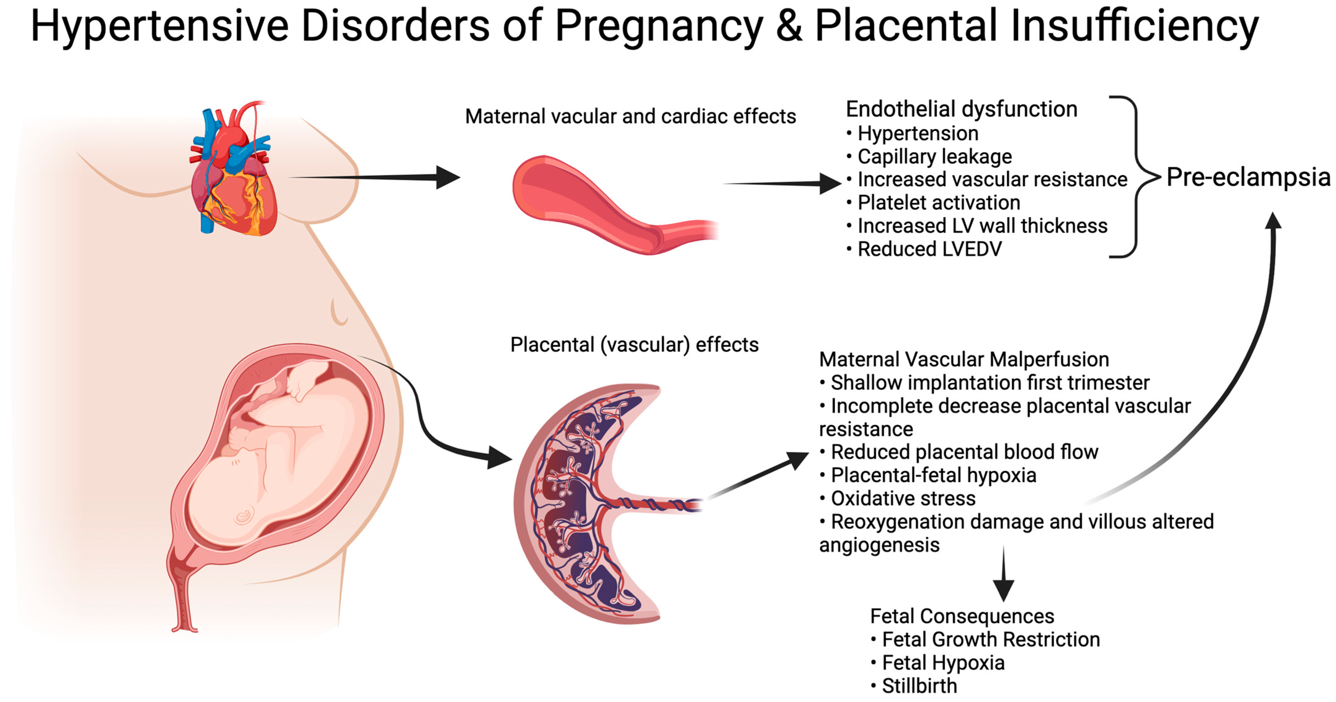
1. Introduction
2. Materials and Methods
2.1. Original Study Design
2.2. Serum Sample Collection
2.3. Laboratory Measurements
2.4. Statistical Analysis
2.5. Ethical Considerations
3. Results
3.1. Cohort Characteristics
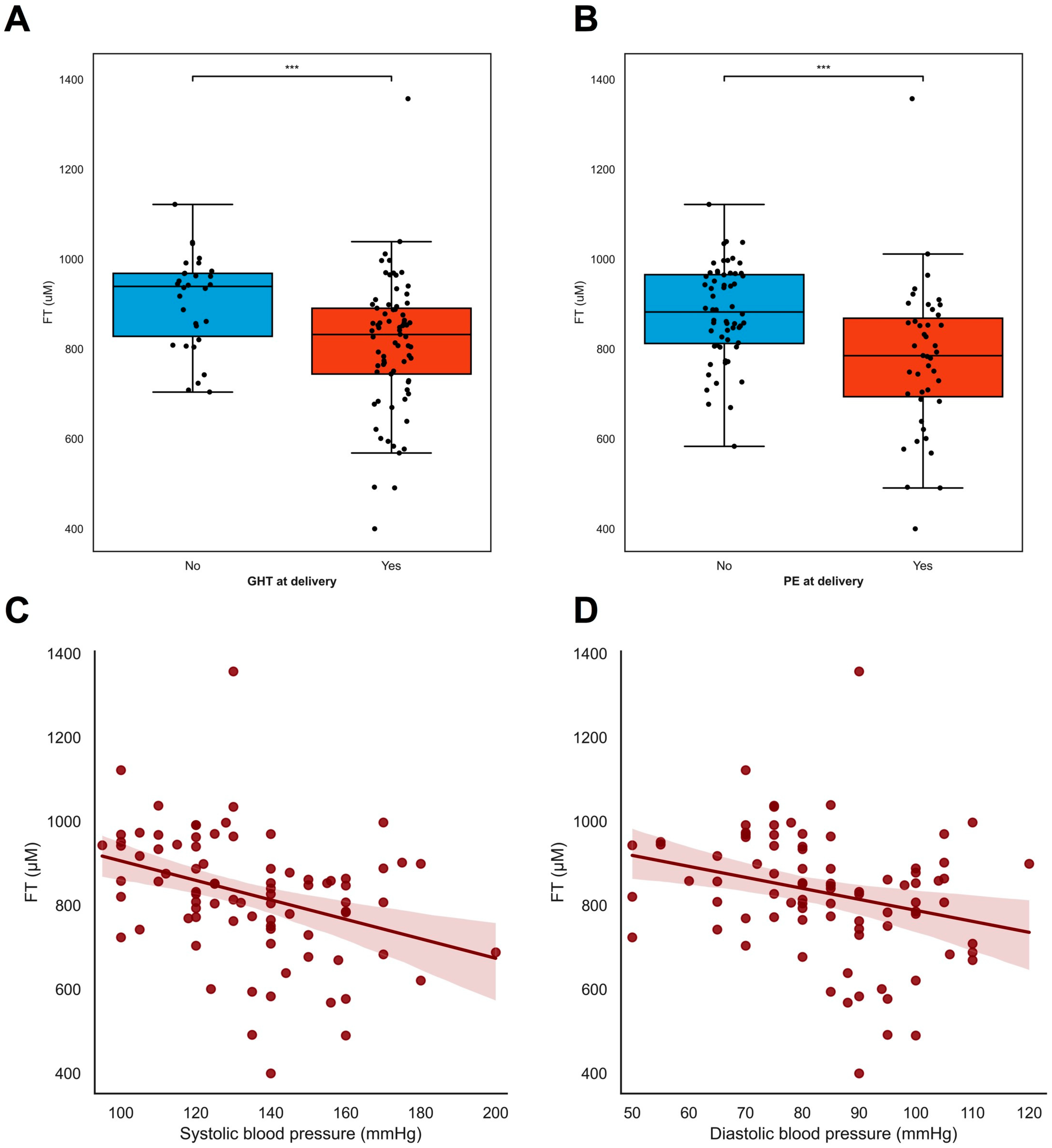
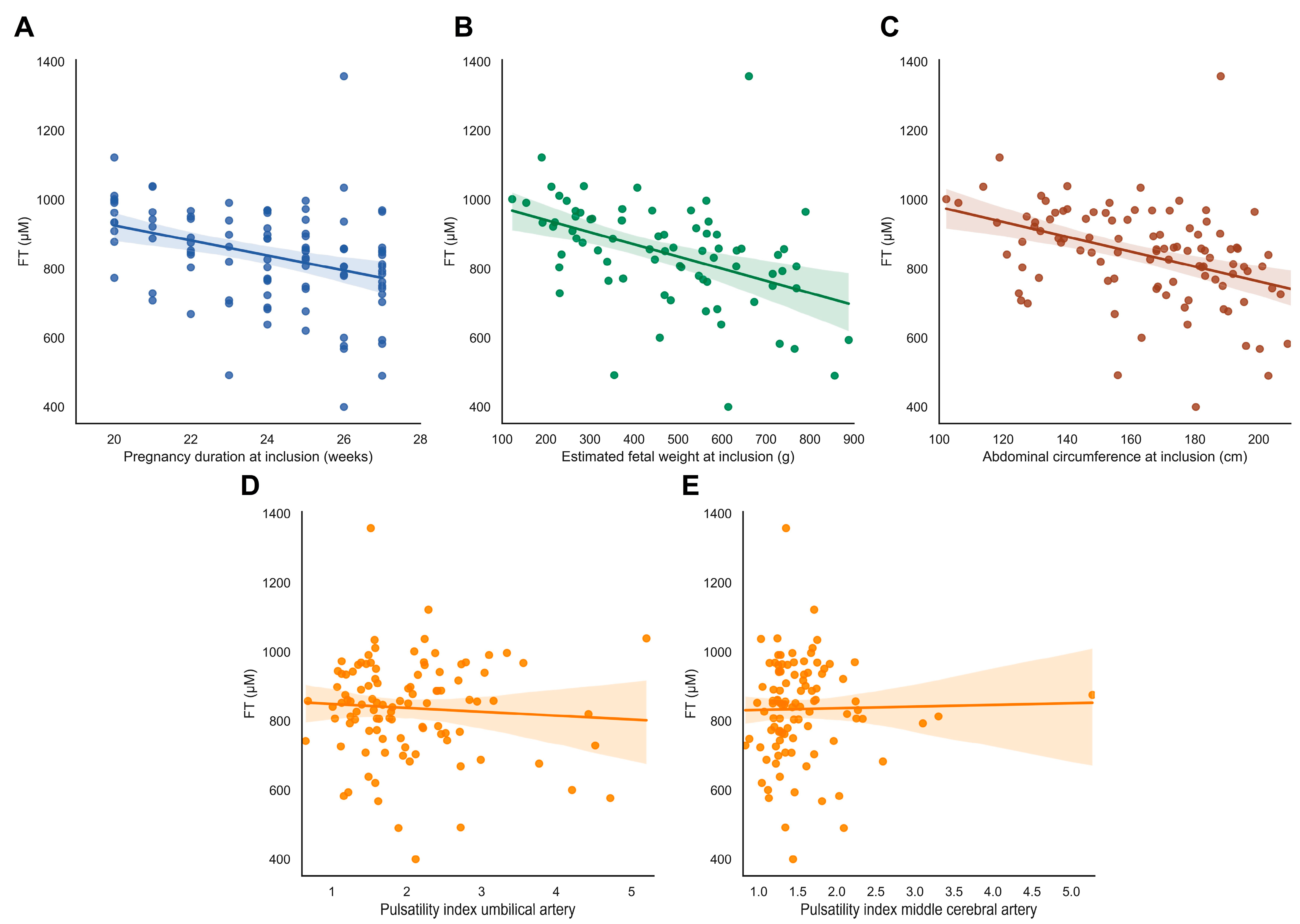
3.2. Oxidative Stress: Plasma Free Thiols (FT) as a Biomarker
4. Discussion
5. Conclusions
Supplementary Materials
Author Contributions
Funding
Institutional Review Board Statement
Informed Consent Statement
Data Availability Statement
Conflicts of Interest
Correction Statement
References
- Spencer, R.; Rossi, C.; Lees, M.; Peebles, D.; Brocklehurst, P.; Martin, J.; Hansson, S.R.; Hecher, K.; Marsal, K.; Figueras, F.; et al. Achieving orphan designation for placental insufficiency: Annual incidence estimations in Europe. BJOG Int. J. Obstet. Gynaecol. 2018, 126, 1157–1167. [Google Scholar] [CrossRef] [PubMed]
- Pels, A.; Beune, I.M.; Van Wassenaer-Leemhuis, A.G.; Limpens, J.; Ganzevoort, W. Early-onset fetal growth restriction: A systematic review on mortality and morbidity. Acta Obstet. Gynecol. Scand. 2019, 99, 153–166. [Google Scholar] [CrossRef] [PubMed]
- Kovo, M.; Schreiber, L.; Ben-Haroush, A.; Gold, E.; Golan, A.; Bar, J. The placental component in early-onset and late-onset preeclampsia in relation to fetal growth restriction. Prenat. Diagn. 2012, 32, 632–637. [Google Scholar] [CrossRef] [PubMed]
- Kovo, M.; Schreiber, L.; Elyashiv, O.; Ben-Haroush, A.; Abraham, G.; Bar, J. Pregnancy Outcome and Placental Findings in Pregnancies Complicated by Fetal Growth Restriction with and without Preeclampsia. Reprod. Sci. 2014, 22, 316–321. [Google Scholar] [CrossRef] [PubMed]
- Beebe, L.A.; Cowan, L.D.; Altshuler, G. The epidemiology of placental features: Associations with gestational age and neonatal outcome. Obstet. Gynecol. 1996, 87, 771–778. [Google Scholar] [CrossRef] [PubMed]
- Kovo, M.; Schreiber, L.; Ben-Haroush, A.; Cohen, G.; Weiner, E.; Golan, A.; Bar, J. The placental factor in early- and late-onset normotensive fetal growth restriction. Placenta 2013, 34, 320–324. [Google Scholar] [CrossRef] [PubMed]
- Silver, R.M. Examining the link between placental pathology, growth restriction, and stillbirth. Best Pract. Res. Clin. Obstet. Gynaecol. 2018, 49, 89–102. [Google Scholar] [CrossRef]
- Spinillo, A.; Gardella, B.; Adamo, L.; Muscettola, G.; Fiandrino, G.; Cesari, S. Pathologic placental lesions in early and late fetal growth restriction. Acta Obstet. Gynecol. Scand. 2019, 98, 1585–1594. [Google Scholar] [CrossRef]
- Burton, G.J.; Jauniaux, E. Pathophysiology of placental-derived fetal growth restriction. Am. J. Obstet. Gynecol. 2018, 218, S745–S761. [Google Scholar] [CrossRef]
- Bustamante Helfrich, B.; Chilukuri, N.; He, H.; Cerda, S.R.; Hong, X.; Wang, G.; Pearson, C.; Burd, I.; Wang, X. Maternal vascular malperfusion of the placental bed associated with hypertensive disorders in the Boston Birth Cohort. Placenta 2017, 52, 106–113. [Google Scholar] [CrossRef]
- Romero, R.; Kim, Y.M.; Pacora, P.; Kim, C.J.; Benshalom-Tirosh, N.; Jaiman, S.; Bhatti, G.; Kim, J.-S.; Qureshi, F.; Jacques, S.M.; et al. The frequency and type of placental histologic lesions in term pregnancies with normal outcome. JPME 2018, 46, 613–630. [Google Scholar] [CrossRef]
- Wright, E.; Audette, M.C.; Ye, X.Y.; Keating, S.; Hoffman, B.; Lye, S.J.; Shah, P.S.; Kingdom, J.C. Maternal Vascular Malperfusion and Adverse Perinatal Outcomes in Low-Risk Nulliparous Women. Obstet. Gynecol. 2017, 130, 1112–1120. [Google Scholar] [CrossRef] [PubMed]
- Vedmedovska, N.; Rezeberga, D.; Teibe, U.; Melderis, I.; Donders, G.G. Placental pathology in fetal growth restriction. Eur. J. Obstet. Gynecol. Reprod. Biol. 2011, 155, 36–40. [Google Scholar] [CrossRef] [PubMed]
- Tajik, P.; van Wyk, L.; Boers, K.E.; le Cessie, S.; Zafarmand, M.H.; Roumen, F.; van der Post, J.A.; Porath, M.; van Pampus, M.G.; Spaanderdam, M.E.; et al. Which intrauterine growth restricted fetuses at term benefit from early labour induction? A secondary analysis of the DIGITAT randomised trial. Eur. J. Obstet. Gynecol. Reprod. Biol. 2013, 172, 20–25. [Google Scholar] [CrossRef] [PubMed][Green Version]
- Aditya, I.; Tat, V.; Sawana, A.; Mohamed, A.; Tuffner, R.; Mondal, T. Use of Doppler velocimetry in diagnosis and prognosis of intrauterine growth restriction (IUGR): A Review. J. Neonatal. Perinatal. Med. 2016, 9, 117–126. [Google Scholar] [CrossRef] [PubMed]
- Burton, G.J.; Hempstock, J.; Jauniaux, E. Oxygen, early embryonic metabolism and free radical-mediated embryopathies. Reprod. Biomed. Online 2003, 6, 84–96. [Google Scholar] [CrossRef] [PubMed]
- Fogarty, N.M.; Ferguson-Smith, A.C.; Burton, G.J. Syncytial Knots (Tenney-Parker Changes) in the Human Placenta: Evidence of loss of transcriptional activity and oxidative damage. Am. J. Pathol. 2013, 183, 144–152. [Google Scholar] [CrossRef]
- Burton, G.J.; Redman, C.W.; Roberts, J.M.; Moffett, A. Pre-eclampsia: Pathophysiology and clinical implications. BMJ 2019, 366, l2381. [Google Scholar] [CrossRef]
- Burton, G.J.; Jauniaux, E. Oxidative stress. Best Pract. Res. Clin. Obstet. Gynaecol. 2011, 25, 287–299. [Google Scholar] [CrossRef]
- Turell, L.; Radi, R.; Alvarez, B. The thiol pool in human plasma: The central contribution of albumin to redox processes. Free. Radic. Biol. Med. 2013, 65, 244–253. [Google Scholar] [CrossRef]
- Raijmakers, M.T.; Roes, E.M.; Poston, L.; Steegers, E.A.; Peters, W.H. The transient increase of oxidative stress during normal pregnancy is higher and persists after delivery in women with pre-eclampsia. Eur. J. Obstet. Gynecol. Reprod. Biol. 2008, 138, 39–44. [Google Scholar] [CrossRef] [PubMed]
- Bos, M.; Schoots, M.H.; Fernandez, B.O.; Mikus-Lelinska, M.; Lau, L.C.; Eikmans, M.; van Goor, H.; Gordijn, S.J.; Pasch, A.; Feelisch, M.; et al. Reactive Species Interactome Alterations in Oocyte Donation Pregnancies in the Absence and Presence of Pre-Eclampsia. Int. J. Mol. Sci. 2019, 20, 1150. [Google Scholar] [CrossRef] [PubMed]
- Abdulle, A.E.; Bourgonje, A.R.; Kieneker, L.M.; Koning, A.M.; Gemert, S.l.B.-V.; Bulthuis, M.L.C.; Dijkstra, G.; Faber, K.N.; Dullaart, R.P.F.; Bakker, S.J.L.; et al. Serum free thiols predict cardiovascular events and all-cause mortality in the general population: A prospective cohort study. BMC Med. 2020, 18, 130. [Google Scholar] [CrossRef] [PubMed]
- Koning, A.M.; Meijers, W.C.; Pasch, A.; Leuvenink, H.G.; Frenay, A.-R.S.; Dekker, M.M.; Feelisch, M.; de Boer, R.A.; van Goor, H. Serum free thiols in chronic heart failure. Pharmacol. Res. 2016, 111, 452–458. [Google Scholar] [CrossRef] [PubMed]
- Frenay, A.-R.S.; De Borst, M.; Bachtler, M.; Tschopp, N.; Keyzer, C.A.; van den Berg, E.; Bakker, S.J.; Feelisch, M.; Pasch, A.; van Goor, H. Serum free sulfhydryl status is associated with patient and graft survival in renal transplant recipients. Free. Radic. Biol. Med. 2016, 99, 345–351. [Google Scholar] [CrossRef]
- Bourgonje, A.R.; Bourgonje, M.F.; Post, A.; Gemert, S.l.B.-V.; Kieneker, L.M.; Bulthuis, M.L.; Gordijn, S.J.; Gansevoort, R.T.; Bakker, S.J.; Mulder, D.J.; et al. Systemic oxidative stress associates with new-onset hypertension in the general population. Free. Radic. Biol. Med. 2022, 187, 123–131. [Google Scholar] [CrossRef] [PubMed]
- Tan, M.Y.; Wright, D.; Syngelaki, A.; Akolekar, R.; Cicero, S.; Janga, D.; Singh, M.; Greco, E.; Wright, A.; Maclagan, K.; et al. Comparison of diagnostic accuracy of early screening for pre-eclampsia by NICE guidelines and a method combining maternal factors and biomarkers: Results of SPREE. Ultrasound Obstet. Gynecol. 2018, 51, 743–750. [Google Scholar] [CrossRef]
- Cuffe, J.S.; Holland, O.; Salomon, C.; Rice, G.E.; Perkins, A.V. Review: Placental derived biomarkers of pregnancy disorders. Placenta 2017, 54, 104–110. [Google Scholar] [CrossRef]
- Tan, M.Y.; Syngelaki, A.; Poon, L.C.; Rolnik, D.L.; O’Gorman, N.; Delgado, J.L.; Akolekar, R.; Konstantinidou, L.; Tsavdaridou, M.; Galeva, S.; et al. Screening for pre-eclampsia by maternal factors and biomarkers at 11-13 weeks’ gestation. Ultrasound Obstet. Gynecol. 2018, 52, 186–195. [Google Scholar] [CrossRef]
- Raijmakers, M.T.M.; Zusterzeel, P.L.M.; Roes, E.M.; Steegers, E.A.P.; Mulder, T.P.J.; Peters, W.H.M. Oxidized and free whole blood thiols in preeclampsia. Obstet. Gynecol. 2001, 97, 272–276. [Google Scholar]
- Pels, A.; Derks, J.; Elvan-Taspinar, A.; van Drongelen, J.; de Boer, M.; Duvekot, H.; van Laar, J.; van Eyck, J.; Al-Nasiry, S.; Sueters, M.; et al. Maternal Sildenafil vs Placebo in Pregnant Women with Severe Early-Onset Fetal Growth Restriction: A Randomized Clinical Trial. JAMA Netw. Open 2020, 3, e205323. [Google Scholar] [CrossRef] [PubMed]
- Groom, K.M.; Mccowan, L.M.; Mackay, L.K.; Lee, A.C.; Gardener, G.; Unterscheider, J.; Sekar, R.; Dickinson, J.E.; Muller, P.; Reid, R.A.; et al. STRIDER NZAus: A multicentre randomised controlled trial of sildenafil therapy in early-onset fetal growth restriction. BJOG Int. J. Obstet. Gynaecol. 2019, 126, 997–1006. [Google Scholar] [CrossRef] [PubMed]
- Sharp, A.; Cornforth, C.; Jackson, R.; Harrold, J.; Turner, M.A.; Kenny, L.C.; Baker, P.N.; Johnstone, E.D.; Khalil, A.; von Dadelszen, P.; et al. Maternal sildenafil for severe fetal growth restriction (STRIDER): A multicentre, randomised, placebo-controlled, double-blind trial. Lancet Child Adolesc. Health 2018, 2, 93–102. [Google Scholar] [CrossRef] [PubMed]
- Schoots, M.H.; Bourgonje, M.F.; Bourgonje, A.R.; Prins, J.R.; van Hoorn, E.G.; Abdulle, A.E.; Kobold, A.C.M.; van der Heide, M.; Hillebrands, J.-L.; van Goor, H.; et al. Oxidative stress biomarkers in fetal growth restriction with and without preeclampsia. Placenta 2021, 115, 87–96. [Google Scholar] [CrossRef]
- Kovo, M.; Bar, J.; Schreiber, L.; Shargorodsky, M. The relationship between hypertensive disorders in pregnancy and placental maternal and fetal vascular circulation. J. Am. Soc. Hypertens. 2017, 11, 724–729. [Google Scholar] [CrossRef]
- Konukoglu, D.; Uzun, H. Endothelial Dysfunction and Hypertension. Adv. Exp. Med. Biol. 2017, 956, 511–540. [Google Scholar]
- Yannoutsos, A.; Levy, B.I.; Safar, M.E.; Slama, G.; Blacher, J. Pathophysiology of hypertension: Interactions between macro and microvascular alterations through endothelial dysfunction. J. Hypertens. 2014, 32, 216–224. [Google Scholar] [CrossRef]


| Baseline | (n = 108) |
|---|---|
| Maternal | |
| Age (years) | 32 (±5) |
| BMI (kg/m2) | 24.5 (±6.1) |
| Ethnicity | |
| 80 (79.2%) 11 (10.9%) 3 (3.0%) 14 (13%) |
| Current smoker | 8 (7.6%) |
| Low-dose aspirin usage | 10 (9.5%) |
| Gestational hypertension | 23 (21.9%) |
| Pre-eclampsia | 21 (20%) |
| Systolic blood pressure (mm Hg) | 134 (±41) |
| Diastolic blood pressure (mm Hg) | 84 (±24) |
| Sildenafil # | 52 (50.5%) |
| FetaI | |
| Gestational age (weeks + days, IQR) | 24 + 3 (23 + 3 to 25 + 3) |
| Estimated fetal weight (grams) | 517.2 (±338.3) |
| Abdominal circumference (mm) | 171.3 (±42.3) |
| Umbilical artery p > p95 | 61 (59.2%) |
| Middle cerebral artery pulsatility index: <p5 | 58 (59.8%) |
| Birth outcomes | |
| Gestational hypertension | 73 (67.6%) |
| Gestational hypertension developed after inclusion | 40 (37.0%) |
| Pre-eclampsia | 43 (39.8%) |
| Pre-eclampsia developed after inclusion | 22 (20.4%) |
| Onset of labor | |
| Spontaneous | 9 (8.6%, 5 stillbirths) |
| Induction of labor | 35 (32.4%, 24 stillbirths) |
| Prelabour cesarean section | 61 (58.1%, 0 stillbirths) |
| Indication for induced delivery (n = 96) | |
| Maternal | 23 (24%) |
| Fetal indication | 73 (76%) |
| Mode of delivery | |
| Spontaneous vaginal delivery | 40 (38.1%, 29 stillbirths) |
| Instrumental vaginal delivery | 1 (1.0%, 0 stillbirths) |
| Caesarean section | 64 (61%, 0 stillbirths) |
| Neonatal outcomes | |
| Gestational age at delivery (weeks + days) | 28 + 3 (21 to 38) |
| Birthweight (g) | 1170 (±1215) |
| Birthweight ratio | 0.5583 (±0.136) |
| Stillbirth | 29 (27.6%) |
| Male sex | 58 (53.7%) |
| Cord pH is arterial | 7.3 (±0.10) |
| Cord pH: venous | 7.4 (±0.13) |
| Birthweight below p10 | 86 (96.6%) |
| Birthweight below p3 | 77 (86.5%) |
| Persistent pulmonary hypertension | 8/79 (10.1%) |
| Necrotizing enterocolitis | 9/79 (11.3%) |
| Early-onset sepsis | 5/79 (6.3%) |
| Late-onset sepsis | 20/79 (25.3%) |
| Periventricular leukomalacia grade ≥ 3 | 0/79 (0%) |
| Intraventricular hemorrhage grade ≥ 3 | 2/79 (2.5%) |
| Neonatal death | 18/79 (22.7%) |
Disclaimer/Publisher’s Note: The statements, opinions and data contained in all publications are solely those of the individual author(s) and contributor(s) and not of MDPI and/or the editor(s). MDPI and/or the editor(s) disclaim responsibility for any injury to people or property resulting from any ideas, methods, instructions or products referred to in the content. |
© 2023 by the authors. Licensee MDPI, Basel, Switzerland. This article is an open access article distributed under the terms and conditions of the Creative Commons Attribution (CC BY) license (https://creativecommons.org/licenses/by/4.0/).
Share and Cite
Feenstra, M.E.; Bourgonje, M.F.; Bourgonje, A.R.; Schoots, M.H.; Hillebrands, J.-L.; Muller Kobold, A.C.; Prins, J.R.; van Goor, H.; Ganzevoort, W.; Gordijn, S.J. Systemic Oxidative Stress in Severe Early-Onset Fetal Growth Restriction Associates with Concomitant Pre-Eclampsia, Not with Severity of Fetal Growth Restriction. Antioxidants 2024, 13, 46. https://doi.org/10.3390/antiox13010046
Feenstra ME, Bourgonje MF, Bourgonje AR, Schoots MH, Hillebrands J-L, Muller Kobold AC, Prins JR, van Goor H, Ganzevoort W, Gordijn SJ. Systemic Oxidative Stress in Severe Early-Onset Fetal Growth Restriction Associates with Concomitant Pre-Eclampsia, Not with Severity of Fetal Growth Restriction. Antioxidants. 2024; 13(1):46. https://doi.org/10.3390/antiox13010046
Chicago/Turabian StyleFeenstra, Marjon E., Martin F. Bourgonje, Arno R. Bourgonje, Mirthe H. Schoots, Jan-Luuk Hillebrands, Anneke C. Muller Kobold, Jelmer R. Prins, Harry van Goor, Wessel Ganzevoort, and Sanne J. Gordijn. 2024. "Systemic Oxidative Stress in Severe Early-Onset Fetal Growth Restriction Associates with Concomitant Pre-Eclampsia, Not with Severity of Fetal Growth Restriction" Antioxidants 13, no. 1: 46. https://doi.org/10.3390/antiox13010046
APA StyleFeenstra, M. E., Bourgonje, M. F., Bourgonje, A. R., Schoots, M. H., Hillebrands, J.-L., Muller Kobold, A. C., Prins, J. R., van Goor, H., Ganzevoort, W., & Gordijn, S. J. (2024). Systemic Oxidative Stress in Severe Early-Onset Fetal Growth Restriction Associates with Concomitant Pre-Eclampsia, Not with Severity of Fetal Growth Restriction. Antioxidants, 13(1), 46. https://doi.org/10.3390/antiox13010046