Article

Chemical Profile and Bioactivity Evaluation of Salvia Species from Eastern Europe

by
Simon Vlad Luca
1,2,
Krystyna Skalicka-Woźniak
3,
Cosmin-Teodor Mihai
4,
Adina Catinca Gradinaru
1,
Alexandru Mandici
1,
Nina Ciocarlan
5,
Anca Miron
1 and
Ana Clara Aprotosoaie
1,*
1
Biothermodynamics, TUM School of Life Sciences, Technical University of Munich, 85354 Freising, Germany
2
Faculty of Pharmacy, “Grigore T. Popa” University of Medicine and Pharmacy of Iasi, 700115 Iasi, Romania
3
Department of Natural Products Chemistry, Medical University of Lublin, 20-093 Lublin, Poland
4
Advanced Research and Development Center for Experimental Medicine (CEMEX), “Grigore T. Popa” University of Medicine and Pharmacy of Iasi, 700454 Iasi, Romania
5
Botanical Garden, Academy of Sciences of Moldova, 2002 Chisinau, Moldova
*
Author to whom correspondence should be addressed.
Antioxidants 2023, 12(8), 1514; https://doi.org/10.3390/antiox12081514
Submission received: 14 June 2023 / Revised: 14 July 2023 / Accepted: 27 July 2023 / Published: 28 July 2023

Abstract

:
The Salvia genus comprises about 1000 species endowed with medicinal, aromatic, cosmetic, and ornamental applications. Even though the genus is one of the most-studied taxa of the Lamiaceae family, data on the chemical composition and biological properties of certain locally used Salvia species are still scarce. The present work aimed to evaluate the phytochemical profile and antimicrobial, antioxidant, and cytotoxic potential of ten Salvia species that grow in Eastern Europe (e.g., the Republic of Moldova). LC-HRMS/MS metabolite profiling allowed for the annotation of 15 phenolic and organic acids, 18 flavonoids, 19 diterpenes, 5 sesterpenes, and 2 triterpenes. Multivariate analysis (e.g., principal component analysis, hierarchical cluster analysis) revealed that S. austriaca, S. nutans, and S. officinalis formed individual clusters, whereas the remaining species had a similar composition. S. officinalis showed the highest activity against Staphylococcus aureus and Streptococcus pneumoniae (MIC = 0.625 mg/mL). As evaluated in DPPH, ABTS, and FRAP assays, S. officinalis was one of the most potent radical scavenging and metal-reducing agents (CE50 values of 25.33, 8.13, and 21.01 μg/mL, respectively), followed by S. verticillata, S. sclarea, S. kopetdaghensis, S. aethiopis, and S. tesquicola. Pearson correlation analysis revealed strong correlations with rosmarinic acid, luteolin-O-glucuronide, and hydroxybenzoic acid. When the cytotoxic activity was evaluated in human breast carcinoma MCF-7 and MDA-MB-231 cells, no significant reduction in cell viability was observed over the concentrations ranging from 25 and 100 μg/mL. The results confirm the potential use of understudied Salvia species as promising sources of antioxidant compounds for developing novel pharmaceutical, nutraceutical, or cosmeceutical products.

1. Introduction

Salvia is one of the largest genera of the Lamiaceae family, consisting of about 1000 species. It includes medicinal, aromatic, culinary, and ornamental plants with many pharmaceutical, food, and cosmetic applications. Although Salvia plants are distributed worldwide, they are predominantly found in temperate and tropical areas (Mediterranean region, Central and South-East Asia, and Central and South America) [1,2]. For a long time, sage species have been traditionally used for their carminative, spasmolytic, antiseptic, astringent, wound-healing, and anti-inflammatory properties [3,4]. In European folk medicine, sage has been used to treat gastrointestinal disorders (dyspepsia, flatulence, abdominal spasms, diarrhea, inflammation of intestinal mucosa), inflammation of the mouth and throat, excessive sweating, coughs, skin inflammations, and galactorrhea [3,4]. In Asia and South America, sage plants have been used to treat various complaints such as rheumatism, gout, ulcers, diarrhea, and hyperglycemia [5]. Although Salvia officinalis (common sage, Dalmatian sage) is the most common representative of the genus, many other Salvia species (S. fruticosa, S. lavandulifolia, S. sclarea, S. tomentosa) are important for the production of essential oils, pharmaceuticals, colorants, cosmetics, perfumes, and biocides [4]. S. miltiorrhiza (Danshen, red sage) is a popular traditional Chinese medicinal product indicated for treating cardiovascular diseases. Furthermore, S. hispanica (Chia) is largely used in South America as an important nutraceutical [4].
Apart from their medicinal uses, some sage species, such as S. officinalis and S. fruticosa (Greek sage), are also popular as culinary plants due to their flavoring, seasoning, and food preservative properties [5,6]. Numerous Salvia species have been extensively shown to possess various biological activities, including antioxidant, anti-inflammatory, neuroprotective, anticancer, and metabolic effects. All these emphasize the promising potential of sage plants and their compounds in developing health-promoting agents. Apart from essential oil and non-volatile terpenes, Salvia species are valuable sources of polyphenols that significantly contribute to their bioactivity. However, the chemical composition is variable depending on genetic and environmental factors. In this regard, data on the chemical composition and associated bioactivity of some locally used Salvia species are still scarce. In the Republic of Moldova, the genus Salvia is represented by 12 species [7], with S. officinalis, S. nemorosa, S. nutans, S. pratensis, and S. aethiopis being the most used in folk medicine [8]. Although some Moldavian sage species (S. officinalis, S. sclarea) have been investigated concerning the chemistry of their essential oils [7,9], the non-volatile composition and biological properties have been poorly studied. To the best of our knowledge, only one study focused on the polyphenolic composition and antioxidant activity of six Salvia species from spontaneous Moldavian flora [10]. This work aimed to evaluate the phytochemical profile of polyphenolic compounds and non-volatile terpenes of ten Moldavian Salvia species from ex situ cultures. In addition, the antioxidant, antimicrobial, and cytotoxic activities were assessed.

2. Materials and Methods

2.1. Chemicals

Gallic acid, 2,2-diphenyl-1-picrylhydrazyl radical (DPPH), 2,2′-azino-bis(3-ethylbenzothiazoline-6-sulfonic acid) diammonium salt (ABTS), potassium ferricyanide, iron (III) chloride, acetonitrile, and formic acid were purchased from Sigma-Aldrich (Steinheim, Germany). Folin–Ciocalteu’s phenol reagent, 3-(4,5-dimethyl-2-thiazolyl)-2-5-diphenyl-2H-tetrazolium bromide (MTT), dimethyl sulfoxide (DMSO), ethanol, and methanol were from Merck (Darmstadt, Germany). Trichloroacetic acid and potassium persulfate were supplied by Riedel-de-Haën (Seelze, Germany). Ultrapure water was obtained using an SGWater Ultra Clear TWF water purification system (Siemens Water Technologies Corp., Warrendale, PA, USA).

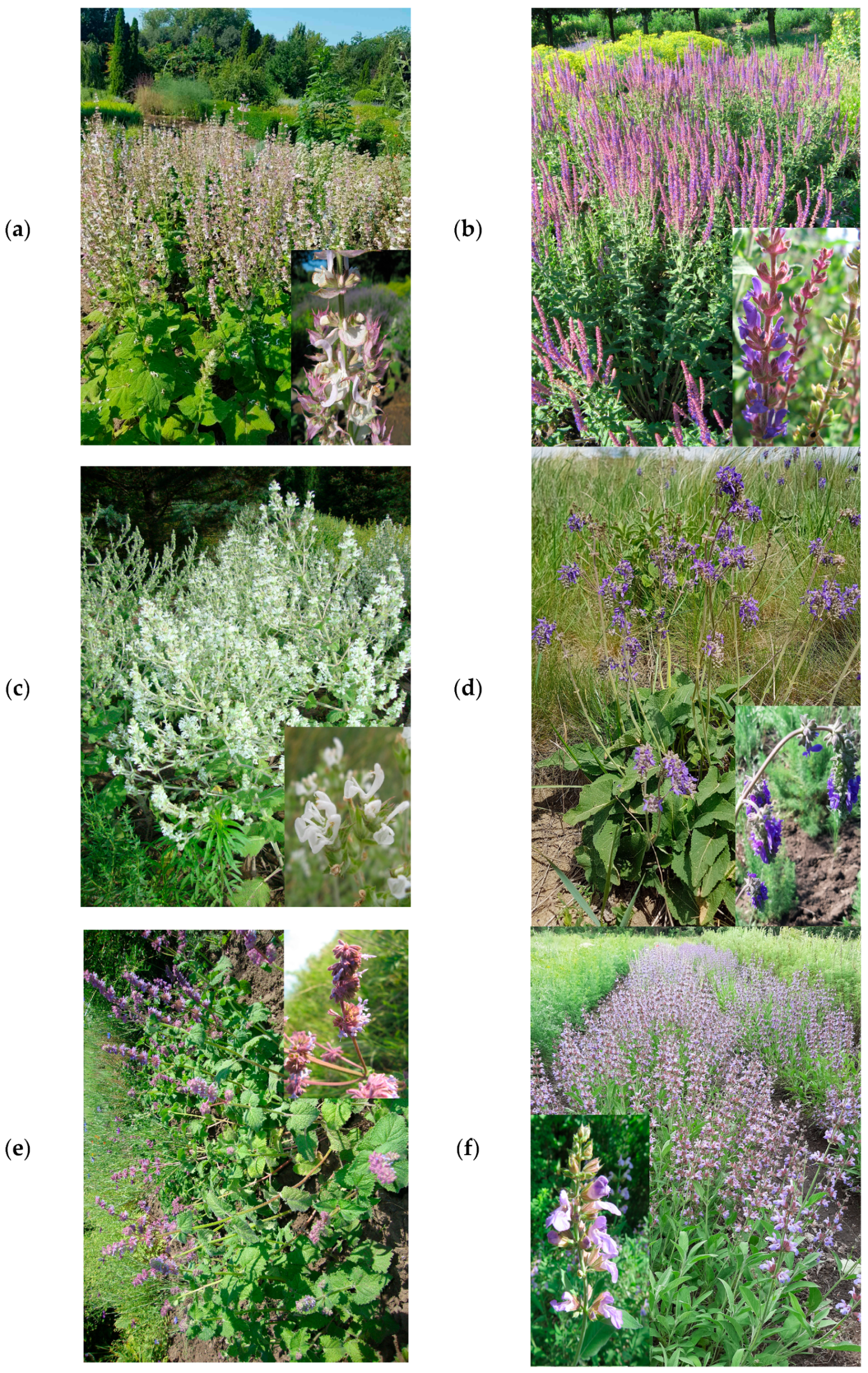
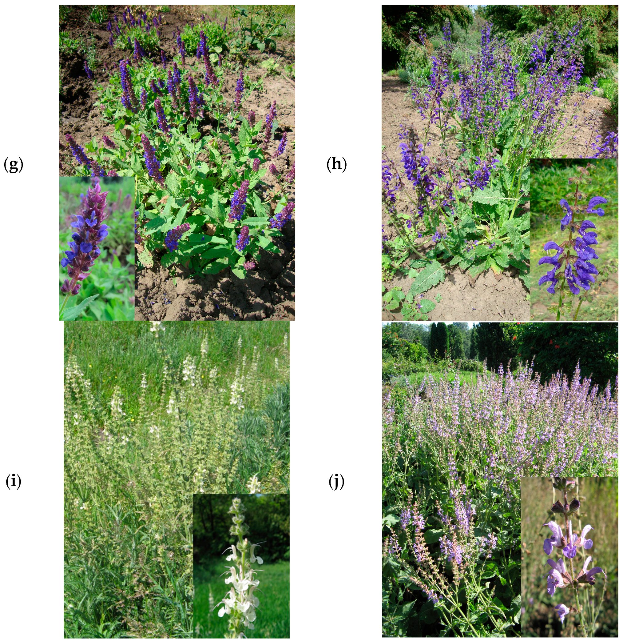
2.2. Plant Material and Extraction

The aerial parts of the ten Salvia species (Figure 1) were harvested from the crop fields of the National Botanical Garden ‘Alexandru Ciubotaru’ Chisinau, Republic of Moldova (GPS: N 46°58′25.43″, E 28°52′47.16″), during the flowering period (July 2019). Voucher specimens (Table 1) were deposited in the Department of Pharmacognosy and Phytotherapy, Grigore T. Popa University of Medicine and Pharmacy Iasi (Romania). The air-dried and powdered aerial parts (4 g) were extracted under reflux with 100 mL of 80% ethanol for 2 h at 60 °C. The extracts were concentrated in a rotary evaporator (Büchi Rotavapor, Flawil, Switzerland) under reduced pressure at 40 °C, and then they were stored in a freezer at −18 °C until analysis.

2.3. Total Phenolics Quantification

The total phenolic content (TPC) of extracts from selected Salvia species was determined with Folin–Ciocalteu reagent according to the method of Singleton, with slight modification [11,12]. In this regard, suitable dilutions in 80% ethanol were prepared for each extract. Gallic acid was used as a reference standard, and TPC was expressed as mg gallic acid equivalents/g dry extract (mg GAE/g extract).

2.4. LC-HRMS/MS Analysis

The LC-HRMS/MS analysis was performed on an Agilent 1200 HPLC (Agilent Technologies, Palo Alto, CA, USA) connected to a quadrupole-time-of-flight MS detector (G6530B). The HPLC separation was accomplished on a Phenomenex Gemini C18 column (100 mm × 2 mm i.d., 3 μm) operated at 20 °C. A linear gradient elution (10–60% B in 0–45 min) was achieved using 0.1% formic acid in water (A) and 0.1% formic acid in acetonitrile (B) at a flow rate of 0.2 mL/min. The sample injection volume was 10 μL. The detection was carried out in negative electrospray ionization mode, with the spectra recorded in the range of m/z 100–1000 Da. The ion source parameters were as follows: carrier gas flow rate—10 L/min; carrier gas temperature—275 °C; sheath gas flow rate—12 L/min; sheath gas temperature—325 °C; nebulizer pressure—35 psi; capillary, fragmentor, skimmer, and octapole radiofrequency voltages—4000 V, 140 V, 65 V, and 750 V, respectively. MS/MS fragmentation was carried out via automated fragmentation, with the collision-induced dissociation energy set at 30 V. Mass Hunter software version B.08.00 (Agilent Technology) was used for data acquisition and processing, including the prediction of chemical formula and exact mass calculation.

2.5. Antimicrobial Assay

2.5.1. Microbial Strains

The antimicrobial activity of Salvia extracts was tested against standard strains (American Type Culture Collection-ATCC) of Gram-positive bacteria (Staphylococcus aureus ATCC 25923, Streptococcus pneumoniae ATCC 49619), Gram-negative bacteria (Escherichia coli ATCC 25922, Pseudomonas aeruginosa ATCC 27853), and one pathogenic fungus (Candida albicans ATCC 10231). They were provided by Liofilchem (Abruzzi, Italy).

2.5.2. Minimum Inhibitory Concentration (MIC)

The broth microdilution method was used to determine MIC values according to the CLSI (Clinical and Laboratory Standards Institute) guidelines [13]. Serial double dilutions of the Salvia extracts ranging from 10 to 0.03 mg/mL were prepared in Mueller–Hinton broth (Biolab Zrt., Budapest, Hungary) using 24-well cell cultures plates (Becton Dickinson Labware Europe, Le Pont De Claix, France). For Streptococcus pneumoniae, the Mueller–Hinton broth was supplemented with 5% (v/v) lysed horse blood (Oxoid, Basingstoke, UK), and for Candida albicans, Sabouraud dextrose agar medium (Liofilchem, Abruzzi, Italy) was used; 10 μL of inoculum (105 CFU/well) were added to each well. The plates were incubated for 24 h, at 35 °C. The growth of microorganisms was monitored by visual assessment of turbidity. Negative controls (microbial strains growth control, sterility control, solvents used to dilute the extracts) were also included in the assay. Stock solutions (20 mg/mL) of extracts in DMSO–ultrapure water (5:5, v/v) were prepared for antimicrobial testing. The MIC is defined as the lowest concentration of extract that inhibits the growth of the tested microbial strains [14].

2.6. Antioxidant Assays

Solutions stock of Salvia extracts (20 mg/mL) in 80% ethanol were prepared and adjusted at suitable working concentrations in each test. All antioxidant assays were performed in triplicate.

2.6.1. DPPH Radical-Scavenging Assay

The test was carried out using the method described by Malterud et al. [15]. First, 2.95 mL of DPPH solution in methanol (A517 nm = 1.02 ± 0.03) was mixed with 0.5 mL of Salvia extract dilutions at different concentrations (20.83–333.33 μg/mL). The absorbance was measured at 517 nm before adding the extract dilution (A0) and after 5 min reaction time (Aend). The percentage of DPPH scavenging activity of each extract was calculated as follows: DPPH scavenging activity (%) = 100 × [(A0 – Aend)/Aend]. Gallic acid (1.30–333.33 μg/mL) was used as the positive control.

2.6.2. ABTS Radical-Cation-Scavenging Assay

The test was performed according to the method of Re et al. [16]. The ABTS radical cation was obtained by incubating ABTS stock solution (7 mM) with potassium persulfate (2.45 mM) at room temperature in the dark for 16 h before use. Then, the ABTS radical-cation solution was diluted with ethanol to yield an absorbance of 0.70 ± 0.02 at 734 nm. Then 0.02 mL of each dilution of Salvia extract (12.5–100 μg/mL) was mixed with 1.98 mL ABTS radical cation solution. The absorbance at 734 nm was determined after a 6 min reaction time. The capacity to scavenge ABTS radical cation was determined using the following equation: ABTS scavenging activity (%) = 100 × [(Acontrol – Asample)/Asample]. Gallic acid (0.39–100 μg/mL) was used as the positive control.

2.6.3. Ferric Ion Reducing Antioxidant Power Assay (FRAP)

The capacity of Salvia extracts to reduce iron (III) to iron (II) was evaluated using the Oyaizu method [17] with minor changes. First, 0.5 mL of Salvia extract dilutions (16.69–267.55 μg/mL) was mixed with 1.2 mL of 0.2 M phosphate buffer (pH 6.6) and 1.25 mL of 1% potassium ferricyanide and incubated at 50 °C for 20 min. Then, 1.25 mL of 10% trichloroacetic acid was added to it. The mixture was centrifuged at 3000 rpm for 10 min, and after that, 1.25 mL of the upper layer was treated with 1.25 mL of ultrapure water and 0.25 mL of 0.1% ferric chloride. The absorbance was measured at 700 nm after 90 s. Gallic acid (0.24–3.84 μg/mL) was used as the positive control. A high absorbance value indicated the potent reducing capacity of the samples.

2.7. Cell Viability Assay

2.7.1. Cell Lines

Human breast carcinoma MCF-7 (ATCC, HTB-22) and MDA-MB-231 (ATCC, CRM-HTB-26) cell lines were maintained in DMEM (Dulbecco’s modified Eagle medium, Biochrom AG, Berlin, Germany), supplemented with 10% FSB (fetal bovine serum, Sigma, Steinheim, Germany), 100 IU/mL penicillin (Biochrom AG, Berlin, Germany), and 100 µg/mL streptomycin (Biochrom AG, Berlin, Germany) at 37 °C in a humidified atmosphere of 5% CO2 in the air. The cell lines were a cordial donation of Prof. Charalambos Anastassiou from the University of Cyprus.

2.7.2. MTT Assay

The cell viability was evaluated using the MTT assay [18]. Briefly, cells were seeded in 96-well plates (TPP Techno Plastic Products AG, Trasadingen, Switzerland) at a density of 5 × 10³ cells/well and allowed to attach and grow overnight. Salvia extracts were added to cell cultures in concentrations of 25–100 µg/mL, using as vehicle agent DMSO with a final concentration of 0.1%. After 48 h, the cells were washed and covered with 100 μL of fresh 10% FBS in DMEM. Then, 10 μL of MTT (5 mg/mL) was added to the medium, and cells were incubated for 3 h. DMSO was used to solve the formed formazan, and the absorbance was recorded at 570 nm (PG Instruments T70, PG Instruments Ltd., Lutterworth, UK). The assay was performed in five replicates. The cell viability (%) was calculated according to the formula: % cell viability = [AbsorbanceSample/[AbsorbanceControl] × 100.

2.8. Data Analysis

Data are presented as mean ± standard deviation of the respective number of replicates. One-way analysis of variance with Tukey’s post hoc test was conducted; p < 0.05 was considered statistically significant. After Pareto scaling, the phytochemical data (peak areas from the base chromatograms) were imported into SPSS 20.0 software (IBM, New York, NY, USA) and used to perform principal component analysis, hierarchical cluster analysis, and Pearson correlation analysis. For antioxidant tests, the EC50 values were calculated by linear interpolation between values above and below 50% activity. In the FRAP assay, the EC50 value represents the concentration of extract/positive control that leads to an absorbance of 0.5.

3. Results and Discussion

3.1. Total Phenolic Content

The values of TPC are shown in Table 1. The highest amounts of polyphenols were determined in S. officinalis (126.91 mg GAE/g extract), followed by S. sclarea (110.90 mg GAE/g extract), S. kopetdaghensis (107.63 mg GAE/g extract), and S. verticillata (107.62 mg GAE/g extract). S. nutans and S. austriaca had the lowest contents of phenolic compounds among the investigated Salvia species (66.12 and 57.87 mg GAE/g extract, respectively). Our results align with the findings of other studies on Salvia phenolics. S. verticillata, S. nemorosa, and S. aethiopis from Turkey contained 167.1, 63.9, and 82.1 mg GAE/g extract, respectively [19]. Furthermore, S. verticillata from Serbia and S. sclarea from Iran contained outstanding amounts of total polyphenols (175.6 and 268 mg GAE/g extract, respectively) [20,21]. A previous study by Mocan et al. [22] showed TPC values of 65.02 mg GAE/ extract for S. officinalis from Romania. Hanganu et al. [10] reported lower values of TPC (22.25–118.75 mg GAE/g dry plant material) for six Salvia species from spontaneous Moldavian flora (S. aethiopis, S. austriaca, S. nemorosa, S. nutans, S. sclarea, S. verticillata). The provenience of plant material, the geographical and pedo-climatic factors that influence the growing and harvesting time, and the mode of results expression (extract/plant, reference standard) could explain the different outcomes. In our study, the plants were collected from field crops, while in the mentioned research, the plants were harvested from spontaneous flora. Mocan et al. [22] reported a similar trend for S. transsylvanica from Romania and pointed out that cultivated plants could produce higher levels of polyphenols than wild plants. In addition, abiotic factors such as rainfall, temperature, and cloud cover play significant roles in sage phenolics biosynthesis [23].

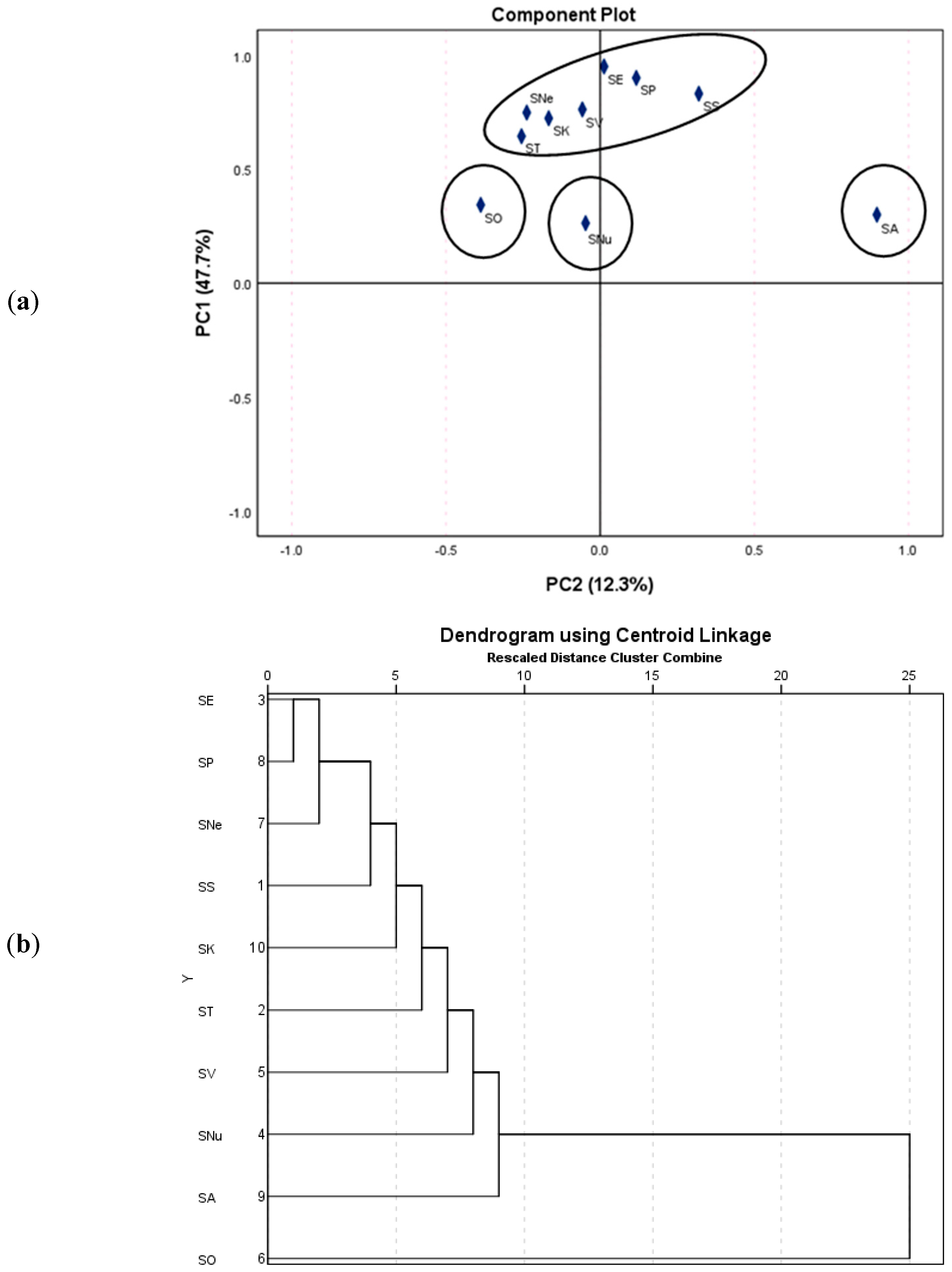
3.2. Metabolite Profiling Using LC-HRMS/MS

LC-MS platforms are extensively used to perform metabolite profiling, not only of common sage species but also of less-investigated or endemic Salvia species. Previously, the comprehensive phytochemical characterization of S. officinalis revealed the presence of more than 40 compounds, such as phenolic acids, flavonoids, diterpenes, and triterpenes [24]. S. miltiorrhiza Bunge was profiled using LC-MS, evidencing two main structural groups, namely, phenolic acids (monomers, dimers, trimers, and tetramers of hydroxycinnamic acids) and diterpenes (tanshinones) [25,26]. Shojaeifard et al. [27] documented the occurrence of flavonoids (e.g., rutin, luteolin-7-O-glucoside, apigenin-7-O-glucoside, cirsimaritin, eupatorin), phenolic acids (e.g., rosmarinic acid, salvianolic acid B), and diterpenes (e.g., carnosol) in 50 Salvia species collected from different regions of Iran, including S. indica L., S. grossheimii Sosn., S nemorosa, S. palaestina Benth., S. spinosa L., S. syriaca L., and S. verticillata. Zengin et al. [28] reported 66 compounds (phenolic acids, flavonoids, sugars, and fatty acids) in three Salvia species endemic to Turkey, namely, S. blepharochlaena Hedge and Hub.-Mor., S. euphratica var. leiocalycina (Rech.f.) Hedge, and S. verticillata subsp. amasiaca (Freyn and Bornm.) Bornm. Salvia species from Pakistan (S. coccinea, S. lanata, S. moocroftiana, S. nubicola, S. plebeiana) [29], Turkey (S. veneris Hedge, S. poculata, S. eriophora Boiss. and Kotschy, S. ceratophylla L., S. sclarea, S. absconditiflora Greuter and Burdet) [30,31,32,33], Greece (S. pomifera L., S. fruticosa Mill.) [34,35], and Poland (S. przewalskii Maxim., S. cadmica Boiss., S. yangii B.T. Drew, S. abrotanoides Kar.) [25,36,37] were also comprehensively characterized using LC-MS.
In the current study, the LC-HRMS/MS-based metabolite profiling of the ten Salvia species allowed for the annotation of 73 compounds belonging to 8 phytochemical classes. Total identification was performed by matching the spectro-chromatographic data with those obtained by standard injection, whereas partial identification was conducted by comparing the acquired data with those from databases (e.g., KNApSACK [38]) or relevant literature reporting on the LC-MS analysis of compounds from Salvia or Lamiaceae species [24,25,27,28,29,30,31,34,36,39,40,41,42,43]. The collected information (e.g., proposed identity, retention time, molecular formulas, fragment ions, sample distribution) is provided in Table 2. Overall, S. aethiopis showed the most complex profile (57 compounds), followed by S. sclarea (55 compounds) and S. verticillata (52 compounds). Next, 48 compounds were assigned in S. austriaca and S. kopetdaghensis, 46 compounds in S. nutans, 44 compounds in S. tesquicola and S. pratensis, 41 compounds in S. officinalis, and 39 compounds in S. nemorosa. To our knowledge, the LC-HRMS/MS-based phytochemical profiling of S. nutans and S. kopetdaghensis was performed herein for the first time, whereas a few studies have profiled S. sclarea [33], S. aethiopis [44], S. verticillata [27,45], S. nemorosa [27], S. pratensis [20], and S. austriaca [46].
Thirteen phenolic acids were labeled in the ten sage species, classified as hydroxybenzoic acids (5 and 6), hydroxycinnamic acids (4, 8, 9, 14, 18, and 34), and hydroxycinnamic acid oligomers (23, 24, 26, 27, and 29). Dihydroxybenzoic acid (5) was present only in S. nemorosa, whereas feruloylmalic acid (18) was identified only in S. aethiopis. Furthermore, salvianolic acid H was annotated only in S. pratensis, while caffeoylthreonic acid (9) was distributed specifically in S nemorosa and S. pratensis. Caffeic acid-O-hexoside (14), a phenolic glycoside, was characteristic of S. nutans and S. officinalis. Interestingly, caffeic acid (8) and rosmarinic acid (23) were ubiquitously found in all species, while hydroxybenzoic acid (6) was absent only in S. austriaca. In a previous study [35], 10 phenolic acids, such as hydroxybenzoic, dihydroxybenzoic, caffeic, ferulic, vanillic, chlorogenic, neochlorogenic, cyrptochlorogenic, and rosmarinic acids, were reported in S. fruticosa. Salvianolic acid B, salvianolic acid K, and chlorogenic, caffeic, ferulic, coumaric, and rosmarinic acids were documented in S. officinalis [47]. Zengin et al. [28] reported at least ten phenolic acids (e.g., danshensu, caffeic acid, caffeic acid-O-hexoside, protocatechuic acid, coutaric acid, coumaric acid) in S. blepharochlaena, S. euphratica var. leiocalycina, and S. verticillata subsp. amasiaca.
A series of 18 flavonoids was next labeled as flavanol aglycons (22), flavones aglycons (31, 36, 41, 48), flavone glycosides (1012, 15, 17, 20, 21, 25, 28, 30, 33), and flavonol glycosides (13, 16). Apigenin-O-pentoside-O-hexoside (10), chrysoeriol-O-acetylglucuronides I (30), and II (33) were observed only in S. nutans. Luteolin-O-hexoside-O-glucuronide (11) was characteristic of S. tesquicola, whereas quercetin-O-hexoside (13) was found only in S. officinalis. Interestingly, luteolin-7-O-glucoside (17) and luteolin-O-glucuronide I (20) were retrieved in all ten sage species. Luteolin-O-acetylglucuronide (28) was found only in S. nutans and S. verticillata, while apigenin was distributed specifically in S. sclarea and S. austriaca. Luteolin-O-hexoside-O-rhamnoside (15) was present in S. tesquicola, S. nutans, and S. officinalis, whereas apigenin-7-O-glucoside (21) was specific to S. sclarea, S. nemora, and S. austriaca. Genkwanin (48), a methoxylated flavone, was distributed in S. sclarea, S. pratensis, and S. austriaca. Previously, flavonoids with similar structures were found in sage species [24,25,27,28,29,30,31,34,36,39,40,41,42,43].
With 19 congeners, diterpenes were the largest phytochemical class in Salvia spp. Most compounds were derivatives of rosmanol (40, 42, 45, 54, 56, and 65) or carnosol (37, 47, 52, 54, 55, 59, and 68). S. officinalis was the only species that contained rosmanol II (42), methoxycarnosol (54), acetylhorminone II (64), and rosmaridiphenol (65). Salvipiliferol (58), hydroxysalviol (62), and salviol (70) were characteristic of S. nemorosa. Acetylhorminone I (51) was shown only in S. verticillata, whereas dihydroxycarnosic acid (55) was specifically found in S. tesquicola and S. kopetdaghensis. S. sclarea and S. tesquicola were the only species containing hydroxcarnosic acid I (37), whereas carnosol (59) was present only in S. officinalis and S. kopetdaghensis. Diterpenes are widely reported in the Salvia genus. For instance, Koutsoulas et al. [34] retrieved seven diterpenes in S. fruticosa (including rosmanol, carnosol, rosmadial, carnosic acid, methoxycarnosol) and two diterpenes in S. pomifera. Rosmanol, rosmaridiphenol, epirosmanol, epiisorosmanol, and methoxycarnosol were found in S. veneris [31], whereas carnosol, rosmanol, galdosol, carnosic acid, salviol, methyl carnosic acid, and 20-hydroxyfemiginol were shown in S. officinalis [24].
Five sesterpenes were putatively assigned as follows: lachnocalyxolides C (43), C’ (46), and A (50) in S. nutans; and salvimirzacolides I (44) and II (57) in S. aethiopis. Previously, lachnocalyxolides were identified in S. lachnocalyx Hedge [43], whereas salvimirzacolide was isolated from S. mirzayanii Rech. f. and Esfand. [38]. Nonetheless, oleanolic acid (72) and ursolic acid (73) were labeled as triterpenes in almost all Salvia species. Oleanolic acid (72) was absent in S. sclarea, S. aethiopis, S. officinalis, and S. kopetdaghensis, whereas ursolic acid (73) was not present in S. sclarea, S. aethiopis, S. austriaca, and S. kopetdaghensis. These triterpenes were previously reported in S. pomifera. and S. fruticosa [34]. Furthermore, two organic acids, namely, malic acid (2), quinic acid (3), and one sugar derivative, sucrose (1), were identified as non-specific metabolites in the polar region of the chromatograms (retention times <5 min, Table 2). In contrast, 13 fatty acid derivatives were found in the non-polar region of the chromatographic elution (retention times between 30 and 55 min, Table 2).
When it comes to the relative abundance of the constituents, it was found that rosmarinic acid (23) was the most predominant compound in S. sclarea, S. tesquicola, S. aethiopis, S. verticillata, S. officinalis, S. nemorosa, S. pratensis, and S. kopetdaghensis. Gallocatechin (22) was dominant in S. nutans, while salvianolic acid B (35) was abundant in S. kopetdaghensis. Luteolin-7-O-glucoside (15) was the major constituent in S. aethiopis, S. nemorosa, and S. austriaca. Caffeic acid (8) was found in relatively high levels in S. aethiopis and S. nutans. Luteolin (31), cirsimaritin (41), and apigenin (36) were significant in S. sclarea and S. austriaca. Carnosol (59), methylcarnosic acid (68), and rosmanol (45) were predominant in S. officinalis, whereas carnosic acid (52) and hydroxycarnosic acid (47) were found in high amounts in S. tesquicola. In an attempt to point out more objectively the chemotaxonomic differences between the ten sage species, a multivariate analytical approach based on principal component analysis and hierarchical cluster analysis was subsequently applied. The semi-quantitative data (peak areas of identified compounds after the Paretto scaling) were used as input information. As shown in the component plot (Figure 2a) and dendrogram (Figure 2b), S. officinalis, S. austriaca, and S. nutans formed individual clusters, whereas the remaining Salvia species could be regarding a single big cluster.

3.3. Assessment of the Antimicrobial Activity

Screening plant sources to determine their antimicrobial properties is an important strategy to find new therapeutic anti-infective solutions and combat the multidrug-resistance phenomenon. It is estimated that about 75% of the pharmaceuticals used in anti-infective therapy are obtained from natural sources [48]. The extracts obtained from the ten sage species were evaluated concerning their antimicrobial properties against a standard panel of human pathogens, including Gram+ bacteria (S. aureus and S. pneumoniae), Gram− bacteria (E. coli and P. aeruginosa), and yeasts (C. albicans). The results (Table 3) showed that the MIC values of most samples were situated between 1.25 and 5 mg/mL. Only S. officinalis showed moderate activity (MIC = 0.625 mg/mL) when tested against S. aureus and S. pneumoniae. Furthermore, Salvia sclarea behaved more actively on the same bacteria (MIC = 1.25 mg/mL). Among the analyzed Salvia species, S. nemorosa and S. pratensis were the most active against Candida albicans (MIC = 1.25 mg/mL).
Previous literature reports revealed comparable antimicrobial properties of Salvia species. For instance, extracts from S. pratensis showed MIC values between 5 and 20 mg/mL against S. aureus, P. aeruginosa, E. coli, and C. albicans [20]. S. verticillata displayed MIC values between 1.25 and 20 mg/mL against a panel of eight bacterial and eight fungal strains [45]. S. aethiopis, S. nemorosa, and S. sclarea also showed MIC values between 1.25 and 20 mg/mL against E. coli, K. pneumoniae, S. typhi, B. subtilis, S. epidermidis, and S. aureus [49]. Mocan et al. [22] showed that extracts derived from S. officinalis exhibited MIC values between 0.01 and 0.18 mg/mL against E. coli, P. aeruginosa, S. typhimurium, L. monocytogenes, E. cloacae, M. flavus, B. cereus, and S. aureus. Gram-positive bacteria are more susceptible to Salvia extracts. The resistance of Gram-negative bacteria can be related to their multi-layered highly complex cell structure consisting of inner and outer membranes [48]. The outer membrane containing mainly lipopolysaccharides acts as an additional selective and impermeable barrier [50]. Although volatile terpenes are better known in terms of antimicrobial properties, many other specific metabolites of Salvia species (flavonoids, phenolic acids, diterpenes, triterpenes) are capable of inhibiting the growth of different pathogens affecting multiple targets of microbial cells. Flavonoids, such as apigenin, luteolin, and quercetin derivatives, may cause cell-membrane damage, and inhibition of nucleic-acid synthesis and of the bacterial respiratory chain [51]. Antimicrobial mechanisms of phenolic acids (rosmarinic, caffeic, ferulic acids) include damage to bacterial membrane integrity and bacterial cell morphology, leakage of cellular electrolytes, and alteration of microbial metabolism [52,53]. Sage diterpenes can inhibit microbial protein synthesis and damage microbial membrane structure and cellular respiration [54].

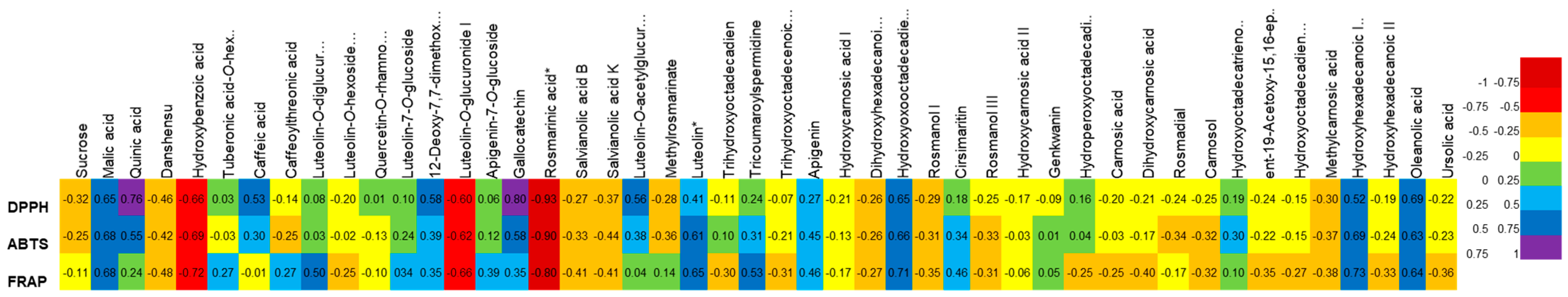
3.4. Assessment of Antioxidant Activity

Reactive oxygen species and oxidative stress are involved in many pathologies (cancer, cardiovascular and neurodegenerative diseases, skin disorders). The antioxidant abilities of plant products may be an important strategy to improve cell responses to injuries, counteract noxious and pathogenic stimuli, and preserve cell health status. The antioxidant capacity of the ten Moldavian Salvia species was assessed in three in vitro tests, namely, DPPH, ABTS, and FRAP. The results, presented as EC50 values (Table 4), show that the extracts obtained from S. officinalis and S. verticillata were the most potent DPPH radical scavengers (EC50 = 25.33 and 27.36 µg/mL, respectively). On the contrary, S. nutans and S. austriaca showed the weakest anti-radical activity. A similar trend was found in the ABTS assay when the EC50 values varied from 8.13 and 59.16 µg/mL. The following decreasing activity order can be concluded: S. officinalis > S. verticilata > S. kopetdaghensis > S. pratensis > S. nemorasa > S. aethiopis ~ S. sclarea > S. tesquicola >> S. nutans > S. austriaca. Lastly, the reducing power revealed that S. kopetdaghensis (EC50 = 19.75 µg/mL), S. verticillata (EC50 = 19.75 µg/mL), and S. officinalis (EC50 = 19.75 µg/mL) were the most active samples.
Our results are comparable with those from the existing literature. For instance, CE50 values between 80.09 and 158.76 µg/mL in DPPH and 1.39 and 8.04 mol Trolox equivalents (TE)/mg in FRAP were reported for the extracts obtained from S. aethiopis, S. austriaca, S. sclarea, S. nutans, S. verticillata, and S. nemorosa [10]. Similarly, S. officinalis, S. sclarea, S. pratensis, S. austriaca, S. nemorosa, and S. verticillata displayed potent DPPH radical scavenging (53.44–189.94 μg TE/mL) and FRAP (1.19–5.89 μmol TE/100 mL) activities [55]. S. verticillata showed good antioxidant activity, as assessed in DPPH (EC50 = 33.04 μg/mL), ABTS (EC50 = 67.01 μg/mL), and NO (EC50 = 73.12 μg/mL) radical scavenging assays [45]. In addition, Matkowski et al. [56] reported potent DPPH radical scavenging (EC50 = 19.84 μg/mL), reducing power (0.671 g quercetin equivalents/g), and Trolox equivalent antioxidant capacity (13.30 mg TE/g) for S. verticillata. Similarly, the aerial parts extract of S. pratensis revealed EC50 values of 50.17 and 90.65 μg/mL in DPPH and ABTS radical scavenging tests, respectively [20]. Tohma et al. [44] also studied the antioxidant properties of S. aethiopis in DPPH, FRAP, and cupric-ion-reducing antioxidant (CUPRAC) assays. Extracts from S. sclarea were shown to scavenge DPPH (85.08 mg TE/g) and ABTS (33 mg TE/g) radicals, reduce ferric (77.06 mg TE/g) and cupric (144.75 mg TE/g) ions, and chelate ferrous ions (37.27 mg EDTAE/g) [57]. The antioxidant effects of other Salvia species (e.g., S. blepharochlaena, S. euphratica var. leiocalycina, S. glutinosa, S. transsylvanica, S. syriaca, S. aegyptiaca, S. palaestina, S. absconditiflora, S. cadmica, S. ceratophylla) are also documented in the literature [22,28,33,36,49,57,58].
In an attempt to correlate the observed antioxidant activity (Table 4) with the phytochemical composition (Table 2), Pearson correlation analysis was subsequently performed. The peak areas from the LC-MS chromatograms were extracted and used as input information. As depicted in Figure 3, most compounds displayed negligible correlations (R values < 0.50). However, rosmarinic acid and, to a lesser extent, hydroxybenzoic acid and luteolin-O-glucuronide correlated well with the DPPH-radical-scavenging, ABTS-radical-scavenging, and metal-reducing activity. Rosmarinic acid and luteolin derivatives are particularly known to exert potent antioxidant effects, as revealed by numerous studies [59,60].

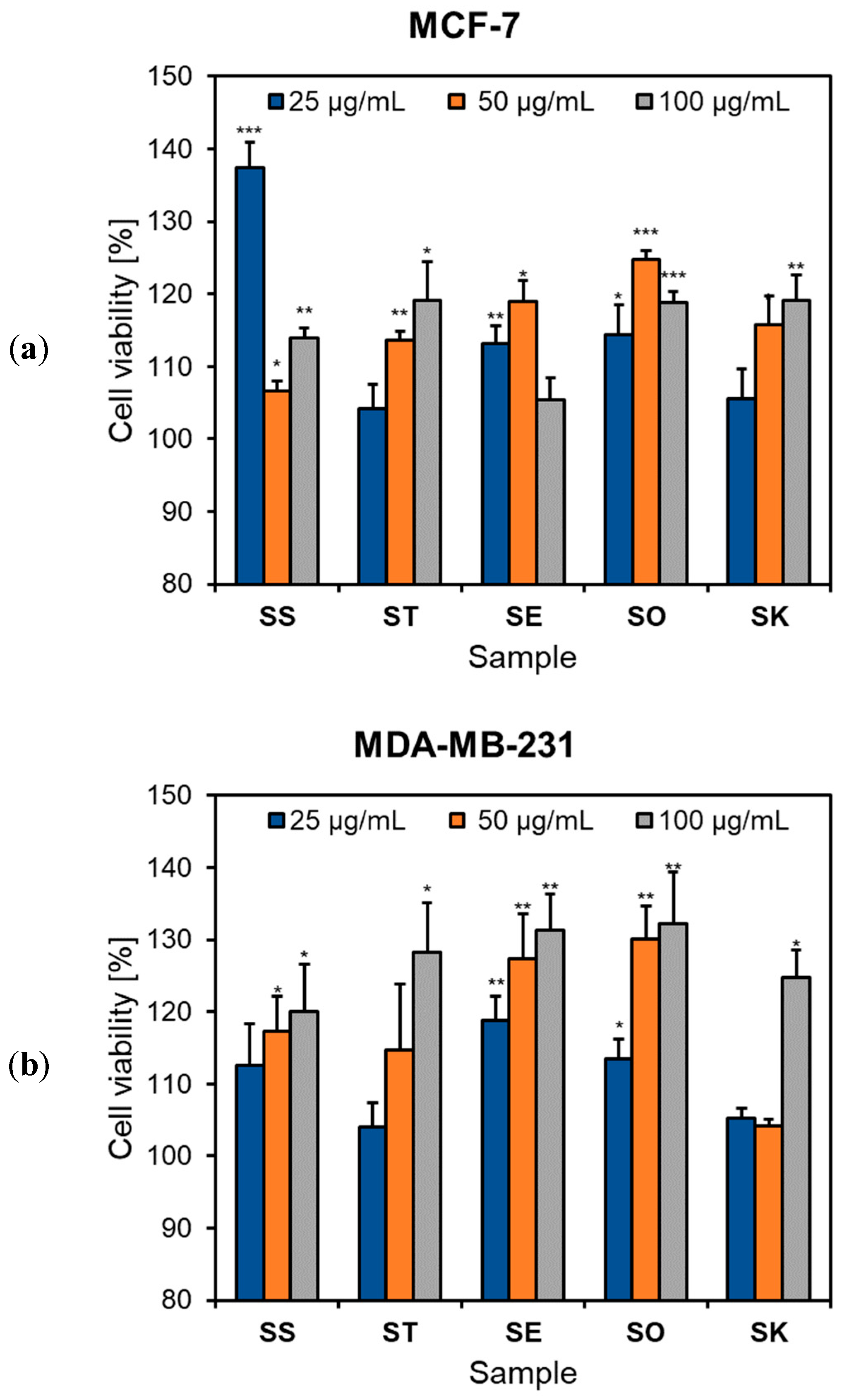
3.5. Assessment of the Cytotoxic Activity

Plant-derived products are highly valuable resources for developing chemopreventive and anticancer agents. Over 60% of anticancer drugs are obtained from natural products (plants, aquatic organisms, and microbial sources) [61]. Salvia species are a rich reservoir of many compounds with multiple bioactivities and have attracted great interest in screening cytotoxic agents. This section presents the influence of five selected sage species on the viability of human breast carcinoma MCF-7 and MDA-MB-231 cell lines. The MCF-7 cell line retains estrogen and progesterone receptors and is highly responsive to chemotherapy. MDA-MB-231 is a highly invasive and aggressive triple-negative breast cancer cell line [62,63]. The five Salvia species were chosen according to their use in Moldavian traditional medicine for different kinds of tumors [8]. Breast cancer, the most common malignancy in women [64], is also one of interest in the research of the cytotoxic potential of Salvia species and their metabolites.
In our study, no sample showed a significant reduction in cell viability over the concentration domain ranging from 25 to 100 μg/mL (Figure 4). On the contrary, we found a slight increase in the number of viable cells at tested doses for all Salvia extracts. A similar effect was reported by Mocan et al. [22] in the case of Romanian S. glutinosa and S. transsylvanica at intermediary doses on MCF-7 and HepG2 cells. Furthermore, some previous studies revealed low cytotoxicity of the Salvia genus. For example, extracts derived from S. verticillata showed no viability reduction in epidermoid carcinoma A431, liver cancer HepG2, and colon carcinoma LoVo cells at concentrations of 5 to 50 μg/mL [45]. Similar outcomes were also reported for S. pratensis, with IC50 values for aerial part extracts >200 μg/mL in A431 cells [20]. Zengin et al. [28] confirmed the lack of antiproliferative activity of S. verticillata subsp. amasiaca, S. euphratica var. leiocalycina, and S. blepharochlaena in human alveolar lung epithelial carcinoma A549 and human breast adenocarcinoma MCF-7 cells. The cytotoxicity of eleven Salvia species (e.g., S. aethiopis, S. nemorosa, S. syriaca, etc.) was tested in MCF-7, acute promyelocytic leukemia HL60, and chronic myelogenous leukemia K562 cells, revealing IC50 values generally higher than 50 μg/mL [49]. Mocan et al. [22] showed a modest cytotoxic effect of S. glutinosa and S. transsylvanica against HepG2, A549, and MCF-7 cell lines with IC50 values greater than 100 μg/mL. Furthermore, Nicolescu et al. [58] evidenced IC50 values between 131.68 and 293.79 μg/mL for various extracts of S. glutinosa in MCF-7, HepG2, non-small cell lung carcinoma NCI H460, and cervical carcinoma HeLa cells.
Although many studies have revealed good cytotoxic potential of certain sage species in lung (S. hispanica, S. pilifera, S. macrosiphon), prostate (S. ballotiflora, S. hispanica, S. pilifera), colon (S. fruticosa), and breast cancer (S. officinalis, S. miltiorrhiza, S. fruticosa, S. verbenaca, S. atropatana, S. macrosiphon, S. rosmarinus, S. chloroleuca) [5,65,66] there are, as we mentioned, opposite results. The type of extract (polar, lipophilic), the presence and concentration of specific components, the ratios between them, the tested concentration, and the exposure period significantly influence the outcome. Perhaps the identification and monitoring of some marker cytotoxic compounds would be helpful to establish the criteria for more practical screening of the complex extracts of sage species.

4. Conclusions

Our study provides data on the chemical profile and potential bioactivities of ten Moldavian Salvia species from ex situ crop cultures (S. officinalis, S. sclarea, S. tesquicola, S. aethiopis, S. austriaca, S. kopetdaghensis, S. nemorosa, S. nutans, S. pratensis, S. verticillata). To the best of our knowledge, this is the first study on Moldavian sage plants from field crops regarding non-volatile chemical composition and biological activity. Moreover, data about LC-HRMS/MS-based phytochemical profiling of S. kopetdaghensis and S. nutans are reported herein for the first time. S. officinalis, S. sclarea, S. kopetdaghensis, and S. verticillata are valuable sources of polyphenols. LC-HRMS/MS metabolite profiling indicated a rich composition of sage plants, including eight chemical classes (phenolic acids, flavonoids, diterpenes, sesterpenes, triterpenes, organic acids, fatty acids, and sugars). S. aethiopis presented the most complex profile. Hydroxybenzoic acids, hydroxycinnamic acids, and their oligomers were identified in sage species having a specific distribution. Caffeic and rosmarinic acids were ubiquitously distributed in all Salvia species. Salvianolic acids were primarily found in S. officinalis, S. tesquicola, S. nemorosa, S. kopetdaghensis, and S. pratensis. Luteolin, apigenin, and quercetin derivatives were the main flavonoids identified, but the composition varied between the tested sages. Luteolin derivatives were present in all species, with luteolin-7-O-glucuronide being the most common flavonoid. Rosmanol and carnosol derivatives were the main diterpenes in the investigated Moldavian sage plants. Among the triterpenes, oleanolic and ursolic acids were present in S. nemorosa, S. nutans, S. verticillata, and S. tesquicola. Multivariate analysis showed that Moldavian S. officinalis, S. austriaca, and S. nutans formed individual clusters. All analyzed sages scavenged free radicals and acted as reducing agents, with S. officinalis, S. verticillata, and S. kopetdaghensis being the most effective antioxidants. The highest antimicrobial activity was found for S. officinalis. The tested Salvia species (S. officinalis, S. sclarea, S. tesquicola, S. aethiopis, S. kopetdaghensis) did not show cytotoxic properties on breast cancer cell lines (MCF-7 and MDA-MB-231) On the contrary, the extracts seemed to show proliferative activity on tested cell lines. In this regard, for a substantiated statement, it is necessary to investigate a broader range of doses and variations of the exposure period.
Our research contributes to knowledge about the chemistry and biological potential of understudied Moldavian Salvia species, providing evidence for future studies that can lead to developing sage-based health-promoting agents in oxidative stress-related disturbances and redox medicine.

Author Contributions

Conceptualization, S.V.L.; methodology, S.V.L., K.S.-W., A.C.A.; software, S.V.L., C.-T.M.; validation, S.V.L.; formal analysis, S.V.L., A.C.A.; investigation, S.V.L., C.-T.M., A.C.G., N.C., A.M. (Alexandru Mandici), A.C.A.; resources, N.C., S.V.L., K.S.-W., A.C.A.; data curation, S.V.L.; writing—original draft preparation, S.V.L., A.C.A.; writing—review and editing, KS-W, C.-T.M., A.C.G., A.M. (Alexandru Mandici), N.C., A.M. (Anca Miron); visualization, S.V.L., A.C.A.; supervision, S.V.L., A.C.A.; project administration, S.V.L., A.C.A.; funding acquisition, A.C.A. All authors have read and agreed to the published version of the manuscript.

Funding

This research received no external funding.

Institutional Review Board Statement

Not applicable.

Informed Consent Statement

Not applicable.

Data Availability Statement

Data is contained within the article.

Conflicts of Interest

The authors declare no conflict of interest.

References

  1. Mervić, M.; Bival Štefan, M.; Kindl, M.; Blažeković, B.; Marijan, M.; Vladimir-Knežević, S. Comparative antioxidant, anti-acetylcholinesterase and anti-α-glucosidase activities of mediterranean Salvia species. Plants 2022, 11, 625. [Google Scholar] [CrossRef]
  2. Afonso, A.F.; Pereira, O.R.; Fernandes, Â.; Calhelha, R.C.; Silva, A.M.; Ferreira, I.C.; Cardoso, S.M. Phytochemical composition and bioactive effects of Salvia africana, Salvia officinalis ‘Icterina’and Salvia mexicana aqueous extracts. Molecules 2019, 24, 4327. [Google Scholar] [CrossRef] [Green Version]
  3. European Medicines Agency (EMA). Assessment report on Salvia officinalis L., folium and Salvia officinalis L., aetheroleum. Available online: https://www.ema.europa.eu/en/documents/herbal-report/final-assessment-report-salvia-officinalis-l-folium-salvia-officinalis-l-aetheroleum-revision-1_en.pdf (accessed on 12 June 2023).
  4. Sharifi-Rad, M.; Ozcelik, B.; Altın, G.; Daşkaya-Dikmen, C.; Martorell, M.; Ramírez-Alarcón, K.; Alarcón-Zapata, P.; Morais-Braga, M.F.B.; Carneiro, J.N.; Leal, A.L.A.B. Salvia spp. plants-from farm to food applications and phytopharmacotherapy. Trends Food. Sci. Technol. 2018, 80, 242–263. [Google Scholar] [CrossRef]
  5. Ghorbani, A.; Esmaeilizadeh, M. Pharmacological properties of Salvia officinalis and its components. J. Trad. Complement. Med. 2017, 7, 433–440. [Google Scholar] [CrossRef]
  6. Grdiša, M.; Jug-Dujaković, M.; Lončarić, M.; Carović-Stanko, K.; Ninčević, T.; Liber, Z.; Radosavljević, I.; Šatović, Z. Dalmatian sage (Salvia officinalis L.): A review of biochemical contents, medical properties and genetic diversity. Agric. Conspec. Sci. 2015, 80, 69–78. [Google Scholar]
  7. Balmus, Z.; Gonceariuc, M.; Cotelea, L.; Butnaras, V. Parfum perfect the new early variety of Salvia sclarea L. (clary sage). In Proceedings of the International Congress of Geneticists and Breeders from the Republic of Moldova, Chisinau, Moldova, 15–16 June 2021; p. 70. [Google Scholar]
  8. Ciocarlan, N. Family Lamiaceae: Main important spontaneous medicinal and aromatic species in the Republic of Moldova. Rev. Bot. 2016, 12, 86–91. [Google Scholar]
  9. Gonceariuc, M. Medicinal and aromatic plant varieties developed in the Republic of Moldova. Olten. Stud. Si Comun. Stiint. Nat. 2014, 30, 29–34. [Google Scholar]
  10. Hanganu, D.; Olah, N.K.; Pop, C.E.; Vlase, L.; Oniga, I.; Ciocarlan, N.; Matei, A.; Puşcaş, C.M.; Silaghi-Dumitrescu, R.; Benedec, D. Evaluation of polyphenolic profile and antioxidant activity for some Salvia species. Farmacia 2019, 67, 801–805. [Google Scholar] [CrossRef] [Green Version]
  11. Aprotosoaie, A.C.; Mihai, C.T.; Vochita, G.; Rotinberg, P.; Trifan, A.; Luca, S.V.; Petreus, T.; Gille, E.; Miron, A. Antigenotoxic and antioxidant activities of a polyphenolic extract from European Dracocephalum moldavica L. Ind. Crops Prod. 2016, 79, 248–257. [Google Scholar] [CrossRef]
  12. Grădinariu, V.; Cioancă, O.; Gille, E.; Aprotosoaie, A.; Hriţcu, L.; Hăncianu, M. The chemical profile of basil biovarieties and its implication on the biological activity. Farmacia 2013, 61, 632–639. [Google Scholar]
  13. M02-A10; Performance Standards for Antimicrobial Disk Susceptibility Tests (10th ed.), Approved Standard. Clinical and Laboratory Standards Institute: Wayne, PA, USA, 2009.
  14. Grădinaru, A.; Trifan, A.; Şpac, A.; Brebu, M.; Miron, A.; Aprotosoaie, A. Antibacterial activity of traditional spices against lower respiratory tract pathogens: Combinatorial effects of Trachyspermum ammi essential oil with conventional antibiotics. Lett. Appl. Microb. 2018, 67, 449–457. [Google Scholar] [CrossRef]
  15. Malterud, K.E.; Farbrot, T.L.; Huse, A.E.; Sund, R.B. Antioxidant and radical scavenging effects of anthraquinones and anthrones. Pharmacology 1993, 47, 77–85. [Google Scholar] [CrossRef]
  16. Re, R.; Pellegrini, N.; Proteggente, A.; Pannala, A.; Yang, M.; Rice-Evans, C. Antioxidant activity applying an improved ABTS radical cation decolorization assay. Free Radic. Biol. Med. 1999, 26, 1231–1237. [Google Scholar] [CrossRef]
  17. Oyaizu, M. Studies on products of browning reaction: Antioxidative activities of products of browning reaction prepared from glucosamine. Jpn. J. Nutr. 1986, 44, 307–315. [Google Scholar] [CrossRef] [Green Version]
  18. Mosmann, T. Rapid colorimetric assay for cellular growth and survival: Application to proliferation and cytotoxicity assays. J. Immun. Met. 1983, 65, 55–63. [Google Scholar] [CrossRef]
  19. Tosun, M.; Ercisli, S.; Sengul, M.; Ozer, H.; Polat, T.; Ozturk, E. Antioxidant properties and total phenolic content of eight Salvia species from Turkey. Biol. Res. 2009, 42, 175–181. [Google Scholar] [CrossRef]
  20. Srećković, N.; Mišić, D.; Gašić, U.; Matić, S.L.; Stanković, J.S.K.; Mihailović, N.R.; Monti, D.M.; D’Elia, L.; Mihailović, V. Meadow sage (Salvia pratensis L.): A neglected sage species with valuable phenolic compounds and biological potential. Ind. Crops Prod. 2022, 189, 115841. [Google Scholar] [CrossRef]
  21. Asadi, S.; Ahmadiani, A.; Esmaeili, M.A.; Sonboli, A.; Ansari, N.; Khodagholi, F. In vitro antioxidant activities and an investigation of neuroprotection by six Salvia species from Iran: A comparative study. Food Chem. Toxicol 2010, 48, 1341–1349. [Google Scholar] [CrossRef]
  22. Mocan, A.; Babotă, M.; Pop, A.; Fizeșan, I.; Diuzheva, A.; Locatelli, M.; Carradori, S.; Campestre, C.; Menghini, L.; Sisea, C.R. Chemical constituents and biologic activities of sage species: A comparison between Salvia officinalis L., S. glutinosa L. and S. transsylvanica (Schur ex griseb. & schenk) schur. Antioxidants 2020, 9, 480. [Google Scholar]
  23. Mekinić, I.G.; Ljubenkov, I.; Možina, S.S.; Abramović, H.; Šimat, V.; Katalinić, A.; Novak, T.; Skroza, D. Abiotic factors during a one-year vegetation period affect sage phenolic metabolites, antioxidants and antimicrobials. Ind. Crops Prod. 2019, 141, 111741. [Google Scholar] [CrossRef]
  24. Gericke, S.; Lübken, T.; Wolf, D.; Kaiser, M.; Hannig, C.; Speer, K. Identification of new compounds from sage flowers (Salvia officinalis L.) as markers for quality control and the influence of the manufacturing technology on the chemical composition and antibacterial activity of sage flower extracts. J. Agric. Food Chem. 2018, 66, 1843–1853. [Google Scholar] [CrossRef] [PubMed]
  25. Ożarowski, M.; Piasecka, A.; Gryszczyńska, A.; Sawikowska, A.; Pietrowiak, A.; Opala, B.; Mikołajczak, P.; Kujawski, R.; Kachlicki, P.; Buchwald, W. Determination of phenolic compounds and diterpenes in roots of Salvia miltiorrhiza and Salvia przewalskii by two LC–MS tools: Multi-stage and high resolution tandem mass spectrometry with assessment of antioxidant capacity. Phytochem. Lett. 2017, 20, 331–338. [Google Scholar] [CrossRef]
  26. Yang, S.; Wu, X.; Rui, W.; Guo, J.; Feng, Y. UPLC/Q-TOF-MS analysis for identification of hydrophilic phenolics and lipophilic diterpenoids from Radix Salviae Miltiorrhizae. Acta Chromatogr. 2015, 27, 711–728. [Google Scholar] [CrossRef] [Green Version]
  27. Shojaeifard, Z.; Hemmateenejad, B.; Jassbi, A.R. Chemometrics-based LC-UV-ESIMS analyses of 50 Salvia species for detecting their antioxidant constituents. J. Pharm. Biomed. Anal. 2021, 193, 113745. [Google Scholar] [CrossRef] [PubMed]
  28. Zengin, G.; Llorent-Martínez, E.J.; Fernández-de Córdova, M.L.; Bahadori, M.B.; Mocan, A.; Locatelli, M.; Aktumsek, A. Chemical composition and biological activities of extracts from three Salvia species: S. blepharochlaena, S. euphratica var. leiocalycina, and S. verticillata subsp. amasiaca. Ind. Crops Prod. 2018, 111, 11–21. [Google Scholar] [CrossRef]
  29. Haq, F.U.; Ali, A.; Akhtar, N.; Aziz, N.; Khan, M.N.; Ahmad, M.; Musharraf, S.G. A high-throughput method for dereplication and assessment of metabolite distribution in Salvia species using LC-MS/MS. J. Adv. Res. 2020, 24, 79–90. [Google Scholar] [CrossRef]
  30. Yener, I. Determination of antioxidant, cytotoxic, anticholinesterase, antiurease, antityrosinase, and antielastase activities and aroma, essential oil, fatty acid, phenolic, and terpenoid-phytosterol contents of Salvia poculata. Ind. Crops Prod. 2020, 155, 112712. [Google Scholar] [CrossRef]
  31. Toplan, G.G.; Kurkcuoglu, M.; Goger, F.; İşcan, G.; Ağalar, H.G.; Mat, A.; Baser, K.H.C.; Koyuncu, M.; Sarıyar, G. Composition and biological activities of Salvia veneris Hedge growing in Cyprus. Ind. Crops Prod. 2017, 97, 41–48. [Google Scholar] [CrossRef]
  32. Bursal, E.; Aras, A.; Kılıç, Ö.; Taslimi, P.; Gören, A.C.; Gülçin, İ. Phytochemical content, antioxidant activity, and enzyme inhibition effect of Salvia eriophora Boiss. & Kotschy against acetylcholinesterase, α-amylase, butyrylcholinesterase, and α-glycosidase enzymes. J. Food Biochem. 2019, 43, e12776. [Google Scholar]
  33. Uysal, S.; Zengin, G.; Sinan, K.I.; Ak, G.; Ceylan, R.; Mahomoodally, M.F.; Uysal, A.; Sadeer, N.B.; Jekő, J.; Cziáky, Z. Chemical characterization, cytotoxic, antioxidant, antimicrobial, and enzyme inhibitory effects of different extracts from one sage (Salvia ceratophylla L.) from Turkey: Open a new window on industrial purposes. RSC Adv. 2021, 11, 5295–5310. [Google Scholar] [CrossRef]
  34. Koutsoulas, A.; Čarnecká, M.; Slanina, J.; Tóth, J.; Slaninová, I. Characterization of phenolic compounds and antiproliferative effects of Salvia pomifera and Salvia fruticosa extracts. Molecules 2019, 24, 2921. [Google Scholar] [CrossRef] [Green Version]
  35. Sarrou, E.; Martens, S.; Chatzopoulou, P. Metabolite profiling and antioxidative activity of Sage (Salvia fruticosa Mill.) under the influence of genotype and harvesting period. Ind. Crops Prod. 2016, 94, 240–250. [Google Scholar] [CrossRef]
  36. Piątczak, E.; Owczarek, A.; Lisiecki, P.; Gonciarz, W.; Kozłowska, W.; Szemraj, M.; Chmiela, M.; Kiss, A.K.; Olszewska, M.A.; Grzegorczyk-Karolak, I. Identification and quantification of phenolic compounds in Salvia cadmica Boiss. and their biological potential. Ind. Crops Prod. 2021, 160, 113113. [Google Scholar] [CrossRef]
  37. Bielecka, M.; Pencakowski, B.; Stafiniak, M.; Jakubowski, K.; Rahimmalek, M.; Gharibi, S.; Matkowski, A.; Ślusarczyk, S. Metabolomics and DNA-based authentication of two traditional Asian medicinal and aromatic species of Salvia subg. Perovskia. Cells 2021, 10, 112. [Google Scholar] [CrossRef]
  38. KNApSAcK. Available online: https://www.knapsackfamily.com/knapsack_core/result.php?sname=all&word=Salvia (accessed on 12 June 2023).
  39. Pacifico, S.; Piccolella, S.; Lettieri, A.; Nocera, P.; Bollino, F.; Catauro, M. A metabolic profiling approach to an Italian sage leaf extract (SoA541) defines its antioxidant and anti-acetylcholinesterase properties. J. Funct. Foods 2017, 29, 1–9. [Google Scholar] [CrossRef]
  40. Gök, H.N.; Luca, S.V.; Ay, S.T.; Komsta, Ł.; Salmas, R.E.; Orhan, I.E.; Skalicka-Woźniak, K. Profiling the annual change of the neurobiological and antioxidant effects of five Origanum species in correlation with their phytochemical composition. Food Chem. 2022, 368, 130775. [Google Scholar] [CrossRef]
  41. Bujor, A.; Miron, A.; Luca, S.V.; Skalicka-Wozniak, K.; Silion, M.; Trifan, A.; Girard, C.; Demougeot, C.; Totoson, P. Vasorelaxant effects of Crataegus pentagyna: Links with arginase inhibition and phenolic profile. J. Ethnopharmacol. 2020, 252, 112559. [Google Scholar] [CrossRef]
  42. Velamuri, R.; Sharma, Y.; Fagan, J.; Schaefer, J. Application of UHPLC-ESI-QTOF-MS in phytochemical profiling of sage (Salvia officinalis) and rosemary (Rosmarinus officinalis). Planta Med. Int. Open 2020, 7, e133–e144. [Google Scholar] [CrossRef]
  43. Farimani, M.M.; Mazarei, Z. Sesterterpenoids and other constituents from Salvia lachnocalyx Hedge. Fitoterapia 2014, 98, 234–240. [Google Scholar] [CrossRef]
  44. Tohma, H.; Köksal, E.; Kılıç, Ö.; Alan, Y.; Yılmaz, M.A.; Gülçin, İ.; Bursal, E.; Alwasel, S.H. RP-HPLC/MS/MS analysis of the phenolic compounds, antioxidant and antimicrobial activities of Salvia L. species. Antioxidants 2016, 5, 38. [Google Scholar] [CrossRef] [Green Version]
  45. Stanković, J.S.K.; Srećković, N.; Mišić, D.; Gašić, U.; Imbimbo, P.; Monti, D.M.; Mihailović, V. Bioactivity, biocompatibility and phytochemical assessment of lilac sage, Salvia verticillata L. (Lamiaceae)-A plant rich in rosmarinic acid. Ind. Crops Prod. 2020, 143, 111932. [Google Scholar] [CrossRef]
  46. Šulniūtė, V.; Pukalskas, A.; Venskutonis, P.R. Phytochemical composition of fractions isolated from ten Salvia species by supercritical carbon dioxide and pressurized liquid extraction methods. Food Chem. 2017, 224, 37–47. [Google Scholar] [CrossRef] [PubMed]
  47. Sotiropoulou, N.S.; Megremi, S.F.; Tarantilis, P. Evaluation of antioxidant activity, toxicity, and phenolic profile of aqueous extracts of chamomile (Matricaria chamomilla L.) and sage (Salvia officinalis L.) prepared at different temperatures. Appl. Sci. 2020, 10, 2270. [Google Scholar] [CrossRef] [Green Version]
  48. Nourbakhsh, F.; Lotfalizadeh, M.; Badpeyma, M.; Shakeri, A.; Soheili, V. From plants to antimicrobials: Natural products against bacterial membranes. Phytother. Res. 2022, 36, 33–52. [Google Scholar] [CrossRef] [PubMed]
  49. Firuzi, O.; Miri, R.; Asadollahi, M.; Eslami, S.; Jassbi, A.R. Cytotoxic, antioxidant and antimicrobial activities and phenolic contents of eleven Salvia species from Iran. Iran J. Pharm. Res. 2013, 12, 801–810. [Google Scholar] [PubMed]
  50. Tavares, T.D.; Antunes, J.C.; Padrão, J.; Ribeiro, A.I.; Zille, A.; Amorim, M.T.P.; Ferreira, F.; Felgueiras, H.P. Activity of specialized biomolecules against gram-positive and gram-negative bacteria. Antibiotics 2020, 9, 314. [Google Scholar] [CrossRef]
  51. Yuan, G.; Guan, Y.; Yi, H.; Lai, S.; Sun, Y.; Cao, S. Antibacterial activity and mechanism of plant flavonoids to gram-positive bacteria predicted from their lipophilicities. Sci. Rep. 2021, 11, 10471. [Google Scholar] [CrossRef]
  52. Liu, J.; Du, C.; Beaman, H.T.; Monroe, M.B.B. Characterization of phenolic acid antimicrobial and antioxidant structure–property relationships. Pharmaceutics 2020, 12, 419. [Google Scholar] [CrossRef]
  53. Miklasińska-Majdanik, M.; Kępa, M.; Wojtyczka, R.D.; Idzik, D.; Wąsik, T.J. Phenolic compounds diminish antibiotic resistance of Staphylococcus aureus clinical strains. Int. J. Environ. Res. Public Health. 2018, 15, 2321. [Google Scholar] [CrossRef] [Green Version]
  54. Mahizan, N.A.; Yang, S.-K.; Moo, C.-L.; Song, A.A.-L.; Chong, C.-M.; Chong, C.-W.; Abushelaibi, A.; Lim, S.-H.E.; Lai, K.-S. Terpene derivatives as a potential agent against antimicrobial resistance (AMR) pathogens. Molecules 2019, 24, 2631. [Google Scholar] [CrossRef] [Green Version]
  55. Oniga, I.; Vlase, L.; Hanganu, D.; Toiu, A.; Benedec, D. Comparative assessment of phenolic profile and antioxidant activity of some indigenous Salvia species. Hop Med. Plants 2018, 26, 76–83. [Google Scholar]
  56. Matkowski, A.; Zielińska, S.; Oszmiański, J.; Lamer-Zarawska, E. Antioxidant activity of extracts from leaves and roots of Salvia miltiorrhiza Bunge, S. przewalskii Maxim., and S. verticillata L. Biores. Technol. 2008, 99, 7892–7896. [Google Scholar] [CrossRef]
  57. Onder, A.; Izgi, M.N.; Cinar, A.S.; Zengin, G.; Yilmaz, M.A. The characterization of phenolic compounds via LC-ESI-MS/MS, antioxidant, enzyme inhibitory activities of Salvia absconditiflora, Salvia sclarea, and Salvia palaestina: A comparative analysis. S. Afr. J. Bot. 2022, 150, 313–322. [Google Scholar] [CrossRef]
  58. Nicolescu, A.; Babotă, M.; Ilea, M.; Dias, M.I.; Calhelha, R.C.; Gavrilaș, L.; Rocchetti, G.; Crișan, G.; Mocan, A.; Barros, L. Potential therapeutic applications of infusions and hydroalcoholic extracts of Romanian glutinous sage (Salvia glutinosa L.). Front. Pharmacol. 2022, 13, 975800. [Google Scholar] [CrossRef]
  59. Guan, H.; Luo, W.; Bao, B.; Cao, Y.; Cheng, F.; Yu, S.; Fan, Q.; Zhang, L.; Wu, Q.; Shan, M. A comprehensive review of rosmarinic acid: From phytochemistry to pharmacology and its new insight. Molecules 2022, 27, 3292. [Google Scholar] [CrossRef]
  60. Muruganathan, N.; Dhanapal, A.R.; Baskar, V.; Muthuramalingam, P.; Selvaraj, D.; Aara, H.; Shiek Abdullah, M.Z.; Sivanesan, I. Recent updates on source, biosynthesis, and therapeutic potential of natural flavonoid luteolin: A review. Metabolites 2022, 12, 1145. [Google Scholar] [CrossRef]
  61. Talib, W.H.; Alsalahat, I.; Daoud, S.; Abutayeh, R.F.; Mahmod, A.I. Plant-derived natural products in cancer research: Extraction, mechanism of action, and drug formulation. Molecules 2020, 25, 5319. [Google Scholar] [CrossRef]
  62. Camarillo, I.G.; Xiao, F.; Madhivanan, S.; Salameh, T.; Nichols, M.; Reece, L.M.; Leary, J.F.; Otto, K.; Natarajan, A.; Ramesh, A. Low and high voltage electrochemotherapy for breast cancer: An in vitro model study. In Electroporation-Based Therapies for Cancer; Elsevier: Amsterdam, The Netherlands, 2014; pp. 55–102. [Google Scholar]
  63. Ghoderao, P.; Sahare, S.; Kulkarni, A.A.; Bhave, T. Paclitaxel conjugated magnetic carbon nanotubes induce apoptosis in breast cancer cells and breast cancer stem cells in vitro. In Paclitaxel; Elsevier: Amsterdam, The Netherlands, 2022; pp. 309–331. [Google Scholar]
  64. Çebi, A.; Akgün, E.; Çelikler, S.; Firat, M.; Özel, M.Z.; Ulukaya, E.; Ari, F. Cytotoxic and genotoxic effects of an endemic plant of Turkey Salvia kronenburgii on breast cancer cell lines. J. Cancer Res. Ther. 2019, 15, 1080–1086. [Google Scholar] [CrossRef]
  65. Ezema, C.A.; Ezeorba, T.P.C.; Aguchem, R.N.; Okagu, I.U. Therapeutic benefits of Salvia species: A focus on cancer and viral infection. Heliyon 2022, 8, e08763. [Google Scholar] [CrossRef]
  66. Zhao, H.; Han, B.; Li, X.; Sun, C.; Zhai, Y.; Li, M.; Jiang, M.; Zhang, W.; Liang, Y.; Kai, G. Salvia miltiorrhiza in breast cancer treatment: A review of its phytochemistry, derivatives, nanoparticles, and potential mechanisms. Front. Pharmacol. 2022, 13, 872085. [Google Scholar] [CrossRef]
Figure 1. Salvia species: (a) Salvia sclarea L., (b) Salvia tesquicola Klok. and Pobed., (c) Salvia aethiopis L., (d) Salvia nutans L., (e) Salvia verticillata L., (f) Salvia officinalis L., (g) Salvia nemorosa L., (h) Salvia pratensis L., (i) Salvia austriaca Jacq., (j) Salvia kopetdaghensis Kudr.
Antioxidants 12 01514 g001aAntioxidants 12 01514 g001b
Figure 2. Overview of the phytochemical differences among the Salvia species based on LC-HRMS/MS data. Principal component analysis (a) and hierarchical cluster analysis (b) of Salvia species.
Antioxidants 12 01514 g002
Figure 3. Correlation between specialized metabolites and antioxidant activity of Salvia species.
Antioxidants 12 01514 g003
Figure 4. Influence of Salvia species on (a) MCF-7 and (b) MDA-MB-231 cell viability. Cells were incubated for 48 h with Salvia extracts (25–100 μg/mL), with the cell viability determined using MTT assay. Sample codes as in Table 1; * p < 0.05, ** p < 0.01, and *** p < 0.001 vs. negative control.
Antioxidants 12 01514 g004
Table 1. Identification, extraction data, and total phenolic content of Salvia species.
No.SpeciesVoucherCodeExtraction Yield
(%)
TPC
(mg GAE/g Extract)
1Salvia sclarea L.SS/2019SS53.35110.90 ± 0.26
2Salvia tesquicola Klok. and Pobed.ST/2019ST51.3971.47 ± 0.16
3Salvia aethiopis L.SE/2019SE47.7181.43 ± 0.25
4Salvia nutans L.SNu/2019SNu55.1166.12 ± 0.15
5Salvia verticillata L.SV/2019SV48.52107.62 ± 0.08
6Salvia officinalis L.SO/2019SO55.33126.91 ± 0.56
7Salvia nemorosa L.SNe/2019SNe47.5698.42 ± 0.38
8Salvia pratensis L.SP/2019SP52.3581.70 ± 0.20
9Salvia austriaca Jacq.SA/2019SA47.6757.87 ± 0.33
10Salvia kopetdaghensis Kudr.SK/2019SK48.67107.63 ± 0.21
Table 2. LC-HRMS/MS identification of specialized metabolites in Salvia species.
No.Proposed IdentityClassTR
(min)
Exp. (m/z)Calcd. (m/z)Δ (ppm)MFMS/MS (-)Ref.Samples §
1SucroseSugars1.8341.1104341.1089−4.28C12H22O11179.0548, 119.0359[28]SS, ST, SE, SNu, SV, SO, SNe, SP, SA, SK
2Malic acidOrganic acid2.4133.0145133.0142−1.89C4H6O5115.0093[41]SE, SNu, SV, SO, SP, SA
3Quinic acidOrganic acid4.7191.0567191.0561−3.06C7H12O6145.0453, 129.0482, 115.0363, 101.0573[41]SNu, SK
4Danshensu/salvianic acidPhenolic acid6.3197.0456197.0455−0.27C9H10O5179.0395, 135.0461, 123.0456[25]SS, ST, SE, SV, SO, SNe, SP, SK
5Dihydroxybenzoic acidPhenolic acid7.9153.0124153.01936.05C7H6O4108.0225[41]SNe
6Hydroxybenzoic acidPhenolic acid10.3137.0249137.0244−3.49C7H6O3108.0185[30]SS, ST, SE, SV, SO, SNe, SP, SK
7Tuberonic acid-O-hexosideFatty acid15.2387.1679387.1661−4.75C18H8O9207.0956, 163.1025, 101.0232[40]ST, SNu, SNe
8Caffeic acid *Phenolic acid17.3179.0359179.0350−5.10C9H8O4161.0434, 135.0470, 107.0488[25]SS, ST, SE, SNu, SV, SO, SNe, SP, SA, SK
9Caffeoylthreonic acidPhenolic acid18.5297.0645297.06163.66C13H14O4179.0326, 161.0227, 135.0323[36]SNe, SP
10Apigenin-O-pentoside-O-hexosideFlavonoid21.1563.1428563.1406−3.85C26H28O14473.1105, 383.0745, 353.0694, 297.0852, 269.0640[40]SNu
11Luteolin-O-hexoside-O-glucuronideFlavonoid21.7623.1243623.12541.72C27H28O17447.0639, 285.0218[28]ST
12Luteolin di-O-glucuronideFlavonoid22.0637.1065637.1046−2.92C27H26O18351.0358, 285.0122[28]ST, SE, SNe, SA
13Quercetin-O-hexosideFlavonoid22.1463.0902463.0902−4.31C21H20O12301.0424, 300.0286[29]SO
14Caffeic acid-O-hexosidePhenolic acid22.4341.0914341.08781.18C15H18O9223.0618, 179.0737, 135.0311[39]SNu, SO
15Luteolin-O-hexoside-O-rhamnosideFlavonoid22.9593.1525593.1512−2.20C27H30O15327.0822, 285.0445, 267.0343[28]ST, SNu, SO
16Quercetin-O-rhamnoside-O-glucosideFlavonoid23.2609.1473609.1461−1.96C27H30O16300.0167, 271.0154, 150.994[28]SNu, SNe, SK
17Luteolin−7-O-glucoside *Flavonoid23.7447.0945447.0933−2.71C21H20O11285.0434, 257.0504, 151.0031[28]SS, ST, SE, SNu, SV, SO, SNe, SP, SA, SK
18Feruloylmalic acidPhenolic acid24.2309.0612309.06161.26C14H13O8193.0522, 133.0381[38]SE
1912-Deoxy-7,7-dimethoxy-6-ketoroyleanoneDiterpene24.3373.2030373.2020−2.55C22H30O5358.1920, 343.1934, 283.1752[38]ST, SNu, SNe
20Luteolin-O-glucuronide IFlavonoid24.9461.0732461.0725−1.41C21H18O12357.0635, 285.0393, 175.0150[31]SS, ST, SE, SV, SO, SNe, SP, SA, SK
21Apigenin-7-O-glucoside *Flavonoid25.5431.1002431.0984−4.23C21H20O10269.0631, 151.0096[28]SS, SNe, SA
22GallocatechinFlavonoid25.6305.0652305.06674.82C15H14O7225.1161[42]SNu, SO, SNe, SA
23Rosmarinic acid *Phenolic acid26.5359.0786359.0772−3.77C18H16O8197.0494, 179.0375, 161.0261, 135.0451[28]SS, ST, SE, SNu, SV, SO, SNe, SP, SA, SK
24Salvianolic acid BPhenolic acid27.4717.1492717.1461−4.30C36H30O16519.0998, 493.1205, 295.0849, 203.0513, 179.0488[28]SS, ST, SO, SNe, SP, SK
25Luteolin-O-glucuronide IIFlavonoid28.2461.0742461.0725−3.57C21H18O12285.0589, 241.0651, 199.0513, 151.0129, 133.0326[31]SS
26Salvianolic acid KPhenolic acid28.4555.1153555.1144−1.59C27H24O13537.1068, 493.1148, 359.0808, 197.0465[28]ST, SV, SO, SNe, SA, SK
27Salvianolic acid HPhenolic acid28.6537.1073537.1038−6.41C27H22O12493.1205, 359.0827, 295.0642, 161.0271[25]SP
28Luteolin-O-acetylglucuronideFlavonoid28.5503.0901503.0831−0.57C23H20O13285.0435, 217.0504, 175.0343[31]SNu, SV
29MethylrosmarinatePhenolic acid29.4373.0909373.09295.32C31H52O11193.0584, 179.0347, 161.0173, 135.0479[28]ST, SE, SV, SO, SNe, SP, SK
30Chrysoeriol-O-acetylglucuronide IFlavonoid30.6517.1053517.0988−2.96C24H22O13299.0594, 217.0362, 175.0267[31]SNu
31Luteolin *Flavonoid30.8285.0407285.0405−0.83C15H10O6151.0060, 133.0314, 107.0130[28]SS, ST, SO, SP, SA, SK
32Trihydroxyoctadecadienoic acidFatty acid31.8327.2187327.2177−3.05C18H32O5309.2011, 239.1382, 229.1456, 211.1312, 171.1039[28]SS, ST, SE, SNu, SV, SO, SNe, SP, SA, SK
33Chrysoeriol-O-acetylglucuronide IIFlavonoid32.6517.1053517.0988−2.96C24H22O13457.0739, 299.0594, 284.0272, 217.0362, 175.0267[31]SNu
34TricoumaroylspermidinePhenolic acid33.1582.2623582.2610−2.3C34H37N3O6462.2030, 342.1455, 316.1757, 145.0278, 119.0521[24]SNe, SP, SA, SK
35Trihydroxyoctadecenoic acidFatty acid33.5329.2335329.2333−0.76C18H34O5229.1436, 211.1309, 171.1004[40]ST, SE, SNu, SV, SO, SNe, SP, SK
36ApigeninFlavonoid33.7329.0678329.0667−3.40C17H14O7314.0447, 299.0225, 285.0452, 271.0294, 243.0318, 227.0390[31]SS, SA
37Hydroxycarnosic acid IDiterpene34.5347.1858347.18641.72C20H28O5303.2015, 259.2180[40]SS, ST
38Dihydroxyhexadecanoic acidFatty acid34.9287.2240287.2228−4.22C16H32O4171.1045[40]SV, SO, SP, SA, SK
39Hydroxyoxooctadecadienoic acidFatty acid36.0309.2080309.2071−2.79C18H30O4291.1957, 251.1660, 171.1045[40]SS, SNu, SNe, SP, SA
40Rosmanol IDiterpene36.4345.1709345.1707−0.44C20H26O5330.1366, 315.1609, 301.1794, 283.1413[34]ST, SO, SNe
41CirsimaritinFlavonoid37.3313.0716313.07180.51C17H14O6289.0486, 283.0281, 255.0338, 227.0375, 163.0053, 135.0085, 117.0363[27]SS, ST, SE, SO, SNe, SP, SA
42Rosmanol IIDiterpene37.6345.1717345.1707−2.75C20H26O5330.1366, 315.1609, 301.1794, 283.1413[34]SO
43Lachnocalyxolide CSesterpene38.0461.2562461.2545−3.73C26H38O7429.2174, 385.2302, 341.2410[43]SNu
44Salvimirzacolide ISesterpene38.5417.2630417.26463.94C25H38O5373.2700, 235.1544, 205.1478, 137.0943[38]SE
45Rosmanol IIIDiterpene38.7345.1698345.17072.74C20H26O5330.1366, 315.1609, 301.1794, 283.1413[34]SO, SP
46Lachnocalyxolide C’Sesterpene39.3461.2581461.2545−7.84C26H38O7385.2133, 341.2381[43]SNu
47Hydroxycarnosic acid IIDiterpene40.0347.1858347.18641.72C20H28O5329.1832, 303.2018, 259.2078[40]ST, SNe, SK
48GenkwaninFlavonoid40.6283.0620283.0612−2.83C16H12O5268.0311, 240.03666, 239.0341, 211.0332, [34]SS, SP, SA
49Hydroperoxyoctadecadienoic acidFatty acid40.7311.2237311.2228−2.94C18H32O4293.2085, 253.1793, 223.1693[40]ST, SNu, SV, SO, SP, SA
50Lachnocalyxolide ASesterpene42.2429.2305429.2283−5.20C25H34O6385.2405, 341.2499, 299.2342, 205.1180[43]SNu
51Acetylhorminone IIDiterpene42.7373.1995373.20205.74C22H30O5313.1384, 193.1266[38]SV
52Carnosic acidDiterpene42.8331.1935331.1915−4.87C20H28O4287.2177, 259.2130[34]ST, SO, SNe, SK
53Hydroxydodecanoic acidFatty acid42.9215.1639215.16536.33C12H24O3171.1045[40]SA
54MethoxycarnosolDiterpene44.5359.1856359.18642.21C21H28O5283.1678[31]SO
55Dihydroxycarnosic acidDiterpene44.9363.1834363.1813−5.73C20H28O6319.1970, 275.2060, 257.1916[31]ST, SK
56RosmadialDiterpene45.3343.1543343.15512.32C20H24O5299.1693, 243.1035, 216.0784[34]SO, SNe, SP, SA
57Salvimirzacolide ISesterpene45.7417.2654417.2646−1.80C25H38O5373.2784, 235.1751, 137.1009[38]SE
58SalvipiliferolDiterpene46.1303.1973303.1966−2.41C19H28O3205.1221[38]SNe
59CarnosolDiterpene46.5329.1761329.1758−0.81C20H26O4285.1871, 201.0936[34]SO, SK
60Hydroxyoctadecatrienoic acidFatty acid46.9293.2125293.2122−0.96C18H30O3275.2143, 224.1450, 195.1416[40]SS, ST, SE, SNu, SV, SNe, SP, SA, SK
61ent-19-Acetoxy-15,16-epoxy-3,13(16),14-clerodatrien-6,18-diolDiterpene48.2375.2177375.21775.31C22H32O5315.2082, 285.2014[38]SV, SK
62HydroxysalviolDiterpene48.3317.2114317.21222.57C20H30O3273.2682, 137.1239[40]SNe
63Oxooctadecatrienoic acidFatty acid48.4291.1980291.1966−2.16C18H28O3211.1170, 109.1020[40]SA
64Acetylhorminone IIIDiterpene48.7373.2028373.2020−2.01C22H30O5313.1384, 193.1266[38]SO
65RosmaridiphenolDiterpene49.0315.1958315.19662.43C20H28O3285.1877, 201.0888[31]SO
66Hydroxytetradecanoic acidFatty acid49.6243.1974243.1966−3.41C14H28O3197.1966[40]SA
67Hydroxyoctadecadienoic acidFatty acid49.7295.2269295.22793.27C18H32O3277.2134, 235.1680, 195.1328, 171.1023[40]SS, ST, SNu, SV, SO, SNe, SP, SK
68Methylcarnosic acidDiterpene51.6345.2084345.2071−3.66C21H30O4301.2190, 286.2012, 191.1768[34]ST, SV, SO, SNe, SP
69Hydroxyhexadecanoic acid IFatty acid52.7271.2260271.2279−0.12C16H32O3225.2151[40]SP, SA, SK
70SalviolDiterpene52.9301.2095301.2173−1.31C20H30O2205.1268, 169.9510[24]SNe
71Hydroxyhexadecanoic acid IIFatty acid53.4271.2270271.22793.19C16H32O3225.2151[40]SP, SK
72Oleanolic acidTriterpene54.1455.3549455.35311.25C30H48O3407.3436[34]ST, SNu, SV, SNe, SP, SA
73Ursolic acidTriterpene54.6455.3551455.3531−4.45C30H48O3408.3315, 373.2988[34]ST, SNu, SV, SO, SNe, SP
* Identified by standard injection; § sample code as in Table 1; TR, retention time; Δ, mass error; MF, molecular formula; MS, mass spectra; in bold the most abundant compounds.
Table 3. Antimicrobial activity of Salvia species.
Sample §SSSTSESNuSVSOSNeSPSASK
Microbial Strain MIC (mg/mL)
S. aureus ATCC 259231.252.51.2551.250.6252.52.52.52.5
S. pneumoniae
ATCC 49619
1.252.52.52.52.50.6252.52.52.52.5
E. coli ATCC 2592255552.555552.5
P. aeruginosa
ATCC 27853
2.52.52.52.52.52.52.52.52.52.5
C. albicans ATCC 102312.52.5552.52.51.251.2552.5
§ Sample codes as in Table 1; MIC, minimum inhibitory concentration.
Table 4. Antioxidant activity of Salvia species.
Sample §DPPHABTSFRAP
Test EC50 (μg/mL)
SS32.23 ± 0.35 a17.20 ± 0.10 a29.67 ± 0.02 a
ST41.16 ± 0.15 b26.50 ± 0.20 b28.51 ± 0.22 b
SE42.00 ± 0.10 b17.00 ± 0.10 a36.94 ± 0.18 c
SNu178.90 ± 1.1 c50.83 ± 0.15 c52.08 ± 0.01 d
SV27.36 ± 0.32 d13.40 ± 0.10 d19.75 ± 0.02 e
SO25.33 ± 0.05 d8.13 ± 0.05 e21.01 ± 0.02 e
SNe57.40 ± 0.40 e16.46 ± 0.15 f55.61 ± 0.33 f
SP39.53 ± 0.15 f15.06 ± 0.05 g40.94 ± 0.07 g
SA146.6 ± 1.1 c59.16 ± 0.05 h80.02 ± 0.05 h
SK38.53 ± 0.25 f14.06 ± 0.05 i19.74 ± 0.09 e
Gallic acid1.60 ± 0.01 g0.60 ± 0.01 j1.57 ± 0.01 i
§ Sample codes as in Table 1; results are presented as mean ± SD from three replicates; different superscript letters indicate significant differences at p < 0.05.
Disclaimer/Publisher’s Note: The statements, opinions and data contained in all publications are solely those of the individual author(s) and contributor(s) and not of MDPI and/or the editor(s). MDPI and/or the editor(s) disclaim responsibility for any injury to people or property resulting from any ideas, methods, instructions or products referred to in the content.

