
Role of Olive Bioactive Compounds in Respiratory Diseases
by
 and
and
Ubashini Vijakumaran
,
Neng-Yao Goh
,
Rabiatul Adawiyah Razali
,
Nur Atiqah Haizum Abdullah
,
Muhammad Dain Yazid
and
Nadiah Sulaiman
* Centre for Tissue Engineering & Regenerative Medicine, Faculty of Medicine, Universiti Kebangsaan Malaysia, Jalan Yaacob Latif, Cheras, Kuala Lumpur 56000, Malaysia
*
Author to whom correspondence should be addressed.
Antioxidants 2023, 12(6), 1140; https://doi.org/10.3390/antiox12061140
Submission received: 27 March 2023
/
Revised: 16 May 2023
/
Accepted: 19 May 2023
/
Published: 23 May 2023
(This article belongs to the Special Issue Olive Tree Products and Antioxidants)
Abstract
:Respiratory diseases recently became the leading cause of death worldwide, due to the emergence of COVID-19. The pathogenesis of respiratory diseases is centred around inflammation and oxidative stress. Plant-based alongside synthetic drugs were considered as therapeutics due to their proven nutraceutical value. One such example is the olive, which is a traditional symbol of the MedDiet. Olive bioactive compounds are enriched with antioxidant, anti-inflammatory, anticancer and antiviral properties. However, there are few studies relating to the beneficial effect of olive bioactive compounds on respiratory diseases. A vague understanding of its molecular action, dosage and bioavailability limits its usefulness for clinical trials about respiratory infections. Hence, our review aims to explore olive bioactive compound’s antioxidant, anti-inflammatory and antiviral properties in respiratory disease defence and treatment. Molecular insight into olive compounds’ potential for respiratory system protection against inflammation and ensuing infection is also presented. Olive bioactive compounds mainly protect the respiratory system by subsiding proinflammatory cytokines and oxidative stress.

1. Introduction
The growing prevalence of chronic respiratory diseases (CRDs) has increased morbidity and mortality rates worldwide [1]. Chronic respiratory diseases include chronic obstructive pulmonary disease (COPD), asthma, pneumoconiosis, pneumonia, lung cancer, chronic bronchitis, pulmonary sarcoidosis and tuberculosis [2]. COPD causes 81.7% of CRD deaths and is the third-leading cause of death worldwide, killing almost 3.2 million people annually. Meanwhile, pneumonia is the leading cause of death among geriatric (>65 years old, elderly) and paediatric (<5 years old, children) patients [3]. The World Health Organization (WHO) reported that around 6.8 million people’s lives abruptly ended mainly due to respiratory illnesses during the COVID-19 pandemic era [4]. Thus, the management of CRDs was given priority, encompassing the invention of new drugs, vaccines, antibiotics, cortisone, ventilation tools, inhalation therapies and advanced lung surgical intervention [5]. However, developing drug-resistant organism strains and variants make available treatments less effective [6,7]. Hence, more efficient and atoxic drugs are preferable to ease CRD management, especially during the pandemic. Scientific interest is supported by the fact that more than thirty per cent of FDA-approved drugs are of natural origin [8]. Historically, natural-based therapies have long been incorporated into CRD treatment. More than 2000 years ago, drug delivery for respiratory diseases was performed via inhalation therapies in ayurvedic medicine [9]. Scientifically, Oriola et al. reviewed the potential of plant-derived natural chemicals, thus supporting their benefits for common respiratory disease treatment [10].
The olive has been one of the most researched plant varieties throughout the decades for its enormous health benefits [11,12]. It is a traditional symbol of Mediterranean culture. This is reflected by a quote from a famous French writer, Georges Duhamel, “There, where the olive tree gives up, is where the Mediterranean ends. The tree of light is the nature and culture of the Mediterranean” [13,14]. The olive fruit and olive oil are the largest products that are commercialised from the olive tree [14], which serve as primary sources of fat in the MedDiet [15]. In 2013, the United Nations Educational, Scientific and Cultural Organization (UNESCO) added the MedDiet to the “Representative List of the Intangible Cultural Heritage of Humanity”. The MedDiet was also specified as being a healthy diet in the 2015–2020 Dietary Guidelines for Americans [16]. Its nutritional values have been correlated with anti-inflammatory [17,18], cardio-protective [19,20,21], anticancer [22,23], anti-ageing [24,25] and neuroprotection [26,27,28] effects. Interestingly, a meta-analysis of cross-sectional studies demonstrated that the MedDiet was associated with longer telomere length and positive ageing [29]. Even though research on olive bioactive compounds has been carried out over the decades, it is much less studied with regard to respiratory diseases. A simple keyword search containing “Olive AND respiratory“ within the past 20 years shows three times fewer articles published compared to “Olive AND cardiovascular”, thus substantiating the hypothesis that the safety, efficacy, and molecular mechanism of olive compounds on respiratory diseases have not been fully comprehended. Therefore, we aimed to review emerging evidence from in vitro, in vivo and clinical studies of olive phytoconstituents in the prevention or impediment of respiratory disease progression.
2. Olive Bioactive Compounds
The olive is botanically known as Olea europaea (L.), predominantly found in the Mediterranean Basin [30] and other temperate regions in Asia, Africa and Europe. There are more than 40 species in this genus and, among these species, Olea europaea (L.) is the only species being harvested for oil [31]. Phenolic compounds in olive trees are categorised into five groups, as shown in Figure 1 [32]. Oleuropein is the most prominent polyphenol in olive leaves, followed by hydroxytyrosol, luteolin-7-glucosides, apigenin-7-glucosides and verbascoside [33].
Olive oil was first produced in Greece around 1500 years BC in Bronze Age Minoan Crete [34]. Apparently, it was derived from olive fruits by grinding or pressing them either mechanically or chemically. According to the International Olive Oil Council, virgin olive oil (VOO) should be obtained in a mechanical way under thermal conditions (cold pressing), which does not cause any alteration in the oil. Besides this, olives are also not allowed to go through any processing other than filtration, centrifugation, washing and decantation [35]. Extra virgin olive oil (EVOO) has free acidity (0.8 g oleic acid per 100 g), followed by VOO, with about 2 g per 100 g, and ordinary virgin olive oil (OVOO) has 3.3.g per 100 g [36]. The refining method produces refined olive oil from VOO without altering its glyceridic chemical structure. However, it tends to lose its phenolic compounds due to the refining process [37]. Olive oil is blended with refined olive oil and EVOO to make it suitable for consumption [36]. Olive oil is mainly composed of monounsaturated fatty acids (MUFAs) at 55–83%, polyunsaturated fatty acids (PUFAs) at 4–20% and saturated fatty acids (SFAs) at 8–14%. Phenolic molecules such as oleuropein, tyrosol (TY), hydroxytyrosol (HT), ligstroside and oleocanthal make up ~90% of olive total phenols. The saponifiable fraction of olive oil comprises 90.0% to 99.0% of total weight, which is mainly composed of phospholipids, mono-/di- and triacylglycerols [38]. Meanwhile, unsaponifiable fractions are composed of heterogenous compounds which are non-chemically related to fatty acids such as aliphatic alcohol, pigment sterols and so on [39]. Active biological compounds under saponifiable and non-saponifiable fractions present in olive oil are shown in Table 1.
Among the bioactive compounds in olive oil, polyphenols are the key compound that exert olive-derived health benefits. A growing interest in olive oil polyphenols is clearly shown in a review by Finicelli et al., who collectively report the latest clinical trials utilising olive oil polyphenols based on biodistribution, absorption and metabolism [40]. Interestingly, polyphenols from extra virgin olive oil absorb better when consumed with high-fat and fibre-rich foods [14]. The quantity of polyphenols is the first factor that determines the degree of health benefits followed by effective distribution and absorption. Polyphenols largely vary in ordinary olive oil (OO), virgin olive oil (VOO) and extra virgin olive oil (EVOO). EVOO has the lowest acidity in terms of oleic acid, with 0.8 g of acid per 100 g of fat, while OO has no more than 3.3% in total [41]. Hydrophilic phenols in VOO exist in the form of phenolic acids (e.g., hydroxyphenyl acetic acid), phenolic alcohols (e.g., HT and TY), hydroxy-isochrons, flavonoids (e.g., luteolin), secoiridoids (e.g., oleuropein, oleuropein aglycone) and lignans [42]. The quantity of polyphenols depends on the extensiveness of olive processing. Hence, EVOO has the highest polyphenol content compared to refined olive oils, which is about 500 mg/L [43,44]. A remarkably high concentration of polyphenols and flavonoids was also reported in olive by-products, i.e., paste, pomace and aqueous extract [42,45,46]. Therefore, due to minimal processing, EVOO had higher levels of polyphenols and was more readily absorbed than refined olive oil [47]. Table 2 summarises the main phenolic compounds in virgin olive oil.
Oleuropein is a secoiridoid phenolic compound derived from raw olive leaves and fruits. It is composed of three structural subunits, secoiridoid (elenolic acid), polyphenol (hydroxytyrosol) and a glucose molecule [48]. It is a major olive bioactive compound alongside demethyloleuropein, nuzhenide oleoside, ligstroside and nuzhenide [49], followed by minor tyrosol and hydroxytyrosol. In 1960, Oleuropein was first isolated and characterised by Panizzi, whereas the chiral centres of the secoiridoid were subsequently discovered by Yoshida et al., in 1970 [50]. The content of oleuropein varies as the olive fruit matures, which accumulates in the olive during the first growth phase and is subsequently reduced in the black maturation phase [51]. Oleuropein makes up 14% dry weight of olives, 61.56 g/kg in leaves and 2.8 mg/kg in oil [52,53]. Interestingly, lyophilised olive fruits could retain oleuropein 20 times more (80.3 g/kg) than fresh olive fruits [54], making them suitable as a nutritional supplement. Hydroxytyrosol (HT) and tyrosol are products of oleuropein hydrolytic breakdown. The exorbitant antioxidant effect of HT is derived from its o-dihydroxyphenyl moiety. It donates a hydrogen atom to peroxyl-radicals (ROO*) and replaces it with an HT radical (*) [55]. Hydroxytyrosol is the polyphenol that occurs in a large quantity in table olives regardless of the processing method, where about 250–760 mg/kg is present in the kalamata olive [56].
Fascinatingly, polyphenol content in food could be extracted from Phenol-Explorer which is a comprehensive database that extracts information from published scientific research [57]. Pure HT is the highest in olives, with 74.3 mg/kg in olive oil and 4133 mg/kg in olive fruits based on representatives of 48 olives from nine different publications [58]. With regard to pharmacokinetics, HT reached its maximum plasma concentration at 13 min (tmax) following administration but was undetected after 1 hour. The approximate half-life of HT was 8 min, and the bioavailability of free HT ranges from 2.4 to 11.8% [59]. An in vivo toxicological evaluation reported that 500 mg/kg/d of HT did not show any mutagenic or genotoxic effects [60], with no genocytotoxity observed in vitro [61]. The promising safety profile thus made HT a potent nutraceutical supplement in the food and pharmaceutical industries.
On the other hand, tyrosol (ty), another compound derived from oleuropein hydrolytic breakdown, is also an eminently researched antioxidant [62,63,64,65]. However, its 1-hydroxyl radical on the phenol ring makes it less effective in radical scavenging than hydroxytyrosol [66]. Nevertheless, tyrosol is still able to provide beneficial effects, as with other antioxidants, once it reaches its effective intracellular concentration [67,68]. The pharmacokinetics of tyrosol metabolites in rats were well studied by Lee et al., who demonstrated that the rapid uptake of tyrosol occurs within an hour. It is rapidly distributed to most organs before being eliminated within 4 h [69].
3. Respiratory Diseases
Respiratory diseases affect the airways, lungs and respiratory muscles surrounding the ribcage. These debilitating disorders can cause chest discomfort, wheezing, coughing and other respiratory symptoms [70]. Respiratory diseases, a significant public health concern, can be acute or chronic, and cause mild to severe reactions, with some being life-threatening. Infections, environmental pollutants, allergens, genetic predisposition and lifestyle factors such as smoking are among the causes of respiratory disease. The underlying cause may be due to viruses, bacteria, fungi or other microorganisms, leading to respiratory illnesses such as pneumonia, tuberculosis (TB) and even COVID-19 [71,72]. Meanwhile, environmental pollutants and allergens such as dust, pollen and smoke can trigger asthma and other respiratory illnesses in susceptible individuals [73]. Genetic factors can also contribute to respiratory diseases such as chronic obstructive pulmonary disease (COPD) and lung cancer [74]. Respiratory diseases can be classified according to their causes, symptoms and other characteristics. Table 3 summarises some standard respiratory disease classifications [70].
The most common respiratory diseases include asthma, pneumonia, tuberculosis, lung cancer, chronic obstructive pulmonary disease (COPD), pulmonary fibrosis, cystic fibrosis, sleep apnoea, allergic rhinitis and acute respiratory distress syndrome (ARDS).
Asthma is a chronic respiratory disease that affects the airways by causing the airways to become narrow and inflamed, with an increased production of mucus [75]. Asthma symptoms include wheezing, coughing, chest tightness and shortness of breath, making breathing difficult for individuals with asthma. Asthma is often triggered by exposure to allergens such as pollen, mould, pet dander, dust mites and certain foods, and its symptoms can vary depending on the individual and the origin of triggers [73]. Asthma attacks can occur suddenly and can be life-threatening if not treated promptly. Pneumonia is a lung infection caused by bacteria, viruses or fungi, which leads to symptoms such as cough, fever, chills, shortness of breath, chest pain and fatigue [76]. This disease typically spreads via respiratory droplets, such as coughing or sneezing. Pneumonia treatment depends on the infection’s underlying cause (a bacterial infection will be treated with antibiotics, etc.). Another example of respiratory disease caused by bacterial infection is tuberculosis (TB). Active TB disease occurs when the bacteria multiply and cause symptoms such as cough, fever, weight loss and night sweats, which can be fatal if left untreated [77]. In 2020–2021, an estimated 1.6 million people died from TB. Measures such as practicing good hygiene, vaccination and avoiding close contact with people who are sick are among good practices in preventing infections [71,72].
Allergic rhinitis is an allergic reaction to airborne substances such as pollen, dust mites, animal dander or mould. Upon a trigger by allergens, the T cells (predominantly T helper 2) will infiltrate the nasal mucosa and release cytokines that promote immunoglobulin E (IgE) production. This event will trigger the release of histamine and other mediators that cause inflammation and irritation of the nasal passages [78]. Symptoms of allergic rhinitis include sneezing, rhinorrhoea (runny nose), itching of the nose or throat, watery or red eyes and postnasal drip [79]. Treatment for allergic rhinitis includes avoiding the allergen triggers and using medications such as antihistamines, intranasal corticosteroids and decongestants to relieve the symptoms [78,79]. Chronic obstructive pulmonary disease (COPD) is a progressive lung disease typically caused by long-term exposure to irritants such as cigarette smoke, air pollution and dust [80,81]. The main symptoms of COPD include cough, shortness of breath, wheezing and chest tightness, which will reduce the patient’s quality of life [80]. However, as the disease progresses, it will lead to other complications such as respiratory infections, heart problems and lung cancer [81]. Treatment for COPD includes medications, such as bronchodilators and inhaled corticosteroids, which can help to open the airways and reduce lung inflammation. Nonetheless, the early diagnosis and treatment of COPD can help to slow its progression and improve one’s quality of life [80].
Pulmonary fibrosis is a chronic lung disease that occurs when lung tissue becomes damaged and scarred, leading to difficulties in the proper functioning of the lungs. Over time, the scarring can progress and lead to respiratory failure [82]. Pulmonary fibrosis symptoms include shortness of breath, cough and fatigue. Pulmonary fibrosis can be caused by various factors, including exposure to environmental irritants, radiation therapy, certain medications, autoimmune diseases, such as rheumatoid arthritis or lupus, and idiopathic pulmonary fibrosis (IPF) [83]. Treatment for pulmonary fibrosis typically focuses on managing the symptoms and slowing the progression of the disease. Medications such as corticosteroids and immunosuppressants can be used to reduce inflammation and scarring in the lungs; however, avoiding exposure to irritants, quitting smoking and good respiratory hygiene should be practiced [84].
Cystic fibrosis (CF) is a genetic disease that can affect the lungs. Mutations in the cystic fibrosis transmembrane conductance regulator (CFTR) gene lead to thick, sticky mucus that can clog the airways and cause infections [85]. Common symptoms of CF include chronic cough, recurrent lung infections, shortness of breath and wheezing. The treatment for CF involves a combination of medications, airway clearance techniques and nutritional support. Drugs such as antibiotics, bronchodilators and mucolytics can help to clear the airways and prevent infections [86]. While there is no cure for CF, early diagnosis and treatment can help to manage symptoms and improve one’s quality of life.
A more progressive respiratory disease is lung cancer. Lung cancer stems from the over-proliferation of lung cells and is among the leading causes of cancer-related deaths worldwide [87]. The two main types of lung cancer are small-cell lung cancer (SCLC) and non-small cell lung cancer (NSCLC) [88]. NSCLC is the most prevalent, making up about 85% of cases [89], and it normally advances more slowly than SCLC. Adenocarcinoma, squamous cell carcinoma and giant cell carcinoma are among the subtypes of NSCLC. Although it tends to spread and develop more quickly than NSCLC, SCLC is less common and makes up only about 10–15% of all lung cancer cases. It is also frequently more responsive to chemotherapy [90]. Exposure to radon, air pollution and other carcinogens can increase the risk of developing lung cancer in addition to smoking [88]. Lung cancer may be characterised by chest pain, wheezing, shortness of breath and weight loss.
Inflammation, oxidative stress and microbial infections are factors that contribute to the development and progression of respiratory diseases. Inflammation induces lung tissue damage and chronic diseases such as asthma and chronic obstructive pulmonary disease (COPD) [91]. Oxidative stress, on the other hand, contributes to the development of respiratory disorders in addition to the further deterioration of lung tissue. ROS cause damage to DNA, proteins and lipids in the respiratory tract, causing inflammation and tissue damage [92]. This oxidative stress has been linked to the development of respiratory diseases such as COPD, asthma and lung cancer [91,93]. Inflammation induced by microbial infections, such as those caused by bacteria or viruses, contributes to the development of respiratory diseases such as pneumonia and bronchitis [94]. All three factors interact to increase the severity of respiratory diseases and their symptoms. Understanding the intricate relationship between inflammation, oxidative stress and microbial infections can aid in developing new treatments and prevention strategies for respiratory disease. Natural compounds that are enriched with anti-inflammatory, antioxidant and infectious properties could possibly mitigate respiratory disease development. Henceforth, a focus on the olive compounds’ action in respiratory health will be uncovered.
4. Olive Bioactive Molecules in Respiratory Inflammation
Inflammation is an essential defence mechanism of the respiratory system which is triggered by foreign pathogens or internal damage to host cells [95]. The airway epithelium is the first line of defence that produces mucins, lysozyme, nitric oxide, defensins and lactoferrin to protect the respiratory tract [96]. Microbial products (i.e., LPS, viral dsRNA and ssRNA) and cytokines (i.e., IL-1β, IL-6 and TNF-α) promote intracellular signalling activation which leads to the production of inflammatory mediators via the interaction with pattern recognition receptors (PRRs) of Toll-like receptors (TLRs) [97,98]. Epithelial cells are able to recruit inflammatory cells by secreting cytokines such as TNF-α, IL-1β, macrophage colony-stimulating factor (GM-CSF) and platelet activators during inflammation [99]. The expression of CD11b in alveolar macrophages is a novel biomarker in obstructive lung disease [100]. Macrophages in COPD patients were polarised towards a pro-inflammatory M1 phenotype, which was associated with increased levels of inflammatory cytokines in the lungs [91,101]. Cigarette smoke also increased the CD11b+ pulmonary macrophages, including monocyte-derived alveolar macrophages [98], and activated macrophage polarisation [102]. Bigagli et al. reported that Hydroxytyrosol inhibited oxidative burst and CD11b expression of human granulocytes and monocytes [103], which potentially could improve obstructive lung diseases too.
On the other hand, biomarkers in respiratory inflammatory diseases can be categorised by (1) inflammatory cells count (e.g., total white blood cells, PMN count); (2) cytokines and chemokines (e.g., TNF-α, IL-6, IL-8); (3) adhesion molecules (e.g., P-selections, VCAM, ICAM-1); (4) inflammatory proteins (e.g., C-reactive protein (CRP), serum amyloid A); (5) inflammatory enzymes (e.g., MMP-9, SOD, iNOS and COX-2); (6) enzymatic antioxidants (e.g., CAT, GPx, SOD) and non-enzymatic antioxidants (e.g., GSH, NPSH, ascorbic acid) and (7) oxidative stress products (e.g., ROS, RNS, MDA, AOPP, peroxynitrite) [98,104,105,106,107]. Luteolin, a flavone from olives, could inhibit ICAM-1 expression, IkappaB kinase (IKK) and nuclear factor-kappab (NF-kb) in respiratory epithelial cells. Luteolin and apigenin suppress mitogen-activated protein kinase (MAPK) pathways [108]. Similarly, essential oil from Abies holophylla leaf inhibits MAPK and NF-kb transcriptional activity, thus salvaging airway inflammation and epithelial hyperplasia [109]. C-reactive protein (CRP) is released during inflammatory events; therefore, CRP levels correlate with disease severity, lung function, volume alterations and pneumonia development [105,110]. A double-blind controlled trial involved 93 ventilated pulmonary failure patients given two high-fat diets, one with olive, and the other with sunflower oil. The olive oil supplementation group had lower CRP levels and increased antioxidants in serum, while the sunflower oil group did not [111]. Interestingly, olive phytochemicals such as oleuropein and oleocanthal showed better interaction with CRP than ibuprofen in molecular docking, which can be exploited as an analgesic and anti-inflammatory drug [112].
In addition, respiratory viral infections such as SARS-CoV-2 trigger a substantial production of chemokines such as IL-6, IL-8 and IL-1β. COVID-19 patients’ blood profiles have been detected with a high level of IL-6 [113]. Studies on COVID-19 have reported that the mortality of patients correlates with a “cytokine storm” [114]. A cytokine storm creates excessive pro-inflammatory cytokines, resulting in tissue damage, multi-organ failure and eventually death [115]. Multiple drugs are utilised to suppress extreme cytokine activity [116]. COVID-19 patients are prescribed cytokine blockers to inhibit cytokine synthesis. In a case report of a 42-year-old COVID-19 male patient, Tocilizumab (TCZ), an IL-6 inhibitor, improved his recovery [117]. Thus, natural compounds that are enriched with anti-inflammatory effects could suppress excessive cytokine production.
4-Hydroxyphenylacetic acid (4-HPA) from olives was observed to inhibit the expression of TNF-α, IL-1β and IL-6 in lung-injured rat tissue [118]. It also decreased hypoxia-triggered host hypoxia-inducible factor-1α (HIF-1α) in alveolar epithelial cells (AECs), where HIF-1α was also found to be a critical factor in promoting SARS-CoV-2 infection and subsequent COVID-19 pro-inflammatory responses [119]. Furthermore, 4-HPA suppressed lung oedema and inflammation in hypoxia-induced rat models. Similarly, luteolin decreased lung oedema in a septic experimental mouse model. An intraperitoneal injection of luteolin significantly decreased pro-inflammatory cytokines IL-6 and IL-1β in lung tissue [120]. It further impedes the expression of ICAM-1 and NF-kb. Neutrophilia was reported to occur in 8 to 10 patients after COVID-19 symptom onset and was highly correlated with lung injury in COVID-19 patients [121]. Oleuropein has also been reported to attenuate neutrophil infiltration by lowering myeloperoxidase (MPO) levels in male mice [122]. The unsaponifiable fraction of extra virgin olive oil (EVOO) attenuated DNA damage in an oxidative-stress-induced mice model. It improved lung histoarchitecture and became a new supplementation strategy to reduce lung inflammation [123]. Similarly, tyrosol has been demonstrated to inhibit pro-inflammatory cytokines such as TNF-α, IL-1β, IL-6 and Cyclooxygenase (COX-2) protein expression in LPS-induced lung inflammation [124]. COX-2 is essential in producing PGE2, which is responsible for pain expression and inflammation [125]. A perfect example is the SARS coronavirus outbreak in 2003, whereby the elevation of PGE production was reported because of direct virus binding to the COX-2 promoter [126]. Lescure et al. proposed that PGE2 plays a crucial role in COVID-19 pathophysiology, hyperinflammatory and immune responses. He hypothesised that PGE2 inhibition can increase the host’s immune response against COVID-19 [127].
Along with tyrosol and hydroxytyrosol, oleuropein was also reported to attenuate COX-2 expression in animal and human models [128]. Maslinic acid from olives was also found to downregulate the production of COX-2/PGE2 in lipopolysaccharide (LPS)-induced cells [129]. Likewise, Hydroxytyrosol (HT) and oleuropein (OLE) reduced COX-2 expression in a TNF-a-induced pre-senescent human lung fibroblast. Interestingly, these olive derivatives protected the cells from senescence by suppressing senescence-associated secretory phenotype (SASP) markers [128]. A molecular docking study by Thangavel et al. with OliveNet™, a directory of Olea Europaea (Oleaceae), found that six different olive secoiridoids could prevent the hyperinflammatory responses of SARS-CoV-2 [130]. Similarly, olive oil consumption is linked to preventing non-communicable diseases and COVID-19, as seen via a detailed search of papers published in the last 30 years [131].
5. Olive Bioactive Molecules in Respiratory Oxidative Stress
Oxidative stress and inflammation are significant events, particularly in chronic obstructive pulmonary disease (COPD) patients [132] and in COVID-19 pathogenesis [133]. The hyperproduction of neutrophils is also responsible for ROS production, which induces oxidative stress and ultimately affects the lung defence system [134]. Derouiche et al. analysed the importance of antioxidant therapeutics to counteract the severity of lung diseases associated with SARS-Cov-2 (COVID-19) via a systematic review [133]. Comparably, Lammi et al. suggested taking on food-derived antioxidants as a new strategy to treat COVID-19 patients to decrease oxidative stress in the respiratory system [135]. Our review reinforced the idea of using antioxidant therapy by collecting experimental findings from related articles. A hydrophilic fraction of extra virgin olive oil (OOHF) diminished lung oxidative stress caused by aluminium and acrylamide. OOHF decreased malondialdehyde (MDA), hydrogen peroxide H2O2 and advanced oxidation protein product (AOPP), and improved the lung histoarchitecture [123].
During acute respiratory distress syndrome (ARDS), the lungs generate inflammatory factors which subsequently increase the production of inducible nitric oxide synthase (iNOS) via the neutrophil and bronchial epithelium. Higher iNOS availability substantially increases NO production in the lung tissue [136,137]. Abundant NO metabolites cause necrosis and pulmonary epithelial cell denaturation in ARDS patients [138,139]. Therefore, bioactive compounds or drugs that could reduce iNOS expression could subsequently be utilised to subside the inflammatory reactions. Tyrosol efficiently reduced iNOS and NO expression in LPS-stimulated macrophages [124] and an LPS-induced acute lung injury (ALI) mouse model by inhibiting NF-κb and activator protein-1 (AP-1) [124]. In parallel, maslinic acid (MA) also downregulated iNOS in the lung tissue of LPS-treated mice via suppressing NF-κB and p-STAT expression [129]. Additionally, Oleuropein aglycone also improved carrageenan-induced pleurisy by inhibiting inflammatory cytokines, NO and lipid peroxidation [122].
Transcription factor nuclear factor erythroid 2-related factor 2 (Nrf2) is an emerging regulator of antioxidant defence. Nrf2 has also been well reviewed in preventing respiratory diseases such as ARDS, COPD, asthma and lung cancer [140]. Most of the olive antioxidants, including MA [129], tyrosol [141] and olive extract [141], activated the Nrf2/HO-1 pathways to prevent oxidative stress. Clear details about the studies conducted on olive benefits in the respiratory system are tabulated in Table 4. The majority of in vivo studies were conducted with regard to lung edema, pneumonia, lung injury and asthma animal models. Olive compounds reversed the clinical manifestations of the diseases mainly by exhibiting antioxidant pathways and inhibiting pro-inflammatory and cytokine production. Additionally, research is also conducted in both in vitro and in vivo models where respiratory epithelial cells, fibroblasts and macrophages represent in vitro models. Olive compounds regulate biomolecules and pathways in respiratory inflammation and oxidative stress, as seen via in vitro studies, illustrated in Figure 2, while Figure 3 represents olive compounds’ bioactivity in vivo.
A randomised clinical trial in China studied the effect of olive-oil-based (OLIVE) and soybean-oil-based (SOYBEAN) parenteral nutrition. The olive is demonstrated to deliver adequate nutrients and is well tolerated in the system. The oxidation and inflammation effects are similar for both the olive and soybean groups. Interestingly, olive-oil-based nutrition groups were found to significantly lower infection incidence. These outcomes signify the immune-boosting capability of the olive-based parenteral nutrition [146]. In addition, the olive extract supplement significantly reduced the sick days of high school athletes suffering from upper respiratory illness. However, there was no significant difference between olive extract intake and the incidence of illness [147]. A high-protein diet including olive oil reduced the arterial CO2 tension serum hs-CRP level in acute pulmonary failure patients [111]. Table 5 shows clinical trials based on olive bioactive compounds in respiratory disease patients, while Table 6 presents unpublished clinical trials.
6. Olive Bioactive Molecules in Infectious Respiratory Diseases
Respiratory infections occur due to bacteria or invading viruses. Antibiotic treatments tend to fail when dealing with antibiotic-resistant bacteria. Respiratory pathogens were reported to exacerbate chronic obstructive pulmonary disease [154]. A meta-analysis of 3338 COVID-19 patients reported that 6.9% of patients were coinfected with bacterial infections [155]. Seven compounds from olive (caffeic acid, verbascoside, oleuropein, luteolin 7-O-glucoside, rutin, apigenin 7-O-glucoside and luteolin 4’-O-glucoside) were found to have an antibacterial effect towards strains such as Bacillus cereus, Staphylococcus aureus, Pseudomonas aeruginosa and Klebsiella pneumoniae and antifungal strains such as Candida albicans [156]. Olive secoiridoides also inhibited five different bacterial strains (Haemophilus influenzae, Moraxella catarrhalis, Salmonella typhi, Vibrio parahaemolyticus and Staphylococcus aureus) that commonly cause intestinal and respiratory tract infections [157]. Moreover, aliphatic aldehydes from olives showed similar antibacterial activity [158,159] where alpha- and beta-unsaturated aldehydes were found to have broad-spectrum antibacterial activity, while saturated aldehydes did not show a significant antibacterial effect. Olive extract was reported as being one of the most potent antimycobacterial agents among 63 Mexican traditional medicines postulated as a potential drug for tuberculosis [160,161].
Viral infection is the major reason for respiratory diseases such as pneumonia, bronchitis and COVID-19. The most common viruses that invade the human respiratory system are human coronavirus, rhinovirus (RV), influenza, adenovirus, respiratory syncytial virus (RSV) and so on [162]. Olive compounds have been well reviewed and preferred as a functional food containing antiviral and immune-boosting effects [163]. Additionally, Hydroxytyrosol has been found to disrupt the viral envelope of influenza A viruses, including H1N1, H3N2, H5N1 and H9N2 [164]. Oleuropein has also been reported to inhibit the herpes simplex virus (HSV-1) via phosphorylating PKR, c-FOS and c-JUN in Hela cells [163]. Furthermore, purified HT from olive and a patented HT, HIDROX®, have been shown to inactivate SARS-CoV-2. They altered the spike protein, significantly impacting the viral genome [165]. A similar effect has been reported in molecular docking by Geromichalou et al. He demonstrated the EVOO compound’s potential to bind to inhibit the SARS-CoV-2 spike protein via targeting angiotensin-converting enzyme 2 (ACE2) and the receptor-binding domain (RBD) [166]. On the other hand, Nrf2 has also been revealed to have the ability to inhibit virus penetration by secreting anti-proteases in COVID-19 patients [167]. Nrf2 activates interferon gene expression to initiate antiviral activity [168]. Our review collectively reported findings of olive-derived phytochemicals’ ability to activate the Nrf2 pathway [129,141,142]. Thus, they certainly could play a role in drug design for COVID-19 treatment.
7. Olive Bioactive Molecules in Over-Proliferation of Respiratory Cells
Lung cancer is a leading cause of cancer death to date, which is due to the over-proliferation of respiratory cells [169]. Olive compounds, especially polyphenols, have been well studied for their anticancer effects [44,170,171]. A systematic search and meta-analysis of 45 studies [22] discovered that olive oil consumption prevents cancer. An olive extract and bromelain combination suppressed Benzo[a]pyrene (BaP)-induced lung carcinogenesis by decreasing the expression of inflammation and oxidative markers (Nrf2, NF-κB) [142]. In another study, oleic acid, and its metabolite oleoyl ethanolamide, induced apoptosis in lung carcinoma cell lines by decreasing programmed death-ligand 1 (PD-L1), the tumorigenesis marker and the phosphorylate STAT pathway [172]. An extract from olive mill wastewater (OMWW A009) limited lung cancer cell propagation by activating apoptosis. The extract was able to reduce CXCL12 and CXCR4 chemokines and STAT3 phosphorylation [173]. Besides, (-)-Oleocanthal (OC) disrupted metastasis by inhibiting the activation of mesenchymal-epithelial transition factor (c-MET) and cyclooxygenase 2 (COX2) in adenocarcinoma cells A549 and NCI-H322M [174]. The same study showed that eight weeks of OC supplementation prevented brain and other organ metastasis in mice models. The c-MET inhibitors showed promising results in lung cancer prevention in both animal models and clinical trials [175,176]. Hydroxytyrosol was also reported to have reversed TGFβ1-induced EMT in respiratory epithelial cells by inhibiting AKT and SMAD2/3 expression [177]. Thus, hydroxytyrosol could be exploited for cancer prevention by targeting c-MET inhibition.
8. Conclusions and Future Perspectives
This review has highlighted several beneficial effects of olive compounds in respiratory diseases. In vivo and in vitro studies and clinical trials have shown promising results. Most of the in vivo and in vitro studies have been conducted in single olive compounds, which is good for elucidating the potential of olive compounds individually. Nevertheless, combined olive polyphenols’ synergic mechanism could accelerate human health. Hence, studies with the combination of different polyphenols will be valuable. Aside from this, the bioaccessibility and bioavailability of the compounds have been neglected in recent study designs, as tabulated in Table 4. Bioavailability varies depending on the chemical structure, purity of compounds, animal models, etc. Therefore, researchers should consider evaluating the bioaccessibility and bioavailability to provide a better understanding of these compounds in the body. There is an apparent gap in the epigenetic regulation of olives in respiratory models. More studies should focus on the elucidation of an olive effect, especially in DNA methylation and histone modification, etc.
To conclude, our review summarised the significance of olive-derived phytochemicals in ameliorating lung diseases. In vitro, in vivo and clinical trials support the notion that olive-derived phytochemicals exert respiratory protection mainly by subsiding the production of pro-inflammatory cytokines and oxidative stress, therefore potentially abating a cytokine storm in COVID-19 patients. The nutraceutical product derived from olives is highly recommended as it provides a protective in vitro and in vivo environment in reducing the incidence of respiratory diseases associated with chronic inflammation and oxidative stress, as summarised in Figure 4.
Author Contributions
N.S. and M.D.Y.: conceptualisation and laid out the broad initial research question for this review. U.V., R.A.R. and N.-Y.G. designed the search and refined the research question. U.V. and N.-Y.G. performed article selection and screening. U.V., R.A.R. and N.-Y.G. completed data collection and extraction, original draft preparation, data analysis and visualisation. N.S. oversaw, reviewed and verified the article selection, data collection and analysis. N.S., M.D.Y. and N.A.H.A. performed the final manuscript proofreading, oversaw the project and conducted funding acquisition. All authors have read and agreed to the published version of the manuscript.
Funding
This research is fully funded by the Faculty of Medicine, National University of Malaysia, FF-2020-295. The funder had no contribution and was not involved in the decision to publish or prepare the manuscript.
Institutional Review Board Statement
Not applicable.
Informed Consent Statement
Not applicable.
Data Availability Statement
Not applicable.
Acknowledgments
All of the authors would like to thank the Faculty of Medicine, UKM, for the guidance and resources to complete this review.
Conflicts of Interest
The authors declare no conflict of interest.
References
- Xie, M.; Liu, X.; Cao, X.; Guo, M.; Li, X. Trends in prevalence and incidence of chronic respiratory diseases from 1990 to 2017. Respir. Res. 2020, 21, 49. [Google Scholar] [CrossRef]
- Baptista, E.A.; Dey, S.; Pal, S. Chronic respiratory disease mortality and its associated factors in selected Asian countries: Evidence from panel error correction model. BMC Public. Health 2021, 21, 53. [Google Scholar] [CrossRef]
- Levine, S.M.; Marciniuk, D.D. Global Impact of Respiratory Disease: What Can We Do, Together, to Make a Difference? Chest 2022, 161, 1153–1154. [Google Scholar] [CrossRef]
- World Health Organization. Available online: https://covid19.who.int/ (accessed on 21 March 2023).
- Geddes, D. The history of respiratory disease management. Medicine 2020, 48, 239–243. [Google Scholar] [CrossRef]
- Behzadi, M.A.; Leyva-Grado, V.H. Overview of Current Therapeutics and Novel Candidates Against Influenza, Respiratory Syncytial Virus, and Middle East Respiratory Syndrome Coronavirus Infections. Front. Microbiol. 2019, 10, 1327. [Google Scholar] [CrossRef]
- He, H.; Wunderink, R.G. Staphylococcus aureus Pneumonia in the Community. Semin. Respir. Crit. Care Med. 2020, 41, 470–479. [Google Scholar] [CrossRef]
- Patridge, E.; Gareiss, P.; Kinch, M.S.; Hoyer, D. An analysis of FDA-approved drugs: Natural products and their derivatives. Drug. Discov. Today 2016, 21, 204–207. [Google Scholar] [CrossRef]
- Sanders, M. Inhalation therapy: An historical review. Prim. Care Respir. J. 2007, 16, 71–81. [Google Scholar] [CrossRef]
- Oriola, A.O.; Oyedeji, A.O. Plant-Derived Natural Products as Lead Agents against Common Respiratory Diseases. Molecules 2022, 27, 3054. [Google Scholar] [CrossRef]
- Garcia-Martinez, O.; Ruiz, C.; Gutierrez-Ibanez, A.; Illescas-Montes, R.; Melguizo-Rodriguez, L. Benefits of Olive Oil Phenolic Compounds in Disease Prevention. Endocr. Metab. Immune Disord. Drug. Targets 2018, 18, 333–340. [Google Scholar] [CrossRef]
- Barazani, O.; Dag, A.; Dunseth, Z. The history of olive cultivation in the southern Levant. Front. Plant. Sci. 2023, 14, 1131557. [Google Scholar] [CrossRef]
- Letendre, D. Les mots de l’insoumis. Liberte 2014, 47. Available online: https://scholar.google.com/scholar_lookup?title=Les+mots+de+l%E2%80%99insoumis&author=Letendre,+D.&publication_year=2014&journal=Liberte&volume=47&pages=278 (accessed on 26 March 2023).
- Jimenez-Lopez, C.; Carpena, M.; Lourenço-Lopes, C.; Gallardo-Gomez, M.; Lorenzo, J.M.; Barba, F.J.; Prieto, M.A.; Simal-Gandara, J. Bioactive Compounds and Quality of Extra Virgin Olive Oil. Foods 2020, 9, 1014. [Google Scholar] [CrossRef]
- Widmer, R.J.; Flammer, A.J.; Lerman, L.O.; Lerman, A. The Mediterranean diet, its components, and cardiovascular disease. Am. J. Med. 2015, 128, 229–238. [Google Scholar] [CrossRef]
- Romagnolo, D.F.; Selmin, O.I. Mediterranean Diet and Prevention of Chronic Diseases. Nutr. Today 2017, 52, 208–222. [Google Scholar] [CrossRef]
- Che Man, R.; Sulaiman, N.; Ishak, M.F.; Bt Hj Idrus, R.; Abdul Rahman, M.R.; Yazid, M.D. The Effects of Pro-Inflammatory and Anti-Inflammatory Agents for the Suppression of Intimal Hyperplasia: An Evidence-Based Review. Int. J. Environ. Res. Public Health 2020, 17, 7825. [Google Scholar] [CrossRef]
- Al-Aubaidy, H.A.; Dayan, A.; Deseo, M.A.; Itsiopoulos, C.; Jamil, D.; Hadi, N.R.; Thomas, C.J. Twelve-Week Mediterranean Diet Intervention Increases Citrus Bioflavonoid Levels and Reduces Inflammation in People with Type 2 Diabetes Mellitus. Nutrients 2021, 13, 1133. [Google Scholar] [CrossRef]
- Martínez-González, M.A.; Gea, A.; Ruiz-Canela, M. The Mediterranean Diet and Cardiovascular Health. Circ. Res. 2019, 124, 779–798. [Google Scholar] [CrossRef]
- Vijakumaran, U.; Yazid, M.D.; Hj Idrus, R.B.; Abdul Rahman, M.R.; Sulaiman, N. Molecular Action of Hydroxytyrosol in Attenuation of Intimal Hyperplasia: A Scoping Review. Front. Pharmacol. 2021, 12, 3266. [Google Scholar] [CrossRef]
- Vijakumaran, U.; Shanmugam, J.; Heng, J.W.; Azman, S.S.; Yazid, M.D.; Haizum Abdullah, N.A.; Sulaiman, N. Effects of Hydroxytyrosol in Endothelial Functioning: A Comprehensive Review. Molecules 2023, 28, 1861. [Google Scholar] [CrossRef]
- Morze, J.; Danielewicz, A.; Przybyłowicz, K.; Zeng, H.; Hoffmann, G.; Schwingshackl, L. An updated systematic review and meta-analysis on adherence to mediterranean diet and risk of cancer. Eur. J. Nutr. 2021, 60, 1561–1586. [Google Scholar] [CrossRef]
- Porciello, G.; Montagnese, C.; Crispo, A.; Grimaldi, M.; Libra, M.; Vitale, S.; Palumbo, E.; Pica, R.; Calabrese, I.; Cubisino, S.; et al. Mediterranean diet and quality of life in women treated for breast cancer: A baseline analysis of DEDiCa multicentre trial. PLoS ONE 2020, 15, e0239803. [Google Scholar] [CrossRef] [PubMed]
- Mazzocchi, A.; Leone, L.; Agostoni, C.; Pali-Schöll, I. The Secrets of the Mediterranean Diet. Does [Only] Olive Oil Matter? Nutrients 2019, 11, 2941. [Google Scholar] [CrossRef]
- Fernández del Río, L.; Gutiérrez-Casado, E.; Varela-López, A.; Villalba, J.M. Olive Oil and the Hallmarks of Aging. Molecules 2016, 21, 163. [Google Scholar] [CrossRef] [PubMed]
- Kamil, K.; Yazid, M.D.; Idrus, R.B.; Kumar, J. Hydroxytyrosol Promotes Proliferation of Human Schwann Cells: An In Vitro Study. Int. J. Environ. Res. Public Health 2020, 17, 4404. [Google Scholar] [CrossRef]
- Naureen, Z.; Dhuli, K.; Medori, M.C.; Caruso, P.; Manganotti, P.; Chiurazzi, P.; Bertelli, M. Dietary supplements in neurological diseases and brain aging. J. Prev. Med. Hyg. 2022, 63, E174–E188. [Google Scholar] [CrossRef]
- Foscolou, A.; Critselis, E.; Panagiotakos, D. Olive oil consumption and human health: A narrative review. Maturitas 2018, 118, 60–66. [Google Scholar] [CrossRef]
- Canudas, S.; Becerra-Tomás, N.; Hernández-Alonso, P.; Galié, S.; Leung, C.; Crous-Bou, M.; De Vivo, I.; Gao, Y.; Gu, Y.; Meinilä, J.; et al. Mediterranean Diet and Telomere Length: A Systematic Review and Meta-Analysis. Adv. Nutr. 2020, 11, 1544–1554. [Google Scholar] [CrossRef]
- Besnard, G.; Khadari, B.; Baradat, P.; Bervillé, A. Olea europaea (Oleaceae) phylogeography based on chloroplast DNA polymorphism. TAG. Theor. Appl. Genet. Theor. Und Angew. Genet. 2002, 104, 1353–1361. [Google Scholar] [CrossRef]
- Mallamaci, R.; Budriesi, R.; Clodoveo, M.L.; Biotti, G.; Micucci, M.; Ragusa, A.; Curci, F.; Muraglia, M.; Corbo, F.; Franchini, C. Olive Tree in Circular Economy as a Source of Secondary Metabolites Active for Human and Animal Health Beyond Oxidative Stress and Inflammation. Molecules 2021, 26, 1072. [Google Scholar] [CrossRef]
- Vogel, P.; Kasper Machado, I.; Garavaglia, J.; Zani, V.T.; de Souza, D.; Morelo Dal Bosco, S. Polyphenols benefits of olive leaf (Olea europaea L.) to human health. Nutr. Hosp. 2014, 31, 1427–1433. [Google Scholar] [CrossRef] [PubMed]
- Farràs, M.; Canyelles, M.; Fitó, M.; Escolà-Gil, J.C. Effects of Virgin Olive Oil and Phenol-Enriched Virgin Olive Oils on Lipoprotein Atherogenicity. Nutrients 2020, 12, 601. [Google Scholar] [CrossRef] [PubMed]
- Riley, F.R. Olive oil production on bronze age crete: Nutritional properties, processing methods and storage life of Minoan olive oil. Oxford J. Archaeol. 2002, 21, 63–75. [Google Scholar] [CrossRef]
- Román, G.C.; Jackson, R.E.; Reis, J.; Román, A.N.; Toledo, J.B.; Toledo, E. Extra-virgin olive oil for potential prevention of Alzheimer disease. Rev. Neurol. 2019, 175, 705–723. [Google Scholar] [CrossRef]
- Aparicio, R.; Harwood, J. Handbook of Olive Oil: Analysis and Properties; Springer: New York, NY, USA, 2013. [Google Scholar]
- Vidal, A.M.; Moya, M.; Alcalá, S.; Romero, I.; Espínola, F. Enrichment of Refined Olive Oils with Phenolic Extracts of Olive Leaf and Exhausted Olive Pomace. Antioxidants 2022, 11, 204. [Google Scholar] [CrossRef] [PubMed]
- Gouvinhas, I.; Machado, N.; Sobreira, C.; Domínguez-Perles, R.; Gomes, S.; Rosa, E.; Barros, A. Critical Review on the Significance of Olive Phytochemicals in Plant Physiology and Human Health. Molecules 2017, 22, 1986. [Google Scholar] [CrossRef]
- Bulotta, S.; Celano, M.; Lepore, S.M.; Montalcini, T.; Pujia, A.; Russo, D. Beneficial effects of the olive oil phenolic components oleuropein and hydroxytyrosol: Focus on protection against cardiovascular and metabolic diseases. J. Transl. Med. 2014, 12, 219. [Google Scholar] [CrossRef]
- Finicelli, M.; Squillaro, T.; Galderisi, U.; Peluso, G. Polyphenols, the Healthy Brand of Olive Oil: Insights and Perspectives. Nutrients 2021, 13, 3831. [Google Scholar] [CrossRef]
- Nocella, C.; Cammisotto, V.; Fianchini, L.; D’Amico, A.; Novo, M.; Castellani, V.; Stefanini, L.; Violi, F.; Carnevale, R. Extra Virgin Olive Oil and Cardiovascular Diseases: Benefits for Human Health. Endocr. Metab. Immune Disord. Drug. Targets 2018, 18, 4–13. [Google Scholar] [CrossRef]
- Ghanbari, R.; Anwar, F.; Alkharfy, K.M.; Gilani, A.H.; Saari, N. Valuable nutrients and functional bioactives in different parts of olive (Olea europaea L.)—A review. Int. J. Mol. Sci. 2012, 13, 3291–3340. [Google Scholar] [CrossRef]
- Naczk, M.; Shahidi, F. Extraction and analysis of phenolics in food. J. Chromatogr. A 2004, 1054, 95–111. [Google Scholar] [CrossRef] [PubMed]
- Gorzynik-Debicka, M.; Przychodzen, P.; Cappello, F.; Kuban-Jankowska, A.; Marino Gammazza, A.; Knap, N.; Wozniak, M.; Gorska-Ponikowska, M. Potential Health Benefits of Olive Oil and Plant Polyphenols. Int. J. Mol. Sci. 2018, 19, 686. [Google Scholar] [CrossRef] [PubMed]
- Cabrera-Bañegil, M.; Schaide, T.; Manzano, R.; Delgado-Adámez, J.; Durán-Merás, I.; Martín-Vertedor, D. Optimization and validation of a rapid liquid chromatography method for determination of the main polyphenolic compounds in table olives and in olive paste. Food Chem. 2017, 233, 164–173. [Google Scholar] [CrossRef] [PubMed]
- Cravotto, C.; Fabiano-Tixier, A.S.; Claux, O.; Rapinel, V.; Tomao, V.; Stathopoulos, P.; Skaltsounis, A.L.; Tabasso, S.; Jacques, L.; Chemat, F. Higher Yield and Polyphenol Content in Olive Pomace Extracts Using 2-Methyloxolane as Bio-Based Solvent. Foods 2022, 11, 1357. [Google Scholar] [CrossRef]
- Kalogeropoulos, N.; Tsimidou, M.Z. Antioxidants in Greek Virgin Olive Oils. Antioxidants 2014, 3, 387–413. [Google Scholar] [CrossRef]
- Ahamad, J.; Toufeeq, I.; Khan, M.A.; Ameen, M.S.M.; Anwer, E.T.; Uthirapathy, S.; Mir, S.R.; Ahmad, J. Oleuropein: A natural antioxidant molecule in the treatment of metabolic syndrome. Phytother. Res. 2019, 33, 3112–3128. [Google Scholar] [CrossRef]
- Cecchi, L.; Migliorini, M.; Zanoni, B.; Breschi, C.; Mulinacci, N. An effective HPLC-based approach for the evaluation of the content of total phenolic compounds transferred from olives to virgin olive oil during the olive milling process. J. Sci. Food Agric. 2018, 98, 3636–3643. [Google Scholar] [CrossRef]
- Gariboldi, P.; Jommi, G.; Verotta, L. Secoiridoids from Olea europaea. Phytochemistry 1986, 25, 865–869. [Google Scholar] [CrossRef]
- Bastoni, L.; Bianco, A.; Piccioni, F.; Uccella, N. Biophenolic profile in olives by nuclear magnetic resonance. Food Chem. 2001, 73, 145–151. [Google Scholar] [CrossRef]
- Lee, O.H.; Lee, B.Y.; Lee, J.; Lee, H.B.; Son, J.Y.; Park, C.S.; Shetty, K.; Kim, Y.C. Assessment of phenolics-enriched extract and fractions of olive leaves and their antioxidant activities. Bioresour. Technol. 2009, 100, 6107–6113. [Google Scholar] [CrossRef]
- Goldsmith, C.; Stathopoulos, C.; Golding, J.; Roach, P. Fate of the phenolic compounds during olive oil production with the traditional press method. Int. Food Res. J. 2014, 21, 101–109. [Google Scholar]
- Cecchi, L.; Migliorini, M.; Cherubini, C.; Innocenti, M.; Mulinacci, N. Whole Lyophilized Olives as Sources of Unexpectedly High Amounts of Secoiridoids: The Case of Three Tuscan Cultivars. J. Agric. Food Chem. 2015, 63, 1175–1185. [Google Scholar] [CrossRef] [PubMed]
- Karković Marković, A.; Torić, J.; Barbarić, M.; Jakobušić Brala, C. Hydroxytyrosol, Tyrosol and Derivatives and Their Potential Effects on Human Health. Molecules 2019, 24, 2001. [Google Scholar] [CrossRef] [PubMed]
- Blekas, G.; Vassilakis, C.; Harizanis, C.; Tsimidou, M.; Boskou, D.G. Biophenols in Table Olives. J. Agric. Food Chem. 2002, 50, 3688–3692. [Google Scholar] [CrossRef]
- Neveu, V.; Perez-Jiménez, J.; Vos, F.; Crespy, V.; du Chaffaut, L.; Mennen, L.; Knox, C.; Eisner, R.; Cruz, J.; Wishart, D. Phenol-Explorer: An online comprehensive database on polyphenol contents in foods. Database 2010, 2010. [Google Scholar] [CrossRef] [PubMed]
- Turck, D.; Bresson, J.L.; Burlingame, B.; Dean, T.; Fairweather-Tait, S.; Heinonen, M.; Hirsch-Ernst, K.I.; Mangelsdorf, I.; McArdle, H.J.; Naska, A.; et al. Safety of hydroxytyrosol as a novel food pursuant to Regulation (EC) No 258/97. Efsa J. 2017, 15, e04728. [Google Scholar] [CrossRef]
- González-Santiago, M.; Fonollá, J.; Lopez-Huertas, E. Human absorption of a supplement containing purified hydroxytyrosol, a natural antioxidant from olive oil, and evidence for its transient association with low-density lipoproteins. Pharm. Res. 2010, 61, 364–370. [Google Scholar] [CrossRef]
- Auñon-Calles, D.; Canut, L.; Visioli, F. Toxicological evaluation of pure hydroxytyrosol. Food Chem. Toxicol. 2013, 55, 498–504. [Google Scholar] [CrossRef]
- Auñon-Calles, D.; Giordano, E.; Bohnenberger, S.; Visioli, F. Hydroxytyrosol is not genotoxic in vitro. Pharm. Res. 2013, 74, 87–93. [Google Scholar] [CrossRef]
- Boronat, A.; Mateus, J.; Soldevila-Domenech, N.; Guerra, M.; Rodríguez-Morató, J.; Varon, C.; Muñoz, D.; Barbosa, F.; Morales, J.C.; Gaedigk, A.; et al. Cardiovascular benefits of tyrosol and its endogenous conversion into hydroxytyrosol in humans. A randomized, controlled trial. Free. Radic. Biol. Med. 2019, 143, 471–481. [Google Scholar] [CrossRef]
- Kutlu, T.; Özkan, H.; Güvenç, M. Tyrosol retards induction of fibrosis in rats. J. Food Biochem. 2021, 45, e13965. [Google Scholar] [CrossRef]
- Li, X.; Wei, T.; Li, J.; Yuan, Y.; Wu, M.; Chen, F.; Deng, Z.Y.; Luo, T. Tyrosol Ameliorates the Symptoms of Obesity, Promotes Adipose Thermogenesis, and Modulates the Composition of Gut Microbiota in HFD Fed Mice. Mol. Nutr. Food Res. 2022, 66, e2101015. [Google Scholar] [CrossRef]
- Chernysheva, G.A.; Smol’yakova, V.I.; Osipenko, A.N.; Plotnikova, T.M.; Plotnikov, M.B. Effect of p-Tyrosol on the Process of Left-Ventricular Remodeling in the Long Period after Myocardial Infarction. Bull. Exp. Biol. Med. 2022, 173, 17–20. [Google Scholar] [CrossRef]
- Dávalos, J.Z.; Valderrama-Negrón, A.C.; Barrios, J.R.; Freitas, V.L.S.; Ribeiro da Silva, M. Energetic and Structural Properties of Two Phenolic Antioxidants: Tyrosol and Hydroxytyrosol. J. Phys. Chem. A 2018, 122, 4130–4137. [Google Scholar] [CrossRef]
- Di Benedetto, R.; Varì, R.; Scazzocchio, B.; Filesi, C.; Santangelo, C.; Giovannini, C.; Matarrese, P.; D’Archivio, M.; Masella, R. Tyrosol, the major extra virgin olive oil compound, restored intracellular antioxidant defences in spite of its weak antioxidative effectiveness. Nutr. Metab. Cardiovasc. Dis. 2007, 17, 535–545. [Google Scholar] [CrossRef] [PubMed]
- De La Cruz, J.P.; Ruiz-Moreno, M.I.; Guerrero, A.; Reyes, J.J.; Benitez-Guerrero, A.; Espartero, J.L.; González-Correa, J.A. Differences in the Neuroprotective Effect of Orally Administered Virgin Olive Oil (Olea europaea) Polyphenols Tyrosol and Hydroxytyrosol in Rats. J. Agric. Food Chem. 2015, 63, 5957–5963. [Google Scholar] [CrossRef] [PubMed]
- Lee, D.-H.; Kim, Y.-J.; Kim, M.J.; Ahn, J.; Ha, T.-Y.; Lee, S.H.; Jang, Y.J.; Jung, C.H. Pharmacokinetics of Tyrosol Metabolites in Rats. Molecules 2016, 21, 128. [Google Scholar] [CrossRef]
- Kritek, P.A.; Levy, B.D. Approach to the Patient with Disease of the Respiratory System. In Harrison’s Principles of Internal Medicine, 20th ed.; Jameson, J.L., Fauci, A.S., Kasper, D.L., Hauser, S.L., Longo, D.L., Loscalzo, J., Eds.; McGraw-Hill Education: New York, NY, USA, 2018. [Google Scholar]
- Visca, D.; Ong, C.; Tiberi, S.; Centis, R.; D’ambrosio, L.; Chen, B.; Mueller, J.; Mueller, P.; Duarte, R.; Dalcolmo, M. Tuberculosis and COVID-19 interaction: A review of biological, clinical and public health effects. Pulmonology 2021, 27, 151–165. [Google Scholar] [CrossRef] [PubMed]
- Ranganathan, S.C.; Sonnappa, S. Pneumonia and other respiratory infections. Pediatr. Clin. N. Am. 2009, 56, 135–156. [Google Scholar] [CrossRef]
- D’amato, G.; Vitale, C.; De Martino, A.; Viegi, G.; Lanza, M.; Molino, A.; Sanduzzi, A.; Vatrella, A.; Annesi-Maesano, I.; D’amato, M. Effects on asthma and respiratory allergy of Climate change and air pollution. Multidiscip. Respir. Med. 2015, 10, 1–8. [Google Scholar] [CrossRef]
- Le Souëf, P.N.; Candelaria, P.; Goldblatt, J. Evolution and respiratory genetics. Eur. Respir. J. 2006, 28, 1258–1263. [Google Scholar] [CrossRef] [PubMed]
- Kaplan, A.G.; Balter, M.S.; Bell, A.D.; Kim, H.; McIvor, R.A. Diagnosis of asthma in adults. CMAJ 2009, 181, E210–E220. [Google Scholar] [CrossRef]
- Lutfiyya, M.N.; Henley, E.; Chang, L.F.; Reyburn, S.W. Diagnosis and treatment of community-acquired pneumonia. Am. Fam. Physician 2006, 73, 442–450. [Google Scholar]
- Byrne, A.L.; Marais, B.J.; Mitnick, C.D.; Lecca, L.; Marks, G.B. Tuberculosis and chronic respiratory disease: A systematic review. Int. J. Infect. Dis. 2015, 32, 138–146. [Google Scholar] [CrossRef] [PubMed]
- Small, P.; Kim, H. Allergic rhinitis. Allergy Asthma Clin. Immunol. 2011, 7, S3. [Google Scholar] [CrossRef] [PubMed]
- Nathan, R.A.; Meltzer, E.O.; Seiner, J.C.; Storms, W. Prevalence of allergic rhinitis in the United States. J. Allergy Clin. Immunol. 1997, 99, S808–S814. [Google Scholar] [CrossRef]
- Mirza, S.; Clay, R.D.; Koslow, M.A.; Scanlon, P.D. COPD Guidelines: A Review of the 2018 GOLD Report. Mayo Clin. Proc. 2018, 93, 1488–1502. [Google Scholar] [CrossRef]
- Berry, C.E.; Wise, R.A. Mortality in COPD: Causes, Risk Factors, and Prevention. COPD J. Chronic Obstr. Pulm. Dis. 2010, 7, 375–382. [Google Scholar] [CrossRef]
- Thannickal, V.J.; Toews, G.B.; White, E.S.; Lynch Iii, J.P.; Martinez, F.J. Mechanisms of pulmonary fibrosis. Annu. Rev. Med. 2004, 55, 395–417. [Google Scholar] [CrossRef]
- Macneal, K.; Schwartz, D.A. The genetic and environmental causes of pulmonary fibrosis. Proc. Am. Thorac. Soc. 2012, 9, 120–125. [Google Scholar] [CrossRef]
- Walter, N.; Collard, H.R.; King, T.E., Jr. Current perspectives on the treatment of idiopathic pulmonary fibrosis. Proc. Am. Thorac. Soc. 2006, 3, 330–338. [Google Scholar] [CrossRef] [PubMed]
- Goetz, D.; Ren, C.L. Review of cystic fibrosis. Pediatr. Ann. 2019, 48, e154–e161. [Google Scholar] [CrossRef]
- Naehrig, S.; Chao, C.-M.; Naehrlich, L. Cystic fibrosis: Diagnosis and treatment. Dtsch. Ärzteblatt Int. 2017, 114, 564. [Google Scholar]
- Kris, M.G.; Gaspar, L.E.; Chaft, J.E.; Kennedy, E.B.; Azzoli, C.G.; Ellis, P.M.; Lin, S.H.; Pass, H.I.; Seth, R.; Shepherd, F.A. Adjuvant systemic therapy and adjuvant radiation therapy for stage I to IIIA completely resected non-small-cell lung cancers: American Society of Clinical Oncology/Cancer Care Ontario clinical practice guideline update. J. Clin. Oncol. 2017, 35, 2960–2974. [Google Scholar] [CrossRef]
- Falk, S.; Williams, C. Lung Cancer; Oxford University Press: Oxford, UK, 2009. [Google Scholar]
- Yang, J.; Lin, J.; Liu, T.; Chen, T.; Pan, S.; Huang, W.; Li, S. Analysis of lncRNA expression profiles in non-small cell lung cancers (NSCLC) and their clinical subtypes. Lung Cancer 2014, 85, 110–115. [Google Scholar] [CrossRef] [PubMed]
- Bunn, P.A., Jr.; Carney, D.N. Overview of chemotherapy for small cell lung cancer. Semin. Oncol. 1997, 24, S7–S69. [Google Scholar] [PubMed]
- Finicelli, M.; Digilio, F.A.; Galderisi, U.; Peluso, G. The Emerging Role of Macrophages in Chronic Obstructive Pulmonary Disease: The Potential Impact of Oxidative Stress and Extracellular Vesicle on Macrophage Polarization and Function. Antioxidants 2022, 11, 464. [Google Scholar] [CrossRef]
- Mittal, M.; Siddiqui, M.R.; Tran, K.; Reddy, S.P.; Malik, A.B. Reactive oxygen species in inflammation and tissue injury. Antioxid. Redox Signal. 2014, 20, 1126–1167. [Google Scholar] [CrossRef] [PubMed]
- Barnes, P.J. Oxidative Stress in Chronic Obstructive Pulmonary Disease. Antioxidants 2022, 11, 965. [Google Scholar] [CrossRef]
- Bello, S.; Mincholé, E.; Fandos, S.; Lasierra, A.B.; Ruiz, M.A.; Simon, A.L.; Panadero, C.; Lapresta, C.; Menendez, R.; Torres, A. Inflammatory response in mixed viral-bacterial community-acquired pneumonia. BMC Pulm. Med. 2014, 14, 123. [Google Scholar] [CrossRef]
- Arora, V.K.; Chopra, K.K. Inflammation plays a central role in respiratory diseases, including tuberculosis. Indian. J. Tuberc. 2018, 65, 103–105. [Google Scholar] [CrossRef]
- Spinelli Oliveira, E.; Hancock, J.T.; Hermes-Lima, M.; Isola, D.A.; Ochs, M.; Yu, J.; Wilhem Filho, D. Implications of dealing with airborne substances and reactive oxygen species: What mammalian lungs, animals, and plants have to say? Integr. Comp. Biol. 2007, 47, 578–591. [Google Scholar] [CrossRef]
- Kay, E.; Scotland, R.S.; Whiteford, J.R. Toll-like receptors: Role in inflammation and therapeutic potential. Biofactors 2014, 40, 284–294. [Google Scholar] [CrossRef] [PubMed]
- Chen, L.; Deng, H.; Cui, H.; Fang, J.; Zuo, Z.; Deng, J.; Li, Y.; Wang, X.; Zhao, L. Inflammatory responses and inflammation-associated diseases in organs. Oncotarget 2018, 9, 7204–7218. [Google Scholar] [CrossRef] [PubMed]
- Adler, K.B.; Fischer, B.M.; Wright, D.T.; Cohn, L.A.; Becker, S. Interactions between respiratory epithelial cells and cytokines: Relationships to lung inflammation. Ann. N. Y. Acad. Sci. 1994, 725, 128–145. [Google Scholar] [CrossRef] [PubMed]
- Duan, M.; Steinfort, D.P.; Smallwood, D.; Hew, M.; Chen, W.; Ernst, M.; Irving, L.B.; Anderson, G.P.; Hibbs, M.L. CD11b immunophenotyping identifies inflammatory profiles in the mouse and human lungs. Mucosal Immunol. 2016, 9, 550–563. [Google Scholar] [CrossRef]
- Chen, X.; Tang, J.; Shuai, W.; Meng, J.; Feng, J.; Han, Z. Macrophage polarization and its role in the pathogenesis of acute lung injury/acute respiratory distress syndrome. Inflamm. Res. 2020, 69, 883–895. [Google Scholar] [CrossRef]
- Zhu, Y.; Zhang, S.; Sun, J.; Wang, T.; Liu, Q.; Wu, G.; Qian, Y.; Yang, W.; Wang, Y.; Wang, W. Cigarette smoke promotes oral leukoplakia via regulating glutamine metabolism and M2 polarization of macrophage. Int. J. Oral. Sci. 2021, 13, 25. [Google Scholar] [CrossRef]
- Bigagli, E.; Cinci, L.; Paccosi, S.; Parenti, A.; D’Ambrosio, M.; Luceri, C. Nutritionally relevant concentrations of resveratrol and hydroxytyrosol mitigate oxidative burst of human granulocytes and monocytes and the production of pro-inflammatory mediators in LPS-stimulated RAW 264.7 macrophages. Int. Immunopharmacol. 2017, 43, 147–155. [Google Scholar] [CrossRef]
- Levy, H.; Kalish, L.A.; Huntington, I.; Weller, N.; Gerard, C.; Silverman, E.K.; Celedón, J.C.; Pier, G.B.; Weiss, S.T. Inflammatory markers of lung disease in adult patients with cystic fibrosis. Pediatr. Pulmonol. 2007, 42, 256–262. [Google Scholar] [CrossRef]
- Heidari, B. The importance of C-reactive protein and other inflammatory markers in patients with chronic obstructive pulmonary disease. Casp. J. Intern. Med. 2012, 3, 428–435. [Google Scholar]
- Zinellu, E.; Zinellu, A.; Fois, A.G.; Fois, S.S.; Piras, B.; Carru, C.; Pirina, P. Reliability and Usefulness of Different Biomarkers of Oxidative Stress in Chronic Obstructive Pulmonary Disease. Oxidative Med. Cell. Longev. 2020, 2020, 4982324. [Google Scholar] [CrossRef] [PubMed]
- Fezai, M.; Senovilla, L.; Jemaà, M.; Ben-Attia, M. Analgesic, Anti-Inflammatory and Anticancer Activities of Extra Virgin Olive Oil. J. Lipids 2013, 2013, 129736. [Google Scholar] [CrossRef] [PubMed]
- Chen, C.C.; Chow, M.P.; Huang, W.C.; Lin, Y.C.; Chang, Y.J. Flavonoids inhibit tumor necrosis factor-alpha-induced up-regulation of intercellular adhesion molecule-1 (ICAM-1) in respiratory epithelial cells through activator protein-1 and nuclear factor-kappaB: Structure-activity relationships. Mol. Pharm. 2004, 66, 683–693. [Google Scholar]
- Park, N.; Park, S.J.; Kim, M.H.; Yang, W.M. Efficacy and mechanism of essential oil from Abies holophylla leaf on airway inflammation in asthma: Network pharmacology and in vivo study. Phytomedicine 2022, 96, 153898. [Google Scholar] [CrossRef]
- Dahl, M.; Vestbo, J.; Lange, P.; Bojesen, S.E.; Tybjærg-Hansen, A.; Nordestgaard, B.G. C-reactive Protein As a Predictor of Prognosis in Chronic Obstructive Pulmonary Disease. Am. J. Respir. Crit. Care Med. 2007, 175, 250–255. [Google Scholar] [CrossRef] [PubMed]
- Nourmohammadi, M.; Moradi Moghdam, O.; Niakan Lahiji, M.; Vahdat Shariatpanahi, Z. High-fat low-carbohydrate enteral feeding enriched with olive oil and acute respiratory failure: A double-blind, randomized, controlled trial. Clin. Nutr. ESPEN 2022, 52, 144–150. [Google Scholar] [CrossRef] [PubMed]
- Rahman, H.U.; Mahmood, M.H.; Sama, N.U.; Afzal, M.; Asaruddin, M.R.; Khan, M.S.A. Impact of Olive Oil Constituents on C-reactive Protein: In silico Evidence. J. Oleo Sci. 2022, 71, 1199–1206. [Google Scholar] [CrossRef] [PubMed]
- Chen, X.; Zhao, B.; Qu, Y.; Chen, Y.; Xiong, J.; Feng, Y.; Men, D.; Huang, Q.; Liu, Y.; Yang, B.; et al. Detectable Serum Severe Acute Respiratory Syndrome Coronavirus 2 Viral Load (RNAemia) Is Closely Correlated With Drastically Elevated Interleukin 6 Level in Critically Ill Patients With Coronavirus Disease 2019. Clin. Infect. Dis. 2020, 71, 1937–1942. [Google Scholar] [CrossRef]
- Tang, Y.; Liu, J.; Zhang, D.; Xu, Z.; Ji, J.; Wen, C. Cytokine Storm in COVID-19: The Current Evidence and Treatment Strategies. Front. Immunol. 2020, 11, 1708. [Google Scholar] [CrossRef]
- Ragab, D.; Salah Eldin, H.; Taeimah, M.; Khattab, R.; Salem, R. The COVID-19 Cytokine Storm; What We Know So Far. Front. Immunol. 2020, 11, 1446. [Google Scholar] [CrossRef]
- Cron, R.Q. COVID-19 cytokine storm: Targeting the appropriate cytokine. Lancet Rheumatol. 2021, 3, e236–e237. [Google Scholar] [CrossRef] [PubMed]
- Michot, J.M.; Albiges, L.; Chaput, N.; Saada, V.; Pommeret, F.; Griscelli, F.; Balleyguier, C.; Besse, B.; Marabelle, A.; Netzer, F.; et al. Tocilizumab, an anti-IL-6 receptor antibody, to treat COVID-19-related respiratory failure: A case report. Ann. Oncol. 2020, 31, 961–964. [Google Scholar] [CrossRef] [PubMed]
- Liu, Z.; Xi, R.; Zhang, Z.; Li, W.; Liu, Y.; Jin, F.; Wang, X. 4-hydroxyphenylacetic acid attenuated inflammation and edema via suppressing HIF-1α in seawater aspiration-induced lung injury in rats. Int. J. Mol. Sci. 2014, 15, 12861–12884. [Google Scholar] [CrossRef] [PubMed]
- Tian, M.; Liu, W.; Li, X.; Zhao, P.; Shereen, M.A.; Zhu, C.; Huang, S.; Liu, S.; Yu, X.; Yue, M.; et al. HIF-1α promotes SARS-CoV-2 infection and aggravates inflammatory responses to COVID-19. Signal. Transduct. Target. Ther. 2021, 6, 308. [Google Scholar] [CrossRef] [PubMed]
- Rungsung, S.; Singh, T.U.; Rabha, D.J.; Kumar, T.; Cholenahalli Lingaraju, M.; Parida, S.; Paul, A.; Sahoo, M.; Kumar, D. Luteolin attenuates acute lung injury in experimental mouse model of sepsis. Cytokine 2018, 110, 333–343. [Google Scholar] [CrossRef] [PubMed]
- Wang, J.; Li, Q.; Yin, Y.; Zhang, Y.; Cao, Y.; Lin, X.; Huang, L.; Hoffmann, D.; Lu, M.; Qiu, Y. Excessive Neutrophils and Neutrophil Extracellular Traps in COVID-19. Front. Immunol. 2020, 11, 2063. [Google Scholar] [CrossRef]
- Impellizzeri, D.; Esposito, E.; Mazzon, E.; Paterniti, I.; Di Paola, R.; Bramanti, P.; Morittu, V.M.; Procopio, A.; Britti, D.; Cuzzocrea, S. The effects of oleuropein aglycone, an olive oil compound, in a mouse model of carrageenan-induced pleurisy. Clin. Nutr. 2011, 30, 533–540. [Google Scholar] [CrossRef]
- Ghorbel, I.; Chaâbane, M.; Boudawara, O.; Kamoun, N.G.; Boudawara, T.; Zeghal, N. Dietary unsaponifiable fraction of extra virgin olive oil supplementation attenuates lung injury and DNA damage of rats co-exposed to aluminum and acrylamide. Env. Sci. Pollut. Res. Int. 2016, 23, 19397–19408. [Google Scholar] [CrossRef]
- Kim, Y.Y.; Lee, S.; Kim, M.J.; Kang, B.C.; Dhakal, H.; Choi, Y.A.; Park, P.H.; Choi, H.; Shin, T.Y.; Choi, H.G.; et al. Tyrosol attenuates lipopolysaccharide-induced acute lung injury by inhibiting the inflammatory response and maintaining the alveolar capillary barrier. Food Chem. Toxicol. 2017, 109, 526–533. [Google Scholar] [CrossRef]
- Das, U.N. Can Bioactive Lipids Inactivate Coronavirus (COVID-19)? Arch. Med. Res. 2020, 51, 282–286. [Google Scholar] [CrossRef]
- FitzGerald, G.A. Misguided drug advice for COVID-19. Science 2020, 367, 1434. [Google Scholar] [CrossRef] [PubMed]
- Lescure, F.X.; Bouadma, L.; Nguyen, D.; Parisey, M.; Wicky, P.H.; Behillil, S.; Gaymard, A.; Bouscambert-Duchamp, M.; Donati, F.; Le Hingrat, Q.; et al. Clinical and virological data of the first cases of COVID-19 in Europe: A case series. Lancet Infect. Dis. 2020, 20, 697–706. [Google Scholar] [CrossRef] [PubMed]
- Menicacci, B.; Cipriani, C.; Margheri, F.; Mocali, A.; Giovannelli, L. Modulation of the Senescence-Associated Inflammatory Phenotype in Human Fibroblasts by Olive Phenols. Int. J. Mol. Sci. 2017, 18, 2275. [Google Scholar] [CrossRef] [PubMed]
- Lee, W.; Kim, J.; Park, E.K.; Bae, J.S. Maslinic Acid Ameliorates Inflammation via the Downregulation of NF-kappaB and STAT-1. Antioxidants 2020, 9, 106. [Google Scholar] [CrossRef]
- Thangavel, N.; Al Bratty, M.; Al Hazmi, H.A.; Najmi, A.; Ali Alaqi, R.O. Molecular Docking and Molecular Dynamics Aided Virtual Search of OliveNet™ Directory for Secoiridoids to Combat SARS-CoV-2 Infection and Associated Hyperinflammatory Responses. Front. Mol. Biosci. 2020, 7, 627767. [Google Scholar] [CrossRef]
- Majumder, D.; Debnath, M.; Sharma, K.N.; Shekhawat, S.S.; Prasad, G.; Maiti, D.; Ramakrishna, S. Olive Oil Consumption can Prevent Non-communicable Diseases and COVID-19: A Review. Curr Pharm Biotechnol 2022, 23, 261–275. [Google Scholar] [CrossRef]
- Barnes, P.J. Oxidative stress-based therapeutics in COPD. Redox Biol. 2020, 33, 101544. [Google Scholar] [CrossRef]
- Derouiche, S. Oxidative stress associated with SARS-CoV-2 (COVID-19) increases the severity of the lung disease-a systematic review. J. Infect. Dis. Epidemiol. 2020, 6, 121. [Google Scholar]
- Wang, Y.; Xu, J.; Meng, Y.; Adcock, I.M.; Yao, X. Role of inflammatory cells in airway remodeling in COPD. Int. J. Chron. Obs. Pulmon Dis. 2018, 13, 3341–3348. [Google Scholar] [CrossRef]
- Lammi, C.; Arnoldi, A. Food-derived antioxidants and COVID-19. J. Food Biochem. 2021, 45, e13557. [Google Scholar] [CrossRef]
- Wallet, F.; Delannoy, B.; Haquin, A.; Debord, S.; Leray, V.; Bourdin, G.; Bayle, F.; Richard, J.-C.; Boussel, L.; Guérin, C. Evaluation of Recruited Lung Volume at Inspiratory Plateau Pressure With PEEP Using Bedside Digital Chest X-ray in Patients With Acute Lung Injury/ARDS. Respir. Care 2013, 58, 416–423. [Google Scholar] [CrossRef] [PubMed]
- Matsuo, N. The role of intrapulmonary nitric oxide generation in the development of adult respiratory distress syndrome. Surg. Today 1999, 29, 1068–1074. [Google Scholar] [CrossRef] [PubMed]
- Tang, K.; Shao, X.; Liu, F.; Zhu, B.; Dong, Z.; Xu, W.; Yang, Q. Correlation between nitric oxide content in exhaled breath condensate and the severity of acute respiratory distress syndrome. Int. J. Clin. Exp. Pathol. 2017, 10, 7350–7355. [Google Scholar] [PubMed]
- Guimarães, L.M.F.; Rossini, C.V.T.; Lameu, C. Implications of SARS-Cov-2 infection on eNOS and iNOS activity: Consequences for the respiratory and vascular systems. Nitric Oxide 2021, 111–112, 64–71. [Google Scholar] [CrossRef]
- Liu, Q.; Gao, Y.; Ci, X. Role of Nrf2 and Its Activators in Respiratory Diseases. Oxid. Med. Cell. Longev. 2019, 2019, 7090534. [Google Scholar] [CrossRef] [PubMed]
- Wang, W.C.; Xia, Y.M.; Yang, B.; Su, X.N.; Chen, J.K.; Li, W.; Jiang, T. Protective Effects of Tyrosol against LPS-Induced Acute Lung Injury via Inhibiting NF-κB and AP-1 Activation and Activating the HO-1/Nrf2 Pathways. Biol. Pharm. Bull. 2017, 40, 583–593. [Google Scholar] [CrossRef]
- Majumder, D.; Debnath, R.; Nath, P.; Libin Kumar, K.V.; Debnath, M.; Tribedi, P.; Maiti, D. Bromelain and Olea europaea (L.) leaf extract mediated alleviation of benzo(a)pyrene induced lung cancer through Nrf2 and NFκB pathway. Env. Sci. Pollut. Res. Int. 2021, 28, 47306–47326. [Google Scholar] [CrossRef]
- Alaçam, H.; Karli, R.; Alici, O.; Avci, B.; Güzel, A.; Kozan, A.; Mertoglu, C.; Murat, N.; Salis, O.; Güzel, A.; et al. The effects of α-tocopherol on oxidative damage and serum levels of Clara cell protein 16 in aspiration pneumonitis induced by bile acids. Hum. Exp. Toxicol. 2013, 32, 53–61. [Google Scholar] [CrossRef]
- Dikmen, N.; Cellat, M.; Etyemez, M.; İşler, C.T.; Uyar, A.; Aydın, T.; Güvenç, M. Ameliorative Effects of Oleuropein on Lipopolysaccharide-Induced Acute Lung Injury Model in Rats. Inflammation 2021, 44, 2246–2259. [Google Scholar] [CrossRef]
- Cellat, M.; Kuzu, M.; İşler, C.T.; Etyemez, M.; Dikmen, N.; Uyar, A.; Gökçek, İ.; Türk, E.; Güvenç, M. Tyrosol improves ovalbumin (OVA)-induced asthma in rat model through prevention of airway inflammation. Naunyn Schmiedebergs Arch. Pharm. 2021, 394, 2061–2075. [Google Scholar] [CrossRef]
- Jia, Z.Y.; Yang, J.; Xia, Y.; Tong, D.N.; Zaloga, G.P.; Qin, H.L. Safety and efficacy of an olive oil-based triple-chamber bag for parenteral nutrition: A prospective, randomized, multi-center clinical trial in China. Nutr. J. 2015, 14, 119. [Google Scholar] [CrossRef] [PubMed]
- Somerville, V.; Moore, R.; Braakhuis, A. The Effect of Olive Leaf Extract on Upper Respiratory Illness in High School Athletes: A Randomised Control Trial. Nutrients 2019, 11, 358. [Google Scholar] [CrossRef] [PubMed]
- Moreno, C.; De San Pedro, B.S.; Millán, C.; Panizo, C.; Martín, S.; Florido, F. Exploratory study of tolerability and immunological effect of a short up-dosing immunotherapy phase with a standardised allergen extract derived from pollen of Olea europaea. Clin. Transl. Allergy 2015, 5, 27. [Google Scholar] [CrossRef] [PubMed]
- Jaudszus, A.; Mainz, J.G.; Pittag, S.; Dornaus, S.; Dopfer, C.; Roth, A.; Jahreis, G. Effects of a dietary intervention with conjugated linoleic acid on immunological and metabolic parameters in children and adolescents with allergic asthma--a placebo-controlled pilot trial. Lipids Health Dis. 2016, 15, 21. [Google Scholar] [CrossRef] [PubMed]
- ClinicalTrials.gov. Oropharyngeal Immunoprophylaxis with High Polyphenolic Olive Oil as Clinical Spectrum Mitigating Factor in COVID-19. (COVID-19). Available online: https://clinicaltrials.gov/ct2/show/record/NCT05685901?term=olive&cond=Respiratory+Disease&draw=2&rank=8 (accessed on 30 April 2023).
- ClinicalTrials.gov. Standardized Olive Leaf Capsules; As a Co-Therapy in the Treatment of COVID-19 Patients. Available online: https://clinicaltrials.gov/ct2/show/NCT04873349?term=olive&cond=Respiratory+Disease&draw=2&rank=9 (accessed on 30 April 2023).
- clinicalTrials.gov. Safety Clinical Trial with Depigopid 50% Grasses/50% Olea Europaea (2000DPP/mL) or Depigoid 50% Grasses/50% Parietaria Judaica (2000DPP/mL). Available online: https://clinicaltrials.gov/ct2/show/record/NCT01734265?term=olive&cond=Respiratory+Disease&draw=3&rank=15 (accessed on 30 April 2023).
- ClinicalTrials.gov. The Effect of Intravenous Lipids on Lung Function in Acute Respiratory Distress Syndrome (ARDS). Available online: https://clinicaltrials.gov/ct2/show/record/NCT01096771?term=olive&cond=Respiratory+Disease&draw=5&rank=33 (accessed on 30 April 2023).
- Euba, B.; López-López, N.; Rodríguez-Arce, I.; Fernández-Calvet, A.; Barberán, M.; Caturla, N.; Martí, S.; Díez-Martínez, R.; Garmendia, J. Resveratrol therapeutics combines both antimicrobial and immunomodulatory properties against respiratory infection by nontypeable Haemophilus influenzae. Sci. Rep. 2017, 7, 12860. [Google Scholar] [CrossRef] [PubMed]
- Langford, B.J.; So, M.; Raybardhan, S.; Leung, V.; Westwood, D.; MacFadden, D.R.; Soucy, J.R.; Daneman, N. Bacterial co-infection and secondary infection in patients with COVID-19: A living rapid review and meta-analysis. Clin. Microbiol. Infect. 2020, 26, 1622–1629. [Google Scholar] [CrossRef] [PubMed]
- Pereira, A.P.; Ferreira, I.C.; Marcelino, F.; Valentão, P.; Andrade, P.B.; Seabra, R.; Estevinho, L.; Bento, A.; Pereira, J.A. Phenolic compounds and antimicrobial activity of olive (Olea europaea L. Cv. Cobrançosa) leaves. Molecules 2007, 12, 1153–1162. [Google Scholar] [CrossRef]
- Bisignano, G.; Tomaino, A.; Lo Cascio, R.; Crisafi, G.; Uccella, N.; Saija, A. On the in-vitro antimicrobial activity of oleuropein and hydroxytyrosol. J. Pharm. Pharm. 1999, 51, 971–974. [Google Scholar] [CrossRef]
- Bisignano, G.; Laganà, M.G.; Trombetta, D.; Arena, S.; Nostro, A.; Uccella, N.; Mazzanti, G.; Saija, A. In vitro antibacterial activity of some aliphatic aldehydes from Olea europaea L. FEMS Microbiol. Lett. 2001, 198, 9–13. [Google Scholar] [CrossRef]
- Waterman, E.; Lockwood, B. Active components and clinical applications of olive oil. Altern. Med. Rev. 2007, 12, 331–342. [Google Scholar]
- Alkhatib, A. Antiviral Functional Foods and Exercise Lifestyle Prevention of Coronavirus. Nutrients 2020, 12, 2633. [Google Scholar] [CrossRef] [PubMed]
- Bertelli, M.; Kiani, A.K.; Paolacci, S.; Manara, E.; Kurti, D.; Dhuli, K.; Bushati, V.; Miertus, J.; Pangallo, D.; Baglivo, M.; et al. Hydroxytyrosol: A natural compound with promising pharmacological activities. J. Biotechnol. 2020, 309, 29–33. [Google Scholar] [CrossRef] [PubMed]
- Troy, N.M.; Bosco, A. Respiratory viral infections and host responses; insights from genomics. Respir. Res. 2016, 17, 156. [Google Scholar] [CrossRef] [PubMed]
- Pennisi, R.; Ben Amor, I.; Gargouri, B.; Attia, H.; Zaabi, R.; Chira, A.B.; Saoudi, M.; Piperno, A.; Trischitta, P.; Tamburello, M.P.; et al. Analysis of Antioxidant and Antiviral Effects of Olive (Olea europaea L.) Leaf Extracts and Pure Compound Using Cancer Cell Model. Biomolecules 2023, 13, 238. [Google Scholar] [CrossRef] [PubMed]
- Yamada, K.; Ogawa, H.; Hara, A.; Yoshida, Y.; Yonezawa, Y.; Karibe, K.; Nghia, V.B.; Yoshimura, H.; Yamamoto, Y.; Yamada, M.; et al. Mechanism of the antiviral effect of hydroxytyrosol on influenza virus appears to involve morphological change of the virus. Antivir. Res. 2009, 83, 35–44. [Google Scholar] [CrossRef]
- de Oliveira, J.R.; Antunes, B.S.; do Nascimento, G.O.; Kawall, J.C.S.; Oliveira, J.V.B.; Silva, K.; Costa, M.A.T.; Oliveira, C.R. Antiviral activity of medicinal plant-derived products against SARS-CoV-2. Exp. Biol. Med. 2022, 247, 1797–1809. [Google Scholar] [CrossRef]
- Geromichalou, E.G.; Geromichalos, G.D. In Silico Approach for the Evaluation of the Potential Antiviral Activity of Extra Virgin Olive Oil (EVOO) Bioactive Constituents Oleuropein and Oleocanthal on Spike Therapeutic Drug Target of SARS-CoV-2. Molecules 2022, 27, 7572. [Google Scholar] [CrossRef]
- Schultz, M.A.; Hagan, S.S.; Datta, A.; Zhang, Y.; Freeman, M.L.; Sikka, S.C.; Abdel-Mageed, A.B.; Mondal, D. Nrf1 and Nrf2 transcription factors regulate androgen receptor transactivation in prostate cancer cells. PLoS ONE 2014, 9, e87204. [Google Scholar] [CrossRef]
- Hassan, S.M.; Jawad, M.J.; Ahjel, S.W.; Singh, R.B.; Singh, J.; Awad, S.M.; Hadi, N.R. The Nrf2 Activator (DMF) and COVID-19: Is there a Possible Role? Med. Arch. 2020, 74, 134–138. [Google Scholar] [CrossRef]
- Demerlé, C.; Gorvel, L.; Olive, D. BTLA-HVEM Couple in Health and Diseases: Insights for Immunotherapy in Lung Cancer. Front. Oncol. 2021, 11, 682007. [Google Scholar] [CrossRef]
- Torić, J.; Marković, A.K.; Brala, C.J.; Barbarić, M. Anticancer effects of olive oil polyphenols and their combinations with anticancer drugs. Acta Pharm. 2019, 69, 461–482. [Google Scholar] [CrossRef] [PubMed]
- Moral, R.; Escrich, E. Influence of Olive Oil and Its Components on Breast Cancer: Molecular Mechanisms. Molecules 2022, 27, 477. [Google Scholar] [CrossRef] [PubMed]
- Yamagata, K.; Uzu, E.; Yoshigai, Y.; Kato, C.; Tagami, M. Oleic acid and oleoylethanolamide decrease interferon-γ-induced expression of PD-L1 and induce apoptosis in human lung carcinoma cells. Eur. J. Pharm. 2021, 903, 174116. [Google Scholar] [CrossRef] [PubMed]
- Gallazzi, M.; Festa, M.; Corradino, P.; Sansone, C.; Albini, A.; Noonan, D.M. An Extract of Olive Mill Wastewater Downregulates Growth, Adhesion and Invasion Pathways in Lung Cancer Cells: Involvement of CXCR4. Nutrients 2020, 12, 903. [Google Scholar] [CrossRef]
- Siddique, A.B.; Kilgore, P.C.S.R.; Tajmim, A.; Singh, S.S.; Meyer, S.A.; Jois, S.D.; Cvek, U.; Trutschl, M.; Sayed, K.A.E. (−)-Oleocanthal as a Dual c-MET-COX2 Inhibitor for the Control of Lung Cancer. Nutrients 2020, 12, 1749. [Google Scholar] [CrossRef] [PubMed]
- Pasquini, G.; Giaccone, G. C-MET inhibitors for advanced non-small cell lung cancer. Expert. Opin. Investig. Drugs 2018, 27, 363–375. [Google Scholar] [CrossRef] [PubMed]
- Recondo, G.; Che, J.; Jänne, P.A.; Awad, M.M. Targeting MET Dysregulation in Cancer. Cancer Discov. 2020, 10, 922–934. [Google Scholar] [CrossRef]
- Razali, R.A.; Yazid, M.D.; Saim, A.; Idrus, R.B.H.; Lokanathan, Y. Approaches in Hydroxytyrosol Supplementation on Epithelial—Mesenchymal Transition in TGFβ1-Induced Human Respiratory Epithelial Cells. Int. J. Mol. Sci. 2023, 24, 3974. [Google Scholar]
Figure 1.
Phenolic compounds in the olive tree.

Figure 2.
Summary of molecular pathways regulated by olive phytocompounds that modulate respiratory disease inflammation against stimuli in vitro. (1) Upregulation of Nrf2/OH-1 antioxidant pathways by MA and Ty. (2) HT, TY and OLE attenuate TNF-α-induced inflammation and downregulation of NF-κb pathway. LPS or TNF-α activates IKKs and triggers the phosphorylation of inactive IκBα-NF-κb complex. Ty, OLE, HT and MA hinder the ubiquitylation and degradation of phosphorylated IκBα along with the prevention of NF-κb, p50 dimer nuclear translocation and the further activation of target genes for chemokines, cytokines, adhesion molecules, Cox-2/prostaglandin, iNOS/nitric oxide and senescence-associated markers. (3) EMT reversion via the olive extract. (4) Downregulation of AP-1 pathway by attenuating LPS-induced overexpression of c-fos/c-jun dimer by TY. (5) Downregulation of HIF-1 pathway. 4-HPA ameliorates hypoxia and hypertonicity-induced hypoxia-inducible factors; 1 alpha activation by promoting pyruvate dehydrogenase and two dependent hypoxia-inducible factors; 1 alpha degradation and inhibits subsequent vascular endothelial growth factor synthesis and a further increase in monolayer permeability.

Figure 3.
Representation of olive and its phytoconstituent bioactivity in vivo. Olive prevents the after-effects of stimuli. Olive attenuates several pathways and biomarker changes (dark grey) and prevents subsequent conditions (light grey).
Figure 3.
Representation of olive and its phytoconstituent bioactivity in vivo. Olive prevents the after-effects of stimuli. Olive attenuates several pathways and biomarker changes (dark grey) and prevents subsequent conditions (light grey).

Figure 4.
Summary of olive compounds and possible disease outcomes.


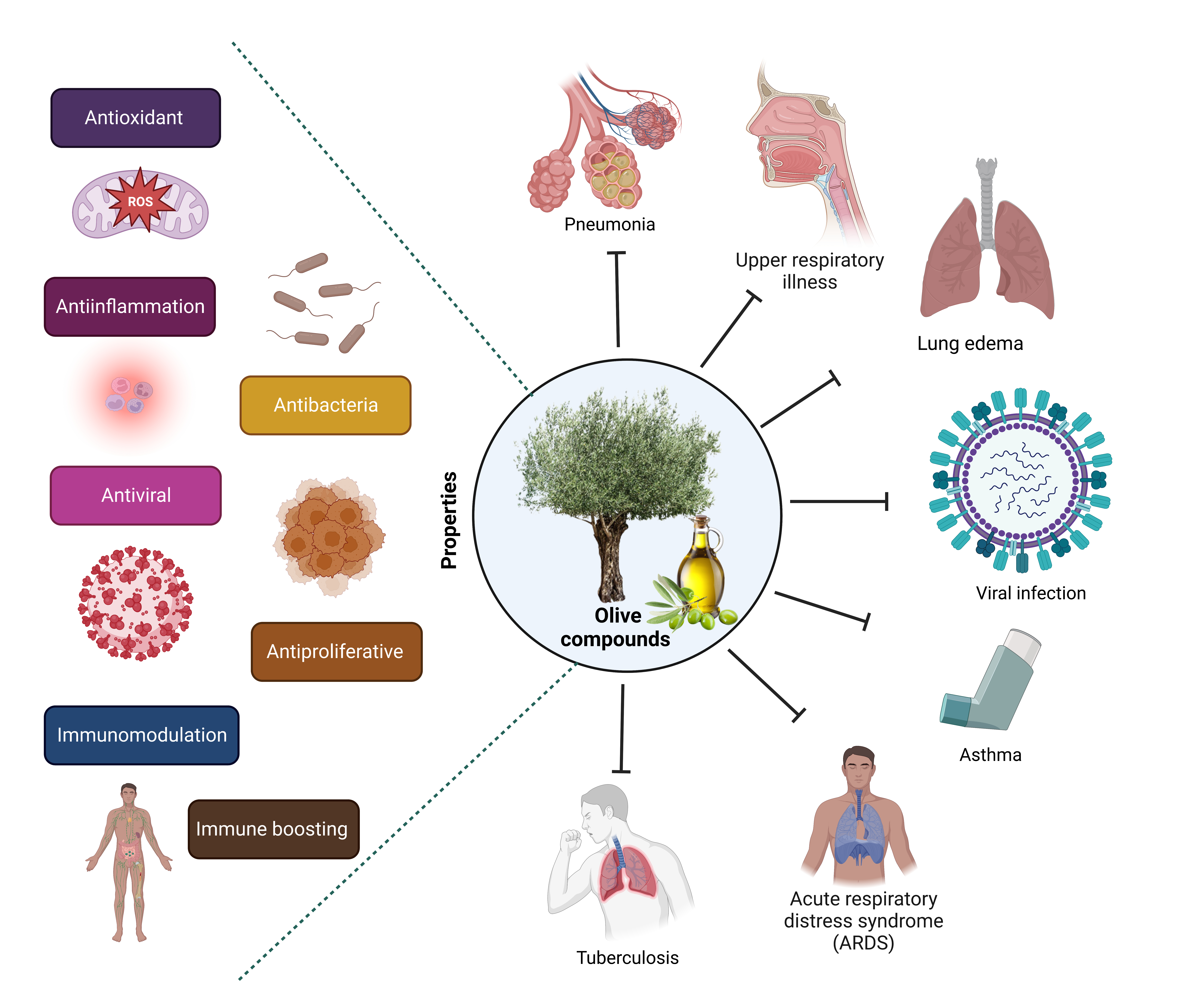
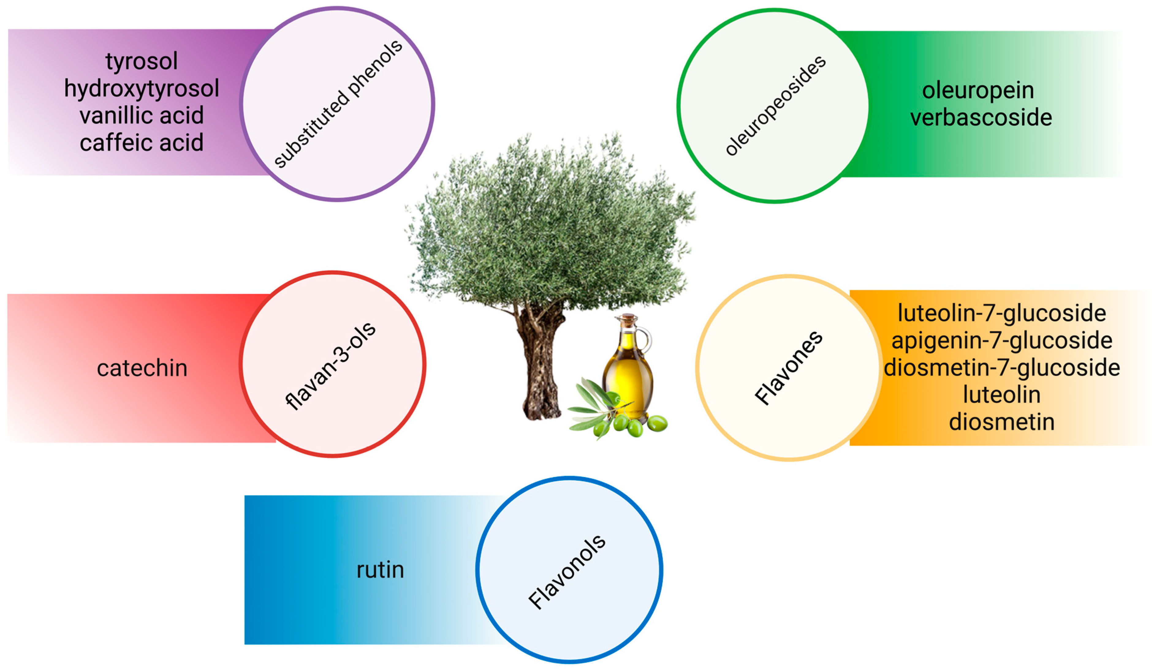
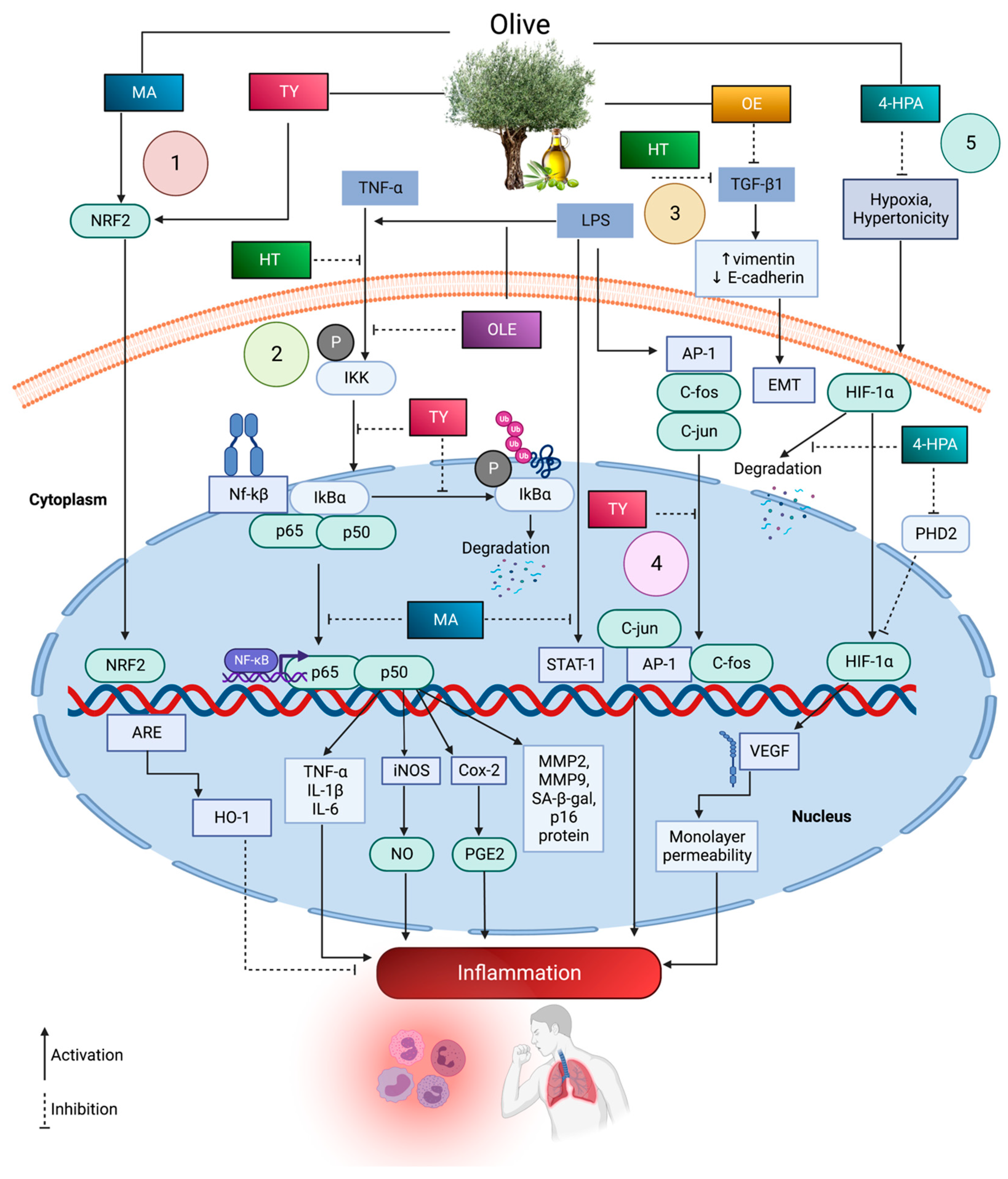
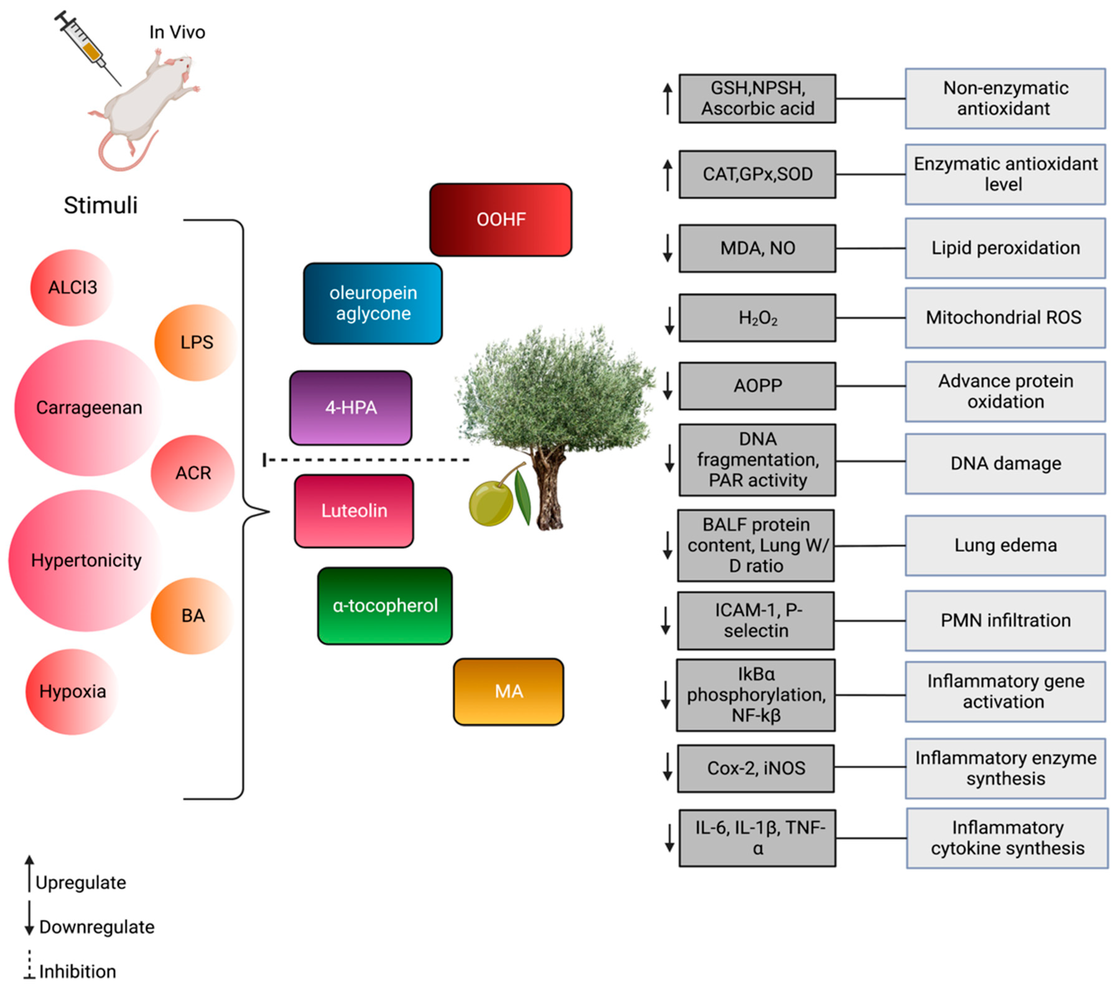
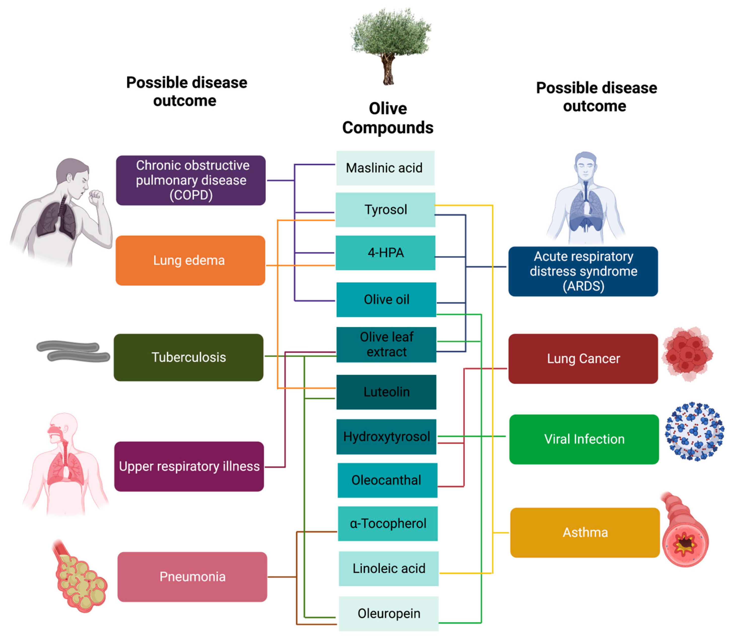
Table 1.
Composition of olive oil.
| Saponifiable Fraction | Unsaponifiable Fraction |
|---|---|
|
|
Table 2.
Main phenolic compounds in virgin olive oil [39].
Table 2.
Main phenolic compounds in virgin olive oil [39].
| Phenolic Acids | Phenolic Alcohols | Flavonoids | Secoroidoids | Lignans | Tocopherols and Tocotrienols |
|---|---|---|---|---|---|
|
|
|
|
| (α, β, γ, δ) |
Table 3.
Categories of respiratory diseases adapted from Kritek et al., 2018 [70].
Table 3.
Categories of respiratory diseases adapted from Kritek et al., 2018 [70].
| Category | Examples |
|---|---|
| Infections Disease caused by various microorganisms, such as bacteria, viruses, fungi and parasites. | Sinusitis, pneumonia, tuberculosis, influenza and COVID-19 |
| Obstructive Disease caused by narrowing or obstruction of the airways, causing difficulty in breathing. | Asthma, chronic obstructive pulmonary disease (COPD), bronchiectasis and bronchiolitis |
| Restrictive Disease affects the lung tissue or chest wall making it difficult for the lungs to expand and contract properly. | COPD, pulmonary fibrosis and chest wall disorders |
| Occupational Disease caused by exposure to various substances in the workplace, such as dust, chemicals and fumes. | Occupational asthma and pneumoconiosis |
| Genetic Disease caused by genetic mutations or abnormalities. | Cystic fibrosis and alpha-1 antitrypsin deficiency |
| Malignancy Disease caused by the uncontrolled growth of abnormal cells in the lungs or airways. | Lung cancer and mesothelioma |
| Vascular Disease affects the blood vessels in the lungs. | Pulmonary hypertension and pulmonary embolism |
Table 4.
Studies conducted on olive compounds in terms of respiratory anti-inflammation and antioxidants.
Table 4.
Studies conducted on olive compounds in terms of respiratory anti-inflammation and antioxidants.
| Compound | Model | Dosage and Duration | Finding(s) | Possible Contribution in Respiratory Disease | Reference |
|---|---|---|---|---|---|
| 4-Hydroxyphenylacetic acid (4-HPA) |
|
|
| Anti-inflammation | [118] |
| Luteolin | Caecal ligation and puncture (CLP)-induced ALI mice | 0.2 mg/kg 1 h |
|
| [120] |
| Oleuropein aglycone | Carrageenan-induced pleurisy mice | 40–100 mM/kg 30 min |
| Anti-inflammation | [122] |
| Hydrophilic fraction from olive oil (OOHF) | Aluminium- and acrylamide-induced rats | (1 mL) by gavage—21 days |
| Antioxidant | [123] |
| Tyrosol |
|
|
| Anti-inflammation | [124] |
| Maslinic acid |
|
|
|
| [129] |
| Pre-senescent human lung cells (MRC5) |
|
| Anti-inflammation | [128] |
| Ethanolic olive leaf extract (EOLE) and bromelain | Benzo(a)pyrene-mediated lung cancer mice |
|
|
| [142] |
| α-tocopherol (AT) | Aspiration-pneumonitis-induced rats |
|
| Antioxidant | [143] |
| Oleuropein | Lipopolysaccharide (LPS)-induced ALI in rats |
|
|
| [144] |
| Tyrosol | Ovalbumin-induced asthma rats |
|
|
| [145] |
↑ = Increases/upregulations, ↓ = decreases/downregulations. PHD2, HIF-prolyl hydroxylase domain; HIF-1α, Hypoxia-inducible factor-1α; TNF-α, tumour necrosis factor α; IL-1β, interleukin-1β; IL-6, interleukin-6; ICAM-1, intercellular adhesion molecule-1; NF-κβ, nuclear factor kappa-light-chain-enhancer of activated B cells; polymorphonuclear leukocyte (PMNs); iNOS, inducible nitric oxide synthase; MDA, malondialdehyde; CAT, catalase; SOD, Superoxide dismutase; GSH, glutathione; MPO, Myeloperoxidase; AlCl3, aluminium chloride; ACR, acrylamide; H2O2, hydrogen peroxide; AOPP, advanced oxidation protein products; LPS, lipopolysaccharide; IκBα, nuclear factor of kappa light polypeptide gene enhancer in B-cell inhibitor, alpha; LPO, lipid peroxidation; HO)-1, heme oxygenase; (Nrf2), nuclear factor erythrocyte 2-related factor 2; GPx, Glutathione peroxidase; ROS, reactive oxygen species; COX-2, cyclooxygenase; PMNs, polymorphonuclear neutrophils; prostaglandin, PGE2; TGFβ1, transforming growth factor; LPS, lipopolysaccharide; AREs, antioxidant response elements; MMP, matrix metalloproteinase; SASP, senescence-associated secretory phenotype; α-SMA, alpha smooth muscle actin; AP-1, activator protein; BALF, bronchoalveolar lavage fluid; NAG, N-acetyl glucosaminidase; LDH, lactate dehydrogenase; GGT, γ-glutamyl transferase; PICI, Peribronchial inflammatory cell infiltration; ASI, alveolar septal infiltration; AED, alveolar edema; AEx, alveolar exudate; CC-16, Clara cell protein 16; STAT-1, signal transducer and activator of transcription.
Table 5.
Clinical trials based on olive bioactive compounds in respiratory diseases.
| ID | Intervention and Control | Disease/ Condition | Study Size | Finding | References |
|---|---|---|---|---|---|
| NCT02421614 |
| Acute respiratory failure | 48 ventilated acute pulmonary failure patients |
| [111] |
| NCT01674595 |
| Allergic rhinoconjunctivitis | 93 allergic rhinoconjunctivitis with/without asthma |
| [148] |
| NCT00876356 |
| Asthma | 29 asthmatic children |
| [149] |
| ACTRN12618000328279 |
| Upper respiratory illness | 32 high school athletes |
| [147] |
| N/A | 226 patients undergoing surgery |
| [146] |
Table 6.
Unpublished clinical trials based on olive bioactive compounds in respiratory diseases.
| ClinicalTrials.gov ID | Intervention and Control | Disease/Condition | Study Size | Findings/Outcome Measures | Status | References |
|---|---|---|---|---|---|---|
| NCT05685901 |
| COVID-19 prevention | 88 adults |
|
| [150] |
| NCT04873349 |
|
| 60 COVID-19 adults |
|
| [151] |
| NCT01734265 |
|
| 63 adults |
|
| [152] |
| NCT01096771 |
| Acute respiratory distress syndrome | 14 |
|
| [153] |
Disclaimer/Publisher’s Note: The statements, opinions and data contained in all publications are solely those of the individual author(s) and contributor(s) and not of MDPI and/or the editor(s). MDPI and/or the editor(s) disclaim responsibility for any injury to people or property resulting from any ideas, methods, instructions or products referred to in the content. |
© 2023 by the authors. Licensee MDPI, Basel, Switzerland. This article is an open access article distributed under the terms and conditions of the Creative Commons Attribution (CC BY) license (https://creativecommons.org/licenses/by/4.0/).
Share and Cite
MDPI and ACS Style
Vijakumaran, U.; Goh, N.-Y.; Razali, R.A.; Abdullah, N.A.H.; Yazid, M.D.; Sulaiman, N. Role of Olive Bioactive Compounds in Respiratory Diseases. Antioxidants 2023, 12, 1140. https://doi.org/10.3390/antiox12061140
AMA Style
Vijakumaran U, Goh N-Y, Razali RA, Abdullah NAH, Yazid MD, Sulaiman N. Role of Olive Bioactive Compounds in Respiratory Diseases. Antioxidants. 2023; 12(6):1140. https://doi.org/10.3390/antiox12061140
Chicago/Turabian StyleVijakumaran, Ubashini, Neng-Yao Goh, Rabiatul Adawiyah Razali, Nur Atiqah Haizum Abdullah, Muhammad Dain Yazid, and Nadiah Sulaiman. 2023. "Role of Olive Bioactive Compounds in Respiratory Diseases" Antioxidants 12, no. 6: 1140. https://doi.org/10.3390/antiox12061140
Note that from the first issue of 2016, this journal uses article numbers instead of page numbers. See further details here.