Article

Higher Reduced State of Fe/S-Signals, with the Suppressed Oxidation of P700, Causes PSI Inactivation in Arabidopsis thaliana

1
Graduate School for Agricultural Science, Kobe University, 1-1 Rokkodai, Nada-ku, Kobe 657-8501, Japan
2
Core Research for Evolutional Science and Technology (CREST), Japan Science and Technology Agency (JST), 7 Gobancho, Tokyo 102-0076, Japan
3
Graduate School for Agriculture, Kyoto University, Kitashirakawa Oiwake-cho, Sakyo-ku, Kyoto 606-8502, Japan
*
Author to whom correspondence should be addressed.
Antioxidants 2023, 12(1), 21; https://doi.org/10.3390/antiox12010021
Submission received: 29 November 2022 / Revised: 17 December 2022 / Accepted: 20 December 2022 / Published: 22 December 2022
(This article belongs to the Special Issue Redox Regulation in Photosynthesis)

Abstract

:
Environmental stress increases the risk of electron accumulation in photosystem I (PSI) of chloroplasts, which can cause oxygen (O2) reduction to superoxide radicals and decreased photosynthetic ability. We used three Arabidopsis thaliana lines: wild-type (WT) and the mutants pgr5hope1 and paa1-7/pox1. These lines have different reduced states of iron/sulfur (Fe/S) signals, including Fx, FA/FB, and ferredoxin, the electron carriers at the acceptor side of PSI. In the dark, short-pulse light was repetitively illuminated to the intact leaves of the plants to provide electrons to the acceptor side of PSI. WT and pgr5hope1 plants showed full reductions of Fe/S during short-pulse light and PSI inactivation. In contrast, paa1-7/pox1 showed less reduction of Fe/S and its PSI was not inactivated. Under continuous actinic-light illumination, pgr5hope1 showed no P700 oxidation with higher Fe/S reduction due to the loss of photosynthesis control and PSI inactivation. These results indicate that the accumulation of electrons at the acceptor side of PSI may trigger the production of superoxide radicals. P700 oxidation, responsible for the robustness of photosynthetic organisms, participates in reactive oxygen species suppression by oxidizing the acceptor side of PSI.

1. Introduction

Oxygen (O2) in the atmosphere exists as triplet molecules (3O2) that have two unpaired electrons. Therefore, O2 easily reacts with radicals and molecules with low redox potential from which it receives electrons, resulting in the production of superoxide radicals (O2) [1]. Superoxide radicals disproportionate to hydrogen peroxide (H2O2) and water (H2O), and H2O2 can further produce hydroxyl radicals (·OH) via a Fenton reaction with reduced iron, i.e., Fe(II). Reactive oxygen species (ROS), such as O2, H2O2, and OH, are highly reactive and can easily oxidize DNA, proteins, and lipids in cells [2,3,4].
In the photosynthetic electron transport reaction, and depending on the difference in the redox potential of the sequential electron carriers, the electrons produced in photosystem (PS) II are transported from the acceptor side of PSII to the donor side of PSI through plastoquinone, the cytochrome (Cyt) b6/f-complex, and plastocyanin (PC), and from the PSI acceptor side to NADP+ through phylloquinone A1, iron/sulfur (Fe/S) clusters, namely Fx and FA/FB, and ferredoxin (Fd). In PSI, the redox potential of these electron carriers is sufficiently low to reduce O2 to O2 [4,5]. The production of H2O2 in PSI was first observed by Mehler [6,7] and Mehler and Brown [8]. Thereafter, Asada and Kiso [9] and Asada et al. [10] identified the primary product of the O2 reduction reaction in PSI to be O2. Furthermore, Takahashi and Asada [11] found that O2 can be reduced to O2 by Fe/S in PSI, and both Kozuleva et al. [12,13] and Kruk [14] reported that phylloquinone can reduce O2 to O2. Khorobrykh et al. [4] suggested the production of ·OH by the reaction of O2 with H2O2 catalyzed by Fe/S in PSI.
The in vivo production of ROS in PSI can be inferred from the fact that PSI inactivation occurs with the dependency of O2 in intact leaves. Repetitive short-pulse (rSP) light illumination of intact leaves under dark conditions induces electron accumulation in the photosynthetic electron transport system [15,16]. During rSP light illumination, PSI, but not PSII, was inactivated, causing a decrease in the carbon dioxide (CO2) assimilation rate. In addition, the inactivation of PSI depends on atmospheric O2 concentration [15]. Takahashi and Asada [11,17,18] reported that short-pulse light illumination produced O2 in the PSI of thylakoid membranes in vitro. Furthermore, the ROS produced in PSI may directly and immediately inactivate PSI, inducing oxidative damage to PSI proteins.
In the present study, we elucidated the relationship between electron accumulation at the acceptor side of PSI and inactivation of PSI in intact leaves of Arabidopsis thaliana. We monitored the reduction-oxidation (redox) states of P700 and Fe/S, including Fx and FA/FB, and Fd in PSI, using the intact leaves of wild-type (WT) and mutant pgr5hope1 and paa1-7/pox1 A. thaliana, which exhibit suppressed and enhanced oxidation levels of P700 [19,20]. These mutants were expected to have different electron accumulation levels on the acceptor side of PSI. We found that a higher reduced state (more electron accumulation) of both Fe/S and Fd accelerated the inactivation rate of PSI. The molecular mechanism of ROS production in PSI and the physiological function of P700 oxidation to suppress PSI inactivation in vivo are discussed.

2. Materials and Methods

2.1. Plant Materials and Growth Conditions

WT and mutant (pgr5hope1 and paa1-7/pox1) A. thaliana (gl-1) were grown in soil pots containing a 2:1.5 ratio of seeding-culture soil (TAKII Co., Ltd., Kyoto, Japan) to vermiculite. The pgr5hope1 and paa1-7/pox1 ethyl methane sulfonate-induced mutants originated from Wada et al. [20] and Furutani et al. [21], respectively. The pots were placed in a controlled chamber (14 h light at 23 °C/10 h darkness at 20 °C; photon flux density: 100–150 µmol photons m−2 s−1; relative humidity: 55–60%). Seeds were planted in the soil after 3 days of vernalization at 4 °C. The plants were watered every 2–3 days, and 1000-fold diluted Hyponex solution (Hyponex, Osaka, Japan) was applied weekly after seeding. Measurements were conducted using rosette leaves of 4–5 week old plants.

2.2. Simultaneous Measurements of Chlorophyll (Chl) Fluorescence, P700, and Fe/S-Signals with Gas-Exchange

Chl fluorescence, P700, Fe/S, including Fx, FA/FB, and Fd, and CO2 exchange were simultaneously measured using Dual/KLAS-NIR [22,23] (Heinz Walz GmbH, Effeltrich, Germany) and infra-red gas analyzer (IRGA) LI-7000 (Li-COR, Lincoln, NE, USA) measuring systems equipped with a 3010-DUAL gas exchange chamber at 40 Pa CO2/21 kPa O2 (Heinz Walz GmbH). The gases were saturated with water vapor at 16 ± 0.1 °C. The leaf temperature was controlled at 25 ± 0.5 °C (relative humidity: 55–60%). The actinic photon flux density at the upper position on the leaf in the chamber was adjusted to the indicated intensity. The net CO2 assimilation rate (A) and the dark respiration rate (Rd) were measured. The Chl fluorescence parameters were calculated [24] as follows: maximum quantum efficiency of PSII photochemistry, Fv/Fm = (FmFo)/Fm; Fo, minimum fluorescence yield; Fm, maximum fluorescence yield. The signals for the oxidized P700 (P700+) and reduced Fe/S (Fe/S) were calculated based on the deconvolution of four pulse-modulated dual-wavelength difference signals in the near-infrared region (780–820, 820–870, 840–965, and 870–965 nm) [22]. P700 was completely reduced and Fe/S was fully oxidized in the dark. To determine the total photo-oxidizable P700 (Pm), a saturation flash was applied after 10 s of illumination with far-red light (740 nm). Total photo-reducible Fe/S was determined by illumination with red actinic light (450 µmol photons m−2 s−1) after plant leaves were adapted to the dark for 5 min [22]. The redox state of P700 under actinic light illumination was evaluated as the ratio of P700+ to total P700. The incident photo-oxidizable P700 obtained by short-pulse light during rSP light illumination treatment was termed Pm’ [15].

2.3. Constant High-Intensity Light Treatment

High-intensity light treatments of both WT and pgr5hope1 plants were conducted under ambient air conditions (40 Pa CO2 and 21 kPa O2). After 15 min of photosynthesis induction (AL, 550 µmol photons m−2 s−1), the leaves were exposed to high-intensity light (1100 µmol photons m−2 s−1) for 120 min. To minimize the effect of rSP light illumination on PSI photoinactivation, the photosynthetic parameter Pm’ was recorded every 15 min [15]. The leaves were left in the dark for 30 min after treatment, and then the Fv/Fm of PSII, Pm, and magnitude of Fe/S were measured.

2.4. Statistical Analysis

Statistical analysis, Welch’s t-test, which are included in Microsoft Excel for Mac (ver. 16.16.27), were performed to detect any significant differences (* p < 0.05, ** p < 0.01, *** p < 0.001).

3. Results

We have previously reported that rSP light illumination induces inactivation of PSI in intact leaves of plants under atmospheric O2 conditions [15,25,26]. Comparing the ratio of PSI inactivation among land plants (liverworts, ferns, gymnosperms, and angiosperms), angiosperms suffered the most severe PSI inactivation [26]. This is because liverworts, ferns, and gymnosperms have flavodiiron proteins (FLV) in their chloroplasts, which produce H2O by reducing O2 using the reducing power (electrons) from the photosynthetic electron transport system, but angiosperms do not. FLV function as electron acceptors from PSI even during short-pulse light (~1000 ms). Therefore, P700 is reduced during short-pulse illumination in angiosperms, while in other plants FLV oxidize P700. This indicates that in angiosperms, including Arabidopsis, the oxidation reaction of the excited P700 to the oxidized form, P700+, is limited during short-pulse light, and electrons should accumulate at the acceptor side of PSI. Simultaneously, the accumulated electrons can flow to O2 producing O2. To confirm electron accumulation at the acceptor side of PSI during short-pulse light, we monitored Fe/S, representing both the Fe/S clusters and Fd, in PSI using DUAL/KLAS-NIR [23,27,28].
Upon illumination with short-pulse light (300 ms, 15,000 µmol photons m−2 s−1) of WT A. thaliana dark-adapted leaves, P700 was rapidly oxidized to P700+, achieving its maximum value at approximately 10 ms, and then decreased to its minimum at approximately 100 ms (Figure 1A). Upon illumination with short-pulse light, Fe/S was reduced to 70%, then transiently oxidized to 65%, and thereafter largely reduced to 100%. As shown in Figure 1B, the time range of the second reduction of Fe/S corresponds to the reduction of P700+ after reaching its maximum oxidation. That is, the reduction of P700+ after approximately 10 ms of short-pulse light resulted in the accumulation of electrons in the Fe/S of PSI. The maximum accumulation of electrons in Fe/S suppressed P700+ to its minimum, indicating the limitation of the oxidation reaction of the excited P700 during short-pulse light. The Arabidopsis mutant pgr5hope1 has an identical point mutation in PGR5 gene (At2g05620) as the pgr5-1 mutant and is therefore deficient in PGR5 protein [19,20]. pgr5hope1 has shown no P700 oxidation under continuous illumination with actinic light [19,20]. Compared to the WT, pgr5hope1 showed a lower proton motive force and proton gradient (ΔpH) across the thylakoid membranes but this did not suppress the plastoquinol oxidation activity of the Cyt b6/f complex. However, photosynthesis control does not function in pgr5hope1 [20]. In the present study, pgr5hope1 did not show any limitation in the reduction reaction of P700+ in the photo-oxidation reduction cycle of P700 in PSI. The illumination of short-pulse light to the dark-adapted leaves of pgr5hope1 and the behaviors of P700+ and Fe/S were all similar to those of the WT (Figure 1). In contrast to continuous illumination by actinic light, short-pulse light did not form a ΔpH across the thylakoid membranes [29], which did not induce photosynthesis control. Therefore, we could not find any difference in the behaviors of P700+ and Fe/S between WT and pgr5hope1 plants under short-pulse light illumination.
The Arabidopsis mutant paa1-7/pox1 was isolated when screening for the P700 oxidation response under single short-pulse illumination; it was identified as a mutant of P-type ATPase, PAA1 (At4g33525), which functions in copper (Cu) ion transport across the chloroplast envelope [21,30]. The paa1-7/pox1, similar to other paa1 mutants, showed a lower amount of PC due to the lack of Cu ion transport into chloroplasts [21,30]. We confirmed a much lower amount of PC compared to WT [21]. Furthermore, the reduction reaction of P700+ was expected to be limited in the photo-oxidation reduction cycle of P700 in PSI because electron transfer would be suppressed from the Cyt b6/f complex to P700 in PSI [21]. The illumination of the short-pulse light to the dark-adapted leaves of paa1-7/pox1 A. thaliana and the behaviors of P700+ and Fe/S were compared to those of the WT (Figure 1). During illumination with short-pulse light, P700 was oxidized for approximately 80 ms after reaching its maximum value, which was different from that of the WT (Figure 1A). The reduction rate of P700+ after reaching the maximum was slower than that of the WT, and the final oxidation state of P700+ at 300 ms was higher than that of the WT. These facts reflect the limitation of P700+ reduction during short-pulse illumination owing to the lower amount of PC. Fe/S took approximately 200 ms for full reduction after the start of the short-pulse light (Figure 1B). During the first 100 ms of the short-pulse light, Fe/S was more oxidized in the paa1-7/pox1 mutant than in the WT. Thereafter, Fe/S was slowly reduced, indicating the accumulation of electrons, leading to the reduction of P700+ after approximately 80 ms of short-pulse light. The kinetics of Fe/S showed that the electrons flowed from the Cyt b6/f complex to PSI at a slower rate in paa1-7/pox1 than in the WT.
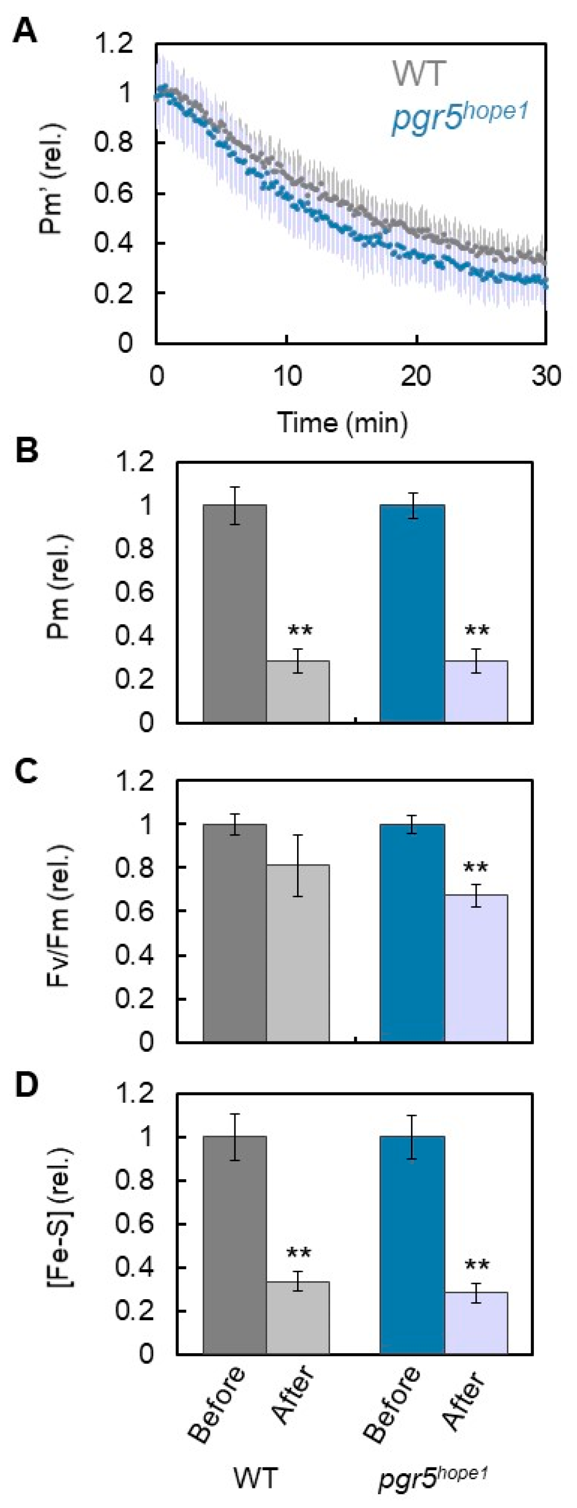
Using three plants of WT, pgr5hope1, and paa1-7/pox1, we analyzed the effects of electron accumulation at the acceptor side of PSI on PSI inactivation. The accumulation of electrons is represented by the reduction of Fe/S, as shown in Figure 1. During short-pulse light, both WT and pgr5hope1 plants showed the same degree of electron accumulation. In the comparison of WT with pgr5hope1, the rSP light treatments (300 ms, 15,000 µmol photons m−2 s−1, every 10 s for 30 min) gradually decreased the Pm’ values in both plant lines (Figure 2A). Pm’ corresponds to the incident photo-oxidizable P700 estimated by short-pulse illumination (see “Materials and Methods”), and a decrease in Pm’ leads to a decrease in Pm. That is, a decrease in Pm’ reflects PSI inactivation [15]. The extent of the Pm’ decrease in WT was similar to that in pgr5hope1. The residual Pm in both plant lines after 30 min of rSP light treatment was approximately 30% (Figure 2B). In contrast, the residual Fv/Fm of PSII was maintained above 80% in WT and approximately 70% in pgr5hope1. PSI was largely damaged by short-pulse light compared to PSII and at similar extent in both WT and pgr5hope1. Furthermore, we found a decrease in Fe/S after the rSP light treatment. The extent of the Fe/S decrease in WT (approximately 30%) was similar to that of pgr5hope1 (Figure 2D). In other words, Pm decreased as Fe/S decreased. This could be due to the lack of difference in electron accumulation in the Fe/S of both WT and pgr5hope1 plant lines during short-pulse light (Figure 1B).
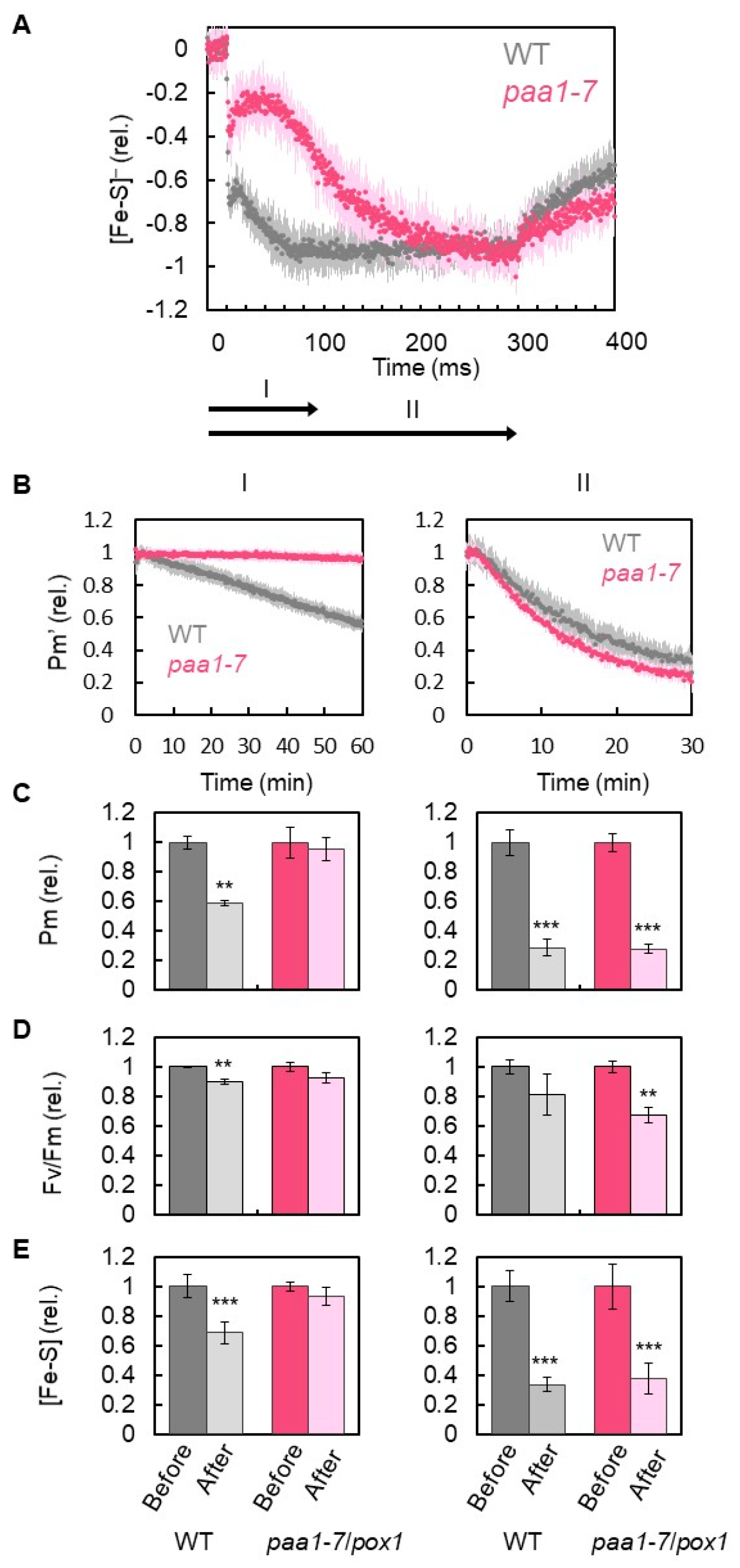
The paa1-7/pox1 mutant showed two time-based steps of Fe/S reduction: in the first step, the mutant showed less accumulation of electrons during the first 100 ms after the onset of the short-pulse light illumination, as compared to the WT; in the second step, there was full accumulation of electrons after 200 ms. Then, we set the illumination time of the short-pulse light to 100 ms (I) and 300 ms (II) in the rSP light illumination treatments I and II, respectively, for the comparison of PSI inactivation between WT and paa1-7/pox1 (Figure 3A). The rSP light illumination treatment I (duration I, 15,000 µmol photons m−2 s−1, every 10 s for 60 min) applied to WT gradually decreased Pm’; however, the rate of decrease was slower than that of the rSP light illumination treatment II (duration II, 15,000 µmol photons m−2 s−1, every 10 s for 30 min) (Figure 3B). The residual Pm in rSP light illumination treatment I was approximately 60% at 60 min, whereas it was approximately 30% at 30 min in rSP light illumination treatment II. In contrast to the WT, the rSP light illumination treatment I of paa1-7/pox1 did not inactivate PSI and PSII, as reflected by the Fv/Fm (Figure 3C). However, rSP light illumination treatment II inactivated PSI and PSII to the same extent as in the WT (Figure 3C). Furthermore, the residual Fe/S in the rSP light illumination treatment I of WT was approximately 70% at 60 min, whereas it was approximately 30% at 30 min in the rSP light illumination treatment II of WT (Figure 3E). In contrast to the WT, the rSP light illumination treatment I of paa1-7/pox1 did not decrease Fe/S (Figure 3D). However, the rSP light illumination treatment II of paa1-7/pox1 decreased Fe/S to a similar extent as in the WT (residual Fe/S, about 30%) (Figure 3D).
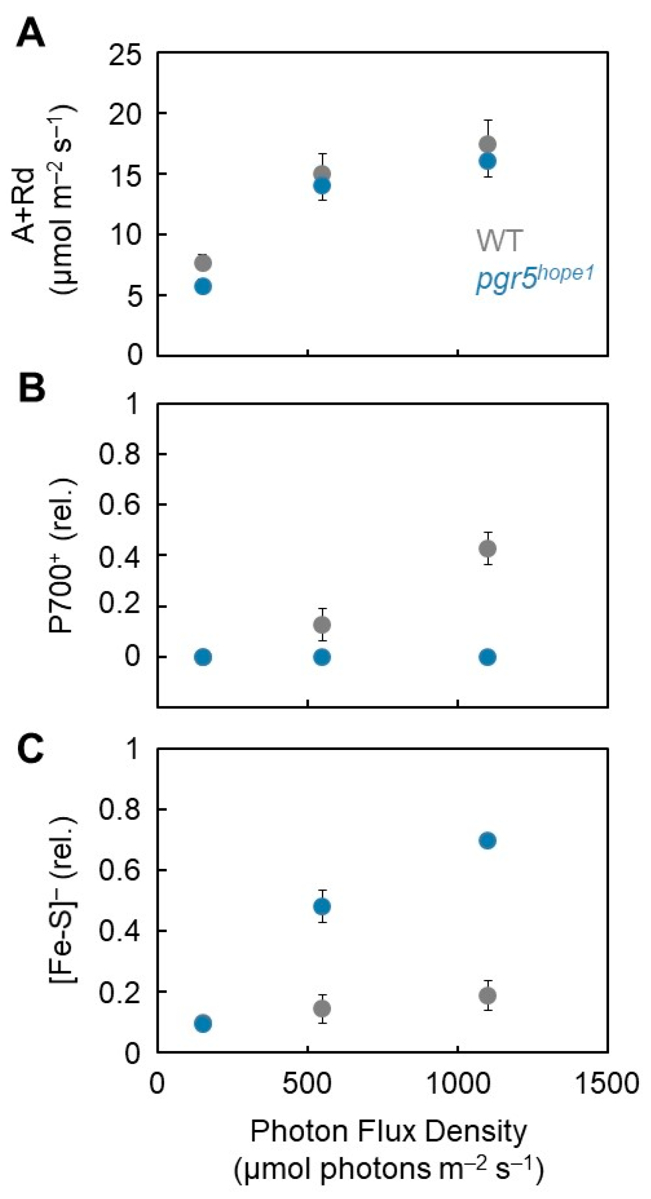
These results indicate that electron accumulation in Fe/S is the reason for PSI inactivation. The kinetics of both P700+ and Fe/S redox reactions during short-pulse light revealed that the preceding reduction of Fe/S and electron accumulation in the acceptor side of PSI induced the reduced state of P700+ (Figure 1). Under continuous illumination with actinic light, pgr5hope1 did not show any oxidation of P700+ owing to the lack of photosynthesis control, as described above [19,20]. Next, we analyzed the relationship between the redox state of P700 and Fe/S in both WT and pgr5hope1 plants under continuous illumination with actinic light (Figure 4). We compared the dependency of the gross CO2 assimilation rate (A + Rd, see “Materials and Methods”) on the photon flux density and found no difference between WT and pgr5hope1 (Figure 4A). However, we found a different dependency of P700 oxidation on the photon flux density (Figure 4B). With an increase in photon flux density, P700 was oxidized to approximately 40% at 1100 μmol photons m−2 s−1 in WT, but not in pgr5hope1. Furthermore, we found an enhanced reduction of Fe/S in pgr5hope1 with an increase in the photon flux density compared to the WT (Figure 4C). The reverse relationship between the oxidation of P700 and the reduced state of Fe/S indicated that photosynthesis limits electron donation to P700 in PSI and induces the oxidation of Fe/S.
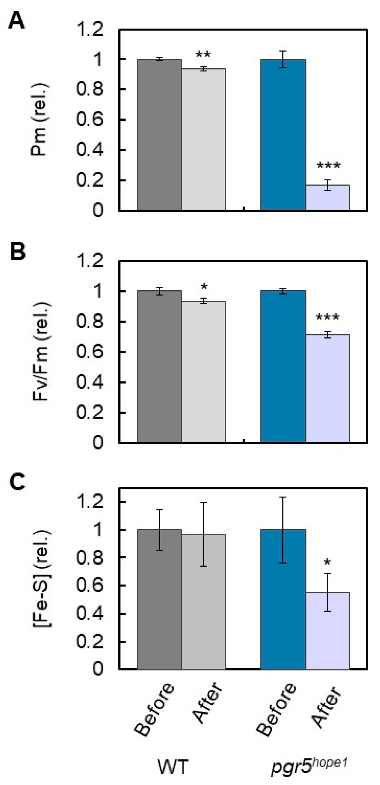
The reduced state of Fe/S, that is, the electron accumulation in Fe/S, might be the cause of PSI inactivation (Figure 2 and Figure 3). After reaching the steady state of the net CO2 assimilation rate induced by the continuous illumination of actinic light (500 µmol photons m−2 s−1, 30 min) in both WT and pgr5hope1, we increased the photon flux density to 1100 μmol photons m−2 s−1 and continued to illuminate intact leaves for 2 h. In contrast to the WT, pgr5hope1 showed PSI inactivation (Figure 5A). After the light treatment, Pm decreased to below 20% and Fv/Fm decreased to approximately 70% (Figure 5B). Furthermore, in contrast to the WT, Fe/S decreased to approximately 50% in pgr5hope1. A positive correlation between electron accumulation in Fe/S and PSI inactivation was therefore confirmed. The electron accumulation in Fe/S could trigger the production of ROS at the acceptor side of PSI and degrade Fe/S and PSI simultaneously.

4. Discussion

In the present study, we elucidated the positive relationship between electron accumulation at the acceptor side of PSI and PSI inactivation in A. thaliana intact leaves. We used three genotypes of Arabidopsis, which have different reduced states of Fe/S signals, including Fx, FA/FB, and Fd in PSI: WT, and the mutants, pgr5hope1 and paa1-7/pox1, which exhibit suppressed and enhanced oxidation levels of P700, respectively [21,30,31,32]. In the dark, where ΔpH across the thylakoid membranes was not induced and photosynthesis control was not activated, rSP light illumination was applied to the intact leaves of plants to provide electrons to the acceptor side of PSI. We confirmed the reduction of Fe/S with the reduction of P700 during short-pulse light in both WT and pgr5hope1 plants and to the same extent. In addition, both plant lines showed PSI inactivation. In contrast, paa1-7/pox1 showed two-phase kinetics of Fe/S characterized by slow and fast reductions, which were discriminated by the short-pulse light illumination time. A shorter illumination time (treatment I) reduced Fe/S by less than 25% with the maximum oxidation of P700; a longer illumination time (treatment II) led to the full reduction of Fe/S with a greater reduction in P700. Compared to the WT, the rSP light illumination treatment I did not inactivate the PSI of paa1-7/pox1. The rSP light illumination treatment I inactivated PSI in both WT and paa1-7/pox1. Furthermore, we compared the effects of the reduction of Fe/S on PSI inactivation under continuous illumination with actinic light in both WT and pgr5hope1. In a previous study, and in contrast to the WT, pgr5hope1 did not induce P700 oxidation [19,20]. This was also observed in the present study. We also found a higher reduction of Fe/S in pgr5hope1 than in the WT. In contrast to pgr5hope1, photosynthesis control suppressed plastoquinol oxidation activity in the WT, limiting the reduction of oxidized P700 in PSI. We confirmed PSI inactivation in pgr5hope1 plants under continuous illumination with actinic light. These results corresponded to those reported by Wada et al. [20]. The above results evidenced that the higher reduced state of Fe/S in PSI and accumulation of electrons at its acceptor side can trigger the production of ROS, which oxidatively damages PSI.
The higher reduced state of photosynthetic electron transport in illuminated thylakoid membranes and chloroplasts [33,34,35,36] and illuminated intact leaves of cucumber, Arabidopsis, and barley at lower temperature [37,38,39,40,41,42,43,44,45] and higher reduced state of P700 in angiosperms during rSP light illumination treatment [15,25,46,47,48], can cause PSI to be oxidatively damaged, depending on the presence of O2. The higher reduced state of Fe/S found in the present study reflected the reduction of phylloquinone, Fx, FA/FB, and Fd at the acceptor side of PSI. Reduced phylloquinone can donate electrons to O2 producing O2 [5,12,13]. Fx, FA/FB, and Fd can reduce O2 to O2 [5,11,12,13], which can oxidize the PSI.
However, regarding the kinetic interactions of these components with O2, the reduction of O2 to O2 would not be easy because the lifetime of phylloquinone A1 is less than 20 ns, the lifetime of Fx is less than 50 ns, and the lifetime of FA/FB is 500 ns to 100 ms [5], which is too short to react with O2. In fact, the reduction rate of O2 to O2 in PSI, the Mehler reaction rate, ranged from 15 to 30 μmol O2 mg Chl−1 h−1 [10,49]. Assuming that the ratio of P700 to Chl is 1:600 in thylakoid membranes, the half time of O2 reduction was estimated to be 150 to 300 ms [4], indicating that O2 would not have the opportunity to react with these electron carriers [4]. In fact, the O2 reduction rate in the Mehler reaction is negligible [50,51]. Ruuska et al. [50] showed no enhancement of the Mehler reaction, even in the reduced electron sinks of transgenic tobacco with reduced amounts of Rubisco.
Asada et al. reported the production of O2 in the aprotic interior of thylakoid membranes [11]. In the experiments using thylakoid membranes, no electron acceptors for PSI were present, and the electron carriers A1, FX, and FA/FB were greatly reduced. However, if the lifetime of the reduced electron carriers A1, FX, and FA/FB were prolonged, they might react with O2. These hypotheses have been recently supported by the following facts: (1) phylloquinones in PSI particles isolated from Chlamydomonas could reduce O2 to O2 [12,13], and (2) Fe/S clusters were the primary targets of PSI photoinhibition [33,34,35,36,52]. These conditions contributed to the accumulation of electrons at the acceptor side of PSI, which enhanced the interactions of these electron carriers with O2. Furthermore, no photosynthesis control functioned to oxidize the P700. Consequently, all these electron carriers were reduced.
In the present study, we demonstrated the reduction of Fe/S in the continuous illumination treatments of actinic light to pgr5hope1 (Figure 4). The reduction state of Fe/S was greater than 40% (Figure 4). The higher reduction of Fe/S in pgr5hope1 prolonged the lifetime of the reduced electron carriers (A1, FX, and FA/FB), which enhanced the reduction of O2 to O2 within the PSI complex. Furthermore, compared to WT, pgr5hope1 induces less luminal acidification of thylakoid membranes [53], thus prolonging the lifetime of the O2 produced in the aprotic interior of thylakoid membranes. In contrast to aqueous conditions, protons are absent in the aprotic interior of thylakoid membranes [54,55]. Hence, O2 cannot be protonated, its lifetime is enhanced, and O2 is more likely to react with PSI proteins located within the thylakoid membranes. At the low pH of the luminal space of thylakoid membranes, O2 can diffuse to the luminal face of the thylakoid membranes and easily react with the protons in the luminal space to dismutate to H2O2 and H2O, which prevents the interaction of O2 with the electron carriers (A1, FX, and FA/FB). If O2 is produced by xanthine oxidase in the dark, PSI is oxidatively damaged [35]. The O2 produced in the aqueous space easily accesses the PSI complex from the stromal side of the thylakoid membranes and can easily oxidize PSI [35]. That is, the acidification of the luminal space of thylakoid membranes contributes to both the oxidation of the electron carriers to suppress the production of O2 and the rapid dismutation of O2 produced in the PSI complex to H2O2 in the luminal space by supporting the diffusion direction of O2 in the aprotic interior space of thylakoid membranes to the luminal space, which is reflected as the O2 gradient from the production site to the luminal space. These observations were attributed to P700 oxidation. In addition, there is a positive relationship between P700 oxidation and the protection of PSI inactivation against the highly reduced state of Fe/S. These protection mechanisms are mainly driven by luminal acidification and photosynthetic control.
As described above, the rSP light illumination treatments decreased Fe/S in PSI (Figure 2, Figure 3 and Figure 5). The degradation of Fe/S clusters, including FX and FA/FB in the photo inactivated PSI has already been reported [36,39,52]. Furthermore, the degradation of PSI, PSI-A, and PSI-B reaction center proteins was observed under the photoinhibition of PSI [44,52], which was triggered by the O2 produced in PSI. O2 can inactivate the Escherichia coli enzymes dihydroxy-acid dehydratase, fumarase A, and fumarase B, as well as mammalian aconitase with rate constants ranging from 106 to 107 m−1 s−1 [56]. These enzymes have a 4Fe-4S cluster in their active sites. One of the irons in the reduced form of 4Fe-4S is attacked by O2 with a negative free energy change (from −10 to −30 kcal/mol) and degraded to 3Fe-4S with the release of one Fe, which cannot catalyze the electron transfer reaction [56]. Simultaneously, H2O2 is formed by the attachment of O2 to the 3Fe-4S cluster. Both Fe and H2O2 produced at the same site initiate the Fenton reaction to produce highly reactive ·OH, which might destroy the PSI-A/PSI-B polypeptide. In contrast, spinach dihydroxy-acid dehydratase containing a 2Fe-2S cluster in the active site showed resistance against O2 attack [56]. That is, the degradation of Fe/S clusters by O2 is specific to the 4Fe-4S clusters. In the present study, we observed a decrease in Fe/S amount in the leaves of A. thaliana WT, pgr5hope1, and paa1-7/pox1 (Figure 2, Figure 3 and Figure 5). Fd contains a 2Fe-2S cluster in its catalytic center [57] and it could not be degraded by the photoinactivation treatment. If Fx and/or FA/FB are degraded by O2, Fd cannot accept electrons from the PSI to be reduced. The remaining Fd could not be measured in our assay system (see “Materials and Methods”). As a result, Fe/S decreased to approximately 30% after rSP illumination (Figure 2, Figure 3 and Figure 5).
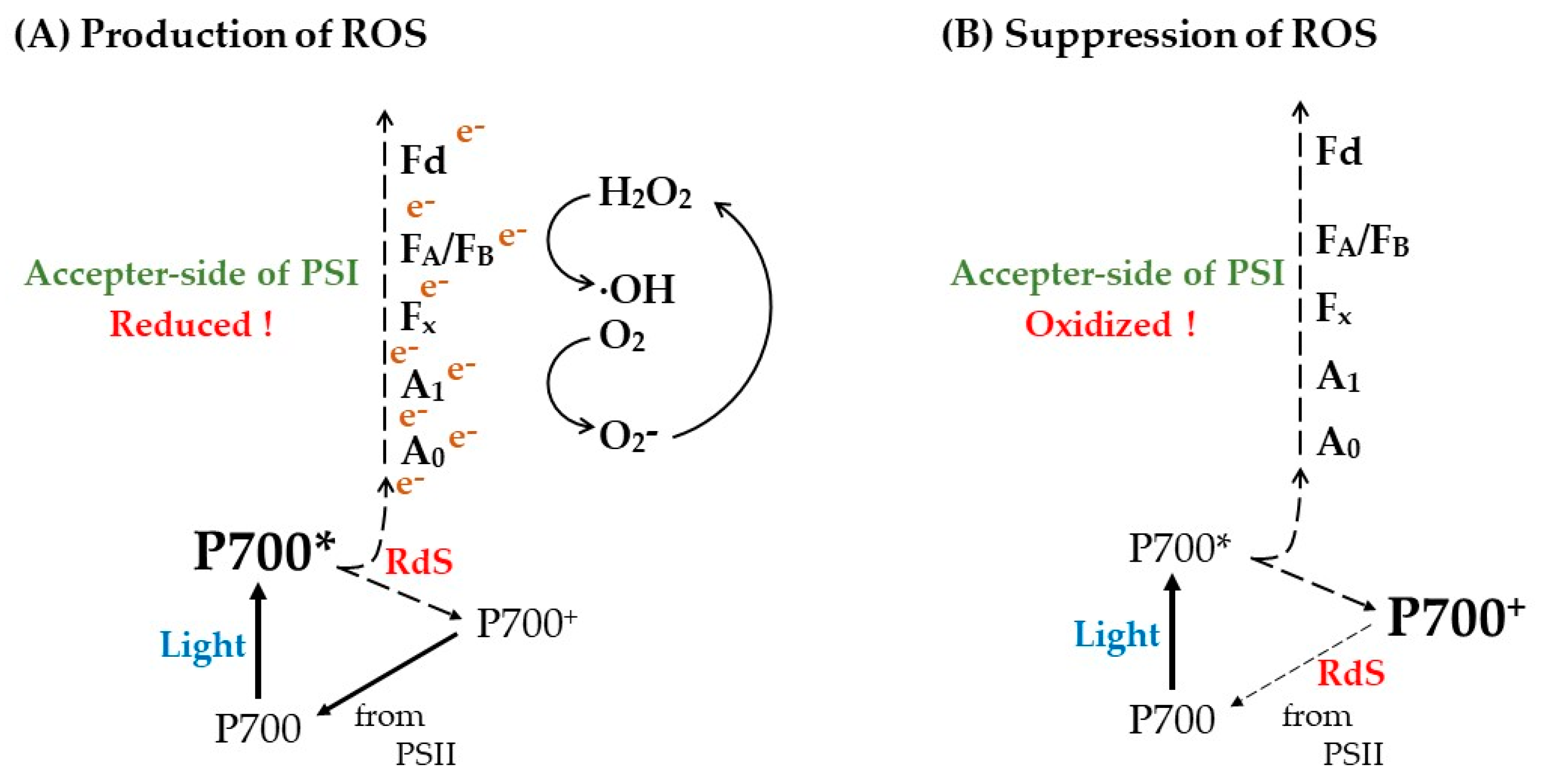
In the present study, we confirmed that the accumulation of electrons at the acceptor side of PSI, observed as the reduction of Fe/S, is the trigger of PSI inactivation. All oxygenic photosynthetic organisms oxidize P700 at low photosynthetic efficiency (e.g., under drought, fluctuating light, fluctuating stomata opening, high light, low temperature) [47,53,58,59,60,61,62,63,64,65,66]. Under these conditions, photosynthesis control driven by luminal acidification of thylakoid membranes downregulates the oxidation activity of plastoquinol in the Cyt b6/f complex [67]. Then, the rate-determining step (RdS) of the P700-photooxidation reduction cycle in PSI is shifted to the reduction reaction of oxidized P700 for the accumulation of P700+ [46,48]. Consequently, the reduction of Fe/S that leads to ROS production can be mitigated. This is the physiological function of P700 oxidation (Figure 6).

Author Contributions

C.M. conceived and designed the experiments; R.F. performed most of the experiments; S.W. and S.M. performed some of the experiments; R.F. and C.M. analyzed the data; R.F., K.I., S.W. and C.M. wrote the manuscript. All authors have read and agreed to the published version of the manuscript.

Funding

This work was supported by Core Research for Evolutional Science and Technology (CREST) of the Japan Science and Technology Agency, Japan (grant number JPMJCR15O3 to C.M.).

Institutional Review Board Statement

Not applicable.

Informed Consent Statement

Not applicable.

Data Availability Statement

Not applicable.

Conflicts of Interest

The authors declare no conflict of interest.

References

  1. Asada, K. The water–water cycle as alternative photon and electron sinks. Philos. Trans. R. Soc. B Biol. Sci. 2000, 355, 1419–1431. [Google Scholar] [CrossRef] [PubMed] [Green Version]
  2. Hamilton, T.L. The trouble with oxygen: The ecophysiology of extant phototrophs and implications for the evolution of oxygenic photosynthesis. Free Radic. Biol. Med. 2019, 140, 233–249. [Google Scholar] [CrossRef] [PubMed]
  3. Mattila, H.; Khorobrykh, S.; Havurinne, V.; Tyystjärvi, E. Reactive oxygen species: Reactions and detection from photosynthetic tissues. J. Photochem. Photobiol. B Biol. 2015, 152, 176–214. [Google Scholar] [CrossRef]
  4. Khorobrykh, S.; Havurinne, V.; Mattila, H.; Tyystjärvi, E. Oxygen and ROS in photosynthesis. Plants 2020, 9, 91. [Google Scholar] [CrossRef] [PubMed] [Green Version]
  5. Cherepanov, D.A.; Milanovsky, G.E.; Petrova, A.A.; Tikhonov, A.N.; Semenov, A.Y. Electron transfer through the acceptor side of photosystem I: Interaction with exogenous acceptors and molecular oxygen. Biochemistry 2017, 82, 1249–1268. [Google Scholar] [CrossRef]
  6. Mehler, A.H. Studies on reactions of illuminated chloroplasts. I. Mechanism of the reduction of oxygen and other Hill reagents. Arch. Biochem. Biophys. 1951, 33, 65–77. [Google Scholar] [CrossRef]
  7. Mehler, A.H. Studies on reactions of illuminated chloroplasts. II. Stimulation and inhibition of the reaction with molecular oxygen. Arch. Biochem. Biophys. 1951, 34, 339–351. [Google Scholar] [CrossRef] [PubMed]
  8. Mehler, A.H.; Brown, A.H. Studies on reactions of illuminated chloroplasts. III. Simultaneous photoproduction and consumption of oxygen studied with oxygen isotopes. Arch. Biochem. Biophys. 1952, 38, 365–370. [Google Scholar] [CrossRef]
  9. Asada, K.; Kiso, K. The photo-oxidation of epinephrine by spinach chloroplasts and its inhibition by superoxide dismutase: Evidence for the formation of superoxide radicals in chloroplasts. Agri. Biol. Chem. 1973, 37, 453–454. [Google Scholar] [CrossRef]
  10. Asada, K.; Kiso, K.; Yoshikawa, K. Univalent reduction of molecular oxygen by spinach chloroplasts on illumination. J. Biol. Chem. 1974, 249, 2175–2181. [Google Scholar] [CrossRef]
  11. Takahashi, M.; Asada, K. Superoxide production in aprotic interior of chloroplast thylakoids. Arch. Biochem. Biophys. 1988, 267, 714–722. [Google Scholar] [CrossRef] [PubMed]
  12. Kozuleva, M.; Petrova, A.; Milrad, Y.; Semenov, A.; Ivanov, B.; Redding, K.E.; Yacoby, I. Phylloquinone is the principal Mehler reaction site within photosystem I in high light. Plant Physiol. 2021, 186, 1848–1858. [Google Scholar] [CrossRef] [PubMed]
  13. Kozuleva, M.A.; Petrova, A.A.; Mamedov, M.D.; Semenov, A.Y.; Ivanov, B.N. O2 reduction by photosystem I involves phylloquinone under steady-state illumination. FEBS Lett. 2014, 588, 4364–4368. [Google Scholar] [CrossRef] [PubMed] [Green Version]
  14. Kruk, J.; Jemioła-Rzemińska, M.; Burda, K.; Schmid, G.H.; Strzałka, K. Scavenging of superoxide generated in photosystem I by plastoquinol and other prenyllipids in thylakoid membranes. Biochemistry 2003, 42, 8501–8505. [Google Scholar] [CrossRef]
  15. Sejima, T.; Takagi, D.; Fukayama, H.; Makino, A.; Miyake, C. Repetitive short-pulse light mainly inactivates photosystem I in sunflower leaves. Plant Cell Physiol. 2014, 55, 1184–1193. [Google Scholar] [CrossRef]
  16. Zivcak, M.; Brestic, M.; Kunderlikova, K.; Olsovska, K.; Allakhverdiev, S.I. Effect of photosystem I inactivation on chlorophyll a fluorescence induction in wheat leaves: Does activity of photosystem I play any role in OJIP rise? J. Photochem. Photobiol. B Biol. 2015, 152, 318–324. [Google Scholar] [CrossRef]
  17. Takahashi, M.; Asada, K. Dependence of oxygen affinity for Mehler reaction on photochemical activity of chloroplast thylakoids. Plant Cell Physiol. 1982, 23, 1457–1461. [Google Scholar]
  18. Takahashi, M.; Asada, K. Superoxide anion permeability of phospholipid membranes and chloroplast thylakoids. Arch. Biochem. Biophys. 1983, 226, 558–566. [Google Scholar] [CrossRef]
  19. Munekage, Y.; Hojo, M.; Meurer, J.; Endo, T.; Tasaka, M.; Shikanai, T. PGR5 is involved in cyclic electron flow around photosystem I and is essential for photoprotection in Arabidopsis. Cell 2002, 110, 361–371. [Google Scholar] [CrossRef] [Green Version]
  20. Wada, S.; Amako, K.; Miyake, C. Identification of a novel mutation exacerbated the PSI photoinhibition in pgr5/pgrl1 mutants; Caution for overestimation of the phenotypes in Arabidopsis pgr5-1 mutant. Cells 2021, 10, 2884. [Google Scholar] [CrossRef]
  21. Furutani, R.; Ohnishi, M.; Mori, Y.; Wada, S.; Miyake, C. The difficulty of estimating the electron transport rate at photosystem I. J. Plant Res. 2021, 135, 565–577. [Google Scholar] [CrossRef] [PubMed]
  22. Klughammer, C.; Schreiber, U. Deconvolution of ferredoxin, plastocyanin, and P700 transmittance changes in intact leaves with a new type of kinetic LED array spectrophotometer. Photosynth. Res. 2016, 128, 195–214. [Google Scholar] [CrossRef] [PubMed] [Green Version]
  23. Sétif, P.; Boussac, A.; Krieger-Liszkay, A. Near-infrared in vitro measurements of photosystem I cofactors and electron-transfer partners with a recently developed spectrophotometer. Photosynth. Res. 2019, 142, 307–319. [Google Scholar] [CrossRef] [PubMed]
  24. Baker, N.R.; Harbinson, J.; Kramer, D. Determining the limitations and regulation of photosynthetic energy transduction in leaves. Plant Cell Environ. 2007, 30, 1107–1125. [Google Scholar] [CrossRef]
  25. Shimakawa, G.; Miyake, C. Oxidation of P700 ensures robust photosynthesis. Front. Plant Sci. 2018, 9, 1617. [Google Scholar] [CrossRef] [Green Version]
  26. Takagi, D.; Ishizaki, K.; Hanawa, H.; Mabuchi, T.; Shimakawa, G.; Yamamoto, H.; Miyake, C. Diversity of strategies for escaping reactive oxygen species production within photosystem I among land plants: P700 oxidation system is prerequisite for alleviating photoinhibition in photosystem I. Physiol. Plant. 2017, 161, 56–74. [Google Scholar] [CrossRef]
  27. Kadota, K.; Furutani, R.; Makino, A.; Suzuki, Y.; Wada, S.; Miyake, C. Oxidation of P700 induces alternative electron flow in Photosystem I in wheat leaves. Plants 2019, 8, 152. [Google Scholar] [CrossRef] [Green Version]
  28. Sétif, P.; Shimakawa, G.; Krieger-Liszkay, A.; Miyake, C. Identification of the electron donor to flavodiiron proteins in Synechocystis sp. PCC 6803 by in vivo spectroscopy. Biochim. Biophys. Acta BBA Bioenerg. 2020, 1861, 148256. [Google Scholar] [CrossRef]
  29. Johnson, M.P.; Ruban, A.V. Rethinking the existence of a steady-state Δψ component of the proton motive force across plant thylakoid membranes. Photosynth. Res. 2013, 119, 233–242. [Google Scholar] [CrossRef] [Green Version]
  30. Shikanai, T.; Müller-Moulé, P.; Munekage, Y.; Niyogi, K.K.; Pilon, M. PAA1, a P-Type ATPase of Arabidopsis, functions in copper transport in chloroplasts. Plant Cell 2003, 15, 1333–1346. [Google Scholar] [CrossRef] [Green Version]
  31. Tikkanen, M.; Grieco, M.; Nurmi, M.; Rantala, M.; Suorsa, M.; Aro, E.-M. Regulation of the photosynthetic apparatus under fluctuating growth light. Philos. Trans. R. Soc. B Biol. Sci. 2012, 367, 3486–3493. [Google Scholar] [CrossRef] [PubMed]
  32. Tikkanen, M.; Rantala, S.; Aro, E.-M. Electron flow from PSII to PSI under high light is controlled by PGR5 but not by PSBS. Front. Plant Sci. 2015, 6, 521. [Google Scholar] [CrossRef] [PubMed] [Green Version]
  33. Satoh, K. Mechanism of photoinactivation in photosynthetic systems II. The occurrence and properties of two different types of photoinactivation. Plant Cell Physiol. 1970, 11, 29–38. [Google Scholar] [CrossRef]
  34. Satoh, K. Mechanism of photoinactivation in photosynthetic systems. III. The site and mode of photoinactivation in photosystem I. Plant Cell Physiol. 1970, 11, 187–197. [Google Scholar] [CrossRef]
  35. Inoue, K.; Fujii, T.; Yokoyama, E.; Matsuura, K.; Hiyama, T.; Sakurai, H. The Photoinhibition site of photosystem I in isolated chloroplasts under extremely reducing conditions. Plant Cell Physiol. 1989, 30, 65–71. [Google Scholar] [CrossRef]
  36. Inoue, K.; Sakurai, H.; Hiyama, T. Photoinactivation sites of photosystem I in isolated chloroplasts. Plant Cell Physiol. 1986, 27, 961–968. [Google Scholar]
  37. Havaux, M.; Davaud, A. Photoinhibition of photosynthesis in chilled potato leaves is not correlated with a loss of Photosystem-II activity. Photosynth. Res. 1994, 40, 75–92. [Google Scholar] [CrossRef]
  38. Sonoike, K.; Terashima, I. Mechanism of photosystem-I photoinhibition in leaves of Cucumis sativus L. Planta 1994, 194, 287–293. [Google Scholar] [CrossRef]
  39. Sonoike, K.; Terashima, I.; Iwaki, M.; Itoh, S. Destruction of photosystem I iron-sulfur centers in leaves of Cucumis sativus L. by weak illumination at chilling temperatures. FEBS Lett. 1995, 362, 235–238. [Google Scholar] [CrossRef] [Green Version]
  40. Terashima, I.; Funayama, S.; Sonoike, K. The site of photoinhibition in leaves of Cucumis sativus L. at low temperatures is photosystem I, not photosystem II. Planta 1994, 193, 300–306. [Google Scholar] [CrossRef]
  41. Ivanov, A.G.; Morgan, R.M.; Gray, G.R.; Velitchkova, M.Y.; Huner, N.P. Temperature/light dependent development of selective resistance to photoinhibition of photosystem I. FEBS Lett. 1998, 430, 288–292. [Google Scholar] [CrossRef] [PubMed] [Green Version]
  42. Takeuchi, K.; Che, Y.; Nakano, T.; Miyake, C.; Ifuku, K. The ability of P700 oxidation in photosystem I reflects chilling stress tolerance in cucumber. J. Plant Res. 2022, 135, 681–692. [Google Scholar] [CrossRef] [PubMed]
  43. Scheller, H.V.; Haldrup, A. Photoinhibition of photosystem I. Planta 2005, 221, 5–8. [Google Scholar] [CrossRef] [PubMed]
  44. Tjus, S.E.; Møller, B.L.; Scheller, H. Photosystem I is an early target of photoinhibition in barley illuminated at chilling Temperatures. Plant Physiol. 1998, 116, 755–764. [Google Scholar] [CrossRef] [Green Version]
  45. Zhang, S.; Scheller, H. Photoinhibition of Photosystem I at chilling temperature and Subsequent Recovery in Arabidopsis thaliana. Plant Cell Physiol. 2004, 45, 1595–1602. [Google Scholar] [CrossRef]
  46. Furutani, R.; Ifuku, K.; Suzuki, Y.; Noguchi, K.; Shimakawa, G.; Wada, S.; Makino, A.; Sohtome, T.; Miyake, C. P700 Oxidation Suppresses the Production of Reactive Oxygen Species in Photosystem I; Hisabori, T., Ed.; Academic Press: Cambridge, MA, USA, 2000; Volume 96, p. 26. [Google Scholar]
  47. Furutani, R.; Makino, A.; Suzuki, Y.; Wada, S.; Shimakawa, G.; Miyake, C. Intrinsic fluctuations in transpiration induce photorespiration to oxidize P700 in photosystem I. Plants 2020, 9, 1761. [Google Scholar] [CrossRef]
  48. Miyake, C. Molecular mechanism of oxidation of P700 and suppression of ROS production in photosystem I in response to electron-sink limitations in C3 Plants. Antioxidants 2020, 9, 230. [Google Scholar] [CrossRef] [Green Version]
  49. Miyake, C.; Schreiber, U.; Hormann, H.; Sano, S.; Asada, K. The FAD-enzyme monodehydroascorbate radical reductase mediates photoproduction of superoxide radicals in spinach thylakoid membranes. Plant Cell Physiol. 1998, 39, 821–829. [Google Scholar] [CrossRef] [Green Version]
  50. Ruuska, S.A.; Badger, M.R.; Andrews, T.J.; von Caemmerer, S. Photosynthetic electron sinks in transgenic tobacco with reduced amounts of Rubisco: Little evidence for significant Mehler reaction. J. Exp. Bot. 2000, 51, 357–368. [Google Scholar] [CrossRef] [Green Version]
  51. Driever, S.M.; Baker, N.R. The water-water cycle in leaves is not a major alternative electron sink for dissipation of excess excitation energy when CO2 assimilation is restricted. Plant Cell Environ. 2011, 34, 837–846. [Google Scholar] [CrossRef]
  52. Sonoike, K. Photoinhibition of photosystem I. Physiol. Plant. 2011, 142, 56–64. [Google Scholar] [CrossRef] [PubMed]
  53. Suorsa, M.; Järvi, S.; Grieco, M.; Nurmi, M.; Pietrzykowska, M.; Rantala, M.; Kangasjärvi, S.; Paakkarinen, V.; Tikkanen, M.; Jansson, S.; et al. PROTON GRADIENT REGULATION5 is essential for proper acclimation of Arabidopsis photosystem I to naturally and artificially fluctuating light conditions. Plant Cell 2012, 24, 2934–2948. [Google Scholar] [CrossRef] [PubMed]
  54. Hormann, H.; Neubauer, C.; Asada, K.; Schreiber, U. Intact chloroplasts display pH 5 optimum of O2-reduction in the absence of methyl viologen: Indirect evidence for a regulatory role of superoxide protonation. Photosynth. Res. 1993, 37, 69–80. [Google Scholar] [CrossRef]
  55. Hormann, H.; Neubauer, C.; Schreiber, U. An active Mehler-peroxidase reaction sequence can prevent cyclic PS I electron transport in the presence of dioxygen in intact spinach chloroplasts. Photosynth. Res. 1994, 41, 429–437. [Google Scholar] [CrossRef] [PubMed]
  56. Flint, D.; Tuminello, J.; Emptage, M. The inactivation of Fe-S cluster containing hydro-lyases by superoxide. J. Biol. Chem. 1993, 268, 22369–22376. [Google Scholar] [CrossRef] [PubMed]
  57. Holden, H.M.; Jacobson, B.L.; Hurley, J.K.; Tollin, G.; Oh, B.-H.; Skjeldal, L.; Chae, Y.K.; Cheng, H.; Xia, B.; Markley, J.L. Structure-function studies of [2Fe-2S] ferredoxins. J. Bioenerg. Biomembr. 1994, 26, 67–88. [Google Scholar] [CrossRef] [PubMed]
  58. Golding, A.J.; Johnson, G.N. Down-regulation of linear and activation of cyclic electron transport during drought. Planta 2003, 218, 107–114. [Google Scholar] [CrossRef] [PubMed]
  59. Harbinson, J.; Genty, B.; Foyer, C.H. Relationship between photosynthetic electron transport and stromal enzyme activity in pea leaves: Toward an understanding of the nature of photosynthetic control. Plant Physiol. 1990, 94, 545–553. [Google Scholar] [CrossRef] [Green Version]
  60. Harbinson, J.; Foyer, C.H. Relationships between the efficiencies of photosystems I and II and stromal redox state in CO2-free air: Evidence for cyclic electron flow in vivo. Plant Physiol. 1991, 97, 41–49. [Google Scholar] [CrossRef] [Green Version]
  61. Harbinson, J.; Hedley, C.L. Changes in P-700 oxidation during the early stages of the induction of photosynthesis. Plant Physiol. 1993, 103, 649–660. [Google Scholar] [CrossRef] [Green Version]
  62. Kono, M.; Noguchi, K.; Terashima, I. Roles of the cyclic electron flow around PSI (CEF-PSI) and O2-dependent alternative pathways in regulation of the photosynthetic electron flow in short-term fluctuating light in Arabidopsis thaliana. Plant Cell Physiol. 2014, 55, 990–1004. [Google Scholar] [CrossRef] [PubMed] [Green Version]
  63. Miyake, C.; Miyata, M.; Shinzaki, Y.; Tomizawa, K.-I. CO2 response of cyclic electron flow around PSI (CEF-PSI) in Tobacco leaves—Relative electron fluxes through PSI and PSII determine the magnitude of non-photochemical quenching (NPQ) of Chl fluorescence. Plant Cell Physiol. 2005, 46, 629–637. [Google Scholar] [CrossRef] [PubMed]
  64. Wada, S.; Miyake, C.; Makino, A.; Suzuki, Y. Photorespiration coupled with CO2 assimilation protects photosystem I from photoinhibition under moderate poly(ethylene glycol)-induced osmotic stress in Rice. Front. Plant Sci. 2020, 11, 1121. [Google Scholar] [CrossRef] [PubMed]
  65. Wada, S.; Takagi, D.; Miyake, C.; Makino, A.; Suzuki, Y. Responses of the photosynthetic electron transport reactions stimulate the oxidation of the reaction center chlorophyll of photosystem I, P700, under drought and high temperatures in Rice. Int. J. Mol. Sci. 2019, 20, 2068. [Google Scholar] [CrossRef] [PubMed] [Green Version]
  66. Shi, Q.; Wang, X.; Zeng, Z.; Huang, W. Photoinhibition of photosystem I induced by different intensities of fluctuating light is determined by the kinetics of ∆pH formation rather than linear electron flow. Antioxidants 2022, 11, 2325. [Google Scholar] [CrossRef]
  67. Tikhonov, A.N. The cytochrome b6f complex at the crossroad of photosynthetic electron transport pathways. Plant Physiol. Biochem. 2014, 81, 163–183. [Google Scholar] [CrossRef]
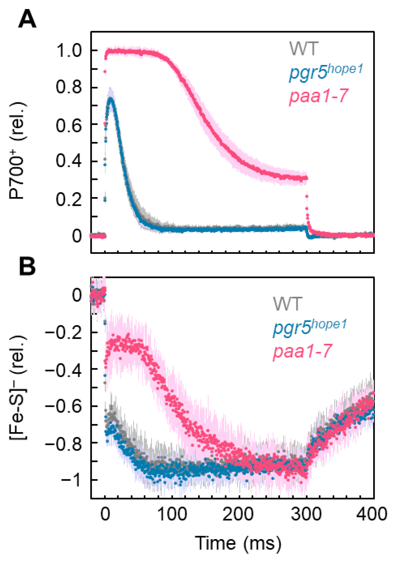
Figure 1. Kinetics of (A) oxidized P700 (P700+) and (B) reduced Fe/S signals (Fe/S) in response to short-pulse light for wild-type (WT), pgr5hope1, and paa1-7 Arabidopsis intact leaves. Short-pulse light (15,000 µmol photons m−2 s−1, 300 ms) was started at 0 ms. The redox reactions of both P700 and Fe/S were monitored simultaneously. Relative values of both P700+ and [Fe/S] were normalized to the maximum oxidation and reduction levels, respectively, as described in the “Materials and Methods”. The negative values of Fe/S show the reduction of Fe/S. The data points for WT (gray), pgr5hope1 (magenta), and paa1-7/pox1 (red) are the means of six biological replicates (darker color) and the shadowed area is the standard deviation (lighter color).
Antioxidants 12 00021 g001
Figure 2. Effects of rSP light illumination on the incident photo-oxidizable P700 (Pm’), photo-oxidizable P700 (Pm), maximum quantum efficiency of PSII (Fv/Fm), and amount of Fe/S in wild-type (WT) and pgr5hope1 Arabidopsis. The leaves were illuminated every 10 s with short-pulse light (300 ms) of 15,000 μmol photons m−2 s−1 under the atmospheric conditions (40 Pa CO2/ 21 kPa O2) for 30 min. The rSP light illumination started at 0 min. (A) Pm’. Black, WT; magenta, pgr5hope1. The mean values were normalized to the primary values before the rSP light treatment; error bars represent the standard deviation; data were acquired from six biological replicates. (B) Pm, (C) Fv/Fm, and (D) Fe/S were compared before and after the rSP light illumination treatment in WT and pgr5hope1. These parameters were evaluated after the illuminated leaves were left for 1 h in the dark. Each value was normalized to the value before the rSP light illumination treatment. ** p < 0.01 (Welch’s t-test).
Antioxidants 12 00021 g002
Figure 3. Effects of rSP light illumination and illumination time on the incident photo-oxidizable P700 (Pm’), photo-oxidizable P700 (Pm), maximum quantum efficiency of PSII (Fv/Fm), and amount of Fe/S in wild-type (WT) and paa1-7/pox1 Arabidopsis. The leaves were illuminated every 10 s with short-pulse light of 15,000 μmol photons m−2 s−1 under atmospheric conditions (40 Pa CO2/ 21 kPa O2) for 30 min. The rSP light illumination started at 0 min. (A) The illumination time was set to the two durations, as indicated by the arrows (I, 100 ms; II, 300 ms), based on the reduction kinetics of Fe/S of both WT and paa1-7/pox1 (redrawn from Figure 1, Black, WT; red, paa1-7/pox1). In experiments I and II, the parameters Pm’, Pm, Fv/Fm, and [Fe/S] were analyzed. (B) Pm’. Black, WT; magenta, pgr5hope1. The values were normalized to the primary values before the rSP light illumination treatment and are shown with standard deviations. The data were obtained from six biological replicates. (C) Pm, (D) Fv/Fm, and (E) Fe/S before and after the rSP light illumination treatments in both WT and pgr5hope1 were compared. These parameters were evaluated after the illuminated leaves were left for 1 h in the dark. Each value was normalized against the value before the rSP light illumination treatment. ** p < 0.01; *** p < 0.001 (Welch’s t-test).
Antioxidants 12 00021 g003
Figure 4. Effect of photon flux density on the gross CO2 assimilation rate (A + Rd), oxidized P700 (P700+), and reduction ratio of Fe/S signals [Fe/S] in wild-type (WT) and pgr5hope1 Arabidopsis. (A) The net CO2 assimilation rates were measured simultaneously with P700+ and Fe/S under atmospheric conditions (40 Pa CO2, 21 kPa O2). The dark respiration rates (Rd) were measured before starting actinic light illumination. After the net CO2 assimilation reached the steady state at the photon flux density of 150 μmol photons m−2 s−1, the intensity was increased to 550 and 1100 sequentially, after reaching each steady-state CO2 assimilation. The gross CO2 assimilation rates are expressed as A + Rd. (B) The oxidized P700 (P700+) is plotted against the photon flux density. (C) The reduction ratio of [Fe/S] is plotted against the photon flux density. The mean of three biological replicates and standard deviation are shown. Gray symbols, WT; Blue symbols, pgr5hope1.
Antioxidants 12 00021 g004
Figure 5. Effects of the continuous illumination of actinic light on the photo-oxidizable P700 (Pm), maximum quantum efficiency of PSII (Fv/Fm), and amount of Fe/S ([Fe/S]) in wild-type (WT) and pgr5hope1 Arabidopsis. After reaching the steady state of the net CO2 assimilation rate induced by the continuous illumination of actinic light (500 μmol photons m−2 s−1, 30 min) in both WT and pgr5hope1, the photon flux density was increased to 1100 μmol photons m−2 s−1, and the illumination was continued for 2 h. The parameters, (A) Pm, (B) Fv/Fm, and (C) [Fe/S] of both WT and pgr5hope1 treated with continuous illumination were evaluated after the illuminated leaves were left for 1 h in the dark, and values obtained before and after the continuous light illumination treatment were compared. The mean of three biological replicates and standard deviation are shown. * p < 0.05; ** p < 0.01; *** p < 0.001 (Welch’s t-test).
Antioxidants 12 00021 g005
Figure 6. Production and suppression mechanism of ROS in PSI. PSI catalyzes the photosynthetic electron transport through a photo-oxidation reduction cycle in the reaction center of P700. P700 is photo-excited to P700* by the absorbed light energy and donates electrons to the primary electron acceptor chlorophyll a (A0). P700+ accepts electrons from PSII through plastoquinol, Cyt b6/f-complex, and plastocyanin (PC), regenerating P700. The electron in A0 flows to ferredoxin (Fd) through phylloquinone A1 and the iron-sulfur (4Fe-4S) clusters Fx and FA/FB. The accumulation of P700+ under the constant photon flux density is determined by the rate-determining step (RdS) of the production-consumption rate of P700+ in the photo-oxidation reduction cycle of P700 in PSI. At the RdS of P700*, oxidation P700+ does not accumulate; at the RdS of P700+ reduction, P700+ accumulates. (A) The RdS of P700* oxidation is caused by the accumulation of electrons at the acceptor-side of PSI, as observed with the reduction of Fe/S clusters (Fe/S), including Fx, FA/FB, and Fd, where the possibilities of the reduction of phylloquinone A1 also increase. These accumulated electrons would flow to O2 to produce superoxide radical (O2), and O2 would degrade FX and FA/FB with the release of Fe and hydrogen peroxide (H2O2). Both H2O2 and the reduced Fe further react to produce hydroxyl radical (OH) through Fenton reaction. This highly reactive oxygen species (ROS) would oxidatively degrade PSI irreversibly, leading to PSI inactivation. (B) The RdS of P700+ reduction is caused by the limitation of photosynthetic electron transport from plastoquinol to P700 through Cyt b6/f-complex and PC [46,48]. Even under low photosynthesis efficiency conditions (drought, high intensity light, low/high temperature, low CO2, etc.), P700 is oxidized to P700+. The RdS of P700+ reduction is induced by the acidification in the luminal side of thylakoid membranes, which suppress the oxidation activity of plastoquinol by the Cyt b6/f-complex, with the oxidation of the electron acceptors A0, A1, FX, FA/FB, and Fd, leading to the suppression of ROS production. This is the physiological function of P700 oxidation to suppress ROS production in PSI.
Antioxidants 12 00021 g006
Disclaimer/Publisher’s Note: The statements, opinions and data contained in all publications are solely those of the individual author(s) and contributor(s) and not of MDPI and/or the editor(s). MDPI and/or the editor(s) disclaim responsibility for any injury to people or property resulting from any ideas, methods, instructions or products referred to in the content.