Article

Role of Plasma Membrane NADPH Oxidase in Response to Salt Stress in Cucumber Seedlings

Department of Plant Molecular Physiology, Faculty of Biological Sciences, University of Wrocław, Kanonia 6/8, 50-328 Wrocław, Poland
*
Author to whom correspondence should be addressed.
Antioxidants 2022, 11(8), 1534; https://doi.org/10.3390/antiox11081534
Submission received: 14 July 2022 / Revised: 3 August 2022 / Accepted: 4 August 2022 / Published: 6 August 2022

Abstract

:
Plasma membrane NADPH oxidases (RBOHs, EC 1.6.3.1) are known as the main ROS generators involved in plant adaptation to stress conditions. In the present work, regulation of NADPH oxidase was analyzed in cucumber (Cucumis sativus L. var. Krak) seedlings exposed to salinity. RBOH activity and gene expression, as well as H2O2 content, were determined in the roots of plants treated with 50 or 100 mM NaCl for 1 h, and 50 mM NaCl for 1 or 6 days. It was found that enzyme activity increased in parallel with an enhancement in the H2O2 level in roots exposed to 100 mM NaCl for 1 h, and to 50 mM NaCl for 1 day. The expression of some CsRboh genes was induced by salt. Moreover, an increase in the activity of G6PDH, providing the substrate for the NADPH oxidase, was observed. In seedlings subjected to salinity for a longer time, antioxidant enzymes—including superoxide dismutase, catalase, and ascorbate peroxidase—were activated, participating in maintaining a steady-state H2O2 content in the root cells. In conclusion, NADPH oxidase and endogenous H2O2 up-regulation seem to be early events in cucumber response to salinity.

1. Introduction

In plants, reactive oxygen species (ROS), including the superoxide radical (O2) and hydrogen peroxide (H2O2), are known to act as harmful compounds responsible for the oxidation and damage of numerous cellular components. On the other hand, when present in low concentrations, they are essential signaling molecules involved in the regulation of multiple physiological processes that affect plant growth and development [1]. ROS production may occur through both enzymatic and non-enzymatic pathways, among which plasma membrane NADPH oxidases (respiratory burst oxidase homologs, RBOHs) are the most studied of enzymes considered to be the main ROS generators [2]. Both ROS and RBOHs have been shown to participate in cell growth via regulation of cell wall loosening and stiffening. RBOH-dependent spatial and temporal control of ROS production is necessary for proper cell elongation. It has been demonstrated that Arabidopsis and barley Rboh mutants display impaired growth [3].
RBOHs are integral membrane proteins containing several functional domains, including haem cofactors, FAD, and calcium-binding (EF-hand) motifs. They catalyze an electron transfer from cytosolic donor NADPH to extracellular acceptor O2 via FAD and two haems, forming O2, followed by its dismutation to H2O2 [4]. Studies using Arabidopsis have revealed that NADPH oxidase activity may be regulated at post-translational level synergistically by phosphorylation and Ca2+ binding [5]. More recently, it has been indicated that NADPH oxidases remain under the control of differential cellular effectors, such as small GTPases, calcium sensors CBL-CIPKs (calcineurin B-like protein-CBL-interacting protein kinases), and phosphatidic acid, the lipid product of phospholipase D [2]. Plant NADPH oxidases are encoded by small multigenic families. Ten Rboh genes have been identified in Arabidopsis, whereas nine isogenes are present in the genomes of rice and cucumber [3,6]. Differential Rboh expression, both in plant organs/tissues and at distinct developmental stages, suggests specific or housekeeping functions for individual RBOH isoforms [3].
In addition to the plasma membrane NADPH oxidases, apoplastic H2O2 generation can also be mediated by oxidative degradation of polyamines (PAs) [7]. The cell wall PA-catabolizing amine oxidases are classified into copper-containing diamine oxidases (DAOs, also known as CuAOs) and flavin-dependent polyamine oxidases (PAOs). DAOs oxidize mainly putrescine, whereas the apoplastic PAOs terminally degrade spermidine and spermine, both yielding aminoaldehydes and H2O2 [7]. Consequently, an increase in the extracellular H2O2 pool appears. It has been suggested that H2O2 generated as a product of PA oxidation may be directly involved in cell signaling processes, as well as in plant adaptation to abiotic stresses [8]. It is worth noting that PAO is highly expressed mainly in monocots, whereas DAO is active at high levels in dicotyledons [8].
In their natural environment, plants are exposed to unfavorable factors, including drought, salt, and extreme temperatures, which are perceived by cells. Tolerance of plants to salinity is governed by numerous physiological and molecular adaptive mechanisms [9]. Some of them involve ROS signaling [3,9]. Several studies have suggested that the plasma membrane NADPH oxidase may play an important role in plant acclimation to salinity, participating in both transcriptional and post-translational regulation. It has been shown, that under salinity, NADPH oxidase activity and endogenous ROS production are involved in the elevation of Ca2+ levels [10], the stabilization of the plasma membrane Na+/H+ antiporter, AtSOS1, mRNA [11], the regulation of Na+/K+ homeostasis via activation of plasma membrane Ca2+-permeable channels as well as inwardly rectifying K+ channels [12], and NaCl-induced activation of antioxidant enzymes [13].
Other studies have revealed that salinity induces the PA catabolism pathway into the apoplast, generating H2O2, and thereby activating tolerance mechanisms [7]. Tanou, et al. [14], observed that PAO and DAO encoding genes were remarkably induced by salinity treatments. They showed that PA degradation by DAO and PAO represents an active source for H2O2 generation. H2O2 produced by PA catabolism may activate antioxidative defense responses [14]. More recently, the interplay between amine oxidases and NADPH oxidases was examined in tobacco subjected to salt stress [15]. RBOH and apoplastic PAO were found to cooperate in controlling the accumulation of H2O2 and superoxides O2. Both enzymes are a part of the same ROS regulatory network, in which NADPH seems to act upstream of apoplastic PAO [15].
The aim of the present study was to understand the role of the plasma membrane NADPH oxidase in the regulation of the H2O2 level, and its interrelation with antioxidant enzymes (superoxide dismutase, catalase, and ascorbate peroxidase) in the roots of cucumber seedlings exposed to salinity. Roots are the first organ exposed to unfavorable factors that get into plants from the soil. It was expected that under conditions of different time-exposure to salt, the plasma membrane NADPH oxidase activity would be modulated in a manner dependent on its gene expression and substrate (NADPH) delivery, and that, consequently, the content of the H2O2 would be modified. Moreover, it was assumed that changes in NADPH oxidase activity are related to the action of antioxidant enzymes.

2. Materials and Methods

Cucumber (Cucumis sativus L. var. Krak) seeds (originating from W. Legutko, Jutrosin, Poland) were germinated in darkness at 25 °C for 2 days. Seedlings were grown in containers (one container for each treatment) filled with nutrient solution (pH 6.5) consisting of 1.7 mM KNO3, 1.7 mM Ca(NO3)2·4H2O, 0.33 mM KH2PO4, 0.33 mM MgSO4·7H2O, and micronutrients: 75 μM ferric citrate, 10 μM MnSO4·5H2O, 5 μM H3BO4, 1 μM CuSO4·5H2O, 0.01 μM ZnSO4·7H2O, 0.05 μM Na2MoO4·2H2O without (control) or with the addition of salt. The plants were treated with 50 or 100 mM NaCl for 1 h immediately before analysis (50/1H and 100/1H, respectively), 50 mM NaCl for 1 day (50/1D; the seedlings were grown for 5 days without NaCl, and then transferred to an NaCl-containing medium for the next 24 h) or 50 mM NaCl for 6 days (50/6D; the seedlings were grown for 6 days in an NaCl-containing medium). Concentration of 5 mM H2O2 was added to the control nutrient solution for 24 h. The plants were grown hydroponically under a 16-h photoperiod at temperatures of 25 °C/22 °C (day/night), photon flux density 180 mol m−2s−1, and about 70% relative humidity. Whole roots were collected from all the cucumber plants in the container (200 plants/1 dm3 or 20 plants/100 cm3 of nutrient solution, depending on the biochemical measurement), and used for analysis.
H2O2 was quantified according to the method of Velikova, et al. [16], with some modifications as described by Kabała, et al. [17], based on the oxidation of potassium iodide (KI). After incubation in darkness at room temperature for 60 min, the absorbance of the reaction product, triiodide (I3), was measured at 390 nm.
The plasma membrane fractions were isolated from the cucumber roots in accordance with the method of Larsson [18], as modified by Kłobus [19], using a 6.2% two-phase system containing PEG (polyethylene glycol) 3350 and dextran T500. The upper phase was enriched in highly purified plasma membrane vesicles.
The catalytic activity of the NADPH oxidase (EC 1.6.3.1) was determined in the plasma membrane fractions, according to the modified method of Sagi and Fluhr [20], as described by Jakubowska, et al. [6], using 0.5 mM XTT (2,3-bis(2-methoxy-4-nitro-5-sulfophenyl)-2H-tetrazolium-5-carboxanilide inner salt) and 0.1 mM NADPH as an electron donor. XTT reduction by an NADPH oxidase-generated O2 radical was determined at 470 nm in the presence and absence of 50 units of CuZn-SOD. NADPH oxidase in-gel assays were performed according to the method described by Sagi and Fluhr [20]. The plasma membrane fractions were subjected to Native PAGE, using 7.5% (w/v) polyacrylamide gel. Electrophoresis was conducted at 110 V and 20 mA for 60 min. The NADPH-dependent O2 production was assayed in gel by a modified NBT (nitroblue tetrazolium) reduction method. After the NADPH addition, the appearance of blue formazan bands was monitored.
Diamine oxidase (DAO, EC 1.4.3.6) activity was assayed in seedling roots spectrophotometrically, using putrescine as a substrate, in accordance with the method of Holmsted, et al. [21], as modified by Federico and Angelini [22]. During the reaction, Δ-pyrroline, formed by the enzymatic oxidation of putrescine, reacted with 2-aminobenzaldehyde to produce a yellowish-colored dihydroquinazolinium derivative. The absorbance was measured at 430 nm (e = 1.86 103 mol−1 cm−1).
The activities of enzymes generating NADPH, 6PGDH (6-phosphogluconate dehydrogenase, EC 1.1.1.44), G6PDH (glucose-6-phosphate dehydrogenase, EC 1.1.1.49), NADP-ICDH (NADP-isocitrate dehydrogenase, EC 1.1.1.42), and NADP-ME (NADP-malic enzyme, EC. 1.1.1.40), were determined in the cucumber roots in accordance with the method of Li, et al. [23], as described by Jakubowska, et al. [6]. Generation of the NADPH was monitored spectrophotometrically by measuring the reduction of the NADP at 340 nm.
Catalase (CAT, EC 1.11.1.6) activity was determined according to the method of Aebi [24], as described by Janicka-Russak, et al. [25]. H2O2 decomposition was monitored spectrophotometrically by measuring the reduction in absorbance at 240 nm. One unit of CAT is defined as the amount of enzyme that breaks down 1 µmol of H2O2 per min.
Ascorbate peroxidase (APX, EC 1.11.1.11) activity was determined in accordance with the method of Chen and Asada [26], as described by Janicka-Russak et al. [25]. The H2O2-dependent oxidation of ascorbic acid was followed by monitoring the decrease of absorbance at 290 nm, assuming an absorption coefficient value of 2.8 mM−1 cm−1. One unit of APX is defined as the amount of enzyme that oxidizes 1 µmol of ascorbic acid per min.
Superoxide dismutase (SOD, EC 1.15.1.1) activity was measured in accordance with the method (xanthine/xanthine oxidase test) of Beauchamp and Fridovich [27]. The photoreduction of NBT and the formation of blue formazan was measured at 560 nm; an inhibition curve was made against different volumes of extract (5–20 µL). One unit of SOD is defined as the enzyme activity that inhibits the reduction of NBT to blue formazan by 50%.
Protein content was determined according to the method of Bradford [28], using bovine serum albumin (BSA) as the standard.
The total RNA was isolated from the cucumber roots using Tri Reagent (Sigma, St. Louis, MO, USA), and cDNA was synthetized with a High-Capacity cDNA Reverse Transcription Kit (Applied Biosystems, Waltham, Massachusetts, USA), in accordance with the manufacturer’s instructions, as described by Jakubowska, et al. [6].
The expression profile of the NADPH oxidase and SOD encoding genes in the cucumber roots was analyzed with the LightCycler 480 system (Roche, Basel, Switzerland). The cDNA was used as a template in a real-time PCR reaction with the Real-Time 2× PCR Master Mix SYBR kit (A&A Biotechnology, Gdańsk, Poland), performed in accordance with the manufacturer’s instructions, as described by Jakubowska, et al. [6]. The amplification conditions were as follow: 30 s at 95 °C; 40–45 cycles of 10 s at 95 °C; 10 s at 58 °C (for SOD genes) and 57 or 59 °C (for RBOH genes); 12 s at 72 °C; and final melting for 15 s at 65 °C. Genes encoding the cucumber tonoplast intrinsic protein, CsTIP41, and the clathrin adaptor complex subunit, CsCACS, were used as the internal standards [29]. Primer sequences specific to amplified genes, designed by LightCycler ProbeDesign software (Roche, Basel, Switzerland), are listed in Table 1. A melting curve analysis was performed to confirm the specificity of amplification and the absence of nonspecific by-products.
All data presented are expressed as the means of at least three biological replicates (each performed with two or three analytical replications) ± standard deviation (SD). One-way ANOVA and Duncan’s post hoc analysis (significant at p < 0.05) were used for statistical analysis, performed with Statistica 13.3 (TIBCO Software Inc., Palo Alto, CA, USA; 2017).

3. Results

Environmental stress factors are known to elevate endogenous levels of small signaling molecules, including hydrogen peroxide, in plant tissues. Accordingly, H2O2 content was measured in the roots of cucumber seedlings exposed to 50 or 100 mM NaCl for 1 hour, and to 50 mM NaCl for 1 or 6 days. It was observed that 1-h-treatment of plants with 100 mM salt resulted in a visible increase in the H2O2 level, whereas 50 mM salt had no significant effect. The H2O2 achieved 130% and 110%, respectively, of the control (Figure 1A). As a result of longer treatment of seedlings with 50 mM NaCl concentration, the H2O2 content increased, reaching about 137% of the control value after 24 h, and then decreased in 6-day exposed plants, to the level observed in 1-h-treated seedlings, i.e., 110% of the control (Figure 1B).
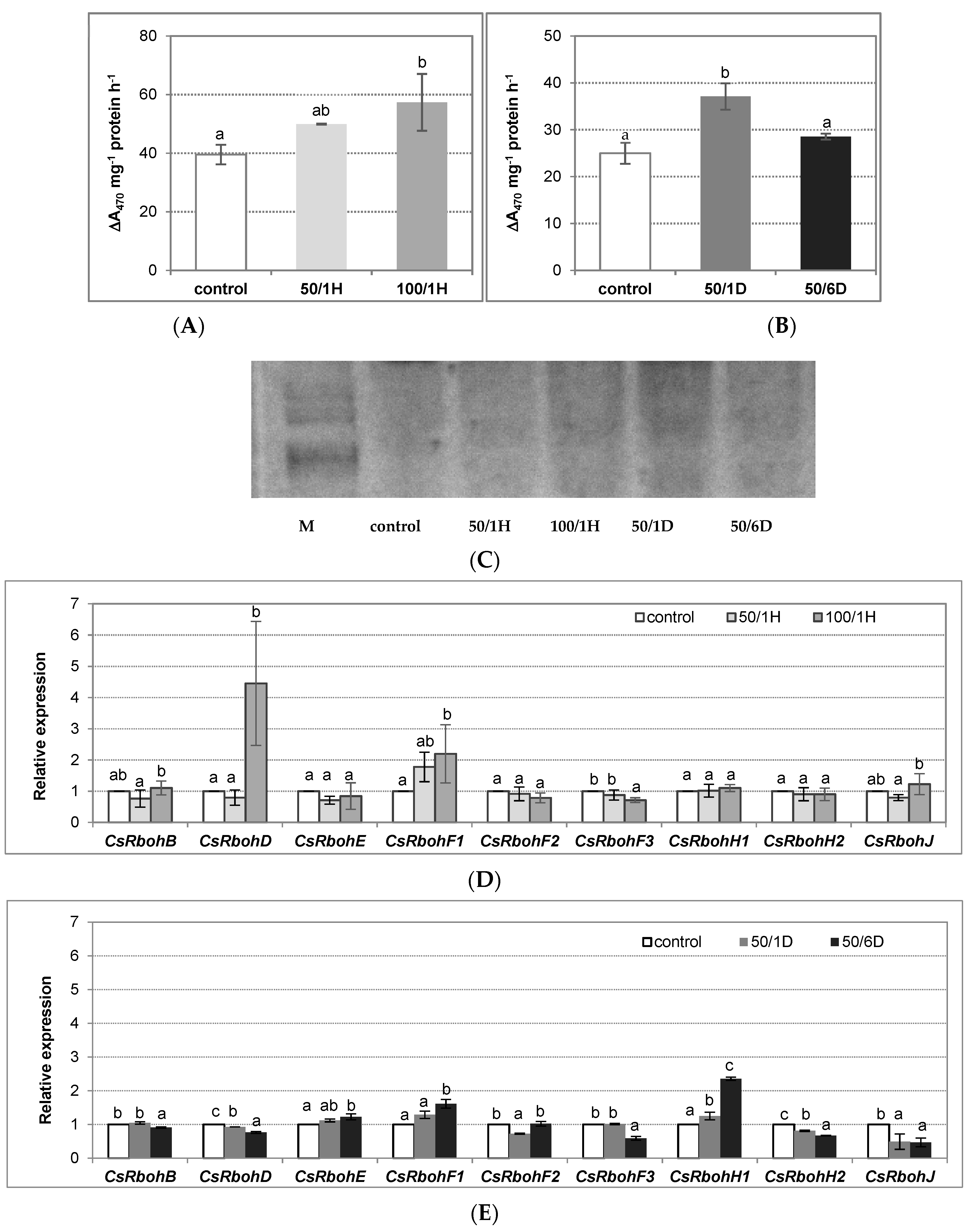
The plant NADPH oxidases are the main enzymes that participate in H2O2 production. Using two methods, we demonstrated that the treatment of seedlings with NaCl stimulated NADPH oxidase activity in the roots. The activity achieved about 126% and 145% of the control level in plants exposed to 50 mM and 100 mM NaCl, respectively, for 1 h (Figure 2A,C). When plants were treated with 50 mM NaCl for a longer time, the enzyme activity also increased, reaching about 149% of the control after 24 h, and consequently decreased to nearly the control value (115% of the control) in roots exposed to salt for 6 days (Figure 2B,C). Thus, the observed increase in the H2O2 level occurred in parallel with the enhanced NADPH oxidase activity. To verify whether the observed modulations of enzyme activity were a result of changes at transcriptional level, the Rboh gene expression was measured in the roots of seedlings exposed to salt stress. It was demonstrated that two of nine cucumber genes, CsRbohD and CsRbohF1, were significantly up-regulated by treatment with 100 mM NaCl for 1 h, reaching a 4,5-fold and 2,2-fold increase, respectively, in comparison to the control (Figure 2D). When the plants were exposed to salt for a longer time, the most pronounced effect of 6-day treatment with 50 mM NaCl on CsRbohH1 expression (a 2,4-fold increase) was observed (Figure 2E).
Apoplastic diamine oxidase (DAO) and polyamine oxidase (PAO) appeared to be the other enzymes responsible for H2O2 generation. Our results indicated that DAO activity remained at a similar level in both the control and the salt-stressed plants (Figure 3A,B). On the other hand, in the cucumber roots, the PAO activity was maintained at a very low level, several times lower than the DAO, regardless of the growing conditions (personal communications: Janicka, M., 2021, available in the repository) and, for this reason, it has not been studied. This suggests that the elevation in the H2O2 amount could be related to the plasma membrane NADPH activity.
The activity of the plasma membrane NADPH oxidase is related to the action of the superoxide dismutase (SOD). It was found in cucumber that SOD activity was enhanced by salt, similarly to NADPH oxidase. However, a significant increase, by about 60%, was observed in the roots of the plants treated with salt for 6 days (Figure 4A,B). Analysis of six genes encoding SOD in the cucumber genome—including CsCSD1, 2, and 3 belonging to Cu/Zn-SODs, CsMSD representing Mn-SODs as well as CsFSD2 and 3 identified as Fe-SOD—indicated that the expression of one of them, CsMSD, was upregulated about twice in seedlings exposed to 50 mM NaCl for both 1 h and 6 days. On the other hand, the transcript level of CsCSD2 significantly increased (a 2,5-fold increase) only after plant exposure to salt for 6 days (Figure 4C,D).
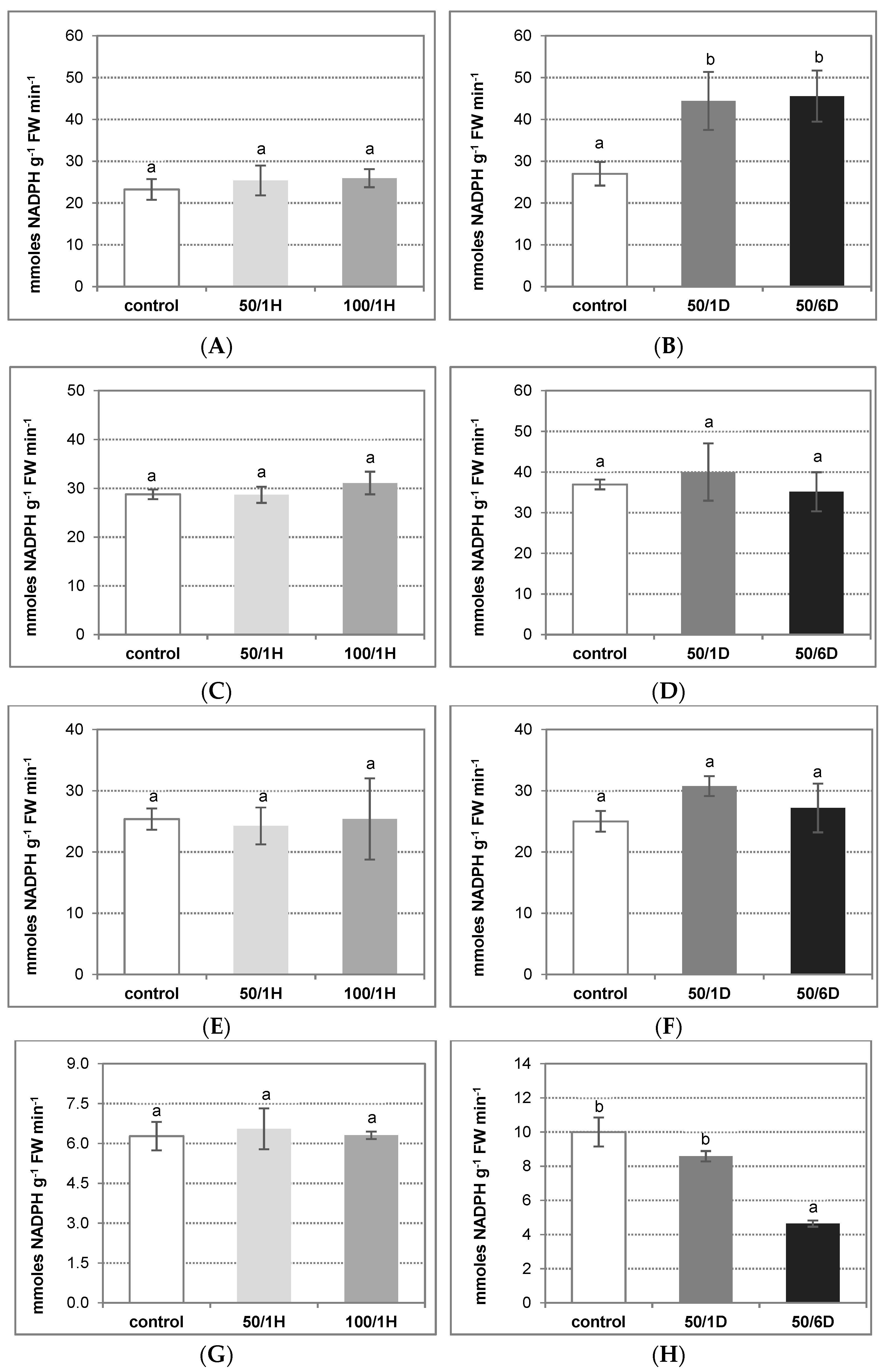
The activity of NADPH oxidase can be dependent on NADPH as its metabolic substrate. For this reason, the role of four NADPH-generating enzymes, i.e., glucose-6-phosphate dehydrogenase (G6PDH), 6-phosphogluconate dehydrogenase (6PGDH), NADP-isocitrate dehydrogenase (NADP-ICDH), and NADP-malic enzyme (NADP-ME), was examined in cucumber plants exposed to salinity (Figure 5). Among the enzymes tested, G6PDH was found to be significantly stimulated by salt applied for a longer period. In roots stressed with 50 mM NaCl for 1 and 6 days, the enzyme activity reached about 160–170% of the control value (Figure 5A,B). On the other hand, the activities of both 6PGDH and NADP-ICDH were relatively unaffected by NaCl, achieving similar levels in the roots of control as well as salt-treated plants (Figure 5C–F). In contrast to the above results, NADP-ME was visibly inhibited under longer salinity stress. The observed decrease in its activity was closely related to the time of NaCl treatment, and reached about 50% of the control in roots stressed with salt for 6 days (Figure 5G,H). Thus, it seems very likely that G6PDH is responsible for providing the substrate for NADPH oxidase under salt stress condition.
To minimize hydrogen peroxide accumulation, plants activate the H2O2-metabolizing enzymes, including catalase (CAT) and ascorbate peroxidase (APX). In the cucumber root tissues, CAT activity was reduced by about 30% after one day of plant treatment with 50 mM NaCl, as compared to the control plants. Longer plant exposure to this salt concentration (6 days) caused a significant increase in enzyme activity, which achieved above 150% of the control value. CAT activity did not change significantly in the roots treated with salt for 1 h (Figure 6A,B). On the other hand, APX activity was stimulated in the cucumber seedlings stressed with NaCl for a longer time period, reaching its highest level (140% of the control) in roots exposed to salt for 6 days (Figure 6C,D).
The possibility that NADPH oxidase can be subjected to regulation by H2O2 was also studied. The study demonstrated that exogenously applied H2O2 significantly stimulated the enzyme. The activity of the NADPH oxidase increased 1.9-fold in the roots of seedlings treated with this signaling molecule for 24 h (Figure 7A). Furthermore, it was indicated that the H2O2-induced increase in the enzyme activity was not related to the activation of CsRboh gene expression in roots (Figure 7B).

4. Discussion

To survive in soils contaminated with high salt concentration, plants have developed different adaptive mechanisms, in which signaling molecules seem to play an essential role. Many studies have indicated that, in response to salinity, enhanced levels of ROS, including hydrogen peroxide, are generated in plant tissues [30]. Consequently, oxidative damage of cellular components occurs, affecting plant cell functioning [30]. On the other hand, an increase in ROS content detected after exposure to salt can act as an early component of salinity sensing (acclimation signal), activating downstream H2O2-dependent signaling pathways, and leading to stress responses [9]. Among others, ROS were found to activate the MAPK cascade and potassium channels, and to modulate ion fluxes in the cells [9]. For this reason, it has been suggested that the coordinated ROS generating and scavenging system plays a crucial role in plant adaptation to salt stress [31].
Although knowledge about salinity sensing by plant cells is still limited, several sensory mechanisms have been proposed [32]. Moreover, it is supposed that different systems can operate in the same cell and at the same time, including the plasma membrane NADPH oxidase [33]. Accordingly, it has been shown in several plant species that salt stress rapidly induces a transient increase in NADPH oxidase activity, and up-regulates the expression of Rboh genes. This increase in enzyme activity is accompanied by H2O2 accumulation [34,35]. Arabidopsis mutants with defect in functional RBOH isoforms (AtrbohD and AtrbohF) exhibit increased Na+ hypersensitivity, and lack of ROS accumulation in the xylem vessels [12,36]. Thus, it has been proposed that RBOH could be involved in the control of sodium loading to the xylem, protecting leaves from the adverse effects of salinity [37].
In contrast to the above results, NaCl can also negatively affect the NADPH oxidase. Srivastava, et al. [38], have demonstrated that enzyme activity decreases in a glycophyte Brassica juncea (low salt accumulator), and that it remains unaffected in a halophyte Sesuvium portulacastrum (high salt accumulator) under salt stress conditions. Decrease in the ROS level related to inhibited RBOH activity has been shown in maize seedlings treated with NaCl [39]. Similarly, in sugar beet, H2O2 content was significantly lower in salt-treated tissues compared to the controls. This correlated with decreased NADPH oxidase activity and down-regulation of RBOH gene transcription [40].
Our results indicate that an enhanced H2O2 level occurred in cucumber roots stressed with 100 mM NaCl for 1 h and 50 mM NaCl for 1 day. At the same time, NADPH oxidase activity was significantly stimulated. The observed increase in enzyme activity was related to the enhanced expression of two Rboh genes, CsRbohD and CsRbohF1. Similarly, Niu, et al. [33], using 75 mM NaCl concentration, indicated that the H2O2 content is rapidly elevated in the roots of salt-treated pumpkin-grafted cucumber plants, reaching a peak at 3 h and decreasing during the 3–12 h period. This increase is related to the NaCl-induced NADPH oxidase activity, as well as enhanced expression of RbohD and RbohF transcription level.
The generation of ROS in the apoplastic space could be related not only to the activation of the cell membrane-bound RBOH but also the apoplastic polyamine or diamine oxidases [41]. In salt-stressed maize leaves, RBOH activity is inhibited. Under such condition, the apoplastic polyamine oxidase, which activity increases, is responsible for the elevation in H2O2 content required for cell wall loosening and leaf growth [42]. In our study, DAO activity remained unchanged in the cucumber seedlings, regardless of the salt concentration and treatment time. Furthermore, PAO activity maintained a very low level in the cucumber roots (personal communications: Janicka M, 2021, available in the repository). This may suggest that RBOH participates in H2O2 generation in the apoplast in cucumber roots under salt stress. As we demonstrated earlier, the H2O2 content only slightly increased in 1-day stressed roots, whereas it was very high in 6-day treated roots of Cucumis sativus var. Wisconsin under salinity condition [43]. It is interesting that such visible differences occurred in two cultivars of the same plant species, suggesting some distinct stress responses. Duan, et al. [44], using two cucumber cultivars with different salt tolerance—Changchun mici (tolerant) and Jinchun No. 2 (more sensitive)—exposed to salinity for 7 days, demonstrated that 50 mM NaCl caused a significant increase in DAO activity in the roots of the former, but only a slight increase (only observed after 3 days of stress) in the roots of the latter. Similar to our results, salt treatment had no effect on DAO activity in the roots of rice seedlings [45]. The authors concluded that DAO does not contribute to H2O2 production in the cell wall of stressed plants. In contrast, salt stress was found to both strongly promote DAO activity and to stimulate polyamine degradation in the root tissues of maize seedlings [46] and lupine seedlings [47]. Thus, it seems obvious that different mechanisms function in plant cells to enhance H2O2 production, depending on the plant species/cultivar and its sensitivity to salt as well as treatment conditions.
The activity of plasma membrane NADPH oxidase can be regulated in several ways [2]. Research using Arabidopsis has revealed some regulatory mechanisms of RBOH proteins, which depend on different signaling effectors. Thus, it has been suggested that differences in regulatory mechanisms may be responsible for diverse RBOH functions in plant cells [48]. A few studies have shown that, under salinity conditions, cytosolic Na+ accumulation followed by Ca2+ elevation can lead to the activation of RBOH and, consequently, to the accumulation of apoplastic H2O2 [37]. In our research, treatment of plants with H2O2 clearly activated NADPH oxidase, indicating that this enzyme remains under feedback control. However, the observed effect did not include the gene expression level, suggesting some post-translational regulatory mechanisms or additional signaling events, such as nitrosylation [49], H2S-dependent persulfidation [50], and reversible phosphorylation [51].
Substrate availability appears to be another factor involved in controlling NADPH oxidase activity. Several enzymes are responsible for NADPH delivery function in plant cells [52]. It has been proposed that various NADPH-generating dehydrogenases could provide this essential cofactor for different NADPH-dependent processes. Moreover, there is evidence that one or more NADP dehydrogenases are regulated under specific stress conditions [52]. In Arabidopsis seedlings, the NADP-ICDH, which activity increased, appeared to act as a barrier in the defense mechanisms activated against salt stress [53]. In contrast, a decrease in the activities of the main NADPH-generating enzymes, especially NADP-ICDH, has been demonstrated in tomato roots under salinity conditions [54]. Among the NADP dehydrogenases (G6PDH, 6PGDH, NADP-ME, and NADP-ICDH) analyzed in the present work, the NADP-ICDH activity remained relatively unaffected, whereas the G6PDH activity was importantly enhanced in the roots of 1-week-old cucumber seedlings treated with NaCl for 1 and 6 days, suggesting that this protein may serve as a source of NADPH for RBOH under stress conditions. On the other hand, treatment of 5-week-old cucumber plants with 100 mM NaCl for 1–3 days significantly reduced G6PDH activity in roots, but stimulated it in leaves [55]. As it was reported in A. thaliana exposed to high salinity, the cytosolic G6PDH isozyme (G6PD6) was subjected to phosphorylation, and this modification was responsible for the observed enzyme activation [56]. Studies using Arabidopsis mutants revealed that both the G6PD enzymatic activity and the expression of G6PD5 and G6PD6 decreased in atrbohD1, atrbohF1, and atrbohD1/F1 mutants in comparison to the WT plants, confirming the involvement of cytosolic G6PDH in RBOH-dependent ROS generation [57]. Scharte, et al. [58], have proposed that cytosolic G6PDH functions as an essential element of plant defense reactions, and that its modulation is important to enhance stress tolerance.
Plasma membrane-localized NADPH oxidases are a major source of cellular O2 radicals, which are transformed by superoxide dismutases to produce less reactive H2O2. Related to the metal cofactor at the active center, three distinct isoenzymes function in plant cells: Mn-SODs are present in the mitochondria and peroxisomes; Cu/Zn-SODs mainly in the cytosol, mitochondria, and plastids; while Fe-SODs are localized in the cytosol, mitochondria, chloroplasts, and peroxisomes [59]. Many studies have confirmed that salt stress triggers an increase in total SOD activity, especially in the leaves of plants treated with high doses of NaCl for several days [59]. In cucumber root cells, total SOD activity as well as CsMSD and CsCSD2 expression increased significantly in the roots of seedlings exposed to 50 mM NaCl for 6 days, when NADPH oxidase activity and H2O2 level were lowered to nearly control values. This suggests that hydrogen peroxide generated by SOD may be effectively decomposed by H2O2-metabolizing systems. Hossain, et al. [40], have shown that in a highly salt-tolerant sugar beet stressed with 300 mM NaCl for up to 14 days, H2O2 content is always significantly lower in the salt-treated leaves in comparison to the control. This was correlated with the suppressed NADPH oxidase activity and gene expression (with the exception of RBOHB), as well as with the up-regulation of SOD activity and increased transcription of genes encoding this enzyme (with the exception of FeSOD1). Such a system, together with enhanced expression of genes for alternative oxidase and plastid terminal oxidase, is responsible for maintaining the low ROS level under severe salinity [40].
It is assumed that early production of H2O2 acts as a signal to induce antioxidant systems and prevent subsequent ROS generation and ROS-dependent cell damage [37]. In Arabidopsis under short-term salt treatments, RbohD and RbohF were suggested to be required in this process [13]. The main enzymes able to break down H2O2 are catalase, with the highest turnover rates, and ascorbate peroxidase, exhibiting higher affinity for H2O2 than CAT [60,61]. In accordance with the above assumption, in this study, both the up-regulation of APX and CAT activity were shown in the roots of cucumber seedlings exposed to salinity for 6 days, confirming the involvement of both enzymes in maintaining the low H2O2 level under prolonged salt exposure. In contrast, catalase activity was inhibited in roots treated with salt for 1 day. Stimulation of both APX and CAT in response to NaCl has been reported in many plant species. It has been shown to be associated with decreased oxidative damage and improved tolerance to salinity. Similar to SOD, a greater increase in CAT activity was observed in salt-tolerant plants/genotypes than in more sensitive ones [60]. Furthermore, in some glycophytes, including rice, the activity of this enzyme decreased after NaCl treatment [62].

5. Conclusions

At the early stages of salt stress (1 h and 1-day-treatment), the plasma membrane NADPH oxidase was activated in the cucumber (Cucumis sativus var. Krak) seedlings. This activation correlated with an observed enhancement in H2O2 content, suggesting the involvement of NADPH oxidase in the generation of H2O2, which may function as a signaling molecule participating in stress response. The observed stimulation of NADPH oxidase may have been at least partially related to the induction of some CsRboh genes, as well as the increased activity of G6PDH, providing NADPH. Another factor which may contribute to positive RBOH regulation at the post-translational level, could be H2O2. This proposal, however, requires additional studies concerning direct or indirect H2O2 action.
After 6-day exposure to salt stress, the plasma membrane NADPH oxidase activity, and consequently the H2O2 level, were diminished. This protected the cells against the toxic effect of high H2O2 concentrations. Additionally, antioxidant enzymes, including SOD, APX, and CAT, were significantly activated to maintain low H2O2 content in cells, and to avoid ROS-dependent damages. However, to explain the precise mechanism of NADH oxidase regulation, and its interrelation with the antioxidant system in cucumber seedlings subjected to salinity, further analysis is needed.

Author Contributions

Conceptualization, M.J.; methodology, K.K., M.R., A.W., and M.J.; formal analysis, K.K., M.R., A.W., and M.J; writing—original draft preparation, K.K.; writing—review and editing, M.R., A.W., and M.J; supervision, M.J. All authors have read and agreed to the published version of the manuscript.

Funding

This research received no external funding.

Institutional Review Board Statement

Not applicable.

Informed Consent Statement

Not applicable.

Data Availability Statement

The data presented are available in this manuscript and in the repository at https://www.repozytorium.uni.wroc.pl/dlibra/publication/139358/edition/128721# (accessed on 10 September 2021).

Acknowledgments

We thank Beata Kuligowska for her excellent technical assistance.

Conflicts of Interest

The authors declare no conflict of interest.

References

  1. Apel, K.; Hirt, H. Reactive oxygen species: Metabolism, oxidative stress, and signal transduction. Annu. Rev. Plant. Biol. 2004, 55, 373–399. [Google Scholar] [CrossRef] [PubMed] [Green Version]
  2. Qu, Y.; Yan, M.; Zhang, Q. Functional regulation of plant NADPH oxidase and its role in signaling. Plant Signal. Behav. 2017, 12, e1356970. [Google Scholar] [CrossRef] [PubMed] [Green Version]
  3. Marino, D.; Dunand, C.; Puppo, A.; Pauly, N. A burst of plant NADPH oxidases. Trends Plant Sci. 2012, 17, 9–15. [Google Scholar] [CrossRef]
  4. Sagi, M.; Fluhr, R. Production of reactive oxygen species by plant NADPH oxidases. Plant Physiol. 2006, 141, 336–340. [Google Scholar] [CrossRef] [PubMed] [Green Version]
  5. Ogasawara, Y.; Kaya, H.; Hiraoka, G.; Yumoto, F.; Kimura, S.; Kadota, Y.; Hishinuma, H.; Senzaki, E.; Yamagoe, S.; Nagata, K.; et al. Synergistic activation of the Arabidopsis NADPH oxidase AtrbohD by Ca2+ and phosphorylation. J. Biol. Chem. 2008, 283, 8885–8892. [Google Scholar] [CrossRef] [PubMed] [Green Version]
  6. Jakubowska, D.; Janicka-Russak, M.; Kabała, K.; Migocka, M.; Reda, M. Modification of plasma membrane NADPH oxidase activity in cucumber seedling roots in response to cadmium stress. Plant Sci. 2015, 234, 50–59. [Google Scholar] [CrossRef]
  7. Angelini, R.; Cona, A.; Federico, R.; Fincato, P.; Tavladoraki, P.; Tisi, A. Plant amine oxidases “on the move”: An update. Plant Physiol. Biochem. 2010, 48, 560–564. [Google Scholar] [CrossRef]
  8. Moschou, P.N.; Paschalidis, A.K.; Delis, I.D.; Andriopoulou, A.H.; Lagiotis, G.D.; Yakoumakis, D.I.; Roubelakis-Angelakis, K.A. Spermidine exodus and oxidation in the apoplast induced by abiotic stress is responsible for H2O2 signatures that direct tolerance responses in tobacco. Plant Cell 2008, 20, 1708–1724. [Google Scholar] [CrossRef] [Green Version]
  9. Isayenkov, S.V.; Maathuis, F.J.M. Plant salinity stress: Many unanswered questions remain. Front. Plant Sci. 2019, 10, e80. [Google Scholar] [CrossRef] [Green Version]
  10. Yang, Y.L.; Xu, S.J.; An, L.Z.; Chen, N.L. NADPH oxidase dependent hydrogen peroxide production, induced by salinity stress, may be involved in the regulation of total calcium in roots of wheat. J. Plant Physiol. 2007, 164, 1429–1435. [Google Scholar] [CrossRef]
  11. Chung, J.S.; Zhu, J.K.; Bressan, R.A.; Hasegawa, P.M.; Shi, H.Z. Reactive oxygen species mediate Na+-induced SOS1 mRNA stability in Arabidopsis. Plant J. 2008, 53, 554–565. [Google Scholar] [CrossRef] [PubMed] [Green Version]
  12. Ma, L.; Zhang, H.; Sun, L.; Jiao, Y.; Zhang, G.; Miao, C.; Hao, F. NADPH oxidase AtrbohD and AtrbohF function in ROS-dependent regulation of Na+/K+ homeostasis in Arabidopsis under salt stress. J. Exp. Bot. 2012, 63, 305–317. [Google Scholar] [CrossRef] [PubMed]
  13. Ben Rejeb, K.; Benzarti, M.; Debez, A.; Bailly, C.; Savouré, A.; Abdelly, C. NADPH oxidase-dependent H2O2 production is required for salt-induced antioxidant defense in Arabidopsis thaliana. J. Plant Physiol. 2015, 174, 5–15. [Google Scholar] [CrossRef]
  14. Tanou, G.; Ziogas, V.; Belghazi, M.; Christou, A.; Filippou, P.; Job, D.; Fotopoulos, V.; Molassiotis, A. Polyamines reprogram oxidative and nitrosative status and the proteome of citrus plants exposed to salinity stress. Plant Cell Environ. 2014, 37, 864–885. [Google Scholar] [CrossRef]
  15. Gémes, K.; Kim, Y.J.; Park, K.Y.; Moschou, P.N.; Andronis, E.; Valassaki, C.; Roussis, A.; Roubelakis-Angelakis, K.A. An NADPH-Oxidase/Polyamine Oxidase feedback loop controls oxidative burst under salinity. Plant Physiol. 2016, 172, 1418–1431. [Google Scholar] [CrossRef] [PubMed] [Green Version]
  16. Velikova, V.; Yordanov, I.; Edreva, A. Oxidative stress and some antioxidant systems in acid rain-treated bean plants. Protective role of exogenous polyamines. Plant Sci. 2000, 151, 59–66. [Google Scholar] [CrossRef]
  17. Kabała, K.; Zboińska, M.; Głowiak, D.; Reda, M.; Jakubowska, D.; Janicka, M. Interaction between the signaling molecules hydrogen sulfide and hydrogen peroxide and their role in vacuolar H+-ATPase regulation in cadmium-stressed cucumber roots. Physiol. Plant. 2019, 166, 688–704. [Google Scholar] [CrossRef] [PubMed] [Green Version]
  18. Larsson, C. Plasma membranes. In Cell Components; Linskens, H.F., Jackson, J.F., Eds.; Springer: Berlin/Heidelberg, Germany, 1985; pp. 85–104. [Google Scholar]
  19. Kłobus, G. The role of plasma membrane-bound activities in nitrate transport into sealed plasma membrane vesicles from Cucumis sativus L. roots. In Developments in Plant and Soil Science; Baluška, F., Ciamporova, M., Gasparicova, O., Barlow, P.W., Eds.; Kluwer Academic Publishers: Dordrecht, The Netherlands, 1995; Volume 58, pp. 133–140. [Google Scholar]
  20. Sagi, M.; Fluhr, R. Superoxide production by plant homologues of the gp91(phox)NADPH oxidase. Modulation of activity by calcium and by tobacco mosaic virus infection. Plant Physiol. 2001, 126, 1281–1290. [Google Scholar] [CrossRef] [Green Version]
  21. Holmsted, B.; Larsson, L.; Tham, R. Further studies on spectrophotometric method for the determination of amine oxidase activity. Biochim. Biophys. Acta 1961, 48, 182–186. [Google Scholar] [CrossRef]
  22. Federico, R.; Angelini, R. Distribution of polyamines and their related catabolic enzyme in etiolated and light-grown leguminosae seedlings. Planta 1988, 173, 317–321. [Google Scholar] [CrossRef]
  23. Li, J.; Chen, G.; Wang, X.; Zhang, Y.; Jia, H.; Bi, Y. Glucose-6-phosphate dehydrogenase-dependent hydrogen peroxide production is involved in the regulation of plasma membrane H+-ATPase and Na+/H+ antiporter protein in salt-stressed callus from Carex moorcroftii. Physiol. Plant. 2011, 141, 239–250. [Google Scholar] [CrossRef] [PubMed]
  24. Aebi, H. Catalase in vitro. Methods Enzymol. 1984, 105, 121–126. [Google Scholar] [PubMed]
  25. Janicka-Russak, M.; Kabała, K.; Burzyński, M. Different effect of cadmium and copper on H+-ATPase activity in plasma membrane vesicles from Cucumis sativus roots. J. Exp. Bot. 2012, 63, 4133–4142. [Google Scholar] [CrossRef] [Green Version]
  26. Chen, G.X.; Asada, K. Ascorbate peroxidase in tea leaves: Occurrence of two isozymes and the differences in their enzymatic and molecular properties. Plant Cell Physiol. 1989, 30, 987–998. [Google Scholar]
  27. Beauchamp, C.; Fridovich, I. Superoxide dismutase: Improved assays and an assay applicable to acrylamide gels. Anal. Biochem. 1971, 44, 276–287. [Google Scholar] [CrossRef]
  28. Bradford, M.M. A rapid and sensitive method for quantitation of microgram quantities of protein utilizing the principles of protein dye binding. Anal. Biochem. 1976, 72, 248–254. [Google Scholar] [CrossRef]
  29. Migocka, M.; Papierniak, A. Identification of suitable reference genes for studying gene expression in cucumber plants subjected to abiotic stress and growth regulators. Mol. Breed. 2010, 28, 343–357. [Google Scholar] [CrossRef]
  30. Gupta, B.; Huang, B. Mechanism of salinity tolerance in plants: Physiological, biochemical, and molecular characterization. Int. J. Genom. 2014, e701596. [Google Scholar] [CrossRef]
  31. Kaur, N.; Dhawan, M.; Sharma, I.; Pati, P.K. Interdependency of Reactive Oxygen Species generating and scavenging system in salt sensitive and salt tolerant cultivars of rice. BMC Plant Biol. 2016, 16, e131. [Google Scholar] [CrossRef] [Green Version]
  32. Shabala, S.; Wu, H.; Bose, J. Salt stress sensing and early signalling events in plant roots: Current knowledge and hypothesis. Plant Sci. 2015, 241, 109–119. [Google Scholar] [CrossRef]
  33. Niu, M.; Huang, Y.; Sun, S.; Sun, J.; Cao, H.; Shabala, S.; Bie, Z. Root respiratory burst oxidase homologue-dependent H2O2 production confers salt tolerance on a grafted cucumber by controlling Na+ exclusion and stomatal closure. J. Exp. Bot. 2017, 69, 3465–3476. [Google Scholar] [CrossRef] [PubMed]
  34. Xie, Y.J.; Xu, S.; Han, B.; Wu, M.Z.; Yuan, X.X.; Han, Y.; Gu, Q.; Xu, D.K.; Yang, Q.; Shen, W.B. Evidence of Arabidopsis salt acclimation induced by up-regulation of HY1 and the regulatory role of RbohD-derived reactive oxygen species synthesis. Plant J. 2011, 66, 280–292. [Google Scholar] [CrossRef] [PubMed]
  35. Liu, L.; Huang, L.; Lin, X.; Sun, C. Hydrogen peroxide alleviates salinity-induced damage through enhancing proline accumulation in wheat seedlings. Plant Cell Rep. 2020, 39, 567–575. [Google Scholar] [CrossRef] [PubMed]
  36. Jiang, C.; Belfield, E.J.; Mithani, A.; Visscher, A.; Ragoussis, J.; Mot, R.; Smith, J.A.; Harberd, N.P. ROS-mediated vascular homeostatic control of root-to-shoot soil Na delivery in Arabidopsis. EMBO J. 2012, 31, 4359–4370. [Google Scholar] [CrossRef] [Green Version]
  37. Kurusu, T.; Kuchitsu, K.; Tada, Y. Plant signaling networks involving Ca2+ and Rboh/Nox-mediated ROS production under salinity stress. Front. Plant Sci. 2015, 6, e427. [Google Scholar] [CrossRef] [Green Version]
  38. Srivastava, A.K.; Srivastava, S.; Lokhande, V.H.; D’Souza, S.F.; Suprasanna, P. Salt stress reveals differential antioxidant and energetics responses in glycophyte (Brassica juncea L.) and halophyte (Sesuvium portulacastrum L.). Front. Environ. Sci. 2015, 3, e19. [Google Scholar] [CrossRef] [Green Version]
  39. Rodríguez, A.A.; Ramiro Lascano, H.; Bustos, D.; Taleisnik, E. Salinity-induced decrease in NADPH oxidase activity in the maize leaf blade elongation zone. J. Plant Physiol. 2007, 64, 223–230. [Google Scholar] [CrossRef]
  40. Hossain, M.S.; ElSayed, A.I.; Moore, M.; Dietz, K.J. Redox and reactive oxygen species network in acclimation for salinity tolerance in sugar beet. J. Exp. Bot. 2017, 68, 1283–1298. [Google Scholar] [CrossRef] [Green Version]
  41. Hossain, M.S.; Dietz, K.J. Tuning of redox regulatory mechanisms, reactive oxygen species and redox homeostasis under salinity stress. Front. Plant Sci. 2016, 7, e548. [Google Scholar] [CrossRef] [Green Version]
  42. Rodríguez, A.A.; Maiale, S.J.; Menéndez, A.B.; Ruiz, O.A. Polyamine oxidase activity contributes to sustain maize leaf elongation under saline stress. J. Exp. Bot. 2009, 60, 4249–4262. [Google Scholar] [CrossRef] [Green Version]
  43. Janicka, M.; Reda, M.; Czyżewska, K.; Kabała, K. Involvement of signalling molecules NO, H2O2 and H2S in modification of plasma membrane proton pump in cucumber roots subjected to salt or low temperature stress. Funct. Plant Biol. 2018, 45, 428–439. [Google Scholar] [CrossRef] [PubMed]
  44. Duan, J.; Li, J.; Guo, S.; Kang, Y. Exogenous spermidine affects polyamine metabolism in salinity-stressed Cucumis sativus roots and enhances short-term salinity tolerance. J. Plant Physiol. 2008, 165, 1620–1635. [Google Scholar] [CrossRef] [PubMed]
  45. Lin, C.C.; Kao, C.H. Cell wall peroxidase activity, hydrogen peroxide level and NaCl-inhibited root growth of rice seedlings. Plant Soil 2001, 230, 135–143. [Google Scholar] [CrossRef]
  46. Kongkiattikajorn, J. Effect of salinity stress on degradation of polyamines and amine oxidase activity in maize seedlings. Kasetsart J. (Nat. Sci.) 2009, 43, 28–33. [Google Scholar]
  47. Legocka, J.; Sobieszczuk-Nowicka, E.; Ludwicki, D.; Lehmann, T. Putrescine catabolism via DAO contributes to proline and GABA accumulation in roots of lupine seedlings growing under salt stress. Acta Soc. Bot. Pol. 2017, 86, e3549. [Google Scholar] [CrossRef] [Green Version]
  48. Baxter, A.; Mittler, R.; Suzuki, N. ROS as key players in plant stress signaling. J. Exp. Bot. 2014, 65, 1229–1240. [Google Scholar] [CrossRef] [PubMed]
  49. Yun, B.W.; Feechan, A.; Yin, M.; Saidi, N.B.; Le Bihan, T.; Yu, M.; Moore, J.W.; Kang, J.G.; Kwon, E.; Spoel, S.H.; et al. S-nitrosylation of NADPH oxidase regulates cell death in plant immunity. Nature 2011, 478, 264–268. [Google Scholar] [CrossRef]
  50. Shen, J.; Zhang, J.; Zhou, M.; Zhou, H.; Cui, B.; Gotor, C.; Romero, L.C.; Fu, L.; Yang, J.; Foyer, C.H.; et al. Persulfidation-based modification of cysteine desulfhydrase and the NADPH oxidase RBOHD controls guard cell abscisic acid signaling. Plant Cell 2020, 32, 1000–1017. [Google Scholar] [CrossRef]
  51. Wu, F.; Chi, Y.; Jiang, Z.; Xu, Y.; Xie, L.; Huang, F.; Wan, D.; Ni, J.; Yuan, F.; Wu, X.; et al. Hydrogen peroxide sensor HPCA1 is an LRR receptor kinase in Arabidopsis. Nature 2020, 578, 577–581. [Google Scholar] [CrossRef]
  52. Corpas, F.J.; Barroso, J.B. NADPH-generating dehydrogenases: Their role in the mechanism of protection against nitro-oxidative stress induced by adverse environmental conditions. Front. Environ. Sci. 2014, 2, e55. [Google Scholar] [CrossRef] [Green Version]
  53. Leterrier, M.; Barroso, J.B.; Valderrama, R.; Palma, J.M.; Corpas, F.J. NADP-dependent isocitrate dehydrogenase from Arabidopsis roots contributes in the mechanism of defence against the nitro-oxidative stress induced by salinity. Sci. World J. 2012, 6, e694740. [Google Scholar]
  54. Manai, J.; Gouia, H.; Corpas, F.J. Redox and nitric oxide homeostasis are affected in tomato (Solanum lycopersicum) roots under salinity-induced oxidative stress. J. Plant Physiol. 2014, 171, 1028–1035. [Google Scholar] [CrossRef] [PubMed]
  55. Hýsková, V.; Plisková, V.; Červený, V.; Ryšlavá, H. NADP-dependent enzymes are involved in response to salt and hypoosmotic stress in cucumber plants. Gen. Physiol. Biophys. 2017, 36, 247–258. [Google Scholar] [CrossRef]
  56. Dal Santo, S.; Stampfl, H.; Krasensky, J.; Kempa, S.; Gibon, Y.; Petutschnig, E.; Rozhon, W.; Heuck, A.; Clausen, T.; Jonak, C. Stress-induced GSK3 regulates the redox stress response by phosphorylating glucose-6-phosphate dehydrogenase in Arabidopsis. Plant Cell 2012, 24, 3380–3392. [Google Scholar] [CrossRef] [Green Version]
  57. Yang, L.; Wang, X.; Chang, N.; Nan, W.; Wang, S.; Ruan, M.; Sun, L.; Li, S.; Bi, Y. Cytosolic glucose-6-phosphate dehydrogenase is involved in seed germination and root growth under salinity in Arabidopsis. Front. Plant Sci. 2019, 10, e182. [Google Scholar] [CrossRef] [PubMed] [Green Version]
  58. Scharte, J.; Schön, H.; Tjaden, Z.; Weis, E.; von Schaewen, A. Isoenzyme replacement of glucose-6-phosphate dehydrogenase in the cytosol improves stress tolerance in plants. Proc. Natl. Acad. Sci. USA 2009, 106, 8061–8066. [Google Scholar] [CrossRef] [Green Version]
  59. Szőllősi, R. Superoxide dismutase (SOD) and abiotic stress tolerance in plants: An overview. In Oxidative Damage to Plants. Antioxidant Networks and Signaling; Ahmad, P., Ed.; Elsevier Academic Publishers: Amsterdam, The Netherlands, 2014; pp. 89–129. [Google Scholar]
  60. Sofo, A.; Scopa, A.; Nuzzaci, M.; Vitti, A. Ascorbate peroxidase and catalase activities and their genetic regulation in plants subjected to drought and salinity stresses. Int. J. Mol. Sci. 2015, 16, 13561–13578. [Google Scholar] [CrossRef] [PubMed] [Green Version]
  61. Filiz, E.; Ozyigit, I.I.; Saracoglu, I.A.; Uras, M.E.; Sen, U.; Yalcin, B. Abiotic stress-induced regulation of antioxidant genes in different Arabidopsis ecotypes: Microarray data evaluation. Biotech. Biotechnol. Equip. 2019, 33, 128–143. [Google Scholar] [CrossRef] [Green Version]
  62. Kibria, M.G.; Hossain, M.; Murata, Y.; Hoque, M.A. Antioxidant defense mechanisms of salinity tolerance in rice genotypes. Rice Sci. 2017, 24, 155–162. [Google Scholar] [CrossRef]
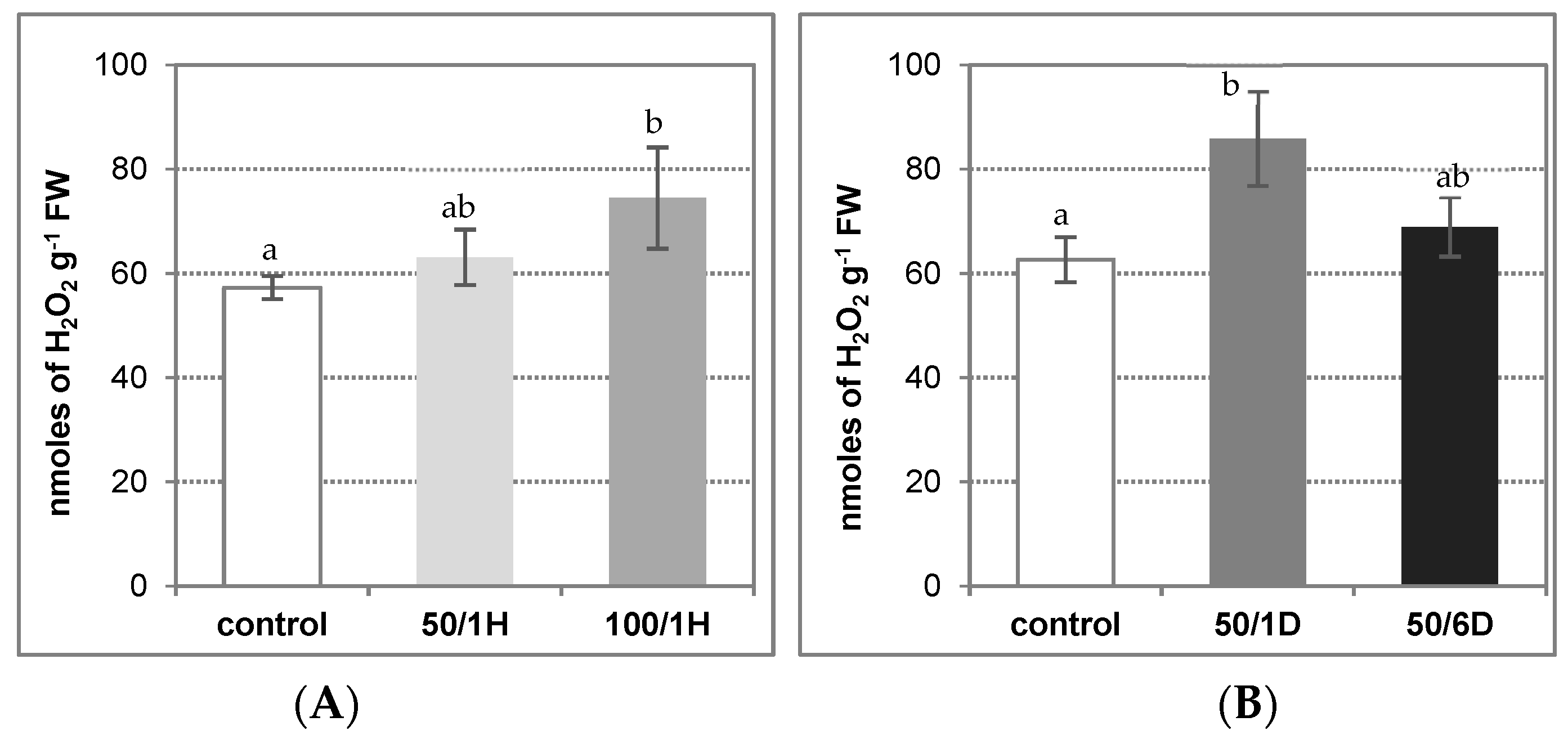
Figure 1. H2O2 level in roots of cucumber seedlings treated with: (A) 50 mM or 100 mM NaCl for 1 h (50/1H and 100/1H, respectively); (B) 50 mM NaCl for 1 or 6 days (50/1D and 50/6D, respectively). The H2O2 level was determined in the roots as described in Materials and Methods. The presented results are the means ± SD of three independent experiments, each run in triplicate. Different letters show different homogeneous groups of means calculated by Duncan’s multiple range test (significant at p < 0.05).
Antioxidants 11 01534 g001
Figure 2. NADPH oxidase catalytic activity (A,B), in-gel activity(C), and relative expression of CsRboh genes (D,E) in the tissues of cucumber roots exposed to salinity. The cucumber seedlings were treated with: (A,C,D) 50 mM or 100 mM NaCl for 1 h (50/1H and 100/1H, respectively) and (B,C,E) 50 mM NaCl for 1 or 6 days (50/1D and 50/6D, respectively). The plasma membrane vesicles were isolated from the cucumber roots to determine both catalytic (A,B) and in-gel (C) NADPH oxidase activity, as described in Materials and Methods. (A,B) The presented results are the means ± SD of three independent experiments, with each experiment performed in triplicate, and are expressed as the ΔA470 of formazan XTT mg−1 protein h−1. (C) Gel image with blue formazan bands detected using the NBT method. The plasma membrane proteins were subjected to native PAGE, and examined for their ability to generate O2 by reduction of nitroblue tetrazolium. The bands detected in the gel were due to NADPH-dependent NBT reduction by O2 radicals. The gel image shown is representative of three independent replications of the experiment; M—molecular weight marker. (D,E) The total RNA was isolated from the cucumber roots, and real-time PCR analysis was performed with CsTIP41 as a reference gene, as described in Materials and Methods. The results are shown as means ± SD of three replications. Different letters show different homogeneous groups of means calculated by Duncan’s multiple range test (significant at p < 0.05).
Antioxidants 11 01534 g002
Figure 3. Diamine oxidase activity in the tissues of cucumber roots treated with: (A) 50 mM or 100 mM NaCl for 1 h (50/1H and 100/1H, respectively); (B) 50 mM NaCl for 1 or 6 days (50/1D and 50/6D, respectively). DAO activity was assayed in the roots, as described in Materials and Methods. The presented results are the means ± SD of three independent experiments, with each experiment performed in triplicate, and are expressed as nmoles of Δ1-pyrroline g−1 FW min−1. Means with the same letter are not significantly different (p < 0.05).
Antioxidants 11 01534 g003
Figure 4. Superoxide dismutase activity (A,B) and relative expression of SOD encoding genes (C,D) in the tissues of cucumber roots exposed to salinity. The cucumber seedlings were treated with: (A,C) 50 mM or 100 mM NaCl for 1 h (50/1H and 100/1H, respectively); (B,D) 50 mM NaCl for 1 or 6 days (50/1D and 50/6D, respectively). (A,B) SOD activity was determined in the roots, as described in Materials and Methods. The presented results are the means ± SD of three independent experiments, each run in triplicate, and are expressed as units g−1 FW. (C,D) The total RNA was isolated from the cucumber roots, and real-time PCR analysis was performed, with CsCACS as a reference gene, as described in Materials and Methods. The results are shown as the means ± SD of three replications. Different letters show different homogeneous groups of means calculated by Duncan’s multiple range test (significant at p < 0.05).
Antioxidants 11 01534 g004
Figure 5. Activity of NADPH-generating enzymes: G6PDH (A,B), 6PGDH (C,D), NADP-ICDH (E,F), and NADP-ME (G,H) in the roots of cucumber seedlings exposed to salinity. The cucumber seedlings were treated with: (A,C,E,G) 50 mM or 100 mM NaCl for 1 h (50/1H and 100/1H, respectively); (B,D,F,H) 50 mM NaCl for 1 or 6 days (50/1D and 50/6D, respectively). The activities of NADP dehydrogenases were determined in the roots, as described in Materials and Methods. The presented results are the means ± SD of three independent experiments, each run in triplicate, and are expressed as nmoles of NADPH g−1 FW min−1. Different letters show different homogeneous groups of means calculated by Duncan’s multiple range test (significant at p < 0.05).
Antioxidants 11 01534 g005
Figure 6. Catalase (A,B) and ascorbate peroxidase (C,D) activity in the tissues of cucumber roots exposed to salinity. The cucumber seedlings were treated with: (A,C) 50 mM or 100 mM NaCl for 1 h (50/1H and 100/1H, respectively); (B,D) 50 mM NaCl for 1 or 6 days (50/1D and 50/6D, respectively). The activities of antioxidant enzymes were determined in the roots, as described in Materials and Methods. The presented results are the means ± SD of three independent experiments, each run in triplicate, and are expressed as units g−1 FW min−1. Different letters show different homogeneous groups of means calculated by Duncan’s multiple range test (significant at p < 0.05).
Antioxidants 11 01534 g006
Figure 7. NADPH oxidase catalytic activity (A) and relative expression of CsRboh genes (B) in cucumber roots treated with H2O2. The cucumber seedlings were treated with 5 mM H2O2 for 24 h, as described in Materials and Methods. (A) The plasma membrane vesicles were isolated from the cucumber roots, as described in Materials and Methods. The presented results are the mean ± SD of three independent experiments, with each experiment performed in triplicate, and are expressed as the ΔA470 of formazan XTT mg−1 protein h−1. (B) The total RNA was isolated from the cucumber roots, and real-time PCR analysis was performed, with CsTIP41 as a reference gene, as described in Materials and Methods. The results are shown as the means ± SD of three replications. Different letters show different homogeneous groups of means calculated by Duncan’s multiple range test (significant at p < 0.05).
Antioxidants 11 01534 g007
Table 1. The list of primers used in qRT-PCR reactions.
GeneForward PrimerReverse Primer
CsTIP41CAACAGGTGATATTGGATTATGATTATACGCCAGCTCATCCTCATATAAG
CsRbohBAGAGTCGGCTTCAGCGGTCTCCTCCTCTGATGTTCTGAACGG
CsRbohDTCTTCTTCTTCTTCCTCCCTCAAAGCCGAAAGTTCAGGGTCTTCAAGAGAGTTG
CsRbohETCACTTTGGAAGTTGAGGATGATTCTGTTGTCGAGAGAGGAGGCAGTGG
CsRbohF1GCTTCGATTTCGAGATCGCCGACAAATCCATTTCCACCACCAGAAGAATGT
CsRbohF2TCGCATCGGTTTCGGCAGAGTTCACGGTTGAAGATGACGC
CsRbohF3AAGAGAAGTCCGAGAATGAGGTTTATCTCCTGAGATTCTGACATTCCAATGC
CsRbohH1AGATTCCGATGTGATAGATGTCATGGTTTGGACTGTCTTCTTCCAAGCCTC
CsRbohH2AAATCCAGGAGAATGCCCACTGAGAAGTCTCAGCGACGG
CsRbohJCTGACGATGGGATTACTCTGCAACAGAAGCAGTCCACTTCAGTATGGTCT
CsCACSTGGGAAGATTCTTATGAAGTGCCTCGTCAAATTTACACATTGGT
CsCSD1AAAGATGGTGAAGGCTGTGGCATGTTGTTTTCCAGCAG
CsCSD2CCCATTCTCCAATTCTTCATTTGGGTGAGCGTGACAA
CsCSD3CCAAAAGAGGGGGAATTTTTGCCTCTGACGTTGGAATCTC
CsMSDCGCTTCGAATTCTAGGCAGGCCTTTGTTGATGGCCTCGTG
CsFSD2TAACACGGTTGACCATCCTTCTGTAGCCTCAAGTCT
CsFSD3TCTGATGGAACCACAAGAGTGTGGCCATATAGAATATCATTT
Publisher’s Note: MDPI stays neutral with regard to jurisdictional claims in published maps and institutional affiliations.