Article

Neuroprotective Profile of Edible Flowers of Borage (Borago officinalis L.) in Two Different Models: Caenorhabditis elegans and Neuro-2a Cells

by
Cristina Moliner
1,†,
Guillermo Cásedas
1,2,†,
Lillian Barros
3,
Tiane C. Finimundy
3,
Carlota Gómez-Rincón
1,2 and
Víctor López
1,2,*
1
Department of Pharmacy, Faculty of Health Sciences, Universidad San Jorge, 50830 Zaragoza, Spain
2
Instituto Agroalimentario de Aragón-IA2, CITA-Universidad de Zaragoza, 50059 Zaragoza, Spain
3
Centro de Investigação de Montanha (CIMO), Instituto Politécnico de Bragança, Campus de Santa Apolónia, 5300-253 Bragança, Portugal
*
Author to whom correspondence should be addressed.
These authors contributed equally to this work.
Antioxidants 2022, 11(7), 1244; https://doi.org/10.3390/antiox11071244
Submission received: 6 May 2022 / Revised: 18 June 2022 / Accepted: 21 June 2022 / Published: 24 June 2022

Abstract

:
The flowers of Borago officinalis L. (Boraginaceae), commonly known as borage, are widely used as a culinary ingredient. The aim of this study was to assess the potential benefits of fresh borage flower extract related to antioxidant, neuroprotective and anti-aging properties. The extract was obtained by Soxhlet extraction with ethanol as a solvent, and fatty acids were detected by GC-FID. The antioxidant activity was evaluated in vitro through the DPPH, FRAP and ORAC assays. Regarding the fatty acid (FA) composition, the extract showed high amounts of polyunsaturated FA. The Neuro-2a cell line was used to determine the cytoprotective capacity of the extract subjected to oxidative stress (H2O2). Moreover, the model organism Caenorhabditis elegans was used to assess antioxidant activity, delayed ageing as well as cytoprotection and reduced β-amyloid toxicity. Cells treated with the extract and H2O2 showed a better response to oxidative stress than the control group, particularly in terms of mitochondrial activity (MTT assay), redox state (ROS formation) and the activity of antioxidant enzymes (catalase and superoxide dismutase). B. officinalis flower extract showed promising antioxidant activity in the selected models, without causing toxicity. Hence, the results obtained support the antioxidant properties of borage flowers in different bioassays using living organisms.

1. Introduction

Borago officinalis L. (Boraginaceae), commonly known as borage, is an annually flowering, herbaceous plant native to the Mediterranean region (Italy, Spain, Middle East, etc.). This species is widely cultivated for consumption of its leaves and flowers, but also for its therapeutic, ornamental and cosmetic uses [1]. Borage flowers have gained interest due to the renewed popularity of edible flowers among consumers. Beyond their aesthetic value, edible flowers are an interesting source of bioactive compounds with positive health-promoting properties. However, this species may contain small amounts of pyrrolizidine alkaloids [2,3,4]. In Europe, there is a long-established use of borage as a mood enhancer, and most of the recent studies of this plant are focused on the seeds, due to the high content of gamma linolenic acids, which have shown anti-inflammatory and anti-fibrotic effects [5,6].
It is well known that neurodegenerative diseases and inflammatory and cardiovascular disorders have a common mechanism, the production of reactive oxygen species (ROS). Phenolic extracts of plant materials have been shown to neutralize free radicals in various model systems and antioxidant activity [7,8], but few researchers have identified the antioxidant profile of borage [9,10]. To improve the knowledge of this edible plant and confirm the absence of toxicity, other techniques have been used. As the literature shows, neuronal and C. elegans models subjected to oxidative stress are suitable for this purpose.
Borage extracts also modulate signaling pathways related to skin photoaging by downregulating collagen degradation and stimulating collagen synthesis, its oil being a beneficial product for eczema and dermatological applications [11].
The aim of this study is to assess the antioxidant and neuroprotective activity in vitro and in vivo, using different methodologies and bioassays in order to establish a scientific basis for future human studies.

2. Materials and Methods

2.1. Standards and Reagents

AAPH (2,2′-azobis(2-methyl-propionamidine)-di- hydrochloride), BSA (bovine serum albumin), DCFH-DA(2′,7′-dichloro-dihy-drofluorescein diacetate), DPPH (2,2-diphenyl-1-picrylhydrazyl), hydrogen peroxide, MTT (3-(4,5-Dimethyl-2-thiazolyl)-2,5-diphenyltetrazolium bromide), TBA (thiobarbituric acid), TCA (trichloroacetic acid), TPTZ (2,4,6-tris(2-pyridyl)-s-triazine), tris-HCl, Trolox (6-hydroxy-2,5,7,8-tetra-methylchromane-2-carboxylic acid) and pyrogallol were purchased from Sigma-Aldrich (St. Louis, MO, USA). DMEM (Dulbecco’s modified Eagle’s medium), FBS (fetal bovine serum) and PBS (phosphate-buffered saline) were acquired from Gibco (Invitrogen, Paisley, UK). DNTB (5,5′-dithiobis(2-nitrobenzoic acid)) and juglone (5-hydroxy-1,4-naphthoquinone) were from Alfa Aesar (Ward Hill, MA, USA), while Folin–Cicolteau reagent was purchased from Chem-Lab (Zeldelgem, Belgium). All other general laboratory reagents were purchased from Panreac Química S.L.U. (Barcelona, Spain). Water was treated in a Milli-Q water purification system (TGI Pure Water Systems, Greenville, SC, USA).

2.2. Plant Material and Preparation of Extract

Fresh flowers of Borago officinalis were provided by Innoflower S.L. (Zaragoza, Spain). The extract was obtained with ethanol as a solvent, using a Soxhlet apparatus at an extraction temperature between 80 °C and 85 °C for 4 h. The solvent was removed from the extract with a rotary flash evaporator and the resulting extract was preserved at −20 °C until its use.

2.3. Fatty Acid Composition

Fatty acids were determined after a trans-esterification procedure, as described previously by Barros et al. [12], using GC-FID (DANI model GC 1000 instrument, Contone, Switzerland) with a Macherey–Nagel (Düren, Germany) column (50% cyanopropyl-methyl–50% phenylmethyl polysiloxane, 30 m 0.32 mm i.d. 0.25 m df). The program used was as follows: the initial temperature of the column was 50 °C, held for 2 min, followed by a 30 °C/min ramp to 125 °C, 5 °C/min ramp to 160 °C, 20 °C/min ramp to 180 °C, 3 °C/min ramp to 200 °C, 20 °C/min ramp to 220 °C and held for 15 min. The carrier gas (hydrogen) flow rate was 4.0 mL/min, measured at 50 °C. Split injection (1:40) was carried out at 250 °C. Fatty acid identification was performed by comparing the relative retention times of FAME (fatty acid methyl esters, reference standard mixture 37, Sigma-Aldrich, St. Louis, MO, USA) peaks from samples with standards. The results were recorded and processed using CSW 1.7 software (DataApex 1.7). The relative percentage of each fatty acid identification was determined by comparing the relative retention times from samples with standards. The calculated oxidizability value (COX) of the borago flower extract was calculated by applying the formula proposed by Fatemi and Hammond [13] (Equation (1)):
COX = C 18 : 1 + 10.3 x C 18 : 2 + 21.6 x C 18 : 3 100
The atherogenic index (AI) of the borago flower extract was evaluated based on Ulbricht and Southgate [14] (Equation (2)):
A I = C 12 : 0 + 4 x C 14 : 0 + C 16 : 0 Σ M U F A + Σ ( ω 3 ) + Σ ( ω 6 )

2.4. In Vitro Antioxidant Activity Assays

2.4.1. DPPH Scavenging Activity

Determination of radical scavenging activity (RSA) was achieved following the protocol described by Lopez et al., with minor modifications [15]. Briefly, 150 µL of a DPPH ethanolic solution (0.04 mg/mL) was added to 150 µL of different extract dilutions in ethanol. After 30 min of incubation in darkness, the absorbance was measured at 517 nm. Radical scavenging activity was calculated as (Equation (3)):
%RSA = ((Abscontrol − Abssample)/Abscontrol) × 100
where the Abscontrol is the absorbance of DPPH· without extract (control) and the Abssample is the absorbance of DPPH· with the extracts.

2.4.2. Ferric Reducing Antioxidant Power (FRAP) Assay

Ferric reducing ability was assessed according to the method of Pulido et al. [16]. The FRAP reagent was freshly prepared by mixing TPTZ (10 mmol/L in 40 mmol/L HCl), FeCl3·6H2O (20 mmol/L), and sodium acetate buffer (300 mmol/L, pH 3.6) in a volume ratio of 1:1:10, respectively. Then, 30 μL of flower extract (1 mg/mL) was mixed with 90 μL of distilled water and 900 μL of FRAP reagent. The absorbance of the reaction mixture was measured at 595 nm after incubation for 30 min at 37 °C. The result of the assay was calculated from a standard curve of FeSO4·7H2O and expressed as μmol Fe2+/g extract.

2.4.3. Oxygen Radical Antioxidant Capacity (ORAC) Assay

To evaluate the capacity of the extract to scavenge peroxyl radicals, the ORAC assay was conducted as described by Davalos [17]. Each well contained 20 μL of a dilution of extract or trolox standard mixed with 120 μL of fluorescein (70 mM). Then, 60 μL of AAHH (12 nM) was added and fluorescence was recorded every 70 s for 93 min at 485 nm and 520 nm (excitation/emission, respectively). The area under the curve (AUC) was calculated. Standard curve was constructed using trolox; therefore, results were expressed as μmol trolox equivalents (TE)/mg extract.

2.5. Neuro-2a Cell Assays

2.5.1. Cell Culture and Treatments

Mouse neuroblastoma cells (N2a cells; ATCC® CCL-131 TM) were obtained from the American Type Culture Collection (ATCC, Manassas, VA, USA). The cells were cultured in DMEM supplemented with 10% FBS and 1% penicillin–streptomycin and maintained in an incubator at 37 °C, 5% CO2, and in a humidified atmosphere.
The treatment was carried out using borago flower extract diluted in sterilized PBS. The sample was filtered with a 0.22 μm syringe filter. Oxidative stress was induced by exposing cells to 300 μM H2O2 for 1 h.

2.5.2. Cell Viability Assay

The cytotoxicity of the extract was evaluated by using the MTT-based colorimetric method [18]. Briefly, the cells were seeded in 96-well plates at 1 × 10⁴ cells/well. After 24 h, cells were treated with various concentrations of the extract for 24 h. On the next day, oxidative stress was induced with 300 μM H2O2 for 60 min. Then, 100 μL of MTT stock solution (2 mg/mL in DMEM) was added to each well and the plate was incubated for 3 h. Finally, MTT solution was removed and replaced with 100 μL of DMSO. The absorbance was read at 550 nm in a Synergy H1 Hybrid Multi-Mode Reader (Biotek, Bad Friedrichshall, Germany). This assay was performed over four days and different weeks and passages.
The cell viability was calculated according to the following formula (Equation (4)):
Cell viability (%) = Abs sample/Abs control × 100

2.5.3. Intracellular ROS Production Assay

The production of intracellular reactive oxygen species was determined using DCFH-DA. Neuro-2a cells were seeded in a 96-well plate for 24 h. After this, the medium was removed and PBS (0,18% glucose) and DCFH-DA (2,7-dichlorodihydrofluorescein diacetate, 0.01 M) were added for 30 min at 37 °C [19]. Cells were washed twice with new PBS and supplemented with Borago officinalis L. extract (15–250 µg/mL) and H2O2 (300 µM). The absorbance was checked at 480 nm (excitation) and 520 nm (emission) wavelengths. The kinetic was performed over 90 min in a Synergy H1 Hybrid Multi-Mode Reader (Winooski, VT, USA). Results were represented as a percentage of intracellular ROS production (100% of control).

2.5.4. Endogenous Antioxidant Enzymes

Protein content of treated Neuro-2a cells was determined by the bicinchoninic acid (BCA) assay in order to normalize each pellet by using a lysis buffer with proteinase inhibitors for 20 min (ethylenediaminetetraacetic acid (EDTA) 1 mM, Tris 25 mM, NaCl 150 mM; leupeptin, pepstatin, phenylmethylsulfonyl fluoride, and 0.1% Triton; pH = 7.4).
Finally, supernatants were stored for antioxidant enzyme determination.
Catalase absorbance was calculated following a 30 sec kinetic at 240 nm; 1970 µL of hydrogen peroxide (15 mM; sodium phosphate buffer pH = 7.5) and 30 µL supernatant were mixed in a in a quartz cuvette, using a Shimadzu Spectrophotometer UV-1800 (Duisburg, Germany) [20]. The activity of the enzyme was calculated as (Equation (5)):
Catalase activity = ((DAbs/min) × 2 × F)/(0.0436 ×Vs × C)
43.6 mL nmol−1 cm−1: molar extinction coefficient of H2O2;
F: dilution factor;
C: protein concentration (mg/mL);
Vs: sample volume (mL);
DAbs/min: activity of the kinetic.
The absorbance of superoxide dismutase (SOD) was determined at 420 nm for one minute by mixing 1555 µL of Tris–DTPA buffer (50 mM, pH 8.2), 20 µL of pyrogallol (23.78 mM) diluted in HCl (10 mM), and 25 µL total cell extract in a quartz cuvette. This order is required due to the rapid oxidation of pyrogallol [20]. A Shimadzu Spectrophotometer UV-1800 (Duisburg, Germany) was required for the assay. The activity of SOD was expressed as (Equation (6)):
% Inhibition = (DAbs control − DAbs sample)/(DAbs control) × 100
SOD activity = (% Inhibition × 2 × F)/(50 × Vs × C)

2.6. C. elegans Assays

2.6.1. Nematode Strain and Maintenance

The following nematode strains were used in this study: N2 (wild type), SS104 glp-4 (bn2), and CL4176 smg-1(cc546). Worms were maintained on Nematode Growth Medium (NGM) agar plates seeded with a lawn of E. coli OP50 (Caenorrhabditis Genetics Center, Minneapolis, MN, USA), according to standardized protocols [21]. N2 was propagated at 20 °C, while transgenic CL4176 and SS104 were maintained at 16 °C. The worm and bacterial strains used were provided by Caenorhabditis Genetics Center (CGC, Minneapolis, MN, USA).

2.6.2. Oxidative Stress Resistance Assay

Oxidative stress in nematodes was induced with juglone following the procedure described by Surco-Laos, with modifications [22]. L1 synchronized using alkaline bleach solution were transferred to NGM agar plates containing different concentrations of extract or in the absence of it and cultivated at 20 °C. Once they reached L4, worms were transferred to NGM agar containing 150 μM juglone. After 24 h of exposure at 20 °C, the numbers of dead and alive nematodes were recorded. Worms were scored as dead if they did not respond to touch stimulus with a platinum wire. The results were expressed as a percentage of survival rate (Equation (7)):
% Survival Rate = Number of alive worms/Total number of worms × 100
Assays were performed with at least 120 nematodes per treatment.

2.6.3. Lifespan Assay

Lifespan assay was performed using C. elegans SS104, a temperature-sensitive sterile strain, as described by Virk et al. [23]. L1 obtained by egg laying were grown at 16 °C for 72 h. At the beginning of the L4 stage, the temperature was upshifted to 25 °C to induce sterility in worms. A total of 25 worms were transferred to NGM agar plates containing different concentrations of extract or in the absence of it on the first day of adulthood, which was counted as day 0. At least 100 worms were used per condition. Nematodes were transferred to fresh plates every 7 days and survival was scored every 2–3 days from the seventh day. The scoring method was the same as that used for the oxidative stress assay (Section 2.6.2.)

2.6.4. Paralysis Assay

The neuroprotective effect of the extract was evaluated as protection against beta amyloid toxicity in C. elegans CL4176 [24]. CL4176 is a transgenic strain with the temperature-inducible muscle expression of human beta amyloid peptide. The accumulation of beta amyloid peptide causes the paralysis of the worms.
L1 synchronized by egg laying were placed on plates containing different concentrations of extract, or in the absence of it, at 16 °C. After 48 h of incubation, the temperature was upshifted to 25 °C to induce transgene expression. Then, 44 h later, the body movement of the worms was scored for 72 h. Nematodes were considered paralyzed if there was no response or they only moved their head after prodding with a platinum wire. For each assay, at least 100 nematodes were studied per treatment.

2.7. Statistics

All experiments were performed at least three times. All data were analyzed using GraphPad Prism v.6 and results were expressed as mean ± standard error (SEM). The 50% inhibitory concentration (IC50) was estimated by means of a linear regression equation. Statistical significance was determined by one-way ANOVA followed by Tukey´s multiple comparison test, while lifespan and paralysis assays were calculated using Kaplan–Meier survival analyses with log-rank test. Differences with p ≤ 0.05 were considered statistically significant.

3. Results

3.1. Extract Yield

The extract was prepared from fresh flowers of B. officinalis. The yield of the extract was 5.45% (mass of extract/mass of fresh flowers).

3.2. Fatty Acid Composition

The results of the main fatty acids (FA) found in the studied B. officinalis flower extract, as well as their relative percentages of saturated fatty acids (SFA), monounsaturated fatty acids (MUFA), and polyunsaturated fatty acids (PUFA), are shown in Table 1. In total, twenty-four FA were identified in the B. officinalis flower extract. PUFA were the most predominante group of FA, with 55% approximately. The major fatty acid found was linoleic acid (53.7%), followed by palmitic acid (17.17%) and oleic acid (15.93%). For the nutritional value estimation, the atherogenicity index (AI) was 0.26. The Cox value was calculated by considering the percentages of unsaturated FA present in the extract. Higher unsaturated FA led to an increase in Cox values. The values are presented in Table 1.

3.3. In Vitro Antioxidant Activity Assays (DPPH·Assay, FRAP, and ORAC)

Three different methods were used to assess the antioxidant activity of B. officinalis flower extract. Results are shown in Table 2.
Selected standard compounds such as gallic acid (1 µg/mL) or ascorbic acid (3 µg/mL) were also measured to detect the scavenger capacity of B. officinalis. IC50 values were calculated by non-linear regression for DPPH, reaching 646 µg/mL. The reducing activity was established by FRAP assay and represented as µmol Fe 2+/g extract; data were extrapolated and 4 µmol Fe2+/g extract was achieved for this extract. Finally, the ORAC assay resulted in 0.54 µmol TE/mg extract.

3.4. Neuro-2a Cells Subjected to Oxidative Stress

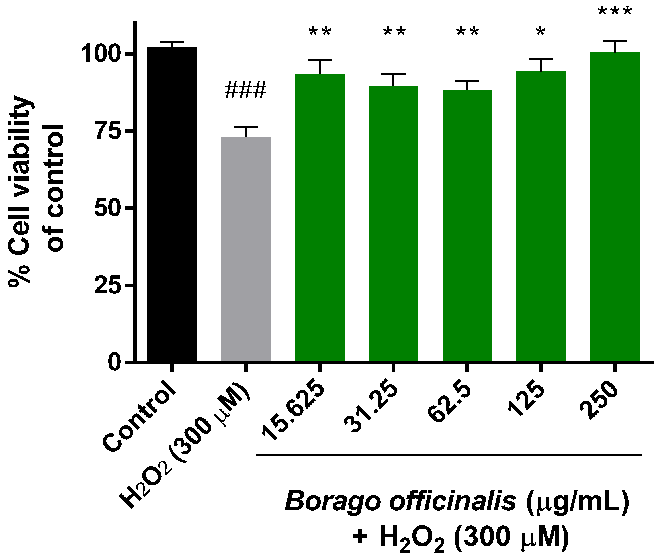
3.4.1. Neuroprotective Effect of B. Officinalis (MTT Assay)

The extensively used MTT assay was required to check the combined effect of hydrogen peroxide and the plant extract over Neuro-2a mitochondria. Previously, the cytotoxicity of the extract was measured for 24 h and none of the concentrations produced any damage to the cells. Once neurons were subjected to hydrogen peroxide (300 µM) for 1 h, the cell viability was reduced by 25% and significant differences were obtained over control cells (Figure 1). In this order, the neuroprotective effect of borago extract was confirmed when cells were subjected to both treatments.

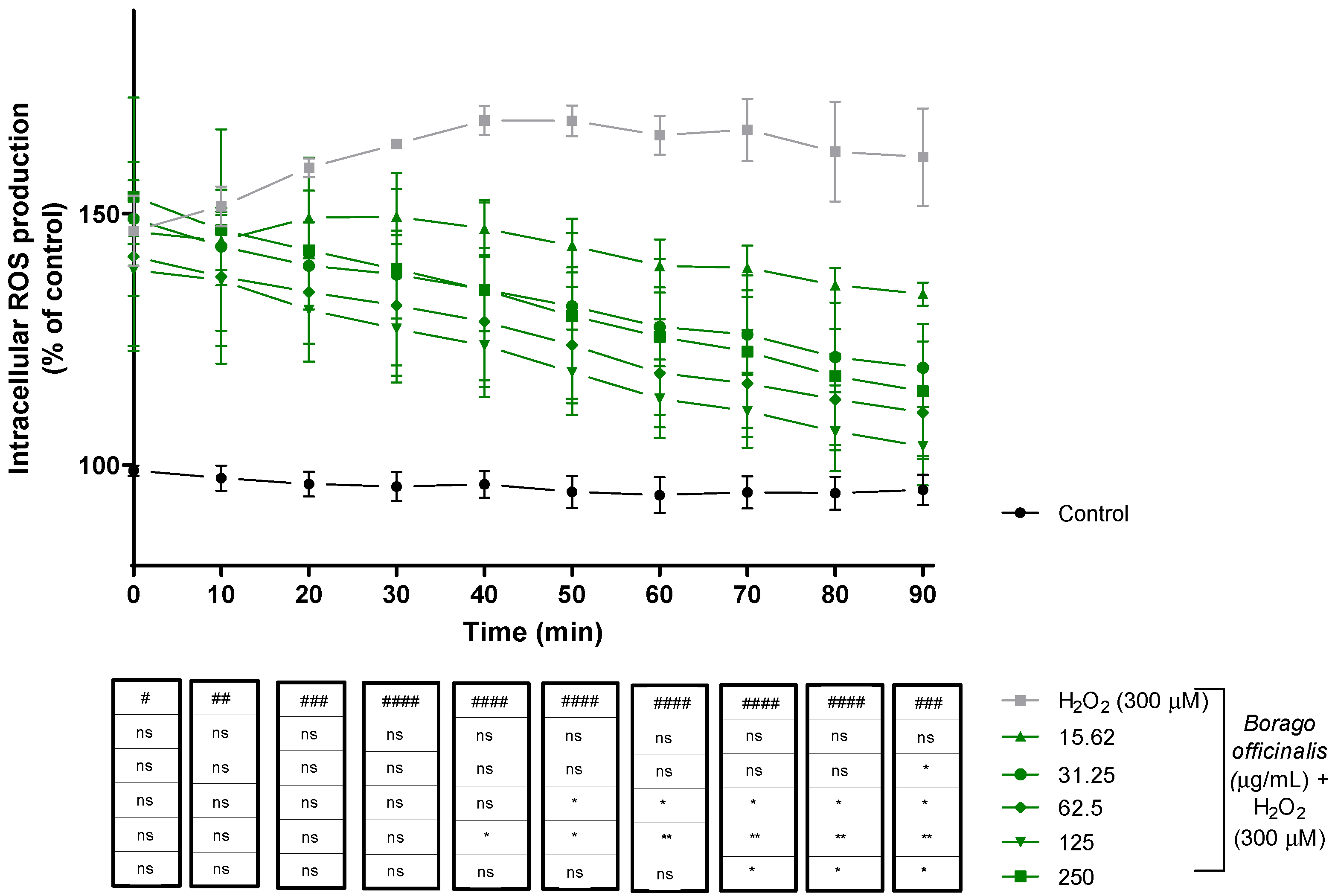
3.4.2. Reactive Oxygen Species (ROS) Production Decreases When Neuro-2a Is Supplemented with B. officinalis

Intracellular ROS production was measured for 90 min. At the beginning of the assay, treated cells displayed similar ROS production while control cells remained at the same levels during the experiment. B. officinalis started to neutralize radicals during the assay, but cells subjected to H2O2 revealed higher levels (over 150%). In the end, cotreated cells showed values close to the control cells rather than hydrogen peroxide cells, and significant differences appeared (Figure 2).

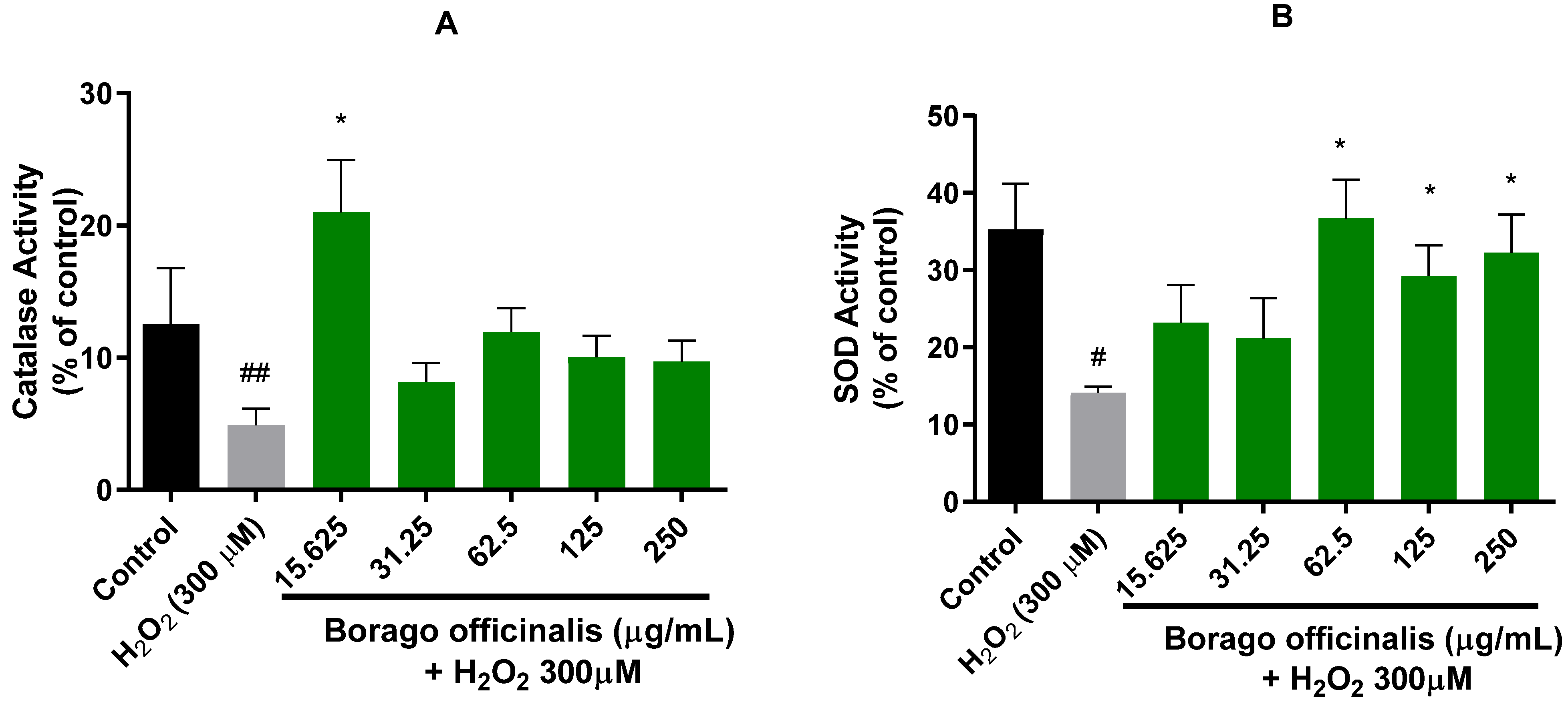
3.4.3. B. officinalis Ameliorates Catalase and Superoxide Dismutase (SOD) Activities

In order to measure the activity of catalase and superoxide dismutase enzymes, previously, the BCA protein assay was selected, and proteins were normalized. Borage extract demonstrated a similar profile for both enzymes (Figure 3). Remarkably, the lowest concentration expressed the highest activity for catalase, even higher than control cells. The rest of the concentrations retained the activity between control and H2O2 cells. In the case of SOD activity, the profile showed a slightly dose-dependent tendency. Furthermore, significant differences were detected in the three highest concentrations.

3.5. Neurodegenerative Model of C. elegans

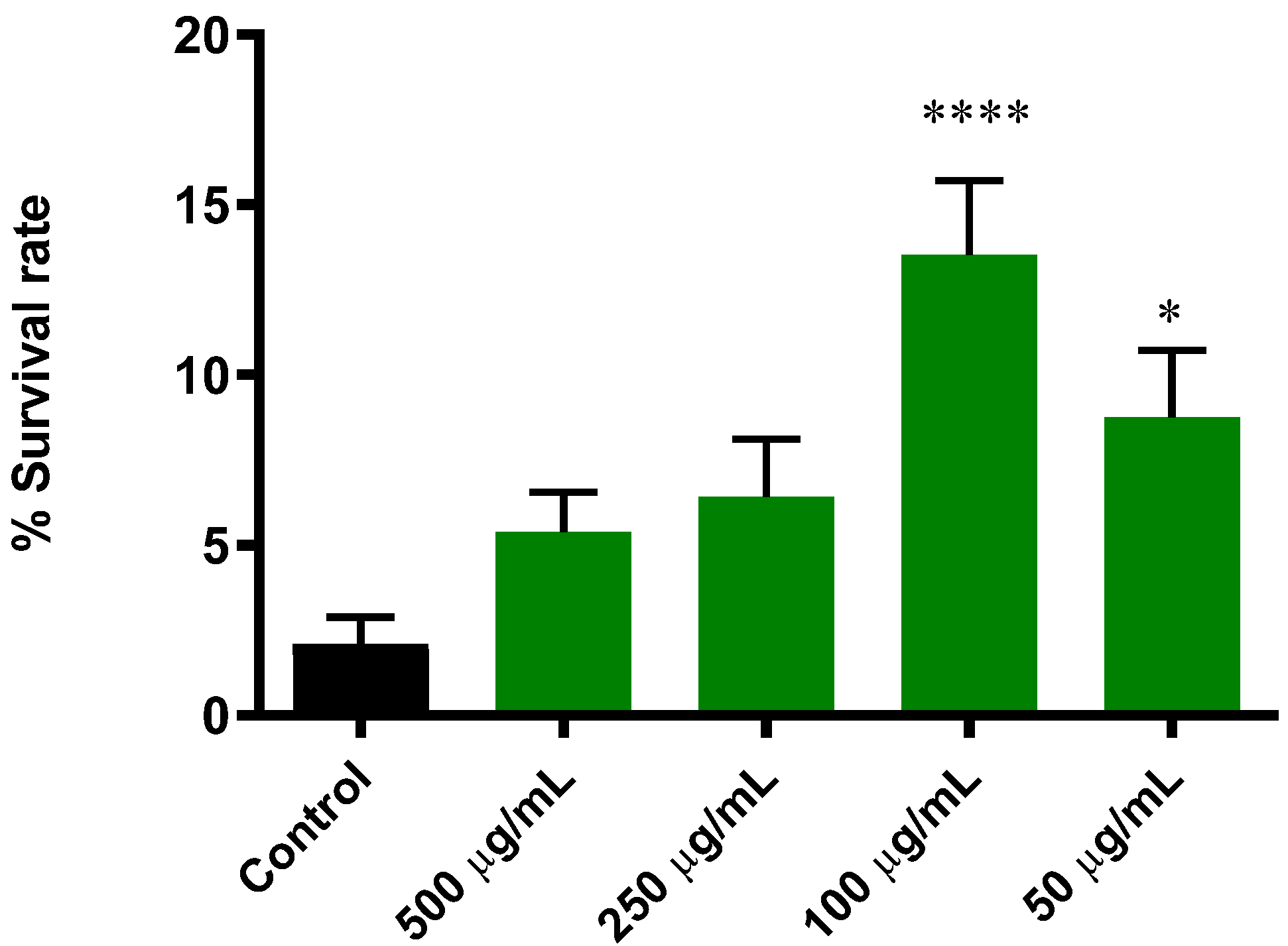
3.5.1. Extract of B. officinalis Enhances Survival Rate in C. elegans Induced with Lethal Oxidative Stress

In this study, we used juglone, a powerful redox cycler, to generate lethal oxidative stress [25]. The results in Figure 4 showed a significantly higher survival rate in worms pretreated with B. officinalis extracts compared to the control group. The best response to oxidative stress was found in 100 µg/mL, for which the survival rate was increased from 2.1% ± 0.8 (control group) to 13.5% ± 2.1 (p ≤ 0.0001).

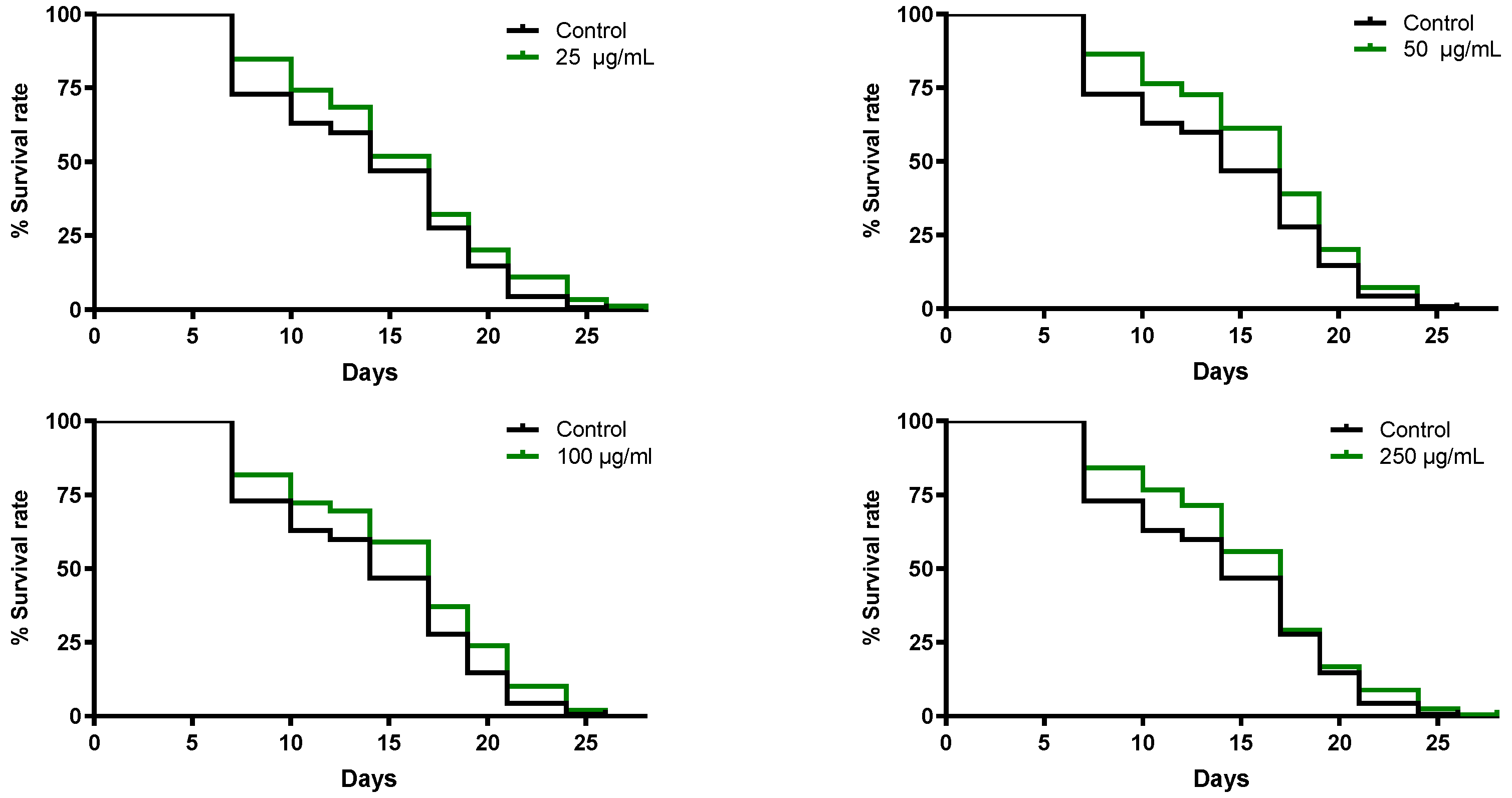
3.5.2. B. officinalis Flower Extract Extends the Lifespan of C. elegans

The effect of B. officinalis extract on lifespan was assessed in SS104 nematodes, a sterile strain, at a restrictive temperature. By analysis of the survival curves, a significant difference in lifespan extension was observed among the treatment groups and control group (Figure 5). All concentrations tested exhibited a lifespan extension of approximately 18% regarding the control group.

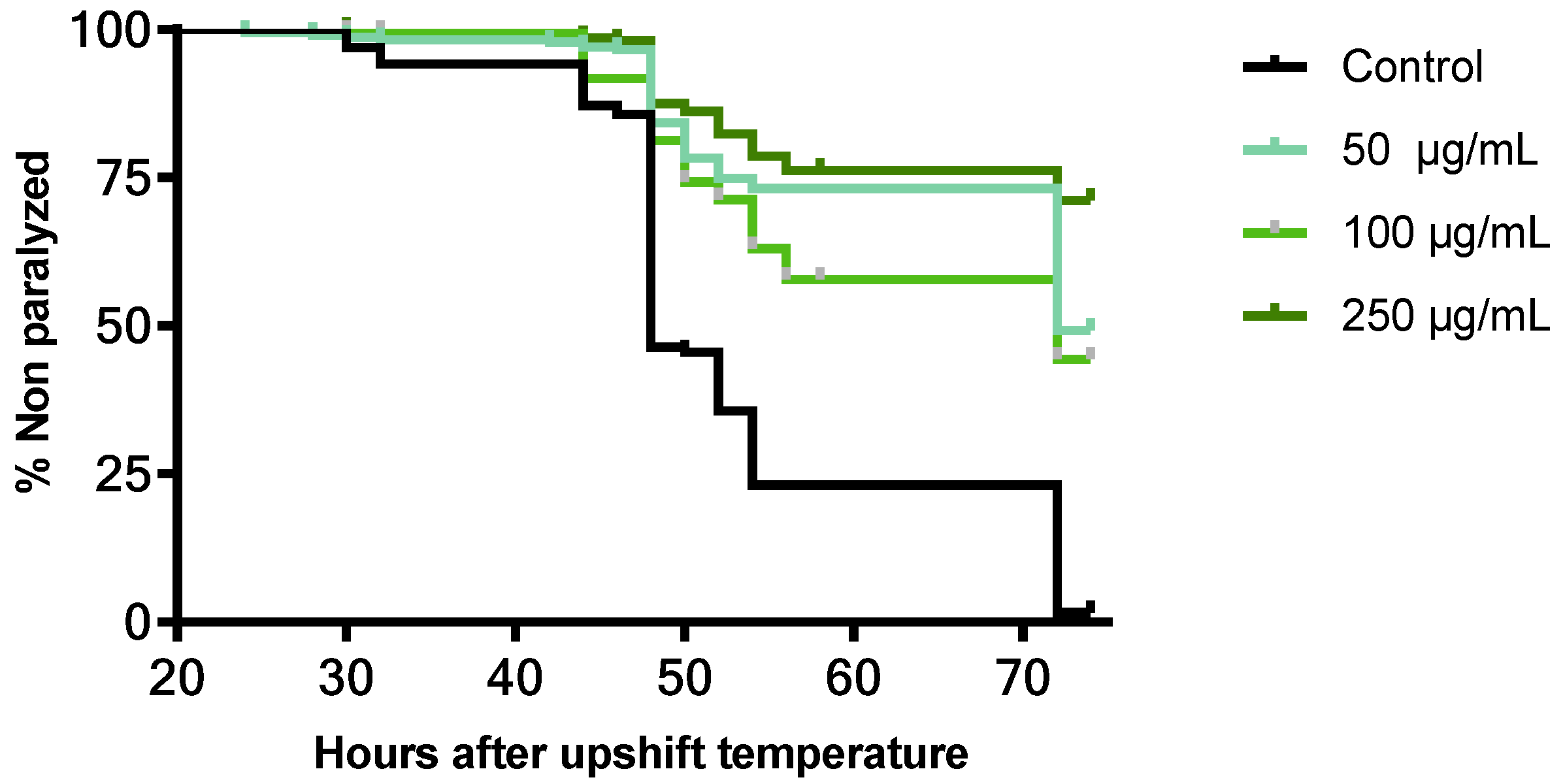
3.5.3. Neuroprotective Effect of B. officinalis Flower Extract in C. elegans

The neuroprotective effect of B. officinalis flower extract against beta amyloid toxicity was assessed using C. elegans CL4176. Figure 6 shows the paralysis curves of CL4176 treated with different concentrations of extract compared with untreated nematodes (control group). After 28 h, after the temperature upshift, the control group started to become paralyzed due to the expression of human β-amyloid peptide. All treatment groups showed a significant delay in body paralysis compared with the control (p ≤ 0.0001). The time within which 50% of the worms were paralyzed or dead (PT50) was 48 h for the control group, while in treated worms with 50 and 100 µg/mL, it was increased until 72 h after upshifting the temperature. For the highest dose tested, the PT50 was not achieved during the experiment.

4. Discussion

B. officinalis has been previously studied in terms of memory function in animal models using the leaves [26]. In this study, the neuroprotective effect of B. officinalis flower extract was assessed on Neuro-2a cells as well as an Alzheimer’s model on C. elegans, since there has been an increase in the consumption of edible flowers worldwide.
To perform an appropriate assessment of the antioxidant activity of borage, several methods based on different mechanisms were used: DPPH, FRAP, and ORAC. Previously, free radical scavenging activity was evaluated by using 2,2-diphenyl-1-picryl-hydrazyl-hydrate (DPPH) in other manuscripts. Jaradat et al. described that Borago officinalis methanolic extract showed an IC50 of 39.92 ± 0.52 μg/mL, while Abu-Qaoud and collaborators studied two different borage cultivars, the wild leaf extract and cultivated leaf extract reaching lower IC50 values of 6.3 μg/mL and 8.7 μg/mL, respectively [27,28]. Another study from Italian researchers revealed that an hydroalcoholic extract (70% aqueous EtOH through maceration) from borage leaves achieved 58 μg/mL (IC50) [10]. Crude borage methanolic extract displayed 89% inhibition of DPPH radicals by HPLC [9]. All of them demonstrated a potential antioxidant profile for borage, whose values were 20–100 times lower than the one obtained in this manuscript, which suggests that the fresh flowers may have a better antioxidant profile than other parts of the plant due to their composition.
Borago officinalis’s ferric reducing antioxidant power (FRAP) was previously determined by an Iranian research group. They also prepared an ethanolic extract and almost reached 3 μmol/g tissue, very similar to the fresh flowers observed in this study (4 μmol/g) [26].
This is the first time that the antioxidant activity of borage has been measured by oxygen radical techniques such as the ORAC assay. This result supports the antioxidant potential evidenced by the previous assays shown in Table 2. Wang et al. carried out a comparison between 12 edible flowers common in China, and reported an ORAC value among 0.86–0.04 µmol TE/mg extract [29]. The ORAC value found in this study for B. officinalis flowers is in the upper part of this range.
Despite the scarce literature about the antioxidant activity of the flowers of B. officinalis, there are compounds present in it with noted antioxidant activity. As described by Fernandes et al., the flowers of B. officinalis are a good source of polyunsaturated fatty acids (PUFAs) and carotenoids; moreover, it contains vitamin E, mainly α-tocopherol [30], and their antioxidant properties are well established [31,32,33].
However, the antioxidant and protective activity in models of oxidative stress in relation to the nervous system has never been established before for these flowers. It is commonly known that neurodegenerative diseases are induced at least in part by free radicals; for this reason, the neuroprotective effect was evaluated in two different models: Neuro-2a and C. elegans. No previous paper has studied the effect of this extract in any in vitro neuronal or glial cell line, making it attractive for testing. Hydrogen peroxide was added to the neurons as a free radical initiator and B. officinalis neutralized its effect and confirmed the antioxidant activity of the extract. Consequently, the role that it plays as a neuroprotective agent was studied by evaluating the response of the physiological antioxidant defense systems, such as catalase or superoxide dismutase and intracellular ROS production, against hydrogen peroxide.
Borage supplementation reversed the synaptic plasticity in the hippocampal dentate gyrus following Aβ treatment, and thus borage consumption may lead to an improvement in AD-induced cognitive dysfunction [34].
Furthermore, the potential of B. officinalis flower extract was challenged on C. elegans, which has proven to be a useful in vivo system to study the bioactivity of nutraceuticals and functional foods due to its short life, ease of handling, wide range of mutant strains, and, more importantly, the high degree of similarity with human genetics (between 60 and 80% of human genes have homologues in the worm´s genome) [35]. Initially, the antioxidant potential of B. officinalis against induced lethal oxidative stress was tested using juglone, a strong prooxidant, to stress the worms. High doses of juglone generate superoxide and H2O2, which causes protein modifications, lipid oxidation, and nuclear DNA damage in C. elegans [36]. The studied extract can alleviate juglone’s toxicity since it increased the survival rate. Moreover, B. officinalis promoted lifespan extension in C. elegans. To the best of our knowledge, this is the first study of B. officinalis on C. elegans regarding its antioxidant and anti-ageing capacity. Some of the compounds described in the flowers of B. officinalis are already reported in the literature for protecting worms against oxidative stress and extending their lifespan, such as polyunsaturated fatty acids (PUFAs) [30,32]. Our results agree with these findings and support the use of this species as a functional ingredient with a lack of toxicity.
Experimental and clinical evidence suggests that oxidative stress plays a crucial role in the pathogenesis of Alzheimer’s disease and other neurogenerative disorders [37,38]. In CL4176, prior to the appearance of symptoms of Alzheimer’s disease (paralysis in nematodes), there is an accumulation of oxidative damage that could be attenuated due to the antioxidant activity showed by the extract [39]. The results obtained from paralysis assays showed a significant delay in the time to develop paralysis in a dose-dependent manner. On the other hand, this finding could also be explained by the capacity of different extracts of B. officinalis shown to inhibit amyloid formation in vitro and in vivo [34].
These results can be explained partially by the high amount of polyunsaturated FAs (PUFA) found in the extract. Several authors consider that FAs are essential for neurogenesis and neurotransmitter production [40,41,42,43]. We can also highlight that the IA in FAs in borage extract was 0.26. According to Montaner et al. [44], this index can range from 0.18 to 0.43, and it is important to consider the different effects that FAs might have on human health, particularly the probability of decreasing the incidence of pathogenic effects of atheroma and brain inflammatory processes. Regarding the calculated oxidizability value (Cox) of 5.73, the tendency of borago flower extract to prevent oxidative deterioration was observed, which supports its significant dietary nutraceutical value.

5. Conclusions

Our study showed that the fresh flowers of B. officinalis exhibit neuroprotective and antioxidant activity in vitro as well as in vivo. The studied extract was able to decrease ROS production and increase antioxidant enzyme activity in neuronal cells. Moreover, B. officinalis attenuated Aβ toxicity in an Alzheimer’s model of C. elegans. Interesting results are here presented; nevertheless, more in vitro experiments in human cells and in vivo models are needed to confirm and develop B. officinalis flowers as functional ingredients for human use.

Author Contributions

Conceptualization, C.M., G.C., C.G.-R. and V.L.; Formal analysis, C.M. and G.C.; Funding acquisition V.L.; Investigation, C.M., G.C., L.B. and T.C.F.; Methodology, C.M., G.C., L.B. and V.L.; Project administration, V.L.; Resources, V.L.; Supervision, L.B., C.G.-R. and V.L.; Visualization, V.L.; Writing—original draft, C.M. and G.C.; Writing—review & editing, C.G.-R. and V.L. All authors have read and agreed to the published version of the manuscript.

Funding

The authors are grateful to the Foundation for Science and Technology (FCT, Portugal) for financial support from national funds FCT/MCTES, to CIMO (UIDB/AGR/00690/2020); for national funding by FCT, P.I., through the institutional scientific employment program contract for L. Barros contracts. Universidad San Jorge and Gobierno de Aragón are also thanked for their financial support (Phyto-Pharm Group B44_20D).

Institutional Review Board Statement

Not applicable.

Informed Consent Statement

Not applicable.

Data Availability Statement

Data is contained within the article.

Conflicts of Interest

The authors declare no conflict of interest. The funders had no role in the design of the study; in the collection, analyses or interpretation of data; in the writing of the manuscript or in the decision to publish the results.

References

  1. Ramezani, M.; Amiri, M.S.; Zibaee, E.; Boghrati, Z.; Ayati, Z.; Sahebkar, A.; Emami, S.A. A Review on the Phytochemistry, Ethnobotanical Uses and Pharmacology of Borago Species. Curr. Pharm. Des. 2019, 26, 110–128. [Google Scholar] [CrossRef]
  2. Takahashi, J.A.; Rezende, F.A.G.G.; Moura, M.A.F.; Dominguete, L.C.B.; Sande, D. Edible flowers: Bioactive profile and its potential to be used in food development. Food Res. Int. 2020, 129, 108868. [Google Scholar] [CrossRef]
  3. El-Shazly, A.; Sarg, T.; Ateya, A.; Abdel Aziz, A.; El-Dahmy, S.; Witte, L.; Wink, M. Pyrrolizidine Alkaloids from Echium setosum and Echium vulgare. J. Nat. Prod. 1996, 59, 310–313. [Google Scholar] [CrossRef]
  4. Bruneton, J. Toxic Plants Dangerous to Humans and Animals; É ditions Tec & Doc; Intercept Limited: Huddersfield, UK, 1999; ISBN 1898298629. [Google Scholar]
  5. Avila, C.; Breakspear, I.; Hawrelak, J.; Salmond, S.; Evans, S. A systematic review and quality assessment of case reports of adverse events for borage (Borago officinalis), coltsfoot (Tussilago farfara) and comfrey (Symphytum officinale). Fitoterapia 2020, 142, 104519. [Google Scholar] [CrossRef]
  6. Lozano-Baena, M.-D.; Tasset, I.; Muñoz-Serrano, A.; Alonso-Moraga, Á.; de Haro-Bailón, A. Cancer Prevention and Health Benefices of Traditionally Consumed Borago officinalis Plants. Nutrients 2016, 8, 48. [Google Scholar] [CrossRef] [Green Version]
  7. Djaoudene, O.; López, V.; Cásedas, G.; Les, F.; Schisano, C.; Bachir Bey, M.; Tenore, G.C. Phoenix dactylifera L. seeds: A by-product as a source of bioactive compounds with antioxidant and enzyme inhibitory properties. Food Funct. 2019, 10, 4953–4965. [Google Scholar] [CrossRef]
  8. Fernández-Moriano, C.; González-Burgos, E.; Divakar, P.K.; Crespo, A.; Gómez-Serranillos, M.P. Evaluation of the antioxidant capacities and cytotoxic effects of ten parmeliaceae lichen species. Evid. Based Complement. Altern. Med. 2016, 201, 11. [Google Scholar] [CrossRef] [Green Version]
  9. Bandoniene, D.; Murkovic, M. The detection of radical scavenging compounds in crude extract of borage (Borago officinalis L.) by using an on-line HPLC-DPPH method. J. Biochem. Biophys. Methods 2002, 53, 45–49. [Google Scholar] [CrossRef]
  10. Conforti, F.; Sosa, S.; Marrelli, M.; Menichini, F.; Statti, G.A.; Uzunov, D.; Tubaro, A.; Menichini, F.; Loggia, R. In vivo anti-inflammatory and in vitro antioxidant activities of Mediterranean dietary plants. J. Ethnopharmacol. 2008, 116, 144–151. [Google Scholar] [CrossRef]
  11. Seo, S.A.; Park, B.; Hwang, E.; Park, S.Y.; Yi, T.H. Borago officinalis L. attenuates UVB-induced skin photodamage via regulation of AP-1 and Nrf2/ARE pathway in normal human dermal fibroblasts and promotion of collagen synthesis in hairless mice. Exp. Gerontol. 2018, 107, 178–186. [Google Scholar] [CrossRef]
  12. Barros, L.; Alves, C.T.; Dueñas, M.; Silva, S.; Oliveira, R.; Carvalho, A.M.; Henriques, M.; Santos-Buelga, C.; Ferreira, I.C.F.R. Characterization of phenolic compounds in wild medicinal flowers from Portugal by HPLC–DAD–ESI/MS and evaluation of antifungal properties. Ind. Crops Prod. 2013, 44, 104–110. [Google Scholar] [CrossRef] [Green Version]
  13. Fatemi, S.H.; Hammond, E.G. Analysis of oleate, linoleate and linolenate hydroperoxides in oxidized ester mixtures. Lipids 1980, 15, 379–385. [Google Scholar] [CrossRef]
  14. Ulbricht, T.L.V.; Southgate, D.A.T. Coronary heart disease: Seven dietary factors. Lancet 1991, 338, 985–992. [Google Scholar] [CrossRef]
  15. López, V.; Akerreta, S.; Casanova, E.; García-mina, J.; Cavero, R.; Calvo, M. Screening of Spanish Medicinal Plants for Antioxidant and Antifungal Activities. Pharm. Biol. 2008, 46, 602–609. [Google Scholar] [CrossRef]
  16. Pulido, R.; Bravo, L.; Saura-Calixto, F. Antioxidant Activity of Dietary Polyphenols As Determined by a Modified Ferric Reducing/Antioxidant Power Assay. J. Agric. Food Chem. 2000, 48, 3396–3402. [Google Scholar] [CrossRef] [Green Version]
  17. Dávalos, A.; Gómez-Cordovéz, C.; Bartolomé, B. Extending applicability of the oxygen radical absorbance capacity (ORAC-fluorescein) assay. J. Agric. Food Chem. 2004, 52, 48–54. [Google Scholar] [CrossRef]
  18. Mosmann, T. Rapid colorimetric assay for cellular growth and survival: Application to proliferation and cytotoxicity assays. J. Immunol. Methods 1983, 65, 55–63. [Google Scholar] [CrossRef]
  19. Cásedas, G.; González-burgos, E.; Smith, C.; López, V.; Pilar, M. Regulation of redox status in neuronal SH-SY5Y cells by blueberry (Vaccinium myrtillus L.) juice, cranberry (Vaccinium macrocarpon A.) juice and cyanidin. Food Chem. Toxicol. 2018, 118, 572–580. [Google Scholar] [CrossRef]
  20. Cásedas, G.; Les, F.; López, V.; Choya-Foces, C.; Hugo, M. The metabolite urolithin-a ameliorates oxidative stress in neuro-2a cells, becoming a potential neuroprotective agent. Antioxidants 2020, 9, 177. [Google Scholar] [CrossRef] [Green Version]
  21. Stiernagle, T. Maintenance of C. elegans. WormBook 1999, 2, 51–67. [Google Scholar] [CrossRef] [Green Version]
  22. Surco-Laos, F.; Cabello, J.; Gómez-Orte, E.; González-Manzano, S.; González-Paramás, A.M.; Santos-Buelga, C.; Dueñas, M. Effects of O-methylated metabolites of quercetin on oxidative stress, thermotolerance, lifespan and bioavailability on Caenorhabditis elegans. Food Funct. 2011, 2, 445–456. [Google Scholar] [CrossRef]
  23. Virk, B.; Jia, J.; Maynard, C.A.; Helliwell, N.; Cipinska, M.; Weinkove, D.; Virk, B.; Jia, J.; Maynard, C.A.; Raimundo, A.; et al. Folate Acts in E. coli to Accelerate C. elegans Aging Independently of Bacterial Biosynthesis. Cell Rep. 2016, 14, 1611–1620. [Google Scholar] [CrossRef] [Green Version]
  24. Dostal, V.; Link, C.D. Assaying β-amyloid Toxicity using a Transgenic C. elegans Model. J. Vis. Exp. 2010, 3791, 4–7. [Google Scholar] [CrossRef] [Green Version]
  25. Pluci, B.; Kuczy, P. The oxidative stress in allelopathy: Participation of prenyllipid antioxidants in the response to juglone in Chlamydomonas reinhardtii. Phytochemistry 2017, 144, 171–179. [Google Scholar] [CrossRef]
  26. Zargooshnia, S.; Shahidi, S.; Ghahremanitamadon, F.; Nikkhah, A.; Mehdizadeh, M.; Soleimani Asl, S. The protective effect of Borago officinalis extract on amyloid β (25-35)-induced long term potentiation disruption in the dentate gyrus of male rats. Metab. Brain Dis. 2015, 30, 151–156. [Google Scholar] [CrossRef]
  27. Jaradat, N.A.; Damiri, B.; Abualhasan, M.N. Antioxidant Evaluation for Urtica urens, Rumex cyprius and Borago officinalis edible Wild Plants in Palestine. Available online: https://pubmed.ncbi.nlm.nih.gov/27005499 (accessed on 12 August 2021).
  28. Abu-Qaoud, H.; Shawarb, N.; Hussen, F.; Jaradat, N.; Shtaya, M. Report: Comparison of qualitative, quantitative analysis and antioxidant potential between wild and cultivated Borago officinalis leaves from palestine. Pak. J. Pharm. Sci. 2018, 31, 953–959. [Google Scholar]
  29. Wang, F.; Miao, M.; Xia, H.; Yang, L.; Wang, S.; Sun, G. Antioxidant activities of aqueous extracts from 12 Chinese edible flowers in vitro and in vivo. Food Nutr. Res. 2017, 61, 1–9. [Google Scholar] [CrossRef] [Green Version]
  30. Fernandes, L.; Pereira, J.A.; Saraiva, J.A.; Ramalhosa, E.; Casal, S. Phytochemical characterization of Borago officinalis L. and Centaurea cyanus L. during flower development. Food Res. Int. 2019, 123, 771–778. [Google Scholar] [CrossRef] [Green Version]
  31. Cardoso, L.A.C.; Karp, S.G.; Vendruscolo, F.; Kanno, K.Y.F.; Zoz, L.I.C.; Carvalho, J.C. Biotechnological Production of Carotenoids and Their Applications in Food and Pharmaceutical Products. Carotenoids 2017, 8, 125–141. [Google Scholar] [CrossRef] [Green Version]
  32. Beaudoin-Chabot, C.; Wang, L.; Smarun, A.V.; Vidović, D.; Shchepinov, M.S.; Thibault, G. Deuterated polyunsaturated fatty acids reduce oxidative stress and extend the lifespan of C. elegans. Front. Physiol. 2019, 10, 1–10. [Google Scholar] [CrossRef] [Green Version]
  33. Benzie, I.; Strain, J. The ferric reducing ability of plasma (FRAP) as a measure of “antioxidant power”: The FRAP assay. Anal. Biochem. 1996, 239, 70–76. [Google Scholar] [CrossRef] [PubMed] [Green Version]
  34. Ghahremanitamadon, F.; Shahidi, S.; Zargooshnia, S.; Nikkhah, A.; Ranjbar, A.; Soleimani Asl, S. Protective effects of Borago officinalis extract on amyloid β-peptide(25-35)-induced memory impairment in male rats: A behavioral study. Biomed Res. Int. 2014, 2014, 798535. [Google Scholar] [CrossRef] [PubMed] [Green Version]
  35. Calvo, D.R.; Martorell, P.; Genovés, S.; Gosálbez, L. Development of novel functional ingredients: Need for testing systems and solutions with Caenorhabditis elegans. Trends Food Sci. Technol. 2016, 54, 197–203. [Google Scholar] [CrossRef]
  36. Przybysz, A.J.; Choe, K.P.; Roberts, L.J.; Strange, K. Increased age reduces DAF-16 and SKN-1 signaling and the hormetic response of Caenorhabditis elegans to the xenobiotic juglone. Mech. Ageing Dev. 2009, 130, 357–369. [Google Scholar] [CrossRef] [Green Version]
  37. Cioffi, F.; Adam, R.H.I.; Broersen, K. Molecular Mechanisms and Genetics of Oxidative Stress in Alzheimer’s Disease. J. Alzheimers Dis. 2019, 72, 981–1017. [Google Scholar] [CrossRef] [Green Version]
  38. Volkert, M.R.; Crowley, D.J. Preventing Neurodegeneration by Controlling Oxidative Stress: The Role of OXR1. Front. Neurosci. 2020, 14, 611904. [Google Scholar] [CrossRef]
  39. Drake, J.; Link, C.D.; Butterfield, D.A. Oxidative stress precedes fibrillar deposition of Alzheimer’s disease amyloid beta-peptide (1–42 ) in a transgenic Caenorhabditis elegans model. Neurobiol. Aging 2003, 24, 415–420. [Google Scholar] [CrossRef]
  40. Ratusz, K.; Symoniuk, E.; Wroniak, M.; Rudzińska, M. Bioactive Compounds, Nutritional Quality and Oxidative Stability of Cold-Pressed Camelina (Camelina sativa L.). Oils 2018, 8, 2606. [Google Scholar] [CrossRef] [Green Version]
  41. Chen, J.; Liu, H. Nutritional Indices for Assessing Fatty Acids: A Mini-Review. Int. J. Mol. Sci. 2020, 21, 5695. [Google Scholar] [CrossRef]
  42. Purohit, S.; Kalita, D.; Barik, C.R.; Sahoo, L.; Goud, V.V. Evaluation of thermophysical, biochemical and antibacterial properties of unconventional vegetable oil from Northeast India. Mater. Sci. Energy Technol. 2021, 4, 81–91. [Google Scholar] [CrossRef]
  43. Hosseini, M.; Poljak, A.; Braidy, N.; Crawford, J.; Sachdev, P. Blood fatty acids in Alzheimer’s disease and mild cognitive impairment: A meta-analysis and systematic review. Ageing Res. Rev. 2020, 60, 101043. [Google Scholar] [CrossRef]
  44. Montaner, C.; Zufiaurre, R.; Movila, M.; Mallor, C. Evaluation of Borage (Borago officinalis L.) Genotypes for Nutraceutical Value Based on Leaves Fatty Acids Composition. Foods 2021, 11, 16. [Google Scholar] [CrossRef]
Figure 1. Mitochondrial activity in Neuro-2a cell culture (MTT assay). Cytoprotective effects of Borago officinalis flower extract versus hydrogen peroxide (300 µM). Note: * p < 0.05 versus H2O2; ** p < 0.01 versus H2O2; *** p < 0.005 versus H2O2; ### p < 0.0005 versus control.
Antioxidants 11 01244 g001
Figure 2. Production of ROS in Neuro-2a cells treated with hydrogen peroxide and Borago officinalis flower extract (15.62–250 µg/mL). Data are expressed as percentage over control cells. Note: # p < 0.05 versus control; ## p < 0.01 versus control; ### p < 0.0005 versus control; #### p < 0.0001 versus control; ns: no significant; * p < 0.05; ** p < 0.01. Borage co-treatment did not show any difference until 40 min (125 µg/mL; p < 0.05). From this point, ROS species were diminishing for all extract treatments and significant differences were increased. At the end of the experiment, only 15.62 µg/mL did not achieve any significant differences. Borago officinalis flower extract (125 µg/mL) reduced ROS production almost to basal levels (control cells).
Antioxidants 11 01244 g002
Figure 3. Neuro-2a endogenous antioxidant enzymes exerted by Borago officinalis flower extract treatments (15.62-250 µg/mL). (A) Catalase activity. (B) Superoxide dismutase activity. Note: * p < 0.05 versus H2O2; # p < 0.05 versus control; ## p < 0.01 versus control.
Antioxidants 11 01244 g003
Figure 4. Effect of B. officinalis flower extract on the response to lethal oxidative stress induced by juglone (150 μM) on C. elegans N2. Differences compared to control group were considered significant at p ≤ 0.05 (*) and p ≤ 0.0001 (****).
Antioxidants 11 01244 g004
Figure 5. Lifespan of C. elegans SS104 subjected to Borago officinalis flower extract. The mean lifespan was 14 days in control group and 17 days for all treatment groups. Result were analyzed using the Kaplan–Meier survival model and for significance by using a log-rank pairwise comparison test between the control and treatment groups.
Antioxidants 11 01244 g005
Figure 6. Effect of B. officinalis flower extract on Aβ-induced paralysis on transgenic C. elegans CL4176. The PT50 were 48 h for the control group and 72 h for 50 and 100 µg/mL treated group. The PT50 was not achieved in the group treated with 250 µg/mL. Statistical significance of the difference between the experiments was analyzed by a log-rank (Kaplan–Meier) statistical test, which compared the survival distributions between the control and treated groups. Differences in survival tests between treatment and the control group were p ≤ 0.0001.
Antioxidants 11 01244 g006
Table 1. Fatty acid composition of B. officinalis flower extract.
Fatty AcidsShorthand NomenclatureRelative Percentage (%)
Caproic acid6:00.24 ± 0.04
Caprylic acid8:00.0802 ± 0.0001
Capric acid10:00.041 ± 0.003
Docosanoic acid22:01.39 ± 0.001
Dodecanoic acid12:00.114 ± 0.002
Heptadecanoic acid17:00.48 ± 0.01
Lignoceric acid24:00.732 ± 0.001
Myristic acid14:00.45 ± 0.04
Pentadecanoic acid15:02.48 ± 0.03
Palmitic acid16:017.17 ± 0.04
Stearic acid18:03.8 ± 0.1
Tricosylic acid23:00.32 ± 0.01
Eicosenoic acid20:10.49 ± 0.02
Erucic acid22:1 ω 90.16 ± 0.01
Myristoleic acid14:10.24 ± 0.01
Nervonic acid24:11.05 ± 0.01
Oleic acid18:1 ω 915.93 ± 0.04
Palmitoleic acid16:10.19 ± 0.01
Eicosadienoic acid20:20.42 ± 0.04
Eicosatrienoic acid20:3 ω 3 + 21:00.276 ± 0.004
Eicosapentaenoic acid20:5 ω 30.12 ± 0.01
γ-Linolenic acid18:3 ω 60.13 ± 0.01
α-Linolenic acid18:3 ω 30.07 ± 0.01
Linoleic acid18:2 ω 653.7 ± 0.2
Σ Saturated fatty acids (SFA)27.27
Σ Monounsaturated fatty acids (MUFA)18.05
Σ Polyunsaturated fatty acids (PUFA)54.68
Σ ω 6 53.83
Σ ω 30.59
Cox value5.73
Atherogenicity Index (AI)0.26
Table 2. Antioxidant activity of B. officinalis flower extract. The results are presented as mean ± SEM.
DPPH
IC50 (µg/mL)
FRAP
µmol Fe2+/g Extract
ORAC
µmol TE/mg Extract
B. officinalis646 ± 354 ± 20.54 ± 0.03
Abbreviation: TE = Trolox equivalent.
Publisher’s Note: MDPI stays neutral with regard to jurisdictional claims in published maps and institutional affiliations.

