Article

Shedding Light into the Connection between Chemical Components and Biological Effects of Extracts from Epilobium hirsutum: Is It a Potent Source of Bioactive Agents from Natural Treasure?

by
Gunes Ak
1,
Gokhan Zengin
1,*,
Mohamad Fawzi Mahomoodally
2,
Eulogio Llorent-Martínez
3,
Giustino Orlando
4,
Annalisa Chiavaroli
4,
Luigi Brunetti
4,
Lucia Recinella
4,
Sheila Leone
4,
Simonetta Cristina Di Simone
4,
Luigi Menghini
4,* and
Claudio Ferrante
4
1
Physiology and Biochemistry Research Laboratory, Department of Biology, Science Faculty, Selcuk University, 42250 Konya, Turkey
2
Department of Health Sciences, Faculty of Medicine and Health Sciences, University of Mauritius, Réduit 80837, Mauritius
3
Department of Physical and Analytical Chemistry, Campus Las Lagunillas S/N, University of Jaén, E-23071 Jaén, Spain
4
Department of Pharmacy, Botanic Garden “Giardino dei Semplici”, Università degli Studi “Gabriele d’Annunzio”, via dei Vestini 31, 66100 Chieti, Italy
*
Authors to whom correspondence should be addressed.
Antioxidants 2021, 10(9), 1389; https://doi.org/10.3390/antiox10091389
Submission received: 25 July 2021 / Revised: 21 August 2021 / Accepted: 27 August 2021 / Published: 30 August 2021
(This article belongs to the Special Issue Antioxidant and Biological Properties of Plant Extracts)

Abstract

:
Epilobium hirsutum is extensively used as a traditional remedy in folk medicine, especially against prostate inflammation. Therefore, we evaluated the chemical profiles and biopharmaceutical potentials of different extracts of E. hirsutum aerial parts and roots. Metabolomic, antioxidant, and enzyme inhibitory profiles were investigated. Human prostate cancer PC3 cells were exposed to the extracts to evaluate antiproliferative effects. Gene expression and bioinformatics analyses were performed to investigate anti-inflammatory mechanisms. Oenothein B and myricetin were prominent compounds in the extracts. In scavenging/reducing assays, the methanol, infusion, and methanol/water extracts exhibited similar activities. We also observed the reduction of PC3 viability occurring following exposure to methanol and methanol/water extracts. According to bioinformatics analysis, myricetin was predicted to interact with COX-2 and TNFα. The interaction between TNFα and oxo-dihydroxy-octadecenoic acid was predicted as well. Intriguingly, the gene expression of COX-2 and TNFα was reduced in PC3 cells after exposure to methanol and methanol/water extracts. These effects were paralleled by the decreased gene expression of IL-8 and NFkB and the inhibition of PGE2 release. Therefore, the present findings suggest the potential use of E. hirsutum for the management of the burden of inflammation and oxidative stress occurring in lower urinary tract diseases, including prostatitis.

1. Introduction

In the past decade, people have been looking for new health-promoting compounds from natural sources. Among the natural sources, plants and plant products are the most important [1]. For example, the determination of antioxidant properties of plant extracts is an index of the efficacy against oxidative stress, which is associated with chronic inflammatory and degenerative diseases [2]. In addition to their antioxidant properties, the enzyme inhibitory, toxic, and anticancer properties of plant extracts have been attracting interest in the scientific community [3,4,5]. With this in mind, scientists are increasingly relying on plants as inspiration for new active ingredients that could be developed in the context of the pharmaceutical industry. Overall, this phenomenon is referred to as the green revolution, and new studies, especially on wild plants that have not yet been investigated, have made a significant contribution to this. However, the possible toxic properties of plants and their synergetic and antagonistic interactions should not be forgotten.
The genus Epilobium belongs to the Onagraceae family and comprises more than 200 species worldwide [6]. The members of the genus are known by different local names such as fireweed, wickopy, and niviaqsiaq [7]. Epilobium species have been widely used in traditional medicine. For example, E. angustifolium and E. hirsutum are known as yaki otu and are used to treat prostate diseases in Turkey [8,9]. Moreover, E. hirsutum is used to stop bleeding [10] in the treatment of gastrointestinal problems, menstrual disorders, and sleeping problems [11,12,13,14]. In addition, the young leaves of the Epilobium species are often used as food and feed [11,15]. To confirm the ethnobotanical uses of the species, several pharmacological and biochemical studies on the members of the genus have been performed [13,16,17,18,19,20,21,22]. In addition to these pharmacological studies, the presence of biologically active compounds including tannins (especially ellagitannins), myricetin, quercetin, and several phenolic acids have been reported in phytochemical studies [18,23,24,25,26,27].
In light of the above information, in the present work, we aimed to unravel the relationship between the chemical components and biological properties of various extracts (ethyl acetate, methanol, methanol/water and infusion) from two parts (aerial parts and roots) of E. hirsutum. The chemical characterization of the tested extracts was assessed using the HPLC-ES-MSn technique. The intrinsic biological properties of the extracts were evaluated in terms of antioxidant and enzyme inhibitory effects, whereas the Artemia salina (brine shrimp) and human prostate cancer PC3 cell line were exposed to the extracts to define the biocompatibility limits and the antiproliferative effects, respectively. Finally, the anti-inflammatory properties of the extracts were studied through bioinformatics analysis and the evaluation of the gene expression of different pro-inflammatory biomarkers, namely TNFα, COX-2, VEGFA, IL-6, IL-8, NFkB, and TIMP1. The obtained results will shed light on the rational phytotherapy use of E. hirsutum, especially against inflammatory diseases in the prostate.

2. Materials and Methods

2.1. Plant Collection and Extract Preparation

Epilobium hirsutum L. samples were collected in the city of Erzurum (Karabıyık village, Turkey; GPS coordinate: 40°08′22″ N, 41°56′16″ E; altitude: 2220 m), in August (collection date: 20 August 2020). The plants were authenticated by a plant taxonomist (Dr. Evren Yıldıztugay) in the Science Faculty, Selcuk University, Konya, Turkey, and voucher specimens (Voucher number: GA-20-002) were kept at the herbarium of the above-mentioned faculty. The plant materials (about thirty samples) were collected in the same population.
The plant materials were packed in plastic bags. Thereafter, the aerial parts and roots were separated, and they were immediately dried in the shade for ten days (25 ± 2 °C). During the drying process, the temperature, humidity, and air flow were controlled to avoid the effects of the phytochemicals in the plants. After the drying process, the dried plant materials were ground using the laboratory mill in the same week. The powdered plant materials were stored in a well-ventilated place in the dark (20 °C). The plant materials were extracted immediately to avoid changing the chemical profile in the plants. After the powder process, the extraction of the plant materials was completed in one week.
Regarding the extraction procedure, the samples were extracted with different solvents, namely ethyl acetate, methanol, methanol/water (70%, v/v), and water. To obtain the ethyl acetate, methanol, and methanol/water extracts, the plant samples (10 g) were macerated with 200 mL of methanol for 24 h at room temperature. Then, the extracts were filtered and evaporated. As for the water extracts, the fruit materials (10 g) were kept in 200 mL of water (boiled) for 15 min. The obtained water extracts were filtered, and a lyophilization process was conducted to remove the water. The obtained extracts were stored at 4 °C until experimentation.

2.2. Total Phenolic and Flavonoid Content

The Folin–Ciocalteu and AlCl3 methods were performed to evaluate the total amount of phenolic and flavonoid content as per our previous publications. Standard compounds (gallic acid equivalent: GAE for phenolic and rutin equivalent: RE for flavonoid) were also tested to explain the contents in the tested extracts [28,29].

2.3. Instrumentation

Chromatographic analyses were performed with an Agilent Series 1100 HPLC system with a G1315B diode array detector (Agilent Technologies) and an ion trap mass spectrometer (Esquire 6000, Bruker Daltonics) with an electrospray interface. Separation was performed in a Luna Omega Polar C18 analytical column (150 × 3.0 mm; 5 µm particle size) with a Polar C18 Security Guard cartridge (4 × 3.0 mm), which were both purchased from Phenomenex. Detailed chromatographic conditions are available in Fernández-Poyatos et al. [30].

2.4. Determination of Antioxidant and Enzyme Inhibitory Effects

The methodology used to determine the antioxidant capacity was performed by using different test systems including phosphomolybdenum, metal chelation, reducing power (FRAP and CUPRAC), and free radical scavenging (DPPH and ABTS). The results of the assays were explained as standard equivalents, namely Trolox (TE) and EDTAE (EDTA). As for the enzyme inhibition abilities, several enzymes, including cholinesterases, α-amylase, α-glucosidase, and tyrosinase, were selected. For each assay, standard compounds (galatamine, acarbose, and kojic acid) were used to explain the results. All of the details of the antioxidant and enzyme inhibition assays were reported in our previous paper [31]. The entire analysis was performed in triplicate, and the results are presented as mean ± SD. In order to identify the differences between the extracts, one-way analysis of variance (ANOVA) followed by Tukey’s test was performed. The level of significance was set at p < 0.05. The statistical analysis was conducted with XLSTAT 2016 software.

2.5. Artemia salina Lethality Test

The cytotoxicity limit of the extracts in the range 0.1–20 mg/mL were evaluated through the brine shrimp Artemia salina lethality bioassay, as previously reported [32]. The experiments were conducted in triplicate.

2.6. Cell Culture and Viability Test

The effects of the extracts (100–300 µg/mL) on human prostate cancer PC3 cell viability were determined through the 3-(4,5-dimethylthiazol-2-yl)-2,5-diphenyltetrazolium bromide (MTT) test. In the same concentration range, cells were treated with the extracts for evaluating prostaglandin E2 (PGE2) level (pg/mL) in cell supernatants via radioimmunoassay [33]. In parallel, the gene expression of pro-inflammatory biomarkers was evaluated as well. The experimental conditions of the cell culture were fully described in our previous paper [34].

2.7. Gene Expression Analysis

Gene expression of TNFα, COX-2, VEGFA, IL-6, IL-8, NFkB, and TIMP1 was conducted as previously reported [35]. Briefly, after extraction through the TRI Reagent, the total RNA was reverse transcribed using a High-Capacity cDNA Reverse Transcription Kit (ThermoFischer Scientific, Waltman, MA, USA). Gene expression was determined by quantitative real-time PCR using TaqMan probes obtained from ThermoFischer Scientific (Waltman, MA, USA). β-actin was used as the housekeeping gene. The data analysis was conducted with the Sequence Detection System (SDS) software version 2.3 (ThermoFischer Scientific, Waltman, MA, USA). A detailed description of the experimental protocol is reported in a previous paper of ours [35].

2.8. Statistical Analysis

The experimental data related to in vitro studies were analyzed through ANOVA followed by the Newman–Keuls post hoc test. The GraphPad Prism software was employed for the statistical analysis. p < 0.05 was considered statistically significant.

3. Results and Discussion

3.1. Total Phenolic and Flavonoid Content

In the current research, the total contents of phenolic and flavonoid in the tested extracts were evaluated through colorimetric assays. As it can be seen from Table 1, in the extracts from the aerial parts, the highest level of the total phenolics was observed in the methanol extracts, with a value of 254.55 mg GAE/g, followed by infusion (211.37 mg GAE/g), methanol/water (202.97 mg GAE/g), and ethyl acetate (43.52 mg GAE/g). In the tested roots, the extracts can be ranked as methanol/water (156.27 mg GAE/g) > infusion (152.64 mg GAE/g) > methanol (109.16 mg GAE/g) > ethyl acetate (32.52 mg GAE/g). Regarding the total flavonoid content, the extracts from the aerial parts contained a higher content of total flavonoids than the root extracts. The methanol extract from the aerial parts was noted as being the richest in total flavonoid content, with a value of 87.66 mg RE/g. The lowest level of total flavonoids was found in the methanol/water (2.41 mg RE/g) and methanol (2.54 mg RE/g) extracts of the roots (p > 0.05). These findings are in agreement with the literature data. For example, it was reported by Ege and colleagues [12] that the total phenol content in the macerated water extracts from the aerial parts of E. hirsutum was 225 mg GAE/g. Similarly, this value was reported by Dzhafar and colleagues [7] as 206.3 mg GAE/g in the ethanol-based lyophilized extract. In another study by Pourmorad et al. [36], however, the total phenolic content in the methanol extract of E. hirsutum was 92.12 mg GAE/g, therefore lower than that showed in our study. In addition, the total phenolic content for the members of the genus Epilobium was reported by several authors [11,16,21]. The observed differences between these studies could be explained by geographical (altitude, soil characteristic, etc.) and climatic conditions (rainfall, dryness, etc). [37,38,39]. In recent reports, the Folin–Ciocalteu test does not reflect the true content of phenols [40]; therefore, the results of this test should be confirmed by other analytical techniques such as HPLC-MS or NMR to allow a real comparison. For these reasons, the tested extracts were characterized using the HPLC-ESI-MSn system.

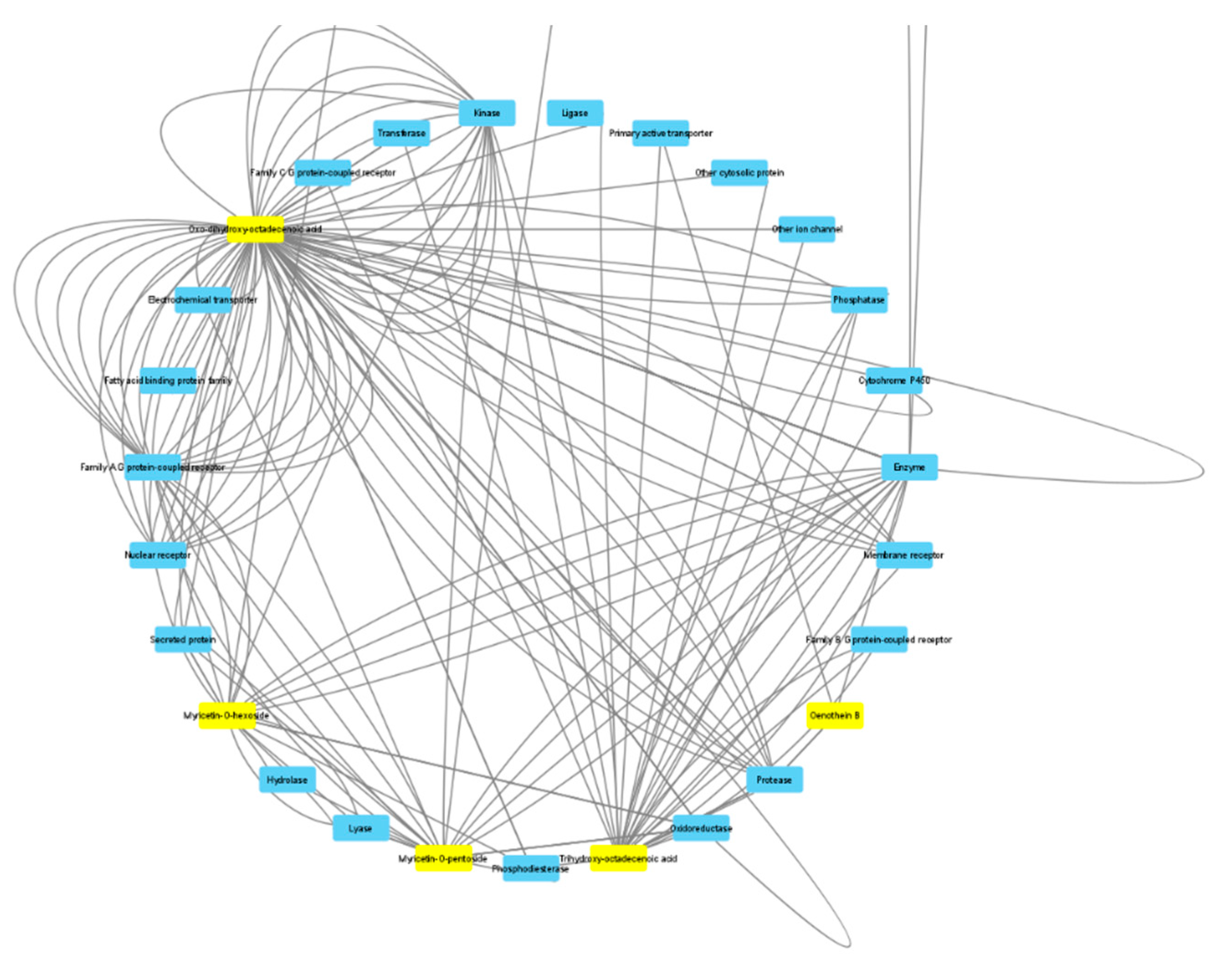
3.2. HPLC-ES-MSn Analysis

The characterization of the phytochemicals was conducted with HPLC-ESI-MSn using the negative ion mode. Identification was performed using analytical standards—citric acid, coumaric acid, ferulic acid, gallic acid, kaempferol, and quercetin—as well as bibliographic information. As an example, the base peak chromatogram of the methanolic extract of aerial parts is shown in Figure S1 (Supplementary Material). The characterization of the compounds in all of the extracts is shown in Table 2. Compounds were numbered according to their elution order, with the same numbering being maintained in all extracts.
Compound 1 was tentatively characterized as a disaccharide (HCl adduct) due to the neutral loss of 162 Da (341179) and the characteristic fragments of hexoside moieties (m/z 179, 161, 119, 113 and 101) [41]. The most plausible structure is a diglucoside.
Compound 2, with deprotonated molecular ion at m/z 195, exhibited the base peak at m/z 129 and other fragment ions at m/z 177 and 159. This fragmentation has been previously reported for gluconic acid in E. angustifolium L. extracts [16].
Compounds 3 and 5 exhibited the same fragmentation pattern, corresponding to (iso)citric acid. The distinction between both isomers was performed by analyzing the analytical standard of citric acid.
Compounds 4 and 11 presented the same molecular ion and similar fragmentation patterns. This fragmentation pattern is consistent with oenothein B, a dimeric ellagitannin that has been reported in E. angustifolium [23].
Compound 7 was identified as gallic acid through comparison with the analytical standard. In addition, several derivatives were also characterized. Compounds 6 and 8 were characterized as digallylglucose isomers, which have been previously reported in E. augustifolium [25]. With additional 152 Da (gallic acid), compounds 15 and 16 were characterized as hexahydroxydiphenoyl-digalloyl-glucose and trigalloylglucose, respectively, as reported in Epilobium species [42].
Compound 10, with [M-H] at m/z 311, showed tartaric and caffeic acids at m/z 149 and 179, respectively. It was identified as caftaric acid [43]. To our knowledge, this compound has not been previously reported in Epilobium species.
Compound 13 suffered a neutral loss of 132 Da (pentoside) to yield ferulic acid at m/z 193, so it was identified as a ferulic acid pentoside.
Compound 14 was tentatively characterized as roseoside (vomifoliolglucoside or drovomifoliol-O-β-D-glucopyranoside) based on bibliographic information [44]. This compound has not been previously reported in Epilobium species.
Compound 26 was identified as myricetin due to the [M-H] at m/z 317 and fragment ions at m/z 179 and 151. Three myricetin glycosides (compounds 17, 18, and 19) were characterized based on the neutral losses of 132 Da (pentoside), 146 Da (deoxyhexoside), and 162 Da (hexoside). Similarly, compounds 20, 22, 23, and 24 were characterized as quercetin glycosides (quercetin aglycone at m/z 301), and compounds 21, 25, and 27 were characterized as kaempferol glycosides (kaempferol aglycone at m/z 285). The presence of myricetin, kaempferol, and quercetin glycosides has been previously reported in Epilobium species [24].
Compounds 28 and 29 were tentatively characterized as saponins due to the similar fragmentation pattern reported for these compounds in other plant species [45].
Compounds 31 and 33 were characterized as the oxylipins oxo-dihydroxy-octadecenoic acid and trihydroxy-octadecenoic acid based on bibliographic information [46].

3.3. Quantification of Phytochemicals

We calculated the relative contribution of all of the compounds using the area normalization method (Table 3 and Table 4). Peak areas of each compound were obtained using the precursor ion [M-H] (Extracted Ion Chromatograms). Then, the relative contribution (in percentage) of each compound was calculated, and the heat map (the darker the color, the higher the abundance) was constructed. There were some differences between the extracts of the aerial parts and roots. The main compounds found in the extracts of the aerial parts were compound 11 (Oenothein B)—except in the EA extract—and compounds 17, 18, and 19 (myricetin glycosides). On the other hand, oenothein B was prominent in all of the root extracts, except in the EA extract, where oxylipins (compounds 31 and 33) were the main contributors to the extract composition.
In the aerial parts, flavonoids accounted for 60–70% of all compounds (only 40% in the infusion extract); most of them were myricetin and its glycosides. However, in the roots, the contribution of flavonoids was very scarce, and oenothein B represented between 43% and 80% of all compounds, except in the EA, where oxylipins accounted for more than 50% of the detected compounds.
The production of phytochemicals in the plant metabolism is a dynamic process [47,48] and depends on several factors, including the vegetative period, collection year and time, and geographic locations [49,50,51,52]. Several studies have shown that both total and individual phenolic compounds have even changed in the same species due to geographical factors [37,39,53]. In addition to geographical factors, the amount of rainfall or drought between years can significantly influence the phytochemical composition of plant extracts [54]. For example, several researchers have reported that the production of secondary metabolites (especially the amount of phenolics) has increased against drought stress [55,56]. The increased content of phenolics can help to inhibit the oxidation of cells under stress conditions [57]. In this sense, the differences in E. hirsutum observed in previous studies could be explained by the factors mentioned above.

3.4. Antioxidant Properties

The evaluation of the antioxidant abilities of plant extracts could be an important signpost on the way from nature to the pharmacy shelf. In this sense, we investigated the antioxidant properties of the E. hirsutum extracts using different chemical approaches, including the quenching of free radicals, reducing power, and the chelation of transition metals. The results are presented in Table 5. DPPH and ABTS radicals are widely used to assess the quenching of radicals by plant extracts. In the tested aerial part extracts, the greatest radical scavenging ability was observed in the methanol extract (798.89 mg TE/g for DPPH and 1382.08 mg TE/g for ABTS). However, in the root extracts, the most active extract was methanol/water (317.72 mg TE/g for DPPH and 552.33 mg TE/g for ABTS). In both the aerial parts and the roots, the ethyl acetate extracts showed the weakest abilities on these radicals. Reduction power assays examine the transformation of Cu2+ to Cu+ and Fe3+ to Fe2+ in CUPRAC and FRAP assays, respectively. In these assays, the infusions obtained from both the aerial parts (1819.57 mg TE/g for CUPRAC and 756.63 mg TE/g for FRAP) and the roots (1065.96 mg TE/g for CUPRAC and 424.87 mg TE/g for FRAP) demonstrated the best abilities. The lowest abilities in both parts were also recorded in the ethyl acetate extracts. The phosphomolybdenum reaction involves the conversion of Mo (VI) to Mo (V) by the antioxidant compounds in the acidic pH. The assay has gained interest in recent studies because it is simple and does not need specific equipment. In the extracts from the aerial parts, the superior ability was demonstrated by methanol extract, with a value of 6.80 mmol TE/g followed by the infusion (4.72 mmol TE/g), methanol/water (4.25 mmol TE/g), and ethyl acetate (3.11 mmol TE/g) extracts. As for the root extracts, the methanol, infusion, and methanol/water extracts had similar abilities (p > 0.05). The chelation of transition metals is considered to be one of the most important antioxidant mechanisms that is able to stop the production of hydroxyl radicals in the Fenton reaction. In the present study, among the tested extracts, the methanol extract of the aerial parts had the strongest chelating ability, with a value of 63.31 mg EDTAE/g followed by infusion of the aerial parts (58.19 mg EDTAE) and the roots (45.22 mg EDTAE/g). When all of the antioxidant results were combined, the results were correlated with the chemical components of the tested extracts. For example, the methanol, methanol/water, and infusion extracts contained higher amounts of phenols and were also most active in the antioxidant assays. As a further attraction, these extracts were rich in oenothein B and myricetin, which are known to be effective antioxidants [58,59]. For example, oenothein B is a unique class of ellagitannis, and its conformational structure with multiple OH- groups has exhibited remarkable biological activities including antioxidant, anticancer, and immunomodulatory effects [60]. Taken together, the observed antioxidant abilities of the tested extracts can be attributed to the presence of these components. In addition, several authors have examined the antioxidant capacities of the members of the genus Epilobium, including E. hirsutum [11,13,21,24,58], and in accordance with our presented study, significant antioxidant effects were observed with higher concentration of phenolics, particularly ellagic acid, oenothein B and its derivatives, and myricetin.

3.5. Enzyme Inhibitory Properties

In recent studies, the prevalence of non-infectious global diseases is increasing day by day, and urgent precautions are needed to control the prevalence of these diseases. In this context, researchers are seeking to find alternative and effective treatment strategies for managing these diseases. With the growing human population, synthetic compounds are still key players in these strategies [61]. However, since most of them have toxic properties, alternative compounds, especially those from natural sources, are gaining a lot of interest in place of these synthetic ones. Among them, natural enzyme inhibitors play an important role in the treatment of the above-mentioned diseases. These natural compounds could inhibit the actions of enzymes associated with different chronic inflammatory and degenerative diseases [62,63,64,65,66]. Therefore, natural extracts and compounds are often tested against enzymes such as cholinesterases, α-amylase, α-glucosidase, lipase, and tyrosinase as pharmaceutical targets.
In this regard, E. hirsutum extracts were tested against cholinesterases, amylase, glucosidase, and tyrosinase, which are deeply involved in Alzheimer’s disease, type-2 disabetes, and hyperpigmentation, respectively. The results are tabulated in Table 6. In AChE inhibition, the methanol extract of the aerial parts had the best inhibitory ability with a value of 4.48 mg GALAE/g followed by methanol/water (2.76 mg GALAE/g) and ethyl acetate (2.69 mg GALAE/g). The infusion of the aerial parts was not active on AChE. In the root extracts, the ability was in the following order: ethyl acetate > methanol > methanol/water > infusion. Taking into account the BChE inhibition, the strongest abilities were shown by the ethyl acetate extracts (4.72 mg GALAE/g for the aerial parts and 5.18 mg GALAE/g for the roots) in both parts, whereas the methanol extracts exhibited the best inhibitory actions against tyrosinase, with the values of 106.68 mg KAE/g (in aerial parts) and 98.31 mg KAE/g (in roots). However, the methanol extract of the aerial parts statistically had a similar efficacy compared to the methanol/water extract (p > 0.05). As for the α-amylase inhibitory effect, the methanol extracts from both parts displayed superior inhibitory potential, while the lowest enzyme inhibitory effects were obtained from the infusions. Interestingly, different results were observed in the α-glucosidase inhibitory assay. The most potent ability was detected in the infusion of the roots, with a value of 1.96 mmol ACAE/g. However, the methanol/water and infusion from aerial parts were not active on α-glucosidase. Overall, the methanol extracts from the tested parts were more active than other extracts. The observed findings could be explained by the presence of chemical components including oenothein B and myricetin. Our observations are consistent with previous studies that reported the significant inhibitory effects of these compounds [67,68,69,70,71]. In addition, remarkable enzyme inhibitory effects have been demonstrated for other plants belonging to the genus [11,21,58]. However, a comparison with our results is difficult because of different methods of expression, including the IC50 values or the percentage of inhibition.

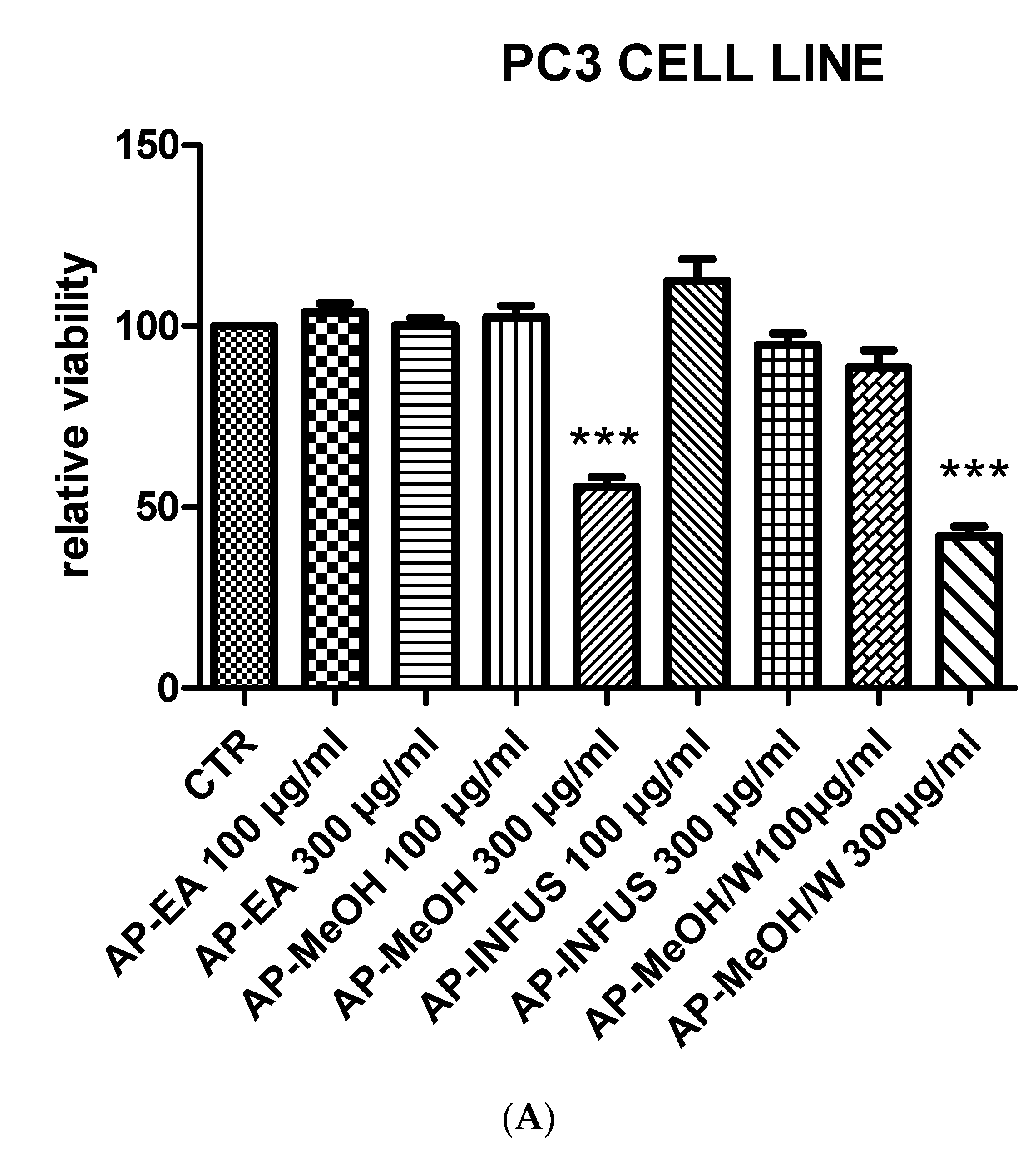
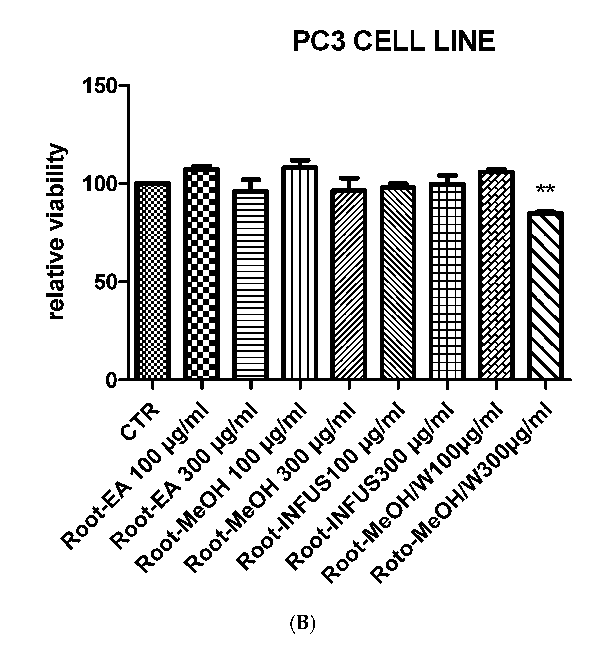
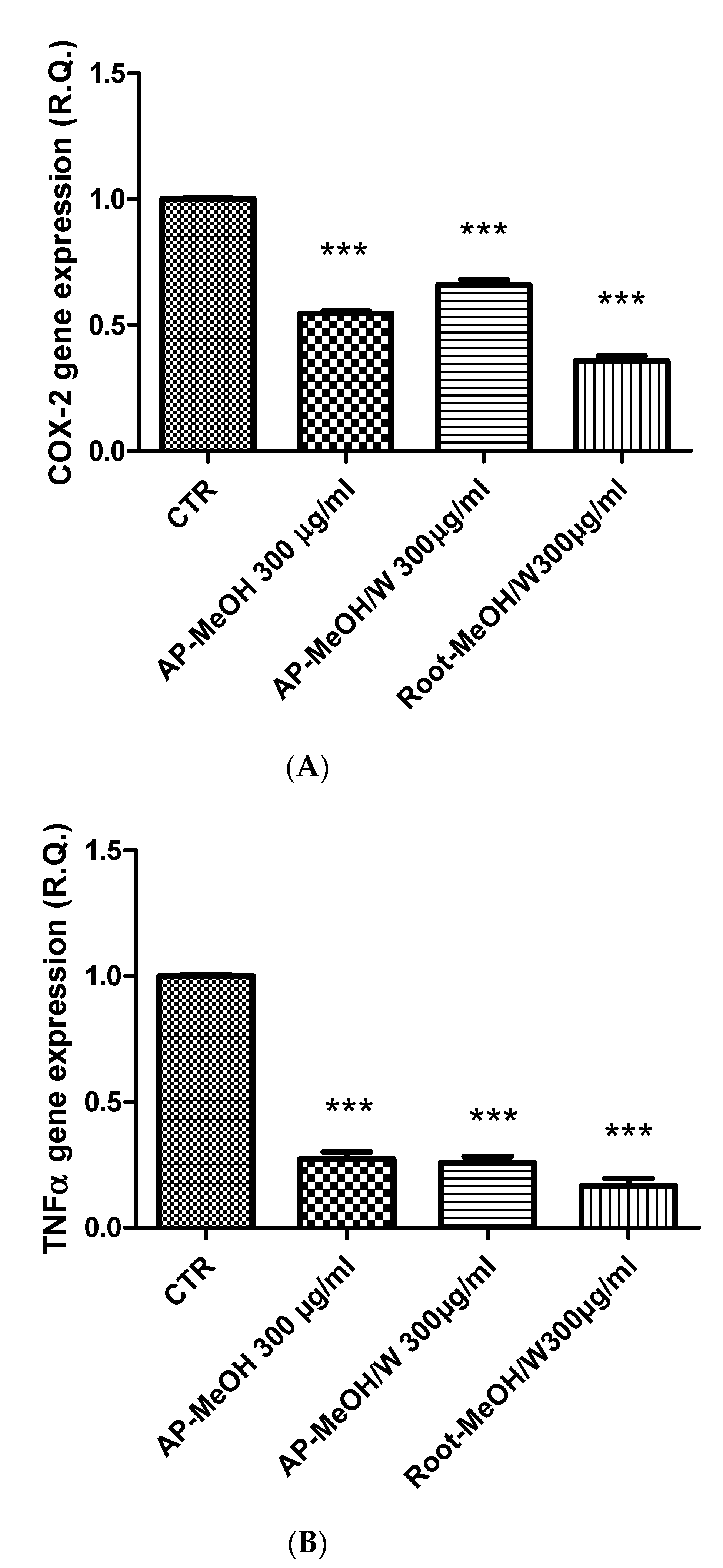
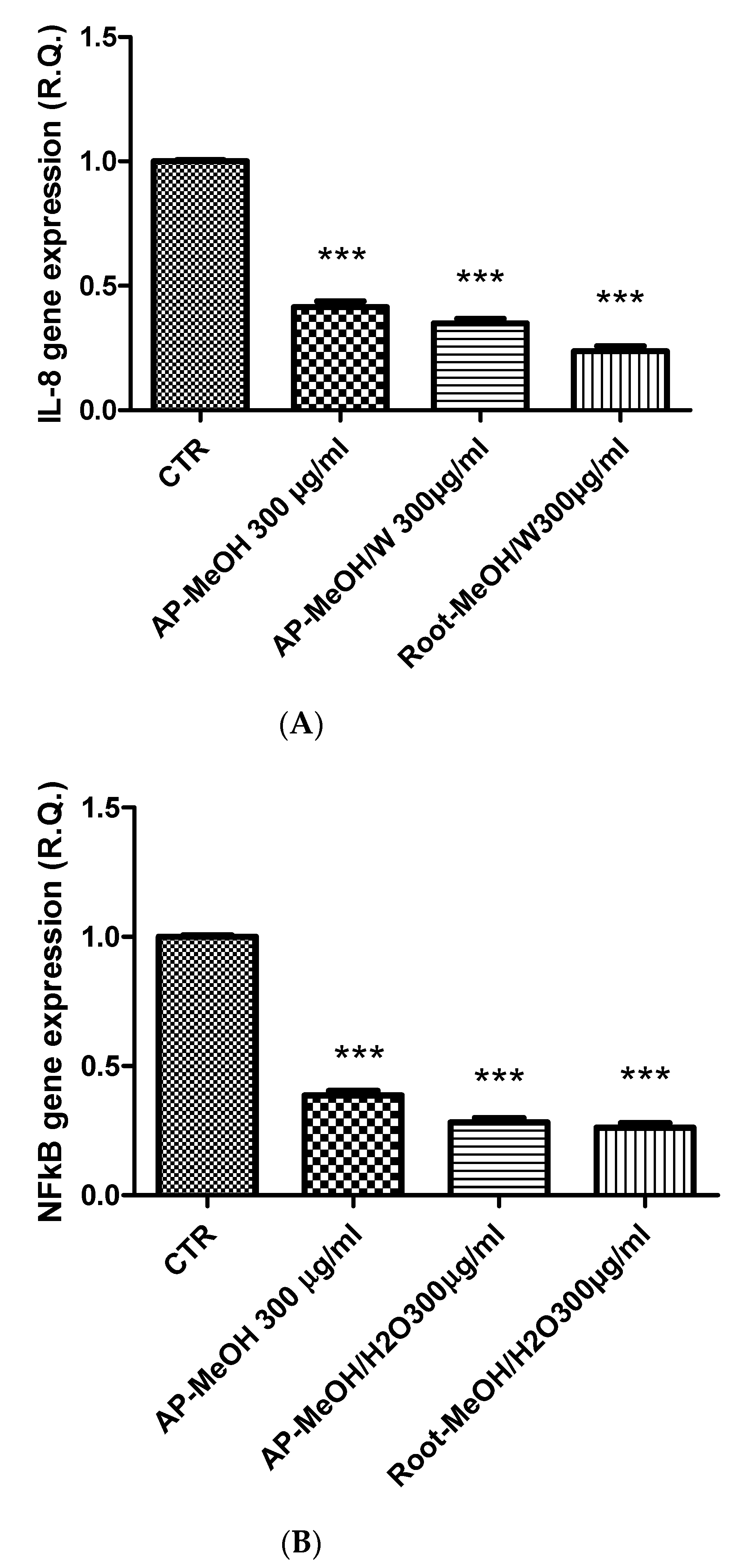
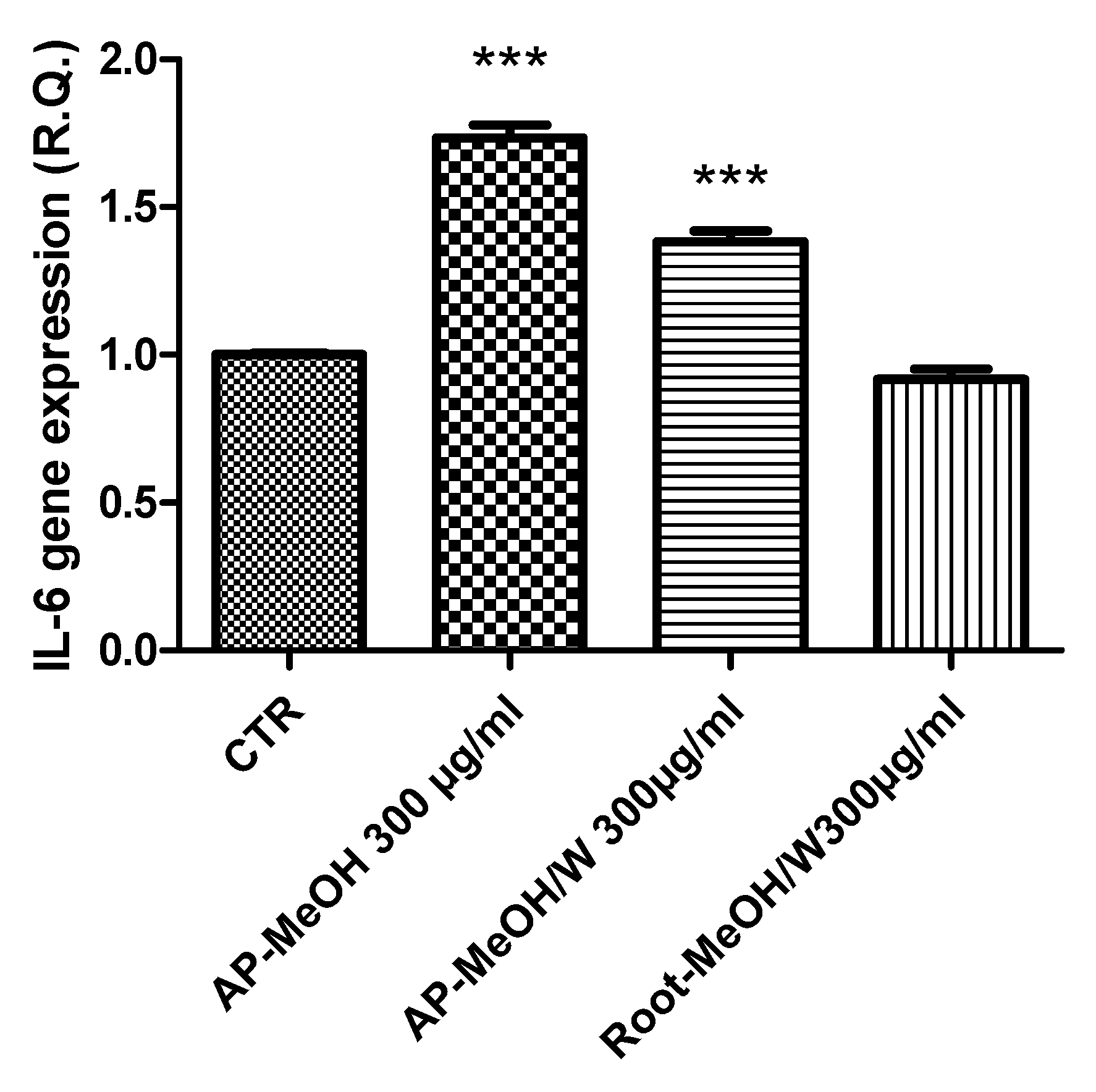
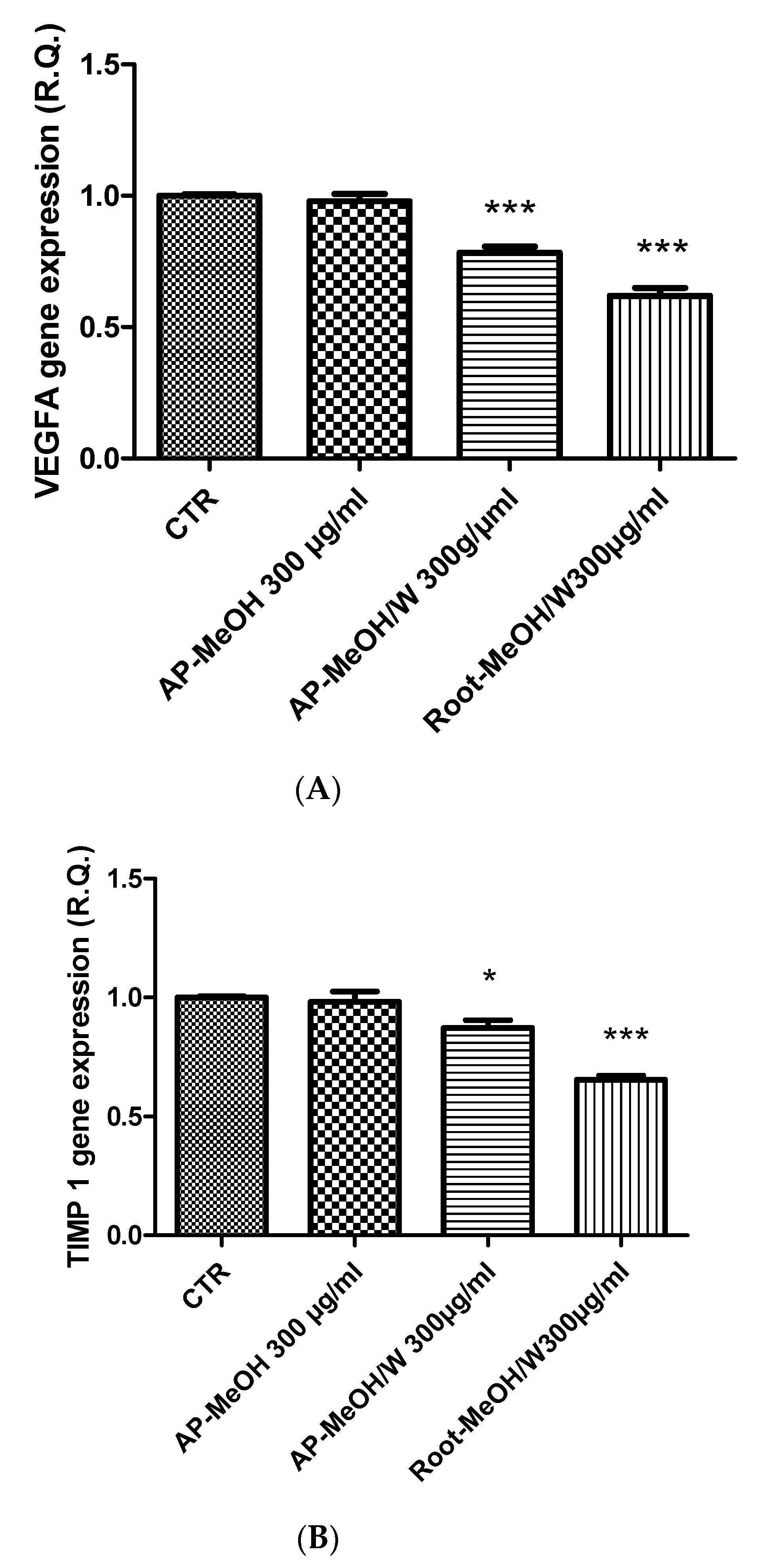
3.6. Toxicological and Pharmacological Evaluation

The Artemia salina (brine shrimp) lethality test was performed to define the biocompatibility limits of the present extracts. This test is widely used as an alternative toxicological model to predict the toxicity limits in eukaryotic cells [72]. In this regard, shrimp nauplii were exposed to the extracts for 24 h (0.1–20 mg/mL), showing LC50 values > 3 mg/mL. Considering these results, a concentration that was at least ten-fold lower was chosen for the pharmacological assays conducted on the human prostate cancer PC3 cell line, which was exposed to the extracts (100–300 µg/mL) to evaluate antiproliferative effects. This is consistent with the study by Stolarczyk and colleagues [42] that showed antiproliferative effects induced by E. hirsutum in hormone-dependent prostate cancer cells (LNCaP). Furthermore, in agreement with our recent finding of antiproliferative effect induced by the water extract of E. angustifolium (250 µg/mL) on PC3 cells [73], we observed the reduction of PC3 viability (Figure 1) occurring at the highest tested concentration (300 µg/mL) of methanol and methanol/water extracts of roots and aerial parts of E. hirsutum. Despite having a lower potency, the antiproliferative effect induced by this extract concentration in PC3 cells was also similar to that induced by daunorubicin 3.12 µg/mL [74]. By contrast, the water (infusion) and ethyl acetate extracts of the analyzed plant materials were completely ineffective in reducing the viability of the prostate cancer cells. The differences between the E. angustifolium and E. hirsutum extracts in inducing antiproliferative effects on the same tumoral cell line could be related, albeit partially, to the different pattern of secondary metabolites measured via HPLC analysis. If the E. angustifolium water extract was particularly rich in catechins, these flavonoids were not detected in the E. hirsutum extracts. On the other hand, the root and aerial part extracts were particularly rich in oenothein B, myricetins, oxo-dihydroxy-octadecenoic acid, and trihydroxy-octadecenoic acid. Therefore, these phytochemicals were selected for a bioinformatics analysis with the aim of predicting the protein targets underlying the antiproliferative effects. As shown in Figure 2, oenothein B, myricetins, oxo-dihydroxy-octadecenoic acid, and trihydroxy-octadecenoic acid were able to interact with a wide number of proteins that are also involved in oxidative stress and inflammation, among which are oxidoreductases and secreted proteins. In detail, myricetins were predicted to interact with cycloxygenase-2 (COX-2) and different cytokines, among which tumor necrosis factor α (TNFα) is present. The interaction between TNFα and oxo-dihydroxy-octadecenoic acid was predicted as well. Considering these results, we also evaluated the gene expression of COX-2 and TNFα in PC3 cells after exposure to the methanol and methanol/water extracts of aerial parts and the methanol/water root extract, finding a significant inhibition of the gene expression of both COX-2 and TNFα (Figure 3A,B). These effects were paralleled by the decreased gene expression of interleukin-8 (IL-8) and NFkB (Figure 4A,B) in the same experimental condition, whereas the inhibition of prostaglandin E2 (PGE2) release (Figure 5) by PC3 cells supports the reduction of COX-2 gene expression as one of the anti-inflammatory mechanisms exerted by the present extracts. However, all of the E. hirsutum extracts were able to increase the gene expression of IL-6 (Figure 6), and this is apparently in contrast with the aforementioned anti-inflammatory properties. However, Eskandari et al. [75] showed that the inhibition of PC3 cell viability induced by ellagic acid was mirrored by the concomitant increase of IL-6 gene expression and release. This is also consistent with the dual role of IL-6, which can exert both pro-inflammatory and anti-inflammatory effects, depending upon tissue and experimental condition [76]. Finally, considering the strict relationships between cancer and inflammation, especially in prostate cancer [77], we also measured the gene expression of TIMP-1 and VEGFA, which are deeply involved in inflammation and cancer [78]. Specifically, TIMP-1 and VEGFA are reported to influence angiogenesis in opposite ways, with inhibitory and stimulatory effects, respectively [78]. The tested extracts were effective in inhibiting the gene expression of VEGFA and TIMP-1 (Figure 7A,B) as well. Regarding the inhibition of VEGFA gene expression, the present results agree with the literature data, strongly indicating the role of this protein as angiogenetic and tumor prognostic factor [79]. On the other hand, the reduction of TIMP1 gene expression is apparently in contrast with the antiproliferative effect on PC3 cells. TIMP1 is known to inhibit angiogenesis in cancer [78]. However, it is also true that TIMP1 gene expression is induced by pro-inflammatory cytokines [80]. In this context, we cannot exclude that the reduction of TIMP1 gene expression following extract treatment could be related, albeit partially, to the inhibition of the gene expression of the cytokines tested in the present experimental paradigm. Intriguingly, the the bioinformatics platform STRINGH showed functional interactions between all of the assayed proteins (Figure 8A), whereas the KEGG analysis indicated the probable (p < 0.05) modulation of different pathways, involved in cancer and inflammation, in the PC3 cells exposed to the extracts (Figure 8B). Overall, the present findings demonstrate the anti-inflammatory and antiproliferative effects of E. hirsutum against human prostate cancer cells, which deserve further investigations with independent experimental models, including in vivo studies.

4. Conclusions

The chemical profile and biological properties of various extracts of E. hirsutum indicated that the plant possessed unique properties. Based on the HPLC-ESI-MSn results, the extracts were rich in phenolic compounds, especially in oenothein B and myricetin derivatives. In particular, the methanol extract of the aerial parts and roots had the greatest antioxidant potentials in the assays that were conducted. However, different results were obtained for each assay with respect to enzyme inhibitory abilities. In the PC3 cancer assay, the methanol and methanol/water extracts of both parts exhibited good reduction on the growth of cancer cells. The pattern of gene expression following PC3 cell exposure to the present extracts also pointed to anti-inflammatory effects that could explain, albeit partially, the antiproliferative effects. In this context, the present findings further strengthen the potential use of plants belonging to the genus Epilobium for the management of the burden of inflammation and oxidative stress occurring in lower urinary tract diseases, among which prostatitis is included [73,81]. The results obtained here can have an impact on understanding the rational phytotherapy use of E. hirsutum.

Supplementary Materials

The following are available online: https://www.mdpi.com/article/10.3390/antiox10091389/s1. Figure S1: Base peak chromatogram of the methanol extract of the aerial parts of Epilobium hirsutum.

Author Contributions

Conceptualization, G.O., C.F., L.M., G.Z. and L.B.; methodology, C.F. and L.M.; software, L.M. and C.F.; validation, C.F., L.M. and G.O.; formal analysis, C.F.; G.Z.; investigation, G.A., E.L.-M., A.C., S.C.D.S., L.R. and S.L.; resources, G.O., L.M. and C.F.; data curation, C.F., G.O. and L.M.; writing—original draft preparation, C.F., G.Z. and M.F.M.; writing—review and editing, C.F., G.O., L.M. and G.Z.; visualization, L.B.; supervision, L.B.; project administration, L.M., G.O. and C.F.; funding acquisition, G.O., C.F. and L.M. All authors have read and agreed to the published version of the manuscript.

Funding

This study was supported with funds from Cristalfarma S.r.l. within the joint project entitled “Il Giardino dei Semplici e lo studio della biodiversità vegetale come modello per la valorizzazione delle filiere sostenibili”. The coordinators of the study are Luigi Menghini, Giustino Orlando, and Claudio Ferrante.

Institutional Review Board Statement

Not applicable.

Informed Consent Statement

Not applicable.

Data Availability Statement

All data is contained within the article and Supplementary Materials.

Acknowledgments

The present article is part of the third mission activities of the Botanic Garden “Giardino dei Semplici” planned for the 20th anniversary of the establishment. Technical and human support provided by CICT of Universidad de Jaén (UJA, MINECO, Junta de Andalucía, FEDER) is gratefully acknowledged.

Conflicts of Interest

The authors declare no conflict of interest.

References

  1. Elkordy, A.A.; Haj-Ahmad, R.R.; Awaad, A.S.; Zaki, R.M. An overview on natural product drug formulations from conventional medicines to nanomedicines: Past, present and future. J. Drug Deliv. Sci. Technol. 2021, 63, 102459. [Google Scholar] [CrossRef]
  2. Pisoschi, A.M.; Pop, A.; Iordache, F.; Stanca, L.; Predoi, G.; Serban, A.I. Oxidative stress mitigation by antioxidants-an overview on their chemistry and influences on health status. Eur. J. Med. Chem. 2020, 209, 112891. [Google Scholar] [CrossRef]
  3. Sudhakar, K.; Mishra, V.; Hemani, V.; Verma, A.; Jain, A.; Jain, S.; Charyulu, R.N. Reverse pharmacology of phytoconstituents of food and plant in the management of diabetes: Current status and perspectives. Trends Food Sci. Technol. 2021, 110, 594–610. [Google Scholar] [CrossRef]
  4. Aumeeruddy, M.Z.; Mahomoodally, M.F. Global documentation of traditionally used medicinal plants in cancer management: A systematic review. S. Afr. J. Bot. 2021, 138, 424–494. [Google Scholar] [CrossRef]
  5. Fakhri, S.; Moradi, S.Z.; Farzaei, M.H.; Bishayee, A. Modulation of dysregulated cancer metabolism by plant secondary metabolites: A mechanistic review. Semin. Cancer Biol. 2020, in press. [Google Scholar] [CrossRef] [PubMed]
  6. Granica, S.; Piwowarski, J.; Czerwińska, M.; Kiss, A. Phytochemistry, pharmacology and traditional uses of different Epilobium species (Onagraceae): A review. J. Ethnopharmacol. 2014, 156, 316–346. [Google Scholar] [CrossRef]
  7. Dzhafar, S.S.; Dalar, A.; Mükemre, M.; Suat, E.; Yildiz, D.; Yunusoğlu, O. Phytochemical Profile and in vitro and in vivo Anticonvulsant and Antioxidant Activities of Epilobium hirsutum. Int. J. Second. Metab. 2020, 7, 63–76. [Google Scholar] [CrossRef]
  8. Korkmaz, M.; Karakuş, S.; Özçelik, H.; Selvi, S. An ethnobotanical study on medicinal plants in Erzincan, Turkey. Indian J. Tradit. Knowl. 2016, 15, 192–202. [Google Scholar]
  9. Şahin, Y.E.; Erbay, M.Ş.; Sezin, A.; Kantar, R.; Kültür, Ş.; Melikoğlu, G. Plants used in traditional treatment of prostate diseases in Turkey. İstanbul J. Pharm. 2019, 49, 191–203. [Google Scholar]
  10. Kawarty, A.M.A.; Behçet, L.; Çakilcioğlu, U. An ethnobotanical survey of medicinal plants in Ballakayati (Erbil, North Iraq). Turk. J. Bot. 2020, 44, 345–357. [Google Scholar] [CrossRef]
  11. Karakaya, S.; Süntar, I.; Yakinci, O.F.; Sytar, O.; Ceribasi, S.; Dursunoglu, B.; Ozbek, H.; Guvenalp, Z. In vivo bioactivity assessment on Epilobium species: A particular focus on Epilobium angustifolium and its components on enzymes connected with the healing process. J. Ethnopharmacol. 2020, 262, 113207. [Google Scholar] [CrossRef]
  12. Ege, T.; Gençler Ozkan, A.; Sen, A.; Adalı, O. Effects of folk medicinal plant epilobium hirsutum l. And its ingredient ellagic acid on rat liver bile acid synthesizing cyps in rats. Pharmacol. Online 2018, 3, 200–215. [Google Scholar]
  13. Sheikh, N.A.; Desai, T.R.; Tirgar, P.R. Evaluation of iron chelating and antioxidant potential of Epilobium hirsutum for the management of iron overload disease. Biomed. Pharmacother. 2017, 89, 1353–1361. [Google Scholar] [CrossRef]
  14. Sõukand, R.; Mattalia, G.; Kolosova, V.; Stryamets, N.; Prakofjewa, J.; Belichenko, O.; Kuznetsova, N.; Minuzzi, S.; Keedus, L.; Prūse, B.; et al. Inventing a herbal tradition: The complex roots of the current popularity of Epilobium angustifolium in Eastern Europe. J. Ethnopharmacol. 2020, 247, 112254. [Google Scholar] [CrossRef]
  15. Kalle, R.; Belichenko, O.; Kuznetsova, N.; Kolosova, V.; Prakofjewa, J.; Stryamets, N.; Mattalia, G.; Šarka, P.; Simanova, A.; Prūse, B. Gaining momentum: Popularization of Epilobium angustifolium as food and recreational tea on the Eastern edge of Europe. Appetite 2020, 150, 104638. [Google Scholar] [CrossRef] [PubMed]
  16. Dacrema, M.; Sommella, E.; Santarcangelo, C.; Bruno, B.; Marano, M.G.; Insolia, V.; Saviano, A.; Campiglia, P.; Stornaiuolo, M.; Daglia, M. Metabolic profiling, in vitro bioaccessibility and in vivo bioavailability of a commercial bioactive Epilobium angustifolium L. extract. Biomed. Pharmacother. 2020, 131, 110670. [Google Scholar] [CrossRef] [PubMed]
  17. Vitali, F.; Fonte, G.; Saija, A.; Tita, B. Inhibition of intestinal motility and secretion by extracts of Epilobium spp. in mice. J. Ethnopharmacol. 2006, 107, 342–348. [Google Scholar] [CrossRef]
  18. Bajer, T.; Šilha, D.; Ventura, K.; Bajerová, P. Composition and antimicrobial activity of the essential oil, distilled aromatic water and herbal infusion from Epilobium parviflorum Schreb. Ind. Crop. Prod. 2017, 100, 95–105. [Google Scholar] [CrossRef]
  19. Wojdyło, A.; Oszmiański, J.; Czemerys, R. Antioxidant activity and phenolic compounds in 32 selected herbs. Food Chem. 2007, 105, 940–949. [Google Scholar] [CrossRef]
  20. Egil, A.C.; Ozdemir, B.; Gok, B.; Kecel-Gunduz, S.; Budama-Kilinc, Y. Synthesis, characterization, biological activities and molecular docking of Epilobium parviflorum aqueous extract loaded chitosan nanoparticles. Int. J. Biol. Macromol. 2020, 161, 947–957. [Google Scholar] [CrossRef] [PubMed]
  21. Szwajgier, D.; Baranowska-Wójcik, E.; Kukula-Koch, W.; Kowalik, K.; Polak-Berecka, M.; Waśko, A. Evolution of the anticholinesterase, antioxidant, and anti-inflammatory activity of Epilobium angustifolium L. infusion during in vitro digestion. J. Funct. Foods 2021, 85, 104645. [Google Scholar] [CrossRef]
  22. Esposito, C.; Santarcangelo, C.; Masselli, R.; Buonomo, G.; Nicotra, G.; Insolia, V.; D’Avino, M.; Caruso, G.; Buonomo, A.R.; Sacchi, R.; et al. Epilobium angustifolium L. extract with high content in oenothein B on benign prostatic hyperplasia: A monocentric, randomized, double-blind, placebo-controlled clinical trial. Biomed. Pharmacother. 2021, 138, 111414. [Google Scholar] [CrossRef] [PubMed]
  23. Baert, N.; Karonen, M.; Salminen, J.P. Isolation, characterisation and quantification of the main oligomeric macrocyclic ellagitannins in Epilobium angustifolium by ultra-high performance chromatography with diode array detection and electrospray tandem mass spectrometry. J. Chromatogr. A 2015, 1419, 26–36. [Google Scholar] [CrossRef] [PubMed]
  24. Hevesi Tóth, B.; Blazics, B.; Kéry, A. Polyphenol composition and antioxidant capacity of Epilobium species. J. Pharm Biomed. Anal. 2009, 49, 26–31. [Google Scholar] [CrossRef] [PubMed]
  25. Agnieszka, G.; Mariola, D.; Anna, P.; Piotr, K.; Natalia, W.; Aneta, S.; Marcin, O.; Bogna, O.; Zdzisław, Ł.; Aurelia, P.; et al. Qualitative and quantitative analyses of bioactive compounds from ex vitro Chamaenerion angustifolium (L.) (Epilobium augustifolium) herb in different harvest times. Ind. Crop. Prod. 2018, 123, 208–220. [Google Scholar] [CrossRef]
  26. Barakat, H.H.; Hussein, S.A.M.; Marzouk, M.S.; Merfort, I.; Linscheid, M.; Nawwar, M.A.M. Polyphenolic metabolites of Epilobium hirsutum. Phytochemistry 1997, 46, 935–941. [Google Scholar] [CrossRef]
  27. Bazylko, A.; Kiss, A.K.; Kowalski, J. High-performance thin-layer chromatography method for quantitative determination of oenothein B and quercetin glucuronide in aqueous extract of Epilobii angustifolii herba. J. Chromatogr. A 2007, 1173, 146–150. [Google Scholar] [CrossRef]
  28. Slinkard, K.; Singleton, V.L. Total phenol analysis: Automation and comparison with manual methods. Am. J. Enol. Vitic. 1977, 28, 49–55. [Google Scholar]
  29. Zengin, G.; Nithiyanantham, S.; Locatelli, M.; Ceylan, R.; Uysal, S.; Aktumsek, A.; Selvi, P.K.; Maskovic, P. Screening of in vitro antioxidant and enzyme inhibitory activities of different extracts from two uninvestigated wild plants: Centranthus longiflorus subsp. longiflorus and Cerinthe minor subsp. auriculata. Eur. J. Integr. Med. 2016, 8, 286–292. [Google Scholar] [CrossRef]
  30. Fernández-Poyatos, M.D.P.; Ruiz-Medina, A.; Zengin, G.; Llorent-Martínez, E.J. Phenolic Characterization, Antioxidant Activity, and Enzyme Inhibitory Properties of Berberis thunbergii DC. Leaves: A Valuable Source of Phenolic Acids. Molecules 2019, 24, 4171. [Google Scholar] [CrossRef] [Green Version]
  31. Uysal, S.; Zengin, G.; Locatelli, M.; Bahadori, M.B.; Mocan, A.; Bellagamba, G.; De Luca, E.; Mollica, A.; Aktumsek, A. Cytotoxic and enzyme inhibitory potential of two Potentilla species (P. speciosa L. and P. reptans Willd.) and their chemical composition. Front. Pharmacol. 2017, 8, 290. [Google Scholar] [CrossRef]
  32. Ferrante, C.; Recinella, L.; Ronci, M.; Menghini, L.; Brunetti, L.; Chiavaroli, A.; Leone, S.; Di Iorio, L.; Carradori, S.; Tirillini, B.; et al. Multiple pharmacognostic characterization on hemp commercial cultivars: Focus on inflorescence water extract activity. Food Chem. Toxicol. 2019, 125, 452–461. [Google Scholar] [CrossRef]
  33. Menghini, L.; Ferrante, C.; Leporini, L.; Recinella, L.; Chiavaroli, A.; Leone, S.; Pintore, G.; Vacca, M.; Orlando, G.; Brunetti, L. An Hydroalcoholic Chamomile Extract Modulates Inflammatory and Immune Response in HT29 Cells and Isolated Rat Colon. Phytother. Res. 2016, 30, 1513–1518. [Google Scholar] [CrossRef]
  34. Orlando, G.; Leone, S.; Ferrante, C.; Chiavaroli, A.; Mollica, A.; Stefanucci, A.; Macedonio, G.; Dimmito, M.P.; Leporini, L.; Menghini, L.; et al. Effects of Kisspeptin-10 on Hypothalamic Neuropeptides and Neurotransmitters Involved in Appetite Control. Molecules 2018, 23, 3071. [Google Scholar] [CrossRef] [Green Version]
  35. Menghini, L.; Ferrante, C.; Leporini, L.; Recinella, L.; Chiavaroli, A.; Leone, S.; Pintore, G.; Vacca, M.; Orlando, G.; Brunetti, L. A natural formula containing lactoferrin, Equisetum arvensis, soy isoflavones and vitamin D3 modulates bone remodeling and inflammatory markers in young and aged rats. J. Biol. Regul. Homeost. Agents 2016, 30, 985–996. [Google Scholar] [PubMed]
  36. Pourmorad, F.; Ebrahimzadeh, M.A.; Mahmoudi, M.; Yasini, S. Antinociceptive activity of methanolic extract of Epilobium hirsutum. Pak. J. Biol. Sci. PJBS 2007, 10, 2764–2767. [Google Scholar]
  37. Herrera Alvarez, L.V.; Zielinski, A.A.F.; Alberti, A.; Nogueira, A. Monitoring of the phenolic compounds and in vitro antioxidant activity of apple beverages according to geographical origin and their type: A chemometric study. LWT 2017, 84, 385–393. [Google Scholar] [CrossRef]
  38. Dittgen, C.L.; Hoffmann, J.F.; Chaves, F.C.; Rombaldi, C.V.; Filho, J.M.C.; Vanier, N.L. Discrimination of genotype and geographical origin of black rice grown in Brazil by LC-MS analysis of phenolics. Food Chem. 2019, 288, 297–305. [Google Scholar] [CrossRef] [PubMed]
  39. Fan, G.; Beta, T. Discrimination of geographical origin of Napirira bean (Phaseolus vulgaris L.) based on phenolic profiles and antioxidant activity. J. Food Compos. Anal. 2017, 62, 217–222. [Google Scholar] [CrossRef]
  40. Sánchez-Rangel, J.C.; Benavides, J.; Heredia, J.B.; Cisneros-Zevallos, L.; Jacobo-Velázquez, D.A. The Folin–Ciocalteu assay revisited: Improvement of its specificity for total phenolic content determination. Anal. Methods 2013, 5, 5990–5999. [Google Scholar] [CrossRef]
  41. Verardo, G.; Duse, I.; Callea, A. Analysis of underivatized oligosaccharides by liquid chromatography/electrospray ionization tandem mass spectrometry with post-column addition of formic acid. Rapid Commun. Mass Spectrom. 2009, 23, 1607–1618. [Google Scholar] [CrossRef] [PubMed]
  42. Stolarczyk, M.; Naruszewicz, M.; Kiss, A.K. Extracts from Epilobium sp. herbs induce apoptosis in human hormone-dependent prostate cancer cells by activating the mitochondrial pathway. J. Pharm. Pharm. 2013, 65, 1044–1054. [Google Scholar] [CrossRef]
  43. Schütz, K.; Kammerer, D.R.; Carle, R.; Schieber, A. Characterization of phenolic acids and flavonoids in dandelion (Taraxacum officinale WEB. ex WIGG.) root and herb by high-performance liquid chromatography/electrospray ionization mass spectrometry. Rapid Commun. Mass Spectrom. 2005, 19, 179–186. [Google Scholar] [CrossRef] [PubMed]
  44. Spínola, V.; Llorent-Martínez, E.J.; Gouveia, S.; Castilho, P.C. Myrica faya: A New Source of Antioxidant Phytochemicals. J. Agric. Food Chem. 2014, 62, 9722–9735. [Google Scholar] [CrossRef] [PubMed]
  45. Gouveia-Figueira, S.C.; Castilho, P.C. Phenolic screening by HPLC–DAD–ESI/MSn and antioxidant capacity of leaves, flowers and berries of Rubus grandifolius Lowe. Ind. Crop. Prod. 2015, 73, 28–40. [Google Scholar] [CrossRef]
  46. Van Hoyweghen, L.; De Bosscher, K.; Haegeman, G.; Deforce, D.; Heyerick, A. In vitro inhibition of the transcription factor NF-κB and cyclooxygenase by bamboo extracts. Phytother. Res. 2014, 28, 224–230. [Google Scholar] [CrossRef]
  47. Berini, J.L.; Brockman, S.A.; Hegeman, A.D.; Reich, P.B.; Muthukrishnan, R.; Montgomery, R.A.; Forester, J.D. Combinations of abiotic factors differentially alter production of plant secondary metabolites in five woody plant species in the boreal-temperate transition zone. Front. Plant. Sci. 2018, 9, 1257. [Google Scholar] [CrossRef] [Green Version]
  48. Usenik, V. The influence of the production system on the composition of phytochemicals in Prunus domestica L. fruit. J. Food Compos. Anal. 2021, 95, 103701. [Google Scholar] [CrossRef]
  49. Farhadi, N.; Babaei, K.; Farsaraei, S.; Moghaddam, M.; Ghasemi Pirbalouti, A. Changes in essential oil compositions, total phenol, flavonoids and antioxidant capacity of Achillea millefolium at different growth stages. Ind. Crop. Prod. 2020, 152, 112570. [Google Scholar] [CrossRef]
  50. Sellami, I.H.; Maamouri, E.; Chahed, T.; Wannes, W.A.; Kchouk, M.E.; Marzouk, B. Effect of growth stage on the content and composition of the essential oil and phenolic fraction of sweet marjoram (Origanum majorana L.). Ind. Crop. Prod. 2009, 30, 395–402. [Google Scholar] [CrossRef]
  51. Ricardez-Miranda, L.E.; Lagunes-Espinoza, L.C.; Hernández-Nataren, E.; Palma-López, D.J.; Conde-Martínez, F.V. Water restriction during the vegetative and reproductive stages of Capsicum annuum var. glabriusculum, and its effect on growth, secondary metabolites and fruit yield. Sci. Hortic. 2021, 285, 110129. [Google Scholar] [CrossRef]
  52. Lorini, A.; Aranha, B.C.; Antunes, B.d.F.; Otero, D.M.; Jacques, A.C.; Zambiazi, R.C. Metabolic profile of olive leaves of different cultivars and collection times. Food Chem. 2021, 345, 128758. [Google Scholar] [CrossRef]
  53. Bouterfas, K.; Mehdadi, Z.; Elaoufi, M.M.; Latreche, A.; Benchiha, W. Antioxidant activity and total phenolic and flavonoids content variations of leaves extracts of white Horehound (Marrubium vulgare Linné) from three geographical origins. Ann. Pharm. Françaises 2016, 74, 453–462. [Google Scholar] [CrossRef] [PubMed]
  54. de Sousa Araújo, T.A.; de Almeida e Castro, V.T.N.; da Silva Solon, L.G.; da Silva, G.A.; das Graças Almeida, M.; da Costa, J.G.M.; de Amorim, E.L.C.; Albuquerque, U.P. Does rainfall affect the antioxidant capacity and production of phenolic compounds of an important medicinal species? Ind. Crop. Prod. 2015, 76, 550–556. [Google Scholar] [CrossRef]
  55. Mechri, B.; Tekaya, M.; Hammami, M.; Chehab, H. Effects of drought stress on phenolic accumulation in greenhouse-grown olive trees (Olea europaea). Biochem. Syst. Ecol. 2020, 92, 104112. [Google Scholar] [CrossRef]
  56. Yadav, B.; Jogawat, A.; Rahman, M.S.; Narayan, O.P. Secondary metabolites in the drought stress tolerance of crop plants: A review. Gene Rep. 2021, 23, 101040. [Google Scholar] [CrossRef]
  57. Yeloojeh, K.A.; Saeidi, G.; Sabzalian, M.R. Drought stress improves the composition of secondary metabolites in safflower flower at the expense of reduction in seed yield and oil content. Ind. Crop. Prod. 2020, 154, 112496. [Google Scholar] [CrossRef]
  58. Kiss, A.K.; Bazylko, A.; Filipek, A.; Granica, S.; Jaszewska, E.; Kiarszys, U.; Kośmider, A.; Piwowarski, J. Oenothein B’s contribution to the anti-inflammatory and antioxidant activity of Epilobium sp. Phytomedicine 2011, 18, 557–560. [Google Scholar] [CrossRef]
  59. Song, X.; Tan, L.; Wang, M.; Ren, C.; Guo, C.; Yang, B.; Ren, Y.; Cao, Z.; Li, Y.; Pei, J. Myricetin: A review of the most recent research. Biomed. Pharmacother. 2021, 134, 111017. [Google Scholar] [CrossRef]
  60. Yoshida, T.; Yoshimura, M.; Amakura, Y. Chemical and biological significance of oenothein B and related ellagitannin oligomers with macrocyclic structure. Molecules 2018, 23, 552. [Google Scholar] [CrossRef] [Green Version]
  61. WHO. World Health Statistics 2021: Monitoring Health for the SDGs, Sustainable Development Goals; World Health Organization: Geneva, Switzerland, 2021. [Google Scholar]
  62. Jawed, A.; Singh, G.; Kohli, S.; Sumera, A.; Haque, S.; Prasad, R.; Paul, D. Therapeutic role of lipases and lipase inhibitors derived from natural resources for remedies against metabolic disorders and lifestyle diseases. S. Afr. J. Bot. 2019, 120, 25–32. [Google Scholar] [CrossRef]
  63. Rathod, P.; Yadav, R.P. Anti-diabesity potential of various multifunctional natural molecules. J. Herb. Med. 2021, 27, 100430. [Google Scholar] [CrossRef]
  64. Sun, L.; Warren, F.J.; Gidley, M.J. Natural products for glycaemic control: Polyphenols as inhibitors of alpha-amylase. Trends Food Sci. Technol. 2019, 91, 262–273. [Google Scholar] [CrossRef]
  65. Seetaloo, A.D.; Aumeeruddy, M.Z.; Rengasamy Kannan, R.R.; Mahomoodally, M.F. Potential of traditionally consumed medicinal herbs, spices, and food plants to inhibit key digestive enzymes geared towards diabetes mellitus management—A systematic review. S. Afr. J. Bot. 2019, 120, 3–24. [Google Scholar] [CrossRef]
  66. Ahmed, S.; Khan, S.T.; Zargaham, M.K.; Khan, A.U.; Khan, S.; Hussain, A.; Uddin, J.; Khan, A.; Al-Harrasi, A. Potential therapeutic natural products against Alzheimer’s disease with Reference of Acetylcholinesterase. Biomed. Pharmacother. 2021, 139, 111609. [Google Scholar] [CrossRef]
  67. Kawakami, K.; Li, P.; Uraji, M.; Hatanaka, T.; Ito, H. Inhibitory Effects of Pomegranate Extracts on Recombinant Human Maltase–Glucoamylase. J. Food Sci. 2014, 79, H1848–H1853. [Google Scholar] [CrossRef]
  68. Khan, H.; Marya; Amin, S.; Kamal, M.A.; Patel, S. Flavonoids as acetylcholinesterase inhibitors: Current therapeutic standing and future prospects. Biomed. Pharmacother. 2018, 101, 860–870. [Google Scholar] [CrossRef] [PubMed]
  69. Wang, B.; Zhong, Y.; Gao, C.; Li, J. Myricetin ameliorates scopolamine-induced memory impairment in mice via inhibiting acetylcholinesterase and down-regulating brain iron. Biochem. Biophys. Res. Commun. 2017, 490, 336–342. [Google Scholar] [CrossRef]
  70. Arumugam, B.; Palanisamy, U.D.; Chua, K.H.; Kuppusamy, U.R. Potential antihyperglycaemic effect of myricetin derivatives from Syzygium malaccense. J. Funct. Foods 2016, 22, 325–336. [Google Scholar] [CrossRef]
  71. Fan, M.; Ding, H.; Zhang, G.; Hu, X.; Gong, D. Relationships of dietary flavonoid structure with its tyrosinase inhibitory activity and affinity. LWT 2019, 107, 25–34. [Google Scholar] [CrossRef]
  72. Radulović, N.S.; Mladenović, M.Z.; Randjelovic, P.J.; Stojanović, N.M.; Dekić, M.S.; Blagojević, P.D. Toxic essential oils. Part IV: The essential oil of Achillea falcata L. as a source of biologically/pharmacologically active trans-sabinyl esters. Food Chem. Toxicol. 2015, 80, 114–129. [Google Scholar] [CrossRef] [PubMed]
  73. Ferrante, C.; Chiavaroli, A.; Angelini, P.; Venanzoni, R.; Angeles Flores, G.; Brunetti, L.; Petrucci, M.; Politi, M.; Menghini, L.; Leone, S.; et al. Phenolic Content and Antimicrobial and Anti-Inflammatory Effects of Solidago virga-aurea, Phyllanthus niruri, Epilobium angustifolium, Peumus boldus, and Ononis spinosa Extracts. Antibiotics 2020, 9, 783. [Google Scholar] [CrossRef] [PubMed]
  74. Zare, E.; Jamali, T.; Ardestani, S.K.; Kavoosi, G. Synergistic effect of Zataria Multiflora essential oil on doxorubicin-induced growth inhibition of PC3 cancer cells and apoptosis. Complement. Ther. Clin. Pract. 2021, 42, 101286. [Google Scholar] [CrossRef]
  75. Eskandari, E.; Heidarian, E.; Amini, S.A.; Saffari-Chaleshtori, J. Evaluating the effects of ellagic acid on pSTAT3, pAKT, and pERK1/2 signaling pathways in prostate cancer PC3 cells. J. Cancer Res. 2016, 12, 1266–1271. [Google Scholar] [CrossRef]
  76. di Giacomo, V.; Recinella, L.; Chiavaroli, A.; Orlando, G.; Cataldi, A.; Rapino, M.; Di Valerio, V.; Politi, M.; Antolini, M.D.; Acquaviva, A.; et al. Metabolomic Profile and Antioxidant/Anti-Inflammatory Effects of Industrial Hemp Water Extract in Fibroblasts, Keratinocytes and Isolated Mouse Skin Specimens. Antioxidants 2021, 10, 44. [Google Scholar] [CrossRef] [PubMed]
  77. Verratti, V.; Brunetti, L.; Ferrante, C.; Orlando, G.; Recinella, L.; Chiavaroli, A.; Leone, S.; Wang, R.; Berardinelli, F. Physiological and pathological levels of prostaglandin E2 in renal parenchyma and neoplastic renal tissue. Prostaglandins Other Lipid Mediat. 2019, 141, 11–13. [Google Scholar] [CrossRef]
  78. Chen, X.; Xu, C.; Hong, S.; Xia, X.; Cao, Y.; McDermott, J.; Mu, Y.; Han, J.J. Immune Cell Types and Secreted Factors Contributing to Inflammation-to-Cancer Transition and Immune Therapy Response. Cell Rep. 2019, 26, 1965–1977. [Google Scholar] [CrossRef] [Green Version]
  79. Hoeben, A.; Landuyt, B.; Highley, M.S.; Wildiers, H.; Van Oosterom, A.T.; De Bruijn, E.A. Vascular endothelial growth factor and angiogenesis. Pharm. Rev. 2004, 56, 549–580. [Google Scholar] [CrossRef]
  80. Ries, C. Cytokine functions of TIMP-1. Cell Mol. Life Sci. 2014, 71, 659–672. [Google Scholar] [CrossRef]
  81. Orlando, G.; Chiavaroli, A.; Ferrante, C.; Recinella, L.; Leone, S.; Brunetti, L.; Di Simone, S.; Menghini, L.; Petrucci, M.; Zengin, G. Protective effects induced by the food supplement Fluxonorm® in the lower urinary tract. Eur. Rev. Med. Pharmacol. Sci. 2021, 25, 3074–3082. [Google Scholar]
Figure 1. Antiproliferative effects induced by the methanol (MeOH), ethyl acetate (EA), water (infusion), methanol/water (MeOH/W) extracts of the aerial parts (AP) (A) and roots (B) from Epilobium hirsutum (100–300 µg/mL). ANOVA, p < 0.0001, ** p < 0.01, *** p < 0.001 vs. CTR (Control) group.
Antioxidants 10 01389 g001aAntioxidants 10 01389 g001b
Figure 2. Target analysis of components underlying the putative interactions of oenothein B, myricetins, oxo-dihydroxy-octadecenoic acid, and trihydroxy-octadecenoic acid with a wide number of proteins, among which are oxidoreductases and secreted proteins, which are also involved in oxidative stress and inflammation. Proteins targeted by extracts were predicted using the bioinformatics platform SwissTargetPrediction, and the network pharmacology analysis was conducted through the software Cytoscape (Version 3.8).
Antioxidants 10 01389 g002
Figure 3. Inhibitory effects of COX-2 (A) and TNFα (B) gene expression induced by the methanol (MeOH) and methanol/water (MeOH/W) extracts of aerial parts (AP) and roots from Epilobium hirsutum (100–300 µg/mL). ANOVA, p < 0.0001, *** p < 0.001 vs. CTR (Control) group.
Antioxidants 10 01389 g003
Figure 4. Inhibitory effects of IL-8 (A) and NFkB (B) gene expression induced by the methanol (MeOH) and methanol/water (MeOH/W) extracts of aerial parts (AP) and roots from Epilobium hirsutum (100–300 µg/mL). ANOVA, p < 0.0001, *** p < 0.001 vs. CTR (Control) group.
Antioxidants 10 01389 g004
Figure 5. Inhibitory effects of PGE2 release induced by the methanol (MeOH) and methanol/water (MeOH/W) extracts of aerial parts (AP) and roots from Epilobium hirsutum (100–300 µg/mL). ANOVA, p < 0.0001, *** p < 0.001 vs. CTR (Control) group.
Antioxidants 10 01389 g005
Figure 6. Stimulatory effects of IL-6 gene expression induced by the methanol (MeOH) and methanol/water (MeOH/W) extracts of aerial parts (AP) and roots from Epilobium hirsutum (100–300 µg/mL). ANOVA, p < 0.0001, *** p < 0.001 vs. CTR (Control) group.
Antioxidants 10 01389 g006
Figure 7. Inhibitory effects of VEGF (A) and TIMP1 (B) gene expression induced by the methanol (MeOH) and methanol/water (MeOH/W) extracts of aerial parts (AP) and roots from Epilobium hirsutum (100–300 µg/mL). ANOVA, p < 0.0001, *** p < 0.001, * p < 0.05 vs. CTR (Control) group.
Antioxidants 10 01389 g007
Figure 8. Protein–protein interactions (A) and KEGG analysis (B) conducted through the bioinformatics platforms STRINGH and g:Profiler, respectively.
Antioxidants 10 01389 g008
Table 1. Total phenolic and flavonoid content of Epilobium hirsutum.
PartsSolventsTPC (mg GAE/g)TFC (mg RE/g)
Aerial partsEA43.52 ± 1.09 d10.49 ± 0.17 d
MeOH254.55 ± 0.72 a87.66 ± 0.38 a
Infusion211.37 ± 1.98 b27.59 ± 4.17 c
MeOH/Water202.97 ± 2.19 c41.80 ± 0.66 b
RootsEA32.52 ± 0.40 d6.36 ± 0.15 a
MeOH109.16 ± 0.05 c2.54 ± 0.16 c
Infusion152.64 ± 1.25 b3.71 ± 0.94 b
MeOH/Water156.27 ± 0.95 a2.41 ± 0.07 c
Values are reported as the mean ± S.D. of three parallel measurements. TPC: total phenolic content; TFC: total flavonoid content; GAE: gallic acid equivalent; RE: rutin equivalent. Different letters in the same column for each plant part indicate significant differences in the tested extracts (p < 0.05).
Table 2. Characterization of the compounds found in the analyzed extracts of Epilobium hirsutum.
No.tR
(min)
[M-H]
m/z
m/z (% Base Peak)Assigned IdentificationAerial PartsRoots
11.8377MS2 [377]: 341 (100), 179 (55), 161 (23)
MS3 [377341]: 179 (100), 119 (35), 113 (9), 101 (11)
Disaccharide (HCl adduct)MeOH
MEOH:H2O
MeOH
MEOH:H2O
EA
21.8195MS2 [195]: 177 (41), 159 (15), 129 (100)Gluconic acidInfInfusion MEOH:H2O
32.1191MS2 [191]: 173 (37), 111 (100)Isocitric acidAll--
42.3783 [M-2H]2−MS2 [783]: 935 (26), 765 (100), 698 (6), 633 (3)
MS3 [783765]: 765 (100), 615 (4), 597 (32), 301 (20), 275 (8)
Oenothein B isomerMeOH
MEOH:H2O
MeOH MEOH:H2O
EA
52.6191MS2 [191]: 173 (28), 111 (100)Citric acidAllİnfusion
63.1483MS2 [483]: 331 (100), 193 (14), 169 (58)
MS3 [483331]: 193 (54), 169 (100)
DigalloylglucoseInf
MEOH:H2O
MeOH
İnfusion MEOH:H2O
73.3169MS2 [169]: 125 (100)Gallic acidAllAll
83.8483MS2 [483]: 331 (100), 169 (52)
MS3 [483331]: 193 (100), 169 (100)
DigalloylglucoseMeOH
Inf
MEOH:H2O
MeOH
İnfusion MEOH:H2O
95.3299MS2 [299]: 179 (100), 161 (38), 143 (44), 119 (98), 113 (44), 101 (56)UnknownAll--
106.0311MS2 [311]: 179 (54), 149 (100), 135 (6)Caftaric acidMeOH
Inf
MEOH:H2O
--
117.7783 [M-2H]2−MS2 [783]: 935 (25), 765 (100), 698 (7)
MS3 [783765]: 765 (100), 597 (14), 301 (16)
Oenothein B isomerMeOH
Inf
MEOH:H2O
All
128.8163MS2 [163]: 119 (100)Coumaric acidMeOH
Inf
MEOH:H2O
Inf
1310.3325MS2 [325]: 193 (100)
MS3 [325193]: 178 (69), 149 (100), 134 (57)
Ferulic acid pentosideMeOH
Inf MEOH:H2O
MeOH
Inf MEOH:H2O
1410.5431MS2 [431]: 385 (100), 223 (11)
MS3 [431385]: 223 (82), 205 (45), 179 (16), 161 (36), 153 (100)
Roseoside (formate adduct)MeOH
Inf MEOH:H2O
MeOH
EA
1511.7785MS2 [785]: 765 (60), 633 (66), 483 (100), 301 (24)hexahydroxydiphenoyl-digalloyl-glucoseMeOH
Inf MEOH:H2O
MeOH
Inf MEOH:H2O
1613.0635MS2 [635]: 465 (100)
MS3 [635465]: 313 (100), 235 (14), 169 (45)
TrigalloylglucoseMeOH
MEOH:H2O
MeOH
MEOH:H2O
1717.1479MS2 [479]: 317 (100), 316 (95)
MS3 [479317]: 271 (94), 179 (100), 151 (36)
Myricetin-O-hexosideAllAll
1819.7449MS2 [449]: 317 (43), 316 (100)
MS3 [449316]: 271 (100), 179 (39), 151 (32)
Myricetin-O-pentosideAllAll
1920.1463MS2 [463]: 317 (66), 316 (100)
MS3 [463316]: 271 (100), 179 (34), 151 (26)
Myricetin-O-deoxyhexosideAllAll
2020.9463MS2 [463]: 301 (100)
MS3 [463301]: 255 (32), 179 (86), 151 (100)
Quercetin-O-hexosideAllMeOH
EA
2123.5447MS2 [447]: 285 (100), 255 (27)
MS3 [447285]: 257 (8), 255 (100), 227 (10)
Kaempferol.-O-hexosideAllMeOH
EA
2223.5433MS2 [433]: 301 (100)
MS3 [433301]: 271 (45), 179 (100), 151 (75)
Quercetin-O-pentosideAllMeOH
EA MEOH:H2O
2324.3433MS2 [433]: 301 (100)
MS3 [433301]: 271 (26), 255 (19), 179 (100), 151 (40)
Quercetin-O-pentosideAllEA
2424.8447MS2 [447]: 301 (100)
MS3 [447301]: 179 (75), 151 (100)
Quercetin-O-deoxyhexosideAllAll
2526.1417MS2 [417]: 285 (70), 284 (100)
MS3 [417284]: 255 (100)
Kaempferol-O-pentosideAllMeOH
EA
2626.9317MS2 [317]: 179 (100), 151 (35)MyricetinMeOH
EA MEOH:H2O
--
2728.9431MS2 [431]: 285 (100)
MS3 [431285]: 255 (100)
Kaempferol-O-deoxyhexosideAllEA
2829.6711MS2 [711]: 665 (62), 503 (100)
MS3 [711503]: 485 (100), 453 (30), 441 (60), 409 (32)
SaponinAllAll
2932.3711MS2 [711]: 665 (75), 503 (100)
MS3 [711503]: 485 (100), 441 (15)
SaponinAllAll
3035.5301MS2 [301]: 179 (100), 151 (99)QuercetinMeOH
EA MEOH:H2O
--
3139.0327MS2 [327]: 291 (56), 229 (100), 211 (87), 171 (96)Oxo-dihydroxy-octadecenoic acidAllAll
3240.2695MS2 [695]: 649 (62), 487 (100)UnknownMeOH
Inf MEOH:H2O
MeOH
Inf MEOH:H2O
3340.5329MS2 [329]: 311 (23), 293 (31), 229 (100), 211 (98), 171 (78)Trihydroxy-octadecenoic acidAllAll
Table 3. Relative peak areas and heat map of extracts of the aerial parts of Epilobium hirsutum *.
PeakCompoundMeOHInfMeOH:H2OEA
1Disaccharide1.010.000.620.00
2Gluconic acid0.000.550.000.00
3Isocitric acid0.210.150.780.38
4Oenothein B isomer3.450.001.070.00
5Citric acid0.160.050.370.29
6Digalloylglucose0.000.510.080.00
7Gallic acid0.130.201.561.41
8Digalloylglucose0.160.750.340.00
9Unknown1.081.860.901.15
10Caftaric acid0.231.160.340.00
11Oenothein B isomer13.4537.9912.680.00
12Coumaric acid0.612.861.030.00
13Ferulic acid pentoside0.793.290.870.00
14Roseoside1.570.921.060.00
15HHDP-digalloyl-glucose0.730.290.770.00
16Trigalloylglucose0.720.000.600.00
17Myricetin-O-hexoside21.3214.4322.887.84
18Myricetin-O-pentoside15.268.8017.0918.20
19Myricetin-O-deoxyhexoside8.806.739.4118.59
20Quercetin-O-hexoside2.561.732.442.38
21Kaempferol.-O-hexoside1.521.231.621.51
22Quercetin-O-pentoside3.211.803.532.08
23Quercetin-O-pentoside2.601.202.123.90
24Quercetin-O-deoxyhexoside5.823.615.2616.55
25Kaempferol-O-pentoside1.460.751.041.27
26Myricetin2.120.001.531.10
27Kaempferol-O-deoxyhexoside0.560.280.571.46
28Saponin1.821.922.091.13
29Saponin1.210.911.080.91
30Quercetin0.680.000.520.74
31Oxo-dihydroxy-octadecenoic acid2.593.222.6811.80
32Unknown2.081.011.430.00
33Trihydroxy-octadecenoic acid2.091.791.667.29
* The darker the color indicates the higher the abundance.
Table 4. Relative peak areas and heat map of extracts of the roots of Epilobium hirsutum *.
PeakCompoundMeOHInfMeOH:H2OEA
1Disaccharide7.380.001.230.24
2Gluconic acid0.003.081.170.00
3Isocitric acid0.000.410.000.00
4Oenothein B isomer4.510.007.031.15
5Citric acid0.000.000.000.00
6Digalloylglucose0.650.890.420.00
7Gallic acid1.050.303.220.38
8Digalloylglucose0.691.771.120.00
9Unknown0.000.000.000.00
10Caftaric acid0.000.000.000.00
11Oenothein B isomer43.1379.9346.242.30
12Coumaric acid0.000.280.000.00
13Ferulic acid pentoside0.121.040.460.00
14Roseoside0.860.000.000.77
15HHDP-digalloyl-glucose6.340.5410.710.00
16Trigalloylglucose3.290.002.930.00
17Myricetin-O-hexoside2.520.452.452.35
18Myricetin-O-pentoside2.070.201.712.46
19Myricetin-O-deoxyhexoside2.260.512.006.39
20Quercetin-O-hexoside0.230.000.001.38
21Kaempferol.-O-hexoside0.200.000.000.52
22Quercetin-O-pentoside0.160.000.240.67
23Quercetin-O-pentoside0.000.000.000.82
24Quercetin-O-deoxyhexoside0.560.100.393.58
25Kaempferol-O-pentoside0.130.000.000.44
26Myricetin0.000.000.000.00
27Kaempferol-O-deoxyhexoside0.000.000.000.66
28Saponin3.521.453.266.14
29Saponin1.850.721.613.76
30Quercetin0.000.000.000.00
31Oxo-dihydroxy-octadecenoic acid7.124.505.1827.11
32Unknown4.470.553.539.27
33Trihydroxy-octadecenoic acid6.893.285.1129.62
* The darker the color indicates the higher the abundance.
Table 5. Antioxidant properties of Epilobium hirsutum extracts.
PartsSolventsDPPH (mg TE/g)ABTS (mg TE/g)CUPRAC (mg TE/g)FRAP (mg TE/g)PBD (mmol TE/g)MCA (mg EDTAE/g)
Aerial partsEA74.54 ± 1.02 d118.95 ± 0.88 d83.28 ± 2.58 d37.43 ± 1.53 d3.11 ± 0.2343.25 ± 0.97 c
MeOH798.89 ± 0.71 a1382.08 ± 0.81 a1642.42 ± 5.42 b677.83 ± 6.13 b6.80 ± 0.3263.31 ± 0.79 a
Infusion751.13 ± 2.73 c1254.58 ± 5.10 b1819.57 ± 65.86 a756.63 ± 8.28 a4.72 ± 0.1558.19 ± 0.24 b
MeOH/Water778.54 ± 2.74 b1078.21 ± 9.41 c1270.20 ± 16.43 c573.81 ± 20.77 c4.25 ± 0.0137.69 ± 3.15 d
RootsEA59.40 ± 0.92 d114.75 ± 0.18 c105.14 ± 7.37 d46.03 ± 1.79 d2.08 ± 0.3225.89 ± 4.62 c
MeOH316.60 ± 0.40 b551.11 ± 0.57 b635.13 ± 11.15 c218.59 ± 9.91 c3.81 ± 0.1939.40 ± 1.40 a
Infusion312.62 ± 0.37 c551.40 ± 0.42 b1065.96 ± 19.02 a424.87 ± 3.33 a3.93 ± 0.0145.22 ± 1.17 a
MeOH/Water317.72 ± 0.11 a552.33 ± 0.29 a845.00 ± 29.67 b376.40 ± 8.80 b3.76 ± 0.0544.01 ± 1.95 ab
Values are reported as the mean ± S.D. of three parallel measurements. TE: Trolox equivalent; EDTAE: EDTA equivalent. Different letters in the same column for each plant part indicate significant differences in the tested extracts (p < 0.05).
Table 6. Enzyme inhibitory properties of Epilobium hirsutum extracts.
PartsSolventsAChE (mg GALAE/g)BChE (mg GALAE/g)Tyrosinase (mg KAE/g)α-Amylase (mmol ACAE/g)α-Glucosidase (mmol ACAE/g)
Aerial partsEA2.69 ± 0.25 b4.72 ± 0.35 a90.24 ± 3.48 b0.95 ± 0.09 a1.57 ± 0.04 b
MeOH4.48 ± 0.35 a2.34 ± 0.46 b106.68 ± 2.02 a1.02 ± 0.08 a1.62 ± 0.03 a
Infusionna1.68 ± 0.10 c49.80 ± 1.54 c0.17 ± 0.01 cna
MeOH/Water2.76 ± 0.11 c1.11 ± 0.18 d103.84 ± 1.27 a0.73 ± 0.13 bna
RootsEA4.35 ± 0.36 a5.18 ± 0.44 a79.30 ± 1.18 b0.62 ± 0.12 b1.66 ± 0.01 c
MeOH3.00 ± 0.57 b4.94 ± 0.69 ab98.31 ± 3.23 a0.83 ± 0.06 a1.86 ± 0.01 b
Infusion1.01 ± 0.06 c4.14 ± 0.59 b34.47 ± 1.09 c0.14 ± 0.01 c1.96 ± 0.01 a
MeOH/Water2.81 ± 0.15 b4.23 ± 0.13 ab96.84 ± 0.48 a0.61 ± 0.06 bna
Values are reported as the mean ± S.D. of three parallel measurements. GALAE: galatamine equivalent; KAE: kojic acid equivalent; ACAE: acarbose equivalent; na: not active. Different letters in the same column for each plant part indicate significant differences in the tested extracts (p < 0.05).
Publisher’s Note: MDPI stays neutral with regard to jurisdictional claims in published maps and institutional affiliations.

