Article

New Unnatural Gallotannins: A Way toward Green Antioxidants, Antimicrobials and Antibiofilm Agents

by
Zuzana Hricovíniová
1,*,
Šárka Mascaretti
2,
Jana Hricovíniová
3,
Alois Čížek
4 and
Josef Jampílek
2,5
1
Institute of Chemistry, Slovak Academy of Sciences, Dúbravská Cesta 9, 845 38 Bratislava, Slovakia
2
Regional Centre of Advanced Technologies and Materials, Czech Advanced Technology and Research Institute, Palacky University, Slechtitelu 27, 783 71 Olomouc, Czech Republic
3
Cancer Research Institute, Biomedical Research Center, Slovak Academy of Sciences, Dúbravská Cesta 9, 845 05 Bratislava, Slovakia
4
Department of Infectious Diseases and Microbiology, Faculty of Veterinary Medicine, University of Veterinary Sciences Brno, Palackého 1946/1, 612 42 Brno, Czech Republic
5
Department of Analytical Chemistry, Faculty of Natural Sciences, Comenius University, Ilkovičova 6, 842 15 Bratislava, Slovakia
*
Author to whom correspondence should be addressed.
Antioxidants 2021, 10(8), 1288; https://doi.org/10.3390/antiox10081288
Submission received: 23 July 2021 / Revised: 9 August 2021 / Accepted: 12 August 2021 / Published: 13 August 2021

Abstract

:
Nature has been a source of inspiration for the development of new pharmaceutically active agents. A series of new unnatural gallotannins (GTs), derived from d-lyxose, d-ribose, l-rhamnose, d-mannose, and d-fructose have been designed and synthesized in order to study the protective and antimicrobial effects of synthetic polyphenols that are structurally related to plant-derived products. The structures of the new compounds were confirmed by various spectroscopic methods. Apart from spectral analysis, the antioxidant activity was evaluated by 1,1-diphenyl-2-picrylhydrazyl (DPPH) radical-scavenging and iron reducing power (FRAP) assays. Antibacterial activity of compounds was tested in vitro against Staphylococcus aureus ATCC 29213, Enterococcus faecalis ATCC 29212 (reference and control strains), three methicillin-resistant isolates of S. aureus, and three isolates of vancomycin-resistant E. faecalis. For screening of antimycobacterial effect, a virulent isolate of Mycobacterium tuberculosis and two non-tuberculous mycobacteria were used. Furthermore, antibiofilm activity of structurally different GTs against S. aureus, and their ability to inhibit sortase A, were inspected. Experimental data revealed that the studied GTs are excellent antioxidants and radical-scavenging agents. The compounds exhibited only a moderate antibacterial effect against Gram-positive pathogens S. aureus and E. faecalis and were practically inactive against mycobacteria. However, they were efficient inhibitors and disruptors of S. aureus biofilms in sub-MIC concentrations, and interacted with the quorum-sensing system in Chromobacterium violaceum. Overall, these findings suggest that synthetic GTs could be considered as promising candidates for pharmacological, biomedical, consumer products, and for food industry applications.

1. Introduction

Tannins are a large sub-class of polyphenolic compounds ubiquitously present in plants. They are found in a variety of species, playing roles in the plant’s natural defence system against environmental stressors and microbial infections [1,2,3]. Natural tannins are widely studied for their prophylactic and therapeutic potential [4,5]. Gallotannins (GTs) from various species have been extensively studied as they exhibit multiple biological activities ranging from antioxidant, radical scavenging, antimicrobial, anti-inflammatory, and immune-modulatory to anticancer effects [2,6,7,8,9]. Moreover, numerous plant polyphenols have exhibited strong antibacterial and antibiofilm activity against staphylococci [10,11]. Among the staphylococci, Staphylococcus aureus is of most clinical concern. Undesirable bacterial S. aureus biofilm layers are formed on indwelling medical devices or food processing contact-surfaces, resulting in microbial communities more resistant to the traditional disinfectants [12,13,14].
The molecular structure of GTs is generally composed of a central carbohydrate core esterified with gallic acid (GA). Structurally related polyphenolic compounds, e.g., 1,2,3,6-tetra-O-galloyl-d-galactose [15], 1-O-galloyl-l-rhamnose [16], 7-O-galloyl- d-sedoheptulose [17], 1,2,3,4,6-penta-O-galloyl-d-glucose [7,18,19], 2,3-di-O-galloyl- d-glucitol, or 2,3,6-tri-O-galloyl-d-glucitol [20], exert interesting biological activities. Among the vast number of bioactive polyphenols, 1,2,3,4,6-penta-O-galloyl-d-glucose (PGG), has been the most widely studied. A number of in vitro and in vivo studies have shown that PGG exhibits diverse pharmacological effects [7,19,21]. Interesting anti-staphylococcal activity was observed for PGG isolated from the Thai mango (Mangifera indica L.). The antibacterial investigation of a crude GT extract on S. aureus revealed that PGG was the most effective component [22]. The effect of the extract was also synergistic with penicillin G. Damaging effects on the cell membrane, leading to an alteration in cell morphology and interference with bacterial division, were suggested as a possible inhibitory mechanism. Moreover, the PGG exhibited a remarkable anti-biofilm activity. It was observed that PGG noticeably inhibited the initial phase of biofilm formation of S. aureus [23]. Natural PGG, isolated from Paeonia suffruticosa, was evaluated for its antifungal activity in vitro, against Candida glabrata. According to the MIC values, PGG was 10-fold more effective than the standard antifungal drug fluconazole. It was demonstrated that the antifungal activity of PGG is mediated by local ruptures in the cell wall, but that these did not affect plasmatic membrane, nucleus or mitochondria [24].
Galloylated branched-chain sugars, are a rare class of naturally occurring GTs. A typical example is 2′,5-di-O-galloyl-2-C-(hydroxymethyl)-d-ribose (hamamelitannin) which is an active component of various plant extracts [25,26,27]. The antioxidant and radical scavenging effect of hamamelitannin have been studied, and the respective molecular mechanisms were described in detail [28,29,30]. Natural hamamelitannin has also been reported as efficient antiviral, antibacterial, antibiofilm, anti-inflammatory, and anticancer agent [31,32,33,34].
The biological effects of galloylated branched-chain sugars have not been investigated in detail due to the difficulties with isolation of the pure compounds from plants. Solution toward this end is the synthesis of naturally identical compounds or their analogues. As a part of our ongoing studies on biologically important sugars as potential drug candidates, we have designed and synthesized new galloyl-derivatives of 2-C-hydroxymethyl-branched sugars derived from d-lyxose, d-ribose, d-mannose, l-rhamnose, d-fructose and compared their biological activities with the unnatural GTs (methyl tetra-O-galloyl-α-d-glucoside, methyl tetra-O-galloyl-α-d-mannoside, methyl tri-O-galloyl-α-l-rhamnoside), penta-O-galloyl-d-glucose, and gallic acid. The effects of GTs (with a degree of galloylation ranging from 1 to 5) on environmental and human pathogens were examined in various experimental systems. Structurally different GTs were screened in vitro for their antimicrobial properties against a spectrum of staphylococci, enterococci, and mycobacteria. Their ability to eradicate pre-formed bacterial biofilms of S. aureus, and quorum sensing (QS) inhibition in Chromobacterium violaceum was also examined.

2. Materials and Methods

2.1. Physico-Chemical Methods

High-resolution NMR spectra were recorded in a 5 mm cryoprobe on a Bruker Avance III HD spectrometer (Bruker, Karlsruhe, Germany) equipped with a CryoProbe (Bruker) at 25 °C in acetone-d6, methanol-d4 or chloroform-d (Sigma-Aldrich, St. Louis, MO, USA). The proton and carbon chemical shifts were referenced to tetramethylsilane (TMS). Optical rotations were determined at 20 °C with an automatic polarimeter PerkinElmer Model 141 (Waltham, MA, USA) using a 10 cm, 1 mL cell. High-resolution mass determination (HRMS) was performed by electrospray ionization mass spectrometry (ESI-MS) on an Orbitrap instrument (Thermo Scientific, West Palm Beach, FL, USA). The progress of the reactions was monitored by thin layer chromatography (TLC) on Merck silica gel 60 or silica gel 60 F254 glass plates (Merck, Darmstadt, Germany). Flash column chromatography was performed with Silica gel (40–100 μm) (Merck Milipore, Burlington, MA, USA). Solvent A (ethyl acetate/hexane/methanol, 6:1:1, v/v/v); solvent B (chloroform/methanol 8:2 v/v); solvent C (ethyl acetate/cyclohexane/hexane, 4:3:1, v/v/v); solvent D (ethyl acetate/hexane, 6:1, v/v); and solvent E (ethyl acetate/hexane, 3:1, v/v). Gallic acid (GA), methyl gallate, N,N-dicyclohexylcarbodiimide (DCC), 4-dimethylaminopyridine (DMAP), 1,2:4,5-di-O-isopropylidene-d-fructopyranose, 1,1-diphenyl-2-picrylhydrazylradical (DPPH), potassium ferricyanide, and 3-(4,5-dimethylthiazol-2-yl)-2,5-diphenyltetrazolium bromide (MTT) (Sigma-Aldrich) were commercial products. All other chemicals and solvents were of analytical grade and were used without further purification.

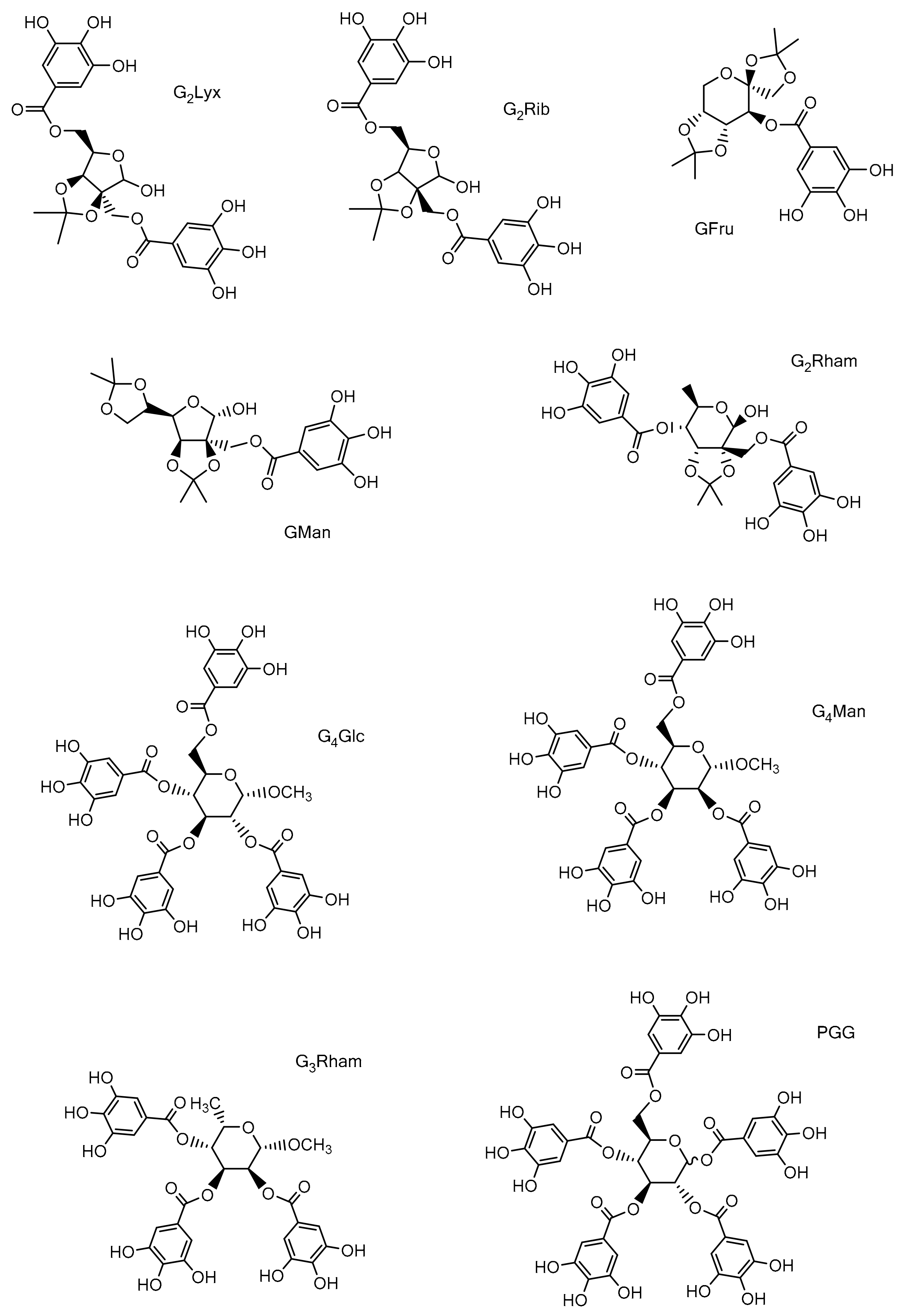
2.2. Studied Compounds

Methyl 2,3,4,6-tetra-O-galloyl-α-d-glucoside (G4Glc), methyl 2,3,4,6-tetra-O-galloyl- α-d-mannoside (G4Man), methyl 2,3,4-tri-O-galloyl-α-l-rhamnoside (G3Rham) were synthesized by Hricovíniová et al., and all spectral data for these GTs were reported in our previous work [35]. 1,2,3,4,6-penta-O-galloyl-αβ-d-glucose (PGG) was synthesized according to Ren et al. [18] and its identity and purity was confirmed by NMR spectroscopy. 1H NMR (600.13 MHz, methanol-d4) δ: 7.21, 7.11, 7.11, 7.05, 6.99, 6.98, 6.95,6.93, 6.92, 6.90 (10 × s, 20 H, galloyl), 6.70 (d, 1H, J1,2 = 3.5 Hz, H-1α), 6.24 (d, 1H, J1,2 = 8.3 Hz, H-1β), 6.13 (dd, 1H, J3,4 = 9.9 Hz, H-3α), 5.90 (dd, 1H, J3,4 = 9.9 Hz, H-3β), 5.71 (dd, 1H, J4,5 = 10.2 Hz, H-4α), 5.62 (dd, 1H, J4,5 = 10.1 Hz, H-4β), 5.59 (dd, 1H, J2,3 = 9.7 Hz, H-2β), 5.50 (dd, 1H, J2,3 = 10.1 Hz, H-2α), 4.57 (m, 1H, H-5α), 4.52 (m, 2H, H-6β), 4.47 (m, 2H, H-6α), 4.41 (m, 1H, H-5β); 13C NMR (150.91 MHz, methanol-d4) δ: 168.08–166.11 (C=O), 146.93–146.43 (CH-Ar), 93.97 (C-1β), 91.13 (C-1α), 74.58 (C-5β), 74.26 (C-3β), 72.35 (C-2β), 72.19 (C-5α), 71.77 (C-2α), 71.74 (C-3α), 69.96 (C-4β), 69.78 (C-4α), 63.27 (C-6β), 63.18 (C-6α).

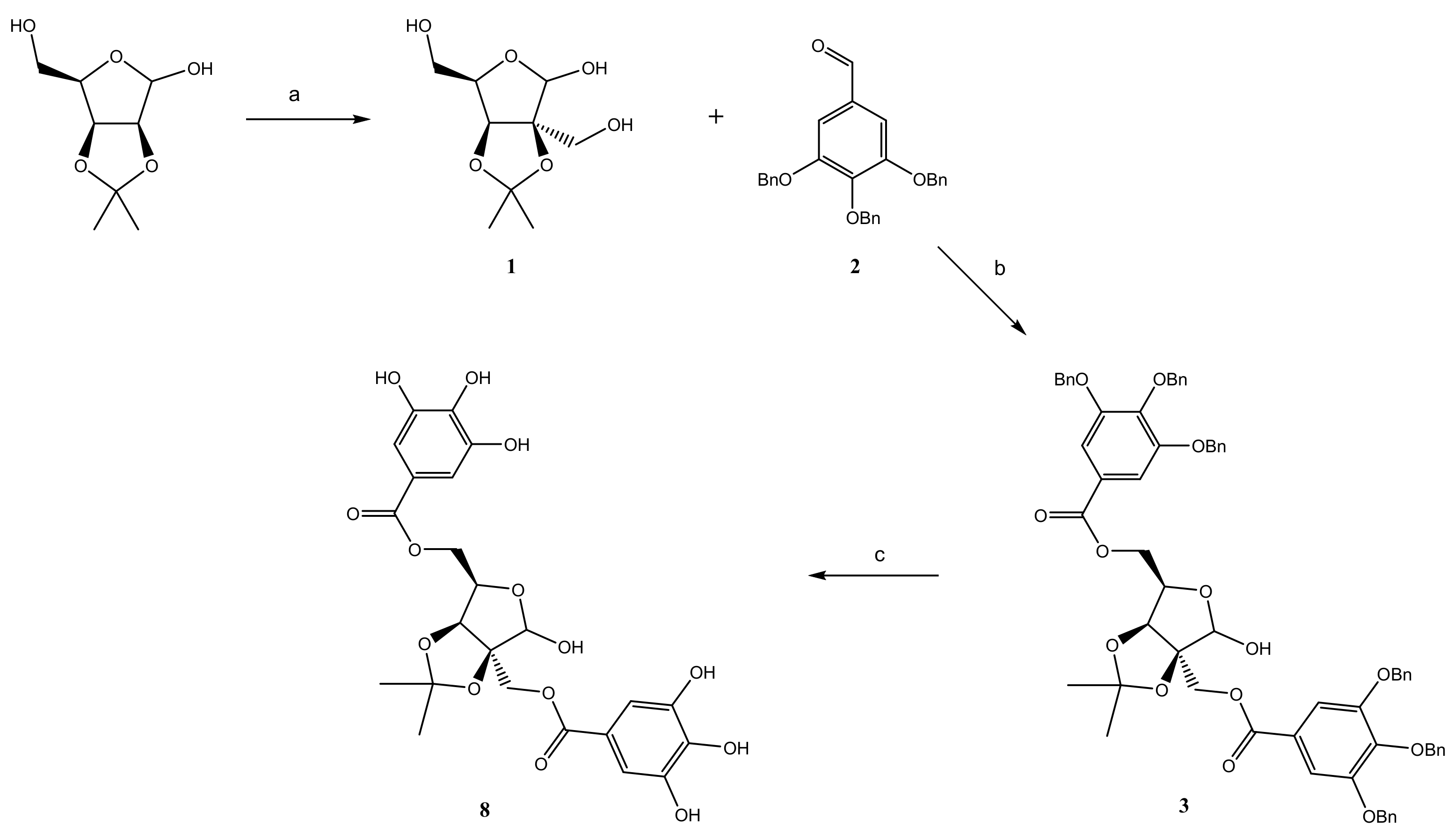
2.3. Synthesis of Galloyl Derivatives of 2-C-(Hydroxymethyl)-branched Saccharides

2.3.1. Preparation of 2-C-(Hydroxymethyl)-2,3-O-isopropylidene-α-d-lyxofuranose (1)

A reaction mixture of 2,3-O-isopropylidene-d-lyxose [36], (1 g; 5.3 mmol), K2CO3 (0.8 g) methanol (15 mL), and 37% aqueous solution of formaldehyde (15 mL; 147 mmol) was refluxed in argon atmosphere at 85 °C for 40 h. The reaction mixture was neutralized with 10% aq sulphuric acid and evaporated. Extraction with chloroform (4 × 30 mL) gave a combined fraction that was dried over anhydrous MgSO4 and concentrated. Crude reaction mixture was purified by column chromatography on silica gel (solvent A). Yield 82%; Rf = 0.58 (solvent A); α d 20 = +3.33→4.6° (c 1, acetone); HRMS: calcd for C9H16O6Na [M+Na]+ 243.0845; found 243.0914 [M+Na]+; 1H NMR (600.13 MHz, acetone-d6) δ: 5.24 (s, 1H, H-1), 4.61(d, 1H, H-3), 4.19 (d, 1H, H-4), 3.83 (m, 2H, CH-2a, CH-5a), 3.73 (2H, CH-2b, CH-5b),1.46, 1.40 (2 × s, 6H, CH3(Ip)); 13C NMR (150.91 MHz, acetone-d6) δ: 104.73 (C-1), 96.14 (C-2), 84.47 (C-4), 82.50 (C-3), 63.52 (CH2(C-2)), 61.45 (CH2(C-5), 29.42, 29.33 (2 × s CH3(Ip).

2.3.2. Preparation of 2-C-(Hydroxymethyl)-2,3-O-isopropylidene-α-d-lyxofuranose (2)

The 3,4,5-tri-O-benzyl protected gallic acid was synthesized from methyl gallate by the method reported previously [35]. Recrystallization from chloroform afforded white cotton-like needles, m.p. 198–199 °C [37]. 1H NMR (600.13 MHz, acetone-d6) δ: 7.27–7.55 (m, 17H, Ar-H), 5.23 (s, 4H, 2 PhCH2), 5.14 (s, 2H, PhCH2).

2.3.3. Preparation of Di-O-(3,4,5-tri-O-benzylgalloyl)-2-C-(hydroxymethyl)-2,3-O-isopro- pylidene Aldoses

The reaction mixture of 2,3-O-isopropylidene derivative of 2-C-(hydroxymethyl)-d-lyxose (1), 2,3-O-isopropylidene-2-C-(hydroxymethyl)-d-ribose [38], or 2,3-O-isopropylidene 2-C-(hydroxymethyl)-l-rhamnose [39] (1 equiv), DMAP (2 equiv), and 2 (2 equiv) in dry dichloromethane (25 mL) was refluxed for 1 h under argon atmosphere. After that DCC (2 equiv) was added and stirring continued at r.t. for 18 h. Then, the reaction mixture was placed into the ice-bath for 1 h and the precipitate was filtered. The filtrate was washed with 3% aq HCl (1 × 30 mL), saturated NaHCO3 (1 × 30 mL), and brine (1 × 30 mL). The combined organic layer was dried over anhydrous MgSO4, evaporated, and flash column chromatography on silica gel afforded the desired products (35).
2′,5-Di-O-(3,4,5-tri-O-benzylgalloyl)-2-C-(hydroxymethyl)-2,3-O-isopropylidene-d-lyxofuranose (3). Yield 73%; α d 20 = +21.0 (c 1, CHCl3); Rf = 0.95 (solvent B); HRMS: calcd for C65H60O14Na [M+Na]+ 1087.1560; found 1087.1584 [M+Na]+; 1H NMR (600.13 MHz, chloroform-d) δ: 7.48–7.14 (m, H-Ar), 6.58 (s, 1H, H-1), 4.79 (m, 1H, H-5a), 4.75 (d, 1H, H-2′a), 4.71 (m, 2H, H-3), 4.48 (m, 1H, H-5b), 4.46 (d, 1H, H-2′b), 4.42 (m, 1H, H-4), 1.59, 1.43 (2 × s, 6H, CH3(Ip)); 13C NMR (150.91 MHz, chloroform-d) δ: 128.52–127.25 (CH Ar), 101.25 (C-1), 93.51 (C-2), 82.80 (C-3), 80.35 (C-4), 64.34 (CH2(C-2)), 62.51 (CH2(C-5)).
2′,5-Di-O-(3,4,5-tri-O-benzylgalloyl)-2-C-(hydroxymethyl)-2,3-O-isopropylidene-d-ribofuranose (4). Yield 69%; α d 20 = −13.5 (c 1, Ac); Rf = 0.95 (solvent B);HRMS: calcd for C65H60O14Na [M+Na]+ 1087.1560; found 1087.1577 [M+Na]+; 1H NMR (600.13 MHz, chloroform-d) δ: 7.38–7.04 (m, H-Ar), 6.72 (s, 1H, H-1), 4.72 (d, 1H, H-2′a), 4.67 (m, 2H, H-3, H-4), 4.52 (m, 1H, H-5a), 4.47 (d, 1H, H-2′b), 4.24 (m, 1H, H-5b), 1.65, 1.51 (2 × s, 6H, CH3(Ip)); 13C NMR (150.91 MHz, chloroform-d) δ: 127.48–126.31 (CH Ar), 101.24 (C-1), 92.29 (C-2), 84.16 (C-4), 83.57 (C-3), 63.89 (CH2(C-2)), 63.13 (CH2(C-5)).
2′,5-Di-O-(3,4,5-tri-O-benzylgalloyl)-2-C-(hydroxymethyl)-2,3-O-isopropylidene-β-l-rhamnopyranose (5). Yield 78%; α d 20 = −36.6 (c 1, Ac); Rf = 0.83 (solvent C); HRMS: calcd for C66H62O14Na [M+Na]+ 1101.4040; found 1101.4062 [M+Na]+; 1H NMR (600.13 MHz, acetone-d6) δ: 7.53–7.22 (m, H-Ar), 6.51 (s, 1H, H-1), 5.29 (m, 1H, H-5), 4.86 (m, 1H, H-3), 4.81 (d, 2H, H-2′a), 4.64 (d,1H, H-2′b), 4.39 (dd, 1H, H-4), 1.48, 1.32 (2 × s, 6H, CH3(Ip)), 1.38 (s, 3H, CH3(Ip)); 13C NMR (150.91 MHz, acetone-d6) δ: 128.75–127.42 (CH Ar), 101.14 (C-1), 93.60 (C-2), 83.98 (C-4), 82.55 (C-3), 68.42 (C-5), 65.29 (CH2(C-2)), 17.02 (CH3).
2′-O-(3,4,5-tri-O-Benzylgalloyl)-2-C-(hydroxymethyl)-2,3:5,6-di-O-isopropylidene-d-mannofuranose (6). The mixture of 2-C-(hydroxymethyl)-2,3:5,6-di-O-isopropylidene- d-mannofuranose [40] (1 equiv), DMAP (1 equiv), and 2 (1 equiv) in dry dichloromethane (25 mL) was refluxed for 1 h under argon atmosphere. After that, DCC (1 equiv) was added and stirring continued at r.t. overnight. The reaction mixture was worked up in exactly the same way as mentioned in previous experiments. Derivative 6 was separated by flash column chromatography on silica gel. Yield 75%; α d 20 = +53.0 (c 1, Ac); Rf = 0.85 (solvent C); HRMS: calcd for C41H44O11Na [M+Na]+ 735.2781; found 735.2815 [M+Na]+; 1H NMR (600.13 MHz, chloroform-d) δ: 7.58–7.47 (m, H-Ar), 6.50 (s, 1H, H-1), 4,73 (d, 1H, H-2′a), 4.71 (d, 1H, H-3), 4.48 (m, 1H, H-2′b), 4.47 (m, 1H, H-5), 4.13 (m, 1H, H-5′a), 4.03 (m, 1H, H-5′b), 4.05 (m, 1H, H-4),1.57, 1.44, 1.41, 1.38 (4 × s, 12H, CH3(Ip)); 13C NMR (150.91 MHz, chloroform-d) δ: 128.53–127.45 (CH Ar), 101.55 (C-1), 93.47 (C-2), 82.72 (C-3, C-4), 72.61 (C-5), 67.05 (CH2(C-5)), 64.49 (CH2(C-2)).
3-O-(3,4,5-tri-O-Benzylgalloyl)-1,2:4,5-di-O-isopropylidene-d-fructopyranose (7). The mixture of 1,2:4,5-di-O-isopropylidene-d-fructopyranose (1 equiv), DMAP (1 equiv), and 2 (1 equiv) in dry dichloromethane (25 mL) was refluxed for 1 h under argon atmosphere. After that, DCC (1 equiv) was added and stirring continued at r.t. overnight. The reaction mixture was worked up as mentioned in previous experiment. Yield 84%; α d 20 = −85.3 (c 1, CHCl3); Rf = 0.91 (solvent D); HRMS: calcd for C40H42O10Na [M+Na]+ 705.2676; found 705.2825 [M+Na]+; 1H NMR (600.13 MHz, chloroform-d) δ: 7.46–7.32 (m, H-Ar), 5.35 (d, 1H, H-3), 4.44 (d, 1H, H-4), 4.28 (d, 1H, H-5), 4.19 (dd, 1H, H-6a),4.14 (dd, 1H, H-6b),4.00 (dd, 1H, H-1a), 3.88 (dd, 1H, H-1b), 1.62 (s, 3H, CH3(Ip)), 1.51 (s, 3H, CH3(Ip)), 1.39 (s, 3H, CH3(Ip)), 1.37 (s, 3H, CH3(Ip)); 13C NMR (150.91 MHz, chloroform-d) δ: 128.56–127.52 (CH Ar), 112.11 (1,2CMe2), 109.77 (4,5CMe2), 75.10 (C-4), 73.82 (C-5), 71.72 (C-1), 70.80 (C-3), 60.49 (C-6), 27.87, 26.47, 26.40, 26.29 (4 × CH3(Ip)).

2.3.4. Debenzylation Procedure

A solution of O-benzylgalloyl-derivative 37 (100 mg) in methanol (6 mL) was stirred for 16 h in the presence of 10% Pd/C (25 mg) in hydrogen atmosphere. The reaction mixture was filtered, washed with methanol (1 × 10 mL) and concentrated in vacuo to give respective per-O-galloyl-derivatives 812 as amorphous solids.
2′,5-Di-O-galloyl-2-C-(hydroxymethyl)-2,3-O-isopropylidene-d-lyxofuranose (8, G2Lyx). Yield 90%; α d 20 = +29.0 (c 1, CHCl3); Rf = 0.73 (solvent A); HRMS: calcd for C23H24O14Na [M+Na]+ 547.1064; found 547.1085 [M+Na]+; 1H NMR (600.13 MHz, methanol-d4) δ: 7.03, 7.01 (2 × s, 4H, galloyl), 6.72 (s, 1H, H-1), 4.94 (d, 1H, H-3), 4.71, 4.66 (m, 2H, H-2′a, H-2′b), 4.60 (m, 1H, H-5′a), 4.58 (m, 1H, H-4), 4.44 (m, 1H, H-5′b), 1.57, 1.42 (2 × s, 6H, CH3(Ip)); 13C NMR (150.91 MHz, methanol-d4) δ: 111.52, 111.43 (2 × CH, galloyl), 103.65 (C-1), 96.39 (C-2), 85.44 (C-3), 82.80 (C-4), 65.61 (CH2(C-2)), 64.68 (CH2(C-5)).
2′,5-Di-O-galloyl-2-C-(hydroxymethyl)-2,3-O-isopropylidene-d-ribofuranose (9, G2Rib). Yield 93%; α d 20 = −49.6 (c 1, Ac); Rf = 0.54 (solvent D); HRMS: calcd for C23H24O14Na [M+Na]+ 547.1064; found 547.1079 [M+Na]+; 1H NMR (600.13 MHz, methanol-d4) δ: 7.04, 7.03 (2 × s, 4H, galloyl), 6.50 (s, 1H, H-1), 4.91 (d, 1H, H-3), 4.68 (m, 2H, H-2′a, H-2′b), 4.56 (m, 1H, H-4), 4.44 (m, 1H, H-5′a), 4.32 (m, 1H, H-5′b), 1.54, 1.44 (2 × s, 6H, CH3(Ip)); 13C NMR (150.91 MHz, methanol-d4) δ: 108.87 (2 × CH, galloyl), 101.90 (C-1), 93.28 (C-2), 85.40 (C-4), 84.65 (C-3), 63.87 (CH2(C-5)), 63.65 (CH2(C-2)).
2′,4-Di-O-galloyl-2-C-(hydroxymethyl)-2,3-O-isopropylidene-β-l-rhamnopyranose (10, G2Rham). Yield 95%; α d 20 = −60.5 (c 1, MeOH); Rf = 0.84 (solvent A); HRMS: calcd for C24H26O14Na [M+Na]+ 561.1220; found 561.1196 [M+Na]+; 1H NMR (600.13 MHz, methanol-d4) δ: 7.07, 7.04 (2 × s, 4H, galloyl), 6.33 (s, 1H, H-1), 5.23 (m, 1H, H-5), 4.91 (d, 1H, H-3), 4.60 (m, 2H, H-2′a, H-2′b), 4.36 (dd, 1H, H-4), 1.56, 1.38 (2 × s, 6H, CH3(Ip)), 1.40 (s, 3H, CH3(Ip)); 13C NMR (150.91 MHz, methanol-d4) δ: 109.27, 109.07 (2 × CH, galloyl), 100.52 (C-1), 93.65 (C-2), 83.77 (C-4), 82.43 (C-3), 67.52 (C-5), 63.27 (CH2(C-2)), 17.06 (CH3).
2′-O-Galloyl-2-C-(hydroxymethyl)-2,3:5,6-di-O-isopropylidene-d-mannofuranose (11, GMan). Yield 89%; α d 20 = +16.3 (c 1, MeOH); Rf = 0.52 (solvent B); HRMS: calcd for C20H26O11Na [M+Na]+ 465.1373; found 465.1396 [M+Na]+; 1H NMR (600.13 MHz, acetone-d6) δ: 7.16 (s, 2H, galloyl), 4.70 (d, 1H, H-3), 4.65 (s, 1H, H-1), 4.55 (d, 1H, H-2′a), 4.37 (m, 1H, H-5), 4.33 (d, 1H, H-2′b), 4.06 (m, 1H, H-5′a), 4.01 (m, 1H, H-5′b), 3.94 (m, 1H, H-4), 1.47, 1.37, 1.35, 1.29 (4 × s, 12H, CH3(Ip)); 13C NMR (150.91 MHz, acetone-d6) δ: 109.93 (CH, galloyl), 105.50 (C-1), 90.67 (C-2), 82.21 (C-3), 78.90 (C-4), 74.35 (C-5), 67.28 (CH2(C-5)), 64.66 (CH2(C-2)).
3-O-Galloyl-1,2:4,5-di-O-isopropylidene-d-fructopyranose (12, GFru). Yield 93%; α d 20 = −102.9 (c 1, Ac); Rf = 0.75 (solvent E); HRMS: calcd for C19H24O10Na [M+Na]+ 435.1267; found 435.1304 [M+Na]+; 1H NMR (600.13 MHz, acetone-d6) δ: 7.19 (s, 2H, galloyl), 5.37 (d, 1H, H-3), 4.06 (dd, 1H, H-4), 4.01 (dd, 1H, H-6a), 3.99 (m, 1H, H-5),3.86 (dd, 2H, H-1a, H-1b), 3.76 (dd, 1H, H-6b),1.43, 1.37, 1.34, 1.29 (4 × s, 12H, CH3(Ip)); 13C NMR (150.91 MHz, acetone-d6) δ:110.33 (CH, galloyl), 72.60 (C-1), 70.92 (C-3), 70.59(C-5), 70.15 (C-4), 65.46 (C-6), 26.97, 26.84, 26.56, 26.24 (4 × CH3(Ip)).

2.4. Determination of Antioxidant Activity

2.4.1. 1,1-Diphenyl-2-picrylhydrazyl Radical Scavenging Activity (DPPH Assay)

The free radical-scavenging capacity was evaluated by DPPH assay [41] with some modifications [35]. The synthesized per-O-galloyl-derivatives of different concentrations (0.1–1 mM) were dissolved in methanol. The reaction mixture consisting of the tested sample (50 µL) and DPPH solution (950 µL) was added to 96-well plate incubated (30 min) and absorbance was measured at 517 nm using an xMarkTM Microplate Spectrophotometer (Bio-Rad Laboratories Inc., Hercules, CA, USA). GA and DPPH solution were used as a positive and negative control. The reaction was conducted at r.t. and experiments were performed in triplicate. The radical-scavenging activity of tested compounds was calculated using the equation:
DPPH radical scavenging activity (%) = 100 × (A0AS)/A0
where A0 is the absorbance of the negative control, and AS is the absorbance of the tested compound.

2.4.2. Iron(III)-Reducing Power Assay (FRAP Assay)

The reducing activity of compounds was evaluated by colorimetric FRAP assay [42] with some modifications described previously [35]. Methanolic solution (200 µL) of tested compounds (0.1–1 mM) were mixed with 0.2 M phosphate buffer (500 µL, pH 6.6) and 1% potassium ferricyanide [K3Fe(CN)6] (500 µL) and incubated at 50 °C for 20 min. The absorbance was recorded at 700 nm using an xMarkTM Microplate Spectrophotometer.

2.5. Biological Studies

2.5.1. Test Microorganisms

Standard reference bacterial strains Staphylococcus aureus ATCC 29213, Enterococcus faecalis ATCC 2921, Mycobacterium tuberculosis H37Ra, and Chromobacterium violaceum ATCC 12472 were obtained from American Type Culture Collection (ATTC). Three vancomycin-resistant isolates of E. faecalis (VRE 342B, VRE 368, VRE 725B) were provided by Oravcová et al. [43]. Clinical isolates of methicillin-resistant S. Aureus (MRSA 63718, MRSA 630, MRSA3202) and non-tuberculoid mycobacteria (Mycobacterium smegmatis and Mycobacterium kansasii) were obtained from a collection of the Department of Infectious Diseases and Microbiology, University of Veterinary Sciences Brno (Czech Republic).

2.5.2. Determination of Antimicrobial Activity

Antimicrobial activity of compounds was tested using microtitration broth method according to Clinical and Laboratory Standards Institute [44] with some modifications described previously [45,46,47]. Compounds were dissolved in DMSO to get concentration 10 mg/mL and diluted in a microtitration plate in an appropriate medium, to reach the final concentration 256–2 µg/mL. Subsequently, a microtitration plate was inoculated with tested microorganisms. The minimum inhibitory concentration (MIC) was evaluated visually as the minimal concentration of tested compound, which completely inhibited the bacterial growth. Each experiment was repeated at least three times.

2.5.3. Biofilm Inhibition

The ability of compounds to prevent biofilm growth was tested according to a previously described method [44,47]. Compounds were diluted in a 96-well microtitration plate in tryptic soya broth (TSB) containing 2% glucose to reach the final concentrations 256–2 µg/mL. The plates were inoculated with inoculum of S. aureus ATCC 29213 in logarithmic growth phase. The final concentration of bacterial cells in the wells was 1 × 10⁵ CFU/mL. The plates were incubated at 37 °C for 48 h, the contents of the wells were removed and washed with sterile phosphate buffered saline (PBS). After drying, 0.5% crystal violet was added to each well, and the plates were incubated at r.t. for 20 min. After that, the dye was removed and the plates were washed three times with sterile PBS. The coloured biofilm was detached from the surface using 33% solution of acetic acid (125 µL). The absorbance at 595 nm was measured (Tecan Infinite 200 PRO, Tecan, Grodig, Austria) and the percentage of inhibition was determined. As a blank, a non-inoculated plate treated in the same way was used [46,48]. The ability to inhibit biofilm formation was evaluated as a percentage inhibition of growth compared to the growth control according to the equation:
% of inhibition = 100 − (OD595S/OD595C) × 100
where OD595S is the absorbance of the sample at 595 nm, and OD595C is the absorbance of the growth control at 595 nm. The minimum biofilm inhibitory concentration was assessed as the lowest concentration of the tested compounds, which inhibited the growth of 80% bacteria compared to the growth control. The experiment was made in duplicate and was repeated at least three times.

2.5.4. Biofilm Disruption

In order to test the ability of the studied GTs to eradicate a matured biofilm, a staphylococcus biofilm was prepared in the same way as for inhibitory activity testing. Biofilms were grown as described in previous experiment, but without the presence of the compounds. The matured biofilms were treated with compounds diluted in CaMH (100 µL) to concentration 256–2 µg/mL. A growth control containing 2.5% of DMSO in CaMH was included. The plates were incubated at 37 °C for 24 h. Bacterial viability was analysed by adding of MTT solution (0.5 mg/mL) in PBS (100 µL) to each well. The plates with MTT were incubated at 37 °C for 1–2 h in the dark, until the blue formazan crystals appeared. The solution in the wells was removed, and the plates were washed with PBS. The formazan crystals were dissolved by using 17% sodium dodecyl sulphate in 40% dimethylformamide [46,49]. The absorbance at 570 nm was measured and the percentage of eradication was determined according to the equation:
% of biofilm eradication = 100 − (OD570S/OD570C) × 100
where OD570S is the absorbance of the sample at 570 nm, and OD570C is the absorbance of the growth control at 570 nm. The minimum biofilm eradication concentration was the lowest concentration of the compound, which reduced the metabolic activity of the biofilm by 80% compared to the growth control. The experiment was made in duplicate and repeated at least three times.

2.5.5. Interaction with Quorum Sensing of C. violaceum

C. violaceum was cultivated on Mueller-Hinton (MH) agar at r.t. for 48 h. Then, several colonies were suspended in Lysogeny broth medium (LB) and cultivated at r.t. overnight. This culture (50 µL) was mixed with dilute 0.1% MH agar (5 mL). The mixture was poured onto pre-prepared Petri dishes with MH agar. When the agar on the plates got solid, wells (diameter of 5 mm) were made sterile in the dish. The wells were at least 25 mm apart and from the edge. The test substance (20 µL) of the desired concentration, prepared from a stock solution of the substance in DMSO (10 mg/mL) diluted in PBS, was added into each well; DMSO blank served as the control. After that, the plates were incubated for 3 days at r.t. until there was a visual growth of C. violaceum [50]. The evaluation was repeated in at least three independent experiments and the results were averaged.

2.5.6. Interaction with sortase A

The activity of sortase A (SrtA) was evaluated by using the SensoLyte® 520 sortase A activity Assay Fluorimetric Kit (AnaSpec Inc., Fremont, CA, USA) in accordance with the manufacturer’s instructions. Compounds were dissolved in DMSO to get concentration 200 µg/mL. StrA was exposed to studied GTs and the results were collected after 10–70 min of ongoing incubation. The inhibitory activities of tested compounds against SrtA were determined as continuous recording of fluorescence intensity relative to the negative control (Tecan Infinite 200 Pro). The known SrtA inhibitor, 4-(hydroxymercuri)- benzoic acid (IC50 = 50.6 nM), was used as a positive control.

2.6. Statistical Analysis

The experimental data were expressed as the mean ± standard deviation (±SD) of three independent experiments. The significance of differences between the means was evaluated by the OriginPro 8 software or Student’s t-test. Differences with p values < 0.05 were considered to be statistically significant.

3. Results and Discussion

3.1. Chemistry

The presence of multiple galloyl units in the GT molecules make them powerful antioxidants as well as effective antimicrobial agents. The strong contribution of the galloyl groups to these properties has been demonstrated several times, but it has also been observed that the carbohydrate moiety also plays an important role as well [21,51]. Carbohydrates are characterized by structural diversity and a multiplicity of nearly equivalent hydroxyl groups. As the 2-C-(hydroxymethyl)-branched-chain aldoses have several different hydroxyl groups, it was important to choose a suitable approach for the synthesis of their galloyl-esters. The synthetic method for the functionalization of branched aldoses is based on the modifications of primary hydroxyls, which tend to react faster, when doing this it is also necessary to protect secondary hydroxyls in order to carry out selective transformation of these polyfunctional molecules. For that reason, 2,3-O-isopropylidene derivatives of the 2-C-(hydroxymethyl)-branched aldoses were chosen as suitable compounds. This leaves the 2-C-hydroxymethyl branch free for galloylation, while keeping aldose in its furanose form.
During the synthesis, all phenolic hydroxyl groups of GA were protected by benzylation to avoid intra- and inter-molecular stacking interactions between galloyl units. Moreover, the benzyl group is most frequently used for such purposes because it allows for quantitative removal by hydrogenation and a simple workup procedure. The O-isopropylidene derivatives, having distinct reactive hydroxyl groups, were used to introduce galloyl groups to the target sites of the sugar molecule. Hence, the appropriately protected 2-C-(hydroxymethyl)-branched aldose was esterified with 2 in the presence of DMAP as a catalyst and DCC as a coupling reagent, in order to obtain 2,3,4-tri-O-benzyl-galloylated 2-C-(hydroxymethyl)-branched aldoses 36 and the 2,3,4-tri-O-benzyl-galloyl derivative of d-fructose (7) in very good yields (69–84%). Subsequent debenzylation of compounds 37 led to the expected galloylated branched-chain aldoses 811 and galloylated d-fructose (12) in excellent yields (89–95%). A representative procedure for the synthesis of the 2′,5-di-O-galloyl-2-C-(hydroxymethyl)- 2,3-O-isopropylidene-d-lyxofuranose (8, G2Lyx) is depicted in Scheme 1. The structures of the new derivatives were determined on the basis of NMR spectroscopy and other analytical methods.

3.2. Antioxidant Activity

The antioxidant activity of compounds is related to their redox properties, which allow them to scavenge free radicals by acting as hydrogen donors or reducing agents. Antioxidant compounds usually contain aromatic rings that enable them to donate protons to free radicals formed during oxidation. A series of structurally different GTs (Figure 1), were screened for their free-radical-scavenging effect and reducing ability using the DPPH and FRAP assays. It is known that GA is a strong antioxidant due to the presence of three hydroxyl groups on the aromatic ring [52]. Thus, the high antioxidant efficiency of these compounds could be attributed to the presence of multiple galloyl groups in their structures. The experimental results indicated that the studied GTs exhibited notably different and concentration-dependent DPPH radical-scavenging effects (Table 1). Compounds PGG, G4Man, G3Rham, and G4Glc exhibited the highest radical-scavenging activity (94–98%), whereas the di-galloylated 2-C-(hydroxymethyl)- branched aldoses (G2Rib, G2Lyx, and G2Rham) manifested a slightly lower effect (85–88%). Moreover, as expected, mono-galloylated derivatives (GMan and GFru) displayed only moderate DPPH radical-scavenging activity (71–73%). Almost equal antioxidant activity was observed for PGG (98%) and G4Glc (96%), where the latter differs from the former by one less galloyl group. Moreover, comparable radical-scavenging activity was also observed for the compounds G4Man (95%) and G4Glc (96%), which have very similar molecular structures. However, in the case of G3Rham (94%) and the derivative G2Rham (85%), a noticeable decrease of antioxidant ability was observed. These compounds differ only by one galloyl group, but the lower antioxidant effect of G2Rham is probably influenced by the presence of isopropylidene groups in the molecule.
The variation in the radical-scavenging effect amongst the tested GTs was probably due to the different stability of the resulting oxygen-centred radical formed in these compounds. It was observed that the presence of isopropylidene groups in galloylated branched-chain aldoses (G2Rib, G2Lyx, and G2Rham) resulted in a decrease of antioxidant activity. The ability of studied GTs to quench DPPH radicals increased in the following order: GMan < GFru < G2Rham < G2Lyx ~ G2Rib ~ GA < G3Rham ~ G4Man ~ G4Glc ~ PGG.
The reduction of metal ions is used as an indicator of the electron-donating activity of compounds. The studied GTs exhibited varying ability to reduce Fe3+/ferricyanide complex to Fe2+/ferrocyanide, as seen in Table 1. Increases of the reducing power were correlated with the concentration of the tested compound, as well as with the number and substitution pattern of the galloyl groups attached to the carbohydrate core. The compounds PGG and unnatural GTs (G4Glc, G4Man, and G3Rham) exhibited remarkable potency for donating electrons to reactive free radicals, transforming them into more stable species. However, the presence of isopropylidene groups in galloylated 2-C-(hydroxymethyl)-branched aldoses (G2Rib, G2Lyx, and G2Rham) resulted in a noticeable decrease of their reducing power. The weakest reducing power (even lower than GA) was observed in the case of the derivatives GMan and GFru, each bearing only one galloyl moiety and two isopropylidene groups in their structure. The reducing power of the studied GTs increased in the following order: GFru < GMan < G2Lyx ~ G2Rham ~ GA ~ G2Rib < G3Rham ~ G4Man ~ G4Glc ~ PGG.
The experimental data thus revealed that the majority of the studied compounds exhibit an excellent radical-scavenging and reducing activity. The results (Table 1) demonstrated that outcomes from the FRAP assay are in good agreement with those of the DPPH assay, and that the overall antioxidant activity of each tested GT can be regarded as the sum of contributions of all the structural features in the molecule, depending on the number of galloyl groups and type of carbohydrate core, and the individual galloyl groups’ contributions varying with their position and spatial arrangement. Our findings are consistent with the previous reports on antioxidant potential of phenolic acids published by other authors [52,53].

3.3. Antibacterial and Antimycobacterial Activity

The antibacterial activity of the unnatural GTs was tested against S. aureus ATCC 29213, three MRSA isolates, E. faecalis ATCC 29212, and three VRE isolates. For screening of antimycobacterial effects a virulent isolate of M. tuberculosis and two non-tuberculous mycobacteria were used. The summary of results from experiments is presented in Table 2.
It can be concluded that most of the tested compounds were ineffective against both staphylococci and enterococci (> 128 µg/mL). PGG and unnatural epimeric GTs (G4Man and G4Glc) showed the highest potency (in the range of 16–64 µg/mL) within the tested series of compounds; however, it is still a moderate effect. In addition, the activity of these derivatives against enterococci is insignificant. Thus, the activity was not significantly affected by the presence of the mecA gene (in MRSA clinical isolates) or vanA (VRE clinical isolates). It can be summarized that unnatural G3Rham, galloylated 2-C-(hydroxymethyl)-branched aldoses (G2Rib, G2Lyx, G2Rham, and GMan), and mono-galloyl fructose (GFru) with isopropylidene groups in their structures were less active than PGG, G4Glc, and G4Man (Table 2). The antimycobacterial activity of the tested GTs was also insignificant. The compounds PGG, G4Glc, and G4Man showed only moderate antimycobacterial effect against M. tuberculosis (MIC ≥ 128 µg/mL). The galloylated 2-C-(hydroxymethyl)-branched aldoses (G2Rib, G2Lyx, G2Rham, and GMan), bearing isopropylidene groups did not show any antimycobacterial activity.
Several studies with similar natural compounds can be found in the literature. The antistaphylococcal activity of Thai mango (M. indica) kernel extract, containing more than 60% PGG and less than 1% gallic acid and methyl gallate, was studied by Jiamboonsri et al. The antibacterial effect was caused by PGG (MIC = 160 µg/mL) and was comparable with our results [22]. The group of Chan et al. investigated the antistaphylococcal activity of galloyl-substituted flavonol-rhamnosides extracted from Calliandra tergemina leaves. Three compounds, kaempferol-3-O-(2,3,4-tri-O-galloyl)-α-l-rhamnopyranoside, quercetin-3-O-(3,4-di-O-galloyl)-α-l-rhamnopyranoside and quercetin-3-O-(2,3,4-tri-O-galloyl)- α-l-rhamnopyranoside, showed only insignificant activity against three MRSA isolates (MIC 256 µg/mL). Compounds bearing only one galloyl group showed no anti-MRSA activity. However, multiple esterification of l-rhamnose molecule increased the antibacterial effect. Scanning electron microscopy (SEM) studies revealed that these compounds interact with the bacterial membrane [54].
Derivatives of gallic acid are able to interact with different bacterial cell structures. As mentioned above, one of the mechanisms of action is the interaction with the plasma membrane [54]. This interaction leads to leakage of cell proteins, nucleic acids, or inorganic compounds. Disruption of the cell membrane subsequently causes cell shrinkage and cell lysis, resulting in cell death [54]. In addition, GTs are also able to chelate metal ions, such as copper and iron, and make them inaccessible to microorganisms. For example, Terminalia chebula extract, containing significant amount of 1,2,6-tri-O-galloyl-d-glucose, demonstrated antibacterial activity against multidrug-resistant uropathogens, which might be due to iron-complexing properties [55,56]. PGG itself is able to interact directly with the bacterial cell wall. Zhao et al. studied PGG activity against Candida glabrata. The compounds were 10 times more active than fluconazole. The antifungal effect was due to the direct interaction of PGG with the fungal cell wall, without any interaction with glucan synthase [24].
The studied PGG analogues and isopropylidene-substituted GTs are new compounds whose antibacterial activity has not yet been described in the literature. However, the presented data demonstrate that the multifaceted GT-induced effects strongly depend on the investigated bacterial strain as well as on the structure of the GTs.

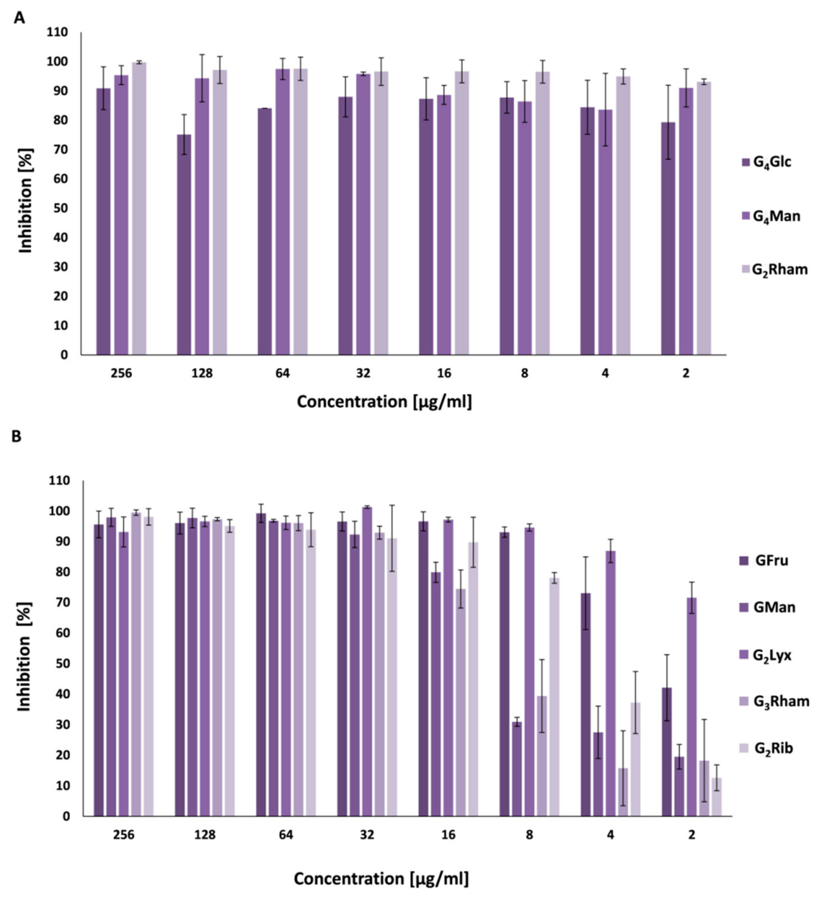
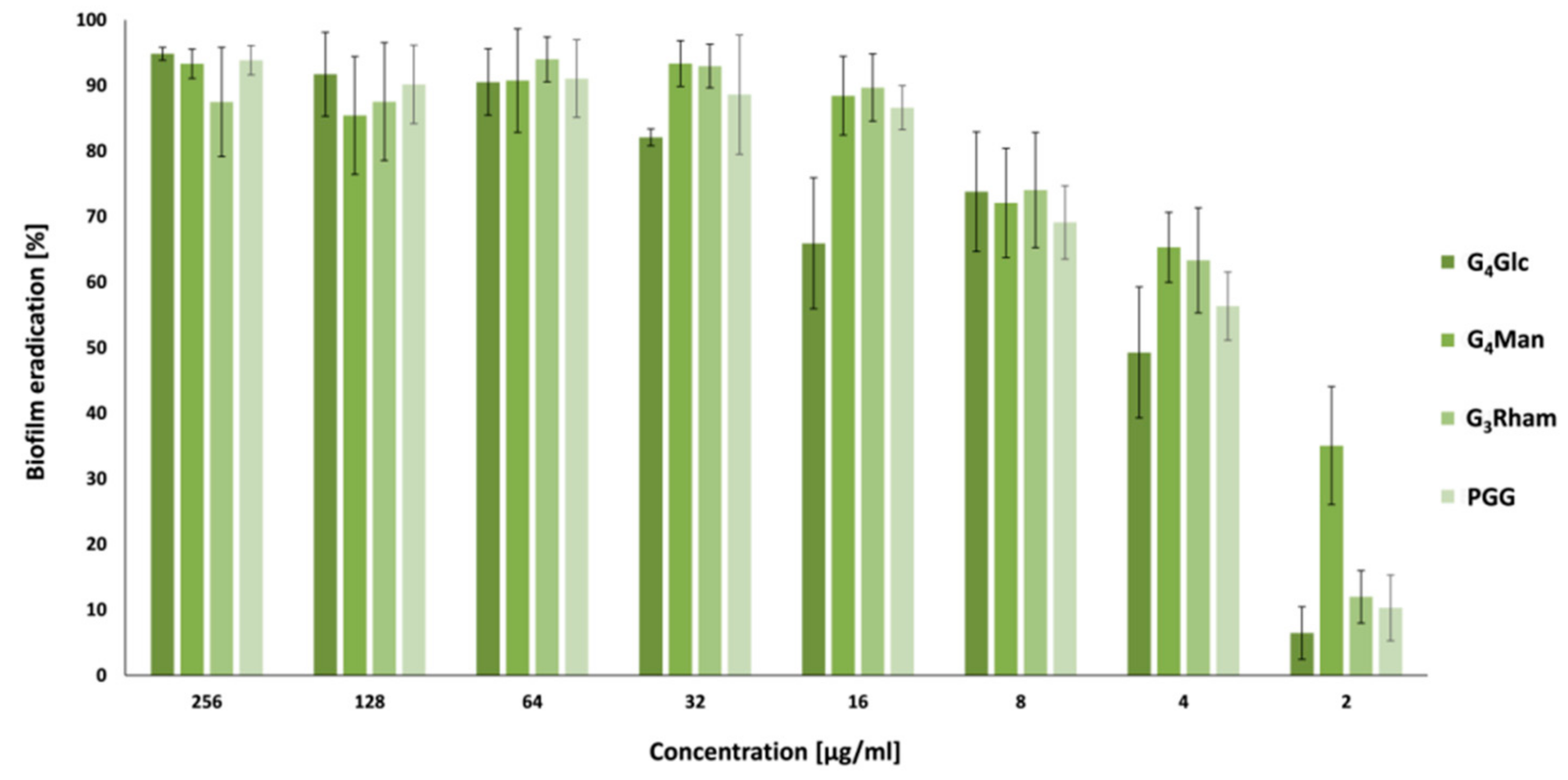
3.4. Antibiofilm Activity

Bacterial biofilms protect the microbial community from external damage and increase the persistence of chronic infections. Selected compounds were examined as inhibitors and disruptors of S. aureus ATCC 29213 biofilms. The results indicate that both groups, PGG analogues (Figure 2A) and galloylated 2-C-(hydroxymethyl)-branched aldoses (Figure 2B), efficiently inhibited S. aureus biofilm formation at concentrations lower than MIC values. For example, the minimum biofilm inhibition concentration (MBIC) of compound G4Glc was 64 times lower than its MIC value, MBIC for G4Man was 32 times lower, and MBIC for G3Rham was 256 times lower than its MIC value. These results identify G3Rham as the most potent inhibitory compound from this series.
New anti-biofilm compounds that can effectively eradicate bacterial biofilms are desirable to prevent various infections. Eradication activity was tested for PGG and its analogues G4Glc, G4Man and G3Rham. All tested compounds eliminated 80% of the matured biofilm of S. aureus at concentrations lower than the MIC value against planktonic cells. In this case, as well, G3Rham was the most active compound. G3Rham eradicated biofilms of S. aureus at a concentration 16 times lower than its MIC value (Figure 3). The interesting biofilm inhibitory and eradication activity against S. aureus make unnatural GTs excellent biofilm-controlling agents for healthcare and food processing facilities.
A number of structurally similar natural GTs with remarkable antimicrobial and antibiofilm properties have been studied. It was reported that extracts of Cytinus possess interesting antimicrobial activity against Gram-positive bacteria (S. aureus, Staphylococcus epidermidis and Enterococcus faecium). Characterization of the tannin profile of Cytinus hypocistis and Cytinus ruber revealed a significant number of GTs, in particular 1-O-galloyl-β-d-glucose and PGG. Three pathogenic species were sensitive to Cytinus extracts, and a significant inhibition of S. epidermidis biofilm formation was also observed [57]. Recently, Bag et al. published two studies investigating the antimicrobial activity of GT, 1,2,6-tri-O-galloyl-β-d-glucopyranose, isolated from T. chebula, against multidrug-resistant E. coli. This tri-O-galloyl derivative of d-glucose, was effective against multidrug-resistant uropathogens, and acted synergistically in combination with gentamicin and trimethoprim and additively in combination with amoxicillin, ciprofloxacin and ceftazidine. This substance was able to eradicate the preformed E. coli biofilms and at the same time act with synergistic antibiofilm activity in combination with gentamicin and trimethoprim [55,58]. This compound differs from the test compound G4Glc only by the number and position of the gallic acid residues in the molecule. Brackman et al. have shown that natural hamamelitannin (which is structurally very similar with test compound G2Rib) interfaces with QS in S. aureus, and thereby increases the susceptibility of S. aureus biofilms to various antibiotics [34].
Overall, our results demonstrate that GTs have sufficient inhibitory effect against S. aureus ATCC 29213 biofilm formation. This preliminary study suggests that the antibiofilm activity of studied compounds did not increase with an increasing number of galloyl units. Furthermore, the activity is affected by the sugar moiety. The most active antibiofilm compound, G3Rham, had one of the lowest activities against the planktonic cells of S. aureus. These experiments showed that the main mechanism of action against biofilm formation is not related to antibacterial activity against planktonic cells. However, as the number of the test compounds was limited, the structure-activity relationship cannot be discussed in more depth.

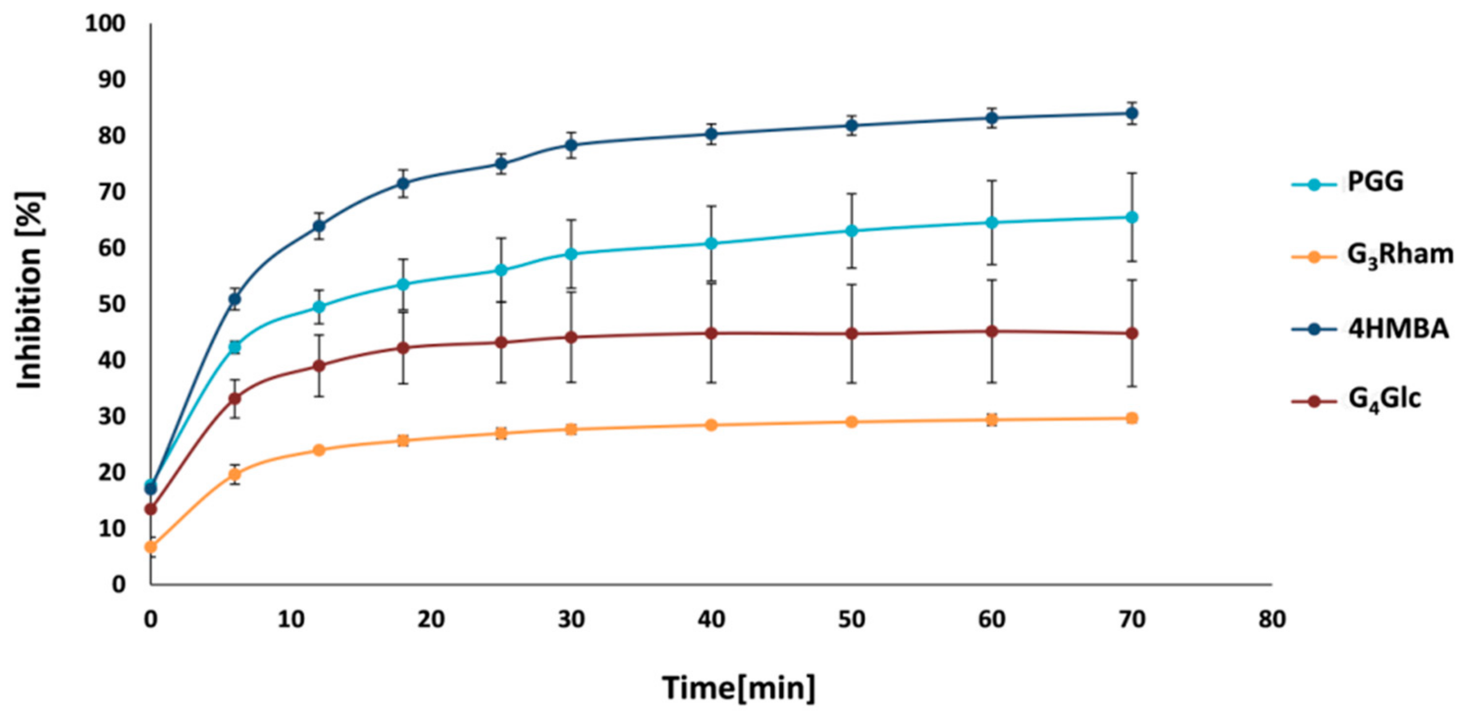
3.5. Quorum Sensing Inhibition

QS is a communication mechanism that regulates bacterial virulence in pathogens. A number of plant phenolics have been identified as important microbial growth and QS inhibitors [14,59]. It was expected that the compounds could work as inhibitors of the QS system, as they interacted with S. aureus biofilms at concentrations lower than MIC values. This activity was tested on the opportunistic bacterium C. violaceum. One particular characteristic of this genus is the synthesis of the purple pigment, violacein, which is regulated by QS. Inhibition of this system blocks violacein production and leads to the growth of white or colourless colonies of C. violaceum. Interestingly, all tested compounds, except for G2Rham, inhibited violacein production (Table 3). However, it was not possible to determine whether the observed effect was related to the sugar moiety or to the number of galloyl groups due to the limited number of the investigated compounds and their slightly different activities. Similarly, it is not possible to explain the complete inactivity of G2Rham. Nevertheless, it is important to be aware of the fact that QS active compounds have non-linear effects [60].

3.6. Inhibition of Sortase A

Since SrtA, which anchors surface proteins to the cell wall, plays a critical role in Gram-positive bacterial pathogenesis, it should be possible to target bacterial virulence and treat infections by inhibiting the activity of SrtA. The structurally different compounds PGG, G4Glc and G3Rham were investigated as potential inhibitors of SrtA. The experiments indicated that these compounds are able to block the pathogenic action of SrtA, and the degree of enzyme inhibition is plotted against time in Figure 4. PGG (bearing five galloyl units in its structure) exhibited the most potent inhibitory effect against SrtA among all tested compounds. It inhibited SrtA by 65.5 ± 7.9% after 70 min. The tetra- and tri-O-galloylated compounds G4Glc and G3Rham had approximately half (44.8 ± 9.5%) and quarter inhibitory activity (29.6 ± 0.8%), respectively. These findings indicate that the ability to inhibit SrtA increases with the degree of galloylation. Thus, the number of galloyl groups attached to the carbohydrate core seems to be a critical factor.
The ability of PGG derivatives to inhibit SrtA has not been described in the literature yet. However, there are many studies examining the effect of other polyphenolic compounds, mainly flavonoids, on bacterial sortases. Wang et al. showed that the flavonoid baicalin binds to sortase B in S. aureus and reduces its virulence [61]. Other flavonoids, especially kurarinol, isolated from the root of Sophora flavescens, acted as inhibitors of SrtA as well. It was observed that the presence of prenyl groups in kurarinol was essential for its strong inhibitory potency [62]. Other sortase inhibitors, which are potentially useful in the treatment of bacterial infections, include isovitexin [63], astilbin [64], and rutin [65].
The findings described above suggest that PGG and its structural analogues G4Glc and G3Rham are able to inhibit the catalytic activity of SrtA in vitro, but have little effect on bacterial growth. The compounds interfered with the adhesion of S. aureus biofilm, and thus hold promise for the development of anti-virulence agents against medical device infections caused by S. aureus. It seems that unnatural GTs may be an important new direction in the field of research into SrtA inhibitors.

4. Conclusions

In summary, new GTs derived from 2-C-(hydroxymethyl)-branched aldoses were synthesized and characterized on the basis of their physical and spectral data. Evaluation of antioxidant, antimicrobial, and antibiofilm activity showed differences among the newly prepared derivatives and three synthetic analogues of PGG and commercial antioxidant gallic acid. Results from the present study indicate that unnatural GTs are promising antioxidants and radical-scavengers. The compounds PGG, G4Glc, and G3Rham inhibited SrtA, and it can be stated that the inhibitory effect increased with the increasing number of galloyl groups. On the other hand, the inhibitory effect was also dependent on the type of sugar. It can be hypothesized that the observed antibacterial activity is related to the inhibition of SrtA, although, there are other mechanisms which are also likely to be involved. All of the investigated GTs proved to be potent inhibitors and disruptors of S. aureus biofilms at concentrations much lower than the MIC values. The promising activity at inhibiting the growth of S. aureus biofilms makes GTs valuable alternatives to synthetic antioxidants and currently used antibiofilm agents suitable for application in the biomedical, pharmaceutical, cosmetics, consumer products, and food industries.

Author Contributions

Conceptualization, Z.H.; data analysis, Š.M., J.H. and A.Č.; investigation, Z.H., Š.M., J.H. and A.Č.; writing—original draft preparation, Z.H., Š.M., J.H., A.Č. and J.J.; writing—review and editing, Z.H. and J.J.; supervision, J.J.; project administration, Z.H. and J.J.; funding acquisition, Z.H. and J.J. All authors have read and agreed to the published version of the manuscript.

Funding

The authors would like to acknowledge financial support from the Slovak Grant Agency VEGA 2/0022/18 and from the Slovak Research and Development Agency APVV-17-0373.

Institutional Review Board Statement

Not applicable.

Informed Consent Statement

Not applicable.

Data Availability Statement

Data is contained within the article.

Conflicts of Interest

The authors declare that they have no known competing financial interests or personal relationships that could have appeared to influence the research reported in this paper.

References

  1. Khanbabaee, K.; van Ree, T. Tannins: Classification and definition. Nat. Prod. Rep. 2001, 18, 641–649. [Google Scholar] [CrossRef]
  2. Quideau, S.; Deffieux, D.; Douat-Casassus, C.; Pouysegu, L. Plant polyphenols: Chemical properties, biological activities, and synthesis. Angew. Chem. Int. Ed. 2011, 50, 586–621. [Google Scholar] [CrossRef] [PubMed]
  3. Barbehenn, R.V.; Constabel, C.P. Tannins in plant–herbivore interactions. Phytochemistry 2011, 72, 1551–1565. [Google Scholar] [CrossRef] [PubMed]
  4. Chung, K.T.; Wong, T.Y.; Wei, C.I.; Huang, Y.W.; Lin, Y. Tannins and human health: A review. Crit. Rev. Food Sci. Nutr. 1998, 38, 421–464. [Google Scholar] [CrossRef]
  5. Kiss, A.K.; Piwowarski, J.P. Ellagitannins, gallotannins and their metabolites—The contribution to the anti-inflammatory effect of food products and medicinal plants. Curr. Med. Chem. 2018, 25, 4946–4967. [Google Scholar] [CrossRef]
  6. Lee, S.J.; Lee, H.K.; Jung, M.K.; Mar, W. In vitro antiviral activity of 1,2,3,4,6-penta-O-galloyl-β-d-glucose against hepatitis B virus. Biol. Pharm. Bull. 2006, 29, 2131–2134. [Google Scholar] [CrossRef] [Green Version]
  7. Zhang, J.; Li, L.; Kim, S.H.; Hagerman, A.E.; Lü, J. Anti-cancer, anti-diabetic and other pharmacologic and biological activities of penta galloyl-glucose. Pharmaceut. Res. 2009, 26, 2066–2080. [Google Scholar] [CrossRef] [PubMed] [Green Version]
  8. Yin, S.; Dong, Y.; Li, J.; Lü, J.; Hu, H. Penta-1,2,3,4,6-O-galloyl-β-d-glucose induces senescence-like terminal S-phase arrest in human hepatoma and breast cancer cells. Mol. Carcinog. 2011, 50, 592–600. [Google Scholar] [CrossRef] [PubMed]
  9. Jourdes, M.; Pouysegu, L.; Deffieux, D.; Teissedre, P.-L.; Quideau, S. Hydrolyzable tannins: Gallotannins and ellagitannins. In Natural Products: Phytochemistry, Botany and Metabolism of Alkaloids, Phenolics and Terpenes; Ramawat, K.G., Merillon, J.M., Eds.; Springer: Berlin, Germany, 2013; pp. 1975–2010. [Google Scholar]
  10. Wu, Y.P.; Liu, X.Y.; Bai, J.R.; Xie, H.C.; Ye, S.L.; Zhong, K.; Huang, Y.N.; Gao, H. Inhibitory effect of a natural phenolic compound, 3-p-trans-coumaroyl-2-hydroxyquinic acid against the attachment phase of biofilm formation of Staphylococcus aureus through targeting sortase A. RSC Adv. 2019, 9, 32453–32461. [Google Scholar] [CrossRef] [Green Version]
  11. Blando, F.; Russo, R.; Negro, C.; De Bellis, L.; Frassinetti, S. Antimicrobial and antibiofilm activity against Staphylococcus aureus of Opuntiaficus indica (L.) Mill. cladode polyphenolic extracts. Antioxidants 2019, 8, 117. [Google Scholar] [CrossRef] [Green Version]
  12. Gotz, F. Staphylococcus and biofilms. Molec. Microbiol. 2002, 43, 1367–1378. [Google Scholar] [CrossRef] [PubMed]
  13. Zheng, Y.; He, L.; Asiamah, T.K.; Otto, M. Colonization of medical devices by staphylococci. Environm. Microbiol. 2018, 20, 3141–3153. [Google Scholar] [CrossRef] [Green Version]
  14. Takó, M.; Kerekes, E.B.; Zambrano, C.; Kotogán, A.; Papp, T.; Krisch, J.; Vágvölgyi, C. Plant phenolics and phenolic-enriched extracts as antimicrobial agents against food contaminating microorganisms. Antioxidants 2020, 9, 165. [Google Scholar] [CrossRef] [PubMed] [Green Version]
  15. Amakura, Y.; Yoshimura, M.; Sugimoto, N.; Yamazaki, T.; Yoshida, T. Marker constituents of the natural antioxidant eucalyptus leaf extract for the evaluation of food additives. Biosci. Biotechnol. Biochem. 2009, 73, 1060–1065. [Google Scholar] [CrossRef] [Green Version]
  16. Abou-Zaid, M.M.; Nozzolillo, C. 1-O-galloyl-α-l-rhamnose from Acer rubrum. Phytochemistry 1999, 52, 1629–1631. [Google Scholar] [CrossRef]
  17. Xie, Y.; Zhao, Y. Synthesis of 7-O-galloyl-d-sedoheptulose. Carbohydr. Res. 2007, 342, 1510–1513. [Google Scholar] [CrossRef]
  18. Ren, Y.; Himmeldirk, K.; Chen, X. Synthesis and structure-activity relationship study of antidiabetic penta-O-galloyl-d-glucopyranose and its analogues. J. Med. Chem. 2006, 49, 2829–2837. [Google Scholar] [CrossRef] [PubMed]
  19. Cao, Y.; Himmeldirk, K.; Qian, Y.; Ren, Y.; Malki, A.; Chen, X. Biological and biomedical functions of penta-O-galloyl-d-glucose and its derivatives. J. Nat. Med. 2014, 68, 465–472. [Google Scholar] [CrossRef] [PubMed]
  20. González-Sarrías, A.; Yuan, T.; Seeram, N.P. Cytotoxicity and structure activity relationship studies of maplexins A–I, gallotannins from red maple (Acer rubrum). Food Chem. Toxicol. 2012, 50, 1369–1376. [Google Scholar] [CrossRef]
  21. Torres-León, C.; Ventura-Sobrevilla, J.; Serna-Cock, L.; Ascacio-Valdés, J.A.; Contreras-Esquivel, J.; Aguilar, C.N. Pentagalloylglucose (PGG): A valuable phenolic compound with functional properties. J. Funct. Foods 2017, 37, 176–189. [Google Scholar] [CrossRef]
  22. Jiamboonsri, P.; Pithayanukul, P.; Bavovada, R.; Chomnawang, M.T. The inhibitory potential of Thai mango seed kernel extract against methicillin-resistant Staphylococcus aureus. Molecules 2011, 16, 6255–6270. [Google Scholar] [CrossRef] [PubMed] [Green Version]
  23. Lin, M.H.; Chang, F.R.; Hua, M.Y.; Wu, Y.C.; Liu, S.T. Inhibitory effects of 1,2,3,4,6-penta-O-galloyl-β-d-glucopyranose on biofilm formation by Staphylococcus aureus. Antimicrob. Agents Chemother. 2011, 55, 1021–1027. [Google Scholar] [CrossRef] [Green Version]
  24. Zhao, Y.; Wang, B.; Zhang, S.; Yang, S.; Wang, H.; Ren, A.; Yi, E. Isolation of antifungal compound from Paeonia suffruticosa and its antifungal mechanism. Chin. J. Integrat. Med. 2014, 21, 211–216. [Google Scholar] [CrossRef] [PubMed]
  25. Shafizadeh, F. Branched-chain sugars of natural occurrence. Adv. Carbohydr. Chem. 1956, 48, 263–283. [Google Scholar] [PubMed]
  26. Grisebach, H.; Schmid, R. Chemistry and biochemistry of branched-chain sugars. Angew. Chem. Internat. Edit. 1972, 1, 159–248. [Google Scholar] [CrossRef]
  27. Beck, E.; Hopf, H. Branched-chain sugars and sugar alcohols. Carbohydrates 1990, 235–289. [Google Scholar] [CrossRef]
  28. Masaki, H.; Atsumi, T.; Sakurai, H. Hamamelitannin as a new potent active oxygen scavenger. Phytochemistry 1994, 37, 337–343. [Google Scholar] [CrossRef]
  29. Masaki, H.; Atsumi, T.; Sakurai, H. Peroxyl radical scavenging activities of hamamelitannin in chemical and biological systems. Free. Rad. Res. 1995, 22, 419–430. [Google Scholar] [CrossRef]
  30. Lizárraga, D.; Touriño, S.; Reyes-Zurita, F.J.; de Kok, T.M.; van Delft, J.H.; Maas, L.M.; Briedé, J.J.; Centelles, J.J.; Torres, J.L.; Cascante, M. Witch hazel (Hamamelis virginiana) fractions and the importance of gallate moieties-electron transfer capacities in their antitumoral properties. J. Agric. Food Chem. 2008, 56, 11675–11682. [Google Scholar] [CrossRef]
  31. Erdelmeier, C.; Cinatl, J.; Rabenau, H.; Doerr, H.; Biber, A.; Koch, E. Antiviral and antiphlogistic activities of Hamamelis virginiana Bark. Planta Med. 1996, 62, 241–245. [Google Scholar] [CrossRef]
  32. Sánchez-Tena, S.; Fernández-Cachón, M.F.; Carreras, A.A.; Mateos-Martín, M.L.; Costoya, N.; Moyer, M.P.; Nuñez, M.J.; Torres, J.L.; Cascante, M. Hamamelitannin from Witch hazel (Hamamelis virginiana) displays specific cytotoxic activity against colon cancer cells. J. Nat. Prod. 2012, 75, 26–33. [Google Scholar] [CrossRef] [PubMed]
  33. Vermote, A.; Brackman, G.; Risseeuw, M.D.P.; Cappoen, D.; Cos, P.; Coenye, T.; Van Calenbergh, S. Novel potentiators for vancomycin in the treatment of biofilm-related MRSA infections via a mix and match approach. ACS Med. Chem. Lett. 2016, 8, 38–42. [Google Scholar] [CrossRef] [PubMed] [Green Version]
  34. Brackman, G.; Breyne, K.; De Rycke, R.; Vermote, A.; Van Nieuwerburgh, F.; Meyer, E.; Van Calenbergh, S.; Coenye, T. The quorum sensing inhibitor hamamelitannin increases antibiotic susceptibility of Staphylococcus aureus bioflms by affecting peptidoglycan biosynthesis and eDNA release. Sci. Rep. 2016, 6, 20321. [Google Scholar] [CrossRef]
  35. Hricovíniová, J.; Ševčovičová, A.; Hricovíniová, Z. Evaluation of the genotoxic, DNA-protective and antioxidant profile of synthetic alkyl gallates and gallotannins using in vitro assays. Toxicol. Vitro 2020, 65, 104789. [Google Scholar] [CrossRef]
  36. Barbat, J.; Gelas, J.; Horton, D. Reactions of D-lyxose and D-xylose with 2-methoxypropene under kinetic conditions. Carbohydr. Res. 1991, 219, 115–121. [Google Scholar] [CrossRef]
  37. Clinton, R.O.; Geissman, T.A. Gallaldehyde tribenzyl ether. J. Am. Chem. Soc. 1943, 65, 85–87. [Google Scholar] [CrossRef]
  38. Ho, P.-T. Branched-chain sugars. Reaction of furanoses with formaldehyde: A stereospecific synthesis of l-dendroketose. Can. J. Chem. 1979, 57, 384–386. [Google Scholar] [CrossRef]
  39. Hricovíniová, Z. Isomerization as a route to rare ketoses. The beneficial effect of microwave irradiation on Mo(VI)-catalyzed stereospecific rearrangement. Tetrahedron Asymmetry 2008, 19, 204–208. [Google Scholar] [CrossRef]
  40. Ho, P.-T. Branched-chain sugars. Reaction of furanoses with formaldehyde: A simple synthesis of d- and l-apiose. Can. J. Chem. 1979, 57, 381–383. [Google Scholar] [CrossRef] [Green Version]
  41. Locatelli, M.; Gindro, R.; Travaglia, F.; Coïsson, J.D.; Rinaldi, M.; Arlorio, M. Study of the DPPH-scavenging activity: Development of a free software for the correct interpretation of data. Food Chem. 2009, 114, 889–897. [Google Scholar] [CrossRef]
  42. Gupta, D. Methods for determination of antioxidant capacity: A review. Int. J. Pharm. Sci. Res. 2015, 6, 546–566. [Google Scholar] [CrossRef]
  43. Oravcová, V.; Zurek, L.; Townsend, A.; Clark, A.B.; Ellis, J.C.; Čížek, A.; Literák, I. American crows as carriers of vancomycin-resistant enterococci with vanA gene. Environ. Microbiol. 2014, 16, 939–949. [Google Scholar] [CrossRef]
  44. CLSI. Clinical and Laboratory Standards Institute Performance Standards for Antimicrobial Susceptibility Testing; The 8th Informational Supplement Document; CLSI: Wayne, PA, USA, 2012; p. M100-S22. [Google Scholar]
  45. Schwalbe, R.; Steele-Moore, L.; Goodwin, A.C. Antimicrobial Susceptibility Testing Protocols; CRC Press: Boca Raton, FL, USA, 2007. [Google Scholar] [CrossRef]
  46. Pospíšilová, Š.; Malík, I.; Bezoušková, K.; Kauerová, T.; Kollár, P.; Csöllei, J.; Oravec, M.; Čížek, A.; Jampílek, J. Dibasic derivatives of phenylcarbamic acid as prospective antibacterial agents interacting with cytoplasmic membrane. Antibiotics 2020, 9, 64. [Google Scholar] [CrossRef] [Green Version]
  47. Pospíšilová, Š.; Malík, I.; Čurillová, J.; Michnová, H.; Černá, L.; Padrtová, T.; Hošek, J.; Pecher, D.; Čížek, A.; Jampílek, J. Insight into antimicrobial activity of substituted phenylcarbamoyloxypiperazinyl propanols. Bioorg. Chem. 2020, 102, 104060. [Google Scholar] [CrossRef]
  48. O’Toole, G.A. Microtiter dish biofilm formation assay. J. Vis. Exp. 2011, 47, 2437. [Google Scholar] [CrossRef]
  49. Merghni, A.; Marzouki, H.; Hentati, H.; Aouni, M.; Mastouri, M. Antibacterial and antibiofilm activities of Laurus nobilis, L. essential oil against Staphylococcus aureus strains associated with oral infections. Curr. Res. Translat. Med. 2016, 64, 29–34. [Google Scholar] [CrossRef] [PubMed]
  50. Szabó, M.Á.; Varga, G.Z.; Hohmann, J.; Schelz, Z.; Szegedi, E.; Amaral, L.; Molnár, J. Inhibition of quorum-sensing signals by essential oils. Phytother. Res. 2010, 24, 782–786. [Google Scholar] [CrossRef]
  51. Fraga-Corral, M.; García-Oliveira, P.; Pereira, A.G.; Lourenço-Lopes, C.; Jimenez-Lopez, C.; Prieto, M.A.; Simal-Gandara, J. Technological application of tannin-based extracts. Molecules 2020, 25, 614. [Google Scholar] [CrossRef] [PubMed] [Green Version]
  52. Siquet, C.; Paiva-Martins, F.; Lima, J.L.F.C.; Reis, S.; Borges, F. Antioxidant profile of dihydroxy- and trihydroxyphenolic acids—A structure-activity relationship study. Free Rad. Res. 2006, 40, 433–442. [Google Scholar] [CrossRef] [PubMed]
  53. Jing, P.; Zhao, S.J.; Jian, W.J.; Qian, B.J.; Dong, Y.; Pang, J. Quantitative studies on structure-DPPH·scavenging activity relationships of food phenolic acids. Molecules 2012, 17, 12910–12924. [Google Scholar] [CrossRef] [PubMed]
  54. Chan, E.W.L.; Gray, A.I.; Igoli, J.O.; Lee, S.M.; Goh, J.K. Galloylated flavonol rhamnosides from the leaves of Calliandra tergemina with antibacterial activity against methicillin-resistant Staphylococcus aureus (MRSA). Phytochemistry 2014, 107, 148–154. [Google Scholar] [CrossRef] [PubMed]
  55. Bag, A.; Bhattacharyya, S.K.; Chattopadhyay, R.R. Isolation and identification of a gallotannin 1,2,6-tri-O-galloyl- β-d-glucopyranose from hydroalcoholic extract of Terminalia chebula fruits effective against multidrug-resistant uropathogens. J. Appl. Microbiol. 2013, 115, 390–397. [Google Scholar] [CrossRef]
  56. Aguilar-Galvez, A.; Noratto, G.; Chambi, F.; Debaste, F.; Campos, D. Potential of tara (Caesalpinia spinosa) gallotannins and hydrolysates as natural antibacterial compounds. Food Chem. 2014, 156, 301–304. [Google Scholar] [CrossRef] [PubMed]
  57. Maisetta, G.; Batoni, G.; Caboni, P.; Esin, S.; Rinaldi, A.C.; Zucca, P. Tannin profile, antioxidant properties, and antimicrobial activity of extracts from two Mediterranean species of parasitic plant Cytinus. BMC Complement. Alternat. Med. 2019, 19, 1–11. [Google Scholar] [CrossRef] [PubMed] [Green Version]
  58. Bag, A.; Chattopadhyay, R.R. Synergistic antibiofilm efficacy of a gallotannin 1,2,6-tri-O-galloyl-β-d-glucopyranose from Terminalia chebula fruit in combination with gentamicin and trimethoprim against multidrug resistant uropathogenic Escherichia coli biofilms. PLoS ONE 2017, 12, e0178712. [Google Scholar] [CrossRef] [Green Version]
  59. Ta, C.; Arnason, J. Mini review of phytochemicals and plant taxa with activity as microbial biofilm and quorum sensing inhibitors. Molecules 2016, 21, 29. [Google Scholar] [CrossRef]
  60. Narla, A.V.; Borenstein, D.B.; Wingreen, N.S. A biophysical limit for quorum sensing in biofilms. Proc. Natl. Acad. Sci. USA 2021, 118, e2022818118. [Google Scholar] [CrossRef] [PubMed]
  61. Wang, G.; Gao, Y.; Wang, H.; Niu, X.; Wang, J. Baicalin weakens Staphylococcus aureus pathogenicity by targeting sortase B. Front. Cell. Infect. Microbiol. 2018, 8, 418. [Google Scholar] [CrossRef] [Green Version]
  62. Oh, I.; Yang, W.-Y.; Chung, S.-C.; Kim, T.-Y.; Oh, K.-B.; Shin, J. In vitro sortase A inhibitory and antimicrobial activity of flavonoids isolated from the roots of Sophora flavescens. Arch. Pharm. Res. 2011, 34, 217–222. [Google Scholar] [CrossRef]
  63. Mu, D.; Xiang, H.; Dong, H.; Wang, D.; Wang, T. Isovitexin, a potential candidate inhibitor of sortase A of Staphylococcus aureus USA 300. J. Microbiol. Biotechnol. 2018, 28, 1426–1432. [Google Scholar] [CrossRef]
  64. Wang, J.; Shi, Y.; Jing, S.; Dong, H.; Wang, D.; Wang, T. Astilbin inhibits the activity of sortase A from Streptococcus mutans. Molecules 2019, 24, 465. [Google Scholar] [CrossRef] [PubMed] [Green Version]
  65. Dong, J.; Zhang, L.; Xu, N.; Zhou, S.; Song, Y.; Yang, Q.; Liu, Y.; Yang, Y.; Ai, X. Rutin reduces the pathogenicity of Streptococcus agalactiae to tilapia by inhibiting the activity of sortase A. Aquaculture 2021, 530, 735743. [Google Scholar] [CrossRef]
Scheme 1. Synthesis of 2′,5-di-O-galloyl-2-C-(hydroxymethyl)-2,3-O-isopropylidene-d-lyxofuranose (8). Reagents and conditions: (a) 37% aq. CHO, K2CO3, methanol, argon atmosphere, reflux, 40 h (b) DCC, DMAP in DCM, reflux, 10 h (c) 10% Pd/C in methanol, H2 atmosphere, r.t., 16 h.
Antioxidants 10 01288 sch001
Figure 1. Studied compounds: 2′,5-di-O-galloyl-2-C-(hydroxymethyl)-2,3-O-isopropylidene- d-lyxose (G2Lyx); 2′,5-di-O-galloyl-2-C-(hydroxymethyl)-2,3-O-isopropylidene-d-ribose (G2Rib); 2′,4-di-O-galloyl-2-C-(hydroxymethyl)-2,3-O-isopropylidene-l-rhamnose (G2Rham); 2′-O-galloyl- 2-C-(hydroxymethyl)-2,3:5,6-di-O-isopropylidene-d-mannose (GMan); 3-O-galloyl- 1,2:4,5-di-O-isopropylidene-d-fructose (GFru); methyl 2,3,4,6-tetra-O-galloyl-α-d-glucoside (G4Glc); methyl 2,3,4,6-tetra-O-galloyl-α-d-mannoside (G4Man); methyl 2,3,4-tri-O-galloyl- α-l-rhamnoside (G3Rham); 1,2,3,4,6-penta-O-galloyl-d-glucose (PGG).
Antioxidants 10 01288 g001
Figure 2. Inhibitory activities of compounds against S. aureus ATCC 29213 biofilm formation. Compounds: (A) methyl 2,3,4,6-tetra-O-galloyl-α-d-glucoside (G4Glc); methyl 2,3,4,6-tetra-O-galloyl- α-d-mannoside (G4Man); methyl 2,3,4-tri-O-galloyl-α-l-rhamnoside (G3Rham). (B) 3-O-galloyl- 1,2:4,5-di-O-isopropylidene-d-fructose (GFru); 2′-O-galloyl-2-C-(hydroxymethyl)-2,3:5,6-di-O-isopro- pylidene-d-mannose (GMan); 2′,5-di-O-galloyl-2-C-(hydroxymethyl)- 2,3-O-isopropylidene-d-lyxose (G2Lyx); 2′,4-di-O-galloyl-2-C-(hydroxymethyl)-2,3-O-isopropylidene- l-rhamnose (G2Rham); 2′,5-di-O-galloyl-2-C-(hydroxymethyl)-2,3-O-isopropylidene-d-ribose (G2Rib).
Antioxidants 10 01288 g002
Figure 3. Eradication activities of compounds against S. aureus ATCC 29213 biofilm formation. Compounds: methyl 2,3,4,6-tetra-O-galloyl-α-d-glucoside (G4Glc); methyl 2,3,4,6-tetra-O-galloyl- α-d-mannoside (G4Man); methyl 2,3,4-tri-O-galloyl-α-l-rhamnoside (G3Rham) and 1,2,3,4,6-penta- O-galloyl-d-glucose (PGG).
Antioxidants 10 01288 g003
Figure 4. Inhibitory activity of compounds against SrtA. Compounds: 1,2,3,4,6-penta-O-galloyl- d-glucose (PGG); methyl 2,3,4-tri-O-galloyl-α-l-rhamnoside (G3Rham) and methyl 2,3,4,6-tetra-O-galloyl-α-d-glucoside (G4Glc); 4-(hydroxymercuri)benzoic acid (4HMBA).
Antioxidants 10 01288 g004
Table 1. Antioxidant activity. DPPH radical-scavenging assay and reducing power of 2′,5-di-O-galloyl-2-C-(hydroxymethyl)-2,3-O-isopropylidene-d-lyxose (G2Lyx); 2′,5-di-O-galloyl- 2-C-(hydroxymethyl)-2,3-O-isopropylidene-d-ribose (G2Rib); 2′,4-di-O-galloyl-2-C-(hydroxymethyl)- 2,3-O-isopropylidene-l-rhamnose (G2Rham); 2′-O-galloyl-2-C-(hydroxymethyl)-2,3:5,6-di-O-isopro- pylidene-d-mannose (GMan); 3-O-galloyl-1,2:4,5-di-O-isopropylidene-d-fructose (GFru); methyl 2,3,4,6-tetra-O-galloyl-α-d-glucoside (G4Glc); methyl 2,3,4,6-tetra-O-galloyl-α-d-mannoside (G4Man); methyl 2,3,4-tri-O-galloyl-α-l-rhamnoside (G3Rham); 1,2,3,4,6-penta-O-galloyl-d-glucose (PGG).
Comp.Concentration
(mM)
DPPH Scavenging Activity (%)Reducing Power
(Absorbance)
G2Lyx0.149.97 ± 1.140.675 ± 0.041
0.2563.98 ± 1.050.886 ± 0.015
0.575.35 ± 1.611.263 ± 0.053
187.36 ± 1.351.699 ± 0.082
G2Rib0.152.67 ± 1.27 *0.698 ± 0.028
0.2567.55 ± 1.08 *0.979 ± 0.015
0.578.39 ± 1.111.185 ± 0.031
187.95 ± 1.391.761 ± 0.102
G2Rham0.149.68 ± 2.130.622 ± 0.036
0.2567.04 ± 1.720.904 ± 0.045
0.579.63 ± 1.211.325 ± 0.018
185.18 ± 1.031.711 ± 0.072
GMan0.133.57 ± 1.66 ***0.534 ± 0.081 *
0.2548.19 ± 2.09 **0.871 ± 0.016 *
0.568.42 ± 1.74 *1.054 ± 0.037 *
171.28 ± 1.16 **1.128 ± 0.057 **
GFru0.127.67± 1.25 ***0.485 ± 0.044 *
0.2539.29 ± 1.08 ***0.621 ± 0.073 **
0.562.18 ± 1.13 ***0.823 ± 0.051 **
173.36± 2.19 ***1.105 ± 0.069 **
G4Glc0.169.72 ± 1.59 ***0.826 ± 0.017 *
0.2575.35 ± 1.98 **1.297 ± 0.044 **
0.584.49 ± 2.27 *1.999 ± 0.081 **
195.96 ± 1.77 **2.267 ± 0.086 **
G4Man0.168.19 ± 1.54 ***0.779 ± 0.051
0.2577.02 ± 2.11 **1.307 ± 0.079 ***
0.583.76 ± 1.99 *1.878 ± 0.068 **
195.01 ± 1.63 **2.238 ± 0.039 **
G3Rham0.171.38 ± 1.74 ***0.801 ± 0.073 *
0.2578.14 ± 1.49 **1.464 ± 0.057 **
0.586.63 ± 2.08 *1.935 ± 0.108 ***
194.13 ± 1.27 **2.197 ± 0.096 **
PGG0.175.32 ± 2.01 ***0.918 ± 0.025 *
0.2580.27 ± 1.14 ***1.506 ± 0.037 **
0.592.35 ± 1.58 **2.104 ± 0.093 ***
197.88 ± 1.22 **2.307 ± 0.034 **
GA0.147.56 ± 1.450.688 ± 0.015
0.2563.03 ± 2.061.002 ± 0.071
0.576.89 ± 1.731.289 ± 0.059
188.14 ± 1.161.736 ± 0.084
Gallic acid (GA) was used as positive control. Data represent the mean values ± SD of three independent experiments; * p < 0.05, ** p < 0.01, *** p < 0.001 indicate significant differences in comparison with positive control.
Table 2. Antimicrobial activities of investigated compounds against Gram-positive bacteria and mycobacteria expressed as a minimum inhibitory concentration (MIC [µg/mL]/[mM]) compared to gallic acid (GA), ampicillin (AMP) and isoniazid (INH).
Comp.MIC ([µg/mL]/[mM])
SAMRSA-1MRSA-2MRSA-3EFVRE-1VRE-2VRE-3MTMKMS
G4Glc256/
318
256/
318
64/
79.7
64/
79.7
32/
39.8
64/
79.7
256/
318
256/
318
>128/
>159
256
318
256/
318
G4Man64/
79.7
256/
318
64/
79.7
64/
79.7
16/
19.9
32/
39.8
256/
318
256/
318
>128/
>159
128/
159
256/
318
G3Rham128/
201
256/
403
128/
201
128/
201
32/
50.4
128/
201
256/
403
256/
403
>128/
>201
128/
201
256/
403
G2Lyx>256/
>504
>256/
>504
>256/
>504
>256/
>504
>256/
>504
>256/
>504
>256/
>504
>256/
>504
>128/
>252
>256/
504
>256/
504
G2Rham256/
456
256/
456
256/
456
256/
456
>256/
>456
>256/
>456
>256/
>456
>256/
>456
>256/
>456
>256/
>456
>256/
>456
G2Rib128/
244
256/
488.2
256/
488
128/
244
>256/
>488
>256/
>488
>256/
>488
>256/
>488
>128/
>244
>256/
>488
>256/
>488
GMan128/
289
256/
578
256/
578
128/
289
>256/
>578
>256/
>578
>256/
>578
>256/
>578
>128/
>289
>256/
>578
>256/
>578
GFru128/
310
128/
310
256/
620
128/
310
>256/
>620
>256/
>620
>256/
>620
>256/
>620
>128/
>310
>256/
>620
>256/
>620
PGG32/
34.0
128/
136
32/
34.0
16/
17.0
16/
17.0
64/
68.0
128/
136
256/
272
>128/
>136
256/
272
>256/
>272
GA256/
1487
64/
371
64/
371
32/
185
>256/
>1487
>256/
>1487
>256/
>1487
>256/
>1487
>128/
>743
256/
1487
>256/
>1487
AMP2/
5.72
16/
45.8
>16/
>45.8
>16/
>45.8
1/
2.86
4/
11.5
4/
11.5
4/
11.5
INH8
58.0
4
29.1
16
117
Staphylococcusaureus ATCC 29213 (SA); MRSA1–3 (clinical isolates of methicillin-resistant S. aureus 63718, SA 630, and SA 3202; National Institute of Public Health, Prague, Czech Republic); Enterococcus faecalis ATCC 29213 (EF), and vancomycin-resistant enterococci VRE 1–3 (VRE 342B, VRE 368, VRE 725B) [43]; Mycobacterium tuberculosis H37Ra/ATCC 25177 (MT); Mycobacterium kansasii DSM 44162 (MK); Mycobacterium smegmatis ATCC 700084 (MS). Compounds: methyl 2,3,4,6-tetra- O-galloyl-α-d-glucoside (G4Glc); methyl 2,3,4,6-tetra-O-galloyl-α-d-mannoside (G4Man); methyl 2,3,4-tri-O-galloyl- α-l-rhamnoside (G3Rham); 2′,5-di-O-galloyl-2-C-(hydroxymethyl)-2,3-O-isopropylidene-d-lyxose (G2Lyx); 2′,4-di-O-galloyl- 2-C-(hydroxymethyl)-2,3-O-isopropylidene-l-rhamnose (G2Rham); 2′,5-di-O-galloyl-2-C-(hydroxymethyl)- 2,3-O-isopropylidene-d-ribose (G2Rib); 2′-O-galloyl-2-C-(hydroxymethyl)-2,3:5,6-di-O-isopropylidene-d-mannose (GMan); 3-O-galloyl-1,2:4,5-di-O-isopropylidene-d-fructose (GFru); 1,2,3,4,6-penta-O-galloyl-d-glucose (PGG).
Table 3. Inhibitory activity of compounds to QS system of C. violaceum.
Comp.
(conc. 10 mg/mL)
Diameter of Inhibition Zone on Agar
[mm]
G4Glc5.00 ± 0.01
G4Man8.50 ± 2.90
G3Rham7.00 ± 1.40
G2Rham
G2Rib8.30 ± 0.62
G2Lyx5.00 ± 1.01
GMan3.00 ± 0.02
GFru6.50 ± 1.31
PGG6.00 ± 0.01
Compounds: methyl 2,3,4,6-tetra-O-galloyl-α-d-glucoside (G4Glc); methyl 2,3,4,6-tetra-O-galloyl- α-d-mannoside (G4Man); methyl 2,3,4-tri-O-galloyl-α-l-rhamnoside (G3Rham); 2′,4-di-O-galloyl- 2-C-(hydroxymethyl)-2,3-O-isopropylidene-l-rhamnose (G2Rham); 2′,5-di-O-galloyl-2-C-(hydroxy- methyl)-2,3-O-isopropylidene-d-ribose (G2Rib); 2′,5-di-O-galloyl-2-C-(hydroxymethyl)-2,3-O-iso- propylidene-d-lyxose (G2Lyx); 2′-O-galloyl-2-C-(hydroxymethyl)-2,3:5,6-di-O-isopropylidene- d-mannose (GMan); 3-O-galloyl-1,2:4,5-di-O-isopropylidene-d-fructose (GFru) and 1,2,3,4,6-penta-O-galloyl-d-glucose (PGG).
Publisher’s Note: MDPI stays neutral with regard to jurisdictional claims in published maps and institutional affiliations.

