Review

Pharmacological Targeting of Heme Oxygenase-1 in Osteoarthritis

1
Medical Center for Translational and Clinical Research, Hiroshima University Hospital, Hiroshima 7348551, Japan
2
Department of Orthopaedic Surgery, Graduate School of Biomedical & Health Sciences, Hiroshima University, Hiroshima 7348551, Japan
*
Author to whom correspondence should be addressed.
Antioxidants 2021, 10(3), 419; https://doi.org/10.3390/antiox10030419
Submission received: 14 January 2021 / Revised: 19 February 2021 / Accepted: 2 March 2021 / Published: 9 March 2021
(This article belongs to the Special Issue Pharmacological and Clinical Significance of Heme Oxygenase-1)

Abstract

:
Osteoarthritis (OA) is a common aging-associated disease that clinically manifests as joint pain, mobility limitations, and compromised quality of life. Today, OA treatment is limited to pain management and joint arthroplasty at the later stages of disease progression. OA pathogenesis is predominantly mediated by oxidative damage to joint cartilage extracellular matrix and local cells such as chondrocytes, osteoclasts, osteoblasts, and synovial fibroblasts. Under normal conditions, cells prevent the accumulation of reactive oxygen species (ROS) under oxidatively stressful conditions through their adaptive cytoprotective mechanisms. Heme oxygenase-1 (HO-1) is an iron-dependent cytoprotective enzyme that functions as the inducible form of HO. HO-1 and its metabolites carbon monoxide and biliverdin contribute towards the maintenance of redox homeostasis. HO-1 expression is primarily regulated at the transcriptional level through transcriptional factor nuclear factor erythroid 2 (NF-E2)-related factor 2 (Nrf2), specificity protein 1 (Sp1), transcriptional repressor BTB-and-CNC homology 1 (Bach1), and epigenetic regulation. Several studies report that HO-1 expression can be regulated using various antioxidative factors and chemical compounds, suggesting therapeutic implications in OA pathogenesis as well as in the wider context of joint disease. Here, we review the protective role of HO-1 in OA with a focus on the regulatory mechanisms that mediate HO-1 activity.

1. Introduction

Osteoarthritis (OA) is the most common joint disorder that mainly affects the knee joints and is linked to an increasing socioeconomic impact owing to an growing aging population [1]. Despite its prevalence, there are currently limited treatment options available for the prevention and slowing of disease progression. OA is a complex and multifaceted whole joint disease that is characterized by articular cartilage degradation with subchondral bone sclerosis and changes in the meniscus and ligaments [2,3]. The pathological process of OA is characterized by an imbalance between receding anabolic processes and accumulating catabolic processes in the joint. Various events, such as aging and joint injury, upregulate the production of pro-inflammatory agents of oxidative stress that contribute to this imbalance.
Oxidative stress is established as a crucial factor driving age-associated diseases. The production of oxygen radicals, collectively known as reactive oxygen species (ROS), is elevated in various tissues including joint tissues with aging and diseases [4,5,6]. Oxidative stress-related imbalances between the production of ROS and the antioxidant capacity of joint cells such as chondrocytes and synovial fibroblasts has been identified as a major component of OA progression [5,7,8,9,10]. Contemporary studies, including those from our group, reported that genetically or chemically-induced antioxidant signals prevent cartilage degeneration and OA severity in aging and surgically-induced OA models [11,12,13,14,15]. Heme oxygenase-1 (HO-1), the rate-limiting enzyme in heme catabolism, is one of the most important antioxidant cytoprotective enzymes involved in the biological response to inflammation stimuli and oxidative stress. Protective functions of HO-1 were reported in numerous diseases, such as in neurodegeneration, rheumatoid arthritis (RA), and OA [10,16,17,18]. We also reported that constitutive expression of HO-1 in menisci and articular cartilage in mice reduce the severity of OA and intervertebral disc degeneration [13,19,20].
HO-1 expression and enzymatic function are mostly regulated at the transcriptional level. The principal HO-1 transcription factor nuclear factor erythroid 2 (NF-E2)-related factor 2 (Nrf2) facilitates a highly potent cellular defense response against oxidative stress by promoting the transcription of an array of genes, including HO-1, that are widely involved in redox homeostasis, xenobiotic detoxification, and metabolism [18,21]. Running counter to Nrf2, the bric-a-brac, tramtrack, and broad complex (BTB), and “cap ‘n’ collar” (CNC) homologue 1 (Bach1) are widely expressed transcriptional repressors of HO-1 belonging to the basic region leucine zipper factor family (CNC-bZIP). Under normal conditions, Bach1 and small Maf proteins form heterodimers that inhibit the transcription of the aforementioned Nrf2-regulated genes [22]. Although the main function of HO-1 is the same in both humans and rodents, previous study indicates that the mechanisms regulating their expression differs. In particular, specificity protein 1 (Sp1) and CCCTC-binding factor (CTCF) are unique to human HO-1 expression regulation [23,24]. Gene expression experiments examining Nrf2 and Bach1 implicate a clear relationship between HO-1 and OA pathogenesis in mouse models [11,13]. In addition, Nrf2/HO-1-inducible drugs have been investigated for their therapeutic potential in OA by assessing their capacity to prevent cartilage degeneration in in vitro and in vivo animal models [14,25,26,27].
In this review, we describe the most up-to-date information relating to the relationship between HO-1 activity and OA pathogenesis, and discuss the findings of drug treatment studies centered around OA prevention via HO-1 induction.

2. Osteoarthritis Pathogenesis and Its Relationship with Oxidative Stress

2.1. Osteoarthritis Development

OA is the most common form of arthritis and is known to manifest in the knee, hip, and hand joints. The condition is frequently associated with functional limitation and physical disability among the elderly [2,28], that often has severe consequences on their quality of life. Cases of OA are often classified into either primary or secondary forms (see Table 1). Primary OA is an intrinsically occurring condition caused by the aging process that universally develops in a joint gradually over a 10–15-year period. Conversely, secondary OA is characterized by traumatic and abnormal inflammatory changes localized to an area of injury in the joint caused by extrinsic risk factors, such as trauma and obesity. Age is a key risk factor for the development of OA. OA treatment is currently limited to pain management by non-steroidal anti-inflammatory drugs (NSAIDs), surgical treatment, and physical therapy, because there are no clinically approved disease-modifying osteoarthritis drugs (DMOADs) currently available [29,30]. Understanding the pathological mechanisms of OA development is needed to develop agents for OA treatment. OA pathogenesis is characterized by an imbalance between dwindling anabolic and escalating catabolic processes in the joint. Mechanistically, metabolic imbalances that manifest during OA progression are driven by numerous factors, including disintegrin-like and metallopeptidase with a thrombospondin type 1 motif 5 (ADAMTS5), matrix metalloproteinase-13 (MMP-13), various pro-oxidant factors, and hedgehog signaling [5,31,32,33,34,35]. MMP-13 and ADAMTS5 are matrix-degradation enzymes that play essential roles in OA development; they are responsible for facilitating the degradation of major extracellular matrix (ECM) components such as type 2 collagen (COL2A1) and aggrecan (ACAN) in articular cartilage [31,32,33,36].
Different mechanisms contribute to OA pathogenesis such as reduced self-renewal ability, increased production of pro-inflammatory mediators, and oxidative stress. Inflammation and oxidative stress are recognized as important risk factors in both primary and secondary OA. The inflammatory cytokines interleukin (IL)-1, IL-6, tumor necrosis factor (TNF)-α, and prostaglandin E2 (PGE2) are mediators of inflammatory states and cartilage degradation in both RA and OA [37]. In particular, IL-1β and TNF-α activate chondrocytes to produce MMP-13 and ADAMTS5 and promote catabolic conditions [38,39]. Previous studies demonstrated that IL-1β activates mitogen-activated protein kinase (MAPK) signaling pathways such as extracellular signal-regulated kinase (ERK), p38, and nuclear factor kappa-light-chain-enhancer of activated B cell (NF-κB) pathways in human articular chondrocytes [40,41,42]. These signaling pathways are well-known local and systemic activators of the inflammatory state and are therefore potent promoters of pathogenesis.

2.2. Oxidative Stress and Antioxidant Signaling in Joints

ROS are free radicals derived from molecular oxygen. Examples of ROS include hydroxyl radicals, hydrogen peroxide (H2O2), superoxide anions, nitric oxide (NO), and hypochlorite ions. Under normal conditions, ROS are generated by a number of typical cell functional process (such as ATP synthesis in the mitochondria) and serve as important cellular messengers and mediators of immune responses during bacterial infection [44,45]. The overaccumulation of ROS is referred to as a state of oxidative stress. In the joint, oxidative stress is induced by aging, mechanical stress, and inflammation. In OA, oxidative stress has also been associated with abnormal chondrocyte death, cellular senescence, and the expression of catabolic factors such as inflammatory cytokines and ECM-degrading proteases [46,47,48,49,50]. Accumulations of ROS activate the NOD-like receptor (NLR) family pyrin domain containing 3 (NLRP3) inflammasome in synovial membrane macrophages and increases the expression of pro-inflammatory cytokines IL-18 and IL-1β [51]. These pro-inflammatory cytokines stimulate chondrocytes and osteoclasts to activate cartilage degradation pathways, thereby driving OA pathogenesis.
Because ROS production and activity play a pivotal role in the maintenance of cellular homeostasis, mammalian cells have acquired adaptive protective mechanisms that regulate the accumulation of ROS through the production of antioxidant factors. These mechanisms have an immense influence on intracellular signaling, with studies indicating that joint cells are highly sensitive to the loss of these regulatory and control systems during OA development [12,52]. Genetic modification studies of antioxidant genes such as superoxide dismutase 2 (SOD2) and HO-1 showed that the dysregulation of antioxidative mechanisms enhances cartilage degradation, synovial inflammation, and chondrocyte senescence [6,12,53,54,55]. Moreover, Nrf2-deficiency augments cartilage injuries and oxidative damage in adjuvant-induced RA joints [56,57]. The role of Nrf2 in RA is summarized and further detailed in several other reviews [10,58]. On the other hand, Nrf2 activation and HO-1 expression using a histone deacetylase inhibitor trichostatin A (TSA) represses IL-1β-induced MMP gene expression in chondrocytes. Nrf2 acetylation is an important upstream regulatory mechanism for Nrf2 transcriptional activation [59,60]. Similarly, in vivo TSA treatment has been reported to decrease OA severity and reduce MMP expression in mice while increasing HO-1 [11]. Although HO-1 expression in knee cartilage is known to decrease with aging, our previous study using Bach1-deficient mice demonstrated that the consecutive induction of HO-1 in chondrocytes can prevent OA development in both aging and surgically-induced preclinical mouse models [13,19]. A number of reports have noted that inducers of antioxidant signals such as N-acetyl cysteine (NAC), S-allyl cysteine, and procyanidins are able to attenuate OA development induced by joint instability in mice models [61,62,63].
Collectively, these studies indicate that the imbalances in redox homeostasis caused by oxidative stress can be corrected by the appropriate application of the induction of antioxidant signals. Therefore, pharmacological modulation of antioxidant signals may represent a novel and promising strategy for the prevention of OA.

3. The Role of Heme Oxygenase-1 in Arthritis

3.1. Properties of Heme Oxygenase-1

Heme oxygenase-1 (HO-1) is a rate-limiting cytoprotective enzyme encoded by the HMOX1 gene that functions to degrade free heme into equimolar amounts of ferrous iron (Fe2+), carbon monoxide (CO), and biliverdin (which is later converted into bilirubin by biliverdin reductase) [64,65,66]. It is a ubiquitously expressed [67] member of the heat shock protein family and is one of two active isozymes that make up the HO system. In contrast to its variant isozyme HO-2 (encoded by the HMOX2 gene), HO-1 expression is inducible, whereas HO-2 is constitutively expressed and not inducible. A catalytically inactive third isoform of HO, HO-3, has also been described. HO-3 shares ~90% of its amino acid sequence identity with HO-2 [68]. Structurally, human HO-1 has a molecular weight of ~32 kDa containing 288 amino acid residues and shares a high level of sequence similarity with rodents. HO-1 is a single compact domain consisting of mostly alpha helical folds. These folds are responsible for aiding substrate orientation within the heme pocket and C-terminal, an essential domain for facilitating anchorage to the smooth endoplasmic reticulum [69].

3.2. Anti-Inflammatory Function of Heme Oxygenase-1

HO-1 has been shown to possess many important immunomodulatory and anti-inflammatory functions. The latest insights into the immunomodulatory functions of HO-1 have been the subject of several reviews [70,71]. A significant component of the interactions between HO-1 and the immune response is defined by its relationship with macrophages. Macrophages are an essential cellular component of the immunomodulatory system and the subsequent inflammatory response. Macrophages adopt different functional programs in response to signals from their microenvironment: they are often classified as having either a pro-inflammatory M1 (classically activated) or anti-inflammatory M2 (alternatively activated) phenotype [72]. Impairment of the balance between M1 and M2 macrophage polarization is thought to be the cause of several inflammatory-related diseases, including arthritis [72]. HO-1 expression in macrophages is dependent on stimulation by multiple transcriptional signals and cytokines. HO-1 induction was observed to direct macrophage polarization towards an M2 phenotype, thereby exerting an anti-inflammatory response [73]. Vitali et al. showed that HO-1-deficient mice-derived macrophages are more sensitive to hypoxia-induced oxidative stress, which was characterized by increased NLRP3 inflammasome signaling [70]. The elevation of HO-1 conversely attenuated complement-dependent inflammation, whereas its inhibition led to a potentiation of the inflammatory response [74]. Significantly, living and autopsy case studies have shown that HO-1-deficient mice exhibit a similar phenotype to genetically HO-1-deficient humans, such as growth retardation, anemia, iron deposition, and vulnerability to stressful injury [75,76]. In contrast, constitutively expressed HO-2-deficient mice retain an intact immune system [77]. These results indicate that inducible HO-1 carries out crucial cytoprotective functions through its immunomodulatory activities.

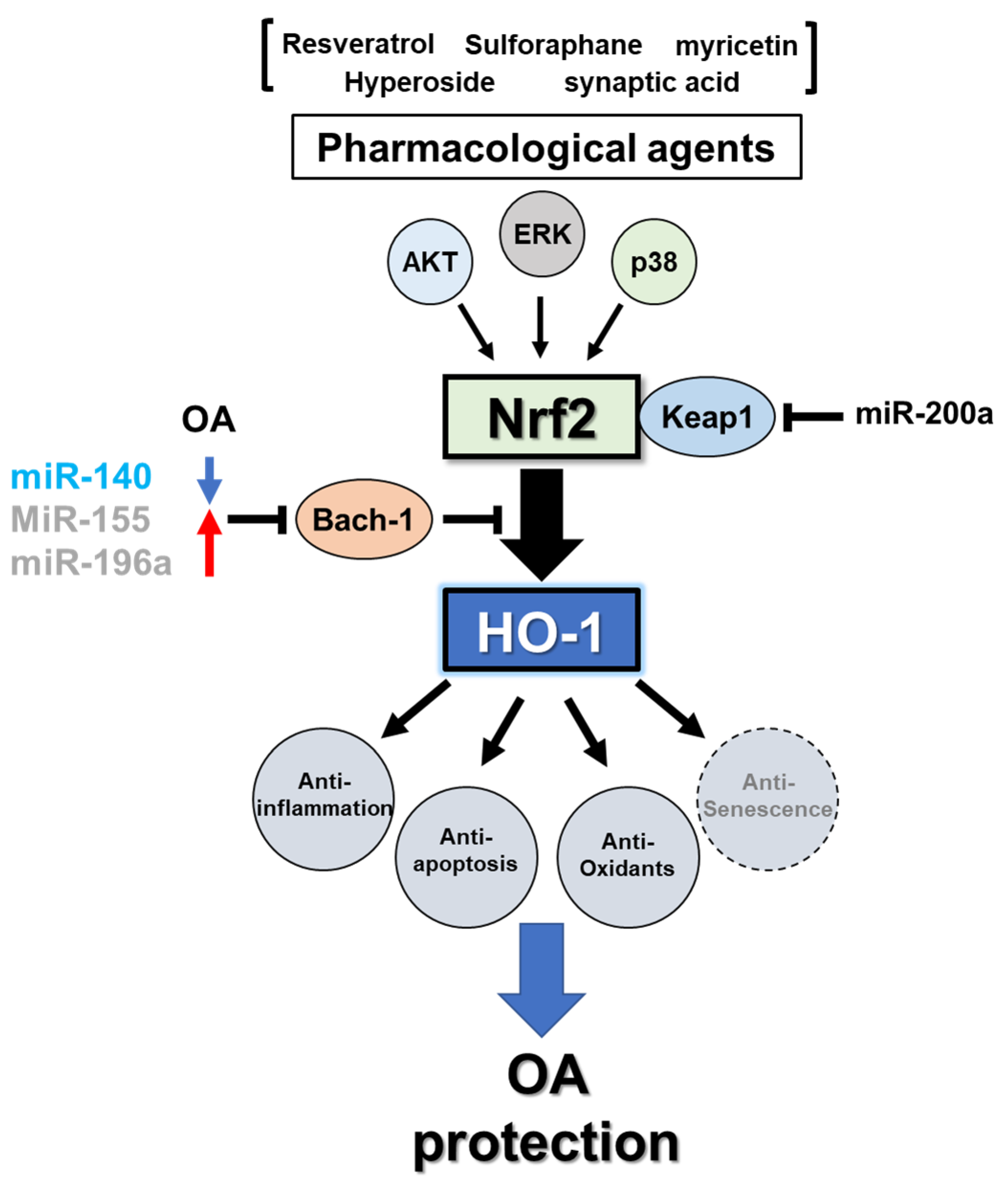
3.3. Regulation of Heme Oxygenase-1

HMOX1 is mostly regulated at the transcriptional level, with the regulatory mechanisms underlying its expression being highly complex and often cell-specific due to the extremely diverse array of stimuli that are able to induce HO-1. For example, HO-1 induction by heme, heavy metals, growth factors, NO, oxidized lipids, and cytokines has been experimentally tested and verified [78,79]. In particular, a large number of studies have reported that the expression of HO-1 is induced by stimuli that increase intracellular levels of ROS, such as heme, heavy metals, UV light, hydrogen peroxide, and lipopolysaccharide, or by stimuli that deplete cellular glutathione stores, including buthionine sulfoximine, sodium arsenite, and iodo-acetamide [80]. The linkage of HO-1-inducible expression to oxidative stress stimuli indicates that it is a component of an adaptive cytoprotective response to environmental stressors. Furthermore, it has been shown that scavengers of ROS, such as NAC, inhibit or reduce the extent of HO-1 induction by oxidative stress [81]. These results indicate that intracellular ROS plays an important role in HMOX1 gene expression regulation: free heme produced in response to oxidative stress is catabolized into a non-cytotoxic catabolite because oxidative stress is coupled to the induction of HO-1 [82].

3.3.1. Transcriptional Regulation

HMOX1 transcription can be induced by various signal transduction pathways that activate different transcription factors [58,83]. Given that the induction of HO-1 is tied to extracellular stimuli, its upstream mitogen-activated protein kinase (MAPK) signaling cascades such as ERK, c-Jun terminal kinase (JNK), and p38 MAPK are known to play a significant role in mediating gene expression [83,84,85,86,87]. However, the molecular regulation of HO-1 induction by stimuli is different between human and rodents [88]. Previous studies using an in vitro and humanized HO-1 transgenic mouse model revealed that several transcriptional factors such as transcription factor jun-B (JunB), Sp1, upstream stimulatory factor (USF) 1/2, and CTCF are uniquely involved in human HO-1 induction mechanisms through chromatin loop formation [23,24].
Nrf2, a 66 kDa protein and a member of the CNC-bZIP family of transcription factors, is considered to be particularly important given its crucial role for the protection of joint destruction through facilitating the induction of target genes such as NAD(P)H:quinone oxidoreductase 1 (NQO1), and, significantly, HO-1 [10,18,21,89,90]. The activity of Nrf2 is functional in a wide-ranging metabolic response to oxidative stress and constitutes a cellular sensor for oxidative stress by a nuclear shuttling mechanism with the cytosolic regulator protein Kelch-like ECH-associated protein 1 (Keap1) [91,92,93]. Kruppel-like factor 2 (KLF2), a member of the zinc finger family, has emerged as an important transcription factor in the development of OA. KLF2 expression is reduced in OA patient-derived cartilage and in IL-1β-stimulated SW1353 human chondrocytes. Genetic and pharmacological overexpression of KLF2 protects against OA progression by increasing the expression of HO-1 and NQO1 through the enhancement of Nrf2 nuclear translocation [94]. Previous studies reported that increasing KLF2 levels reduced the expression of MMP-3 and MMP-13 in monocytes, which attenuates the cartilage degradation process associated with OA [95]. Moreover, in human endothelial cells, KLF2 has been shown to play a crucial role in protecting against oxidative damage through the activation of Nrf2/HO-1 signaling [96,97].
The heme-binding protein Bach1 is a transcriptional repressor of HMOX1 due to its competitive relationship with Nrf2 for binding to ARE [98]. During oxidative stress or increasing heme concentrations, Bach1 is displaced from ARE and exported out of the nucleus to be degraded so that Nrf2 can associate with Maf and bind to ARE sequences [98]. Thus, cellular HO-1 inducive mechanisms are tightly regulated by extracellular conditions through the described Nrf2/Keap1/Bach1 system.

3.3.2. MicroRNA-Mediated Post-Transcriptional Regulation

MicroRNAs (miRNAs) are a major class of small noncoding RNAs found in animals, plants, and some viruses, which function as negative regulators of gene expression. They suppress messenger RNA (mRNA) translation by promoting their degradation via the RNA-induced silencing complex (RISC) [99,100]. miRNA expression levels are frequently altered by aging-related disorders such as OA and cancer [101,102,103,104,105]. miRNAs exist that indirectly modulate HO-1 upstream regulatory factors, such as Bach1, Nrf2, and Keap1. Eades et al. demonstrated, using miRNA-based microarray analysis, that miR-200a expression is silenced in breast cancer cells, and that ectopic re-expression increases Nrf2 nuclear translocation by directly binding to the Keap1 mRNA 3′-untranslated region (3′-UTR) and facilitating its degradation [106]. Kim et al. found that hypoxia-inducible miR-101 induces HO-1 expression in endothelial cells. Upregulated miR-101 targets the E3 ubiquitin ligase Cullin3 and stabilizes Nrf2, resulting in the enhancement of Nrf2 translocation into the nucleus [107]. On the other hand, ectopic expression of miR-28 in mammalian endothelial cells directly downregulates Nrf2 protein expression via 3′-UTR binding of Nrf2 mRNA independently from the Keap1 pathway [108]. Sangokoya et al. identified that patients of sickle cell disease highly express miR-144 in erythrocytes and abrogate antioxidant signals. miR-144 regulates Nrf2 expression through binding to the 3′-UTR of Nrf2 mRNA and modulates the oxidative stress response in K562 and primary erythroid progenitor cells [109].
miRNAs also modulate HO-1 activity by directly regulating HMOX1 expression. Beckman et al. performed in silico analysis of the human HMOX1-3′ UTR and identified two candidate miRNAs, miR-377 and miR-217, as possible inhibitors of HMOX1. Subsequent experiments found that co-transfection of miR-377 and miR-217 downregulated the luciferase activity of HMOX1-3′ UTR and HO-1 protein expression levels [110]. Our previous study established that miR-140 regulates HO-1 expression by binding to the 3′-UTR of BACH1 in human primary chondrocytes [14]. Similar investigations reported that miR-155 and miR-196a regulates the expression of HO-1 through the reduction in BACH1 expression in endothelial cells or hepatoma cells [111,112]. miR-155 is a pro-inflammatory miRNA that is significantly upregulated in OA and RA joints [113,114,115,116]. Moreover, upregulated miR-155 inhibits the expression of a number of core proteins in the autophagy cascade and promotes the cartilage degradation pathway in chondrocytes [113]. Autophagy is a critical evolutionarily conserved eukaryotic process that functions to maintain cellular homeostasis in response to changes to the environment, including in chondrocyte protection [117,118]. While autophagic activity is decreased in aging and OA-induced mouse knee cartilage, HO-1 overexpression is able to rescue the suppression of autophagic activity [13,50]. The pro-inflammatory cytokine TNF-α induces the expression of HO-1 and miR-155 in endothelial cells [111]. In contrast, IL-1β stimuli downregulates the expression of HO-1 and miR-140 in articular chondrocytes [14]. miR-140 is highly expressed in normal cartilage but is significantly reduced in OA cartilage. miR-140 is a cartilage-specific miRNA and is the most important regulatory factor in cartilage homeostasis [102,103]. Previously, we found that carnosic acid (CA), a natural flavonoid, raises the expression of HO-1 and miR-140 in articular chondrocytes through the transcriptional downregulation of BACH1. In addition, we observed that CA treatment alleviates IL-1β-induced cartilage damage. These results suggest that the regulation of Bach1-mediated HO-1 expression by miRNA may depend on cell type and context.

3.4. Heme Oxygenase-1 in Osteoarthritis

Elevation of oxidative stress in joint tissue has long been established as a crucial factor mediating the articular cartilage degradation process during OA [5,119]. Given its antioxidant properties, several studies have suggested a protective role of HO-1 in OA pathogenesis. It has been reported that HO-1 is significantly upregulated in human and mouse models of OA, with higher levels of expression being observed in areas of cartilage damage [120,121]. However, discrepancies exist regarding Nrf2/HO-1 expression between OA cartilage and IL-1β-induced OA-like chondrocytes [122,123]. Our previous study showed that HO-1 protein expression levels in IL-1β-primed normal human primary chondrocytes are significantly decreased compared with a control [14]. Further investigation is required to determine if there is a relationship between Nrf2/HO-1 signaling and OA.
HO-1 is able to confer a protective effect in OA chondrocytes by inhibiting the pro-catabolic effects of IL-1β on the ECM components MMP-1 and MMP-13 [124,125] while reducing the production of pro-inflammatory cytokines such as TNF-α, IL-1β, IL-6, and IL-18 [126]. Conversely, HO-1 simultaneously enhances the synthesis of anabolic factors such as IGF-1, proteoglycan, and COL2A1 [127], and increases anti-inflammatory IL-10 levels [124]. A wide array of studies has investigated the protective effects of HO-1 in OA chondrocytes through its regulatory relationship with the Nrf2/Bach1 system. Nrf2 activation in OA chondrocytes has been reported to inhibit mitochondrial dysfunction, ROS production, and apoptosis induced by IL-1β [120]. Nrf2 activity also inhibits inflammation and ECM degradation in OA chondrocytes [51,94,128] and regulates cellular differentiation in chondrocytes [129,130]. Bach1 deficiency significantly elevates HO-1 expression in healthy and aged articular cartilage and reduces the severity of age-related OA-like changes [13]. Moreover, Bach1 deficiency significantly inhibits the severity of OA-like changes, such as meniscus degradation, inflammatory changes in synovium, osteophyte formation, and subchondral bone thickening [13,19]. All in all, these results suggest that HO-1 expressed in whole joint cells plays a critical role in the maintenance of joint homeostasis.

3.5. Heme Oxygenase-1 and Osteoarthritis-Associated Cellular Senescence

Cellular senescence is a major instigator of both aging-associated and post-traumatic OA pathogenesis [131,132]. In particular, aging-induced cellular senescence is known to be the most important risk factor for primary OA progression. As such, senolytic drugs have rapidly captured the interest of academic research and business venture as a potential avenue for attenuating OA pathogenesis and pathology [133]. Senescent cells secrete a robust pro-inflammatory secretome, known as a senescence-associated secretory phenotype (SASP). A SASP consists of a variety of cytokines, growth factors, and other soluble and insoluble material that continuously alters the structure and function of surrounding cells and tissues in a paracrine manner [134]. Joint tissues (comprising articular cartilage, subchondral bone, synovium, and infrapatellar fat pad) suffering from OA induced by aging or trauma are likely to harbor senescent cells that secrete an OA-propagating SASP [134]. In addition, although the mechanism of action is not fully understood, it is known that oxidative stress induces senescence in chondrocytes through multiple complex signaling pathways [135]. ROS can further propagate this effect by eroding protective telomeres, resulting in accelerated senescence and chondrocyte apoptosis. Several studies have shown that high shear stress alone can induce chondrocyte senescence [136,137,138].
Although still a largely unexplored avenue, a few studies have investigated the interplay between HO-1 induction and chondrocyte senescence. Principally, HO-1 has been documented to protect articular cartilage against cellular senescence. One study reported that cilostazol-induced senescence significantly attenuated HO-1 expression in human chondrocytes and, conversely, that HO-1 overexpression exerts a protective effect against cilostazol-induced senescence [53]. A follow-up study found that a regulator of HO-1 in stress-induced chondrocytes known as protein kinase casein kinase 2 (CK2) is associated with senescence in primary articular chondrocytes, and that the downregulation of CK2 induces cellular senescence by inhibiting the expression of HO-1 [139]. A separate investigation demonstrated that the expression of senescence markers such as senescence-associated β-galactosidase, p21, and caveolin1 were significantly decreased after HO-1 induction [53,140]. Taken together, while the mechanism by which HO-1 can suppress cellular senescence progression remains to be fully understood, these studies indicate that HO-1 may be a potential therapeutic target for aging-related OA development.

4. Pharmacological Treatment for OA Protection

4.1. OA Animal Models for Drug Development

Although aging has become the most common cause of OA, the mechanisms that mediate the effect of age on OA have not yet been completely elucidated. Therefore, pharmacological approaches for neither OA prevention nor disease-modification are presently available. Efficient pharmacological testing is needed to develop novel OA-modifying drugs. Many OA animal models such as those using mice, rats, rabbits, guinea pigs, and large animals have been developed to simulate human OA in pharmacological trials. These models apply various methods for inducing OA-like conditions, including aging, surgical, chemical, and genetic modifications [141,142]. Although aging-derived oxidative stress is the most important OA risk factor, most in vivo studies use either surgically-induced (such as the destabilization of the medial meniscus (DMM) method with transection of ligaments) or chemically-induced OA models [141,142,143]. Antioxidants such as NAC and procyanidins attenuate the development of OA induced by DMM surgery [61,62]. These models are useful for studying post-traumatic secondary OA, but may not be a valid method for studying the mechanisms and treatment of spontaneous aging-associated primary OA.
Spontaneous OA in mice without intervention develops much more slowly than the aforementioned induced OA models. In C57BL/6J mice, OA-like changes become detectable only by 12–18 months. Dunkin-Hartley guinea pigs are a widely used model of spontaneous aging-associated OA in the knee and other joints. Histological changes in the joints can be observed by three months of age, with disease severity increasing as time passes. Eventually, moderate to severe OA is observed around 18 months of age [144,145,146]. Although these models are advantageous, in that they are founded on pathological changes rather than post-traumatic alterations, their application is limited by the significant amount of time it takes for them to develop desirable characteristics. STR/ort mice have been established as a practically convenient mouse model of naturally occurring OA due to their relatively early onset of proteoglycan loss, articular cartilage fibrillation, ECM degradation, osteophyte formation, and subchondral sclerosis in the medial tibial condyle [147,148]. In STR/ort mice, serum levels of malondialdehyde (MDA; an oxidative stress marker) and C-terminal telopeptide of collagen type II (CTX-II; a COL2A1 degradation marker) are both higher than in control CBA mice prior to OA onset, suggesting that oxidative stress is linked to cartilage degradation [119]. Moreover, STR/ort mice tend to exhibit a higher incidence and severity of OA from 18 weeks of age, with severe lesions affecting the majority of animals by 15 months [148,149]. However, there are variations in the incidence and severity of OA within and among mouse models. The development of animal models is therefore required to further our understanding of the therapeutic efficacy of novel treatment modalities for primary OA.
In Table 2, we list OA animal models that have been used for the pharmacological testing of various antioxidant inducers. Most studies utilize methods for inducing secondary OA in animal models, such as through surgical-based DMM or anterior cruciate ligament transection (ACLT), or through chemical-based mono-iodoacetate (MIA) or papain. These animal models establish the OA phenotype in a simple, rapid, and reproducible manner. On the other hand, STR/ort mice, which are characterized by their spontaneous OA phenotype, have only been used in a single evaluation of HO-1 function in joint homeostasis via an HO-1-inducing adenovirus expression model. Furthermore, although several genetically modified mice have been developed to understand the correlation between redox signals such as Nrf2/HO-1 and OA development (see Table 3), such animal models have not yet been applied in the pharmacological study of antioxidants.

4.2. Identifying a Role for HO-1 from a Genetic Modification Mouse Model

A variety of genetically modified animal models that mimic the pathophysiology of OA have been developed to improve our understanding of the condition. In this way, numerous genetically modified animal models have been developed to examine the molecular mechanisms of Nrf2/HO-1 signaling in chondroprotective function (see Table 3). Nrf2-deficient mice that have undergone MIA or DMM exhibit severe cartilage degradation due to reductions in antioxidant signaling activity [11]. The use of histone deacetylase inhibitor TSA inhibits OA progression in DMM and MIA-induced OA via Nrf2 acetylation and activation of the downstream cascade. These effects are abolished by Nrf2 deficiencies in OA-induced mice. Moreover, Nrf2−/− primary chondrocytes exhibited higher responsiveness to IL-1β stimuli in high glucose conditions due to the reduced expression of antioxidant genes [154]. In K/BxN spontaneous inflammatory arthritis and antigen-induced arthritis (AIA) mouse models, Nrf2 deficiencies exhibited more severe structural alterations in the joints such as synovitis, cartilage destruction, and bone erosion compared with wild type arthritic mice [56,57]. On the other hand, Nrf2 constitutively accumulated in the nuclei of Keap1-null mice, leading to the overproduction of cytoprotective target factors. Although Keap1-null mice generate high levels of cytoprotective factors, these mice exhibit severe abnormal keratinization and cornification in the esophagus and forestomach. Keap1 and Nrf2 double knock out (KO) mice reversed the aberrant phenotypic Keap1 deficiencies [156]. These results suggest that adequate Keap1-mediated Nrf2 activation and the regulation of downstream target genes exert a major protective function in OA development and joint arthritis pathogenesis.
The effects of HO-1 deficiency on pathology and pathogenesis have been evaluated in K/BxN mouse models. HO-1-deficient mice exhibit growth retardation, hepatic and renal iron accumulation, and chronic inflammation, thereby sharing a similar phenotype to genetically HO-1-deficient humans [75,76,157]. The incidence and severity of arthritis in arthritic mouse models was higher in HO-1-deficient groups compared with wild type groups [158]. The effects of the pro-inflammatory glucan zymosan on the acute inflammatory response of myeloid-specific HO-1 deficient mouse (HO-1M-KO mouse) was investigated. It was found that zymosan stimulus induced higher serum inflammatory cytokine levels in HO-1M-KO mice compared with wild type mice [159]. Conversely, the consecutive induction of HO-1 via Bach-1 KO protects against the development of experimental- and aging-induced OA [13]. Mild induction of HO-1 expression was ubiquitously observed in Bach-1 KO mice, including in chondrocytes, menisci, and bone marrow macrophages. Moreover, the consecutive induction of HO-1 in Bach-1 KO mice inhibited degeneration in the intervertebral disc after puncture [20]. Oxidative stress accelerates osteoclastogenesis and bone resorption. Abnormal bone resorption in the subchondral bone promotes bone deposition and leads to subchondral sclerosis. Bach-1 KO exhibited resistance to TNF-α-induced inflammatory bone loss in calvarial tissue [160]. In vitro Bach-1 KO bone marrow-derived macrophages exhibited suppressed mature osteoclast differentiation capacity, with knockdown of HO-1 partially reversing the suppressive effects of osteoclastogenesis in the presence of Bach1 deficiency. These results indicate that HO-1 has crucial roles in homeostasis, and that the consecutive mild induction of HO-1 via natural inducers may be able to prevent spontaneous OA development.
Findings from these animal studies are useful information for identifying the functions of Nrf2/HO-1 signaling in OA. However, further application of tissue-specific genetically modified mice using the Cre recombinase system is needed to better our understanding of the detailed molecular mechanisms of the Nrf2/HO-1 axis in joint homeostasis.

4.3. Pharmacological Treatment with HO-1

HMOX1 expression is highly transcriptionally regulated by several injurious stress conditions such as ischemia, atherosclerosis, and inflammation [74,161]. Takeda et al. showed that the pharmacological induction of HO-1 by chemical compounds ameliorates lupus nephritis (an autoimmune disease of the kidney) by suppressing NO-dependent inflammatory responses [162]. Moreover, overexpression of HO-1 by adenovirus vector-mediated gene transfer has protective functions on lipopolysaccharide-induced lung injury [163]. These effects were produced by a systemic or local induction of HO-1. Induction of HO-1 reduces the activity of inflammatory pathways and the production of matrix degradation enzymes in in vitro OA synoviocytes and chondrocytes [54,124]. Antioxidant signal inducers such as resveratrol [126], Sauchinone [164], Licochalcone A [165,166], sinapic acid [167], Monascin [168], wogonin [169], Protandim and 6-Gingerol [122] and Nomilin [170] have all been shown to exert anti-inflammatory and chondroprotective properties in joint tissues through activation of Nrf2/HO-1 pathways both in vivo and in vitro.
Searching the following keywords (Nrf2 or HO-1) and (Osteoarthritis) in the PubMed database produced 88 articles and reviews. In this review, we selected pharmacological animal studies and categorized them based on drug name, administered method, animal model (primary or secondary), and whether Nrf2/HO-1 was expressed in the joint (see Table 4).

4.3.1. Intra-Articular Injection-Based Delivery

The chosen route of administration is one of the most important factors affecting the results of in vivo animal studies. Therapeutics that can be delivered directly into the joint space offer the advantage of providing a high local concentration at the disease site while reducing the potential for adverse effects associated with systemic delivery. Corticosteroids and hyaluronate are potent anti-inflammatory agents with long-standing use in OA treatment. Intra-articular (IA) corticosteroid or hyaluronate injections are usually applied to patients who exhibit an inadequate response to initial therapy [183]. Thus, IA space injections are a safe and useful method for applying a local OA treatment.
IA injection is also able to deliver genetic modifications in specific nonvascular local areas such as cartilage. Previous studies have demonstrated that the adenovirus system effectively delivers genes to knee joint cartilage [94,184,185]. Gao et al. showed that KLF2 overexpression in SW1353 human chondrocytes protects against IL-1β-induced ROS accumulation, catabolic gene expression, and apoptosis, via activation of the Nrf2/ARE/HO-1 signaling cascade [94]. OA rat models have been established through IA injections of MIA (1 mg per cavity in 50 μL of sterile saline), an inhibitor of glycolysis. After injection of MIA on day three, either adenovirus-Klf2 (Ad-Klf2) or adenovirus-control was injected into the knee joints for three consecutive weeks (109 plaque-forming units in a total volume of 10 μL). Immunohistochemical analysis revealed that MIA-injected OA rat knees featured increased quantities of TUNEL and MMP-13-positive cells in cartilage along with a dramatic reduction in KLF2 expression. Ad-Klf2 IA injection increased KLF2 expression in MIA-induced OA cartilage, which resulted in elevated Nrf2 activation in cartilage tissue and protected against proteoglycan loss. Conversely, IA injection of ML385, an Nrf2 inhibitor, significantly abrogated the KLF2-mediated suppression of apoptosis and MMP-13 elevation induced by MIA in vivo [94]. These results indicate that the KLF2/Nrf2/HO-1 axis forms a chondroprotective mechanism that induces anti-inflammation and anti-apoptosis and decreases catabolic factors in joint cartilage tissues. IA therapy applying gene modification methods might be a promising strategy for OA treatment.
Adeno-associated virus (AAV)-based gene delivery is an effective method for transducing genetic material into both systemic and local tissue, including into MIA-induced OA rat cartilage [117,186,187]. While gene therapy has been tested in human joints in clinical trials for RA and OA patients [188], the potential application of AAV-based gene transduction in spontaneous OA knee cartilage remains unclear. Kyostio-Moore et al. performed AAV-based HO-1 injections into spontaneous OA STR/ort mouse knee joints and observed robust transduction in synovial tissues and skeletal muscle, but not in cartilage. Similarly, they also found that AAV1/LacZ-treated joints exhibited β-gal activity in synovial tissue but not in cartilage [182]. Importantly, these AAV/HO-1 injections did not show an effective role for cartilage degradation in the STR/ort mouse. AAV-based gene therapy is therefore a useful tool for the delivery of treatments for arthritis and cartilage degradation. However, the efficiency of the transfer of genetic material into cartilage in spontaneous OA needs to be studied further in the context of a clinical trial.
Yang et al. have developed a new drug delivery system for targeting synovium-activated macrophages that is based on CO gas therapy [181]. CO has multifunctional anti-inflammatory functions through the regulation of numerous cellular signaling pathways including the NF-κB, toll-like receptor (TLR), and MAPK pathways [189,190,191]. CORM-401, a soluble oxidant-sensitive CO-releasing molecule, can be used to deplete H2O2 secreted by activated macrophages and elicit the responsive release of CO. To efficiently deliver CORM-401 into activated macrophages, CORM-401 was encapsulated in peptide dendrimer nanogel (PDN) carriers that were modified by folic acid (FA; a ligand of folate receptor beta) and hyaluronic acid (HA; a ligand of HA receptor (CD44)). PDN-encapsuled CORM-401 (CPHs) were injected into an MIA-induced OA rat knee joint to generate a new type of anti-inflammatory drug. Injection of MIA-induced experimental OA can easily and quickly reproduce loss of articular cartilage with severe synovitis induced by activated macrophage infiltration, ROS accumulation, and functional impairment in ratssimilar to that observed in human disease [152,153,192]. CPH treatment significantly suppressed cartilage degradation through CO release and elicited CO-induced HO-1 activation in the activated macrophage present in the synovial membrane [181]. These results indicated that targeting activated macrophages in the synovial membrane can inhibit OA progression and exert an anti-inflammatory response in joints. However, most triggers of OA pathogenesis are due to non-inflammatory changes to cartilage. As such, we must consider the function of HO-1 in the context of cartilage-specific non-inflammatory cellular protection.
Antioxidant IA injections have been shown to produce beneficial effects in joints outside the knee, such as the temporomandibular (TMJ) joint [180]. Resveratrol is a one of the most studied non-flavonoid polyphenol compounds with a stilbene structure. Resveratrol has been shown to suppress the activation of several transcription factors, including NF-κB, AP-1, and Egr-1, and to downregulate inflammatory products such as COX-2, VEGF, IL-1, IL-6, and TNF-α [193,194]. Imbalances in redox homeostasis leads to the accumulation of ROS in the cartilage, which results in the elevated secretion of pro-inflammatory cytokines due to the activity of pathological factors expressed through the activation of NF-κB signaling such as TNF-α and IL-1β [195,196]. To determine the effects of resveratrol on inflammatory damage in OA cartilage, Yulong et al. performed resveratrol IA injections in MIA-induced OA rat knee joints [126]. Resveratrol was administered to the articular space at 50 mg/kg every three days for eight weeks. Resveratrol treatment ameliorated rat OA clinical scoring, with reductions in swelling and inflammatory cytokine expression. Moreover, resveratrol treatment increased the expression of antioxidant factors SOD2 and HO-1 in joints and reduced ROS accumulation, leading to the inhibition of caspase3/9 activation in cartilage [126].
Resveratrol is well-known as a potent silent information regulator 2 type 1 (SIRT1) activator, which has crucial roles during the inhibition of NF-κB signaling in OA chondrocytes [155,197,198]. SIRT1 is associated with cartilage homeostasis and numerous cellular signaling pathways that respond to cellular energy and redox status, including anti-inflammation, senescence, apoptosis, and autophagy [197]. SIRT1 expression is decreased by aging in arthritis joint cartilage [176,179]. Genetic or chemical induction of SIRT1 suppressed inflammatory signaling and cartilage degradation, but enhanced microtubule-associated proteins 1A/1B-light chain 3B (LC3) expression and ECM synthesis [13]. Importantly, consecutive HO-1 overexpression via Bach1 knockout (Bach1−/−) also enhanced LC3 expression and autophagy activity in the aging cartilage. On the other hand, HO-1 siRNA transfection into primary Bach1−/− chondrocytes abolished LC3 activation in IL-1β-stimulated chondrocytes [13]. These observations suggest that the resveratrol-related cartilage protective function is partially due to the SIRT1/HO-1/LC3 axis. Moreover, injecting high doses of resveratrol (100 μg once a week) into DMM-induced knee IA spaces prevents cartilage degeneration through SIRT1 induction in cartilage [178]. Thus, local HO-1 induction by resveratrol IA injection represents a promising pharmacological strategy for OA with reduced side-effects.
Because IA space injections are a safe and useful method for local OA treatment, clinicians usually select HA IA injection for inducing pain relief in OA patients. However, at a clinical level, serial IA injections for an extended period present practical and inconvenient difficulties.

4.3.2. Intraperitoneal Injection-Based Delivery

There are two methods that are commonly used to model intermittent pharmacological tests: intraperitoneal (IP) injection and intragastric delivery by oral gavage (PO). PO is known to be a safe option for administering drugs because they are metabolized in the liver. Conversely, IP injection directly affects compound concentrations in serum levels, which can manifest as complications later on. Furthermore, there is an added risk of causing internal organ injury or of misplaced injection into the intestinal tract. This route of administration in animal studies is rarely used in repeated dose studies.
Sinomenine (SIN), a traditional Chinese medicinal herb, and scutellarin (SCU), a flavonoid glycoside, were administered once a day to DMM surgical model mice via IP injection. SIN IP injections were started at two weeks after surgery, while SCU IP injections were carried out immediately after surgery. These models exhibited reduced levels of cartilage destruction in DMM knee joints through the suppression of pro-inflammatory gene expression in cartilage without inducing noticeable toxic side effects [199,200]. SIN and SCU treatments enhanced Nrf2 translocation into the nucleus and increased HO-1 expression in mouse primary chondrocytes. Interestingly, SCU was able to bind to the Nrf2 binding site and activate its nuclear translocation. Peiminine (Pm), the active component of the Asian herb Fritillaria verticillata, was also IP-injected into DMM-induced mice. Pretreatment with Pm in IL-1β-induced mouse primary chondrocytes inhibited the expression of ECM degradation enzymes and pro-inflammatory cytokines. Moreover, Pm suppressed NF-κB signaling through the inhibition of AKT phosphorylation by activating Nrf2/HO-1 signaling in chondrocytes. In in vivo experiments, Pm treatment reduced the cartilage loss and increased Nrf2-positive chondrocyte numbers in DMM-induced OA cartilage [177]. Although the application of IP treatment in animal studies seems to be a useful method for OA prevention, its application in clinical medicine is unusual.
Non-irritating saline or phosphate buffer saline (PBS) solvents may be used to reduce IP-associated complications. Sun et al. investigated the anti-arthritic effects of saline-dissolved hyperoside (Hyp), a bioactive flavonol glycoside [201]. Hyp treatment inhibited nitric oxide synthase (iNOS), cyclooxygenase-2 (COX2), and metalloproteinase protein expression in IL-1β-induced mouse chondrocytes. IL-1β-induced ROS accumulation can induce mitochondria dysfunction, eventually leading to chondrocyte apoptosis. Hyp treatment significantly reduced ROS accumulation and protein expression of downstream pro-apoptotic factors Bcl-2-associated X (BAX), cytochrome c, cleaved caspase-9, and cleaved caspase-3. Hyp increased Nrf2 and HO-1 expression, and Nrf2 siRNA transfection partially abolished the anti-apoptotic role of Hyp treatment in IL-1β-induced chondrocytes. Moreover, Hyp prevented the phosphorylation of PI3K, NF-κB, AKT, ERK, JNK, and c-Jun. These results directly or indirectly indicate that Hyp has an anti-inflammatory role via in the inhibition of PI3K/AKT/NF-κB and MAPK signaling pathways in chondrocytes. In an in vivo study, Hyp IP injection was performed every other day in DMM-induced OA mice for four or eight weeks. Knee joint histological analysis indicated that Hyp ameliorated cartilage destruction caused by surgically-induced chondrocyte abnormalities via the enhancement of Nrf2/HO-1 expression and PI3K/AKT/NF-κB and MAPK signaling inhibition [201].

4.3.3. Oral Administration

OA is an aging-associated whole joint disease, which means that the whole-body regulation of the aging process by continuous consumption of antioxidant signal inducers (antioxidants) extracted from natural compounds may represent a more efficient method for delaying OA development. Antioxidants can be orally administered by ingesting supplements and natural food sources such as fruits and vegetables. Genistein, an isoflavone extracted from soybean, was administered through standard feeding (40 mg/kg) in ACLT-induced OA rat models [172]. The compound downregulated IL-1β-induced MMPs, NOS2, and COX2 expression, while stimulating HO-1 expression in human primary chondrocytes. Cartilage degeneration in ACLT-induced OA rat knees was significantly inhibited by oral administration of genistein. Sulforaphane (SFN; 1-isothiocyanato-4-methylsulphinylbutane) is a plant-derived isothiocyanate obtained in the diet through the consumption of cruciferous vegetables (particularly broccoli) [202]. SFN protects against oxidative stress through the Nrf2-mediated induction of phase II detoxification and is known to reduce the risk of contracting various cancers. SFN inhibits the cytokine-induced expression of numerous metalloproteinases, such as MMP-1, MMP-3, ADAMTS4, and ADAMTS5, in human articular chondrocytes, fibroblast-like synoviocytes, and the SW-1353 chondrosarcoma cell line. These beneficial effects are abolished by the transfection of Nrf2 siRNA into in vitro articular joint cells. SFN-mediated activation of the Nrf2 pathway and the subsequent increase in HO-1 expression inhibits cytokine-induced MAPK and NF-κB signaling in human primary chondrocytes. To confirm these results in an in vivo murine model of cartilage destruction, an SFN-rich diet (3 μmol/day) was fed to mice that had underwent DMM. The SNF-rich diet-fed mice exhibited significantly reduced cartilage destruction at 12 weeks after DMM surgery compared to those fed a control diet [175]. Genistein and SFN are potent inducers of Nrf2/HO-1 expression in several joint cell types. Nrf2 activation enhances antioxidative signaling while inhibiting MAPKs and NF-κB signaling in joint cells. However, while genistein and SFN reduced the severity of surgically-induced OA in in vivo experiments, no significant changes to Nrf2/HO-1 signaling were observed in joint tissues.
To directly confirm drug functions in vivo, several experiments were performed using intragastric gavage administration. Chemically-induced OA models, such as papain injection into the IA space, were also used to investigate pharmacological effects [150]. Papain-induced OA knee joints exhibited severe cartilage degradation and synovitis, characterized by high levels of inflammatory immune cell invasion [203,204]. Primary rat fibroblast-like synoviocytes (OA-FLS) were isolated from papain-induced OA joints and used for in vitro study. The dihydroartemisinin derivative DC32 [(9α,12α-dihydroartemisinyl) bis (2′-chlorocinnmate)] was added into cultured OA-FLS with or without the pro-inflammatory cytokine TNF-α. DC32 suppressed the mRNA expression of pro-inflammatory cytokines as well as OA-FLS migration that normally accompanies TNF-α stimulation. To investigate the pharmacological mechanisms of DC32, MAPKs and NF-κB signaling and the Nrf2/HO-1 pathway were evaluated by Western blot and qPCR analysis. DC32 significantly inhibited the phosphorylation of ERK, but not JNK. Moreover, NF-κB signaling was also suppressed by DC32 treatment. These positive effects were abolished by Nrf2 siRNA transfection, indicating that they are tied to the Nrf2/HO-1 pathway [174]. Oral administration of DC32 in papain-induced OA models showed that the treatment ameliorates pain relief, joint swelling, and cartilage destruction when compared with a control. Chemical injection into articular space was observed to cause cartilage degradation, severe synovitis, and ROS accumulation in the synovium. Chemically-induced OA models therefore not only induce extracellular loss in cartilage, but also an RA-like inflammatory phenotype. Nrf2/HO-1-inducing compounds such as NAC and resveratrol are known to have potent anti-inflammatory functions in rheumatic synovium and OA-FLS [205,206].
Surgically-induced OA models such as DMM and ACLT are the most commonly applied method of OA induction because of the ease by which the condition can be induced [143,151]. Moracin, a natural flavonoid compound extracted from Cortex Mori (the root bark of Morus alba), is known for its anti-inflammatory activity via the suppression of NF-κB signaling pathway activation [207,208]. Moracin inhibits IL-1β-induced catabolic gene expression in rat primary chondrocytes through the inhibition of NF-κB signaling via Nrf2/HO1 activation. ACLT-induced OA rats were given 30 mg/kg moracin by gavage once every two days for eight weeks. Moracin administration prevented cartilage degradation and increased Nrf2- and Col2a1-positive cell numbers in cartilage tissue [173]. Piceatannol, a hydroxystilbene derived from the Euphorbia lagascae seeds, is present in various fruits and vegetables and has been reported to possess anti-cancerous and anti-inflammatory activities by the blocking of NF-κB signaling [209,210,211]. In human primary chondrocytes, piceatannol suppresses p65 phosphorylation through the stimulation of IL-1β, inhibits iNOS and COX2 mRNA and protein levels, and reduces NO generation and PGE2 expression in a dose-dependent manner. To investigate the protective effect of piceatannol in OA development in vivo, piceatannol (10 mg/kg in 0.5% carboxymethylcellulose) was administered intragastrically to DMM-induced OA mice once daily for eight weeks. The µCT and safranin-O staining results indicated that piceatannol treatment prevents cartilage degeneration in OA mouse models. Moreover, immunohistochemical analyses using Nrf2 and COL2A1 antibodies indicated elevated levels of Nrf2 and COL2A1 and increased ECM synthesis due to piceatannol treatment in OA cartilage [171]. Altogether, the administration of antioxidants derived from natural sources by oral gavage can prevent surgically-induced OA progression through the enhancement of Nrf2 antioxidant signaling.
Although HO-1 is one of the most important target genes regulated by Nrf2, the aforementioned studies using various antioxidant compounds do not directly indicate the molecular mechanisms of Nrf2 activation in vivo. Cai et al. showed that Nrf2/HO-1 protein expression levels in both sham and OA knee cartilage were increased by the intragastric administration of sinapic acid (SA; 10 mg/kg every two days) in DMM-induced OA mice. Moreover, SA treatment significantly decreased the mRNA expression levels of cartilage degradation enzymes, Mmp1, Mmp3, Mmp13, and Adamts5, and pro-inflammatory cytokines Il-1, Il-6, and Tnf-α at eight weeks after DMM surgery. The OsteoArthritis Research Society International (OARSI) scoring system was used to quantify the histopathological changes. The result showed that scores of the SA treatment group were much lower than the control group. In contrast, co-IA injection with sn-protoporphyrin (SnPP), an important HO-1 inhibitor, abolished the therapeutic effects of SA treatment [121]. These results indicate that oral administration of SA was protective against the progressive cartilage damage of osteoarthritis through HO-1 activation in knee cartilage.
Myricetin, a naturally occurring flavanol, is consumed in most diets or dietary supplements [212]. Myricetin treatment of human primary chondrocytes pre-conditioned with IL-1β inflammatory stimuli suppressed the generation of inflammatory mediators, cartilage degradation enzymes, and p65 phosphorylation. On the other hand, myricetin enhanced Nrf2 nuclear translocation and subsequent HO-1 expression in IL-1β-conditioned human chondrocytes in a dose-dependent manner. The observed chondroprotective functions were partly abolished via the co-treatment of Ly294002, a PI3K/Akt inhibitor. After DMM surgery, 20 mg/kg of myricetin was administered via intragastric administration every two days for eight weeks. The cartilage safranin-O staining area was significantly decreased in DMM mice that did not receive myricetin treatment. Conversely, myricetin-treated mice exhibited significantly improved surface structure and suppressed ECM degradation. Immunohistochemical analysis was used to evaluate the PI3K/Akt mediated Nrf2/HO-1 signaling activation in cartilage cells. In the myricetin treatment group, Nrf2 translocation to the nucleus and phosphorylated Akt expression was found to be significantly higher than in the untreated DMM group [128].
Collectively, these results suggest that the administration of antioxidants prevents OA development in OA-induced models via mechanisms that enhance Nrf2/HO-1 activation in cartilage (Figure 1). The cells involved possess similar antioxidant signaling cascades that activate Nrf2 nuclear translocation and bind to AREs upstream of HMOX1. Moreover, Nrf2 can directly stimulate ERK signaling to inhibit apoptosis. Therefore, to understand the critical roles of Nrf2/HO-1 signaling in spontaneous OA development, we must evaluate the precise activating location of Nrf2/HO-1 signaling in pharmacological trials using spontaneous OA animal models. Several groups, including our own, have reported that senescence accelerated mice (SAM), a rapidly aging mouse derived from the AKR/J strain, feature early spontaneous OA in the knee joints [213,214]. These animal models will be useful tools for understanding and evaluating the effective roles of antioxidants compounds. However, surgically- or chemically-induced OA pathogenesis does not sufficiently consider the characteristics of spontaneous OA development, which is uniquely caused by whole joint disabilities involving chondrocyte or bone metabolism and changes to the synovium and muscle.

5. Perspectives and Conclusions

OA is a global health issue marked by substantial disability and cost of medical care [1,43]. Besides contemporary surgical options (mostly through joint replacement surgery), there are no clinically available drugs that delay onset or the progression of the disease. Many age-related disabilities, including OA, are underpinned by oxidative stress and redox imbalance. Nrf2 and HO-1 are key factors that regulate redox homeostasis. The role of Nrf2/HO-1 signaling in OA has been highlighted in multiple studies, with the regulatory mechanisms of HO-1 being described as a highly promising avenue for attenuating oxidative stress at the cellular level. It is, however, unlikely that brute induction of Nrf2/HO-1 could pose as a solution; a number of reports have found evidence that permanent overactivation of Nrf2/HO-1 leads to numerous undesirable consequences, such as the dysregulation of hematopoietic regeneration [215], and neonatal jaundice [216,217]. Moreover, there are differences between HO-1 inductive mechanisms in human and rodents, making it difficult to directly apply the principles of animal studies to the clinical level. Conversely, mild HO-1 induction (3–5-fold increase in whole tissue) via Bach-1 deficiency has been documented to reduce the severity of OA-like changes in mice without debilitating effects [13]. An overall assessment of pharmacological animal studies gives the impression that a complete study of the regulatory mechanisms of HO-1 and its targets in whole joints has not yet been achieved. The link between cartilage homeostasis and Nrf2/HO-1 signaling has, to date, mostly been explored only in secondary OA animal models (see Table 1 and Table 3). A fuller picture of the detailed mechanisms explaining how HO-1 affects joint homeostasis is therefore required before potent drugs for primary OA management can be developed. In addition, understanding of the adequate level of HO-1 expression for attenuating OA pathology and pathogenesis in the whole joint including cartilage is required. While pharmacological inducers of HO-1 may be an effective therapeutic avenue for treating OA, it is more likely that clinical approaches of Nrf2/HO-1 will manifest in preventative healthcare and anti-aging therapies rather than as DMOADs. We therefore think that clinical research exploring prophylactic options (such as dietary modifications, etc.) of Nrf2/HO-1 induction is more likely to yield significant results.

Author Contributions

Conceptualization, Y.S., N.A. and S.M.; writing—original draft preparation, Y.S., S.J.O.T. and S.M.; writing—review and editing, Y.S., S.J.O.T. and S.M. All authors have read and agreed to the published version of the manuscript.

Funding

This research was supported by the Mishima Kaiun Memorial Foundation (Y.S.) and MEXT/JSPS KAKENHI for Grant-in-Aid for Young Scientists (B) 17K16693 (Y.S.).

Conflicts of Interest

The authors declare no conflict of interest.

References

  1. Hoy, D.G.; Smith, E.; Cross, M.; Sanchez-Riera, L.; Blyth, F.M.; Buchbinder, R.; Woolf, A.D.; Driscoll, T.; Brooks, P.; March, L.M. Reflecting on the global burden of musculoskeletal conditions: Lessons learnt from the Global Burden of Disease 2010 Study and the next steps forward. Ann. Rheum. Dis. 2015, 74, 4–7. [Google Scholar] [CrossRef] [PubMed] [Green Version]
  2. Loeser, R.F.; Goldring, S.R.; Scanzello, C.R.; Goldring, M.B. Osteoarthritis: A disease of the joint as an organ. Arthritis Rheum. 2012, 64, 1697–1707. [Google Scholar] [CrossRef] [PubMed] [Green Version]
  3. Burr, D.B.; Gallant, M.A. Bone remodelling in osteoarthritis. Nat. Rev. Rheumatol. 2012, 8, 665–673. [Google Scholar] [CrossRef]
  4. Del Carlo, M.; Loeser, R.F. Increased Oxidative Stress with Aging Reduces Chondrocyte Survival: Correlation with Intracellular Glutathione Levels. Arthritis Rheum. 2003, 48, 3419–3430. [Google Scholar] [CrossRef]
  5. Lepetsos, P.; Papavassiliou, A.G. ROS/oxidative stress signaling in osteoarthritis. Biochim. Biophys. Acta Mol. Basis Dis 2016, 1862, 576–591. [Google Scholar] [CrossRef]
  6. Scott, J.L.; Gabrielides, C.; Davidson, R.K.; Swingler, T.E.; Clark, I.M.; Wallis, G.A.; Boot-Handford, R.P.; Kirkwood, T.B.L.; Talyor, R.W.; Young, D.A. Superoxide dismutase downregulation in osteoarthritis progression and end-stage disease. Ann. Rheum. Dis. 2010, 69, 1502–1510. [Google Scholar] [CrossRef] [Green Version]
  7. Balaban, R.S.; Nemoto, S.; Finkel, T. Mitochondria, oxidants, and aging. Cell 2005, 120, 483–495. [Google Scholar] [CrossRef] [Green Version]
  8. Finkel, T. Signal transduction by reactive oxygen species. J. Cell Biol. 2011, 194, 7–15. [Google Scholar] [CrossRef] [Green Version]
  9. Loeser, R.F.; Carlson, C.S.; Del Carlo, M.; Cole, A. Detection of nitrotyrosine in aging and osteoarthritic cartilage: Correlation of oxidative damage with the presence of interleukin-1β and with chondrocyte resistance to insulin-like growth factor 1. Arthritis Rheum. 2002, 46, 2349–2357. [Google Scholar] [CrossRef]
  10. Alcaraz, M.J.; Ferrándiz, M.L. Relevance of Nrf2 and heme oxygenase-1 in articular diseases. Free Radic. Biol. Med 2020, 157, 83–93. [Google Scholar] [CrossRef] [PubMed]
  11. Cai, D.; Yin, S.; Yang, J.; Jiang, Q.; Cao, W. Histone deacetylase inhibition activates Nrf2 and protects against osteoarthritis. Arthritis Res. Ther. 2015, 17. [Google Scholar] [CrossRef] [Green Version]
  12. Koike, M.; Nojiri, H.; Ozawa, Y.; Watanabe, K.; Muramatsu, Y.; Kaneko, H.; Morikawa, D.; Kobayashi, K.; Saita, Y.; Sasho, T.; et al. Mechanical overloading causes mitochondrial superoxide and SOD2 imbalance in chondrocytes resulting in cartilage degeneration. Sci. Rep. 2015, 5, 11722. [Google Scholar] [CrossRef] [PubMed] [Green Version]
  13. Takada, T.; Miyaki, S.; Ishitobi, H.; Hirai, Y.; Nakasa, T.; Igarashi, K.; Lotz, M.K.; Ochi, M. Bach1 deficiency reduces severity of osteoarthritis through upregulation of heme oxygenase-1. Arthritis Res. Ther. 2015, 17. [Google Scholar] [CrossRef] [PubMed] [Green Version]
  14. Ishitobi, H.; Sanada, Y.; Kato, Y.; Ikuta, Y.; Shibata, S.; Yamasaki, S.; Lotz, M.K.; Matsubara, K.; Miyaki, S.; Adachi, N. Carnosic acid attenuates cartilage degeneration through induction of heme oxygenase-1 in human articular chondrocytes. Eur. J. Pharmacol. 2018, 830, 1–8. [Google Scholar] [CrossRef]
  15. Sun, J.; Wei, X.; Lu, Y.; Cui, M.; Li, F.; Lu, J.; Liu, Y.; Zhang, X. Glutaredoxin 1 (GRX1) inhibits oxidative stress and apoptosis of chondrocytes by regulating CREB/HO-1 in osteoarthritis. Mol. Immunol. 2017, 90, 211–218. [Google Scholar] [CrossRef] [PubMed]
  16. Morse, D.; Choi, A.M.K. Heme oxygenase-1: The “emerging molecule” has arrived. Am. J. Respir. Cell Mol. Biol 2002, 27, 8–16. [Google Scholar] [CrossRef]
  17. Kuo, S.J.; Yang, W.H.; Liu, S.C.; Tsai, C.H.; Hsu, H.C.; Tang, C.H. Transforming growth factor β1 enhances heme oxygenase 1 expression in human synovial fibroblasts by inhibiting microRNA 519b synthesis. PLoS ONE 2017, 12, e0176052. [Google Scholar] [CrossRef] [Green Version]
  18. Loboda, A.; Damulewicz, M.; Pyza, E.; Jozkowicz, A.; Dulak, J. Role of Nrf2/HO-1 system in development, oxidative stress response and diseases: An evolutionarily conserved mechanism. Cell. Mol. Life Sci. 2016, 73, 3221–3247. [Google Scholar] [CrossRef] [PubMed] [Green Version]
  19. Ochiai, S.; Mizuno, T.; Deie, M.; Igarashi, K.; Hamada, Y.; Ochi, M. Oxidative stress reaction in the meniscus of bach 1 deficient mice: Potential prevention of meniscal degeneration. J. Orthop. Res. 2008, 26, 894–898. [Google Scholar] [CrossRef]
  20. Ohta, R.; Tanaka, N.; Nakanishi, K.; Kamei, N.; Nakamae, T.; Izumi, B.; Fujioka, Y.; Ochi, M. Heme oxygenase-1 modulates degeneration of the intervertebral disc after puncture in Bach 1 deficient mice. Eur. Spine J. 2012, 21, 1748–1757. [Google Scholar] [CrossRef] [Green Version]
  21. Kobayashi, M.; Yamamoto, M. Nrf2-Keap1 regulation of cellular defense mechanisms against electrophiles and reactive oxygen species. Adv. Enzyme Regul. 2006, 46, 113–140. [Google Scholar] [CrossRef]
  22. Okada, S.; Muto, A.; Ogawa, E.; Nakanome, A.; Katoh, Y.; Ikawa, S.; Aiba, S.; Igarashi, K.; Okuyama, R. Bach1-dependent and -independent regulation of heme oxygenase-1 in keratinocytes. J. Biol. Chem. 2010, 285, 23581–23589. [Google Scholar] [CrossRef] [Green Version]
  23. Deshane, J.; Kim, J.; Bolisetty, S.; Hock, T.D.; Hill-Kapturczak, N.; Agarwal, A. Sp1 regulates chromatin looping between an intronic enhancer and distal promoter of the human heme oxygenase-1 gene in renal cells. J. Biol. Chem. 2010, 285, 16476–16486. [Google Scholar] [CrossRef] [Green Version]
  24. Kim, J.; Zarjou, A.; Traylor, A.M.; Bolisetty, S.; Jaimes, E.A.; Hull, T.D.; George, J.F.; Mikhail, F.M.; Agarwal, A. In vivo regulation of the heme oxygenase-1 gene in humanized transgenic mice. Kidney Int. 2012, 82, 278–291. [Google Scholar] [CrossRef] [Green Version]
  25. Sun, K.; Luo, J.; Jing, X.; Guo, J.; Yao, X.; Hao, X.; Ye, Y.; Liang, S.; Lin, J.; Wang, G.; et al. Astaxanthin protects against osteoarthritis via Nrf2: A guardian of cartilage homeostasis. Aging 2019, 11, 10513–10531. [Google Scholar] [CrossRef] [PubMed]
  26. Cai, D.; Feng, W.; Liu, J.; Jiang, L.; Chen, S.; Yuan, T.; Yu, C.; Xie, H.; Geng, D.; Qin, J. 7,8-Dihydroxyflavone activates Nrf2/HO-1 signaling pathways and protects against osteoarthritis. Exp. Ther. Med. 2019, 18. [Google Scholar] [CrossRef] [PubMed] [Green Version]
  27. Lin, Z.; Fu, C.; Yan, Z.; Wu, Y.; Zhan, J.; Lou, Z.; Liao, X.; Pan, J. The protective effect of hesperetin in osteoarthritis: An: In vitro and in vivo study. Food Funct. 2020, 11, 2654–2666. [Google Scholar] [CrossRef] [PubMed]
  28. Palazzo, C.; Nguyen, C.; Lefevre-Colau, M.M.; Rannou, F.; Poiraudeau, S. Risk factors and burden of osteoarthritis. Ann. Phys. Rehabil. Med 2016, 59, 134–138. [Google Scholar] [CrossRef] [PubMed]
  29. Van Manen, M.D.; Nace, J.; Mont, M.A. Management of primary knee osteoarthritis and indications for total knee arthroplasty for general practitioners. J. Am. Osteopath. Assoc. 2012, 112, 709–715. [Google Scholar] [CrossRef]
  30. Peat, G.; Thomas, E.; Duncan, R.; Wood, L.; Hay, E.; Croft, P. Clinical classification criteria for knee osteoarthritis: Performance in the general population and primary care. Ann. Rheum. Dis. 2006, 65, 1363–1367. [Google Scholar] [CrossRef] [PubMed] [Green Version]
  31. Glasson, S.S.; Askew, R.; Sheppard, B.; Carito, B.; Blanchet, T.; Ma, H.L.; Flannery, C.R.; Peluso, D.; Kanki, K.; Yang, Z.; et al. Deletion of active ADAMTS5 prevents cartilage degradation in a murine model of osteoarthritis. Nature 2005, 434, 644–648. [Google Scholar] [CrossRef] [PubMed]
  32. Stanton, H.; Rogerson, F.M.; East, C.J.; Golub, S.B.; Lawlor, K.E.; Meeker, C.T.; Little, C.B.; Last, K.; Farmer, P.J.; Campbell, I.K.; et al. ADAMTS5 is the major aggrecanase in mouse cartilage in vivo and in vitro. Nature 2005, 434, 648–652. [Google Scholar] [CrossRef] [PubMed]
  33. Roach, H.I.; Yamada, N.; Cheung, K.S.C.; Tilley, S.; Clarke, N.M.P.; Oreffo, R.O.C.; Kokubun, S.; Bronner, F. Association between the abnormal expression of matrix-degrading enzymes by human osteoarthritic chondrocytes and demethylation of specific CpG sites in the promoter regions. Arthritis Rheum. 2005, 52, 3110–3124. [Google Scholar] [CrossRef] [PubMed]
  34. Neuhold, L.A.; Killar, L.; Zhao, W.; Sung, M.L.A.; Warner, L.; Kulik, J.; Turner, J.; Wu, W.; Billinghurst, C.; Meijers, T.; et al. Postnatal expression in hyaline cartilage of constitutively active human collagenase-3 (MMP-13) induces osteoarthritis in mice. J. Clin. Investig. 2001, 107, 35–44. [Google Scholar] [CrossRef] [PubMed] [Green Version]
  35. Lin, A.C.; Seeto, B.L.; Bartoszko, J.M.; Khoury, M.A.; Whetstone, H.; Ho, L.; Hsu, C.; Ali, A.S.; Alman, B.A. Modulating hedgehog signaling can attenuate the severity of osteoarthritis. Nat. Med. 2009, 15, 1421–1425. [Google Scholar] [CrossRef]
  36. Little, C.B.; Barai, A.; Burkhardt, D.; Smith, S.M.; Fosang, A.J.; Werb, Z.; Shah, M.; Thompson, E.W. Matrix metalloproteinase 13-deficient mice are resistant to osteoarthritic cartilage erosion but not chondrocyte hypertrophy or osteophyte development. Arthritis Rheum. 2009, 60, 3723–3733. [Google Scholar] [CrossRef]
  37. Wang, T.; He, C. Pro-inflammatory cytokines: The link between obesity and osteoarthritis. Cytokine Growth Factor Rev. 2018, 44, 38–50. [Google Scholar] [CrossRef]
  38. Goldring, M.B. Osteoarthritis and cartilage: The role of cytokines. Curr. Rheumatol. Rep. 2000, 2, 459–465. [Google Scholar] [CrossRef]
  39. Kapoor, M.; Martel-Pelletier, J.; Lajeunesse, D.; Pelletier, J.P.; Fahmi, H. Role of proinflammatory cytokines in the pathophysiology of osteoarthritis. Nat. Rev. Rheumatol. 2011, 7, 33–42. [Google Scholar] [CrossRef]
  40. Fan, Z.; Söder, S.; Oehler, S.; Fundel, K.; Aigner, T. Activation of interleukin-1 signaling cascades in normal and osteoarthritis articular cartilage. Am. J. Pathol. 2007, 171, 938–946. [Google Scholar] [CrossRef] [Green Version]
  41. Fan, Z.; Yang, H.; Bau, B.; Söder, S.; Aigner, T. Role of mitogen-activated protein kinases and NFκB on IL-1β-induced effects on collagen type II, MMP-1 and 13 mRNA expression in normal articular human chondrocytes. Rheumatol. Int. 2006, 26, 900–903. [Google Scholar] [CrossRef] [PubMed]
  42. Zhang, Z.; Bryan, J.L.; DeLassus, E.; Chang, L.W.; Liao, W.; Sandell, L.J. CCAAT/enhancer-binding protein β and NF-κB mediate high level expression of chemokine genes CCL3 and CCL4 by human chondrocytes in response to IL-1β. J. Biol. Chem. 2010, 285, 33092–33103. [Google Scholar] [CrossRef] [Green Version]
  43. Valdes, A.M.; Spector, T.D. Genetic epidemiology of hip and knee osteoarthritis. Nat. Rev. Rheumatol. 2011, 7, 23–32. [Google Scholar] [CrossRef]
  44. Thannickal, V.J.; Fanburg, B.L. Reactive oxygen species in cell signaling. Am. J. Physiol. Lung Cell. Mol. Physiol 2000, 279, L1005-28. [Google Scholar] [CrossRef] [PubMed] [Green Version]
  45. Turrens, J.F. Mitochondrial formation of reactive oxygen species. J. Physiol. 2003, 552, 335–344. [Google Scholar] [CrossRef]
  46. Loeser, R.F. The Effects of Aging on the Development of Osteoarthritis. HSS J. 2012, 8, 18–19. [Google Scholar] [CrossRef] [Green Version]
  47. Musumeci, G.; Castrogiovanni, P.; Trovato, F.M.; Weinberg, A.M.; Al-Wasiyah, M.K.; Alqahtani, M.H.; Mobasheri, A. Biomarkers of chondrocyte apoptosis and autophagy in osteoarthritis. Int. J. Mol. Sci. 2015, 16, 20560–20575. [Google Scholar] [CrossRef] [PubMed] [Green Version]
  48. Chang, S.H.; Mori, D.; Kobayashi, H.; Mori, Y.; Nakamoto, H.; Okada, K.; Taniguchi, Y.; Sugita, S.; Yano, F.; Chung, U.I.; et al. Excessive mechanical loading promotes osteoarthritis through the gremlin-1–NF-κB pathway. Nat. Commun. 2019, 10. [Google Scholar] [CrossRef] [Green Version]
  49. Loeser, R.F. Aging and osteoarthritis: The role of chondrocyte senescence and aging changes in the cartilage matrix. Osteoarthr. Cartil 2009, 17, 971–979. [Google Scholar] [CrossRef] [Green Version]
  50. Loeser, R.F.; Collins, J.A.; Diekman, B.O. Ageing and the pathogenesis of osteoarthritis. Nat. Rev. Rheumatol. 2016, 12, 412–420. [Google Scholar] [CrossRef] [Green Version]
  51. Chen, Z.; Zhong, H.; Wei, J.; Lin, S.; Zong, Z.; Gong, F.; Huang, X.; Sun, J.; Li, P.; Lin, H.; et al. Inhibition of Nrf2/HO-1 signaling leads to increased activation of the NLRP3 inflammasome in osteoarthritis. Arthritis Res. Ther. 2019, 21. [Google Scholar] [CrossRef] [PubMed] [Green Version]
  52. Davies, C.M.; Guilak, F.; Weinberg, J.B.; Fermor, B. Reactive nitrogen and oxygen species in interleukin-1-mediated DNA damage associated with osteoarthritis. Osteoarthr. Cartil. 2008, 16, 624–630. [Google Scholar] [CrossRef] [Green Version]
  53. Kim, K.M.K.; Park, S.E.; Lee, M.S.; Kim, K.M.K.; Park, Y.C. Induction of heme oxygenase-1 expression protects articular chondrocytes against cilostazol-induced cellular senescence. Int. J. Mol. Med. 2014, 34, 1335–1340. [Google Scholar] [CrossRef] [Green Version]
  54. García-Arnandis, I.; Guillén, M.I.; Castejón, M.A.; Gomar, F.; Alcaraz, M.J. Haem oxygenase-1 down-regulates high mobility group box 1 and matrix metalloproteinases in osteoarthritic synoviocytes. Rheumatology 2010, 49, 854–861. [Google Scholar] [CrossRef] [Green Version]
  55. Gavriilidis, C.; Miwa, S.; Von Zglinicki, T.; Taylor, R.W.; Young, D.A. Mitochondrial dysfunction in osteoarthritis is associated with down-regulation of superoxide dismutase 2. Arthritis Rheum. 2013, 65, 378–387. [Google Scholar] [CrossRef]
  56. Wruck, C.J.; Fragoulis, A.; Gurzynski, A.; Brandenburg, L.O.; Kan, Y.W.; Chan, K.; Hassenpflug, J.; Freitag-Wolf, S.; Varoga, D.; Lippross, S.; et al. Role of oxidative stress in rheumatoid arthritis: Insights from the Nrf2-knockout mice. Ann. Rheum. Dis. 2011, 70, 844–850. [Google Scholar] [CrossRef] [Green Version]
  57. Maicas, N.; Ferrándiz, M.L.; Brines, R.; Ibáñez, L.; Cuadrado, A.; Koenders, M.I.; Van Den Berg, W.B.; Alcaraz, M.J. Deficiency of Nrf2 accelerates the effector phase of arthritis and aggravates joint disease. Antioxidants Redox Signal. 2011, 15, 889–901. [Google Scholar] [CrossRef] [Green Version]
  58. Ferrándiz, M.L.; Nacher-Juan, J.; Alcaraz, M.J. Nrf2 as a therapeutic target for rheumatic diseases. Biochem. Pharmacol. 2018, 152, 338–346. [Google Scholar] [CrossRef]
  59. Sun, Z.; Chin, Y.E.; Zhang, D.D. Acetylation of Nrf2 by p300/CBP Augments Promoter-Specific DNA Binding of Nrf2 during the Antioxidant Response. Mol. Cell. Biol. 2009, 29, 2658–2672. [Google Scholar] [CrossRef] [Green Version]
  60. Kawai, Y.; Garduño, L.K.; Theodore, M.; Yang, J.; Arinze, I.J. Acetylation-deacetylation of the transcription factor Nrf2 (nuclear factor erythroid 2-related factor 2) regulates its transcriptional activity and nucleocytoplasmic localization. J. Biol. Chem. 2011, 286, 7629–7640. [Google Scholar] [CrossRef] [PubMed] [Green Version]
  61. Nakagawa, S.; Arai, Y.; Mazda, O.; Kishida, T.; Takahashi, K.A.; Sakao, K.; Saito, M.; Honjo, K.; Imanishi, J.; Kubo, T. N-acetylcysteine prevents nitric oxide-induced chondrocyte apoptosis and cartilage degeneration in an experimental model of osteoarthritis. J. Orthop. Res. 2010, 28, 156–163. [Google Scholar] [CrossRef]
  62. Aini, H.; Ochi, H.; Iwata, M.; Okawa, A.; Koga, D.; Okazaki, M.; Sano, A.; Asou, Y. Procyanidin B3 prevents articular cartilage degeneration and heterotopic cartilage formation in a mouse surgical osteoarthritis model. PLoS ONE 2012, 7, e37728. [Google Scholar] [CrossRef] [PubMed] [Green Version]
  63. Shao, Z.; Pan, Z.; Lin, J.; Zhao, Q.; Wang, Y.; Ni, L.; Feng, S.; Tian, N.; Wu, Y.; Sun, L.; et al. S-allyl cysteine reduces osteoarthritis pathology in the tert-butyl hydroperoxide-treated chondrocytes and the destabilization of the medial meniscus model mice via the Nrf2 signaling pathway. Aging 2020, 12, 19254–19272. [Google Scholar] [CrossRef]
  64. Maines, M.D. The heme oxygenase system: A regulator of second messenger gases. Annu. Rev. Pharmacol. Toxicol. 1997, 37, 517–554. [Google Scholar] [CrossRef] [PubMed]
  65. Tenhunen, R.; Marver, H.S.; Schmid, R. The enzymatic conversion of heme to bilirubin by microsomal heme oxygenase. Proc. Natl. Acad. Sci. USA 1968, 61, 748–755. [Google Scholar] [CrossRef] [PubMed] [Green Version]
  66. Kutty, R.K.; Daniel, R.F.; Ryan, D.E.; Levin, W.; Maines, M.D. Rat liver cytochrome P-450b, P-420b, and P-420c are degraded to biliverdin by heme oxygenase. Arch. Biochem. Biophys. 1988, 260, 638–644. [Google Scholar] [CrossRef]
  67. Elbirt, K.K.; Bonkovsky, H.L. Heme oxygenase: Recent advances in understanding its regulation and role. Proc. Assoc. Am. Physicians 1999, 111, 438–447. [Google Scholar] [CrossRef] [PubMed]
  68. Hayashi, S.; Omata, Y.; Sakamoto, H.; Higashimoto, Y.; Hara, T.; Sagara, Y.; Noguchi, M. Characterization of rat heme oxygenase-3 gene. Implication of processed pseudogenes derived from heme oxygenase-2 gene. Gene 2004, 336, 241–250. [Google Scholar] [CrossRef]
  69. Dennery, P.A. Signaling function of heme oxygenase proteins. Antioxid. Redox Signal. 2014, 20, 1743–1753. [Google Scholar] [CrossRef] [Green Version]
  70. Vitali, S.H.; Fernandez-Gonzalez, A.; Nadkarni, J.; Kwong, A.; Rose, C.; Alex Mitsialis, S.; Kourembanas, S. Heme oxygenase-1 dampens the macrophage sterile inflammasome response and regulates its components in the hypoxic lung. Am. J. Physiol. Lung Cell. Mol. Physiol. 2020, 318, L125–L134. [Google Scholar] [CrossRef]
  71. Ryter, S.W.; Choi, A.M.K. Targeting heme oxygenase-1 and carbon monoxide for therapeutic modulation of inflammation. Transl. Res 2016, 167, 7–34. [Google Scholar] [CrossRef] [PubMed] [Green Version]
  72. Gordon, S. Alternative activation of macrophages. Nat. Rev. Immunol 2003, 3, 23–35. [Google Scholar] [CrossRef]
  73. Naito, Y.; Takagi, T.; Higashimura, Y. Heme oxygenase-1 and anti-inflammatory M2 macrophages. Arch. Biochem. Biophys. 2014, 564, 83–88. [Google Scholar] [CrossRef] [PubMed] [Green Version]
  74. Willis, D.; Moore, A.R.; Frederick, R.; Willoughby, D.A. Heme oxygenase: A novel target for the modulation of the inflammatory response. Nat. Med. 1996, 2, 87–90. [Google Scholar] [CrossRef]
  75. Yachie, A.; Niida, Y.; Wada, T.; Igarashi, N.; Kaneda, H.; Toma, T.; Ohta, K.; Kasahara, Y.; Koizumi, S. Oxidative stress causes enhanced endothelial cell injury in human heme oxygenase-1 deficiency. J. Clin. Investig. 1999, 103, 129–135. [Google Scholar] [CrossRef] [Green Version]
  76. Kawashima, A.; Oda, Y.; Yachie, A.; Koizumi, S.; Nakanishi, I. Heme oxygenase-1 deficiency: The first autopsy case. Hum. Pathol. 2002, 33, 125–130. [Google Scholar] [CrossRef]
  77. Burnett, A.L.; Johns, D.G.; Kriegsfeld, L.J.; Klein, S.L.; Calbin, D.C.; Demas, G.E.; Schramm, L.P.; Tonegawa, S.; Nelson, R.J.; Snyder, S.H.; et al. Ejaculatory abnormalities in mice with targeted disruption of the gene for heme oxygenase-2. Nat. Med. 1998, 4, 84–87. [Google Scholar] [CrossRef]
  78. Naito, Y.; Takagi, T.; Uchiyama, K.; Yoshikawa, T. Heme oxygenase-1: A novel therapeutic target for gastrointestinal diseases. J. Clin. Biochem. Nutr. 2011, 48, 126–133. [Google Scholar] [CrossRef] [Green Version]
  79. Sikorski, E.M.; Hock, T.; Hill-Kapturczak, N.; Agarwal, A. The story so far: Molecular regulation of the heme oxygenase-1 gene in renal injury. Am. J. Physiol. Ren. Physiol. 2004, 286. [Google Scholar] [CrossRef] [Green Version]
  80. Ann Applegate, L.; Luscher, P.; Tyrrell, R.M. Induction of Heme Oxygenase: A General Response to Oxidant Stress in Cultured Mammalian Cells. Cancer Res. 1991, 51, 974–978. [Google Scholar]
  81. Zhou, J.; Terluk, M.R.; Basso, L.; Mishra, U.R.; Orchard, P.J.; Cloyd, J.C.; Schröder, H.; Kartha, R.V. N-acetylcysteine provides cytoprotection in murine oligodendrocytes through heme oxygenase-1 activity. Biomedicines 2020, 8, 240. [Google Scholar] [CrossRef]
  82. Gozzelino, R.; Jeney, V.; Soares, M.P. Mechanisms of cell protection by heme Oxygenase-1. Annu. Rev. Pharmacol. Toxicol. 2010, 50, 323–354. [Google Scholar] [CrossRef] [Green Version]
  83. Alam, J.; Cook, J.L. How many transcription factors does it take to turn on the heme oxygenase-1 gene? Am. J. Respir. Cell Mol. Biol. 2007, 36, 166–174. [Google Scholar] [CrossRef]
  84. Zhang, X.; Bedard, E.L.; Potter, R.; Zhong, R.; Alam, J.; Choi, A.M.K.; Lee, P.J. Mitogen-activated protein kinases regulate HO-1 gene transcription after ischemia-reperfusion lung injury. Am. J. Physiol. Lung Cell. Mol. Physiol. 2002, 283. [Google Scholar] [CrossRef] [Green Version]
  85. Roux, P.P.; Blenis, J. ERK and p38 MAPK-Activated Protein Kinases: A Family of Protein Kinases with Diverse Biological Functions. Microbiol. Mol. Biol. Rev. 2004, 68, 320–344. [Google Scholar] [CrossRef] [Green Version]
  86. Wei, Z.; Liu, H.T. MAPK signal pathways in the regulation of cell proliferation in mammalian cells. Cell Res. 2002, 12, 9–18. [Google Scholar]
  87. Naidu, S.; Vijayan, V.; Santoso, S.; Kietzmann, T.; Immenschuh, S. Inhibition and Genetic Deficiency of p38 MAPK Up-Regulates Heme Oxygenase-1 Gene Expression via Nrf2. J. Immunol. 2009, 182, 7048–7057. [Google Scholar] [CrossRef] [Green Version]
  88. Salom, M.G.; Bonacasa, B. Humanized HO-1 BAC transgenic mice: A new model for the study of HO-1 gene regulation. Kidney Int. 2012, 82, 253–255. [Google Scholar] [CrossRef] [Green Version]
  89. Baird, L.; Dinkova-Kostova, A.T. The cytoprotective role of the Keap1-Nrf2 pathway. Arch. Toxicol. 2011, 85, 241–272. [Google Scholar] [CrossRef]
  90. Buscher, C.; Weis, A.; Wohrle, M.; Bretzel, R.G.; Cohen, A.M.; Federlin, K. Islet transplantation in experimental diabetes of the rat. XII. Effect on diabetic retinopathy. Morphological findings and morphometrical evaluation. Horm. Metab. Res. 1989, 21, 227–231. [Google Scholar] [CrossRef]
  91. Tu, W.; Wang, H.; Li, S.; Liu, Q.; Sha, H. The Anti-Inflammatory and Anti-Oxidant Mechanisms of the Keap1/Nrf2/ARE Signaling Pathway in Chronic Diseases. Aging Dis. 2019, 10, 637. [Google Scholar] [CrossRef] [PubMed] [Green Version]
  92. Shaw, P.; Chattopadhyay, A. Nrf2–ARE signaling in cellular protection: Mechanism of action and the regulatory mechanisms. J. Cell. Physiol. 2020, 235, 3119–3130. [Google Scholar] [CrossRef]
  93. Li, W.; Kong, A.N. Molecular mechanisms of Nrf2-mediated antioxidant response. Mol. Carcinog. 2009, 48, 91–104. [Google Scholar] [CrossRef] [Green Version]
  94. Gao, X.; Jiang, S.; Du, Z.; Ke, A.; Liang, Q.; Li, X. KLF2 Protects against Osteoarthritis by Repressing Oxidative Response through Activation of Nrf2/ARE Signaling in Vitro and in Vivo. Oxid. Med. Cell. Longev. 2019, 2019, 8564681. [Google Scholar] [CrossRef] [Green Version]
  95. Das, M.; Laha, D.; Kanji, S.; Joseph, M.; Aggarwal, R.; Iwenofu, O.H.; Pompili, V.J.; Jain, M.K.; Das, H. Induction of Krüppel-like factor 2 reduces K/BxN serum-induced arthritis. J. Cell. Mol. Med. 2019, 23, 1386–1395. [Google Scholar] [CrossRef] [Green Version]
  96. Wu, W.D.; Geng, P.B.; Zhu, J.; Li, J.W.; Zhang, L.; Chen, W.L.; Zhang, D.F.; Lu, Y.; Xu, X.H. KLF2 regulates eNOS uncoupling via Nrf2/HO-1 in endothelial cells under hypoxia and reoxygenation. Chem. Biol. Interact. 2019, 305, 105–111. [Google Scholar] [CrossRef]
  97. Fledderus, J.O.; Boon, R.A.; Volger, O.L.; Hurttila, H.; Ylä-Herttuala, S.; Pannekoek, H.; Levonen, A.L.; Horrevoets, A.J.G. KLF2 primes the antioxidant transcription factor Nrf2 for activation in endothelial cells. Arterioscler. Thromb. Vasc. Biol. 2008, 28, 1339–1346. [Google Scholar] [CrossRef] [PubMed] [Green Version]
  98. Reichard, J.F.; Motz, G.T.; Puga, A. Heme oxygenase-1 induction by NRF2 requires inactivation of the transcriptional repressor BACH1. Nucleic Acids Res. 2007, 35, 7074–7086. [Google Scholar] [CrossRef] [Green Version]
  99. Bartel, D.P. MicroRNAs: Genomics, Biogenesis, Mechanism, and Function. Cell. 2004, 116, 281–297. [Google Scholar] [CrossRef] [Green Version]
  100. Siomi, H.; Siomi, M.C. Posttranscriptional Regulation of MicroRNA Biogenesis in Animals. Mol. Cell. 2010, 38, 323–332. [Google Scholar] [CrossRef]
  101. Miyaki, S.; Nakasa, T.; Otsuki, S.; Grogan, S.P.; Higashiyama, R.; Inoue, A.; Kato, Y.; Sato, T.; Lotz, M.K.; Asahara, H. MicroRNA-140 is expressed in differentiated human articular chondrocytes and modulates interleukin-1 responses. Arthritis Rheum. 2009, 60, 2723–2730. [Google Scholar] [CrossRef] [PubMed] [Green Version]
  102. Miyaki, S.; Sato, T.; Inoue, A.; Otsuki, S.; Ito, Y.; Yokoyama, S.; Kato, Y.; Takemoto, F.; Nakasa, T.; Yamashita, S.; et al. MicroRNA-140 plays dual roles in both cartilage development and homeostasis. Genes Dev. 2010, 24, 1173–1185. [Google Scholar] [CrossRef] [PubMed] [Green Version]
  103. Miyaki, S.; Asahara, H. Macro view of microRNA function in osteoarthritis. Nat. Rev. Rheumatol. 2012, 8, 543–552. [Google Scholar] [CrossRef] [PubMed] [Green Version]
  104. Calin, G.A.; Croce, C.M. MicroRNA signatures in human cancers. Nat. Rev. Cancer 2006, 6, 857–866. [Google Scholar] [CrossRef] [PubMed]
  105. Munk, R.; Panda, A.C.; Grammatikakis, I.; Gorospe, M.; Abdelmohsen, K. Senescence-Associated MicroRNAs. Int. Rev. Cell Mol. Biol. 2017, 334, 177–205. [Google Scholar] [CrossRef]
  106. Eades, G.; Yang, M.; Yao, Y.; Zhang, Y.; Zhou, Q. miR-200a regulates Nrf2 activation by targeting Keap1 mRNA in breast cancer cells. J. Biol. Chem. 2011, 286, 40725–40733. [Google Scholar] [CrossRef] [Green Version]
  107. Kim, J.H.; Lee, K.S.; Lee, D.K.; Kim, J.; Kwak, S.N.; Ha, K.S.; Choe, J.; Won, M.H.; Cho, B.R.; Jeoung, D.; et al. Hypoxia-responsive MicroRNA-101 promotes angiogenesis via heme oxygenase-1/vascular endothelial growth factor axis by targeting cullin 3. Antioxidants Redox Signal. 2014, 21, 2469–2482. [Google Scholar] [CrossRef] [Green Version]
  108. Yang, M.; Yao, Y.; Eades, G.; Zhang, Y.; Zhou, Q. MiR-28 regulates Nrf2 expression through a Keap1-independent mechanism. Breast Cancer Res. Treat. 2011, 129, 983–991. [Google Scholar] [CrossRef]
  109. Sangokoya, C.; Telen, M.J.; Chi, J.T. microRNA miR-144 modulates oxidative stress tolerance and associates with anemia severity in sickle cell disease. Blood 2010, 116, 4338–4348. [Google Scholar] [CrossRef] [Green Version]
  110. Beckman, J.D.; Chen, C.; Nguyen, J.; Thayanithy, V.; Subramanian, S.; Steer, C.J.; Vercellotti, G.M. Regulation of heme oxygenase-1 protein expression by miR-377 in Combination with miR-217. J. Biol. Chem. 2011, 286, 3194–3202. [Google Scholar] [CrossRef] [Green Version]
  111. Pulkkinen, K.H.; Ylä-Herttuala, S.; Levonen, A.L. Heme oxygenase 1 is induced by miR-155 via reduced BACH1 translation in endothelial cells. Free Radic. Biol. Med. 2011, 51, 2124–2131. [Google Scholar] [CrossRef] [PubMed]
  112. Hou, W.; Tian, Q.; Zheng, J.; Bonkovsky, H.L. MicroRNA-196 represses Bach1 protein and hepatitis C virus gene expression in human hepatoma cells expressing hepatitis C viral proteins. Hepatology 2010, 51, 1494–1504. [Google Scholar] [CrossRef] [PubMed] [Green Version]
  113. D’Adamo, S.; Alvarez-Garcia, O.; Muramatsu, Y.; Flamigni, F.; Lotz, M.K. MicroRNA-155 suppresses autophagy in chondrocytes by modulating expression of autophagy proteins. Osteoarthr. Cartil. 2016, 24, 1082–1091. [Google Scholar] [CrossRef] [Green Version]
  114. Li, H.; Liu, P.; Gong, Y.; Liu, J.; Ruan, F. Expression and function of miR-155 in rat synovial fibroblast model of rheumatoid arthritis. Exp. Ther. Med. 2019, 18. [Google Scholar] [CrossRef] [PubMed]
  115. Su, L.C.; Huang, A.F.; Jia, H.; Liu, Y.; Xu, W.D. Role of microRNA-155 in rheumatoid arthritis. Int. J. Rheum. Dis. 2017, 20, 1631–1637. [Google Scholar] [CrossRef]
  116. Kurowska-Stolarska, M.; Alivernini, S.; Ballantine, L.E.; Asquith, D.L.; Millar, N.L.; Gilchrist, D.S.; Reilly, J.; Ierna, M.; Fraser, A.R.; Stolarski, B.; et al. MicroRNA-155 as a proinflammatory regulator in clinical and experimental arthritis. Proc. Natl. Acad. Sci. USA 2011, 108, 11193–11198. [Google Scholar] [CrossRef] [Green Version]
  117. Galluzzi, L.; Pietrocola, F.; Levine, B.; Kroemer, G. Metabolic Control of Autophagy. Cell. 2014, 159, 1263–1276. [Google Scholar] [CrossRef] [Green Version]
  118. Vinatier, C.; Domínguez, E.; Guicheux, J.; Caramés, B. Role of the inflammation-autophagy-senescence integrative network in Osteoarthritis. Front. Physiol. 2018, 9, 706. [Google Scholar] [CrossRef]
  119. Watari, T.; Naito, K.; Sakamoto, K.; Kurosawa, H.; Nagaoka, I.; Kaneko, K. Evaluation of the effect of oxidative stress on articular cartilage in spontaneously osteoarthritic STR/OrtCrlj mice by measuring the biomarkers for oxidative stress and type II collagen degradation/synthesis. Exp. Ther. Med. 2011, 2, 245–250. [Google Scholar] [CrossRef] [Green Version]
  120. Khan, N.M.; Ahmad, I.; Haqqi, T.M. Nrf2/ARE pathway attenuates oxidative and apoptotic response in human osteoarthritis chondrocytes by activating ERK1/2/ELK1-P70S6K-P90RSK signaling axis. Free Radic. Biol. Med. 2018, 116, 159–171. [Google Scholar] [CrossRef]
  121. Cai, D.; Huff, T.W.; Liu, J.; Yuan, T.; Wei, Z.; Qin, J. Alleviation of cartilage destruction by sinapic acid in experimental osteoarthritis. BioMed Res. Int. 2019, 2019, 5689613. [Google Scholar] [CrossRef]
  122. Abusarah, J.; Benabdoune, H.; Shi, Q.; Lussier, B.; Martel-Pelletier, J.; Malo, M.; Fernandes, J.C.; de Souza, F.P.; Fahmi, H.; Benderdour, M. Elucidating the Role of Protandim and 6-Gingerol in Protection Against Osteoarthritis. J. Cell. Biochem. 2017, 118, 1003–1013. [Google Scholar] [CrossRef]
  123. Fernández, P.; Guillén, M.I.; Gomar, F.; Alcaraz, M.J. Expression of heme oxygenase-1 and regulation by cytokines in human osteoarthritic chondrocytes. Biochem. Pharmacol. 2003, 66, 2049–2052. [Google Scholar] [CrossRef]
  124. Guillén, M.I.; Megías, J.; Gomar, F.; Alcaraz, M.J. Haem oxygenase-1 regulates catabolic and anabolic processes in osteoarthritic chondrocytes. J. Pathol. 2008, 214, 515–522. [Google Scholar] [CrossRef] [PubMed]
  125. Rousset, F.; Nguyen, M.V.C.; Grange, L.; Morel, F.; Lardy, B. Heme Oxygenase-1 Regulates Matrix Metalloproteinase MMP-1 Secretion and Chondrocyte Cell Death via Nox4 NADPH Oxidase Activity in Chondrocytes. PLoS ONE 2013, 8, e0066478. [Google Scholar] [CrossRef]
  126. Wei, Y.; Jia, J.; Jin, X.; Tong, W.; Tian, H. Resveratrol ameliorates inflammatory damage and protects against osteoarthritis in a rat model of osteoarthritis. Mol. Med. Rep. 2018, 17, 1493–1498. [Google Scholar] [CrossRef]
  127. Clérigues, V.; Murphy, C.L.; Guillén, M.I.; Alcaraz, M.J. Haem oxygenase-1 induction reverses the actions of interleukin-1β on hypoxia-inducible transcription factors and human chondrocyte metabolism in hypoxia. Clin. Sci. 2013, 125, 99–108. [Google Scholar] [CrossRef]
  128. Pan, X.; Chen, T.; Zhang, Z.; Chen, X.; Chen, C.; Chen, L.; Wang, X.; Ying, X. Activation of Nrf2/HO-1 signal with Myricetin for attenuating ECM degradation in human chondrocytes and ameliorating the murine osteoarthritis. Int. Immunopharmacol. 2019, 75. [Google Scholar] [CrossRef]
  129. Hinoi, E.; Takarada, T.; Fujimori, S.; Wang, L.; Iemata, M.; Uno, K.; Yoneda, Y. Nuclear factor E2 p45-related factor 2 negatively regulates chondrogenesis. Bone 2007, 40, 337–344. [Google Scholar] [CrossRef]
  130. Murakami, S.; Motohashi, H. Roles of Nrf2 in cell proliferation and differentiation. Free Radic. Biol. Med. 2015, 88, 168–178. [Google Scholar] [CrossRef] [Green Version]
  131. Jeon, O.H.; Kim, C.; Laberge, R.M.; Demaria, M.; Rathod, S.; Vasserot, A.P.; Chung, J.W.; Kim, D.H.; Poon, Y.; David, N.; et al. Local clearance of senescent cells attenuates the development of post-traumatic osteoarthritis and creates a pro-regenerative environment. Nat. Med. 2017, 23, 775–781. [Google Scholar] [CrossRef]
  132. Coryell, P.R.; Diekman, B.O.; Loeser, R.F. Mechanisms and therapeutic implications of cellular senescence in osteoarthritis. Nat. Rev. Rheumatol. 2021, 17, 47–57. [Google Scholar] [CrossRef] [PubMed]
  133. Freund, A.; Orjalo, A.V.; Desprez, P.Y.; Campisi, J. Inflammatory networks during cellular senescence: Causes and consequences. Trends Mol. Med. 2010, 16, 238–246. [Google Scholar] [CrossRef] [Green Version]
  134. Jeon, O.H.; David, N.; Campisi, J.; Elisseeff, J.H. Senescent cells and osteoarthritis: A painful connection. J. Clin. Investig. 2018, 128, 1229–1237. [Google Scholar] [CrossRef]
  135. Brandl, A.; Hartmann, A.; Bechmann, V.; Graf, B.; Nerlich, M.; Angele, P. Oxidative stress induces senescence in chondrocytes. J. Orthop. Res. 2011, 29, 1114–1120. [Google Scholar] [CrossRef] [PubMed]
  136. Martin, J.A.; Brown, T.D.; Heiner, A.D.; Buckwalter, J.A. Chondrocyte senescence, joint loading and osteoarthritis. Clin. Orthop. Relat. Res. 2004, 427, S96–S103. [Google Scholar] [CrossRef] [PubMed]
  137. Wang, P.; Zhu, F.; Tong, Z.; Konstantopoulos, K. Response of chondrocytes to shear stress: Antagonistic effects of the binding partners Toll-like receptor 4 and caveolin-1. FASEB J. 2011, 25, 3401–3415. [Google Scholar] [CrossRef] [PubMed] [Green Version]
  138. Zhu, F.; Wang, P.; Lee, N.H.; Goldring, M.B.; Konstantopoulos, K. Prolonged Application of High Fluid Shear to Chondrocytes Recapitulates Gene Expression Profiles Associated with Osteoarthritis. PLoS ONE 2010, 5, e0015174. [Google Scholar] [CrossRef] [PubMed] [Green Version]
  139. Kim, K.M.K.; Sohn, D.H.; Kim, K.M.K.; Park, Y.C. Inhibition of protein kinase CK2 facilitates cellular senescence by inhibiting the expression of HO-1 in articular chondrocytes. Int. J. Mol. Med. 2019, 43, 1033–1040. [Google Scholar] [CrossRef] [Green Version]
  140. Zhou, H.; Liu, H.; Porvasnik, S.L.; Terada, N.; Agarwal, A.; Cheng, Y.; Visner, G.A. Heme oxygenase-1 mediates the protective effects of rapamycin in monocrotaline-induced pulmonary hypertension. Lab. Investig. 2006, 86, 62–71. [Google Scholar] [CrossRef] [PubMed] [Green Version]
  141. Kim, J.E.; Song, D.H.; Kim, S.H.; Jung, Y.; Kim, S.J. Development and characterization of various osteoarthritis models for tissue engineering. PLoS ONE 2018, 13, e0194288. [Google Scholar] [CrossRef]
  142. Kuyinu, E.L.; Narayanan, G.; Nair, L.S.; Laurencin, C.T. Animal models of osteoarthritis: Classification, update, and measurement of outcomes. J. Orthop. Surg. Res. 2016, 11. [Google Scholar] [CrossRef] [Green Version]
  143. Glasson, S.S.; Blanchet, T.J.; Morris, E.A. The surgical destabilization of the medial meniscus (DMM) model of osteoarthritis in the 129/SvEv mouse. Osteoarthr. Cartil. 2007, 15, 1061–1069. [Google Scholar] [CrossRef] [PubMed] [Green Version]
  144. Jimenez, P.A.; Glasson, S.S.; Trubetskoy, O.V.; Haimes, H.B. Spontaneous Osteoarthritis in Dunkin Hartley Guinea Pigs: Histologic, Radiologic, and Biochemical Changes. Lab. Anim. Sci. 1997, 47, 598–601. [Google Scholar] [PubMed]
  145. Lampropoulou-Adamidou, K.; Lelovas, P.; Karadimas, E.V.; Liakou, C.; Triantafillopoulos, I.K.; Dontas, I.; Papaioannou, N.A. Useful animal models for the research of osteoarthritis. Eur. J. Orthop. Surg. Traumatol. 2014, 24, 263–271. [Google Scholar] [CrossRef]
  146. Yan, J.Y.; Zhang, Y.Z.; Tian, F.M.; Wang, W.Y.; Cheng, Y.; Xu, H.F.; Song, H.P.; Zhang, L. Age dependent changes in cartilage matrix, subchondral bone mass, and estradiol levels in blood serum, in naturally occurring osteoarthritis in guinea pigs. Int. J. Mol. Sci. 2014, 15, 13578–13595. [Google Scholar] [CrossRef]
  147. Mason, R.M.; Chambers, M.G.; Flannelly, J.; Gaffen, J.D.; Dudhia, J.; Bayliss, M.T. The STR/ort mouse and its use as a model of osteoarthritis. Osteoarthr. Cartil. 2001, 9, 85–91. [Google Scholar] [CrossRef] [PubMed] [Green Version]
  148. Staines, K.A.; Poulet, B.; Wentworth, D.N.; Pitsillides, A.A. The STR/ort mouse model of spontaneous osteoarthritis–an update. Osteoarthr. Cartil. 2017, 25, 802–808. [Google Scholar] [CrossRef] [Green Version]
  149. Bapat, S.; Hubbard, D.; Munjal, A.; Hunter, M.; Fulzele, S. Pros and cons of mouse models for studying osteoarthritis. Clin. Transl. Med. 2018, 7, 36. [Google Scholar] [CrossRef] [PubMed] [Green Version]
  150. Van der Kraan, P.M.; Vitters, E.L.; Van de Putte, L.B.A.; Van den Berg, W.B. Development of osteoarthritic lesions in mice by “metabolic” and “mechanical” alterations in the knee joints. Am. J. Pathol. 1989, 135, 1001–1014. [Google Scholar]
  151. Hayami, T.; Pickarski, M.; Zhuo, Y.; Wesolowski, G.A.; Rodan, G.A.; Duong, L.T. Characterization of articular cartilage and subchondral bone changes in the rat anterior cruciate ligament transection and meniscectomized models of osteoarthritis. Bone 2006, 38, 234–243. [Google Scholar] [CrossRef] [PubMed]
  152. Guingamp, C.; Gegout-Pottie, P.; Philippe, L.; Terlain, B.; Netter, P.; Gillet, P. Mono-iodoacetate-induced experimental osteoarthritis: A dose-response study of loss of mobility, morphology, and biochemistry. Arthritis Rheum. 1997, 40, 1670–1679. [Google Scholar] [CrossRef] [PubMed]
  153. Guzman, R.E.; Evans, M.G.; Bove, S.; Morenko, B.; Kilgore, K. Mono-Iodoacetate-Induced Histologic Changes in Subchondral Bone and Articular Cartilage of Rat Femorotibial Joints: AN Animal Model of Osteoarthritis. Toxicol. Pathol. 2003, 31, 619–624. [Google Scholar] [CrossRef]
  154. Vaamonde-Garcia, C.; Courties, A.; Pigenet, A.; Laiguillon, M.C.; Sautet, A.; Houard, X.; Kerdine-Römer, S.; Meijide, R.; Berenbaum, F.; Sellam, J. The nuclear factor-erythroid 2-related factor/heme oxygenase-1 axis is critical for the inflammatory features of type 2 diabetes–associated osteoarthritis. J. Biol. Chem. 2017, 292, 14505–14515. [Google Scholar] [CrossRef] [PubMed] [Green Version]
  155. Matsuzaki, T.; Matsushita, T.; Takayama, K.; Matsumoto, T.; Nishida, K.; Kuroda, R.; Kurosaka, M. Disruption of Sirt1 in chondrocytes causes accelerated progression of osteoarthritis under mechanical stress and during ageing in mice. Ann. Rheum. Dis. 2014, 73, 1397–1404. [Google Scholar] [CrossRef] [PubMed]
  156. Wakabayashi, N.; Itoh, K.; Wakabayashi, J.; Motohashi, H.; Noda, S.; Takahashi, S.; Imakado, S.; Kotsuji, T.; Otsuka, F.; Roop, D.R.; et al. Keap1-null mutation leads to postnatal lethality due to constitutive Nrf2 activation. Nat. Genet. 2003, 35, 238–245. [Google Scholar] [CrossRef] [PubMed]
  157. Poss, K.D.; Tonegawa, S. Heme oxygenase 1 is required for mammalian iron reutilization. Proc. Natl. Acad. Sci. USA 1997, 94, 10919–10924. [Google Scholar] [CrossRef] [PubMed] [Green Version]
  158. Brines, R.; Maicas, N.; Ferrándiz, M.L.; Loboda, A.; Jozkowicz, A.; Dulak, J.; Alcaraz, M.J. Heme Oxygenase-1 Regulates the Progression of K/BxN Serum Transfer Arthritis. PLoS ONE 2012, 7, e0052435. [Google Scholar] [CrossRef] [Green Version]
  159. Brines, R.; Catalán, L.; Alcaraz, M.J.; Ferrándiz, M.L. Myeloid heme oxygenase-1 regulates the acute inflammatory response to zymosan in the mouse air pouch. Oxid. Med. Cell. Longev. 2018, 2018, 5053091. [Google Scholar] [CrossRef] [Green Version]
  160. Hama, M.; Kirino, Y.; Takeno, M.; Takase, K.; Miyazaki, T.; Yoshimi, R.; Ueda, A.; Itoh-Nakadai, A.; Muto, A.; Igarashi, K.; et al. Bach1 regulates osteoclastogenesis in a mouse model via both heme oxygenase 1-dependent and heme oxygenase 1-independent pathways. Arthritis Rheum. 2012, 64, 1518–1528. [Google Scholar] [CrossRef] [Green Version]
  161. Lee, P.J.; Jiang, B.H.; Chin, B.Y.; Iyer, N.V.; Alam, J.; Semenza, G.L.; Choi, A.M.K. Hypoxia-inducible factor-1 mediates transcriptional activation of the heme oxygenase-1 gene in response to hypoxia. J. Biol. Chem. 1997, 272, 5375–5381. [Google Scholar] [CrossRef] [PubMed] [Green Version]
  162. Takeda, Y.; Takeno, M.; Iwasaki, M.; Kobayashi, H.; Kirino, Y.; Ueda, A.; Nagahama, K.; Aoki, I.; Ishigatsubo, Y. Chemical induction of HO-1 suppresses lupus nephritis by reducing local iNOS expression and synthesis of anti-dsDNA antibody. Clin. Exp. Immunol. 2004, 138, 237–244. [Google Scholar] [CrossRef] [PubMed]
  163. Inoue, S.; Suzuki, M.; Nagashima, Y.; Suzuki, S.; Hashiba, T.; Tsuburai, T.; Ikehara, K.; Matsuse, T.; Ishigatsubo, Y. Transfer of heme oxygenase 1 cDNA by a replication-deficient adenovirus enhances interleukin 10 production from alveolar macrophages that attenuates lipopolysaccharide-induced acute lung injury in mice. Hum. Gene Ther. 2001, 12, 967–979. [Google Scholar] [CrossRef] [PubMed]
  164. Wu, D.; Jin, S.S.; Lin, Z.; Chen, R.; Pan, T.; Kang, X.; Huang, H.; Lin, C.; Pan, J. Sauchinone inhibits IL-1β induced catabolism and hypertrophy in mouse chondrocytes to attenuate osteoarthritis via Nrf2/HO-1 and NF-κB pathways. Int. Immunopharmacol. 2018, 62, 181–190. [Google Scholar] [CrossRef] [PubMed]
  165. Jia, T.; Qiao, J.; Guan, D.; Chen, T. Anti-Inflammatory Effects of Licochalcone A on IL-1β-Stimulated Human Osteoarthritis Chondrocytes. Inflammation 2017, 40, 1894–1902. [Google Scholar] [CrossRef]
  166. Yan, Z.; Qi, W.; Zhan, J.; Lin, Z.; Lin, J.; Xue, X.; Pan, X.; Zhou, Y. Activating Nrf2 signalling alleviates osteoarthritis development by inhibiting inflammasome activation. J. Cell. Mol. Med. 2020, 24, 13046–13057. [Google Scholar] [CrossRef]
  167. Li, X.; Lin, J.; Ding, X.; Xuan, J.; Hu, Z.; Wu, D.; Zhu, X.; Feng, Z.; Ni, W.; Wu, A. The protective effect of sinapic acid in osteoarthritis: In vitro and in vivo studies. J. Cell. Mol. Med. 2019, 23, 1940–1950. [Google Scholar] [CrossRef] [Green Version]
  168. Zheng, G.; Zhan, Y.; Tang, Q.; Chen, T.; Zheng, F.; Wang, H.; Wang, J.; Wu, D.; Li, X.; Zhou, Y.Y.; et al. Monascin inhibits IL-1β induced catabolism in mouse chondrocytes and ameliorates murine osteoarthritis. Food Funct. 2018, 9, 1454–1464. [Google Scholar] [CrossRef]
  169. Khan, N.M.; Haseeb, A.; Ansari, M.Y.; Devarapalli, P.; Haynie, S.; Haqqi, T.M. Wogonin, a plant derived small molecule, exerts potent anti-inflammatory and chondroprotective effects through the activation of ROS/ERK/Nrf2 signaling pathways in human Osteoarthritis chondrocytes. Free Radic. Biol. Med. 2017, 106, 288–301. [Google Scholar] [CrossRef] [Green Version]
  170. Xue, X.H.; Xue, J.X.; Hu, W.; Shi, F.L.; Yang, Y. Nomilin targets the Keap1-Nrf2 signalling and ameliorates the development of osteoarthritis. J. Cell. Mol. Med. 2020, 24, 8579–8588. [Google Scholar] [CrossRef]
  171. Tang, Q.; Feng, Z.; Tong, M.; Xu, J.; Zheng, G.; Shen, L.; Shang, P.; Zhang, Y.; Liu, H. Piceatannol inhibits the IL-1β-induced inflammatory response in human osteoarthritic chondrocytes and ameliorates osteoarthritis in mice by activating Nrf2. Food Funct. 2017, 8, 3926–3937. [Google Scholar] [CrossRef]
  172. Liu, F.C.; Wang, C.C.; Lu, J.W.; Lee, C.H.; Chen, S.C.; Ho, Y.J.; Peng, Y.J. Chondroprotective effects of genistein against osteoarthritis induced joint inflammation. Nutrients 2019, 11, 1180. [Google Scholar] [CrossRef] [Green Version]
  173. Zhou, S.; Shi, J.; Wen, H.; Xie, W.; Han, X.; Li, H. A chondroprotective effect of moracin on IL-1ß-induced primary rat chondrocytes and an osteoarthritis rat model through Nrf2/HO-1 and NF-κB axes. Food Funct. 2020, 11, 7935–7945. [Google Scholar] [CrossRef]
  174. Li, Y.N.; Fan, M.L.; Liu, H.Q.; Ma, B.; Dai, W.L.; Yu, B.Y.; Liu, J.H. Dihydroartemisinin derivative DC32 inhibits inflammatory response in osteoarthritic synovium through regulating Nrf2/NF-κB pathway. Int. Immunopharmacol. 2019, 74. [Google Scholar] [CrossRef] [PubMed]
  175. Davidson, R.K.; Jupp, O.; De Ferrars, R.; Kay, C.D.; Culley, K.L.; Norton, R.; Driscoll, C.; Vincent, T.L.; Donell, S.T.; Bao, Y.; et al. Sulforaphane represses matrix-degrading proteases and protects cartilage from destruction in vitro and in vivo. Arthritis Rheum. 2013, 65, 3130–3140. [Google Scholar] [CrossRef] [Green Version]
  176. Fujita, N.; Matsushita, T.; Ishida, K.; Kubo, S.; Matsumoto, T.; Takayama, K.; Kurosaka, M.; Kuroda, R. Potential involvement of SIRT1 in the pathogenesis of osteoarthritis through the modulation of chondrocyte gene expressions. J. Orthop. Res. 2011, 29, 511–515. [Google Scholar] [CrossRef]
  177. Luo, Z.; Zheng, B.; Jiang, B.; Xue, X.; Xue, E.; Zhou, Y. Peiminine inhibits the IL-1β induced inflammatory response in mouse articular chondrocytes and ameliorates murine osteoarthritis. Food Funct. 2019, 10, 2198–2208. [Google Scholar] [CrossRef] [PubMed]
  178. Li, W.; Cai, L.; Zhang, Y.; Cui, L.; Shen, G. Intra-articular resveratrol injection prevents osteoarthritis progression in a mouse model by activating SIRT1 and thereby silencing HIF-2α. J. Orthop. Res. 2015, 33, 1061–1070. [Google Scholar] [CrossRef] [PubMed]
  179. Li, Y.; Xiao, W.; Wu, P.; Deng, Z.; Zeng, C.; Li, H.; Yang, T.; Lei, G. The expression of SIRT1 in articular cartilage of patients with knee osteoarthritis and its correlation with disease severity. J. Orthop. Surg. Res. 2016, 11. [Google Scholar] [CrossRef] [PubMed] [Green Version]
  180. Jiang, C.; Luo, P.; Li, X.; Liu, P.; Li, Y.; Xu, J. Nrf2/ARE is a key pathway for curcumin-mediated protection of TMJ chondrocytes from oxidative stress and inflammation. Cell Stress Chaperones 2020, 25, 395–406. [Google Scholar] [CrossRef]
  181. Yang, G.; Fan, M.; Zhu, J.; Ling, C.; Wu, L.; Zhang, X.; Zhang, M.; Li, J.; Yao, Q.; Gu, Z.; et al. A multifunctional anti-inflammatory drug that can specifically target activated macrophages, massively deplete intracellular H2O2, and produce large amounts CO for a highly efficient treatment of osteoarthritis. Biomaterials 2020, 255. [Google Scholar] [CrossRef]
  182. Kyostio-Moore, S.; Bangari, D.S.; Ewing, P.; Nambiar, B.; Berthelette, P.; Sookdeo, C.; Hutto, E.; Moran, N.; Sullivan, J.; Matthews, G.L.; et al. Local gene delivery of heme oxygenase-1 by adeno-associated virus into osteoarthritic mouse joints exhibiting synovial oxidative stress. Osteoarthr. Cartil. 2013, 21, 358–367. [Google Scholar] [CrossRef] [PubMed] [Green Version]
  183. Hochberg, M.C.; Altman, R.D.; April, K.T.; Benkhalti, M.; Guyatt, G.; McGowan, J.; Towheed, T.; Welch, V.; Wells, G.; Tugwell, P.; et al. American College of Rheumatology 2012 recommendations for the use of nonpharmacologic and pharmacologic therapies in osteoarthritis of the hand, hip, and knee. Arthritis Care Res. 2012, 64, 465–474. [Google Scholar] [CrossRef] [Green Version]
  184. Son, Y.O.; Kim, H.E.; Choi, W.S.; Chun, C.H.; Chun, J.S. RNA-binding protein ZFP36L1 regulates osteoarthritis by modulating members of the heat shock protein 70 family. Nat. Commun. 2019, 10. [Google Scholar] [CrossRef]
  185. Son, Y.O.; Park, S.; Kwak, J.S.; Won, Y.; Choi, W.S.; Rhee, J.; Chun, C.H.; Ryu, J.H.; Kim, D.K.; Choi, H.S.; et al. Estrogen-related receptor γ causes osteoarthritis by upregulating extracellular matrix-degrading enzymes. Nat. Commun. 2017, 8. [Google Scholar] [CrossRef] [PubMed] [Green Version]
  186. Ulrich-Vinther, M. Gene therapy methods in bone and joint disorders. Evaluation of the adeno-associated virus vector in experimental models of articular cartilage disorders, periprosthetic osteolysis and bone healing. Acta Orthop. Suppl. 2007, 78, 1–64. [Google Scholar] [CrossRef] [PubMed] [Green Version]
  187. Payne, K.A.; Lee, H.H.; Haleem, A.M.; Martins, C.; Yuan, Z.; Qiao, C.; Xiao, X.; Chu, C.R. Single intra-articular injection of adeno-associated virus results in stable and controllable in vivo transgene expression in normal rat knees. Osteoarthr. Cartil. 2011, 19, 1058–1065. [Google Scholar] [CrossRef] [Green Version]
  188. Evans, C.H.; Ghivizzani, S.C.; Robbins, P.D. Gene Delivery to Joints by Intra-Articular Injection. Hum. Gene Ther. 2018, 29, 2–14. [Google Scholar] [CrossRef]
  189. Lee, C.W.; Chi, M.C.; Hsu, L.F.; Yang, C.M.; Hsu, T.H.; Chuang, C.C.; Lin, W.N.; Chu, P.M.; Lee, I.T. Carbon monoxide releasing molecule-2 protects against particulate matter-induced lung inflammation by inhibiting TLR2 and 4/ROS/NLRP3 inflammasome activation. Mol. Immunol. 2019, 112, 163–174. [Google Scholar] [CrossRef]
  190. Otterbein, L.E.; Bach, F.H.; Alam, J.; Soares, M.; Lu, H.T.; Wysk, M.; Davis, R.J.; Flavell, R.A.; Choi, A.M.K. Carbon monoxide has anti-inflammatory effects involving the mitogen- activated protein kinase pathway. Nat. Med. 2000, 6, 422–428. [Google Scholar] [CrossRef]
  191. Tabe, H.; Shimoi, T.; Boudes, M.; Abe, S.; Coulibaly, F.; Kitagawa, S.; Mori, H.; Ueno, T. Photoactivatable CO release from engineered protein crystals to modulate NF-κB activation. Chem. Commun. 2016, 52, 4545–4548. [Google Scholar] [CrossRef] [PubMed] [Green Version]
  192. Marker, C.L.; Pomonis, J.D. The monosodium iodoacetate model of osteoarthritis pain in the rat. Methods Mol. Biol. 2012, 851, 239–248. [Google Scholar] [CrossRef] [PubMed]
  193. Aggarwal, B.B.; Bhardwaj, A.; Aggarwal, R.S.; Seeram, N.P.; Shishodia, S.; Takada, Y. Role of resveratrol in prevention and therapy of cancer: Preclinical and clinical studies. Anticancer Res. 2004, 24, 2783–2840. [Google Scholar] [PubMed]
  194. Pan, S.; Li, S.; Hu, Y.; Zhang, H.; Liu, Y.; Jiang, H.; Fang, M.; Li, Z.; Xu, K.; Zhang, H.; et al. Resveratrol post-treatment protects against neonatal brain injury after hypoxia-ischemia. Oncotarget 2016, 7, 79247–79261. [Google Scholar] [CrossRef] [Green Version]
  195. Hsu, D.Z.; Chu, P.Y.; Jou, I.M. Daily sesame oil supplement attenuates joint pain by inhibiting muscular oxidative stress in osteoarthritis rat model. J. Nutr. Biochem. 2016, 29, 36–40. [Google Scholar] [CrossRef] [PubMed]
  196. Scanzello, C.R. Chemokines and inflammation in osteoarthritis: Insights from patients and animal models. J. Orthop. Res. 2017, 35, 735–739. [Google Scholar] [CrossRef] [Green Version]
  197. Deng, Z.; Li, Y.; Liu, H.; Xiao, S.; Li, L.; Tian, J.; Cheng, C.; Zhang, G.; Zhang, F. The role of sirtuin 1 and its activator, resveratrol in osteoarthritis. Biosci. Rep. 2019, 39. [Google Scholar] [CrossRef] [Green Version]
  198. Lei, M.; Wang, J.G.; Xiao, D.M.; Fan, M.; Wang, D.P.; Xiong, J.Y.; Chen, Y.; Ding, Y.; Liu, S.L. Resveratrol inhibits interleukin 1β-mediated inducible nitric oxide synthase expression in articular chondrocytes by activating SIRT1 and thereby suppressing nuclear factor-κB activity. Eur. J. Pharmacol. 2012, 674, 73–79. [Google Scholar] [CrossRef]
  199. Wu, Y.; Lin, Z.; Yan, Z.; Wang, Z.; Fu, X.; Yu, K. Sinomenine contributes to the inhibition of the inflammatory response and the improvement of osteoarthritis in mouse-cartilage cells by acting on the Nrf2/HO-1 and NF-κB signaling pathways. Int. Immunopharmacol. 2019, 75. [Google Scholar] [CrossRef]
  200. Luo, Z.; Hu, Z.; Bian, Y.; Su, W.; Li, X.; Li, S.; Wu, J.; Shi, L.; Song, Y.; Zheng, G.; et al. Scutellarin Attenuates the IL-1β-Induced Inflammation in Mouse Chondrocytes and Prevents Osteoarthritic Progression. Front. Pharmacol. 2020, 11. [Google Scholar] [CrossRef] [Green Version]
  201. Sun, K.; Luo, J.; Jing, X.; Xiang, W.; Guo, J.; Yao, X.; Liang, S.; Guo, F.; Xu, T. Hyperoside ameliorates the progression of osteoarthritis: An in vitro and in vivo study. Phytomedicine 2021, 80, 153387. [Google Scholar] [CrossRef] [PubMed]
  202. Juge, N.; Mithen, R.F.; Traka, M. Molecular basis for chemoprevention by sulforaphane: A comprehensive review. Cell. Mol. Life Sci. 2007, 64, 1105–1127. [Google Scholar] [CrossRef] [PubMed]
  203. Panicker, S.; Borgia, J.; Fhied, C.; Mikecz, K.; Oegema, T.R. Oral glucosamine modulates the response of the liver and lymphocytes of the mesenteric lymph nodes in a papain-induced model of joint damage and repair. Osteoarthr. Cartil. 2009, 17, 1014–1021. [Google Scholar] [CrossRef] [Green Version]
  204. Wang, Y.J.; Shen, M.; Wang, S.; Wen, X.; Han, X.R.; Zhang, Z.F.; Li, H.; Wang, F.; Wu, D.M.; Lu, J.; et al. Inhibition of the TGF-β1/Smad signaling pathway protects against cartilage injury and osteoarthritis in a rat model. Life Sci. 2017, 189, 106–113. [Google Scholar] [CrossRef] [PubMed]
  205. Chi, P.L.; Chen, Y.W.; Der Hsiao, L.; Chen, Y.L.; Yang, C.M. Heme oxygenase 1 attenuates interleukin-1β-induced cytosolic phospholipase A2 expression via a decrease in NADPH oxidase/reactive oxygen species/activator protein 1 activation in rheumatoid arthritis synovial fibroblasts. Arthritis Rheum. 2012, 64, 2114–2125. [Google Scholar] [CrossRef] [PubMed]
  206. Glehr, M.; Fritsch-Breisach, M.; Lohberger, B.; Walzer, S.M.; Moazedi-Fuerst, F.; Rinner, B.; Gruber, G.; Graninger, W.; Leithner, A.; Windhager, R. Influence of resveratrol on rheumatoid fibroblast-like synoviocytes analysed with gene chip transcription. Phytomedicine 2013, 20, 310–318. [Google Scholar] [CrossRef]
  207. Jin, S.E.; Ha, H.; Shin, H.K.; Seo, C.S. Anti-allergic and anti-inflammatory effects of Kuwanon G and Morusin on MC/9 mast cells and HaCaT keratinocytes. Molecules 2019, 24. [Google Scholar] [CrossRef] [Green Version]
  208. Gu, R.; Huang, Z.; Liu, H.; Qing, Q.; Zhuan, Z.; Yang, L.; Su, Z.; Huang, W. Moracin attenuates LPS-induced inflammation in nucleus pulposus cells via Nrf2/HO-1 and NF-κB/TGF-β pathway. Biosci. Rep. 2019, 39. [Google Scholar] [CrossRef]
  209. Kershaw, J.; Kim, K.H. The Therapeutic Potential of Piceatannol, a Natural Stilbene, in Metabolic Diseases: A Review. J. Med. Food 2017, 20, 427–438. [Google Scholar] [CrossRef] [Green Version]
  210. Jin, C.Y.; Moon, D.O.; Lee, K.J.; Kim, M.O.; Lee, J.D.; Choi, Y.H.; Park, Y.M.; Kim, G.Y. Piceatannol attenuates lipopolysaccharide-induced NF-κB activation and NF-κB-related proinflammatory mediators in BV2 microglia. Pharmacol. Res. 2006, 54, 461–467. [Google Scholar] [CrossRef]
  211. Jeong, S.O.; Son, Y.; Lee, J.H.; Cheong, Y.K.; Park, S.H.; Chung, H.T.; Pae, H.O. Resveratrol analog piceatannol restores the palmitic acid-induced impairment of insulin signaling and production of endothelial nitric oxide via activation of anti-inflammatory and antioxidative heme oxygenase-1 in human endothelial cells. Mol. Med. Rep. 2015, 12, 937–944. [Google Scholar] [CrossRef] [PubMed] [Green Version]
  212. Wu, S.; Yue, Y.; Peng, A.; Zhang, L.; Xiang, J.; Cao, X.; Ding, H.; Yin, S. Myricetin ameliorates brain injury and neurological deficits via Nrf2 activation after experimental stroke in middle-aged rats. Food Funct. 2016, 7, 2624–2634. [Google Scholar] [CrossRef] [PubMed]
  213. Malaise, O.; Tachikart, Y.; Constantinides, M.; Mumme, M.; Ferreira-Lopez, R.; Noack, S.; Krettek, C.; Noël, D.; Wang, J.; Jorgensen, C.; et al. Mesenchymal stem cell senescence alleviates their intrinsic and senosuppressive paracrine properties contributing to osteoarthritis development. Aging 2019, 11, 9128–9146. [Google Scholar] [CrossRef] [PubMed]
  214. Nagira, K.; Ikuta, Y.; Shinohara, M.; Sanada, Y.; Omoto, T.; Kanaya, H.; Nakasa, T.; Ishikawa, M.; Adachi, N.; Miyaki, S.; et al. Histological scoring system for subchondral bone changes in murine models of joint aging and osteoarthritis. Sci. Rep. 2020, 10. [Google Scholar] [CrossRef]
  215. Murakami, S.; Suzuki, T.; Harigae, H.; Romeo, P.-H.; Yamamoto, M.; Motohashi, H. NRF2 Activation Impairs Quiescence and Bone Marrow Reconstitution Capacity of Hematopoietic Stem Cells. Mol. Cell. Biol. 2017, 37. [Google Scholar] [CrossRef] [PubMed] [Green Version]
  216. Drummond, G.S.; Kappas, A. Chemoprevention of neonatal jaundice: Potency of tin-protoporphyrin in an animal model. Science 1982, 217, 1250–1252. [Google Scholar] [CrossRef] [PubMed]
  217. Kappas, A. A Method for Interdicting the Development of Severe Jaundice in Newborns by Inhibiting the Production of Bilirubin. Pediatrics 2004, 113, 119–123. [Google Scholar] [CrossRef]
Figure 1. Schematic illustration of the osteoarthritis-protective mechanism of the Nrf2/HO-1 axis by pharmacological treatment of antioxidant signal inducers.
Antioxidants 10 00419 g001
Table 1. Causes of osteoarthritis (OA) pathogenesis in human patients.
Risk FactorPrimary or SecondaryMechanism
AgingPrimary cellular senescence, mitochondrial dysfunction
Genetics PrimaryGDF5, DVWA etc, See (review [43])
ObesitySecondarymechanical stress, inflammatory mediators
TraumaSecondaryjoint instability, pro-inflammation
OveruseSecondarywear and tear, pro-inflammation
Varus and valgus alignmentSecondarychronic overload, wear and tear
Table 2. List of experimental osteoarthritis animal models used in this study.
C57/B6 MouseMethodTime for OA DevelopmentMechanismSecondary or PrimaryRef
C57/B6 mouseDMM surgery8 weeksJoint instability and inflammationSecondary OA[143]
C57/B6 mouseMIA injection4~6 weeksInflammation and synovitisSecondary OA[150]
C57/B6 mousePapain injection4~6 weeksInflammation and synovitisSecondary OA[150]
STR/ORT mouseAging28 weeks~Spontaneous (prone to obesity)Primary OA[147,148]
SD ratACLT surgery8 weeksJoint instability and inflammationSecondary OA[151]
SD ratMIA injection4~6 weeksInflammation and synovitisSecondary OA[152,153]
Table 3. List of antioxidants signal-related gene modification models.
Target GeneModificationMethodsTarget JointOA ProgressionMechanismsRef
Nrf2KOMIA injecrtion or DMMKnee jointPromoteReduction in HO-1, NQO1[11]
Bach-1KOAging or DMM Knee jointPreventmild induction of HO-1 [13]
Nrf2KOAIAKnee jointPromoteReduction in HO-1, NQO1[56,57]
Bach-1KOAgingMeniscusPreventMild induction of HO-1 [19]
Bach-1KOAging or punctureIntervertebral discPreventMild induction of HO-1 [20]
Nrf2KOIL-1β stimuli in a high glucose conditionMouse primary chondrocyteHigher sensitivityReduction in HO-1[154]
SOD2cKO (Col2a1 Cre)Aging or DMM Knee jointPromoteMitochondrial dysfunction[12]
SIRT1cKO (Col2a1 Cre)Aging or DMM Knee jointPromoteActivation of NF-κB signaling[155]
MIA (mono-iodoacetate), DMM (destabilization of the medial meniscus), AIA (antigen-induced arthritis).
Table 4. Pharmacological treatment for OA by Nrf2/HO-1 induction in cartilage.
Pharmacological Treatment in Secondary OA Model
DrugExperimental ModelDose and MethodsNrf2/HO-1 Expression in CartilageDetectionRef.
Oral gavagePiceatannolDMM mouse10 mg/kg/day, p.o for 8weeksNrf2 (immunohisto) in cartilageMMP13, Col2 (immunohisto) in cartilage[171]
SauchinoneDMM mouse10 mg/kg/day, p.o for 8weeks--[164]
MyricetinDMM mouse20 mg/kg/2day, p.o for 8 weeksNrf2 (immunohisto) in cartilagep-Akt (immunohisto) in cartilage[128]
Sinapic acidDMM (with fat pad resection) mouse10 mg/kg/2day, p.o for 8 weeksHO-1 (qPCR, WB), Nrf2 (WB) in cartilageMMP13, ADAMTS5 (qPCR) in cartilage[121]
Licochalcone A (Lico A)DMM mouse10 mg/kg/day, p.o for 8 weeksNrf2 (immunohisto) in cartilageIL-1β, IL18 (ELISA)[165,166]
GenisteinACLT Rat Standard feeding with oral genistein (40 mg/kg) --[172]
MoracinACLT Rat30 mg/kg/2day, p.o for 8 weeksNrf2 (immunohisto) in cartilageCol2 (immunohisto) in cartilage[173]
DC32 [(9α,12α-dihydroartemisinyl) bis(2-chlorocinnmate)Papain-induced OA mouse6.25 mg, 12.5 mg, 25 mg/kg/day, p.o for 4 weeks-Col2a1, MMP13 (qPCR), TNF-α (q-PCR, WB) in cartilage[174]
SulforaphaneDMM mousefeeding with AIN-93G containing 0.18 or 0.6 mg/kg-Col2, Col10 (immunohisto) in cartilage[175]
S-allyl cysteineDMM mouse100 mg/kg/day, p.o. for 8 weeksNrf2 (immunohisto) in cartilagep16 (immunohisto) in cartilage[63]
hesperetin DMM mouse10 mg/kg/day, p.o for 8 weeksNrf2 (immunohisto) in cartilage-[27]
Sinapic acid DMM mouse20 mg/kg/day, p.o for 14 daysNrf2 (immunohisto) in cartilageMMP13, Col2a1 (immunohisto) in cartilage[167]
MonascinDMM mouse10 mg kg/day, p.o for 8 weeksNrf2 (immunohisto) in cartilage-[168]
Nomilin DMM mouse20 mg/kg/day, p.o for 8 weeksNrf2 (immunohisto) in cartilage-[170]
Intrapenetorial injectionSinomenineDMM mouse10 mg/kg/day, i.p for 2 months after 1 month surgery--[176]
PeiminineDMM mouse5 mg/kg/day, i.p for 8 weeks.Nrf2 (immunohisto) in cartilage-[177]
7,8-dihydroxyflavone (7,8-DHF)DMM mouse5 mg/kg/week, for 8 weeksNrf2, HO-1 (qPCR, WB) in cartilage MMP1, 3, 13, IL-1β, TNF-α (qPCR) in cartilage [26]
HyperosideDMM mouse20 mg/kg/2days, for 4 or 8 weeksNrf2 (immunohisto) in cartilage-[178]
ScutellarinDMM mouse50 mg/kg/day, i.p for 8 weeks- PGE2, Cox2 (qPCR) in cartilage[179]
Intra-articular injectionResveratorolMIA-induced arthritis Rat50mg/kg/3days, for 8 weeksNrf2/HO-1 (WB) in jointCas3/9 (ELISA) in joint[126]
Adenovirus-KLF2MIA-nduced OA Rat1 × 109 PHUs/10 μL for 3 weeks Nrf2 (immunohisto) in cartilageTunel staining, MMP13 (immunohisto) in cartilage[94]
CurcumineFreund’s adjuvant injection TMJ OA Rat40 μM/week, for 1 or 4 weeks Nrf2 (immunohisto) in TMJMMP13, 9, IL-1β, iNOS (immunohisto) in TMJ cartilage[180]
FA-HA modified CORMsMIA-nduced OA Rat1 mg, 1.5 mg, 2.5 mg/mL/4days, for 23 days-TNF-α, IL-1β, IL-6 (ELISA) in joint[181]
AstaxanthinDMM mouse20 mg/kg/2 week, for 8 weeksNrf2 (immunohisto) in cartilage-[25]
Pharmacological treatment in primary OA model
DrugExperimental modelDose and MethodsNrf2/HO-1 expression in cartilageDetectionRef
Intra-articular injectionrAAV/HO-1 (adenovirus)STR/ORT OA model mouse (13–15 weeks ~25–27 week)5 × 10¹⁰ drp rAAV (one shot)HO1 in synovium (immunohisto)β-gal staining [182]
Publisher’s Note: MDPI stays neutral with regard to jurisdictional claims in published maps and institutional affiliations.

