Article

Celastrol and Melatonin Modify SIRT1, SIRT6 and SIRT7 Gene Expression and Improve the Response of Human Granulosa-Lutein Cells to Oxidative Stress

by
Rita Martín-Ramírez
1,
Rebeca González-Fernández
1,
Jairo Hernández
2,
Pablo Martín-Vasallo
1,
Angela Palumbo
2 and
Julio Ávila
1,*
1
Laboratorio de Biología del Desarrollo, UDI de Bioquímica y Biología Molecular and Centro de Investigaciones Biomédicas de Canarias (CIBICAN), Universidad de La Laguna, 38206 La Laguna, Spain
2
Centro de Asistencia a la Reproducción Humana de Canarias, 38202 La Laguna, Spain
*
Author to whom correspondence should be addressed.
Antioxidants 2021, 10(12), 1871; https://doi.org/10.3390/antiox10121871
Submission received: 21 October 2021 / Revised: 19 November 2021 / Accepted: 22 November 2021 / Published: 24 November 2021
(This article belongs to the Special Issue Oxidative Stress, Antioxidants and Gene Regulation)

Abstract

:
An excess of oxidative stress (OS) may affect several physiological processes fundamental to reproduction. SIRT1, SIRT6 and SIRT7 are involved in protection stress systems caused by OS, and they can be activated by antioxidants such as celastrol or melatonin. In this study, we evaluate SIRT1, SIRT6 and SIRT7 gene expression in cultured human granulosa-lutein (hGL) cells in response to OS inductors (glucose or peroxynitrite) and/or antioxidants. Our results show that celastrol and melatonin improve cell survival in the presence and absence of OS inductors. In addition, melatonin induced SIRT1, SIRT6 and SIRT7 gene expression while celastrol only induced SIRT7 gene expression. This response was not altered by the addition of OS inductors. Our previous data for cultured hGL cells showed a dual role of celastrol as a free radical scavenger and as a protective agent by regulating gene expression. This study shows a direct effect of celastrol on SIRT7 gene expression. Melatonin may protect from OS in a receptor-mediated manner rather than as a scavenger. In conclusion, our results show increased hGL cells survival with melatonin or celastrol treatment under OS conditions, probably through the regulation of nuclear sirtuins’ gene expression.

1. Introduction

Oxidative stress (OS) or the imbalance between reactive oxygen species (ROS) and antioxidants, causes damage to proteins through aggregation and/or denaturation, lipid peroxidation and nucleotide changes in the DNA structure [1]. OS may affect many physiological processes, including those involved in reproduction as folliculogenesis, fertilization or implantation [2,3].
Sirtuins are a family of proteins with NAD+-dependent deacylase and/or ADP ribosyltransferase activity [4]. In mammals, this family includes seven sirtuins (SIRT1-SIRT7) that play an important role in many cellular biological processes such as transcriptional regulation, inflammatory response, oxidative stress, cell survival, DNA repair or energy metabolism [5]. Sirtuins share a conserved core catalytic domain but differ in catalytic activities, subcellular localization, protein targets, and biological functions [6].
Nuclear sirtuins include SIRT1, SIRT6 and SIRT7 [7]. SIRT1 shuttles to the cytoplasm to act on cytoplasmic targets [8]. In mouse knockout models, deficiencies of Sirt1, Sirt6, and Sirt7 are associated with premature aging syndromes [9,10,11]. OS is an important factor in inducing cell senescence because it leads to DNA damage or decreased telomerase activity [12]. The peroxisome proliferator activated receptor g (PPAR-g) is a non-histone protein target of SIRT1 and plays a role in the antioxidant stress system, inducing the expression of antioxidant enzymes [13]. Deacetylation of FOXO3a increases catalase expression, providing protection from damage caused by OS [14].
SIRT6 plays an important role in genome maintenance and DNA repair [15]. Specifically, under OS conditions, Sirt6 is recruited to DNA damage sites and modulates repair of DNA double strand breaks (DSBs) [16]. SIRT6 maintains genomic and telomeric integrity in mammalian cells through a complex that includes MutY homologue (MYH) DNA glycosylase under oxidative DNA damage [17].
SIRT7 regulates transcription of rDNA interacting with RNA Polymerase I and histones [18]. SIRT7 is also a modulator of stress response by adapting cells to environmental challenges [19,20].
Celastrol is a natural triterpenoid isolate from Tripterygium wilfordii [21] with preventive and therapeutic properties for metabolic dysregulations such as obesity, cancer or inflammatory and neurodegenerative diseases [22,23]. Celastrol also has a protective effect against oxidative effects via activation of NRF2 signaling pathway [24] or decreasing lipid synthesis in the liver and improving anti-oxidative status by increasing SIRT1 expression [25].
Melatonin is a hormone synthesized by a wide variety of animal cells and tissues [26] with antioxidant and free radical scavenger activity [27,28]. There is evidence that melatonin as an anti-oxidative regulator, plays a role in the reproductive system during oocyte maturation [29], steroidogenesis capacity acquisition [30] or embryo implantation [31]. In addition, melatonin has been involved in the clearance of free radicals in oocytes during ovulation [32]. Melatonin effects could be mediated by sirtuins because its action seems to be associated with SIRT1 upregulation [33] and activation of SIRT6 and AMPK-PGC-1α-AKT pathways of signaling after long-term melatonin administration [34].
The aims of this study were to determine the expression of nuclear sirtuins, SIRT1, SIRT6 and SIRT7, in response to oxidative stress induced by glucose or peroxynitrite in cultured human granulosa-lutein cells and to evaluate the effect of the antioxidants, celastrol and melatonin as protective agents.

2. Materials and Methods

2.1. Subjects

Human granulosa-lutein (hGL) cells were obtained from 109 healthy women between 18 and 27 years of age participating in an oocyte donation program (OD). All the procedures and the informed consent from patients were approved by the Ethics Committee of the Universidad de La Laguna (CEIBA2012-0044).

2.2. Ovulation Induction Protocol

Ovulation induction was performed using recombinant FSH (Gonal F, Serono, Madrid, Spain), combined with recombinant LH (Luveris, Serono, Madrid, Spain) or human menopausal gonadotropins (Menopur, Ferring, Madrid, Spain) [35]. The hormonal doses administered to each patient were adjusted according to their individual response. Mature oocytes and follicular fluid (FF) were obtained by ultrasound-guided egg retrieval 36 h after the administration of 0.4 mg of leuprolide acetate (Procrin solution, Abbvie, Madrid, Spain).

2.3. Isolation of hGL Cells

hGL cells were isolated from FF of each woman by light centrifugation and washed in ‘‘isolation medium’’ (Medium 199, supplemented with sodium bicarbonate [3.7 g/L], penicillin [59 mg/L], streptomycin [100 mg/L], amphotericin B [25 mg/L], L-glutamine [0.29 g/L], and bovine serum albumin [0.1%]). Red blood cells and leukocytes were removed by 50% Percoll gradient and anti-CD45-coated magnetic beads (Dynabeads M-450 CD45; Dynal ASA, Oslo, Norway), respectively. Cellular viability (minimum 95%) was confirmed by trypan blue exclusion test.

2.4. Cell Culture and Treatments

Approximately 2.5 × 10⁵ viable cells per well were plated in sterile 6-well dishes (Thermo Fisher Scientific, New York, NY, USA) and cultured for 48 h at 37 °C under 5% CO2 in McCoy’s 5A medium supplemented with l-glutamine (0.29 g/L), BSA (0.1%), penicillin (59 mg/L), streptomycin (100 mg/L), and amphotericin (25 mg/L).

2.4.1. Glucose Treatment

In 17 cell cultures, glucose was added alone or in combination with FSH (added after 24 h) according to the following conditions: control, +20 mM glucose, +20 mM glucose + 100 ng/mL FSH, +100 ng/mL FSH.

2.4.2. Peroxynitrite Treatment

Peroxynitrite were added after 19 h to 20 cell cultures according to the following conditions: control, +0.1 mM peroxynitrite, +0.1 mM peroxynitrite + 100 ng/mL FSH, +100 ng/mL FSH. Peroxynitrite was incubated for 10 min and then fresh medium was added. FSH was added after the first 24 h of culture.

2.4.3. Antioxidant Treatment

Glucose and peroxynitrite treatments were replicated with the addition of celastrol 1 μM (glucose + celastrol n = 19; peroxynitrite + celastrol n = 18) or melatonin 10 μg/mL (glucose + melatonin n = 18, peroxynitrite + melatonin n = 17). The effect of celastrol or melatonin alone was tested in the same donors.

2.5. Gene Expression Analysis by qRT-PCR

Forty-eight hours after seeding, total RNA from each cell culture was isolated using Aurum total RNA mini kit (Bio-Rad Laboratories, Hercules, CA, USA) and reverse transcribed using iScript cDNA Synthesis kit (Bio-Rad Laboratories, Hercules, CA, USA), adding 1 µg RNA per reaction, following the manufacturer’s instructions. Relative gene expression was performed using the following primers: SIRT1 (CTATACCCAGAACATAGACACG, ACAAATCAGGCAAGATGC), SIRT6 (AGGGACAAACTGGCAGAGC, TTAGCCACGGTGCAGAGC) and SIRT7 (GCAGAGCAGACACCATCC, GTTCACGATGTAAAGCTTCG) and β-actin (CTTCCTTCCTGGGCATGG, GCCGCCAGACAGCACTGT) as a housekeeping gene. All amplification reactions were carried out in a BioRad CFX96 real-time PCR system (Bio-Rad Laboratories, Hercules, CA, USA) and performed with 2× Sso Fast Eva Green Supermix (Bio-Rad Laboratories, Hercules, CA, USA) and 0.4 µmol/L of each primer in a final volume of 10 µL.
All samples were analyzed in triplicate using the following thermal profile: 30 s at 95 °C, 45 cycles at 95 °C for 5 s plus 59 °C for 5 s. The melting curve program was performed at 65 °C to 95 °C with a heating rate of 0.1 °C/s and read every 0.5 °C. Gene expression levels are presented as individual data points using the mean of triplicates to calculate 2ΔCT [36].

2.6. DNA Damage Assay

Isolated hGL cells were seeded on poly l-lysine (Sigma, St. Louis, MO, USA) pre-coated 15 mm coverslips and fixed in methanol 100% for 6 min at −20 °C. The blocking step was performed using universal blocking buffer (PBS buffer supplemented with 1% BSA, 0.1% gelatin, 0.5% Triton X-100, 0.05% sodium azide) after washing three times with PBS buffer (Na2HPO4 1.09 g, NaH2PO4 0.32 g, NaCl 9 g, H2O 1000 mL, pH 7.4). Anti-8-OHdG (15A3) antibody (dilution, 1:50; Santa Cruz Biotechnology Inc., Dallas, TX, USA) diluted in blocking solution was incubated for 1 h at room temperature and then washed three times with PBS buffer. Secondary anti-mouse IgG FITC conjugate antibody (dilution, 1:125; Boehringer Mannheim, Baden-Wurttemberg, Germany) was incubated for 1 h. Finally, the coverslips were mounted using ProLong Diamond Antifade Mountant with DAPI (Thermo Fisher Scientific Inc., Waltham, MA, USA) and analyzed using Leica SP8 confocal microscope (Leica Microsystems CMS, Mannheim, Germany). DNA damage was quantified by levels of 8-hydroxy-2′-deoxyguanosine (8-OHdG) staining and cell survival was estimated by DAPI staining in all cell culture conditions as described above. Images were analyzed using Image J 1.53 software (https://imagej.nih.gov/ij/index.html). Cells were counted in 20 different fields and classified in three groups with respect to their fluorescence level: high (cells fluorescent at 100% maximum brightness intensity), medium (cells fluorescent at 60% brightness intensity) and low (cells fluorescent at 20% brightness intensity). DNA damage was calculated as the percentage of cells with different 8-OHdG staining levels in each condition tested. Cell survival was estimated based on the total number of cells (DAPI staining) relative to number of cells in each control.

2.7. Oxidative Stress Assay

2′,7′-dichlorofluorescin diacetate (DCFDA), also known as the H2DCFDA-Cellular Reactive Oxygen Species Detection Assay Kit (Abcam, Cambridge, England) was used for measure the cellular OS level. This kit evaluates the oxidation of DCFDA by ROS or reactive nitrogen species (RNS) [37]. OS level was measured in cell cultures with a 25 × 103 cells per well density treated with 10 μg/mL melatonin using 45 μM DCFDA at 24 and 48 h, following the manufacturer’s instructions.

2.8. Statistical Analysis

SPSS 23 software (IBM Corporation, Somers, NY, USA) was used to perform the statistical analysis using the Student’s t-test to carry out comparisons between each cell culture conditions considering a p value of <0.05 as statistically significant. Mean and standard error (SE) are reported.

3. Results

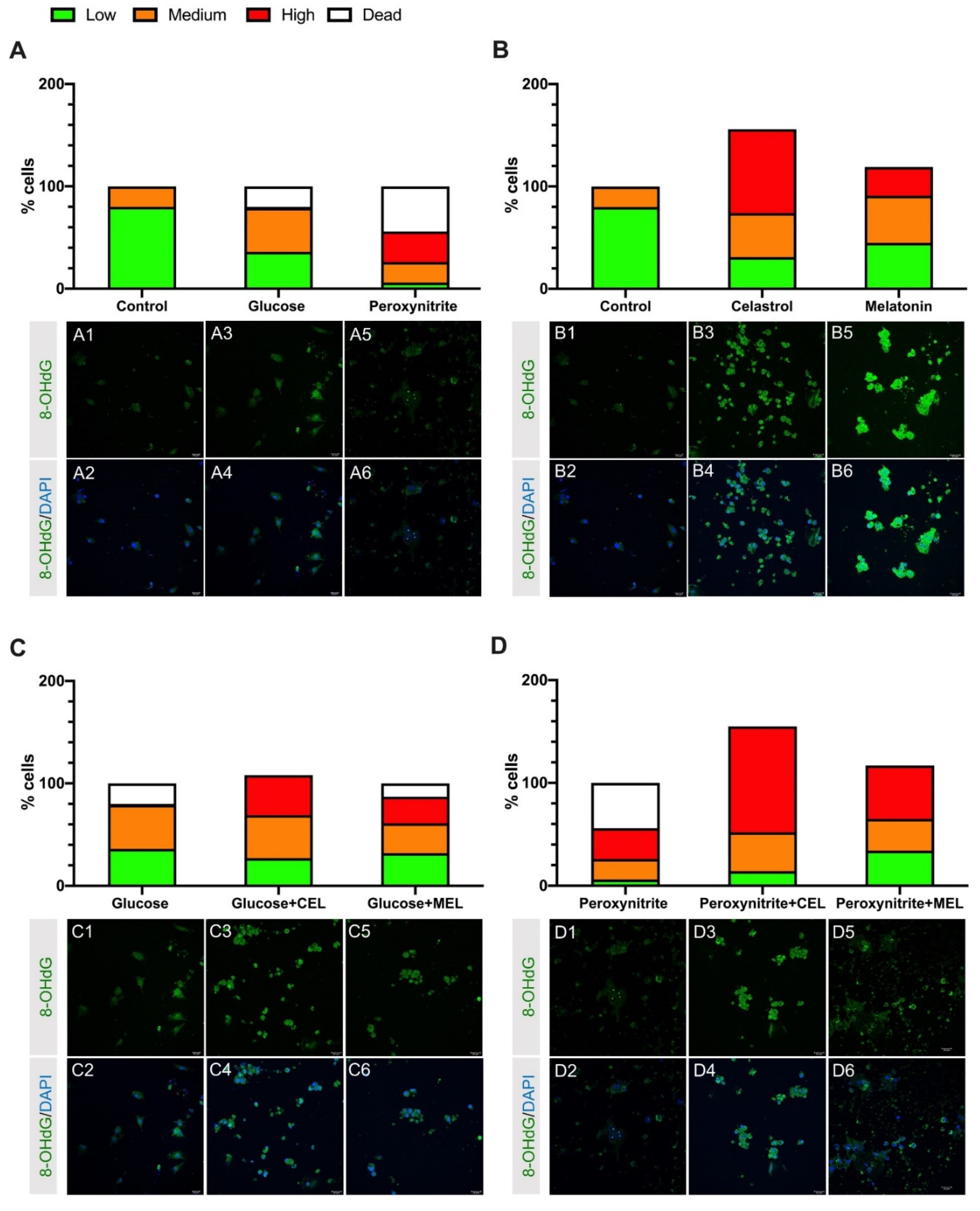
3.1. DNA Damage in Control and Treated Cells

Treatment with glucose, and especially, peroxynitrite increased the percentage of the high and medium intensity levels of 8-OHdG staining. Both the number of damaged cells and dead cells increased compared to control cells (Figure 1A).
Human GL cells treated with celastrol or melatonin showed an increase in the high and medium 8-OHdG staining levels compared to control. This increase was higher in celastrol-treated cells. Celastrol and melatonin treatment increased survival of hGL cells by 56% and 20%, respectively (Figure 1B).
Addition of celastrol or melatonin to hGL cells treated with glucose increased the intensity of staining from high and medium 8-OHdG staining levels. Complementarily, the addition of celastrol or melatonin to cell cultures increased survival by 7% or lowered mortality, respectively (Figure 1C). Peroxynitrite-treated cells showed a lower percentage of highly damaged cells, compared to cells treated with a combination of peroxynitrite and celastrol or peroxynitrite and melatonin. Moreover, we found a survival increase of 55% in peroxynitrite + celastrol and 17% in peroxynitrite + melatonin (Figure 1D).

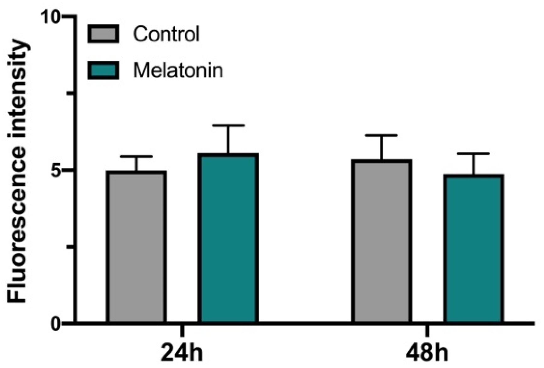
3.2. Effect of Melatonin on hGL Cultured Cells

ROS/RNS levels in hGL cells treated with melatonin were measured by DCFDA at 24 and 48 h (Figure 2). Statistically significant differences were not found.

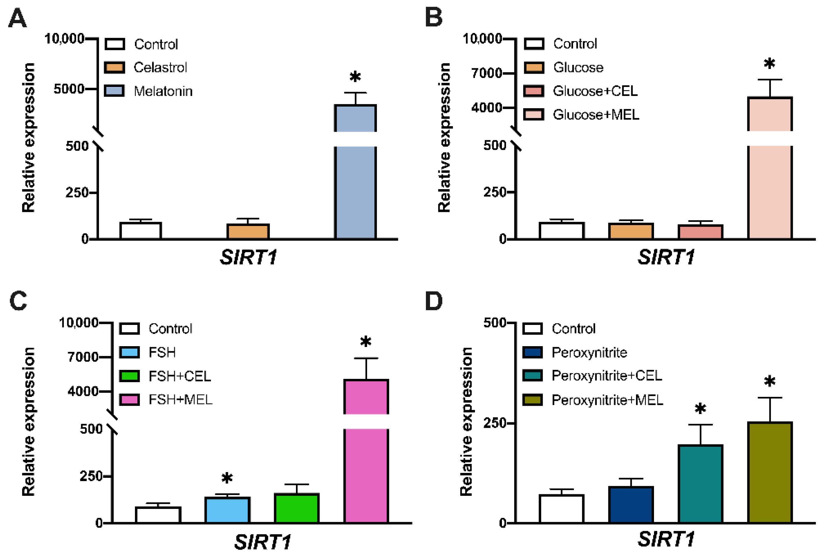
3.3. SIRT1 Expression in Control and Treated Cells

Melatonin-treated cells showed an increased gene expression of SIRT1 compared to control (Figure 3A). The addition of glucose or glucose in combination with celastrol did not modify SIRT1 mRNA level but the combination of glucose and melatonin increased SIRT1 expression (Figure 3B). FSH treatment increased SIRT1 expression compared to control (Figure 3C) but FSH + celastrol addition did not modify gene expression. However, we observed an increased expression of SIRT1 in cells treated with FSH + melatonin compared to control. Peroxynitrite treatment did not affect SIRT1 expression but the combination of peroxynitrite with celastrol or melatonin increased SIRT1 expression (Figure 3D).

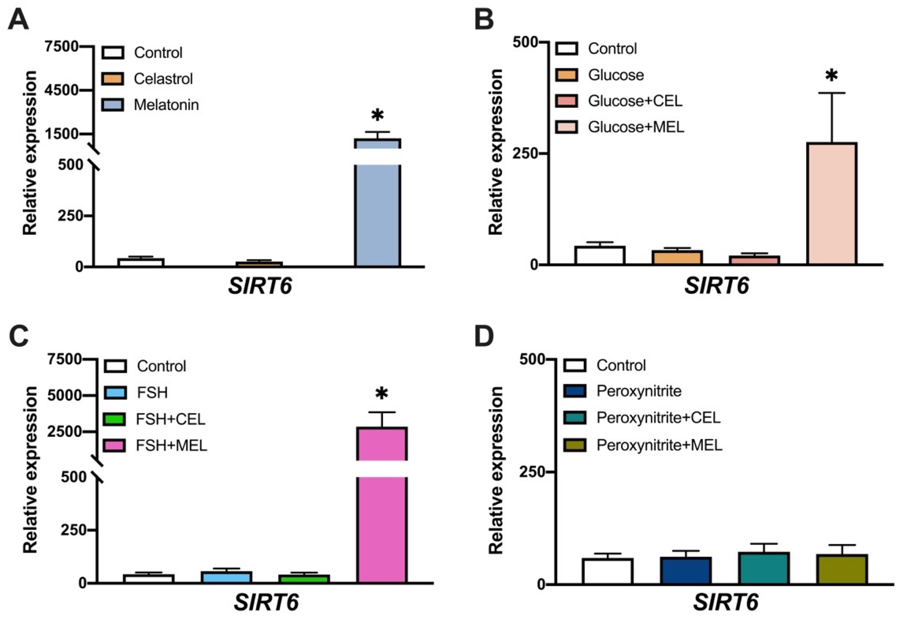
3.4. SIRT6 Expression in Control and Treated Cells

Gene expression analysis showed higher SIRT6 expression in melatonin-treated cells compared to control (Figure 4A). The addition of glucose or FSH, alone or in combination with celastrol, showed no differences in SIRT6 expression compared to control (Figure 4B,C). However, addition of glucose or FSH combined with melatonin increased SIRT6 mRNA levels (Figure 4B,C), probably because of the effect of melatonin alone observed before (Figure 4A). Peroxynitrite treatment and combination with celastrol or melatonin did not modify SIRT6 gene expression compared to control (Figure 4D).

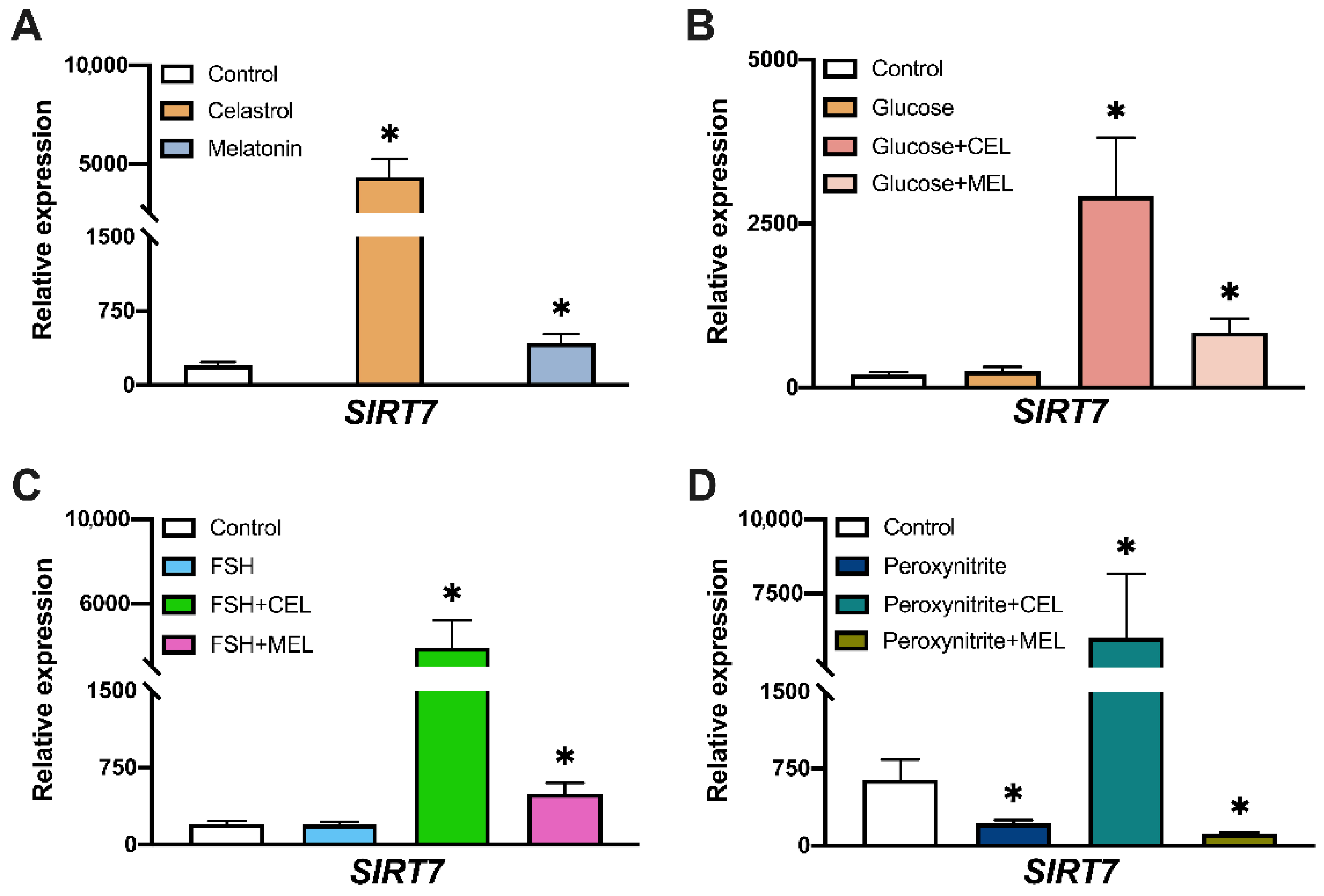
3.5. SIRT7 Expression in Control and Treated Cells

The analysis of gene expression showed that the addition of both celastrol and melatonin increased SIRT7 mRNA levels compared to control (Figure 5A). Glucose or FSH treatment did not modify SIRT7 expression. However, the combined addition of both celastrol or melatonin increased SIRT7 mRNA levels (Figure 5B,C), mimicking the effect observed in cells treated with celastrol or melatonin alone (Figure 5A). This effect of celastrol was also observable in peroxynitrite + celastrol (Figure 5D) whereas in peroxynitrite treatment, lower amounts of SIRT7 mRNA were observed (Figure 5D). SIRT7 expression also decreased in peroxynitrite + melatonin-treated cells (Figure 5D).

4. Discussion

Oxidative stress may be harmful in human reproduction processes and investigation of antioxidant agents that may prevent OS damage in reproductive tissues and processes is of paramount importance to improve both natural fertility and the results of infertility treatments. This research was performed to investigate the effects of celastrol and melatonin in preventing the impact of OS generated by glucose or peroxynitrite on cultured hGL cells.
The addition of OS inductors (glucose or peroxynitrite) to cultured hGL cells increased DNA damage and the percentage of dead cells compared to control, most prominently under peroxynitrite treatment (Figure 1A). Previous studies from our laboratory showed that higher ROS/RNS levels in cells treated with glucose or peroxynitrite [38] increased OS levels, leading to DNA damage and cell death. In this article, we report that the addition of antioxidants (celastrol or melatonin) to culture medium elicits an increase in the amount of high/medium level of 8-OHdG staining (a measure of DNA damage) compared to the amount found in control cells, but in contrast, we found an increase in the total number of surviving cells (Figure 1B). In the case of celastrol, this pro-survival effect could be related to the decrease in ROS/RNS level as upregulation of SIRT7 gene expression was observed in cultured cells treated with antioxidant [38]. The pro-survival response of melatonin is lower and consistent with the fact that the addition of melatonin to cultured medium did not substantially modify ROS/RNS levels (Figure 2). Interestingly, these results support the role of melatonin as an OS protector in a receptor-dependent manner, instead of a scavenger molecule under the conditions tested here. The effects observed after the addition of celastrol and melatonin were maintained in cells treated with the combination of OS inductor + antioxidant (Figure 1C,D).
Analysis of gene expression in hGL cells treated with glucose showed no variation in sirtuins’ gene expression (Figure 3B, Figure 4B and Figure 5B), even though previous studies demonstrated an increased ROS/RNS by the addition of glucose to culture medium [38]. An increase in DNA damage and in the number of dead cells was observed (Figure 2). Taken together, these data indicate that the addition of glucose increased OS and DNA damage with no variation in sirtuins’ gene expression.
To analyze the effect of antioxidants on hormones that play an important role in oocyte maturation, FSH was added to cultured hGL cells. We observed an increase in SIRT1 expression in cells treated with FSH, suggesting that SIRT1 could be a mediator of FSH action. To our knowledge, this is the first time that a relationship between FSH and SIRT1 expression has been described in hGL cells, suggesting a proliferative FSH-effect mediated by SIRT1. This relationship between hFSH and SIRT1 expression was previously described in porcine ovarian cells [39].
The addition of peroxynitrite decreased SIRT7 expression levels but did not modify SIRT1 or SIRT6 expression (Figure 3D, Figure 4D and Figure 5D). Peroxynitrite is a potent oxidant that produces a high percentage of DNA damage and cell death. SIRT7 expression is related to cell proliferation and impaired G1/S progression [40]. It is possible that decreased mRNA levels of SIRT7 in the presence of peroxynitrite induced cell cycle arrest in order to activate DNA repair systems. A post-translational modification of SIRT6 by peroxynitrite-induced OS controlling Sirt6 enzymatic activity has been reported [41], with no alteration in gene expression. This agrees with our data showing the non-variation in SIRT6 expression with peroxynitrite treatment.
Interestingly, we observed changes in gene expression elicited by OS in cultured hGL cells independently of the OS inductor. The addition of melatonin alone or in combination with any of the other substances, induced the expression of the three nuclear sirtuins studied here. The activation of SIRT1 by melatonin has been widely described in many conditions [42,43,44]. In the mouse ovary, melatonin reduces OS and apoptotic damage by activating SIRT1 signaling in a melatonin receptor-dependent manner [45]. Since our results support a protective effect of melatonin without a reduction in ROS/RNS levels, it is possible that the increased survival observed in hGL cells treated with melatonin could be related to SIRT1 inhibition of apoptosis through Foco1 deacetylation [46]. Previous data also described melatonin-dependent regulation of SIRT6 expression in endothelial cells [47] proposing SIRT6-AMPK-PGC-1α-AKT signaling as a novel downstream target of melatonin membrane receptors [34]. To our knowledge, this is the first report of an effect of melatonin on SIRT7 gene expression. Since Akt pathway promotes cell survival in response to extracellular signals and a direct role of SIRT7 in Akt regulation has been described [48], it is possible that melatonin could regulate SIRT7 expression through the PI3K/AKT/mTOR pathway similarly to SIRT6 regulation. This hypothesis is supported by the results of our DNA damage experiments; we found that melatonin treatment increased cell survival (Figure 1B1–B6). The role of SIRT7 in reproduction is not clear, although in female mice, SIRT7 expression is related to ovarian reserve by playing a role in repairing double-strand DNA breaks [49].
The addition of celastrol, alone or with the addition of OS inductors increased SIRT7 gene expression under the treatment conditions tested in these experiments. Several studies point to celastrol as a substance with multiple activities such as anti-tumor [50,51], anti-inflammatory [52,53] and antioxidant [54,55], and it participates in cell processes as a molecule affecting signaling of several pathways such as the ERK pathway [56,57]. In this sense, celastrol has been reported to bind Shoc2, a scaffold protein involved in processes such as cell motility, invasion, and proliferation through the ERK pathway [58]. SIRT7 activates ERK1/2 signaling [59]. Taken together, our results may show a compensatory mechanism where SIRT7 is overexpressed to revert the ERK pathway inhibition by celastrol, leading to maintenance of the cell proliferation status.
In conclusion, our results show that melatonin and celastrol treatments increase hGL cells survival under oxidative stress conditions, probably by regulating the expression levels of nuclear sirtuins. Since sirtuins’ expression is related to the control of several female reproductive processes, modulation of sirtuins’ gene expression may contribute to improved oxidative stress status in normal ovaries and pathologic states.

5. Conclusions

Female reproductive functions may be altered by oxidative stress with resulting infertility or pregnancy complications. Research on the ovarian environment under oxidative stress conditions could yield information to prevent female reproduction failure. The aim of this paper was to study the response of nuclear sirtuins to oxidative stress induction and to evaluate the effect of antioxidants. Our results show that treatment with celastrol or melatonin improves oxidative stress effects and both compounds are differently involved in nuclear sirtuins’ expression.

Author Contributions

Conceptualization, A.P. and J.Á.; Formal analysis, R.M.-R., R.G.-F., J.H., P.M.-V., A.P. and J.Á.; Funding acquisition, J.Á.; Investigation, R.M.-R. and R.G.-F.; Methodology, R.M.-R.; Project administration, J.Á.; Resources, J.H.; Supervision, P.M.-V.; Writing—original draft, R.M.-R. and R.G.-F.; Writing—review & editing, P.M.-V., A.P. and J.Á. All authors have read and agreed to the published version of the manuscript.

Funding

This research was funded by Instituto de Salud Carlos III, grant number PI12/00729 and Gobierno de Canarias (cofounded by FEDER), grant number ProID2020010073 to J.A.; by Universidad de La Laguna, grant Proyecto Puente al Plan Estatal 2018 to J.A., and by a FPI PhD fellowship from the Agencia Canaria de Investigación, Innovación y Sociedad de la Información (ACIISI) cofounded by the European Social Fund, to R.M.-R., and by a Postdoctoral contract from the Agustin de Betancourt program cofounded by Cabildo Insular de Tenerife, TF Innova, FEDECAN, MEDI, to R.G-F.

Institutional Review Board Statement

The study was conducted according to the guidelines of the Declaration of Helsinki, and approved by the Ethics Committee of the Universidad de La Laguna, CEIBA2012-0044 (02/05/2012).

Informed Consent Statement

Informed consent was obtained from all subjects involved in the study.

Data Availability Statement

All data presented in this article can be obtained from Authors, upon request. The data are not publicly available due to privacy and/or ethical restrictions.

Acknowledgments

The authors thank the laboratory and paramedical staff of Centro de Asistencia a la Reproducción Humana de Canarias for their collaboration.

Conflicts of Interest

The authors declare no conflict of interest.

References

  1. Jakubczyk, K.; Dec, K.; Kałduńska, J.; Kawczuga, D.; Kochman, J.; Janda, K. Reactive Oxygen Species—Sources, Functions, Oxidative Damage. Pol. Merkur. Lek. Organ. Pol. Tow. Lek. 2020, 48, 124–127. [Google Scholar]
  2. Agarwal, A.; Gupta, S.; Sharma, R.K. Role of Oxidative Stress in Female Reproduction. Reprod. Biol. Endocrinol. 2005, 3, 21–25. [Google Scholar] [CrossRef] [PubMed] [Green Version]
  3. Du Plessis, S.S.; Agarwal, A.; Halabi, J.; Tvrda, E. Contemporary Evidence on the Physiological Role of Reactive Oxygen Species in Human Sperm Function. J. Assist. Reprod. Genet. 2015, 32, 509–520. [Google Scholar] [CrossRef] [PubMed] [Green Version]
  4. Dang, W. The Controversial World of Sirtuins. Drug Discov. Today Technol. 2014, 12, e9–e17. [Google Scholar] [CrossRef] [Green Version]
  5. Morris, B.J. Seven Sirtuins for Seven Deadly Diseases Ofaging. Free Radic. Biol. Med. 2013, 56, 133–171. [Google Scholar] [CrossRef]
  6. Cantó, C.; Sauve, A.A.; Bai, P. Crosstalk between Poly(ADP-Ribose) Polymerase and Sirtuin Enzymes. Mol. Asp. Med. 2013, 34, 1168–1201. [Google Scholar] [CrossRef] [Green Version]
  7. Ford, E. Mammalian Sir2 Homolog SIRT7 Is an Activator of RNA Polymerase I Transcription. Genes Dev. 2006, 20, 1075–1080. [Google Scholar] [CrossRef] [Green Version]
  8. Tanno, M.; Sakamoto, J.; Miura, T.; Shimamoto, K.; Horio, Y. Nucleocytoplasmic Shuttling of the NAD+-Dependent Histone Deacetylase SIRT1. J. Biol. Chem. 2007, 282, 6823–6832. [Google Scholar] [CrossRef] [Green Version]
  9. Cheng, H.-L.; Mostoslavsky, R.; Saito, S.; Manis, J.P.; Gu, Y.; Patel, P.; Bronson, R.; Appella, E.; Alt, F.W.; Chua, K.F. Developmental Defects and P53 Hyperacetylation in Sir2 Homolog (SIRT1)-Deficient Mice. Proc. Natl. Acad. Sci. USA 2003, 100, 10794–10799. [Google Scholar] [CrossRef] [Green Version]
  10. Mostoslavsky, R.; Chua, K.F.; Lombard, D.B.; Pang, W.W.; Fischer, M.R.; Gellon, L.; Liu, P.; Mostoslavsky, G.; Franco, S.; Murphy, M.M.; et al. Genomic Instability and Aging-like Phenotype in the Absence of Mammalian SIRT6. Cell 2006, 124, 315–329. [Google Scholar] [CrossRef] [Green Version]
  11. Vazquez, B.N.; Thackray, J.K.; Simonet, N.G.; Kane-Goldsmith, N.; Martinez-Redondo, P.; Nguyen, T.; Bunting, S.; Vaquero, A.; Tischfield, J.A.; Serrano, L. SIRT 7 Promotes Genome Integrity and Modulates Non-homologous End Joining DNA Repair. EMBO J. 2016, 35, 1488–1503. [Google Scholar] [CrossRef]
  12. Kim, D.H.; Jung, I.H.; Kim, D.H.; Park, S.W. Knockout of Longevity Gene Sirt1 in Zebrafish Leads to Oxidative Injury, Chronic Inflammation, and Reduced Life Span. PLoS ONE 2019, 14, e0220581. [Google Scholar] [CrossRef] [Green Version]
  13. Lim, J.H.; Kim, E.N.; Kim, M.Y.; Chung, S.; Shin, S.J.; Kim, H.W.; Yang, C.W.; Kim, Y.-S.; Chang, Y.S.; Park, C.W.; et al. Age-Associated Molecular Changes in the Kidney in Aged Mice. Oxidative Med. Cell. Longev. 2012, 2012, 171383. [Google Scholar] [CrossRef] [PubMed] [Green Version]
  14. Hasegawa, K.; Wakino, S.; Yoshioka, K.; Tatematsu, S.; Hara, Y.; Minakuchi, H.; Washida, N.; Tokuyama, H.; Hayashi, K.; Itoh, H. Sirt1 Protects against Oxidative Stress-Induced Renal Tubular Cell Apoptosis by the Bidirectional Regulation of Catalase Expression. Biochem. Biophys. Res. Commun. 2008, 372, 51–56. [Google Scholar] [CrossRef] [PubMed]
  15. Toiber, D.; Erdel, F.; Bouazoune, K.; Silberman, D.M.; Zhong, L.; Mulligan, P.; Sebastian, C.; Cosentino, C.; Martinez-Pastor, B.; Giacosa, S.; et al. SIRT6 Recruits SNF2H to DNA Break Sites, Preventing Genomic Instability through Chromatin Remodeling. Mol. Cell 2013, 51, 454–468. [Google Scholar] [CrossRef] [PubMed] [Green Version]
  16. Mao, Z.; Hine, C.; Tian, X.; van Meter, M.; Au, M.; Vaidya, A.; Seluanov, A.; Gorbunova, V. SIRT6 Promotes DNA Repair under Stress by Activating PARP1. Science 2011, 332, 1443–1446. [Google Scholar] [CrossRef] [PubMed] [Green Version]
  17. Hwang, B.-J.; Jin, J.; Gao, Y.; Shi, G.; Madabushi, A.; Yan, A.; Guan, X.; Zalzman, M.; Nakajima, S.; Lan, L.; et al. SIRT6 Protein Deacetylase Interacts with MYH DNA Glycosylase, APE1 Endonuclease, and Rad9–Rad1–Hus1 Checkpoint Clamp. BMC Mol. Biol. 2015, 16, 12. [Google Scholar] [CrossRef] [PubMed] [Green Version]
  18. Grummt, I.; Pikaard, C.S. Epigenetic Silencing of RNA Polymerase I Transcription. Nat. Rev. Mol. Cell Biol. 2003, 4, 641–649. [Google Scholar] [CrossRef]
  19. Chen, S.; Blank, M.F.; Iyer, A.; Huang, B.; Wang, L.; Grummt, I.; Voit, R. SIRT7-Dependent Deacetylation of the U3-55k Protein Controls Pre-RRNA Processing. Nat. Commun. 2016, 7, 10734. [Google Scholar] [CrossRef] [Green Version]
  20. Shin, J.; He, M.; Liu, Y.; Paredes, S.; Villanova, L.; Brown, K.; Qiu, X.; Nabavi, N.; Mohrin, M.; Wojnoonski, K.; et al. SIRT7 Represses Myc Activity to Suppress ER Stress and Prevent Fatty Liver Disease. Cell Rep. 2013, 5, 654–656. [Google Scholar] [CrossRef] [Green Version]
  21. Yadav, P.; Jaswal, V.; Sharma, A.; Kashyap, D.; Tuli, H.S.; Garg, V.K.; Das, S.K.; Srinivas, R. Celastrol as a Pentacyclic Triterpenoid with Chemopreventive Properties. Pharm. Pat. Anal. 2018, 7, 155–168. [Google Scholar] [CrossRef] [PubMed]
  22. Abu Bakar, M.H.; Sarmidi, M.R.; Tan, J.S.; Mohamad Rosdi, M.N. Celastrol Attenuates Mitochondrial Dysfunction and Inflammation in Palmitate-Mediated Insulin Resistance in C3A Hepatocytes. Eur. J. Pharmacol. 2017, 799, 73–83. [Google Scholar] [CrossRef]
  23. Ng, S.W.; Chan, Y.; Chellappan, D.K.; Madheswaran, T.; Zeeshan, F.; Chan, Y.L.; Collet, T.; Gupta, G.; Oliver, B.G.; Wark, P.; et al. Molecular Modulators of Celastrol as the Keystones for Its Diverse Pharmacological Activities. Biomed. Pharmacother. 2019, 109, 1785–1792. [Google Scholar] [CrossRef]
  24. Zhou, Y.; Zhou, L.; Zhou, K.; Zhang, J.; Shang, F.; Zhang, X. Celastrol Protects RPE Cells from Oxidative Stress-Induced Cell Death via Activation of Nrf2 Signaling Pathway. Curr. Mol. Med. 2019, 19, 172–182. [Google Scholar] [CrossRef]
  25. Zhang, Y.; Geng, C.; Liu, X.; Li, M.; Gao, M.; Liu, X.; Fang, F.; Chang, Y. Celastrol Ameliorates Liver Metabolic Damage Caused by a High-Fat Diet through Sirt1. Mol. Metab. 2017, 6, 138–147. [Google Scholar] [CrossRef] [PubMed]
  26. Reiter, R.; Rosales-Corral, S.; Manchester, L.; Tan, D.-X. Peripheral Reproductive Organ Health and Melatonin: Ready for Prime Time. Int. J. Mol. Sci. 2013, 14, 7231–7272. [Google Scholar] [CrossRef] [Green Version]
  27. Lan, M.; Han, J.; Pan, M.-H.; Wan, X.; Pan, Z.-N.; Sun, S.-C. Melatonin Protects against Defects Induced by Deoxynivalenol during Mouse Oocyte Maturation. J. Pineal Res. 2018, 65, e12477. [Google Scholar] [CrossRef] [PubMed]
  28. Suwannakot, K.; Sritawan, N.; Prajit, R.; Aranarochana, A.; Sirichoat, A.; Pannangrong, W.; Wigmore, P.; Welbat, J.U. Melatonin Protects against the Side-Effects of 5-Fluorouracil on Hippocampal Neurogenesis and Ameliorates Antioxidant Activity in an Adult Rat Hippocampus and Prefrontal Cortex. Antioxidants 2021, 10, 615. [Google Scholar] [CrossRef]
  29. Fan, H.; Wang, S.; Wang, H.; Sun, M.; Wu, S.; Bao, W. Melatonin Ameliorates the Toxicity Induced by Deoxynivalenol in Murine Ovary Granulosa Cells by Antioxidative and Anti-Inflammatory Effects. Antioxidants 2021, 10, 1045. [Google Scholar] [CrossRef]
  30. Liu, Y.; Yang, Y.; Li, W.; Ao, H.; Zhang, Y.; Zhou, R.; Li, K. Effects of Melatonin on the Synthesis of Estradiol and Gene Expression in Pig Granulosa Cells. J. Pineal Res. 2019, 66, e12546. [Google Scholar] [CrossRef]
  31. He, C.; Wang, J.; Li, Y.; Zhu, K.; Xu, Z.; Song, Y.; Song, Y.; Liu, G. Melatonin-Related Genes Expressed in the Mouse Uterus during Early Gestation Promote Embryo Implantation. J. Pineal Res. 2015, 58, 300–309. [Google Scholar] [CrossRef]
  32. Reiter, R.J.; Tamura, H.; Tan, D.X.; Xu, X.-Y. Melatonin and the Circadian System: Contributions to Successful Female Reproduction. Fertil. Steril. 2014, 102, 293–307. [Google Scholar] [CrossRef]
  33. Hardeland, R. Melatonin and Inflammation-Story of a Double-Edged Blade. J. Pineal Res. 2018, 65, e12525. [Google Scholar] [CrossRef] [PubMed] [Green Version]
  34. Yu, L.; Dong, X.; Xue, X.; Xu, S.; Zhang, X.; Xu, Y.; Wang, Z.; Wang, Y.; Gao, H.; Liang, Y.; et al. Melatonin Attenuates Diabetic Cardiomyopathy and Reduces Myocardial Vulnerability to Ischemia-reperfusion Injury by Improving Mitochondrial Quality Control: Role of SIRT6. J. Pineal Res. 2021, 70, e12698. [Google Scholar] [CrossRef] [PubMed]
  35. Nargund, G.; Datta, A.K.; Fauser, B.C.J.M. Mild Stimulation for in Vitro Fertilization. Fertil. Steril. 2017, 108, 558–567. [Google Scholar] [CrossRef] [PubMed] [Green Version]
  36. Schmittgen, T.D.; Livak, K.J. Analyzing Real-Time PCR Data by the Comparative CT Method. Nat. Protoc. 2008, 3, 309–315. [Google Scholar] [CrossRef]
  37. Kalyanaraman, B.; Darley-Usmar, V.; Davies, K.J.A.; Dennery, P.A.; Forman, H.J.; Grisham, M.B.; Mann, G.E.; Moore, K.; Roberts, L.J.; Ischiropoulos, H. Measuring Reactive Oxygen and Nitrogen Species with Fluorescent Probes: Challenges and Limitations. Free Radic. Biol. Med. 2012, 52, 1–6. [Google Scholar] [CrossRef] [Green Version]
  38. Martín-Ramírez, R.; González-Fernández, R.; Rotoli, D.; Hernández, J.; Martín-Vasallo, P.; Palumbo, A.; Ávila, J. Celastrol Prevents Oxidative Stress Effects on FSHR, PAPP, and CYP19A1 Gene Expression in Cultured Human Granulosa-Lutein Cells. Int. J. Mol. Sci. 2021, 22, 3596. [Google Scholar] [CrossRef]
  39. Sirotkin, A.; Dekanová, P.; Harrath, A. FSH, Oxytocin and IGF-I Regulate the Expression of Sirtuin 1 in Porcine Ovarian Granulosa Cells. Physiol. Res. 2020, 69, 461–466. [Google Scholar] [CrossRef]
  40. Kim, J.K.; Noh, J.H.; Jung, K.H.; Eun, J.W.; Bae, H.J.; Kim, M.G.; Chang, Y.G.; Shen, Q.; Park, W.S.; Lee, J.Y.; et al. Sirtuin7 Oncogenic Potential in Human Hepatocellular Carcinoma and Its Regulation by the Tumor Suppressors MiR-125a-5p and MiR-125b. Hepatology 2013, 57, 1055–1067. [Google Scholar] [CrossRef]
  41. Hu, S.; Liu, H.; Ha, Y.; Luo, X.; Motamedi, M.; Gupta, M.P.; Ma, J.-X.; Tilton, R.G.; Zhang, W. Posttranslational Modification of Sirt6 Activity by Peroxynitrite. Free Radic. Biol. Med. 2015, 79, 176–185. [Google Scholar] [CrossRef] [Green Version]
  42. Hardeland, R. Melatonin and the Pathologies of Weakened or Dysregulated Circadian Oscillators. J. Pineal Res. 2017, 62, e12377. [Google Scholar] [CrossRef]
  43. Hardeland, R. Extended Signaling by Melatonin. Cell Cell. Life Sci. J. 2018, 3, 000123. [Google Scholar] [CrossRef]
  44. Hardeland, R. Recent Findings in Melatonin Research and Their Relevance to the CNS. Cent. Nerv. Syst. Agents Med. Chem. 2018, 18, 102–114. [Google Scholar] [CrossRef] [PubMed]
  45. Ma, M.; Chen, X.-Y.; Li, B.; Li, X.-T. Melatonin Protects Premature Ovarian Insufficiency Induced by Tripterygium Glycosides: Role of SIRT1. Am. J. Transl. Res. 2017, 9, 1580. [Google Scholar] [PubMed]
  46. Zhang, M.; Zhang, Q.; Hu, Y.; Xu, L.; Jiang, Y.; Zhang, C.; Ding, L.; Jiang, R.; Sun, J.; Sun, H.; et al. MiR-181a Increases FoxO1 Acetylation and Promotes Granulosa Cell Apoptosis via SIRT1 Downregulation. Cell Death Dis. 2017, 8, e3088. [Google Scholar] [CrossRef]
  47. Lee, F.-Y.; Sun, C.-K.; Sung, P.-H.; Chen, K.-H.; Chua, S.; Sheu, J.-J.; Chung, S.-Y.; Chai, H.-T.; Chen, Y.-L.; Huang, T.-H.; et al. Daily Melatonin Protects the Endothelial Lineage and Functional Integrity against the Aging Process, Oxidative Stress, and Toxic Environment and Restores Blood Flow in Critical Limb Ischemia Area in Mice. J. Pineal Res. 2018, 65, e12489. [Google Scholar] [CrossRef] [PubMed]
  48. Yu, J.; Qin, B.; Wu, F.; Qin, S.; Nowsheen, S.; Shan, S.; Zayas, J.; Pei, H.; Lou, Z.; Wang, L. Regulation of Serine-Threonine Kinase Akt Activation by NAD+-Dependent Deacetylase SIRT7. Cell Rep. 2017, 18, 1229–1240. [Google Scholar] [CrossRef]
  49. Vazquez, B.N.; Blengini, C.S.; Hernandez, Y.; Serrano, L.; Schindler, K. SIRT7 Promotes Chromosome Synapsis during Prophase I of Female Meiosis. Chromosoma 2019, 128, 369–383. [Google Scholar] [CrossRef]
  50. Si, H.; Wang, H.; Xiao, H.; Fang, Y.; Wu, Z. Anti-Tumor Effect of Celastrol on Hepatocellular Carcinoma by the Circ_SLIT3/MiR-223-3p/CXCR4 Axis. Cancer Manag. Res. 2021, 13, 1099. [Google Scholar] [CrossRef]
  51. Liu, P.; Wang, M.; Tang, W.; Li, G.; Gong, N. Circ_SATB2 Attenuates the Anti-Tumor Role of Celastrol in Non-Small-Cell Lung Carcinoma Through Targeting MiR-33a-5p/E2F7 Axis. OncoTargets Ther. 2020, 13, 11899. [Google Scholar] [CrossRef] [PubMed]
  52. Wang, Y.; Li, C.; Gu, J.; Chen, C.; Duanmu, J.; Miao, J.; Yao, W.; Tao, J.; Tu, M.; Xiong, B.; et al. Celastrol Exerts Anti-Inflammatory Effect in Liver Fibrosis via Activation of AMPK-SIRT3 Signalling. J. Cell. Mol. Med. 2020, 24, 941–953. [Google Scholar] [CrossRef] [Green Version]
  53. Dai, W.; Wang, X.; Teng, H.; Li, C.; Wang, B.; Wang, J. Celastrol Inhibits Microglial Pyroptosis and Attenuates Inflammatory Reaction in Acute Spinal Cord Injury Rats. Int. Immunopharmacol. 2019, 66, 215–223. [Google Scholar] [CrossRef] [PubMed]
  54. Chen, X.; Zhao, Y.; Luo, W.; Chen, S.; Lin, F.; Zhang, X.; Fan, S.; Shen, X.; Wang, Y.; Liang, G. Celastrol Induces ROS-Mediated Apoptosis via Directly Targeting Peroxiredoxin-2 in Gastric Cancer Cells. Theranostics 2020, 10, 10290. [Google Scholar] [CrossRef]
  55. Allison, A.C.; Cacabelos, R.; Lombardi, V.R.M.; Álvarez, X.A.; Vigo, C. Celastrol, a Potent Antioxidant and Anti-Inflammatory Drug, as a Possible Treatment for Alzheimer’s Disease. Prog. Neuro-Psychopharmacol. Biol. Psychiatry 2001, 25, 108–114. [Google Scholar] [CrossRef]
  56. Cheng, M.; Wu, G.; Song, Y.; Wang, L.; Tu, L.; Zhang, L.; Zhang, C. Celastrol-Induced Suppression of the MiR-21/ERK Signalling Pathway Attenuates Cardiac Fibrosis and Dysfunction. Cell. Physiol. Biochem. 2016, 38, 1928–1938. [Google Scholar] [CrossRef]
  57. Kim, Y.; Kim, K.; Lee, H.; Han, S.; Lee, Y.-S.; Choe, J.; Kim, Y.-M.; Hahn, J.-H.; Ro, J.Y.; Jeoung, D. Celastrol Binds to ERK and Inhibits FcεRI Signaling to Exert an Anti-Allergic Effect. Eur. J. Pharmacol. 2009, 612, 131–142. [Google Scholar] [CrossRef]
  58. Huang, X.-P.; Chen, J.-K.; Wei, X.; Dong, Y.-F.; Lang, Y.; Zhang, X.-F.; Pan, Y.-M.; Chang, W.-J.; Zhu, J.-B. Systematic Identification of Celastrol-Binding Proteins Reveals That Shoc2 Is Inhibited by Celastrol. Biosci. Rep. 2018, 38, BSR20181233. [Google Scholar] [CrossRef] [Green Version]
  59. Zhao, Y.; Ye, X.; Chen, R.; Gao, Q.; Zhao, D.; Ling, C.; Qian, Y.; Xu, C.; Tao, M.; Xie, Y. Sirtuin 7 Promotes Non-small Cell Lung Cancer Progression by Facilitating G1/S Phase and Epithelial-mesenchymal Transition and Activating AKT and ERK1/2 Signaling. Oncol. Rep. 2020, 44, 959–972. [Google Scholar] [CrossRef] [PubMed]
Figure 1. Effect of glucose, peroxynitrite, celastrol (CEL), melatonin (MEL) and their combinations on DNA damage and cell survival in hGL cells. Histograms represent percentage of cells with different 8-OHdG staining intensity defined as low (green), medium (orange) and high (red). Percentage of dead cells is included (white). Representative images of each culture condition are showed below corresponding bar. (A) Glucose and peroxynitrite treatments compared to control. (B) Celastrol and melatonin compared to control. (C) Glucose + CEL and glucose + MEL treatments compared to glucose treatment. (D) Peroxynitrite + CEL and peroxynitrite + MEL treatments compared to peroxynitrite treatment. n = 3.
Antioxidants 10 01871 g001
Figure 2. ROS/RNS levels in hGL cells treated with melatonin. Histogram representing total OS measures (n = 3) in hGL cells after 24 and 48 h of melatonin exposure compared to control.
Antioxidants 10 01871 g002
Figure 3. Effect of glucose, peroxynitrite, FSH, celastrol (CEL), melatonin (MEL) and their combinations on SIRT1 gene expression. SIRT1 relative expression levels in hGL cells treated with: (A) celastrol (n = 19) or melatonin (n = 18), (B) glucose (n = 17) and in combination with celastrol (n = 19) or melatonin (n = 18), (C) FSH (n = 17) and in combination with celastrol (n = 19) or melatonin (n = 18), (D) peroxynitrite (n = 20) and in combination with celastrol (n = 18) or melatonin (n = 17). Asterisks (*) indicate statistically significant differences compared to control.
Antioxidants 10 01871 g003
Figure 4. Effect of glucose, peroxynitrite, FSH, celastrol (CEL), melatonin (MEL) and their combinations on SIRT6 gene expression. SIRT6 relative expression levels in hGL cells treated with: (A) celastrol (n = 19) or melatonin (n = 18), (B) glucose (n = 17) and in combination with celastrol (n = 19) or melatonin (n = 18), (C) FSH (n = 17) and in combination with celastrol (n = 19) or melatonin (n = 18), (D) peroxynitrite (n = 20) and in combination with celastrol (n = 18) or melatonin (n = 17). Asterisks (*) indicate statistically significant differences compared to control.
Antioxidants 10 01871 g004
Figure 5. Effect of glucose, peroxynitrite, FSH, celastrol (CEL), melatonin (MEL) and their combinations on SIRT7 gene expression. SIRT7 relative expression levels in hGL cells treated with: (A) celastrol (n = 19) or melatonin (n = 18), (B) glucose (n = 17) and in combination with celastrol (n = 19) or melatonin (n = 18), (C) FSH (n = 17) and in combination with celastrol (n = 19) or melatonin (n = 18), (D) peroxynitrite (n = 20) and in combination with celastrol (n = 18) or melatonin (n = 17). Asterisks (*) indicate statistically significant differences compared to control.
Antioxidants 10 01871 g005
Publisher’s Note: MDPI stays neutral with regard to jurisdictional claims in published maps and institutional affiliations.

