Article

Strigolactones Modulate Cellular Antioxidant Defense Mechanisms to Mitigate Arsenate Toxicity in Rice Shoots

by
Mohammad Golam Mostofa
1,2,†,
Chien Van Ha
2,†,
Md. Mezanur Rahman
2,
Kien Huu Nguyen
3,
Sanjida Sultana Keya
2,
Yasuko Watanabe
4,
Misao Itouga
5,
Abeer Hashem
6,
Elsayed Fathi Abd_Allah
7,
Masayuki Fujita
8 and
Lam-Son Phan Tran
2,9,*
1
Department of Biochemistry and Molecular Biology, Bangabandhu Sheikh Mujibur Rahman Agricultural University, Gazipur 1706, Bangladesh
2
Institute of Genomics for Crop Abiotic Stress Tolerance, Department of Plant and Soil Science, Texas Tech University, Lubbock, TX 79409, USA
3
National Key Laboratory for Plant Cell Biotechnology, Agricultural Genetics Institute, Vietnam Academy of Agricultural Science, Hanoi 100000, Vietnam
4
Bioproductivity Informatics Research Team, RIKEN Center for Sustainable Resource Science, Yokohama 230-0045, Japan
5
Synthetic Genomics Research Group, RIKEN Center for Sustainable Resource Science, Yokohama 230-0045, Japan
6
Botany and Microbiology Department, College of Science, King Saud University, P.O. Box 2460, Riyadh 11451, Saudi Arabia
7
Plant Production Department, College of Food and Agricultural Sciences, King Saud University, P.O. Box 2460, Riyadh 11451, Saudi Arabia
8
Laboratory of Plant Stress Responses, Department of Applied Biological Science, Faculty of Agriculture, Kagawa University, Takamatsu 761-0795, Kagawa, Japan
9
Institute of Research and Development, Duy Tan University, Da Nang 550000, Vietnam
*
Author to whom correspondence should be addressed.
Equal contribution.
Antioxidants 2021, 10(11), 1815; https://doi.org/10.3390/antiox10111815
Submission received: 28 September 2021 / Revised: 6 November 2021 / Accepted: 8 November 2021 / Published: 15 November 2021
(This article belongs to the Special Issue Antioxidant Mechanisms in Plants)

Abstract

:
Metalloid contamination, such as arsenic poisoning, poses a significant environmental problem, reducing plant productivity and putting human health at risk. Phytohormones are known to regulate arsenic stress; however, the function of strigolactones (SLs) in arsenic stress tolerance in rice is rarely investigated. Here, we investigated shoot responses of wild-type (WT) and SL-deficient d10 and d17 rice mutants under arsenate stress to elucidate SLs’ roles in rice adaptation to arsenic. Under arsenate stress, the d10 and d17 mutants displayed severe growth abnormalities, including phenotypic aberrations, chlorosis and biomass loss, relative to WT. Arsenate stress activated the SL-biosynthetic pathway by enhancing the expression of SL-biosynthetic genes D10 and D17 in WT shoots. No differences in arsenic levels between WT and SL-biosynthetic mutants were found from Inductively Coupled Plasma-Mass Spectrometry analysis, demonstrating that the greater growth defects of mutant plants did not result from accumulated arsenic in shoots. The d10 and d17 plants had higher levels of reactive oxygen species, water loss, electrolyte leakage and membrane damage but lower activities of superoxide dismutase, ascorbate peroxidase, glutathione peroxidase and glutathione S-transferase than did the WT, implying that arsenate caused substantial oxidative stress in the SL mutants. Furthermore, WT plants had higher glutathione (GSH) contents and transcript levels of OsGSH1, OsGSH2, OsPCS1 and OsABCC1 in their shoots, indicating an upregulation of GSH-assisted arsenic sequestration into vacuoles. We conclude that arsenate stress activated SL biosynthesis, which led to enhanced arsenate tolerance through the stimulation of cellular antioxidant defense systems and vacuolar sequestration of arsenic, suggesting a novel role for SLs in rice adaptation to arsenic stress. Our findings have significant implications in the development of arsenic-resistant rice varieties for safe and sustainable rice production in arsenic-polluted soils.

Graphical Abstract

1. Introduction

Plant growth and developmental processes are regulated by several endogenous molecules, including plant hormones such as strigolactones (SLs) [1,2]. SLs are a new class of phytohormones synthesized from plant pigment carotenoids by the actions of a series of enzymes depending on the types of plant species [1,3,4]. In rice (Oryza sativa), three SL-biosynthetic enzymes β-carotene isomerase, carotenoid cleavage dioxygenase (CCD)7 (CCD7) and CCD8 are encoded by DWARF (D)27 (D27), D17 and D10, respectively [5] (Figure 1). The sequential actions of these three enzymes convert β-carotene into carlactone [5]. Carlactone then undergoes further modification by two cytochrome P450 proteins, namely Os01g0700900 (carlactone oxidase) and Os01g0701400 (orobanchol synthase), to be converted to 4-deoxyprobanchol and orobanchol, respectively [6]. When SLs are absent, transcriptional repressor D53, in cooperation with TOPLESS (TPL)/TOPLESS-RELATED (TPR) repressors, represses downstream signaling of SLs [7]. In the presence of SLs, an α/β hydrolase receptor, namely D14, detects SLs, binds and is activated (Figure 1). SL-activated D14 interacts with the F-box protein D3, leading to the formation of a Skp1-Cullin-F-box (SCF)D3 type of E3 ubiquitin ligase complex that acts to degrade D53, thereby enabling the expression of SL-responsive genes for various developmental, physiological and stress survival functions [3,8] (Figure 1).
SLs were primarily identified as signaling molecules for parasitic seed germination and the establishment of a symbiotic connection between plants and arbuscular mycorrhizal fungi (AMF) in natural environments [9]. Apart from these, SLs can control various developmental traits in both aboveground and belowground parts of plants. SLs positively regulate plant height, leaf senescence, stem thickness, root hair elongation and primary root length, whereas they negatively affect shoot gravitropism and branching, adventitious rooting and lateral root development under normal growth conditions [1,7]. Various loss-of-function studies have used SL-related mutants and synthetic GR24 to demonstrate that SLs played crucial regulatory roles in plant responses to environmental perturbations [10,11,12,13]. For example, SL-deficient Arabidopsis (Arabidopsis thaliana) and rice mutants, and SL-depleted transgenic Lotus japonicus and tomato (Solanum lycopersicum) lines showed higher susceptibility to drought than their respective wild-type (WT) plants [13,14,15,16]. Foliar application of GR24 alleviated the deleterious effects of several abiotic stresses, including low light stress in tomato, drought in wheat (Triticum aestivum) and salinity in rice [12,17,18]. SL-mediated positive effects on plant tolerance to abiotic stresses mainly pertain to the regulation of various physiological and biochemical processes, including photosynthetic efficiency, leaf senescence, cell wall biogenesis, stomatal closure, flavonoid production, antioxidant defense and nutrient homeostasis [1]. Recently, Qiu et al. [11] reported that GR24 application improved cadmium (Cd) toxicity tolerance in contrasting barley (Hordeum vulgare) genotypes by inhibiting Cd uptake, balancing nutrient levels, and activating reactive oxygen species (ROS)-scavenging systems. Although this study pinpoints the likely involvement of SLs in heavy metal tolerance, the genetic and molecular insights into crop tolerance to excessive metal stresses are still obscure. Furthermore, the role of GR24 in stress tolerance should be cautiously interpreted as the sole role of SLs because GR24 is well-known to have both SL and karrikin effects [19]. Thus, to obtain a firm understanding of how SLs potentiate metal-defense networks in plants, it is crucial to use loss-of-function mutants to figure out the SL-modulated genetic basis that governs physiological and biochemical changes for making plants more resilient to metal-induced harsh environments.
Arsenic is the most hazardous metalloid for plant growth and development when it is uptaken in large quantities from the arsenic-contaminated environments [20,21]. Arsenite (AsIII) and arsenate (AsV) are two inorganic forms of arsenic that are predominantly found in anaerobic and aerobic soils, respectively [22]. In plants, aquaglyceroporins and phosphate transporters, respectively, facilitate the uptake of AsV and AsIII from arsenic-contaminated soils [21,22]. Once accumulated inside the plant tissues, arsenic can inhibit seed germination and seedling establishment, induce oxidative stress, inhibit photosynthesis, suppress growth, and reduce seed quality [23,24,25,26]. To protect themselves, plants deploy several coordinated defense processes, such as restriction of arsenic uptake and transportation, synthesis of arsenic chelating metabolites such as glutathione (GSH) and phytochelatins (PCs), stimulation of antioxidant defense, and vacuolar sequestration of arsenic to reduce arsenic-induced toxicity effects [25,27,28].
Agricultural land contamination with arsenic has become a global problem as it has a detrimental influence on all forms of life. Rice is a significant grain crop that feeds half of the world’s population and can be cultivated in a variety of soils, including arsenic-laden soil [23]. As a result, rice and rice-related products account for the majority of arsenic consumed by humans [29]. Moreover, arsenic contamination of soils can escalate the adverse effects of other environmental stresses on rice productivity [30]. Thus, understanding the mechanisms driving arsenic accumulation and detoxification in rice plants is critical for developing future crops that are safe for humans. We have recently reported the involvement of SLs in the regulation of rice root tolerance to excessive AsV stress [27]. However, how SLs deal with the arsenic toxicity in aboveground shoots of rice is currently unknown. Because various plant organs evoke diverse responses to metal stress, including arsenic stress [20,31], we were further interested in deciphering the roles of SLs in counteracting the negative effects of arsenic in the shoots of rice plants subjected to AsV. We evaluated comparative responses of WT and SL-biosynthetic mutants d10 and d17 in the presence of various concentrations of AsV. We have investigated arsenic-metal homeostasis, oxidative stress induction, antioxidant defense response, mineral balance and vacuolar sequestration of arsenic to understand the mechanistic aspects of SL-mediated AsV tolerance in rice shoots. Our integrated findings have provided evidence on how SL deficiency exposes rice shoots to vulnerability of arsenic toxicity, and the plausible roles of SLs in alleviation of AsV stress in an aboveground organ of rice.

2. Materials and Methods

2.1. Plant Materials, Plant Cultivation and Stress Treatments

To carry out the current investigation, we used the SL-deficient dwarf mutants, namely d10-1 and d17-1 (d10 and d17 hereafter) and the relevant WT of ‘Shiokari’ background as plant materials [32]. Rice seeds were sterilized with sodium hypochlorite, then allowed for germination in an incubator under dark conditions at 28 ± 2 °C [33,34]. The uniformly germinated rice seeds were raised in a growth chamber under controlled conditions (16-h/8-h light/dark at 25 ± 2 °C; photon flux density of 100 μmol m−2 s1) in a hydroponic culture (plastic beaker: 250 mL; seedling number in each beaker: 60). The nutrients were supplied to the beakers according to the company’s instructions (Hyponex-all-purpose plant foods, Osaka, Japan), and they were replaced every three days. Fourteen-day-old WT and SL mutant plants were subjected to three levels of sodium arsenate (Na2AsO4, AsV) (0, 125 and 250 μM, hereafter referred to as As0, As1 and As2) in the nutrient solution to evaluate the effect of AsV stress. Following their exposure to AsV treatments, rice plants were allowed to grow for an extra five days in the above-mentioned conditions. Rice plants were harvested after three days of AsV treatment (17-day-old plants), and shoot sections were immediately detached for physiological and biochemical parameter measurements, as well as molecular analysis. Each treatment was replicated thrice under the identical experimental circumstances.

2.2. Assessment of Shoot Phenotypes, Shoot Height, Shoot Dry Weight, Photosynthetic Pigment Contents, Electrolyte Leakage and Water Status

The impact of AsV-induced toxicity on rice performance was first assessed by recording shoot phenotype and measuring shoot height and shoot dry weight (DW) after five days of AsV treatments (19-day-old plants). To illustrate the phenotypic changes, a digital camera was used to photograph the entire set of shoots from each treatment. Rice plants’ shoot heights were measured manually on a meter scale and presented in millimeter (mm) seedling−1. Shoot DW, electrolyte leakage and water status in terms of relative water content (RWC) of rice plants were determined as per the reported procedures [31]. Extraction of photosynthetic pigments and the collection of supernatants were carried out according to a published protocol [31]. The contents of total chlorophylls (Chls) and carotenoids in rice shoots were calculated based on the formulae reported in [35,36], respectively.

2.3. Quantification of the Levels of Arsenic, Phosphorous and Other Minerals in Rice Shoots

After harvesting, shoot samples were immediately freeze-dried for three days, followed by estimation of DW using an analytical balance. Pre-digestion of dried shoot samples were carried out by treatment with 5 mL of strong nitric acid for 1 h at room temperature. Wet-ashing was utilized to fully decompose the organic compounds in samples using a microwave digestion apparatus (Multi Wave 3000; Perkin Elmer, Waltham, MA, USA). The contents of arsenic, phosphorous (P), zinc (Zn), magnesium (Mg) and calcium (Ca) were determined using Inductively Coupled Plasma-Mass Spectrometry (ICP-MS, NexION300, Perkin Elmer, Waltham, MA, USA) following the procedure reported by Itouga et al. [37].

2.4. Histochemical Staining of ROS and Cuticle Damage in Rice Leaves

Histochemical detections of superoxide (O2) and hydrogen peroxide (H2O2) in rice leaves were carried out using nitroblue tetrazolium (NBT) and 3,3′-diaminobenzidine (DAB), respectively [31]. Toluidine blue (TB) staining was conducted to detect the AsV-induced cuticle damage in rice leaves following the described protocol [38].

2.5. Estimation of H2O2 and Malondialdehyde Contents in Rice Shoots

The ‘OxiSelectTM Hydrogen Peroxide/Peroxidase Assay Kit (Fluorometric)’ (Cell Biolabs, Inc., San Diego, CA, USA) was used to extract H2O2 and determine its concentration in rice shoots as described by Nguyen et al. [39]. The technique of Heath and Packer [40] was used to determine the level of malondialdehyde (MDA) in rice shoots. MDA levels in rice shoots were calculated using an extinction coefficient of 155 mM−1 cm−1.

2.6. Extraction and Estimation of Total GSH, Antioxidant Enzyme Activities and Total Soluble Protein Contents

For the extraction of GSH, rice shoots were homogenized in an extraction buffer containing ethylenediaminetetraacetic acid and metaphosphoric acid. The method described by Griffith [41] was adopted to determine total GSH content using a standard graph developed with a series of GSH concentrations. The technique for extracting soluble proteins and preparing supernatants for antioxidant enzyme assays was followed exactly as described previously [31]. Following the procedure of Bradford [42], the Coomassie Protein Assay Kit (ThermoFisher Scientific, Rockford, IL, USA) was utilized to determine soluble protein concentrations in the supernatants. The activity of superoxide dismutase (SOD, EC 1.15.1.1) was measured using a modified version [31] of a previously published technique [43]. The activity of ascorbate peroxidase (APX, EC 1.11.1.11) was measured using the method described in Nakano and Asada [44], while that of glutathione (GSH) reductase (GR, EC 1.6.4.2) according to a prior procedure of Foyer and Halliwell [45]. For the measurement of GSH peroxidase (GPX, EC: 1.11.1.9) and GSH S-transferase (GST, EC 2.5.1.18) activities, the comprehensive protocols published in [46,47], respectively, were used.

2.7. Gene Expression Analysis

The RNeasy Mini Kit was utilized to extract total RNA from rice leaves, which was then used for cDNA preparation (ReverTra Ace qPCR RT Master Mix, Toyobo, Osaka, Japan). The qRT-PCR assay (Mx3000P qPCR system, Agilent Technologies, Santa Clara, CA, USA) was carried out following the previously published procedure [48]. The transcript levels of various genes in rice shoots were determined using the specific primer pairs shown in Supplementary Table S1. OsUbiquitin (OsUBQ) was explored as a reference gene in the analysis of the qRT-PCR data.

2.8. Data Analysis

The Statistix 10 software was used to carry out a two-way analysis of variance (ANOVA) on all of the data. Arithmetical data are provided as means with standard errors (SEs). For physiological and biochemical parameters, and expression of associated genes, the least significant difference (LSD) post hoc test was carried out to identify significant variations among the treatments (p < 0.05). For analyzing expression data of D10 and D17 genes, the Student’s t-test (** p < 0.01) was conducted to identify significant variations among As0, As1 and As2 treatments.

3. Results

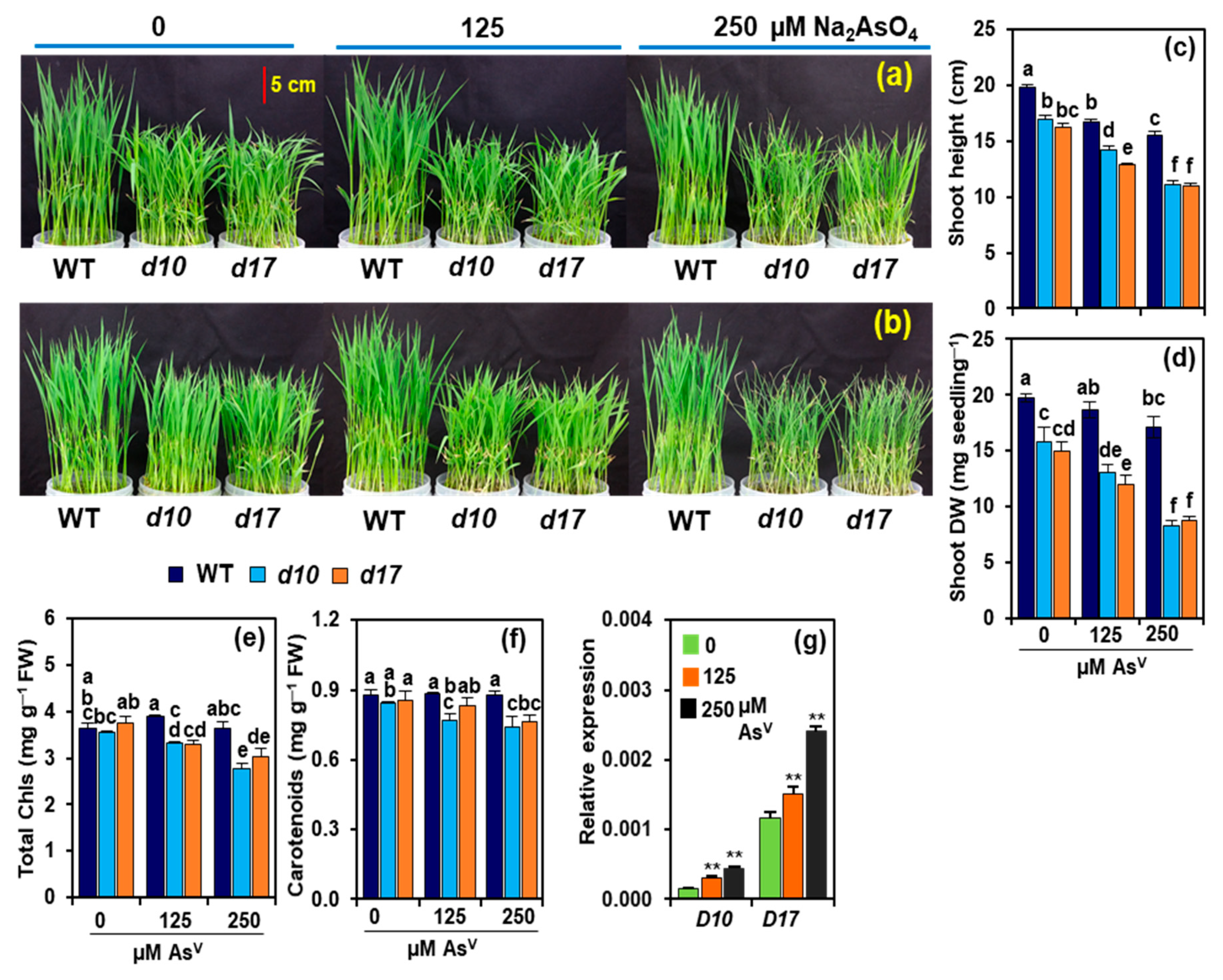
3.1. SL Deficiency Leads to Severe Arsenic Stress on Rice Growth and Biomass Production

The shoots of AsV-exposed d10 and d17 mutants showed severe phenotypic aberrations, including rolling and yellowing of leaves, as well as burning of leaf tips, as compared with WT (Figure 2a,b). At As1 and As2 doses, the shoot heights of WT plants were lowered by 15.3 and 21.9%, respectively, when compared with As0 (Figure 2c). By contrast, the shoot heights of d10 and d17 plants markedly decreased by 16.5 and 20.4% at As1, and more highly declined by 34.4 and 32.2%, respectively, at As2 in relation to the values at As0 (Figure 2c). Comparable shoot DWs were observed in WT plants under As0 and As1, while it was reduced by 13.4% at As2 (Figure 2d). In contrast, As1 and As2 reduced shoot DW by 17.2 and 47.7% in d10, and by 19.9 and 41.3% in d17 plants, respectively, in comparison with their respective value at As0 (Figure 2d). The photosynthetic pigment data showed that AsV stress did not significantly alter the levels of total Chls and carotenoids in WT plants when compared with their respective value at As0 (Figure 2e,f). At As1 and As2, the contents of total Chls were reduced by 6.6 and 21.8%, while the levels of carotenoids were reduced by 8.7 and 12.0% in d10 mutant, respectively, compared with their respective value at As0 (Figure 2e,f). In d17 mutant shoots, the levels of total Chls decreased by 11.9 and 19.1% at As1 and As2, respectively, relative to control conditions (Figure 2e). On the other hand, in the shoots of d17 mutant, carotenoid content remained unaltered at As1, but it showed a significant decline by 11%, at As2 compared with the content observed at As0 (Figure 2f).

3.2. Arsenic Induces the Expression of D10 and D17 in the Shoots of WT Plants

To see whether AsV stress affected the expression of genes involved in the SL-biosynthetic pathway, we assessed the transcript levels of D10 and D17 in the shoots of WT rice plants after their exposure to AsV doses for three days. In contrast to those observed at As0, dramatically elevated transcript levels of D10 (by 96.6 and 184.1%) and D17 (by 30.8 and 107.9%, respectively) were noted at As1 and As2 in the shoots of WT plants (Figure 2g).

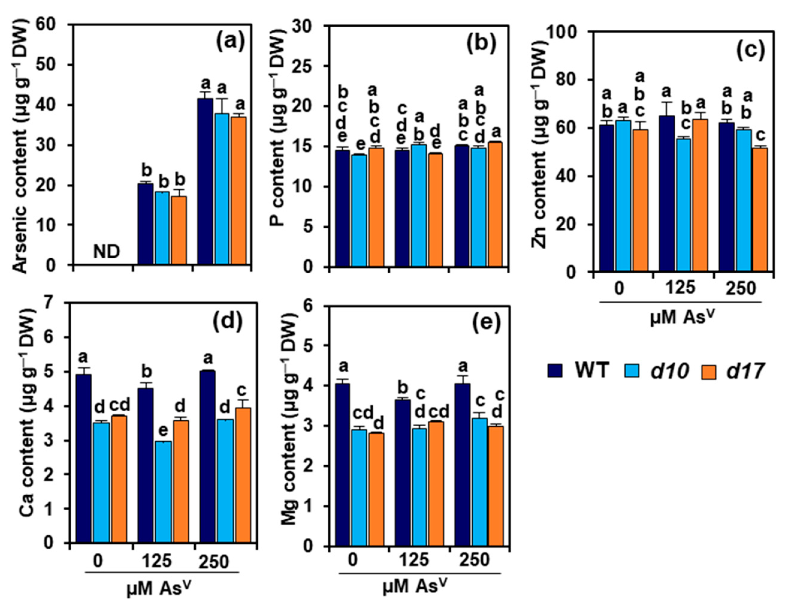
3.3. SL Deficiency Does Not Alter Arsenic, P and Zn Levels in Rice Shoots

All control plants that were not exposed to AsV treatments had no detectable level of arsenic in their shoots (Figure 3a). Arsenic contents steadily and markedly increased in the shoots of WT and SL mutant plants after their exposure to As1 and As2 (Figure 3a). On the other hand, P contents in WT and d17 shoots remained comparable at As1 and As2 in relation to their levels at As0 (Figure 3b). In d10 plants, the shoot-P contents displayed an elevation by 10 and 6.3% at As1 and As2, respectively, in comparison with the respective value at As0 (Figure 3b). Nevertheless, there were no significant differences in the arsenic and P contents between the WT and SL-deficient mutants (Figure 3a,b). Zn contents were diminished by 12.3 and 5.8% in d10 shoots at As1 and As2, respectively, compared with the content recorded at As0 (Figure 3c). In WT and d17 shoots, Zn contents remained nearly unaltered at As0, As1 and As2 treatments (Figure 3c). In general, SL deficiency did not significantly alter Zn contents under both AsV free and AsV stress conditions (Figure 3c).

3.4. SL Deficiency Alters Ca and Mg Contents in Rice Shoots

In comparison with As0, a significant reduction of Ca content was recorded in the shoots of WT (by 8.1%) and d10 (by 15.3%) at As1; however, comparable levels of Ca were observed in the shoots of the two genotypes at As2 (Figure 3d). No significant changes in Ca level were observed in the shoots of AsV-stressed d17 plants at both As1 and As2 compared with that at As0 (Figure 3d). In relation to As0, As1 displayed a notable decrease (by 10.0%), while As2 showed no significant change in the level of Mg in WT shoots (Figure 3e). Nonetheless, Mg levels remained unchanged in the shoots of mutant plants in responses to As1 and As2 when contrasted with their respective levels obtained at As0 (Figure 3e). Interestingly, under control conditions (As0), WT shoots showed significantly higher contents of Ca and Mg than d10 and d17 shoots, and this elevated trend of shoot Ca and Mg contents in WT sustained even under the AsV treatments (As1 and As2) (Figure 3d,e).

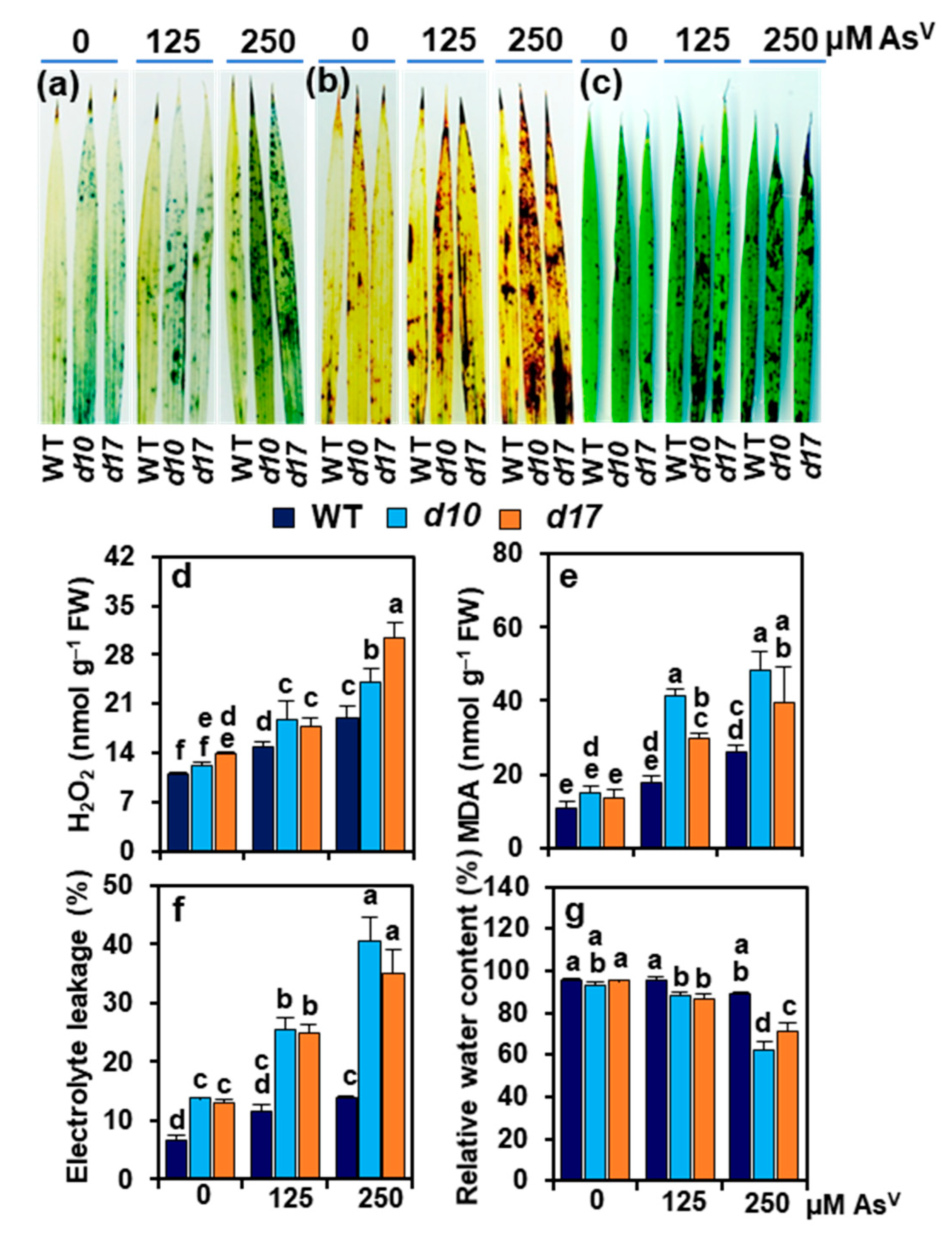
3.5. SL Deficiency Evokes Arsenic-Induced Oxidative Stress, Cuticle Damage, Electrolyte Leakage and Water Loss

NBT and DAB staining analyses showed that the leaves of d10 and d17 mutants developed more deep blue spots and dark brown spots than WT plants under AsV stress, indicating that such stress induced higher accumulation of O2 and H2O2, respectively, in the SL-deficient mutants than WT (Figure 4a,b). Similarly, TB staining showed that the leaves of SL-depleted mutant plants exhibited more intense dark spots than WT under AsV stress, which was an indication of greater cuticle damage by SL deficiency (Figure 4c).
In the shoots of WT, a significant increase in H2O2 was noted by 35.9 and 72.7% at As1 and As2, respectively, over the value found at As0 (Figure 4d). This rise range was even higher in the SL-deficient mutants; specifically, by 56.3 and 99.0% in d10, and 29.0 and 121.1% in d17 shoots at As1 and As2, respectively, when compared with the corresponding values at As0 (Figure 4d). In comparison with As0, MDA contents increased by 62.8 and 141.8% at As1 and As2, respectively, in the shoots of WT. On the other hand, more highly increased MDA contents were noted in the SL-deficient mutants; namely, by 174.2 and 221.7% in d10 shoots and 116.2 and 188.5% in d17 shoots at As1 and As2, respectively, over the control values at As0 (Figure 4e). At As1 and As2, WT shoots showed significant increases by 75.3 and 107.2%, respectively, in EL relative to that at As0 (Figure 4f). EL was more severe by SL deficiency, showing the increased values of 82.3 and 90.4% at As1, and 189.7 and 169.1% at As2 in the shoots of d10 and d17 mutant plants, respectively, in contrast to their data at As0 (Figure 4f). WT plants had comparable RWC in the shoots at all levels of AsV treatments, while SL-deficient mutants displayed lower RWC than WT at As1 and As2 (Figure 4g). In d10 plants, RWC remained unaffected at As1; however, it showed a reduction by 33.3% at As2 in relation to As0 (Figure 4g). In d17 plants, the RWC was reduced by 8.8 and 25.2% at As1 and As2, respectively, in comparison with As0 (Figure 4g).

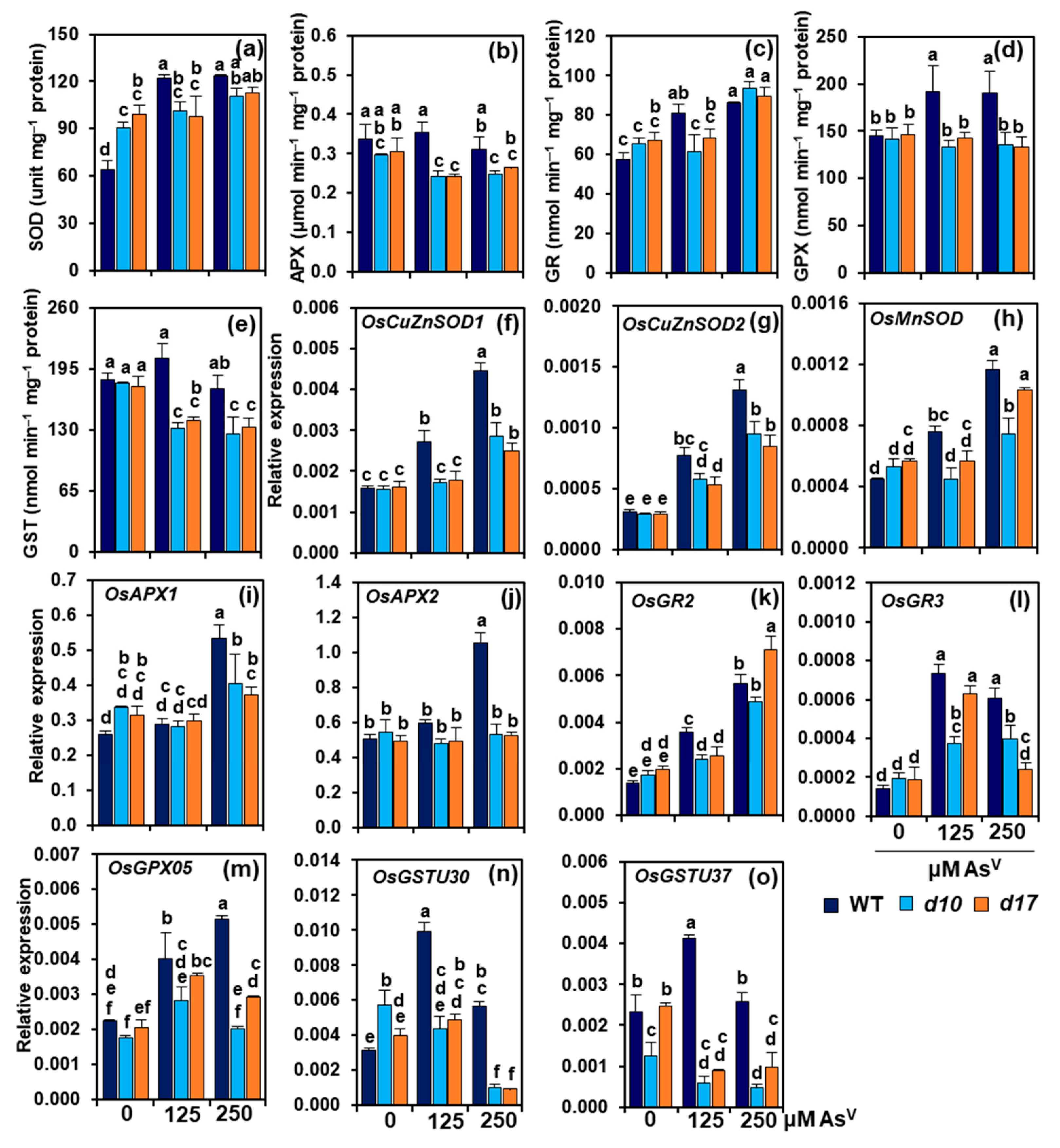
3.6. SL Deficiency Compromises Antioxidant Defense System under AsV Stress

In comparison with As0, As1 and As2 enhanced SOD activities by 91 and 92.7%, respectively, in the shoots of WT (Figure 5a). On the other hand, SOD activities in d10 shoots did not show significant alteration at As1 but an increase by 22.4% at As2, when compared with As0. In d17 shoots, SOD activities remained comparable at both levels of AsV when contrasted with As0 (Figure 5a). APX activities showed insignificant change at As1 but a decrease by 7.6% in WT shoots at As2 relative to As0. However, in comparison with As0, As1 and As2 decreased APX activities by 18.2 and 16.1% in d10 shoots, and 20.7 and 13.6% in d17 shoots, respectively (Figure 5b). WT plants treated with As1 and As2 showed an increase in GR activity by 40.2 and 49.2%, respectively, over the control value obtained at As0 (Figure 5c). By contrast, shoot-GR activities in d10 and d17 plants were not altered at As1 but enhanced by 42.6 and 32.7% at As2, respectively, when contrasted with the data at As0 (Figure 5c). WT plants treated with As1 and As2 showed significant increases in shoot-GPX activity, by 32.3 and 31.3%, respectively, over the data at As0 (Figure 5d). However, GPX activities showed an unaltered trend in the shoots of d10 and d17 plants raised at any levels of AsV (Figure 5d). In WT shoots, GST activities remained unaltered at both As1 and As2 doses when compared with As0 (Figure 5e). On the other hand, As1 and As2 decreased GST activities in shoots of d10 plants by 26.4 and 30.0%, and d17 plants by 20.4 and 24.9%, respectively, in comparison with As0 (Figure 5e).
In WT shoots, the transcript levels of SOD-encoding gene OsCuZnSOD1 were notably elevated by 69.9 and 181.2% at As1 and As2, respectively, whereas in d10 and d17 shoots, its expression levels increased by 82.8% and 55.7%, respectively, only at As2, in relation to As0 (Figure 5f). The OsCuZnSOD2 transcripts were more remarkably enhanced in WT shoots (by 151.6 and 329.2%, respectively) than in d10 (by 96.7 and 223.0%) and d17 (by 80 and 189.9%) shoots at both As1 and As2, when compared with As0 (Figure 5g). Likewise, the expression levels of OsMnSOD more highly increased in the shoots of WT (by 69.4 and 160.8% at As1 and As2, respectively) than in d10 (by 39.9% at As2) and d17 plants (by 81.9% at As2) (Figure 5h). In comparison with As0, the expression levels of APX-encoding genes APX1 and APX2 remained unaltered at As1 in WT, d10 and d17 shoots. However, at As2, the transcript levels of APX1 and APX2 showed a greater enhancement, by 106.9 and 109.9%, respectively, in WT compared with the respective values obtained at As0. Nonetheless, the d10 and d17 shoots did not show significant alteration in APX1 and APX2 transcripts at both As1 and As2 compared with As0 (Figure 5i,j).
The transcript levels of OsGR2 in the shoots of WT plants dramatically increased by 156.9 and 306.7% at As1 and As2 over the value found at As0 (Figure 5k). However, OsGR2 transcript levels in d10 and d17 plants did not alter at As1 but significantly increased by 180.3 and 258.0% at As2, respectively, compared with the data at As0 (Figure 5k). OsGR3 expression levels more highly increased in WT shoots (by 411.3 and 323.8%, respectively) than in d10 (by 91.2 and 103.8%) and d17 shoots (by 240.3% and 30.8%) in responses to As1 and As2 compared with As0 (Figure 5l). The expression levels of the GPX-encoding gene OsGPX05 improved by 78.6 and 128.8% in WT, and 71.8 and 42.7% in d17 shoots at As1 and As2, respectively, in comparison with As0 (Figure 5m). Nevertheless, in d10 shoots, the transcript level of OsGPX05 was raised by 61.3% at As1, but it was not significantly altered at As2 compared with As0 (Figure 5m).
The expression levels of GST-encoding gene OsGSTU30 in WT shoots increased by 214.9 and 79.2% in response to As1 and As2, respectively, in relation to As0 (Figure 5n). However, the OsGSTU30 transcript levels in d10 shoots dramatically decreased in response to As1 (by 23.9%) and As2 (by 82.2%) when compared with As0 (Figure 5n). Additionally, OsGSTU30 expression in d17 shoots displayed an insignificant increase at As1, but its transcript level drastically declined by 76.8% at As2, in relation to As0 (Figure 5n). When compared with As0, OsGSTU37 expression level increased by 78.1% at As1 but its transcript level remained unchanged at As2 in WT shoots (Figure 5o). In d10 shoots, OsGSTU37 expression levels declined upon exposure to AsV stress, but this decline was significant only at As2 (by 61.4%) when contrasted with As0 (Figure 5o). Furthermore, the expression levels of shoot-OsGSTU37 decreased by 63.8 and 60.3% in d17 plants at As1 and As2, respectively, when compared with As0 (Figure 5o).

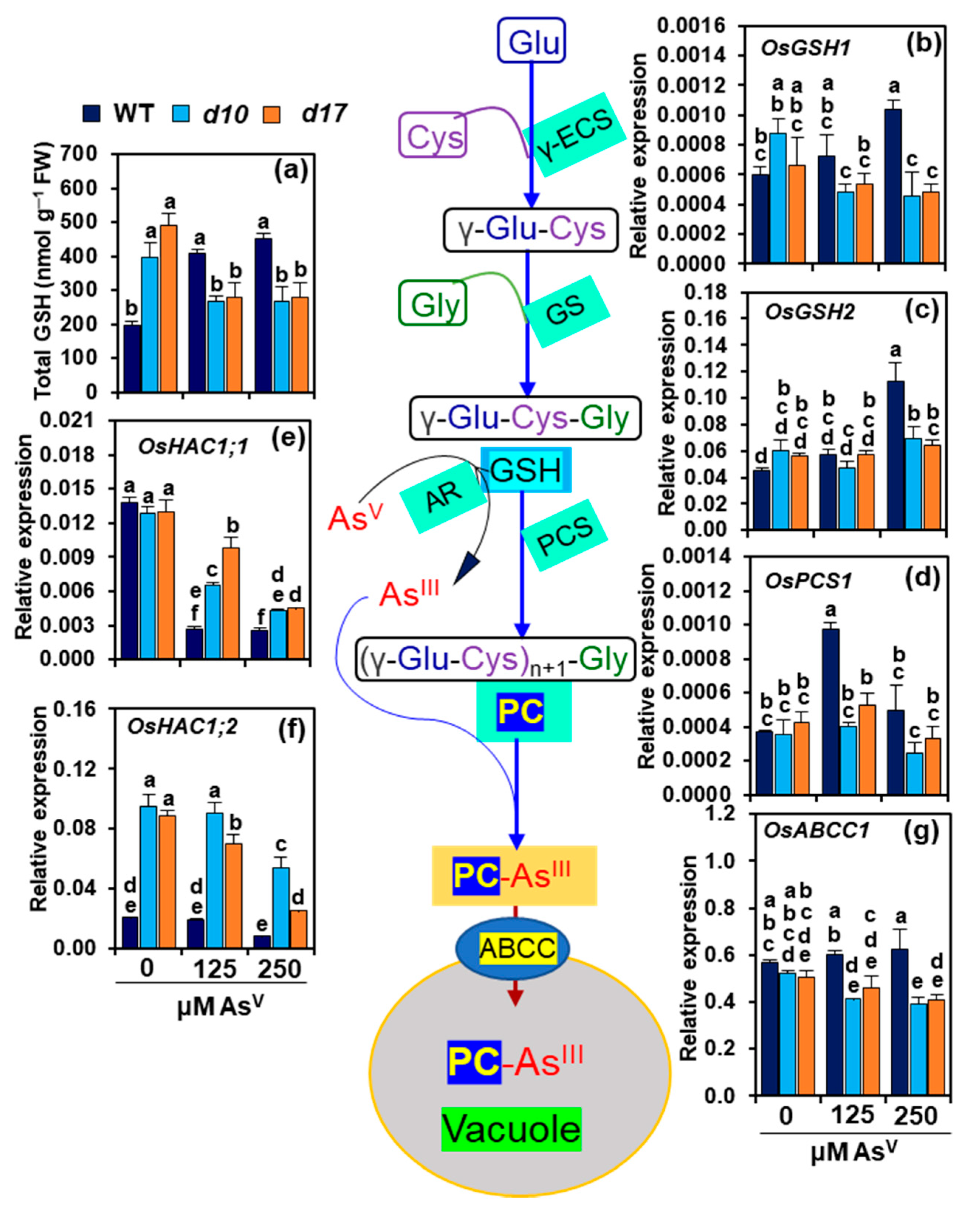
3.7. SL Deficiency Negatively Affects GSH-Assisted Arsenic Detoxification

In WT plants, As1 and As2 led to a considerable rise in shoot contents of total GSH by 104.8 and 127.1%, respectively, when compared with As0 (Figure 6a). On the other hand, SL-depleted mutants stressed with As1 and As2 showed a substantial decline in the total GSH levels by 32.4 and 33.1% in d10 shoots, and 43.2 and 42.6%, respectively, in d17 shoots compared with the respective data at As0 (Figure 6a). We also investigated the expression of genes involved in the biosynthesis of GSH (γ-glutamyl cysteine synthetase 1, OsGSH1 and glutathione synthetase 2, OsGSH2), phytochelatin (phytochelatin synthase 1, OsPCS1), arsenate reductase (high arsenate content 1, OsHAC1;1 and high arsenate content 2, OsHAC1;2) and AsIII-PC ABC transporter (C-type ATP-binding cassette (ABC) transporter, OsABCC1) in the shoots of all studied genotypes (Figure 6b–g). At As1, WT shoots had no significant changes in OsGSH1 and OsGSH2 transcript levels; however, at As2, their transcript levels were considerably raised by 74.75 and 151.9%, respectively, as compared with As0 (Figure 6b,c). In d10 shoots, while OsGSH1 expression levels were significantly reduced by 45.1 and 48.3% at As1 and As2, respectively, those of OsGSH2 remained unaltered under both two stress conditions in comparison with As0. The d17 shoots, on the other hand, showed no significant changes in OsGSH1 and OsGSH2 transcript levels at both As1 and As2 compared with As0 (Figure 6b,c).
In the shoots of WT plants, OsPCS1 expression levels increased by 163.1 and 34.4% at As1 and As2, respectively, relative to the data obtained at As0. However, d10 and d17 shoots did not show significant alteration in the expression levels of OsPCS1 at As1 and As2 when compared with As0 (Figure 6d). The transcript levels of OsHAC1;1 displayed significant attenuation in the shoots of WT (by 80.4 and 81.7%), d10 (by 49.8 and 66.8%) and d17 (by 24.8 and 65.1%, respectively) at As1 and As2 when compared with the respective values at As0 (Figure 6e). The expression levels of OsHAC1;2 remained unaltered in WT shoots at both As1 and As2 compared with As0. In comparison with As0, As2 decreased the transcript levels of OsHAC1;2 by 43.2 and 71.8% in d10 and d17 mutants, while As1 showed a decrease by 21.2% in d17 mutant only (Figure 6f). The expression levels of OsABCC1 remained at the control As0 level in WT shoots at As1 and As2. However, As1 and As2 caused a reduction in OsABCC1 expression levels in the shoots of d10 (by 21.2 and 24.8%) and d17 (by 9.3 and 19.3%, respectively) compared with As0 (Figure 6g).

4. Discussion

In a recent work, it was found that SLs helped rice roots cope with AsV stress by limiting arsenic uptake and accumulation, while also reducing Pi uptake [27]. Following this report, we looked at the amounts of arsenic and P in rice shoots and discovered that despite having equal levels of arsenic and P, SL-depleted mutants (d10 and d17) displayed more damage in their shoots than WT (Figure 1a,b and Figure 2a,b). We were then curious as to why these mutants were more vulnerable to arsenic and what mechanisms might be responsible for WT’s superior performance under high AsV stress circumstances. Because arsenic is a non-essential, hazardous element for plant growth and metabolism, its accumulation in tissues, even at low concentrations, can impair developmental processes in crop plants, including rice, wheat and mustard (Brassica juncea) [49,50,51]. Indeed, it was noticed that both the WT and SL-depleted mutants displayed growth defects in an AsV concentration-dependent manner. However, phenotypic abnormalities, growth reduction, and biomass loss were far more severe in SL mutants than WT (Figure 2a–d). These findings showed that d10 and d17 shoots were more sensitive to AsV than WT shoots, indicating that SL depletion led to a more arsenic-susceptible phenotype of rice shoots. It was also found that AsV addition resulted in a greater reduction of photosynthetic pigment contents in both d10 and d17 plants, which supports the SL-deficient mutant plants’ inferior growth performance when compared with WT (Figure 2e,f). These findings corroborated numerous earlier studies in which photosynthetic components, such as Chls and carotenoids, were the primary targets of arsenic toxicity [26,52,53].
To check whether AsV stress had any modulatory effect on SL biosynthesis, the expression patterns of SL-biosynthetic genes D10 and D17 were investigated in the shoots of WT rice under all doses of AsV (Figure 2g). It was found that exposure of WT plants to AsV increased the amounts of D10 and D17 transcripts, suggesting that the SL-biosynthetic pathway was favorably activated in WT shoots by AsV, as it was observed in the AsV-stressed rice roots earlier [27]. Elevated transcript levels of SL-biosynthetic genes have been reported in rice stems (e.g., D27, D17 and D10) under drought stress, and in Arabidopsis leaves (e.g., MORE AXILLARY BRANCHING (MAX)3 (MAX3) and MAX4, the orthologous genes of rice D17 and D10, respectively) following dehydration treatment [13,54], indicating that different types of abiotic stresses generally induce the expression of SL-biosynthetic genes in plant shoots (Figure 2g and Figure 7). These results, along with those of others, point to the involvement of SLs in the aboveground organs of many plant species to aid their survival under stressful situations.
Next, we searched for a connection between AsV-induced damage and arsenic levels in the shoots of WT and SL-deficient mutant plants. Surprisingly, no significant variation in shoot-arsenic content among WT, d10 and d17 plants was found, although SL-depleted mutants appeared to be more negatively impacted by AsV than WT (Figure 3a). These findings raised the question of why SL-deficient mutants were sensitive to AsV, while the shoots of both WT and mutants accumulated comparable amounts of arsenic. We then checked whether AsV exposure affected the homeostasis of other minerals in the shoots, such as P, Zn, Ca and Mg (Figure 3b–e). Despite increasing the amount of AsV doses, P, Zn, Ca and Mg contents remained relatively stable at the control level in WT shoots. On the other hand, d10 and d17 shoots contained almost equal levels of P and Zn but similarly lower levels of Ca and Mg when compared with their respective content in WT shoots at both normal and AsV stress conditions (Figure 3b–e). These results pinpointed that SL deficiency did not affect the homeostases of P and Zn in the shoots under AsV stress conditions. However, the reduced levels of Ca and Mg in d10 and d17 shoots under normal conditions suggest that SL deficiency causes an imbalance in their homeostasis. Thus, we inferred that reduced basal Ca and Mg levels may be one of the factors contributing to the poor growth performance of d10 and d17 mutants when being challenged with excessive AsV (Figure 3d,e and Figure 7). Under both normal and AsV stress conditions, P and Zn showed comparable contents in the shoots of both the mutants and WT, whereas Ca and Mg exhibited similarly lower levels in the shoots of the mutants than WT (Figure 3b–e). These data suggest that AsV stress does not have any effect on the contents of all four investigated elements.
Excess arsenic in plant tissues has many detrimental effects on cellular metabolism. For example, AsV can substitute P from key cellular metabolites such as ATP due to its analogy with P, causing energy metabolism to be disrupted [55]. AsIII can bind to the sulfhydryl (SH) groups of different sulfur-containing peptides and proteins, preventing them from forming active conformation and functioning as part of the cellular defense system [56]. Both types of arsenic (AsV and AsIII) can disrupt the electron transport system in chloroplasts and mitochondria, which are the primary sites of ROS production in cells [57,58]. The transformation of AsV to AsIII can also directly contribute to ROS production via a Haber–Weiss reaction [59]. As a result, ROS generation and oxidative damage in plant tissues are considered typical hallmarks of arsenic-induced toxic effects. In the current study, we concentrated on how WT and SL-deficient mutant plants dealt with ROS and their detoxification systems under arsenic stress. Results revealed that the mutant plants’ shoots accumulated large amounts of O2 and H2O2 in response to AsV stress (Figure 4a,b,d). This increased production of ROS was positively correlated with the increased levels of lipid peroxidation product MDA and cuticle damage in the shoots of SL mutant plants exposed to AsV stress (Figure 4c,e). Furthermore, the increased levels of MDA and cuticle damage by AsV stress corresponded with increased electrolyte leakage and water loss in d10 and d17 shoots (Figure 4f,g). These findings clearly showed that AsV treatments caused considerable oxidative damage in SL-deficient mutant plants, confirming that ROS-induced oxidative stress is one of AsV’s lethal effects in rice (Figure 7), as also observed in many arsenic-susceptible cultivars such as maize (Zea mays) and barley [49,52,60].
The cellular antioxidant system comprising both enzymatic and non-enzymatic arsenals plays crucial roles in relieving the oxidative burden in plant tissues under stressful conditions. To investigate the SLs’ influence on the antioxidant system at the genetic level, the activities of several key antioxidant enzymes, including APX, SOD, GR, GST and GPX, were evaluated in the shoots of both WT and SL-deficient plants under normal and AsV stress conditions (Figure 5a–e). During AsV stress, the SL-deficient mutants had considerably lower activities of SOD, APX, GR (except at As2), GST and GPX in their shoots than WT plants. qRT-PCR analysis of the expression of relevant genes associated with the biosyntheses of the studied enzymes also displayed that the transcript levels of OsCuZnSOD1, OsCuZnSOD2 and OsMnSOD (encoding SOD), OsAPX1 and OsAPX2 (encoding APX), OsGPX05 (encoding GPX), and OsGSTU30 and OsGSTU37 (encoding GST) positively correlated with the suppressed activities of respective enzymes in the SL-depleted mutant plants (Figure 5f–m). These findings highlighted that the compromised antioxidant systems in SL-deficient mutant plants were unable to handle the excessive ROS induced by AsV, thereby these mutants suffered from severe oxidative stress. These results clearly demonstrated the likely roles of SLs in boosting the plant antioxidant defense system against arsenic-induced ROS (Figure 7).
GSH is a powerful non-enzymatic antioxidant that plays various critical roles in plant protection against heavy metal toxicity. GSH, either directly or by the formation of PCs, can aid in the chelation of AsIII for vacuolar sequestration in the cells [25]. Ideally, PC synthase (PCS) catalyzes the synthesis of PCs from GSH followed by complexation with AsIII (AsIII-PC complex), which is then sequestrated into the vacuoles by ABCC transporter [20] (Figure 6). GSH can also assist GSH-dependent enzymes such as GPX and GST to detoxify toxic aldehydes generated by ROS-induced lipid oxidation [61]. Thus, any depletion in cellular GSH levels might expose plant cells to vulnerable situations, especially under metal-induced adverse conditions. The present study critically evaluated the GSH-assisted arsenic detoxification by estimating GSH content and analyzing the expression levels of several associated genes, including OsGSH1, OsGSH2, OsHAC1;1, OsHAC1;2, OsPCS1 and OsABCC1 (Figure 6a–g). It was observed that AsV stress resulted in a significant reduction of GSH in the shoots of both SL-deficient mutants. It is plausible that AsV stress-mediated inhibition of the expression of GSH biosynthesis-associated genes OsGSH1 and OsGSH2 was responsible for the decrease in GSH pool in the shoots of the AsV-stressed SL-deficient mutants (Figure 6b,c). AR catalyzes the conversion of AsV to AsIII by utilizing GSH [62]. High expression levels of AR-encoding genes OsHAC1;1 and OsHAC1;2 also indicated that reduced levels of GSH might have resulted from the consumption of a large quantity of GSH by AR for the transformation of AsV to AsIII in the shoots of the mutants (Figure 6e,f). It is worth noting that AsIII is far more destructive to cells than AsV [63]. We assumed that the decreased transcript levels of OsPCS1 and OsABCC1 genes resulted in a reduced complexation of AsIII with PC and sequestration of AsIII-PC to vacuoles, respectively, leading to increased AsIII toxicity in the mutant shoots (Figure 2a–f and Figure 6d,g). On the other hand, the elevated GSH content and increased expression levels of OsPCS1 and OsABCC1 in WT shoots suggest that GSH-assisted AsIII detoxification was well executed to reduce arsenic toxicity at the cellular level (Figure 1b–e, Figure 4d,g and Figure 7).

5. Conclusions

Based on the results, we conclude that SL deficiency did not influence the level of arsenic in the shoots of rice plants, and high susceptibility of SL-deficient mutants to AsV resulted from the weakened cellular defense mechanisms responsible for arsenic detoxification (Figure 7). The identified most critical defense strategies that contributed to better resistance of WT against AsV stress include (i) protection of photosynthetic pigments and high basal levels of minerals (e.g., Ca and Mg) for better growth response, (ii) enhanced detoxification of ROS through a stimulated antioxidant system for safeguarding membrane structure and cuticles, and (iii) increased biosynthesis of GSH for efficient GSH-assisted arsenic detoxification (Figure 7). Overall, these findings revealed that SLs can function as an important growth regulator for enhancing arsenic detoxification mechanisms, including stimulation of cellular antioxidant defense systems and vacuolar sequestration of arsenic in rice shoots, thereby assisting rice plants to overcome excessive arsenic-induced toxic consequences at the cellular level.

Supplementary Materials

The following are available online at https://www.mdpi.com/article/10.3390/antiox10111815/s1, Table S1: Primer sequences of reference and target genes used in the current study.

Author Contributions

Conceptualization, M.G.M. and L.-S.P.T.; methodology, M.G.M.; software, M.G.M., C.V.H. and M.M.R.; validation, M.G.M., C.V.H., M.M.R., Y.W., K.H.N. and S.S.K.; formal analysis, M.G.M., C.V.H., M.M.R., S.S.K. and M.I.; investigation, M.G.M., Y.W. and M.I. resources, M.F. and L.-S.P.T.; data curation, M.G.M. and K.H.N.; writing—original draft preparation, M.G.M.; writing—review and editing, M.G.M., C.V.H., L.-S.P.T., A.H. and E.F.A.; visualization, M.G.M., M.M.R. and K.H.N.; supervision, L.-S.P.T. and M.F; project administration, L.-S.P.T.; funding acquisition, L.-S.P.T., A.H. and E.F.A. All authors have read and agreed to the published version of the manuscript.

Funding

The authors would like to extend their sincere appreciation to the Researchers Supporting Project Number (RSP-2021/134), King Saud University, Riyadh, Saudi Arabia.

Institutional Review Board Statement

Not applicable.

Informed Consent Statement

Not applicable.

Data Availability Statement

Data is contained within the article and Supplementary Material.

Acknowledgments

M.G.M. thankfully acknowledges the postdoc fellowship from the Japan Society for the Promotion of Science (JSPS). The authors are grateful to M. Umehara (Toyo University, Japan) for providing us with the seeds of wild type and strigolactone-deficient d10 and d17 mutants. The authors would like to extend their sincere appreciation to the Researchers Supporting Project Number (RSP-2021/134), King Saud University, Riyadh, Saudi Arabia.

Conflicts of Interest

The authors declare no conflict of interest. The funders had no role in the design of the study; in the collection, analyses, or interpretation of data; in the writing of the manuscript, or in the decision to publish the results.

References

  1. Mostofa, M.G.; Li, W.; Nguyen, K.H.; Fujita, M.; Tran, L.-S.P. Strigolactones in plant adaptation to abiotic stresses: An emerging avenue of plant research. Plant Cell Environ. 2018, 41, 2227–2243. [Google Scholar] [CrossRef]
  2. Gupta, A.; Sinha, R.; Fernandes, J.L.; Abdelrahman, M.; Burritt, D.J.; Tran, L.-S.P. Phytohormones regulate convergent and divergent responses between individual and combined drought and pathogen infection. Crit. Rev. Biotechnol. 2020, 40, 320–340. [Google Scholar] [CrossRef]
  3. Waters, M.T.; Gutjahr, C.; Bennett, T.; Nelson, D.C. Strigolactone signaling and evolution. Annu. Rev. Plant Biol. 2017, 68, 291–322. [Google Scholar] [CrossRef] [PubMed]
  4. Al-Babili, S.; Bouwmeester, H.J. Strigolactones, a novel carotenoid-derived plant hormone. Annu. Rev. Plant Biol. 2015, 66, 161–186. [Google Scholar] [CrossRef]
  5. Alder, A.; Jamil, M.; Marzorati, M.; Bruno, M.; Vermathen, M.; Bigler, P. The path from beta-carotene to carlactone, a strigolactone-like plant hormone. Science 2012, 335, 1348–1351. [Google Scholar] [CrossRef] [Green Version]
  6. Zhang, Y.X.; van Dijk, A.D.J.; Scaffidi, A.; Flematti, G.R.; Hofmann, M.; Charnikhova, T. Rice cytochrome P450 MAX1 homologs catalyze distinct steps in strigolactone biosynthesis. Nat. Chem. Biol. 2014, 10, 1028–1033. [Google Scholar] [CrossRef] [PubMed]
  7. Chesterfield, R.J.; Vickers, C.E.; Beveridge, C.A. Translation of strigolactones from plant hormone to agriculture: Achievements, future perspectives, and challenges. Trends Plant Sci. 2020, 25, 1087–1106. [Google Scholar] [CrossRef]
  8. Zhou, F.; Lin, Q.B.; Zhu, L.H.; Ren, Y.L.; Zhou, K.N.; Shabek, N. D14-SCFD3-dependent degradation of D53 regulates strigolactone signalling. Nature 2013, 504, 406–410. [Google Scholar] [CrossRef] [PubMed] [Green Version]
  9. López-Ráez, J.A.; Shirasu, K.; Foo, E. Strigolactones in plant interactions with beneficial and detrimental organisms: The Yin and Yang. Trends Plant Sci. 2017, 22, 527–537. [Google Scholar] [CrossRef]
  10. Cooper, J.W.; Hu, Y.; Beyyoudh, L.; Yildiz, D.H.; Kunert, K.; Beveridge, C.A. Strigolactones positively regulate chilling tolerance in pea and in Arabidopsis. Plant Cell Environ. 2018, 41, 1298–1310. [Google Scholar] [CrossRef]
  11. Qiu, C.-W.; Zhang, C.; Wang, N.-H.; Mao, W.; Wu, F. Strigolactone GR24 improves cadmium tolerance by regulating cadmium uptake, nitric oxide signaling and antioxidant metabolism in barley (Hordeum vulgare L.). Environ. Pollut. 2021, 273, 116486. [Google Scholar] [CrossRef] [PubMed]
  12. Sedaghat, M.; Emam, Y.; Mokhtassi-Bidgoli, A.; Hazrati, S.; Lovisolo, C.; Visentin, I. The potential of the synthetic strigolactone analogue GR24 for the maintenance of photosynthesis and yield in winter wheat under drought: Investigations on the mechanisms of action and delivery modes. Plants 2021, 10, 1223. [Google Scholar] [CrossRef]
  13. Ha, C.V.; Leyva-González, M.A.; Osakabe, Y.; Tran, U.T.; Nishiyama, R.; Watanabe, Y. Positive regulatory role of strigolactone in plant responses to drought and salt stress. Proc. Natl. Acad. Sci. USA 2014, 111, 851–856. [Google Scholar] [CrossRef] [PubMed] [Green Version]
  14. Liu, J.; He, H.; Vitali, M.; Visentin, I.; Charnikhova, T.; Haider, I. Osmotic stress represses strigolactone biosynthesis in Lotus japonicus roots: Exploring the interaction between strigolactones and ABA under abiotic stress. Planta 2015, 241, 1435–1451. [Google Scholar] [CrossRef] [PubMed]
  15. Haider, I.; Andreo-Jimenez, B.; Bruno, M.; Bimbo, A.; Floková, K.; Abuauf, H. The interaction of strigolactones with abscisic acid during the drought response in rice. J. Exp. Bot. 2018, 69, 2403–2414. [Google Scholar] [CrossRef] [PubMed]
  16. Visentin, I.; Vitali, M.; Ferrero, M.; Zhang, Y.; Ruyter-Spira, C.; Novák, O. Low levels of strigolactones in roots as a component of the systemic signal of drought stress in tomato. New Phytol. 2016, 212, 954–963. [Google Scholar] [CrossRef] [PubMed]
  17. Ling, F.; Su, Q.; Jiang, H.; Cui, J.; He, X.; Wu, Z.; Zhang, Z.; Liu, J.; Zhao, Y. Effects of strigolactone on photosynthetic and physiological characteristics in salt-stressed rice seedlings. Sci. Rep. 2020, 10, 6183. [Google Scholar] [CrossRef]
  18. Lu, T.; Yu, H.; Li, Q.; Chai, L.; Jiang, W. Photosynthetic inhibition and oxidative stress from low-light stress with exogenous GR24 in tomato (Solanum lycopersicum L.) seedlings. Front. Plant Sci. 2019, 10, 490. [Google Scholar] [CrossRef]
  19. Scaffidi, A.; Waters, M.T.; Sun, Y.K.; Skelton, B.W.; Dixon, K.W.; Ghisalberti, E.L. Strigolactone hormones and their stereoisomers signal through two related receptor proteins to induce different physiological responses in Arabidopsis. Plant Physiol. 2014, 165, 1221–1232. [Google Scholar] [CrossRef] [Green Version]
  20. Abbas, G.; Murtaza, B.; Bibi, I.; Shahid, M.; Niazi, N.K.; Khan, M.I.; Amjad, M. Arsenic uptake, toxicity, detoxification, and speciation in plants: Physiological, biochemical, and molecular aspects. Int. J. Environ. Res. Public Health 2018, 15, 59. [Google Scholar] [CrossRef] [Green Version]
  21. Khan, I.; Awan, S.A.; Rizwan, M.; Ali, S.; Zhang, X.; Huang, L. Arsenic behavior in soil-plant system and its detoxification mechanisms in plants: A review. Environ. Pollut. 2021, 286, 117389. [Google Scholar] [CrossRef]
  22. Tang, Z.; Zhao, F.-J. The roles of membrane transporters in arsenic uptake, translocation and detoxification in plants. Crit. Rev. Environ. Sci. Technol. 2020, 51, 2449–2484. [Google Scholar] [CrossRef]
  23. Saifullah; Dahlawi, S.; Naeem, A.; Iqbal, M.; Farooq, M.A.; Bibi, S. Opportunities and challenges in the use of mineral nutrition for minimizing arsenic toxicity and accumulation in rice: A critical review. Chemosphere 2018, 194, 171–188. [Google Scholar] [CrossRef]
  24. Peña-Garcia, Y.; Shinde, S.; Natarajan, P.; Lopez-Ortiz, C.; Balagurusamy, N.; Chavez, A.C.D. Arsenic Stress-Related F-Box (ASRF) gene regulates arsenic stress tolerance in Arabidopsis thaliana. J. Hazard. Mater. 2021, 407, 124831. [Google Scholar] [CrossRef]
  25. Vezza, M.E.; Luna, D.F.; Agostini, E.; Talano, M.A. Glutathione, a key compound for As accumulation and tolerance in soybean plants treated with AsV and AsIII. Environ. Exp. Bot. 2019, 162, 272–282. [Google Scholar] [CrossRef]
  26. Bali, A.S.; Sidhu, G.P.S. Arsenic acquisition, toxicity and tolerance in plants-From physiology to remediation: A review. Chemosphere 2021, 283, 131050. [Google Scholar] [CrossRef]
  27. Mostofa, M.G.; Rahman, M.M.; Nguyen, K.H.; Li, W.; Watanabe, Y.; Tran, C.D. Strigolactones regulate arsenate uptake, vacuolar-sequestration and antioxidant defense responses to resist arsenic toxicity in rice roots. J. Hazard. Mater. 2021, 415, 125589. [Google Scholar] [CrossRef] [PubMed]
  28. Dixit, G.; Singh, A.P.; Kumar, A.; Mishra, S.; Dwivedi, S.; Kumar, S. Reduced arsenic accumulation in rice (Oryza sativa L.) shoot involves sulfur mediated improved thiol metabolism, antioxidant system and altered arsenic transporters. Plant Physiol. Biochem. 2016, 99, 86–96. [Google Scholar] [CrossRef]
  29. Sarwar, T.; Khan, S.; Muhammad, S.; Amin, S. Arsenic speciation, mechanisms, and factors affecting rice uptake and potential human health risk: A systematic review. Environ. Technol. Innov. 2021, 22, 101392. [Google Scholar] [CrossRef]
  30. Muehe, E.M.; Wang, T.; Kerl, C.F.; Planer-Friedrich, B.; Fendorf, S. Rice production threatened by coupled stresses of climate and soil arsenic. Nat. Commun. 2019, 10, 4985. [Google Scholar] [CrossRef] [PubMed] [Green Version]
  31. Mostofa, M.G.; Rahman, M.M.; Siddiqui, M.N.; Fujita, M.; Tran, L.-S.P. Salicylic acid antagonizes selenium phytotoxicity in rice: Selenium homeostasis, oxidative stress metabolism and methylglyoxal detoxification. J. Hazard. Mater. 2020, 394, 122572. [Google Scholar] [CrossRef] [PubMed]
  32. Ishikawa, S.; Maekawa, M.; Arite, T.; Onishi, K.; Takamure, I.; Kyozuka, J. Suppression of tiller bud activity in tillering dwarf mutants of rice. Plant Cell Physiol. 2005, 46, 79–86. [Google Scholar] [CrossRef]
  33. Mostofa, M.G.; Fujita, M. Salicylic acid alleviates copper toxicity in rice (Oryza sativa L.) seedlings by up-regulating antioxidative and glyoxalase systems. Ecotoxicology 2013, 22, 959–973. [Google Scholar] [CrossRef] [PubMed]
  34. Mostofa, M.G.; Hossain, M.A.; Fujita, M. Trehalose pretreatment induces salt tolerance in rice (Oryza sativa L.) seedlings: Oxidative damage and co-induction of antioxidant defense and glyoxalase systems. Protoplasma 2015, 252, 461–475. [Google Scholar] [CrossRef] [PubMed]
  35. Arnon, D.I. Copper enzymes in isolated chloroplasts. polyphenoloxidase in Beta Vulgaris. Plant Physiol. 1949, 24, 1–15. [Google Scholar] [CrossRef] [Green Version]
  36. Lichtenthaler, H.K.; Wellburn, A.R. Determinations of total carotenoids and chlorophylls a and b of leaf extracts in different solvents. Biochem. Soc. Trans. 1983, 11, 591–592. [Google Scholar] [CrossRef] [Green Version]
  37. Itouga, M.; Kato, Y.; Sakakibara, H. Phenotypic plasticity and mineral nutrient uptake of the moss Polymeric commune Hew. during acclimation to a change in light intensity. Hikobia 2014, 16, 459–466. [Google Scholar]
  38. Tanaka, T.; Tanaka, H.; Machida, C.; Watanabe, M.; Machida, Y. A new method for rapid visualization of defects in leaf cuticle reveals five intrinsic patterns of surface defects in Arabidopsis. Plant J. 2004, 37, 139–146. [Google Scholar] [CrossRef]
  39. Nguyen, K.H.; Mostofa, M.G.; Watanabe, Y.; Tran, C.D.; Rahman, M.M.; Tran, L.-S.P. Overexpression of GmNAC085 enhances drought tolerance in Arabidopsis by regulating glutathione biosynthesis, redox balance and glutathione-dependent detoxification of reactive oxygen species and methylglyoxal. Environ. Exp. Bot. 2019, 161, 242–254. [Google Scholar] [CrossRef]
  40. Heath, R.L.; Packer, L. Photoperoxidation in isolated chloroplasts: I. Kinetics and stoichiometry of fatty acid peroxidation. Arch. Biochem. Biophys. 1968, 125, 189–198. [Google Scholar] [CrossRef]
  41. Griffith, O.W. Determination of glutathione and glutathione disulfide using glutathione reductase and 2-vinylpyridine. Anal. Biochem. 1980, 106, 207–212. [Google Scholar] [CrossRef]
  42. Bradford, M.M. A rapid and sensitive method for the quantitation of microgram quantities of protein utilizing the principle of protein-dye binding. Anal. Biochem. 1976, 72, 248–254. [Google Scholar] [CrossRef]
  43. El-Shabrawi, H.; Kumar, B.; Kaul, T.; Reddy, M.K.; Singla-Pareek, S.L.; Sopory, S.K. Redox homeostasis, antioxidant defense, and methylglyoxal detoxification as markers for salt tolerance in Pokkali rice. Protoplasma 2010, 245, 85–96. [Google Scholar] [CrossRef] [PubMed]
  44. Nakano, Y.; Asada, K. Hydrogen peroxide is scavenged by ascorbate-specific peroxidase in spinach chloroplasts. Plant Cell Physiol. 1981, 22, 867–880. [Google Scholar]
  45. Foyer, C.H.; Halliwell, B. The presence of glutathione and glutathione reductase in chloroplasts: A proposed role in ascorbic acid metabolism. Planta 1976, 133, 21–25. [Google Scholar] [CrossRef]
  46. Hossain, M.A.; Hasanuzzaman, M.; Fujita, M. Up-regulation of antioxidant and glyoxalase systems by exogenous glycinebetaine and proline in mung bean confer tolerance to cadmium stress. Physiol. Mol. Biol. Plants 2010, 16, 259–272. [Google Scholar] [CrossRef] [Green Version]
  47. Elia, A.C.; Galarini, R.; Taticchi, M.I.; Dörr, A.J.M.; Mantilacci, L. Antioxidant responses and bioaccumulation in Ictalurus melas under mercury exposure. Ecotoxicol. Environ. Saf. 2003, 55, 162–167. [Google Scholar] [CrossRef]
  48. Le, D.T.; Nishiyama, R.; Watanabe, Y.; Mochida, K.; Yamaguchi-Shinozaki, K.; Shinozaki, K. Genome-wide expression profiling of soybean two-component system genes in soybean root and shoot tissues under dehydration stress. DNA Res. 2011, 18, 17–29. [Google Scholar] [CrossRef] [Green Version]
  49. Yadav, P.; Srivastava, S.; Patil, T.; Raghuvanshi, R.; Srivastava, A.K.; Suprasanna, P. Tracking the time-dependent and tissue-specific processes of arsenic accumulation and stress responses in rice (Oryza sativa L.). J. Hazard. Mater. 2021, 406, 124307. [Google Scholar] [CrossRef]
  50. Pandey, C.; Augustine, R.; Panthri, M.; Zia, I.; Bisht, N.C.; Gupta, M. Arsenic affects the production of glucosinolate, thiol and phytochemical compounds: A comparison of two Brassica cultivars. Plant Physiol. Biochem. 2017, 111, 144–154. [Google Scholar] [CrossRef]
  51. Hossain, M.M.; Khatun, M.A.; Haque, M.N.; Bari, M.A.; Alam, M.F.; Mandal, A. Silicon alleviates arsenic-induced toxicity in wheat through vacuolar sequestration and ROS scavenging. Int. J. Phytoremediat. 2018, 20, 796–804. [Google Scholar] [CrossRef] [PubMed]
  52. Anjum, S.A.; Tanveer, M.; Hussain, S.; Ashraf, U.; Khan, I.; Wang, L. Alteration in growth, leaf gas exchange, and photosynthetic pigments of maize plants under combined cadmium and arsenic stress. Water Air Soil Pollut. 2016, 228, 13. [Google Scholar] [CrossRef]
  53. Singh, R.; Jha, A.B.; Misra, A.N.; Sharma, P. Differential responses of growth, photosynthesis, oxidative stress, metals accumulation and NRAMP genes in contrasting Ricinus communis genotypes under arsenic stress. Environ. Sci. Pollut. Res. 2019, 26, 31166–31177. [Google Scholar] [CrossRef] [PubMed]
  54. Du, H.; Huang, F.; Wu, N.; Li, X.; Hu, H.; Xiong, L. Integrative regulation of drought escape through ABA-dependent and -independent pathways in rice. Mol. Plant 2018, 11, 584–597. [Google Scholar] [CrossRef] [PubMed] [Green Version]
  55. Tawfik, D.S.; Viola, R.E. Arsenate replacing phosphate: Alternative life chemistries and ion promiscuity. Biochemistry 2011, 50, 1128–1134. [Google Scholar] [CrossRef] [PubMed] [Green Version]
  56. Finnegan, P.M.; Chen, W. Arsenic toxicity: The effects on plant metabolism. Front. Physiol. 2012, 3, 182. [Google Scholar] [CrossRef] [Green Version]
  57. Farnese, F.S.; Oliveira, J.A.; Paiva, E.A.S.; Menezes-Silva, P.E.; da Silva, A.A.; Campos, F.V. The involvement of nitric oxide in integration of plant physiological and ultrastructural adjustments in response to arsenic. Front. Plant Sci. 2017, 8, 516. [Google Scholar] [CrossRef] [Green Version]
  58. Mostofa, M.G.; Rahman, M.M.; Ansary, M.M.U.; Keya, S.S.; Abdelrahman, M.; Miah, M.G. Silicon in mitigation of abiotic stress-induced oxidative damage in plants. Crit. Rev. Biotechnol. 2021, 41, 918–934. [Google Scholar] [CrossRef]
  59. Mascher, R.; Lippmann, B.; Holzinger, S.; Bergmann, H. Arsenate toxicity: Effects on oxidative stress response molecules and enzymes in red clover plants. Plant Sci. 2002, 163, 961–969. [Google Scholar] [CrossRef]
  60. Zvobgo, G.; LwalabaWaLwalaba, J.; Sagonda, T.; Mutemachani, M.J.; Muhammad, N.; Haider Shamsi, I. Phosphate alleviates arsenate toxicity by altering expression of phosphate transporters in the tolerant barley genotypes. Ecotoxicol. Environ. Saf. 2018, 147, 832–839. [Google Scholar] [CrossRef]
  61. Csiszár, J.; Horváth, E.; Bela, K.; Gallé, Á. Glutathione-related enzyme system: Glutathione reductase (GR), glutathione transferases (GSTs) and glutathione peroxidases (GPXs). In Redox State as a Central Regulator of Plant-Cell Stress Responses; Gupta, D.K., Palma, J.M., Corpas, F.J., Eds.; Springer International Publishing: Cham, Switzerland, 2016; pp. 137–158. [Google Scholar]
  62. Shi, S.; Wang, T.; Chen, Z.; Tang, Z.; Wu, Z.; Salt, D.E. OsHAC1;1 and OsHAC1;2 Function as arsenate reductases and regulate arsenic accumulation. Plant Physiol. 2016, 172, 1708–1719. [Google Scholar] [CrossRef] [PubMed] [Green Version]
  63. Coelho, D.G.; Marinato, C.S.; de Matos, L.P.; de Andrade, H.M.; da Silva, V.M.; Santos-Neves, P.H. Is arsenite more toxic than arsenate in plants? Ecotoxicology 2020, 29, 196–202. [Google Scholar] [CrossRef] [PubMed]
Figure 1. Biosynthesis and signaling of strigolactones (SLs) in rice. SLs are synthesized from all-trans-β-carotene by consecutive actions of β-carotene isomerase (D27), carotenoid cleavage dioxygenase (CCD)7 (D17), CCD8 (D10) and cytochrome P450 (Cyt P450) proteins. SLs bind to receptor D14 followed by interaction with D3, leading to the formation of a Skp1-Cullin-F-box (SCF)D3 type of E3 ubiquitin ligase complex. D53 is ubiquitinated by this SCF-protein complex, which triggers proteasomal degradation of D53, resulting in SL signaling events for activation of various physiological functions in rice. E, ubiquitin ligase; U, ubiquitin.
Antioxidants 10 01815 g001
Figure 2. (af) Growth-related features of wild-type (WT) and d10 and d17 mutant plants exposed to different concentrations of sodium arsenate (Na2AsO4; 0, 125 and 250 μM AsV). Shoot phenotypes were recorded on day 3 (a) and day 5 (b) of AsV treatments. Shoot height (c), shoot dry weight (d), and the levels of total chlorophylls (e) and carotenoids (f) were assessed on day 5 of AsV treatments. (g) Relative expression of D10 and D17 genes was examined in the shoots of WT on day 3 of AsV treatments. Represented numerical data are the means with standard errors (n = 4 biological repeats). Significant differences (p < 0.05) among the treatments are denoted by distinct alphabetical letters according to a least significant difference test. Student’s t-test (** p < 0.01) was conducted to identify significant variations among 0, 125 and 250 μM AsV treatments (g). Chls, chlorophylls; DW, dry weight; FW, fresh weight.
Antioxidants 10 01815 g002
Figure 3. Arsenic and mineral nutrient contents in wild-type (WT) and d10 and d17 mutant plants exposed to different concentrations of sodium arsenate (Na2AsO4; 0, 125 and 250 μM AsV). The contents of arsenic (a), phosphorus (P) (b), zinc (Zn) (c), calcium (Ca) (d) and magnesium (Mg) (e) in rice shoots were determined on day 3 of AsV treatments. Represented numerical data are the means with standard errors (n = 4 biological repeats). Significant differences (p < 0.05) among the treatments are denoted by distinct alphabetical letters according to a least significant difference test. DW, dry weight; ND, not detected.
Antioxidants 10 01815 g003
Figure 4. (ag) Oxidative stress-related parameters in wild-type (WT) and d10 and d17 mutant plants exposed to different concentrations of sodium arsenate (Na2AsO4; 0, 125 and 250 μM AsV). (ac) Leaf staining for detection of superoxide (O2) by nitroblue tetrazolium (a), hydrogen peroxide (H2O2) by diaminobenzidine (b), and cuticle damage by toluidine blue (c). (dg) The levels of H2O2 (d), malondialdehyde (MDA), electrolyte leakage (f) and relative water contents (g) in the shoots of the three genotypes were recorded on day 3 of AsV treatments. Represented numerical data are the means with standard errors (n = 3 biological repeats). Significant differences (p < 0.05) among the treatments are denoted by distinct alphabetical letters according to a least significant difference test. FW, fresh weight.
Antioxidants 10 01815 g004
Figure 5. (ao) Antioxidant enzyme activities and transcript levels of related genes in the shoots of wild-type (WT) and d10 and d17 mutant plants exposed to different concentrations of sodium arsenate (Na2AsO4; 0, 125 and 250 μM AsV). (ae) Superoxide dismutase (SOD) (a), ascorbate peroxidase (APX) (b), glutathione reductase (GR) (c), glutathione peroxidase (GPX) (d) and glutathione S-transferase (GST) (e) activities in the shoots of three genotypes on day 3 of AsV treatments. (fo) Relative expression of biosynthetic genes of SOD (OsCuZnSOD1, OsCuZnSOD2 and OsMnSOD) (fh), APX (OsAPX1 and OsAPX2) (i,j), GR (OsGR2 and OsGR3) (k,l), GPX (OsGPX05) (m) and GST (OsGSTU30 and OsGSTU37) (n,o) enzymes in the shoots of WT, d10 and d17 on day 3 of the AsV treatments. Represented numerical data are the means with standard errors (n = 3 biological repeats). Significant differences (p < 0.05) among the treatments are denoted by distinct alphabetical letters according to a least significant difference test.
Antioxidants 10 01815 g005
Figure 6. (ag) Glutathione (GSH) biosynthesis and GSH-assisted arsenic detoxification in the shoots of wild-type (WT) and d10 and d17 mutant plants exposed to different concentrations of sodium arsenate (Na2AsO4; 0, 125 and 250 μM AsV). Total GSH content (a), and relative expression of genes involved in the biosyntheses of GSH (OsGSH1 (b) and OsGSH2 (c)), phytochelatin synthase, PCS (OsPCS1 (d)), arsenate reductase, AR (OsHAC1;1 (e) and OsHAC1;2 (f)) and C-type ATP-binding cassette, ABCC (OsABCC1 (g)) were determined in the shoots of three genotypes on day 3 of AsV treatments. Represented numerical data are the means with standard errors (n = 3 biological repeats). Significant differences (p < 0.05) among the treatments are denoted by distinct alphabetical letters according to a least significant difference test. AsIII, arsenite; Cys, cysteine; FW, fresh weight; Glu, glutamate; Gly, glycine; γ-ECS, γ-glutamyl cysteine synthetase; GS, glutathione synthetase, HAC, high arsenate content; PC, phytochelatin.
Antioxidants 10 01815 g006
Figure 7. Illustration of strigolactones (SLs)-mediated resistance mechanisms against arsenate toxicity in the shoots of rice plants. Exposure of wild-type (WT) and SL-depleted d10 and d17 mutants to arsenate (AsV) stress resulted in a similar level of arsenic in the shoots of both WT and SL-deficient mutant genotypes. Despite having similar levels of arsenic in the shoots, SL mutants suffered severe AsV toxicity because of compromised cellular defense mechanisms. On the other hand, AsV stress-activated SL biosynthesis (e.g., AsV-induced expression of D10 and D17) in WT shoots, leading to stimulation of several cellular defense strategies, which helped WT plants to reduce AsV toxicity and perform better under excessive AsV stress.
Antioxidants 10 01815 g007
Publisher’s Note: MDPI stays neutral with regard to jurisdictional claims in published maps and institutional affiliations.

