Article

Sulfane Sulfur Is a Strong Inducer of the Multiple Antibiotic Resistance Regulator MarR in Escherichia coli

1
State Key Laboratory of Microbial Technology, Shandong University, 72 Binhai Road, Qingdao 266237, China
2
School of Molecular Biosciences, Washington State University, Pullman, WA 991647520, USA
*
Authors to whom correspondence should be addressed.
The authors contributed equally.
Antioxidants 2021, 10(11), 1778; https://doi.org/10.3390/antiox10111778
Submission received: 11 October 2021 / Revised: 1 November 2021 / Accepted: 3 November 2021 / Published: 6 November 2021
(This article belongs to the Special Issue Redox-Based Regulation in Prokaryotes)

Abstract

:
Sulfane sulfur, including persulfide and polysulfide, is produced from the metabolism of sulfur-containing organic compounds or from sulfide oxidation. It is a normal cellular component, participating in signaling. In bacteria, it modifies gene regulators to activate the expression of genes involved in sulfur metabolism. However, to determine whether sulfane sulfur is a common signal in bacteria, additional evidence is required. The ubiquitous multiple antibiotic resistance regulator (MarR) family of regulators controls the expression of numerous genes, but the intrinsic inducers are often elusive. Recently, two MarR family members, Pseudomonas aeruginosa MexR and Staphylococcus aureus MgrA, have been reported to sense sulfane sulfur. Here, we report that Escherichia coli MarR, the prototypical member of the family, also senses sulfane sulfur to form one or two disulfide or trisulfide bonds between two dimers. Although the tetramer with two disulfide bonds does not bind to its target DNA, our results suggest that the tetramer with one disulfide bond does bind to its target DNA, with reduced affinity. An MarR-repressed mKate reporter is strongly induced by polysulfide in E. coli. Further investigation is needed to determine whether sulfane sulfur is a common signal of the family members, but three members sense cellular sulfane sulfur to turn on antibiotic resistance genes. The findings offer additional support for a general signaling role of sulfane sulfur in bacteria.

1. Introduction

Sulfane sulfur is a common cellular component, and it exists in several forms, including hydrogen polysulfide (H2Sn, n ≥ 2), organic polysulfide (RSSnH, RSSnR, n ≥ 2), and elemental sulfur (S8) [1,2]. Cellular sulfane sulfur is produced from sulfide (H2S and HS) oxidation by quinone oxidoreductases and flavocytochrome c sulfide dehydrogenases [3,4], or from the metabolism of l-cysteine by 3-mercaptopyruvate sulfurtransferase and cysteinyl-tRNA synthetase [5,6,7]. Excessive sulfane sulfur is removed either by enzymes, including persulfide dioxygenase [8], thioredoxin, and glutaredoxin [9], or by chemical reduction to H2S with cellular thiols like glutathione [10]. Although H2S has been recognized as a potential signaling molecule, recent evidence suggests that H2S signaling takes place when sensing proteins detect sulfane sulfur [11,12]. Cellular sulfane sulfur in bacteria is maintained in a micromolar range, changing with growth phases and likely regulating biological behaviors associated with growth phases [2,12].
The signaling role of H2S and sulfane sulfur has been studied extensively in mammalian cells [13,14], but limited research has been conducted on microorganisms. Initially, the signaling function of sulfane sulfur is identified with gene regulators, such as FisR [15], CstR [16], SqrR [17], and CsoR [18], which regulate sulfur metabolism. Recently, other gene regulators that sense sulfane sulfur have been reported. The bacterial gene regulator OxyR, which responds to oxidative stress, also senses high levels of cellular sulfane sulfur to activate the expression of thioredoxin, glutaredoxin, and catalase, which remove sulfane sulfur [19]. The activity of the quorum-sensing regulator LasR is enhanced by sulfane sulfur, which reaches its maximal level at the early stationary phase in Pseudomonas aeruginosa PAO1 [20]. Sulfane sulfur also deactivates MexR to express an efflux pump (MexAB) for antibiotic resistance when P. aeruginosa PAO1 enters the stationary phase [12]. MgrA is modified by sulfane sulfur when Staphylococcus aureus is under H2S stress to derepress genes involved in antibiotic resistance and virulence [21]. Sulfane sulfur modifies the Cys residues in the above regulators, producing persulfide (RSSH), trisulfide (RSSSH), disulfide (RSSR), or multiple sulfur links (RSnR, n ≥ 3).
Both MexR and MgrA belong to the MarR (multiple antibiotic resistance regulator) family. MarR family transcriptional regulators are ubiquitous in bacteria, modulating numerous cellular processes, and they are well known to sense and exert resistance against multiple antibiotics, detergents, and oxidative reagents [22,23,24]. The E. coli MarR is the prototypical member of the MarR family [25]. MarR mutations allow E. coli to develop resistance to multiple antibiotics [26], oxidative stress agents [27], and organic solvents [28]. MarR represses the expression of the marRAB operon, encoding itself (MarR), the global gene regulator MarA, and the unknown function protein MarB. MarA activates the expression of genes for resistance to multiple antibiotics [29]. Salicylate at high concentrations is able to fully induce the expression of the marRAB operon [25], but the high concentration makes it physiologically irrelevant. Recently, Cu2+ has been observed to catalyze the formation of two disulfide bonds between four Cys80 residues of two MarR dimers, leading to the formation of a tetramer with disrupted DNA binding [30]. Free Cu2+ is proposed to be released from copper-containing proteins upon exposure to multiple antibiotics.
Since sulfane sulfur is sensed by two MarR family regulators, MexR and MgrA [12,21], we tested whether sulfane sulfur modified MarR. As expected, inorganic polysulfide (HSnH, n ≥ 2) readily induced the formation of the disulfide bond between Cys80 residues to produce MarR tetramers. HSnH modification reduced MarR’s affinity to its target DNA. An MarR reporter system showed that MarR responded to exogenously added HSnH and salicylate.

2. Materials and Methods

2.1. Bacterial Strains, Culture Conditions, and Reagents

Strains and plasmids used in this work are listed in Table 1. E. coli BL21(DE3) was grown in LB medium at 37 °C with shaking. Kanamycin (50 μg/mL) and gentamicin (30 μg/mL) were added when required. All the primers used in this study are listed in Table 2. The sulfide (NaHS, H2S donor) was purchased from Sigma-Aldrich (St. Louis, MO, USA).

2.2. H2Sn Preparation

H2Sn was prepared according to a previous report [12]. Briefly, 25.6 mg of sulfur powder, 32 mg of NaOH, and 44.8 mg of NaHS were added to 20 mL of anoxic distilled water under argon gas. The bottle was sealed and incubated at 37 °C until sulfur powder was completely dissolved. A cyanolysis method was used to determine H2Sn concentration, with thiosulfate as the standard for calibration [31].

2.3. Reporter Construction and Test

The reporter plasmid pBBR5-MarR-PmarR-mKate was constructed by placing MarR under Plac of the vector and the mkate gene after the marR promoter (PmarR) from E. coli in pBBR1MCS5 [25,32]. PmarR had two MarR-binding sites. The LacI-binding site (the lac operator) of Plac was deleted during plasmid construction to allow constitutive expression of MarR. We used an established method for plasmid construction [33]. The constructed plasmid was transformed into E. coli BL21(DE3).
The reporter strains were grown at 37 °C with shaking in LB medium to OD600nm of 0.6, and 600 µM H2Sn, 600 µM NaHS, 600 µM H2O2, or 4 mM salicylate was added. After incubating at 37 °C for 2 h to produce mKate, the culture OD was adjusted to 1, and 0.2 mL was transferred to a 96-well plate for mKate fluorescence measurement. For this measurement, we used the SynergyH1 microplate reader. The excitation wavelength was set at 588 nm, and the emission wavelength was set at 633 nm.

2.4. Protein Expression and Purification

The marR gene was amplified and cloned into pET-30a vector between NdeI and XhoI sites. The recombinant MarR had an N-terminal His tag for purification. Cloning was conducted using the reported method [33], and site-directed mutagenesis of the cloned genes was conducted using a modified QuikChange method [34] to generate MarR-5XS, in which 5 Cys residues were changed to Ser residues. E. coli BL21(DE3) samples carrying the expression plasmids were cultured in LB medium at 37 °C until OD600nm reached about 0.4–0.6, and then 0.5 mM isopropyl b-d-1-thiogalactopyranoside (IPTG) was added. The temperature was changed to 25 °C for overnight cultivation. Cells were harvested by centrifugation and disrupted through a high-pressure crusher, SPCH-18 (STANSTED), at 4 °C, in an ice-cold lysis buffer (50 mM NaH2PO4, 300 mM NaCl and 20 mM imidazole, pH 8.0). The sample was centrifuged and the supernatant was loaded onto the nickel–nitrilotriacetic acid (Ni-NTA) agarose resin (Invitrogen, Waltham, MA, USA). The target protein was purified following the manufacturer’s instructions. The eluted protein was loaded onto a PD-10 desalting column (GE) for buffer exchange to a 20 mM sodium phosphate buffer (pH 7.6). Protein purification was performed under anaerobic conditions and all buffers used were fully degassed. Purity of the proteins was examined via sodium dodecyl sulphate–polyacrylamide gel electrophoresis (SDS-PAGE). Non-reducing SDS-PAGE was down by omitting the reducing agent dithiothreitol (DTT) in the loading buffer.

2.5. Electrophoretic Mobility Shift Assay (EMSA)

A 100-bp DNA probe containing the marRAB promoter sequence and ending at 25 bp before the marR gene was obtained by using PCR from the E. coli MG1655 genomic DNA. The EMSA reaction mixtures were set up in a final volume of 15 μL containing different amounts of MarR or MarR-5XS, 20 nM DNA probe, and the binding buffer (10 mM Tris, 50 mM KCl, 5% glycerin, pH 8.0). After incubating at 25 °C for 30 min, the reaction mixture was loaded onto a 6% native polyacrylamide gel and electrophoresed at 180 V for 1.5 h. The gel was stained with SYBR green I and photographed with a FlourChemQ system (Alpha Innotech, San Leandro, CA, USA).

2.6. Size Exclusion Chromatography

Freshly purified protein of MarR-5XS (25 µM) was incubated with 400 μM H2Sn for 20 min and analyzed with size exclusion chromatography by using a Superdex-200 (GE Healthcare, Chicago, IL, USA) column equilibrated with gel filtration buffer (10 mm Tris-HCl, pH 8.0, 100 mm NaCl). The molecular mass of the eluting tetramer species was estimated with bovine serum albumin (Mw: 67 KDa) and T7 RNA polymerase (Mw: 99 KDa) as the reference.

2.7. LC–MS/MS Analysis

The purification of MarR used for MS analysis was performed in an anaerobic chamber containing 95% N2 and 5% H2. The protein was reacted with 10-fold (molar ratio) H2Sn under anaerobic conditions at room temperature for 30 min. The denaturing buffer (0.5 M Tris-HCl, 2.75 mM EDTA, 6 M Guanidine-HCl, pH 8.1) and excess iodoacetamide (IAM) were added to denaturalize protein and block free thiols. Samples were digested by trypsin (Promega, Madison, WI, USA) for 12 h at 37 °C and were subject to C18 Zip-Tip (Millipore, Burlington, MA, USA) purification for desalting. Following this, they were analyzed by HPLC–tandem mass spectrometry (LC–MS) through use of a Prominence nano-LC system (Shimadzu, Kyoto, Japan) and an LTQ-OrbitrapVelos Pro CID mass spectrometer (Thermo Scientific, Waltham, MA, USA). A linear gradient of solvent A (0.1% formic acid in 2% acetonitrile) and solvent B (0.1% formic acid in 98% acetonitrile) from 0% to 100% (solvent B) in 100 min was used for elution. Full-scan MS spectra (from 400 to 1800 m/z) were detected with a resolution of 60,000 at 400 m/z.

3. Results

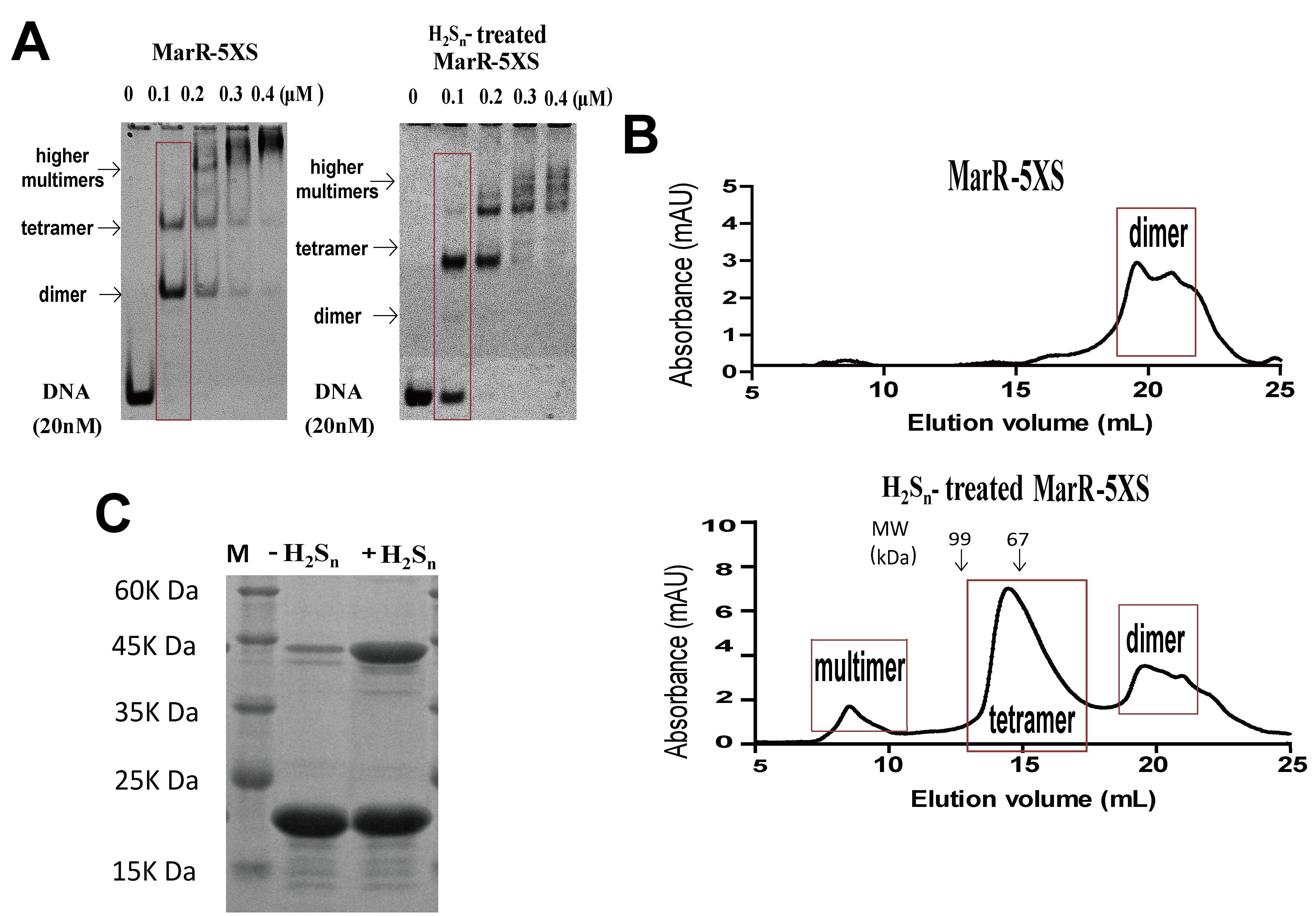
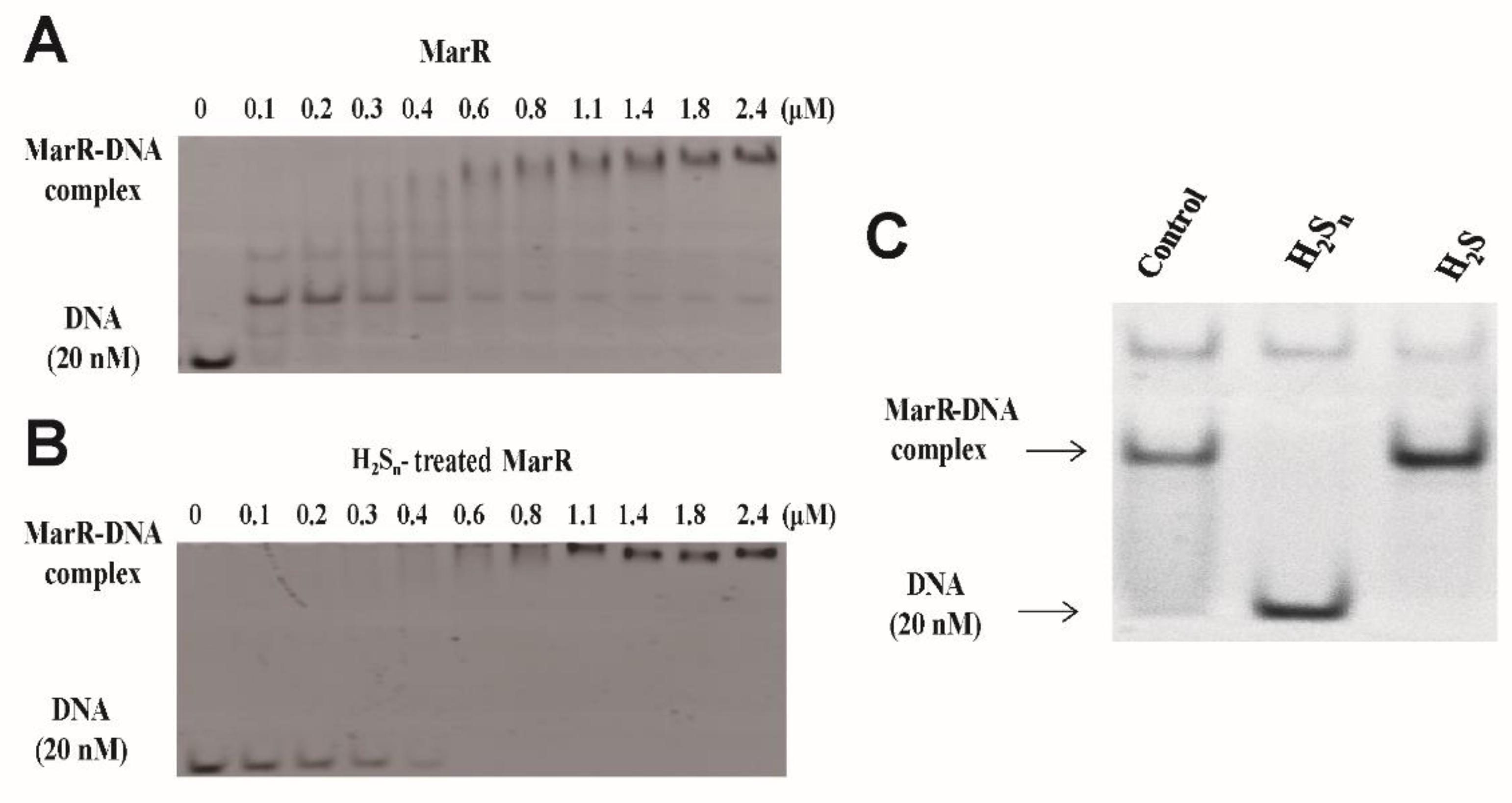
3.1. MarR Sensed Sulfane Sulfur and Decreased Its DNA Binding Affinity

MarR from E. coli MG1655 was overproduced in E. coli BL21 with a N-terminal His-tag and subsequently purified. In EMSA, a 100 nM MarR (monomer) shifted a DNA probe containing a single MarR-binding site at 20 nM (Figure 1A) [35]. A complex band of an MarR dimer and the probe were dominant, and a complex band of two dimers was also present. When the MarR concentration increased, the bands shifted higher, suggesting that complexes with several MarR dimers were formed (Figure 1A). The findings are consistent with previous reports that MarR forms dimers, tetramers, and higher multimers with target DNA probes [25,32]. H2Sn treatment significantly decreased MarR’s affinity to its DNA probe, and a complete shift of the probe occurred with higher H2Sn-treated MarR at 600 nM (Figure 1B). The complex of a dimer MarR with the probe was not detected, but a complex of the probe with multiple MarR dimers was detected (Figure 1B). When repeated with a 200 nM MarR, H2Sn-treated MarR, and H2S-treated MarR, the dimer complex again disappeared after H2Sn treatment, but not after H2S treatment (Figure 1C).
MarR contains six cysteines (Cys47, Cys51, Cys54, Cys80, Cys108, and Cys111). Cys80 is responsible for Cu2+ sensing, and others can be mutated [30]. We constructed and purified the MarR-5XS mutant (with the exception of Cys80, the remaining five Cys residues were mutated to Ser). In EMSA, two bands corresponding to one MarR dimer and two MarR dimers that bound to the DNA probe were detected with the untreated MarR-5XS, and higher multimers were formed with increasing amounts of MarR-5XS (Figure 2A). H2Sn treatment decreased MarR-5XS’s affinity to its probe, as the unshifted probe was detectable in the presence of 100 nM H2Sn-treated MarR-5XS, but not in the presence of 100 nM untreated MarR-5XS (Figure 2A). H2Sn treatment eliminated the probe complexed with an MarR-5XS dimer but enhanced the probe complexed with two dimers. The probe formed complexes with multiple dimers, with increased MarR-5XS and H2Sn-treated MarR-5XS (Figure 2A). Size exclusion chromatography revealed that MarR-5XS was primarily present as a dimer in the solution, but H2Sn treatment produced tetramers (Figure 2B). The peak at around 9 min may represent multimers of MarR-5XS after H2Sn-treatment (Figure 2B). On the basis of the peak areas, the dimer, tetramer, and multimer represented 37.4%, 54.1%, and 8.5% of the H2Sn-treated MarR proteins, respectively. In a non-reducing SDS-PAGE gel, untreated MarR-5XS mainly produced a monomer band, and H2Sn-treated MarR-5XS showed both bands of monomers and dimers (Figure 2C). On the basis of the band intensities, the monomer and dimer accounted for 33.0% and 67.0% of the H2Sn-treated MarR proteins, respectively. These results suggest that the H2Sn treatment forms MarR tetramers that have reduced affinity to their probes.
H2Sn-treated MarR was digested by trypsin and analyzed by using liquid chromatography–mass spectrometry (LC–MS). A 2+-charged peak (m/z: 461.27) matching the disulfide (Cys80-Cys80)-containing peptide (theoretical molecular mass: 921.53Da) and a 2+-charged peak (m/z: 477.26) matching the trisulfide (Cys80-S-Cys80)-containing peptide (theoretical molecular mass: 953.53Da) were identified from the MS data of H2Sn-treated MarR (Figure 3A,B). These results indicate that H2Sn induces the formation of disulfide and trisulfide bonds between two MarR dimers via Cys80.

3.2. MarR Responded to H2Sn in An E. Coli Reporter System

We constructed a reporter plasmid, pBBR5-MarR-PmarR-mKate, in which mKate is repressed by constitutively expressed MarR, and transformed it into E. coli BL21(DE3). The mKate expression was induced by H2Sn in a dose-dependent response (Figure 4A). We also checked mKate induction by a known inducer, salicylate, and potential inducers, H2S and H2O2. The mKate fluorescence was increased significantly by H2Sn, salicylate, and H2O2, but not by H2S when added to the cell suspension (Figure 4B). Among the tested inducers, H2Sn was the strongest for inducing mKate expression.

4. Discussion

As illustrated in Figure 5, we demonstrated that MarR senses sulfane sulfur. After reacting with H2Sn, MarR forms a disulfide bond (Cys80-Cys80′) or a trisulfide bond (Cys80-S-Cys80′) between two dimers, generating a tetramer in solution (Figure 2B). When the tetramer is formed by Cu2+ in the presence of O2, two disulfide bonds are formed between the two dimers, burying the alpha helix containing the Cys80 residue for DNA binding. Therefore, the structure suggests that the tetramer does not bind to its target DNA [30]. The H2Sn treatment may induce the formation of tetramers with one disulfide bond as well as two disulfide bonds (Figure 5), and the tetramer with a single disulfide bond may still bind to its DNA probe, as evidenced by the presence of the tetramer–probe complex and lack of the dimer–probe complex (Figure 1 and Figure 2A). Size exclusion chromatography showed the dominance of tetramers, but the non-reducing SDS-PAGE analysis indicated an abundance of monomers of the H2Sn-treated MarR-5XS (Figure 2), further suggesting the presence of tetramers with a single disulfide bond (Figure 5). The formation of disulfide and trisulfide bonds reduced their affinities to the target DNA, derepressing the repressed genes in both EMSA and in vivo reporter assays (Figure 1 and Figure 4). These results are similar to those regarding the response of P. aeruginosa MexR to sulfane sulfur, forming interprotomer disulfide and trisulfide bonds that covalently link the monomers within a dimer that fails to bind to its cognate DNA [12]. The cellular level of sulfane sulfur varies according to the growth phases of bacteria in liquid media [2]. P. aeruginosa contains the highest level of sulfane sulfur in the early stationary phase, which inactivates MexR for the expression of drug resistant genes [12]. MarR may also sense cellular levels of sulfane sulfur to regulate its repressed genes.
MarR family proteins are small proteins with a winged helix–turn–helix (wHTH) DNA-binding domain and the N- and C-termini participating in the dimer formation [23]. The MarR family proteins are common in bacteria, averaging seven different genes per sequenced bacterial genome [9]. Some are involved in the regulation of efflux pumps for the resistance of antibiotics and organic solvents [23,30,36,37]. Other members display diverse regulatory functions. For example, MgrA is involved in the regulation of biofilm formation and virulence in S. aureus [38,39]. OhrR regulates resistance to organic hydroperoxides in Pseudomonas aeruginosa [40]. HcaR regulates the utilization of hydroxycinnamate in Acinetobacter sp. ADP1 [41]. SlyA and homologues in Enterobacteriaceae have evolved to facilitate the expression of horizontally acquired genes [42]. Their mechanisms of sensing are either binding salicylate or other aromatic compounds in a crevice between the DNA binding domain and the dimer interface [41], or using Cys thiols to detect oxidative stress [30]. Some members, such as MarR, use both mechanisms for inducer sensing (Figure 4B).
Three MarR family proteins that use Cys thiols to sense oxidative stress also sense sulfane sulfur. E. coli MarR is oxidized by Cu2+ to form two disulfide bonds between two dimers (Cys80-Cys80′), turning on the expression of the marRAB operon for further activation of other genes, including a multiple drug efflux pump for antibiotic resistance [30]. When exposed to antibiotics, Cu2+ is released from membrane proteins under oxidative stress. P. aeruginosa MexR responds to H2O2 as well as disulfides GSSG and 2,2′-dithiodipyridine to form interprotomer disulfide bonds between Cys30 and Cys62 in an MexR dimer [36,37]. S. aureus MgrA contains a single Cys residue, Cys12, which is modified by hydrogen peroxide, and the modified MgrA has reduced affinity to its target DNA [43]. These MarR family proteins, which use Cys thiols for sensing, can be deactivated by sulfane sulfur to derepress their regulated genes [12,21]. Further studies are necessary to determine which other members of the MarR family can sense sulfane sulfur.
Sulfane sulfur is the inducer of several gene regulators involved in sulfur metabolism [44]. After reacting with sulfane sulfur, Cupriavidus pinatubonensis FisR forms a tetrasulfide, crosslinking between C53 and C64 [15]. S. aureus CstR generates a mixture of di-, tri-, and tetra-sulfide links between C31 and C60 [16]. Rhodobacter capsulatus SqrR produces a tetrasulfide link between C41 and C107 [17]. These modifications may be induced specifically by sulfane sulfur, as other oxidative reagents, such as H2O2, organic peroxides, and disulfide, cannot induce the formation of multiple sulfur links between two Cys residues. This concept has been applied to design sulfane sulfur-specific fluorescence proteins which, due to the space restriction of the Cys residues involved, cannot form disulfide links, but can form trisulfide and tetrasulfide links [45,46]. Disulfide formation can be achieved by the induction of sulfane sulfur as well as H2O2 and other oxidative reagents. Previously, Cu2+ induced the formation of a disulfide bond by MarR, but H2O2 did not [30]. However, we detected H2O2 as a reducer for MarR (Figure 4B). Given the chemistry of disulfide bond formation, H2O2, H2Sn, and Cu2+ should all induce the formation of the MarR disulfide bond (Figure 5).

5. Conclusions

Sulfane sulfur is the specific inducer that modified gene regulators involved in sulfur metabolism [44]. It is also an effector of OxyR [19], MexR [12], MgrA [21], and MarR. Furthermore, it may sense other agents, such as H2O2, as long as they can induce disulfide formation or modify key Cys thiols. MarR regulates diverse activities in E. coli, including resistance to antibiotics, oxidative stress agents, and organic solvents. Although several inducers have been reported, sulfane sulfur, a common cellular component in bacteria that reaches the maximum in E. coli cells at the late log phase and the early stationary phases of growth [2], is likely a key inducer regulating MarR activity. This discovery further establishes the general signaling role of sulfane sulfur.

Author Contributions

H.X. and G.X. acquired and analyzed the data. H.L. and Y.X. supervised the research. G.X., Y.X. and L.X. designed the study and wrote the manuscript. All authors have read and agreed to the published version of the manuscript.

Funding

The work was financially supported by grants from the National Natural Science Foundation of China (91951202).

Institutional Review Board Statement

Not applicable.

Informed Consent Statement

Not applicable.

Data Availability Statement

Data are contained within the article.

Conflicts of Interest

The authors declare no conflict of interest.

References

  1. Fukuto, J.M.; Ignarro, L.J.; Nagy, P.; Wink, D.A.; Kevil, C.G.; Feelisch, M.; Cortese-Krott, M.M.; Bianco, C.L.; Kumagai, Y.; Hobbs, A.J.; et al. Biological hydropersulfides and related polysulfides—A new concept and perspective in redox biology. FEBS Lett. 2018, 592, 2140–2152. [Google Scholar] [CrossRef] [PubMed] [Green Version]
  2. Ran, M.; Wang, T.; Shao, M.; Chen, Z.; Liu, H.; Xia, Y.; Xun, L. Sensitive method for reliable quantification of sulfane sulfur in biological samples. Anal. Chem. 2019, 91, 11981–11986. [Google Scholar] [CrossRef] [PubMed]
  3. Xin, Y.; Liu, H.; Cui, F.; Liu, H.; Xun, L. Recombinant Escherichia coli with sulfide:quinone oxidoreductase and persulfide dioxygenase rapidly oxidises sulfide to sulfite and thiosulfate via a new pathway. Environ. Microbiol. 2016, 18, 5123–5136. [Google Scholar] [CrossRef]
  4. Lü, C.J.; Xia, Y.Z.; Liu, D.X.; Zhao, R.; Gao, R.; Liu, H.L.; Xun, L.Y. Cupriavidus necator H16 Uses Flavocytochrome c Sulfide Dehydrogenase To Oxidize Self-Produced and Added Sulfide. Appl. Environ. Microbiol. 2017, 83, e01610-17. [Google Scholar] [CrossRef] [PubMed] [Green Version]
  5. Li, K.; Xin, Y.F.; Xuan, G.H.; Zhao, R.; Liu, H.W.; Xia, Y.Z.; Xun, L.Y. Escherichia coli uses separate enzymes to produce H2S and reactive sulfane sulfur from L-cysteine. Front. Microbiol. 2019, 10, 298. [Google Scholar] [CrossRef] [Green Version]
  6. Akaike, T.; Ida, T.; Wei, F.Y.; Nishida, M.; Kumagai, Y.; Alam, M.M.; Ihara, H.; Sawa, T.; Matsunaga, T.; Kasamatsu, S.; et al. Cysteinyl-tRNA synthetase governs cysteine polysulfidation and mitochondrial bioenergetics. Nat. Commun. 2017, 8, 1177. [Google Scholar] [CrossRef] [Green Version]
  7. Nagahara, N.; Koike, S.; Nirasawa, T.; Kimura, H.; Ogasawara, Y. Alternative pathway of H2S and polysulfides production from sulfurated catalytic-cysteine of reaction intermediates of 3-mercaptopyruvate sulfurtransferase. Biochem. Biophys. Res. Commun. 2018, 496, 648–653. [Google Scholar] [CrossRef]
  8. Xia, Y.Z.; Lu, C.J.; Hou, N.K.; Xin, Y.F.; Liu, J.H.; Liu, H.L.; Xun, L.Y. Sulfide production and oxidation by heterotrophic bacteria under aerobic conditions. ISME J. 2017, 11, 2754–2766. [Google Scholar] [CrossRef] [Green Version]
  9. Doka, E.; Pader, I.; Biro, A.; Johansson, K.; Cheng, Q.; Ballago, K.; Prigge, J.R.; Pastor-Flores, D.; Dick, T.P.; Schmidt, E.E.; et al. A novel persulfide detection method reveals protein persulfide- and polysulfide-reducing functions of thioredoxin and glutathione systems. Sci. Adv. 2016, 2, e1500968. [Google Scholar] [CrossRef] [Green Version]
  10. Sato, I.; Shimatani, K.; Fujita, K.; Abe, T.; Shimizu, M.; Fujii, T.; Hoshino, T.; Takaya, N. Glutathione reductase/glutathione is responsible for cytotoxic elemental sulfur tolerance via polysulfide shuttle in fungi. J. Biol. Chem. 2011, 286, 20283–20291. [Google Scholar] [CrossRef] [Green Version]
  11. Toohey, J.I. Sulfur signaling: Is the agent sulfide or sulfane? Anal. Biochem. 2011, 413, 1–7. [Google Scholar] [CrossRef]
  12. Xuan, G.; Lu, C.; Xu, H.; Chen, Z.; Li, K.; Liu, H.; Liu, H.; Xia, Y.; Xun, L. Sulfane Sulfur is an intrinsic signal activating MexR-regulated antibiotic resistance in Pseudomonas aeruginosa. Mol. Microbiol. 2020, 114, 1038–1048. [Google Scholar] [CrossRef]
  13. Lau, N.; Pluth, M.D. Reactive sulfur species (RSS): Persulfides, polysulfides, potential, and problems. Curr. Opin. Chem. Biol. 2019, 49, 1–8. [Google Scholar] [CrossRef] [PubMed]
  14. Kimura, H. Production and physiological effects of hydrogen sulfide. Antioxid. Redox Signal. 2014, 20, 783–793. [Google Scholar] [CrossRef] [Green Version]
  15. Li, H.; Li, J.; Lu, C.; Xia, Y.; Xin, Y.; Liu, H.; Xun, L.; Liu, H. FisR activates sigma(54)-dependent transcription of sulfide-oxidizing genes in Cupriavidus pinatubonensis JMP134. Mol. Microbiol. 2017, 105, 373–384. [Google Scholar] [CrossRef] [Green Version]
  16. Luebke, J.L.; Shen, J.C.; Bruce, K.E.; Kehl-Fie, T.E.; Peng, H.; Skaar, E.P.; Giedroc, D.P. The CsoR-like sulfurtransferase repressor (CstR) is a persulfide sensor in Staphylococcus aureus. Mol. Microbiol. 2014, 94, 1343–1360. [Google Scholar] [CrossRef] [PubMed] [Green Version]
  17. Shimizu, T.; Shen, J.; Fang, M.; Zhang, Y.; Hori, K.; Trinidad, J.C.; Bauer, C.E.; Giedroc, D.P.; Masuda, S. Sulfide-responsive transcriptional repressor SqrR functions as a master regulator of sulfide-dependent photosynthesis. Proc. Natl. Acad. Sci. USA 2017, 114, 2355–2360. [Google Scholar] [CrossRef] [PubMed] [Green Version]
  18. Lu, T.; Cao, Q.; Pang, X.; Xia, Y.; Xun, L.; Liu, H. Sulfane sulfur-activated actinorhodin production and sporulation is maintained by a natural gene circuit in Streptomyces coelicolor. Microb. Biotechnol. 2020, 13, 1917–1932. [Google Scholar] [CrossRef]
  19. Hou, N.; Yan, Z.; Fan, K.; Li, H.; Zhao, R.; Xia, Y.; Xun, L.; Liu, H. OxyR senses sulfane sulfur and activates the genes for its removal in Escherichia coli. Redox Biol. 2019, 26, 101293. [Google Scholar] [CrossRef] [PubMed]
  20. Xuan, G.; Lv, C.; Xu, H.; Li, K.; Liu, H.; Xia, Y.; Xun, L. Sulfane Sulfur regulates LasR-mediated quorum sensing and virulence in Pseudomonas Aeruginosa PAO1. Antioxidants 2021, 10, 1498. [Google Scholar] [CrossRef] [PubMed]
  21. Peng, H.; Zhang, Y.; Palmer, L.D.; Kehl-Fie, T.E.; Skaar, E.P.; Trinidad, J.C.; Giedroc, D.P. Hydrogen Sulfide and Reactive Sulfur Species impact Proteome S-Sulfhydration and global virulence regulation in Staphylococcus aureus. ACS Infect. Dis. 2017, 3, 744–755. [Google Scholar] [CrossRef] [PubMed] [Green Version]
  22. Gupta, A.; Pande, A.; Sabrin, A.; Thapa, S.S.; Gioe, B.W.; Grove, A. MarR family transcription factors from Burkholderia species: Hidden clues to control of virulence-associated genes. Microbiol. Mol. Biol. Rev. MMBR 2019, 83, e00039-18. [Google Scholar] [CrossRef] [Green Version]
  23. Deochand, D.K.; Grove, A. MarR family transcription factors: Dynamic variations on a common scaffold. Crit. Rev. Biochem. Mol. Biol. 2017, 52, 595–613. [Google Scholar] [CrossRef]
  24. Alekshun, M.N.; Levy, S.B. The mar regulon: Multiple resistance to antibiotics and other toxic chemicals. Trends Microbiol. 1999, 7, 410–413. [Google Scholar] [CrossRef]
  25. Martin, R.G.; Rosner, J.L. Binding of purified multiple antibiotic-resistance repressor protein (MarR) to mar operator sequences. Proc. Natl. Acad. Sci. USA 1995, 92, 5456–5460. [Google Scholar] [CrossRef] [PubMed] [Green Version]
  26. George, A.M.; Levy, S.B. Amplifiable resistance to tetracycline, chloramphenicol, and other antibiotics in Escherichia coli: Involvement of a non-plasmid-determined efflux of tetracycline. J. Bacteriol. 1983, 155, 531–540. [Google Scholar] [CrossRef] [Green Version]
  27. Ariza, R.R.; Cohen, S.P.; Bachhawat, N.; Levy, S.B.; Demple, B. Repressor mutations in the marRAB operon that activate oxidative stress genes and multiple antibiotic resistance in Escherichia coli. J. Bacteriol. 1994, 176, 143–148. [Google Scholar] [CrossRef] [Green Version]
  28. White, D.G.; Goldman, J.D.; Demple, B.; Levy, S.B. Role of the acrAB locus in organic solvent tolerance mediated by expression of marA, soxS, or robA in Escherichia coli. J. Bacteriol. 1997, 179, 6122–6126. [Google Scholar] [CrossRef] [Green Version]
  29. Gambino, L.; Gracheck, S.J.; Miller, P.F. Overexpression of the MarA positive regulator is sufficient to confer multiple antibiotic resistance in Escherichia coli. J. Bacteriol. 1993, 175, 2888–2894. [Google Scholar] [CrossRef] [PubMed] [Green Version]
  30. Hao, Z.Y.; Lou, H.B.; Zhu, R.F.; Zhu, J.H.; Zhang, D.M.; Zhao, B.X.S.; Zeng, S.Z.; Chen, X.; Chan, J.; He, C.; et al. The multiple antibiotic resistance regulator MarR is a copper sensor in Escherichia coli. Nat. Chem. Biol. 2014, 10, 21–28. [Google Scholar] [CrossRef]
  31. Kamyshny, A. Improved cyanolysis protocol for detection of zero-valent sulfur in natural aquatic systems. Limnol. Oceanogr. Methods 2009, 7, 442–448. [Google Scholar] [CrossRef] [Green Version]
  32. Notka, F.; Linde, H.J.; Dankesreiter, A.; Niller, H.H.; Lehn, N. A C-terminal 18 amino acid deletion in MarR in a clinical isolate of Escherichia coli reduces MarR binding properties and increases the MIC of ciprofloxacin. J. Antimicrob. Chemother. 2002, 49, 41–47. [Google Scholar] [CrossRef] [PubMed]
  33. Xia, Y.; Li, K.; Li, J.; Wang, T.; Gu, L.; Xun, L. T5 exonuclease-dependent assembly offers a low-cost method for efficient cloning and site-directed mutagenesis. Nucleic Acids Res. 2019, 47, e15. [Google Scholar] [CrossRef] [Green Version]
  34. Xia, Y.Z.; Chu, W.Q.; Qi, Q.S.; Xun, L.Y. New insights into the QuikChange (TM) process guide the use of Phusion DNA polymerase for site-directed mutagenesis. Nucleic Acids Res. 2015, 43, e12. [Google Scholar] [CrossRef] [Green Version]
  35. Domain, F.; Levy, S.B. GyrA Interacts with MarR To Reduce Repression of the marRAB Operon in Escherichia coli (Retraction of vol 192, pg 942, 2010). J. Bacteriol. 2011, 193, 2674. [Google Scholar] [CrossRef] [Green Version]
  36. Chen, H.; Yi, C.Q.; Zhang, J.; Zhang, W.R.; Ge, Z.Y.; Yang, C.G.; He, C.A. Structural insight into the oxidation-sensing mechanism of the antibiotic resistance of regulator MexR. EMBO Rep. 2010, 11, 685–690. [Google Scholar] [CrossRef]
  37. Chen, H.; Hu, J.; Chen, P.R.; Lan, L.; Li, Z.; Hicks, L.M.; Dinner, A.R.; He, C. The Pseudomonas aeruginosa multidrug efflux regulator MexR uses an oxidation-sensing mechanism. Proc. Natl. Acad. Sci. USA 2008, 105, 13586–13591. [Google Scholar] [CrossRef] [PubMed] [Green Version]
  38. Crosby, H.A.; Schlievert, P.M.; Merriman, J.A.; King, J.M.; Salgado-Pabon, W.; Horswill, A.R. The Staphylococcus aureus global regulator MgrA modulates clumping and virulence by controlling surface protein expression. PloS Pathog. 2016, 12, e1005604. [Google Scholar] [CrossRef]
  39. Jiang, Q.; Jin, Z.; Sun, B. MgrA negatively regulates biofilm formation and detachment by repressing the expression of psm operons in Staphylococcus aureus. Appl. Environ. Microbiol. 2018, 84, e01008-18. [Google Scholar] [CrossRef] [Green Version]
  40. Fuangthong, M.; Helmann, J.D. The OhrR repressor senses organic hydroperoxides by reversible formation of a cysteine-sulfenic acid derivative. Proc. Natl. Acad. Sci. USA 2002, 99, 6690–6695. [Google Scholar] [CrossRef] [PubMed] [Green Version]
  41. Kim, Y.; Joachimiak, G.; Bigelow, L.; Babnigg, G.; Joachimiak, A. How Aromatic Compounds block DNA binding of HcaR catabolite regulator. J. Biol. Chem. 2016, 291, 13243–13256. [Google Scholar] [CrossRef] [Green Version]
  42. Will, W.R.; Brzovic, P.; Le Trong, I.; Stenkamp, R.E.; Lawrenz, M.B.; Karlinsey, J.E.; Navarre, W.W.; Main-Hester, K.; Miller, V.L.; Libby, S.J.; et al. The evolution of SlyA/RovA transcription factors from repressors to Countersilencers in Enterobacteriaceae. mBio 2019, 10, e00009-19. [Google Scholar] [CrossRef] [PubMed] [Green Version]
  43. Chen, P.R.; Bae, T.; Williams, W.A.; Duguid, E.M.; Rice, P.A.; Schneewind, O.; He, C. An oxidation-sensing mechanism is used by the global regulator MgrA in Staphylococcus aureus. Nat. Chem. Biol. 2006, 2, 591–595. [Google Scholar] [CrossRef]
  44. Giedroc, D.P. A new player in bacterial sulfide-inducible transcriptional regulation. Mol. Microbiol. 2017, 105, 347–352. [Google Scholar] [CrossRef] [PubMed]
  45. Hu, X.; Li, H.J.; Zhang, X.; Chen, Z.G.; Zhao, R.; Hou, N.K.; Liu, J.H.; Xun, L.Y.; Liu, H.W. Developing polysulfide-sensitive GFPs for real-time analysis of polysulfides in live cells and subcellular organelles. Anal. Chem. 2019, 91, 3893–3901. [Google Scholar] [CrossRef] [PubMed]
  46. Li, Z.M.; Wang, Q.D.; Xia, Y.Z.; Xun, L.Y.; Liu, H.W. A red fluorescent protein-based probe for detection of intracellular reactive sulfane sulfur. Antioxidants 2020, 9, 985. [Google Scholar] [CrossRef]
Figure 1. MarR reacts with sulfane sulfur, decreasing its affinity to its DNA probe. (A,B) EMSA analysis of MarR and H2Sn-treated MarR in the presence of 20 nM the DNA probe (100 bp) containing the MarR-binding sequence. (C) The purified MarR (9 µM) was treated with 800 µM H2Sn and H2S for 20 min. Then 20 nM DNA probe was incubated with 0.2 µM MarR for the shift assay.
Antioxidants 10 01778 g001
Figure 2. MarR sensed sulfane sulfur via Cys80. (A) EMSA analysis of H2Sn-treated and untreated MarR-5XS in the presence of its DNA probe (20 nM). (B) Size-exclusion chromatography revealed the formation of disulfide-stabilized tetramer of MarR-5XS (25 µM) after 400 μM H2Sn treatment for 20 min. The molecular weight (MW) of tetramers was calibrated with BSA (67 kDa) and T7 RNA polymerase (99 kDa). (C) Non-reducing SDS-PAGE analysis of MarR-5XS (25 µM). The protein was treated with H2Sn (400 μM) for 20 min, terminated by adding 500 μM iodoacetamide before analysis.
Antioxidants 10 01778 g002
Figure 3. LC–MS/MS analysis of H2Sn-treated MarR. (A) Left: The 2+ charged peak (m/z: 461.27) corresponding to the disulfide (Cys80-Cys80)-containing peptide (theoretical molecular mass: 921.53Da). Right: MS/MS fragmentation of the 2+ charged peptide (m/z: 461.27). (B) Left: The 2+ charged peak (m/z: 477.26) corresponding to the trisulfide (Cys80-S-Cys80)-containing peptide (theoretical molecular mass: 953.53 Da). Right: MS/MS fragmentation of the 2+ charged peptide (m/z: 477.26). Peptide mass was calculated on the following website: http://db.systemsbiology.net:8080/proteomicsToolkit/FragIonServlet.html (accessed on 11 October 2021).
Antioxidants 10 01778 g003
Figure 4. Testing MarR inducers with an E. coli reporter system. (A) A dose-dependent induction of mKate. (B) mKate induction by several tested inducers. E. coli BL21(DE3) containing pBBR5-MarR-PmarR-mKate grew in LB medium to OD600nm of 0.6, inducers were added, and the cultures were incubated at 37 °C for 2 h before mKate fluorescence analysis. The control was E. coli BL21(DE3) with the empty vector pBBR1MCS5. Student’s t-test was used to calculate the p-value. Symbol * indicates the sample is different (0.05 < p < 0.01) and symbol *** indicates the sample is significantly different from the control (p < 0.001).
Antioxidants 10 01778 g004
Figure 5. Schematic representation of MarR, which senses sulfane sulfur, forming disulfide and trisulfide crosslinks between Cys80. A mixture of tetramer with one and two disulfide bonds may coexist after H2Sn treatment. The tetramer with two disulfide bonds does not bind to its target DNA [30], but our results suggest that the tetramer with one disulfide bond does bind to its target DNA.
Antioxidants 10 01778 g005
Table 1. Strains and plasmids used in this study.
Strain/PlasmidCharacteristicSource
Escherichia coli strains
DH5aCloning strainInvitrogen
BL21(DE3)Cloning strainInvitrogen
Plasmids
pBBR1mcs5Gmr, broad host range[12]
pBBR5-MarR-PmarR-mKatepBBR1mcs5 vector with marR, marR promotor, and mkate genesThis study
pET30aKmr, expression vectorInvitrogen
pET30-MarRpET30a containing MarR with N terminal his-tagThis study
pET30-MarR/5CSpET30-MarR with Cys47Ser, Cys51Ser, Cys54Ser, Cys108Ser, Cys111SerThis study
Table 2. Primers used in this study.
Primers aSequence (5′-3′)Usage
marR-1CGACGACGACAAGGCCATGGCTGATGTGAAAAGTACCAGCGATCTGFor pET30-MarR construction
marR-2CAGTGGTGGTGGTGGTGGTGCTCGATTACGGCAGGACTTTCTTAAG
MarR-5CS-1TAATACTCGCCGCGCTGCGGATAGAGCTGAGCFor MarR-5XS mutant construction
MarR-5CS-2GCTCAGCTCTATCCGCAGCGCGGCGAGTATTA
MarR-5CS-3GGCGCGGCAATAAGTGAACAAAGCCAT
MarR-5CS-4ATGGCTTTGTTCACTTATTGCCGCGCC
marR-F aTCTAGAGAAAGAGGAGAAATACTAGGTGAAAAGTACCAGCGATCTGTTCAATFor pBBR5-MarR-PmarR-mKate construction
marR-RTTGACGGTGGTATTACGGCAGGACTTTCTTAAGCAAATAC
Pmar-FCCTGCCGTAATACCACCGTCAAAAAAAACGGCGCTTTTTAGCGCCGTTTTTATTTTTCATGAACCGATTTAGCAAAACGTGGC
Pmar-RCTAGTATTTCTCCTCTTTCTCTAGAATTAGTTGCCCTGGCAAGTAATTAGTT
mKate-F bTCTAGAGAAAGAGGAGAAATACTAGATGTCAGAATTAATTAAAGAAAATATGCACATG
mKate-RCTTACAATTTCCATTCGCCATTTCAACGATGTCCTAATTTCGACG
pBBR5-F cCTAGTATTTCTCCTCTTTCTCTAGACAACATACGAGCCGGAAGCATAAAG
pBBR5-R cAATGGCGAATGGAAATTGTAAGCG
MarR-emsa-1CATCGCATTGAACAAAACTTGAACFor amplifying the EMSA probe
MarR-emsa-1GTTGCAGGGGATAATATTGCC
a The gene accession numbers of marR and mKate are b1530 and MN623117, respectively. The EMSA probe contained one MarR-binding site, ending at 25 bp before the marR gene. b An rbs sequence (gaggaga) was inserted before the start codon (GTG or ATG) of marR and mkate. c The lac operator sequence (ttgtgagcggataacaa) was not amplified with this primer pair.
Publisher’s Note: MDPI stays neutral with regard to jurisdictional claims in published maps and institutional affiliations.

