Article

Antihyperlipidemic and Antioxidant Capacities, Nutritional Analysis and UHPLC-PDA-MS Characterization of Cocona Fruits (Solanum sessiliflorum Dunal) from the Peruvian Amazon

1
Laboratorio de Química de Productos Naturales, Instituto de Investigaciones de la Amazonía Peruana, Av. Abelardo Quiñones km 2.5, Iquitos 16001, Peru
2
Facultad de Farmacia y Bioquímica, Universidad Nacional de la Amazonía Peruana, Iquitos 16001, Peru
3
Laboratorio de Química de Productos Naturales, Instituto de Química de Recursos Naturales, Universidad de Talca, Casilla 747, Talca 3460000, Chile
4
Laboratorio de Química Orgánica y Productos Naturales, Facultad de Ciencias Agronómicas, Universidad de Tarapacá, Av. General Velásquez 1775, Arica 1000000, Chile
5
Instituto de Farmacia, Facultad de Ciencias, Universidad Austral de Chile, Valdivia 509000, Chile
*
Authors to whom correspondence should be addressed.
Antioxidants 2021, 10(10), 1566; https://doi.org/10.3390/antiox10101566
Submission received: 15 July 2021 / Revised: 27 September 2021 / Accepted: 28 September 2021 / Published: 30 September 2021
(This article belongs to the Special Issue Advances in Natural Antioxidants for Food Improvement)

Abstract

:
Cocona fruits are a popular food and medicinal fruit used mainly in the Amazon and several countries of South America for the preparation of several food products such as drinks, jams and milk shakes. In this study five ecotypes of cocona native to Peru have been studied regarding their nutritional and antioxidants values plus antihyperlipidemic activities. Seventy bioactive compounds have been detected in Peruvian cocona ecotypes including several phenolic acids, aminoacids and flavonoids; of those six were spermidines, (peaks 1, 2, 25, 26, 38 and 39), thirteen were aminoacids, (peaks 3–9, 11–13, 16, 17, 22–24), eighteen flavonoids (peaks 28, 30–32 45,46, 48–53 56, 57, 61 and 64–66), twelve were phenolics (peaks 19, 21, 27, 29, 34, 35, 36, 42, 43, 44, 54, and 59), two carotenoids, (peak 62 and 63), eight were lipid derivatives (peaks 37, 55, 58, 60 and 67–70), one sugar (peak 47), four terpenes (peaks 33, 40, 41 and 47), two amides, (peaks 10 and 18), one aldehyde, (peak 15), and three saturated organic acids, (peaks 4, 5 and 20). Hypercholesterolemic rats administered with pulp of the ecotypes CTR and SRN9 showed the lowest cholesterol and triglyceride levels after treatment (126.74 ± 6.63; 102.11 ± 9.47; 58.16 ± 6.64; 61.05 ± 4.00 mg/dL, for cholesterol, triglycerides, high-density lipoprotein and low-density lipoprotein respectively, for the group treated with SRN9 pulp, and 130.09 ± 8.55; 108.51 ± 10.04; 57.30 ± 5.72; and 65.41 ± 7.68 mg/dL, for cholesterol, triglycerides, HDL and LDL lipoproteins respectively for the group treated with CTR pulp). The ecotypes proved to be good sources of natural antioxidants and their consumption represent an alternative for the prevention of atherosclerosis.

1. Introduction

In recent decades, global interest has increased in search of the chemical composition and biological activities of natural sources since many of the compounds present in biological sources such as local plants and marine organisms are important for the protection of human health. The fruit of cocona (Solanum sessiliflorum Dunal; Solanaceae) are native of the Amazonian tropic. Just like other plants in the genus Solanum, they exhibit a morphological diversity corresponding to the variability in habitat and ecology changes, as well as with the process of domestication of the species; However, little is known about its chemical composition and the implications of the terroir and climate in its morphology and nutrient and health beneficial properties. The common name of the fruit in Spanish or Portuguese speaking countries is cocona, topiro or cubiu, and is known as “Orinoco apple” and “peach tomato” in English speaking countries. It is an endemic species to Amazon cultivated by natives and settlers in agroforestry arrangements and chagras in their settlement sites which improves it on a traditional food from this area. The pulp is the edible part of the fruit and is known for its refreshing flavor to produce refreshments, milk shakes, jams, and jellies and for the medicinal properties, including amelioration of itching produced by insect bites, elimination of parasites, it is also use as topical to heal burns and for the control of cholesterol, diabetes, and uric acid. The studies carried out on the chemistry of the species are few and no complete, mainly limited to the report of some phenolic compounds and volatile metabolites which cannot depict the differentiation in the variations in the chemical composition between the different morphotypes of the fruit which is very important to give added values to the different products and ecotypes. This study aimed to differentiate five varieties of cocona (Figure 1) with the use of UHPLC coupled to high resolution mass spectrometry (UHPLC-PDA-ESI-OT-MS) grown in Peru, called oval, small round, large round big oval round and big square morphotypes, by means of the metabolite fingerprinting of the secondary metabolites present in mature fruits of the species. These adaptive morphotypes of the fruit depends directly on various biological factors and has been diversifying during the evolution and natural selection and can have the ability to produce different biologically important metabolites. Several carotenoids, alkaloids, organic acids, phenolic acids, flavonoid glycosides, coumarins, tannins, and volatile and fixed acids were reported to occur in fresh cocona fruits from Brazil [1,2] and recently, caffeoyl quinic acid was reported as the main important phenolic in the fruits [3], whereas determination of volatile organic compounds (VOCs) was done by HS-SPME/GC-MS in some ecotypes from Brazil [4], besides, extracts of those Brazilian ecotypes presented high concentrations of caffeic and gallic acids, beta-carotene, catechin, quercetin, and rutin and showed low density lipoproteins oxidation; cytotoxic and antiproliferative effect on breast (MCF-7) and colorectal (HT-29) cancer cell lines [5]. The purpose of this work is to contribute to the full phytochemical study of the Peruvian ecotype species through UHPLC-PDA-ESI-OT-MS for full untargeted metabolomic analyses to promote in fruit growers and fruit processors in the Peruvian Amazon the adoption of strategies for the sustainable use of the more promising ones based on its phenolic content and intrinsic health related properties, such as antihyperlipidemic capacities of the studied five ecotypes of this highly consumed fruit from Peru. Finally, proximal composition and mineral contents, plus the antioxidant activities thorough different methods and total carotene and phenolic contents of all ecotype pulps were tested and compared. The UHPLC full MS and PDA fingerprint analysis, was also performed for the ecotypes.

2. Materials and Methods

2.1. Chemicals

Ultra-pure water (<5 µg/L TOC, (total organic carbon)) was obtained from a water purification system Arium 126 61316-RO, plus an Arium 611 UV unit (Sartorius, Goettingen, Germany). Methanol (HPLC grade) and formic acid (puriss. p.a. for mass spectrometry) from J. T. Baker (Phillipsburg, NJ, USA) were obtained. Dichloromethane (HPLC grade) were from Merck (Santiago, Chile). Commercial Folin–Ciocalteu (FC) reagent, 2,2-diphenyl-1-picrylhydrazyl (DPPH), ferric chloride hexahydrate, 2,4,6-tris(2-pyridyl)-s-triazine, trolox, quercetin, gallic acid, atorvastatin, Triton WR-1339 and DMSO were purchased from Sigma-Aldrich Chem. Co. (St Louis, MO, USA).

2.2. Plant Material and Sample Treatment

The study was carried out with five ecotypes of Solanum sessiliflorum, whose seeds were obtained from the ex situ conservation gene bank of the Peruvian Amazon Research Institute, cultivated in the Research and Production Center of Tulumayo of the National Agrarian University of the Jungle (09°06′20″ S and 75°54′15″ W, 565 meters altitude) Huanuco region, Peru. The ecotypes were selected based on their morphotypic difference, which were: CTR, collected in cascas, Lambayeque region, SRN9, collected in Supte Black river, Huanuco region, UNT2, collected in Padre Abad, Ucayali region, NMA1, collected in Leoncio Prado, Huanuco region, and CD1, collected in Bagua, Amazonas region. The selected fruits were washed, brushed, disinfected using a 200-ppm sodium hypochlorite solution for 30 min and rinsed with water. The epicarp was then removed. Then thermal bleaching was carried out at 90 °C for 20 min. The pulp was then extracted in a pulping machine removing the husks and seeds. Finally, the extracted pulp was lyophilized. The lyophilized pulp was packed in plastic bags and stored in a freezer at −20 °C until use (for some days only). Lyophilized pulp (2.0 g) was extracted two times in 5 mL of methanol/water (MeOH/H2O) solutions (80/20, v/v) under sonication (130 kHz, 10 min) at room temperature. Then, samples were centrifuged (5000 rpm, 15 min, 5 °C) and the supernatants were combined after filtration and stored at −20 °C before analyses.

2.3. UHPLC-PDA-ESI-OT-MS Instrument

UHPLC-PDA-ESI-OT-MS analysis was carried out as described by [6], with some modifications, Briefly, a Thermo Scientific Dionex Ultimate 3000 UHPLC system hyphenated with a Thermo Q exactive plus machine was used. For the analysis 5 mg of the extract were dissolved in 2 mL of methanol, filtered (PTFE filter) and 10 µL were injected in the instrument, using an UHPLC C-18 column (Luna© Omega C-18 100 Å, Phenomenex 150 mm × 2.1 mm × 1.6 µm), operated at 25 °C. The detection wavelengths were 254, 280, 330 and 354 nm, and PDA was recorded from 200 to 800 nm for peak characterization. Mobile phases were 1% formic aqueous solution (A) and 1% formic acid in acetonitrile (B). The gradient program (time (min), % B) was: (0.00 min, 5% B); (1.500 min, 15% B); (1.5 min, 5% B); (35.00 min, 95% B); (36.00, 95% B); (38.00 min, 5% B); and 15 min for column equilibration before each injection. The flow rate was 0.300 mL min−1, and the injection volume was 10 µL. Standards and the pulp extract dissolved in methanol were kept at 10 °C during storage in the autosampler. Parameters for Full MS scan: Maximum IT: 80 ms, AGC target: 5 × 10⁶ Resolution: 35,000 Range: 100–1500 m/z, Microscans: 1, Parameters MS2 Maximum IT: 100 ms AGC target: 1 × 10⁶ Resolution: 17,500, Ionization source parameters: ESI (positive and negative) spray volt: 3.5/2.5 KV, Capillary temperature: 260 °C, Carrier gas: N2 (Sheath gas flow rate: 48, Sweep gas flow rate: 2) Gas heater temp: 280/280 °C, S-lens RF level: 100.

2.4. Antioxidant Activity Assays

2.4.1. DPPH Test

The DPPH• radical was assayed by the decolorization method [7]. Briefly, 9 μL of extract, (2 mg/mL), plus 341 μL of methanol DPPH solution (400 μM) were adjusted with the solvent methanol to an absorbance of 1.10 ± 0.02 at 517 nm. The mixture was homogenized using a vortex, allowed to react in the dark at room temperature for 20 min, after which time absorbance was measured at 517 nm in a Synergy HTX monochromator (Biotek, USA). The percentage of decoloration of the DPPH moiety was obtained by measuring the change in absorbance at 517 nm, the values obtained converted to percent inhibition of the DPPH moiety. The results are expressed in TEAC, that is, antioxidant activity equivalent to Trolox (μmol Trolox/g of lyophilized pulp). The synthetic antioxidant reference Trolox, at a concentration of 5–30 µM in 80% methanol solution, is tested under the same conditions.

2.4.2. ABTS Method

The ABTS assay was performed by bleaching of the cationic radical ABTS•+ as described by [8]. For the preparation of the radical ABTS•+ 2.5 mL of the 7 mM ABTS solution, it was mixed with 2.5 mL of 2.45 μM sodium persulfate for 12 hours in the dark at 4 °C. Then, the resulting solution was diluted with absolute ethanol until an initial absorbance of approximately 0.70 ± 0.03 was obtained at 734 nm. The radical discoloration was initiated by adding 50 µL of the extract to 150 µL of the ABTS•+ solution. After 15 minutes of incubation at 25 °C, the absorbance was measured at 734 nm and compared with a calibration curve using Trolox as standard and ethanol as blank. Results were expressed as micromoles of Trolox equivalents per gram of dry sample (µmol TE/g).

2.5. Polyphenol (Folin-Ciocalteau)

Total phenolic compounds (TPC) were analyzed based on [6] and [9]. To 12 μL of extract to be measured, 168 μL of the 1% Folin-Ciocalteu reagent (Merck, Santiago, Chile) were added to well of a microplate reader. The mixture could react for 5 min, then 120 μL of 10% sodium carbonate was added. The mixture was incubated at room temperature for 30 min in darkness. Absorbance was then taken at 765 nm using an UV-Visible multiplate reader (Synergy HTX, Biotek, Winooski, VT, USA). The obtained absorbance values were replaced in the equation of the standard curve of gallic acid (μmol/L). The content regarding phenolic compounds was then expressed as gallic acid milligrams per gram of dry weight (mg GAE/g extract).

2.6. Determination of Proximal Composition

Water content was determined by oven drying the sample up to a constant weight, the crude protein content by the Kjeldahl method (N × 6.25), the fiber content by gravimetric method after acidic hydrolysis of the samples, the total lipid extracted in a Soxhlet apparatus using petroleum ether as solvent, the ash content by incineration in a muffle furnace at 550 ± 15 °C. AOAC procedures were used in all determinations [10].
Total carbohydrates were calculated as difference: 100 − (g water + g protein + g fiber + g fat + g ash). Results were expressed in g per 100 g fresh weight (g/100 g fw).

2.7. Mineral Analysis

For the mineral analysis [11], the fresh fruits pulps were dry ashed at 550 °C. The ash in each case was boiled with 10 ml of 20% hydrochloric acid in a beaker, and then filtered into a 100 mL standard flask and made up to 100 ml with distilled deionized water. Levels of minerals, sodium (Na), potassium (K), magnesium (Mg), manganese (Mn), copper (Cu), zinc (Zn), iron (Fe) and calcium (Ca) were determined from the resulting solution using atomic absorption spectroscopy (Varian AA240). The values obtained for each parameter are averages of three determinations for a given food sample.

2.8. Total Carotene Content

Total carotenoids were extracted with hexane, acetone and ethanol. The supernatant from the hexane phase was extracted, rich in carotenoids, and its absorbance at 450 nm was determined. The calculation of total carotenoids was performed by comparison with a calibration curve obtained with a certified β-carotene standard [12]. The results were expressed as μg β-carotene g−1 sample.

2.9. Induction of Hypercholesterolemia

For experimental induction of hypercholesterolemia, male albino rats of the Wistar strain (150–200 g) were housed under conditions of controlled temperature (25 ± 2 °C) with a 12 h/12 h day-night cycle, during which time they had free access to food and water ad libitum. Animals were maintained per national guidelines and protocols of guide for the handling and care of laboratory of the National Institute of health. Hypercholesterolemia was induced experimentally in 12 h-fasted rats by a single intraperitoneal injection of Triton WR-1339 (300 mg/kg Body weight (b.wt.)) dissolved in 0.89% saline [13]. Forty-eight hours after administration of Triton WR-1339, rats exhibited elevated serum levels of total cholesterol and triglycerides; these rats were deemed to be hypercholesterolemic and use for further investigation. The experimental rats were divided into eight treatment groups, each comprising six rats. Group I. Control rats (not hypercholesterolemic and did not receive any treatment). Group II. Hypercholesterolemic rats that received only saline orally for three days. Group III. Hypercholesterolemic rats that received atorvastatin (10 mg/kg b.wt./day) in an aqueous suspension orally for three days. Group IV. Hypercholesterolemic rats that received the NMA1 lyophilized pulp ecotype (500 mg/ b.wt./day) in an aqueous suspension orally for 3 days. Group V. Hypercholesterolemic rats that received the CD1 lyophilized pulp ecotype (500 mg/b.wt./day) in an aqueous suspension orally for 3 days.
Group VI. Hypercholesterolemic rats that received the CTR lyophilized pulp ecotype (500 mg/b.wt./day) in an aqueous suspension orally for 3 days. Group VII. Hypercholesterolemic rats that received the SRN9 lyophilized pulp ecotype (500 mg/b.wt./day) in an aqueous suspension orally for 3 days. Group VIII. Hypercholesterolemic rats that received the UNT2 lyophilized pulp ecotype (500 mg/b.wt./day) in an aqueous suspension orally for 3 days. Saline, atorvastatin and NMA1, CD1, CTR, SRN9, UNT2 lyophilized pulp ecotypes were administered orally by gastric intubation once daily for 3 days. Blood samples were collected from all experimental rats on day 6 (3 days after start of treatment), and, subsequently, serum was separated for subsequent analysis of serum lipid profile parameters. Mean levels of total cholesterol, triglycerides, high-density lipoprotein (HDL) cholesterol and low-density lipoprotein (LDL) cholesterol were determined by standard kits (Stambio, Boerne, TX, USA) following the manufacturer’s instructions. The units being expressed as milligrams per deciliter (mg/dL).

2.10. Statistical Analysis

The statistical analysis was carried out using the originPro 9.1 software packages (Originlab Corporation, Northampton, MA, USA). The determination was repeated at least three times for each sample solution. The Tukey comparison test determined significant differences between means (p values ≤ 0.05 were regarded as significant).

3. Results and Discussion

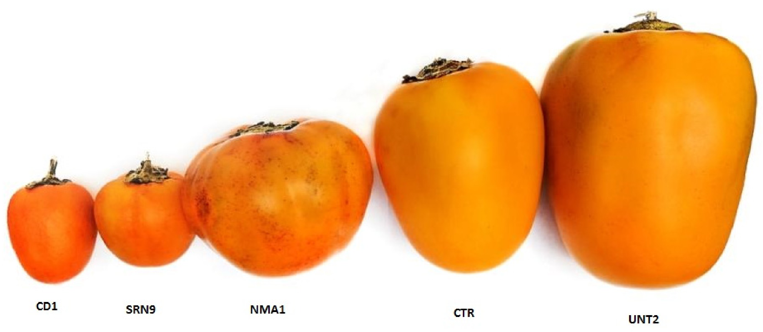
3.1. Nutritional and Physicochemical Properties of 5 Cocona Ecotypes

Table 1 shows proximal composition such as the humidity, ashes, protein, lipids, carbohydrates, and fiber, while Table 2 shows the mineral contents of five ecotypes of cocona. Proximal composition of cocona fruits NMA1, SRN9, CD1, CTR, UNT2 ecotypes was performed. The results of physicochemical properties showed that the proximal composition and the caloric value of this fruit are similar to that showed for other species, but with a high fiber (from 1.08 to 1.93) and carbohydrate content (from 3.12–4.24). The edible portion of cocona ecotypes were analyzed for mineral content (Ca, Na, Mg, K, Cu, Mn, Zn, and Fe). The foods were generally high in K (570.83–2382.24 mg K/100 g edible portion) and low in sodium (3.25–6.87 mg Na/100 g edible portion). The five ecotypes had the highest contents in most of the elements, especially in calcium (17.85–70.07 mg Ca/100 g edible portion) and iron (52–71 mg Fe/100 g edible portion).

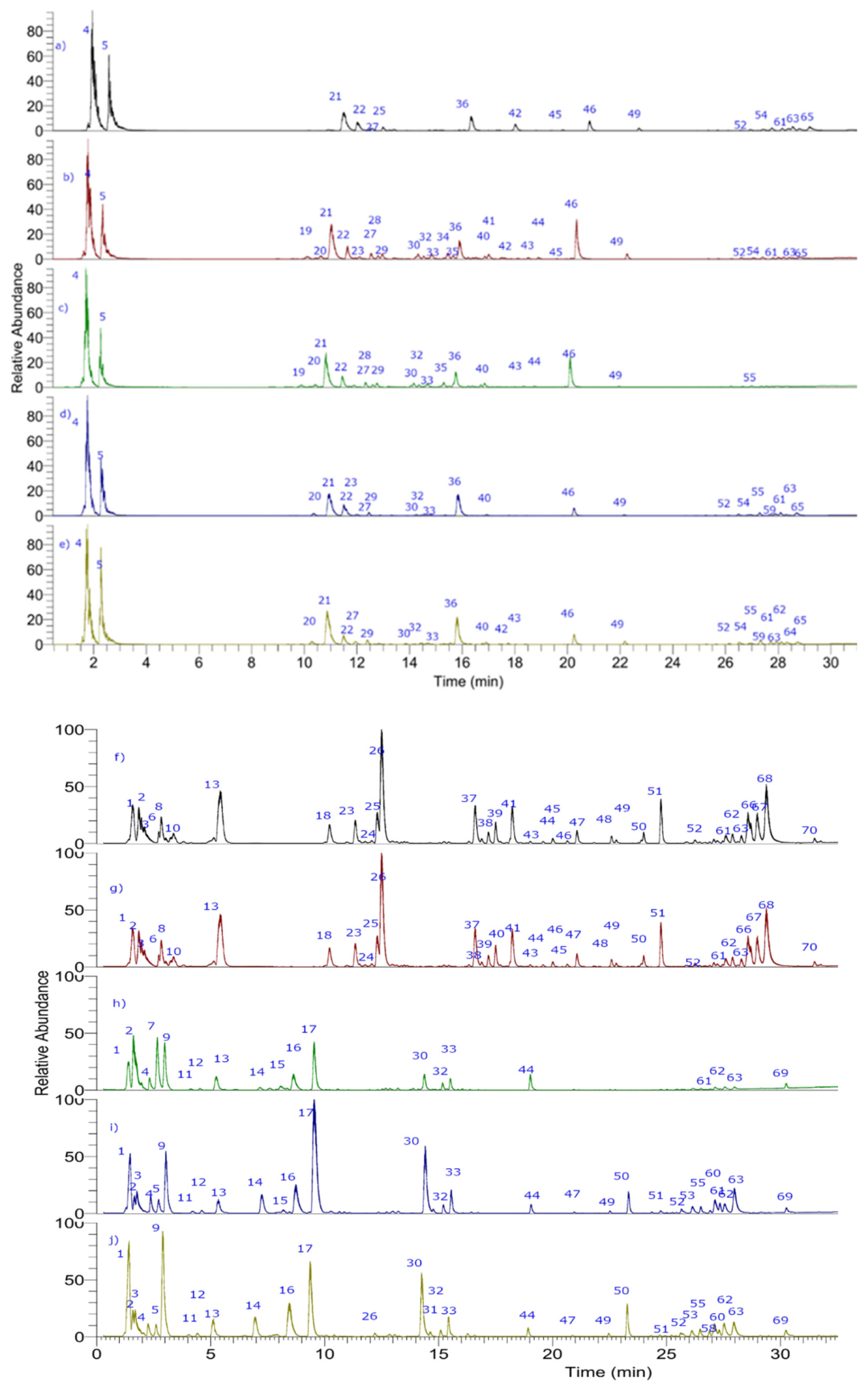
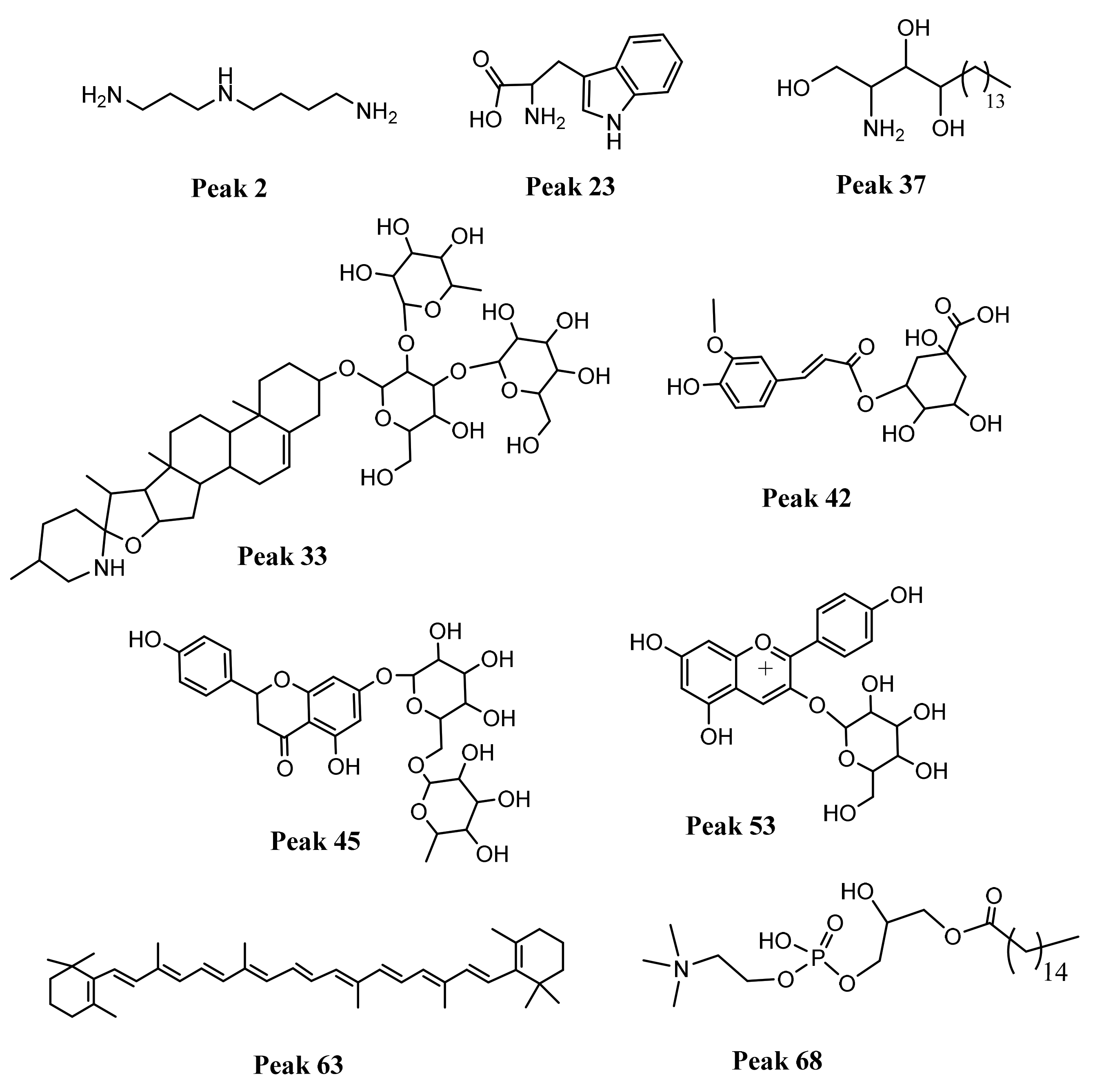
3.2. Metabolite Profiling using UHPLC-PDA-ESI-OT-MS

The metabolite profiling was comprehensively performed by UHPLC-PDA-ESI-OT-MS, while Figure 2 shows the base peak UHPLC-mass chromatograms of ecotypes of cocona fruits and Table 3 shows the tentative identification of metabolites detected in the five ecotypes. Below are the detailed analyses, while Figure 3 show the structures of some representative compounds.

3.2.1. Spermidines

Spermidine derivatives were previously reported by UHPLC MS by some of us in edible goji fruits [14]. Peak 1 with a [M+H] + ion at m/z 203.2229 is identified as spermine (C10H26N4), the parent ion producing some diagnostic daughter ions at m/z 129.1385, 112.1122, 84.0812 and 73.0813 and peak 2 as spermidine (C7H19N3), [14] peak 25 with a [M+H]+ ion at m/z 472.2441 as N-caffeoyl-N-(dihydrocaffeoyl) spermidine (C25H33N3O6) common components of potato tubers [15] and peak 26 as N-caffeoyl-N-(dihydrocaffeoyl)spermidine (C26H37N3O6) while peaks 38 and 39 as N,N″-Bis[3-(4-hydroxy-3-methoxyphenyl)propanoyl] spermidine (C27H39N3O6), and N,N,N-tris(dihydrocaffeoyl) spermidine (C34H43O9N3), Bioactive amines—such as spermidine and spermine-showed antioxidant activity in food through radical scavenging plus metal chelating properties, and were linked to reduced blood pressure and low incidence of cardiovascular diseases [16].

3.2.2. Amines or Aminoacids

Several aminoacids were previously reported using orbitrap mass spectrometry by some of us [17]. Peak 3 was identified as histamine (C5H9N3), peak 6 as asparagine (C4H8N2O3), peak 7 with a [M+H]+ ion at m/z 175.1188 as the aminoacid arginine (C6H14N4O2), peak 9 as nicotinamide (C5H7NO3), Peak 10 as N-phenyl ethyl amide (C8H10N) and peak 11 with a protonated molecule at m/z 294.1544 and MS ions at m/z 276.1436, 258.1332, 230.1383, 212.1278 and 86.0968 as N-fructosyl isoleucine (C12H23NO7) and peak 12 as norleucine (C6H13NO2) and peak 13 as tyrosine (C9H11NO3), peak 14 as adenosine (C10H13N5O4) and peak 15 as phenylacetaldehyde (C8H8O), peak 16–18 and 22–23 as the aminoacids guanosine, phenylalanine, panthotenic acid and tryptophan, respectively. Peak 24 with a protonated molecule at m/z 144.0807 [M-H2O+H] as tryptophol while peak 18 as aminobutyl benzamide (C11H16N2O). Peak 55 was identified as 1-hexadecanoyl-sn-glycero-3-phosphoethanolamine (C21H44NO7P).

3.2.3. Fatty Acids and Derivatives

Some oxylipins or unsaturated fatty acids were previously reported in edible fruits by some of us using TOF mass spectrometry [18]. In this study peak 37 with a pseudomolecular ion at m/z 318.3000, and daughter ions at m/z 318.2995, 300.2890, 282.2785, 270.2785, 60.0450, was identified as phytosphingosine (C18H39NO3) [19], and peak 54 as 1-(9Z,12Z-octadecadienoyl)-glycero-3-phospho-(1′-myo-inositol) (C27H49O12P), peak 58 as 1-hexadecanoyl-sn-glycero-3-phospho-(1′-myo-inositol) (C25H49O12P), peaks 66–70 as 1-(9Z,12Z-octadecadienoyl)-glycero-3-phospho-(1′-myo-inositol) (C27H49O12P), 1-(9Z-octadecenoyl)-sn-glycero-3-phosphoethanolamine (C23H46NO7P), 1-hexadecanoyl-sn-glycero-3-phosphocholine (C24H50NO7P), 1-(9Z-Octadecenoyl)-sn-glycero-3-phosphocholine (C26H52NO7P), and 1-Oleoyl-2-palmitoyl-sn-glycero-3-phosphocholine, (C42H82NO8P), respectively, and finally, peak 60 was identified as 1-(9Z-octadecenoyl)-sn-glycero-3-phospho-(1′-myo-inositol) (C27H51O12P).

3.2.4. Phenolic Acids

Peak 19 was tentatively identified as chlorogenic acid (C8H15O4) and peak 20 as quinic acid, Peak 21 as 3-O-diglucosyl-4-methoxy-3-hydroxybenzoic acid and peak 27 as 3-O-diglucosyl-4-methoxy-3-hydroxybenzoic acid (C20H28O14), peak 29 as apiosyl-glucosyl-hydroxybenzoate (C18H24O12), peak 34 as 1-O-Sinapoyl-glucoside (C17H22O10), and peak 43 as 2-O-sinapoyl-glucoside (C17H22O10) [20], peak 35 as protocatechuic acid 5-O-apiofuranosyl-glucopyranoside (C19H26O13), peak 36 as 4-O-(3′-O-glucopyranosyl)-caffeoyl quinic acid (C22H28O14), peak 42 as 3-O-feruloylquinic acid (C17H20O9) [21], and peak 44 as syringaresinol 4-gentiobioside (C34H46O18), peak 53 as naringenin-7-O-glucoside (C21H22O10), [22] and peak 54 as methyl chlorogenate (C17H20O9) [23], finally, peak 59 was assigned as syringaresinol-glucoside (C28H36O13), and peak 65 as phloretin (C15H14O5). Several acyl quinic acids such as ferulic, sinapic and chlorogenic acid and derivatives showed different interesting bioactivities such as anti-inflammatory, vasorelaxant in mice aorta, platelet activation in human blood samples neuroprotective effects among others [24], while the glycoside derivative of the lignan syringaresinol enhanced ß-endorphin levels in rat plasma, [25] while syringaresinol aglycone was cytotoxic against human breast and lung cancer cells [26].

3.2.5. Flavonoids

Peak 28 with was assigned as the flavonol glycoside rutin (C27H30O16) [27] with diagnostic ions at m/z 463.0920, 343.0465, 300.0280, 271.0252, 178.9982, and 151.0031, while peak 30 was assigned the structure naringenin-5,7-di-O-glucoside [28], (flavanone, Uv max 280 nm, C27H31O15) and peak 31 as genistein 5-O-glucoside (isoflavone, UV max 280 nm, C21H20O10) and peak 32 as the flavanol isoquercitrin (C21H20O12) [27]. In the same way, peak 45 was identified as naringenin 7-O-rutinoside or neohesperidoside (C27H31O14) [29], and peak 46 as quercetin 3-galactoside (C12H19O5) Peak 48 was assigned to the isoflavone biochanin A 7-O-rutinoside (C28H32O14) [30], peaks 56 and 57 as the flavanones naringenin-5-O-glucoside (C21H22O10) and eriodictyol-7-O-glucoside (C21H22O10), [31] respectively. Peak 61 with a ion at m/z 579.2084 as the flavonol quercetin 3-O-malonylglucoside (C24H22O15) [32], while peaks 64 was assigned as naringenin, respectively. Some flavonoids showed antioxidant properties and inhibition of enzymes such as xanthine oxidase and others important such as anti-inflammatory activities [33,34,35].

3.2.6. Terpenes

Peak 33 showing peaks at m/z 884.4987 and 928.4909 [M+FA-H], was tentatively assigned as the derivative of the terpene spirosol-5-en-3-ol,[36] 3-O-[rhamnosyl- glucosyl-galactoside] (C45H73NO16), and peak 40 as spirosol-5-en-3-ol, O-[rhamnosyl-[xylosyl- rhamnosyl-galactoside and peak 41 as cholest-5-ene-3,16,22,26-tetrol, 3-O-[Rhamnosyl- -rhamnosyl-glucoside], 26-O-glucoside (C51H86O22), and finally peak 47 as spirosol-5-en-3-ol, 3-O-[rhamnosyl- [rhamnosyl-glucoside] (C45H73NO15).

3.2.7. Cyanidins

Peak 52 with a positive ion at m/z 595.1656 was assigned to the pigment pelargonidin 3-sophoroside (C27H31O15) showing diagnostic ions at m/z 433.1080, 271.0595, 215.0695, 163.0596, and 127.0389, and peak 51 as pelargonidin 3-glucoside (C21H20O10) [37]. Those cyanidins possess antioxidant and anti-inflammatory activity [38].

3.2.8. Citric Acid

Peaks 5 and 6 were assigned as citric acid and isocitric acid (C6H8O7) respectively.

3.2.9. Carotenoids

Peaks 62 and 63 were tentatively identified as lutein (C40H56O2) and β-carotene (C40H56), respectively. In this study other carotene compounds were not detected, probably due to destruction by the pulping procedure. Lutein showed antioxidant and anti-inflammatory capacities, [39], and carotene showed anti-inflammatory, antioxidant and anticancer activity [40] and their presence in cocona fruits is in concordance with the medicinal.

3.3. Antioxidant Activity, Total Carotenes and Total Polyphenols Content

In this study the antioxidant capacities of the five ecotypes were assessed by the trapping of ABTS and DPPH and expressed as µmol Trolox/g dry matter (Table 4). In addition, total phenolic contents by the Folin and Ciocalteau’s method plus the total carotenes was assessed and correlated with the antioxidant capacities. Trapping of ABTS and DPPH radicals showed similar values in the different ecotypes and correlated with carotene and phenolic contents for the dry pulps. Strong correlation was found between total phenolics and DPPH antioxidant assays (r = 0.847, p < 0.0001). Moderated correlation was found between total carotenoids and ABTS antioxidant assays used (r = 0.640, p < 0.011). Ecotype UNT2 showed the highest DPPH• radical scavenging capacity in terms of content of Trolox equivalents (23.29 ± 1.07 µmol Trolox/g), but interestingly, in ABTS• radical scavenging activity the most powerful was the CD1 ecotype (25.67 ± 0.28 µmol Trolox/g).

3.4. Antihyperlipidemic Activities

Table 5 shows the variations in cholesterol, triglycerides, HDL and LDL in the different study groups. The highest cholesterol and triglyceride values correspond to the hypercholesteremic group that only received saline treatment (group II), with a statistically significant difference (p < 0.05) with the group control (group I) for the variables considered, thus corroborating the effectiveness of the model used. The increases obtained with Triton were 370 and 600 % for cholesterol and triglycerides, respectively. Several authors report similar elevations in the lipid profile of rodents treated with this agent. For instance, Harnafi et al. [41], obtained elevations of 270% for cholesterol and up to more than 1000% for triglycerides; while Khanna et al. [42], reported an elevation of cholesterol by 134%, these differences seem to depend on the strain of animals used.
When comparing the groups treated with the pulps, we noticed that samples which has received the CTR and SRN9 pulps showed the lowest cholesterol and triglyceride levels after treatment, however there is no clear correlations with the compounds detected and bioactivity showed by pulps. In agreement to the above, there isn’t a statistically significant difference (p < 0.05) between the cholesterol and triglyceride levels of the hypercholesterolemic group that received treatment with atorvastatin and the groups that received treatment with CTR and SRN9 pulps; which suggests that the treatment of the experimental animals with two pulps brings cholesterol levels closer to their basal values; this result is very important since it is an indicator of the effectiveness of the pulps tested. Regarding carotene compounds present in cocona fruits, it has been proved that diet with carotenes at a dose 80 mg/kg in rats decreased the lipid serum concentrations and its effect was comparable to that of simvastatin [43]. Regarding anthocyanins, also detected in cocona fruits, a juice from the fruit Aronia containing 106.8 mg cyanidin-3-glucoside equivalents/100 mL juice and 709.3 mg gallic acid equivalents/100 mL juice, showed an antihyperlipidemic effect in rats with hyperlipidemia and was proved to be valuable in reducing factors of cardiovascular risks [44]. Moreover, mulberry (Morus alba L.) lyophilized fruit administered to rats proved to have significant decrease in levels of serum and liver triglycerides, total cholesterol, serum low-density lipoprotein cholesterol, and a decrease in the atherogenic index, while the serum high-density lipoprotein cholesterol was significantly increased [45]. Several flavonoids and extracts rich in flavonoids were attributed anti hyperlipidemia activities; Pterocarpus marsupium heartwood and its flavonoid constituents, marsupsin, pterosupin, and liquiritigenin were able to effect a significant fall in serum cholesterol, LDL-cholesterol, and atherogenic index[46], also extracts full of glycosylated flavonoids from Cardoncellus marioticus showed antioxidant and antihyperlipidemic activities [47] moreover, using our same triton induced hyperlipidemia system extracts from the Mediterranean buckthorn, Rhamnus alaternus full of flavonoids decreased blood levels of cholesterol and triacylglycerols in hyperlipidemic rats (by 60% and 70%, respectively, at 200 mg CME/kg) [48]. In this study, all groups treated with the pulps showed a significant reduction in LDL-C (p < 0.05) with respect to the negative control group (group II), with the SRN9 group showing the best results. Despite the reduction in total cholesterol obtained with the different pulp treatments, we can observe that none of the treatments achieved lower values than the group treated with atorvastatin. In all the groups evaluated, an increase in HDL-C concentration was observed, the increase being significant in the groups that received CTR and SRN9 pulp (p < 0.05); the best result obtained was in the group that received SRN9 pulp after 96 hours of treatment. From the above mentioned we can say that SRN9 pulp, presents the best result of inhibition of the surfactant used (Triton WR-1339), by significantly decreasing the serum concentrations of cholesterol and triglycerides, corroborating the traditional use of this species and previous reports on its antihyperlipidemic activity [49,50]. This allows us to affirm that SRN9 pulp at the doses tested shows an antihyperlipidemic effect and could be an option in the management of people with high lipid levels. The presence of phenolic compounds, including flavonoids, could explain the hypolipidemic effect shown, presumably due to their proven antioxidant activity, as a result of a combination of their iron chelating and free radical scavenging properties [51,52]. During the experimental phase, no toxic effects were evidenced in the experimental specimens treated with NMA1; CD1; CTR; SRN9 and UNT2 pulps.

4. Conclusions

In this study, 70 compounds were detected in special Peruvian ecotypes of cocona fruits. Of those, six were spermidines, (peaks 1, 2, 25, 26, 38 and 39), thirteen were aminoacids, (peaks 3, 9, 11–13, 16, 17, 22–24), eighteen flavonoids (peaks 28, 30–32, 45, 46, 48–53, 56, 57, 61, and 64–66), twelve were phenolics (peaks 19, 21, 27, 29, 34, 35, 36, 42, 43, 44, 54, and 59), two carotenoids, (peak 62 and 63), eight were lipid derivatives (peaks 37, 55, 58, 60 and 67–70), one sugar (peak 47), four terpenes (peaks 33, 40, 41 and 47) two amides, (peaks 10 and 18), one aldehyde, (peak 15), and three saturated organic acids, (peaks 4, 5 and 20). Hypercholesterolemic mice administered with pulp of the ecotypes CTR and SRN9 showed the lowest cholesterol and triglyceride levels after treatment (126.74 ± 6.63; 102.11 ± 9.47; 58.16 ± 6.64; 61.05 ± 4.00 mg/dL, for cholesterol, triglycerides, HDL and LDL respectively, for the group treated with SRN9 pulp, and 130.09 ± 8.55; 108.51 ± 10.04; 57.30 ± 5.72; and 65.41 ± 7.68 mg/dL, for cholesterol, triglycerides, HDL and LDL respectively for the group treated with CTR pulp. Our study showed that the chemistry plus the bioactivity results obtained with five different ecotypes of Solanaceae Cocona fruits opens the door for the potential use of this plant to manage chronic diseases such as hyperlipidemia, especially for the SRN9 and CTR ecotypes, which presented better results in the antihyperlipidemic activity. The cultivation of these ecotypes using different conditions provides this common food plant with the ability to be a rich source of bioactive substances that can boost its consumption, not only as a fruit but also as natural medicine.

Author Contributions

Conceptualization, M.J.S. and G.V.-A.; methodology, validation and biological activities in rats, G.V.-A., M.R.-P., H.D.-W. and L.N.-R.; formal UHPLC-PDA-ESI-OT-MS analysis, C.P. and M.J.S.; in vitro antioxidant assays, M.W.P., C.M.-Z. and C.P.; resources, G.V.-A. and M.J.S.; writing—original draft preparation, M.R.-P., C.M.-Z., L.N.-R., M.J.S. and G.V.-A.; writing—review and editing, M.J.S. and G.V.-A.; supervision and project administration, G.V.-A.; funding acquisition G.V.-A. All authors have read and agreed to the published version of the manuscript.

Funding

This research was funded by the Project Concytec—Banco Mundial “Mejoramiento y Ampliación de los Servicios del Sistema Nacional de Ciencia Tecnología e Innovación Tecnológica” 8682, through its executing unit Fondecyt [Contrato No. 119-2018-FONDECYT-BM-IADT-MU]. M.J.S. acknowledge fondecyt 1180059.

Institutional Review Board Statement

Not applicable.

Informed Consent Statement

Not applicable.

Data Availability Statement

The data presented in this study are available in this article.

Conflicts of Interest

The authors declare no conflict of interest.

References

  1. Rodrigues, E.; Mariutti, L.R.B.; Mercadante, A.Z. Carotenoids and phenolic compounds from Solanum sessiliflorum, an unexploited amazonian fruit, and their scavenging capacities against reactive oxygen and nitrogen species. J. Agric. Food Chem. 2013, 61, 3022–3029. [Google Scholar] [CrossRef] [PubMed]
  2. Mascato, D.R.D.L.H.; Monteiro, J.B.; Passarinho, M.M.; Galeno, D.M.L.; Cruz, R.J.; Ortiz, C.; Morales, L.; Lima, E.S.; Carvalho, R.P. Evaluation of Antioxidant Capacity of Solanum sessiliflorum (Cubiu) Extract: An in Vitro Assay. J. Nutr. Metab. 2015, 2015, 1–8. [Google Scholar] [CrossRef] [PubMed] [Green Version]
  3. Vargas-Muñoz, D.P.; Cardoso da Silva, L.; Neves de Oliveira, L.A.; Teixeira Godoy, H.; Kurozawa, L.E. 5-caffeoylquinic acid retention in spray drying of cocona, an Amazonian fruit, using hydrolyzed collagen and maltodextrin as encapsulating agents. Dry. Technol. 2020, 39, 1854–1868. [Google Scholar] [CrossRef]
  4. Faria, J.V.; Valido, I.H.; Paz, W.H.P.; da Silva, F.M.A.; de Souza, A.D.L.; Acho, L.R.D.; Lima, E.S.; Boleti, A.P.A.; Marinho, J.V.N.; Salvador, M.J.; et al. Comparative evaluation of chemical composition and biological activities of tropical fruits consumed in Manaus, central Amazonia, Brazil. Food Res. Int. 2021, 139, 109836. [Google Scholar] [CrossRef]
  5. Dos Santos Montagner, G.F.F.; Barbisan, F.; Ledur, P.C.; Bolignon, A.; De Rosso Motta, J.; Ribeiro, E.E.; De Souza Praia, R.; Azzolin, V.F.; Cadoná, F.C.; Machado, A.K.; et al. In Vitro Biological Properties of Solanum sessiliflorum (Dunal), an Amazonian Fruit. J. Med. Food 2020, 23, 978–987. [Google Scholar] [CrossRef]
  6. Barrientos, R.; Fernández-Galleguillos, C.; Pastene, E.; Simirgiotis, M.; Romero-Parra, J.; Ahmed, S.; Echeverría, J. Metabolomic Analysis, Fast Isolation of Phenolic Compounds, and Evaluation of Biological Activities of the Bark from Weinmannia trichosperma Cav. (Cunoniaceae). Front. Pharmacol. 2020, 11, 780. [Google Scholar] [CrossRef]
  7. Gómez, J.; Simirgiotis, M.J.; Manrique, S.; Piñeiro, M.; Lima, B.; Bórquez, J.; Feresin, G.E.; Tapia, A. Uhplc-esi-ot-ms phenolics profiling, free radical scavenging, antibacterial and nematicidal activities of “yellow-brown resins” from larrea spp. Antioxidants 2021, 10, 185. [Google Scholar] [CrossRef]
  8. Jiménez-González, A.; Quispe, C.; Bórquez, J.; Sepúlveda, B.; Riveros, F.; Areche, C.; Nagles, E.; García-Beltrán, O.; Simirgiotis, M.J. UHPLC-ESI-ORBITRAP-MS analysis of the native Mapuche medicinal plant palo negro (Leptocarpha rivularis DC.—Asteraceae) and evaluation of its antioxidant and cholinesterase inhibitory properties. J. Enzyme Inhib. Med. Chem. 2018, 33, 936–944. [Google Scholar] [CrossRef]
  9. Cobos, M.; Pérez, S.; Braga, J.; Vargas-Arana, G.; Flores, L.; Paredes, J.D.; Maddox, J.D.; Marapara, J.L.; Castro, J.C. Nutritional evaluation and human health-promoting potential of compounds biosynthesized by native microalgae from the Peruvian Amazon. World J. Microbiol. Biotechnol. 2020, 36, 1–14. [Google Scholar] [CrossRef]
  10. Official Methods of Analysis of AOAC International—18th Edition, Revision 3. Available online: https://www.techstreet.com/standards/official-methods-of-analysis-of-aoac-international-18th-edition-revision-3?product_id=1678986 (accessed on 12 July 2021).
  11. Asaolu, M.F.; Asaolu, S.S. Proximate and mineral compositions of cooked and uncooked Solanum melongena. Int. J. Food Sci. Nutr. 2002, 53, 103–107. [Google Scholar] [CrossRef]
  12. Barreto, G.P.M.; Benassi, M.T.; Mercadante, A.Z. Bioactive compounds from several tropical fruits and correlation by multivariate analysis to free radical scavenger activity. J. Braz. Chem. Soc. 2009, 20, 1856–1861. [Google Scholar] [CrossRef]
  13. Venkadeswaran, K.; Muralidharan, A.R.; Annadurai, T.; Ruban, V.V.; Sundararajan, M.; Anandhi, R.; Thomas, P.A.; Geraldine, P. Antihypercholesterolemic and antioxidative potential of an extract of the plant, piper betle, and its active constituent, eugenol, in triton WR-1339-Induced hypercholesterolemia in experimental rats. Evid. -Based Complement. Altern. Med. 2014, 2014, 478973. [Google Scholar] [CrossRef] [Green Version]
  14. Mocan, A.; Zengin, G.; Simirgiotis, M.; Schafberg, M.; Mollica, A.; Vodnar, D.C.; Crişan, G.; Rohn, S. Functional constituents of wild and cultivated Goji (L. barbarum L.) leaves: Phytochemical characterization, biological profile, and computational studies. J. Enzyme Inhib. Med. Chem. 2017, 32, 153–168. [Google Scholar] [CrossRef] [Green Version]
  15. Narváez-Cuenca, C.E.; Vincken, J.P.; Zheng, C.; Gruppen, H. Diversity of (dihydro) hydroxycinnamic acid conjugates in Colombian potato tubers. Food Chem. 2013, 139, 1087–1097. [Google Scholar] [CrossRef]
  16. Dala-Paula, B.M.; Deus, V.L.; Tavano, O.L.; Gloria, M.B.A. In vitro bioaccessibility of amino acids and bioactive amines in 70% cocoa dark chocolate: What you eat and what you get. Food Chem. 2021, 343, 128397. [Google Scholar] [CrossRef]
  17. Guerrero-Castillo, P.; Reyes, S.; Robles, J.; Simirgiotis, M.J.; Sepulveda, B.; Fernandez-Burgos, R.; Areche, C. Biological activity and chemical characterization of Pouteria lucuma seeds: A possible use of an agricultural waste. Waste Manag. 2019, 88, 319–327. [Google Scholar] [CrossRef]
  18. Simirgiotis, M.J.; Ramirez, J.E.; Schmeda Hirschmann, G.; Kennelly, E.J. Bioactive coumarins and HPLC-PDA-ESI-ToF-MS metabolic profiling of edible queule fruits (Gomortega keule), an endangered endemic Chilean species. Food Res. Int. 2013, 54, 532–543. [Google Scholar] [CrossRef]
  19. Do Amaral, B.S.; da Silva, L.R.G.; Valverde, A.L.; de Sousa, L.R.F.; Severino, R.P.; de Souza, D.H.F.; Cass, Q.B. Phosphoenolpyruvate carboxykinase from T. cruzi magnetic beads affinity-based screening assays on crude plant extracts from Brazilian Cerrado. J. Pharm. Biomed. Anal. 2021, 193, 113710. [Google Scholar] [CrossRef]
  20. Pereira, A.P.A.; Angolini, C.F.F.; Adani, H.B.; Usberti, F.C.S.; Paulino, B.N.; Clerici, M.T.P.S.; Neri-numa, I.A.; Moro, T.d.M.A.; Eberlin, M.N.; Pastore, G.M. Impact of ripening on the health-promoting components from fruta-do-lobo (Solanum lycocarpum St. Hill). Food Res. Int. 2021, 139, 109910. [Google Scholar] [CrossRef]
  21. Simirgiotis, M.J.; Quispe, C.; Areche, C.; Sepúlveda, B. Phenolic compounds in chilean mistletoe (quintral, Tristerix tetrandus) analyzed by UHPLC-Q/Orbitrap/MS/MS and its antioxidant properties. Molecules 2016, 21, 245. [Google Scholar] [CrossRef] [Green Version]
  22. Barrientos, R.E.; Ahmed, S.; Cortés, C.; Fernández-Galleguillos, C.; Romero-Parra, J.; Simirgiotis, M.J.; Echeverría, J. Chemical Fingerprinting and Biological Evaluation of the Endemic Chilean Fruit Greigia sphacelata (Ruiz and Pav.) Regel (Bromeliaceae) by UHPLC-PDA-Orbitrap-Mass Spectrometry. Molecules 2020, 25, 3750. [Google Scholar] [CrossRef]
  23. Yin, J.; Heo, J.H.; Hwang, Y.J.; Le, T.T.; Lee, M.W. Inhibitory activities of phenolic compounds isolated from adina rubella leaves against 5α-reductase associated with benign prostatic hypertrophy. Molecules 2016, 21, 887. [Google Scholar] [CrossRef] [PubMed] [Green Version]
  24. Clifford, M.N.; Jaganath, I.B.; Ludwig, I.A.; Crozier, A. Chlorogenic acids and the acyl-quinic acids: Discovery, biosynthesis, bioavailability and bioactivity. Nat. Prod. Rep. 2017, 34, 1391–1421. [Google Scholar] [CrossRef] [Green Version]
  25. Nishibe, S. Bioactive Phenolic Compounds for Cancer Prevention from Herbal Medicines. Food Factors Cancer Prev. 1997, 276–279. [Google Scholar] [CrossRef]
  26. Yeh, Y.-T.; Huang, J.-C.; Kuo, P.-L.; Chen, C.-Y. Bioactive Constituents from Michelia champaca. Nat. Prod. Commun. 2011, 6, 1251–1252. [Google Scholar] [CrossRef] [Green Version]
  27. Ramirez, J.E.; Zambrano, R.; Sepúlveda, B.; Kennelly, E.J.; Simirgiotis, M.J. Anthocyanins and antioxidant capacities of six Chilean berries by HPLC–HR-ESI-ToF-MS. Food Chem. 2015, 176, 106–114. [Google Scholar] [CrossRef]
  28. Iwashina, T.; Kitajima, J.; Shiuchi, T.; Itou, Y. Chalcones and other flavonoids from Asarum sensu lato (Aristolochiaceae). Biochem. Syst. Ecol. 2005, 33, 571–584. [Google Scholar] [CrossRef]
  29. Brito, A.; Ramirez, J.E.; Areche, C.; Sepúlveda, B.; Simirgiotis, M.J. Molecules HPLC-UV-MS Profiles of Phenolic Compounds and Antioxidant Activity of Fruits from Three Citrus Species Consumed in Northern Chile. Molecules 2014, 19, 17400–17421. [Google Scholar] [CrossRef]
  30. Khan, A.; Bresnick, A.; Cahill, S.; Girvin, M.; Almo, S.; Quinn, R. Advantages of Molecular Weight Identification during Native MS Screening. Planta Med. 2018, 84, 1201–1212. [Google Scholar] [CrossRef]
  31. Mubashir, N.; Fatima, R.; Naeem, S. Identification of Novel Phyto-chemicals from Ocimum basilicum for the Treatment of Parkinson’s Disease using In Silico Approach. Curr. Comput. Aided. Drug Des. 2020, 16, 420–434. [Google Scholar] [CrossRef]
  32. Lin, Y.C.; Wu, C.J.; Kuo, P.C.; Chen, W.Y.; Tzen, J.T.C. Quercetin 3-O-malonylglucoside in the leaves of mulberry (Morus alba) is a functional analog of ghrelin. J. Food Biochem. 2020, 44, e13379. [Google Scholar] [CrossRef] [PubMed]
  33. Montoro, P.; Braca, A.; Pizza, C.; De Tommasi, N. Structure–antioxidant activity relationships of flavonoids isolated from different plant species. Food Chem. 2005, 92, 349–355. [Google Scholar] [CrossRef]
  34. Fan, P.; Terrier, L.; Hay, A.E.; Marston, A.; Hostettmann, K. Antioxidant and enzyme inhibition activities and chemical profiles of Polygonum sachalinensis F. Schmidt ex Maxim (Polygonaceae). Fitoterapia 2010, 81, 124–131. [Google Scholar] [CrossRef] [PubMed]
  35. Farzaei, M.H.; Singh, A.K.; Kumar, R.; Croley, C.R.; Pandey, A.K.; Coy-Barrera, E.; Patra, J.K.; Das, G.; Kerry, R.G.; Annunziata, G.; et al. Targeting Inflammation by Flavonoids: Novel Therapeutic Strategy for Metabolic Disorders. Int. J. Mol. Sci. 2019, 20, 4957. [Google Scholar] [CrossRef] [Green Version]
  36. Quan, H.J.; Koyanagi, J.; Ohmori, K.; Uesato, S.; Tsuchido, T.; Saito, S. Preparations of heterospirostanols and their pharmacological activities. Eur. J. Med. Chem. 2002, 37, 659–669. [Google Scholar] [CrossRef]
  37. Kołota, A.; Głabska, D.; Oczkowski, M.; Gromadzka-Ostrowska, J. Analysis of association between intake of red wine polyphenols and oxidative stress parameters in the liver of growing male rats. Appl. Sci. 2020, 10, 6389. [Google Scholar] [CrossRef]
  38. Wang, H.; Nair, M.G.; Strasburg, G.M.; Chang, Y.C.; Booren, A.M.; Gray, J.I.; DeWitt, D.L. Antioxidant and antiinflammatory activities of anthocyanins and their aglycon, cyanidin, from tart cherries. J. Nat. Prod. 1999, 62, 294–296. [Google Scholar] [CrossRef]
  39. Pap, R.; Pandur, E.; Jánosa, G.; Sipos, K.; Agócs, A.; Deli, J. Lutein Exerts Antioxidant and Anti-Inflammatory Effects and Influences Iron Utilization of BV-2 Microglia. Antioxidants 2021, 10, 363. [Google Scholar] [CrossRef]
  40. Talero, E.; García-Mauriño, S.; Ávila-Román, J.; Rodríguez-Luna, A.; Alcaide, A.; Motilva, V. Bioactive Compounds Isolated from Microalgae in Chronic Inflammation and Cancer. Mar. Drugs 2015, 13, 6152. [Google Scholar] [CrossRef]
  41. Harnafi, H.; Bouanani, N.e.H.; Aziz, M.; Serghini Caid, H.; Ghalim, N.; Amrani, S. The hypolipidaemic activity of aqueous Erica multiflora flowers extract in Triton WR-1339 induced hyperlipidaemic rats: A comparison with fenofibrate. J. Ethnopharmacol. 2007, 109, 156–160. [Google Scholar] [CrossRef]
  42. Khanna, A.K.; Rizvi, F.; Chander, R. Lipid lowering activity of Phyllanthus niruri in hyperlipemic rats. J. Ethnopharmacol. 2002, 82, 19–22. [Google Scholar] [CrossRef]
  43. Sundaram, R.; Ayyakkannu, P.; Muthu, K.; parveen Nazar, S.; Palanivelu, S.; Panchanatham, S. Acyclic Isoprenoid Attenuates Lipid Anomalies and Inflammatory Changes in Hypercholesterolemic Rats. Indian J. Clin. Biochem. 2019, 34, 395–406. [Google Scholar] [CrossRef]
  44. Valcheva-Kuzmanova, S.; Kuzmanov, K.; Mihova, V.; Krasnaliev, I.; Borisova, P.; Belcheva, A. Antihyperlipidemic effect of Aronia melanocarpa fruit juice in rats fed a high-cholesterol diet. Plant Foods Hum. Nutr. 2007, 62, 19–24. [Google Scholar] [CrossRef]
  45. Yang, X.; Yang, L.; Zheng, H. Hypolipidemic and antioxidant effects of mulberry (Morus alba L.) fruit in hyperlipidaemia rats. Food Chem. Toxicol. 2010, 48, 2374–2379. [Google Scholar] [CrossRef]
  46. Jahromi, M.A.F.; Ray, A.B.; Chansouria, J.P.N. Antihyperlipidemic effect of flavonoids from pterocarpus marsupium. J. Nat. Prod. 1993, 56, 989–994. [Google Scholar] [CrossRef]
  47. Shabana, M.M.; El-Sherei, M.M.; Moussa, M.Y.; Sleem, A.A.; Abdallah, H.M. Flavonoid constituents of Carduncellus mareoticus (Del.) Hanelt and their biological activities. Nat. Prod. Commun. 2008, 3, 779–784. [Google Scholar] [CrossRef] [Green Version]
  48. Tacherfiout, M.; Petrov, P.D.; Mattonai, M.; Ribechini, E.; Ribot, J.; Bonet, M.L.; Khettal, B. Antihyperlipidemic effect of a Rhamnus alaternus leaf extract in Triton-induced hyperlipidemic rats and human HepG2 cells. Biomed. Pharmacother. 2018, 101, 501–509. [Google Scholar] [CrossRef]
  49. Maia, J.R.P.; Schwertz, M.C.; Sousa, R.F.S.; Aguiar, J.P.L.; Lima, E.S. Efeito hipolipemiante da suplementação dietética com a farinha do cubiu (solanum sessiliforum dunal) em ratos hipercolesterolêmicos. Rev. Bras. Plantas Med. 2015, 17, 112–119. [Google Scholar] [CrossRef] [Green Version]
  50. Pardo, M.A. Efecto de Solanum sessiliflorum dunal sobre el metabolismo lipídico y de la glucosa. Cienc. Investig. 2004, 7, 43–48. [Google Scholar] [CrossRef]
  51. Zeashan, H.; Amresh, G.; Singh, S.; Rao, C.V. Hepatoprotective activity of Amaranthus spinosus in experimental animals. Food Chem. Toxicol. 2008, 46, 3417–3421. [Google Scholar] [CrossRef]
  52. Nicholson, S.K.; Tucker, G.A.; Brameld, J.M. Effects of dietary polyphenols on gene expression in human vascular endothelial cells. Proc. Nutr. Soc. 2008, 67, 42–47. [Google Scholar] [CrossRef]
Figure 1. Pictures of cocona fruits NMA1, SRN9, CD1, CTR, UNT2 ecotypes.
Antioxidants 10 01566 g001
Figure 2. UHPLC-PDA-ESI-OT-MS chromatograms (TIC, total ion current) of cocona fruits NMA1, SRN9, CD1, CTR, UNT2 ecotypes: (ae) positive mode and (fj) negative mode.
Antioxidants 10 01566 g002
Figure 3. Structures of some representative compounds detected in cocona ecotypes: spermidine, peak 2, the aminoacid triptophan, peak 23, phytoesphingosine peak 37, spirosol-5-en-3-ol, 3-O-[rhamnosyl-glucosyl]-galactoside, peak 33, 3-O-feruloylquinic acid, peak 42, naringenin 7-O-rutinoside, peak 45, pelargonidin 3-O-glucoside, peak 53, B-carotene, peak 63 and 1-hexadecanoyl-sn-glycero-3-phosphocholine, peak 68.
Antioxidants 10 01566 g003
Table 1. Proximal composition of cocona fruits NMA1, SRN9, CD1, CTR, UNT2 ecotypes.
EcotypesHumidityAshesTotal LipidsCrude proteinCrude fiberCarbohydrates
NMA191.85 ± 0.09 a0.75 ± 0.01 a0.65 ± 0.00 a1.08 ± 0.04 a1.68 ± 0.04 a3.99
CD186.64 ± 0.36 b1.24 ± 0.06 b0.88 ± 0.00 b1.93 ± 0.05 b5.03 ± 0.15 b4.28
CTR92.82 ± 0.03 c0.71 ± 0.03 a0.45 ± 0.01 c1.09 ± 0.04 a1.03 ± 0.05 c3.9
SRN986.67 ± 0.12 b0.94 ± 0.02 c0.93 ± 0.01 d2.72 ± 0.04 c4.76 ± 0.17 b3.98
UNT293.52 ± 0.08 d0.79 ± 0.02 a0.19 ± 0.00 e1.64 ± 0.06 d0.76 ± 0.03 c3.1
Each value represents the means ± SEM of three replicates, n = 3, while different letters on the same column indicate significant difference using Tukey test at 0.05 level of significance (p < 0.05).
Table 2. Mineral content (mg/100 g fresh pulp) of cocona fruits NMA1, SRN9, CD1, CTR, UNT2 ecotypes.
EcotypesFeZnMnCuMgKNaCa
NMA171.17 ± 2.69 a26.33 ± 0.95 a8.18 ± 0.30 a12.27 ± 0.52 a42.54 ± 0.93 a846.47 ± 19.85 a4.09 ± 0.13 a28.63 ± 0.86 a
CD170.07 ± 2.65 a67.05 ± 1.56 b34.35 ± 1.16 b41.22 ± 0.73 b164.88 ± 6.55 b2382.24 ± 29.95 b6.87 ± 0.13 b70.07 ± 1.62 b
CTR40.70 ± 1.82 b17.83 ± 0.84 c7.85 ± 0.15 ac11.42 ± 0.42 ac38.56 ± 0.69 ac638.10 ± 6.28 c3.57 ± 0.09 c17.85 ± 0.46 c
SRN958.09 ± 1.15 c45.93 ± 2.19 d9.46 ± 0.08 d14.86 ± 0.65 d91.87 ± 1.54 d2004.88 ± 33.34 d6.76 ± 0.18 b58.09 ± 1.05 d
UNT252.02 ± 1.17 d18.85 ± 0.90 c8.45 ± 0.21 acd10.40 ± 0.30 c41.60 ± 0.84 ac570.83 ± 12.13 e3.25 ± 0.04 c41.60 ± 0.72 e
Each value represents the means ± SEM of three replicates, n = 3, and different letters on the same column indicate significant difference using Tukey test at 0.05 level of significance (p < 0.05).
Table 3. High resolution UHPLC-PDA-ESI-OT-MS identification of metabolites in fractions of cocona fruits (a–e): NMA1, SRN9, CD1, CTR, UNT2 ecotypes, respectively.
Peak#Retention Time (min.)UV MaxTentative
Identification
Molecular FormulaTheoretical Mass (m/z)Measured Mass [M-H] or [M+H]+ (m/z)Accuracy
(ppm)
MSn IonsEcotype
11.25-SpermineC10H26N4203.22302203.2229−2.448129.1385, 112.1122, 84.0812, 73.0813a–e
21.33-SpermidineC7H19N3146.16517146.1651−1.96129.1385, 112.1122, 84.0812, 73.0813a–e
31.35-HistamineC5H9N3112.08692112.08720.7995.0606, 83.0608, 68.0500, 55.0549a–e
41.69-Citric acidC6H8O7191.01944191.018634.24129.1385c–e
51.75-Isocitric acidC6H8O7191.01944191.019473.25111.00794d–e
61.97-Asparagine C4H8N2O3131.0449131.04543.81114.0187, 113.0337, 95.0251, 88.0394
70.0288
a–b
72.29-ArginineC6H14N4O2175.1181175.11883.99158.0920, 140.0702, 130.0972, 116.0706
97.0661
70.0656
c
82.43-Pyroglutamic acidC5H7NO3130.0493130.04994.61102.0251, 84.0448, 56.0551, a–b
93.01-Nicotinamide C6H6N2O123.0550123.05532.43106.0289, 96.0446, 80.0499, 53.0391c–e
103.15-N-phenyl ethyl amide C8H10N120.08810120.080802.4285.02876a–b
114.1-N-Fructosyl isoleucine C12H23NO7294.1541294.15441.01276.1436, 258.1332, 230.1383, 212.1278
161.0681, 144.1017, 132.1017, 86.0968
c–e
124.32-NorleucineC6H13NO2132.1016132.10192.27105.0696, 86.0968, 69.0704c–e
135.21-TyrosineC9H11NO3188.0210188.02110.53165.0554, 147.0438, 136.0755, 123.0441
105.0337
a–e
147.03-AdenosineC10H13N5O4268.1037268.10390.74178.0730, 136.0616, 57.0341c–e
158.12-PhenylacetaldehydeC8H8O121.0642121.06495.48103.0544, 93.0702, 91.0546, 77.0387
53.0392
c–e
168.53-GuanosineC10H13N5O5284.0983284.09881.75152.0564, 133.0494, 121.0647, 95.0609c–e
179.62-PhenylalanineC9H11NO2166.0859166.08611.20149.0594, 131.0491, 120.0808, 103.0543
93.0703
c–e
1810.23-Aminobutyl benzamideC11H16N2O193.1332193.13362.07176.1067, 134.0599, 105.0337, 72.0813a–b
1910.34290–335Chlorogenic acidC8H14O4353.0863353.08824.21191.05574, 707.18678b–c
2010.5208Quinic acidC7H11O6191.0550191.05572.34135.04477, 85.02844b–e
2110.882353-O-diglucosyl-4-methoxy-3-hydroxybenzoic acidC20H27O14491.1395491.14123.46 a–d
2211.25-Pantothenic acidC9H17NO5220.1173220.11792.72202.1069, 184.0965, 160.0965, 142.0860
124.0756
a–d
2311.43-TryptophanC11H12N2O2205.0961205.09683.41188.0701, 170.0596, 159.0914, 146.0597
132.0804
118.0650
a–b
2411.75-TryptopholC10H11NO144.0802
[M-
H2O+H]
144.08073.47128.0491, 117.0699, 103.0506, 91.0547a–b
2511.97330N-Caffeoyl-N-(dihydrocaffeoyl)spermidine C25H33N3O6472.2439472.24410.42455.2163, 310.2118, 293.1852, 222.1120
163.0386, 72.0813
a
2612.03330N-Caffeoyl-N-(dihydrocaffeoyl)spermidineC26H37N3O6488.2751488.27561.02471.2478, 324.2273, 293.1844, 236.1275
222.1119, 165.0542
a–b
2712.023253-O-Diglucosyl-4-methoxy-3-hydroxybenzoic acidC20H27O14491.13953491.141243.46 a–d
2812.24254–354RutinC27H30O16609.14702609.147090.11463.0920, 343.0465, 301.0254, 300.0280, 271.0252
178.9982, 151.0031
b–c
2912.46240Apiosyl-(1→6)-glucosyl 4-hydroxybenzoateC18H24O12431.0980431.09830.46431.1196, 299.0768, 281.0679, 137.0237
93.0336
b–d
3014.03280Naringenin-5,7-di-O-D-glucopyranosideC27H31O15595.16575595.167723.32271.06152, 153.01845, 147.04482, 119.05661c–e
3114.21280Genistein 5-O-glucoside C21H20O10431.0980431.0984
433.1123
0.46414.3355, 271.0595, 269.0390, 253.0485, 215.0698
146.0598, 127.0389, 85.0288
d–e
3215.02254–354IsoquercitrinC21H20O12463.1022463.10271.08300.0280, 271.0251, 255.0301, 178.9982
151.0032
c–e
3315.21-Spirosol-5-en-3-ol, 3-O-[Rhamnosyl-(1→2)-
glucosyl-(1→3)]-galactoside
C45H73NO16884.4982884.4987
928.4909 [M+FA-H]
0.56722.4060, 576.3906, 414.3356b–e
3415.243291-O-Sinapoyl-glucosideC17H22O10385.1142385.11471.29247.0612, 223.0611, 205.0504, 190.0269
164.0704, 119.0342,
b
3515.36280Protocatechuic acid 5-O-[apiofuranosyl-(1→6)-glucopyranoside]C19H26O13461.1301461.13020.21329.0872, 167.0344, 152.0108, 123.0443
108.0208
b–c
3615.573254-O-(3′-O-Glucopyranosyl)-caffeoyl quinic acidC22H28O14515.1401515.14071.16395.0990, 353.0876, 191.0557, 179.0344
161.0238, 135.0444
a–e
3715.87-PhytosphingosineC18H39NO3318.2990318.29951.57300.2890, 282.2785, 270.2785, 60.0450a, b, d, e
3816.23280N,N″-Bis[3-(4-hydroxy-3-methoxyphenyl)propanoyl] spermidineC27H39N3O6502.2902502.29070.99485.2633, 307.1996, 236.1275, 179.0698
137.0594
a–b
3916.73325N,N,N-tris(dihydrocaffeoyl) spermidineC34H43O9N3638.3059638.30620.46474.2588, 456.2484, 293.1852, 222.1120
165.0543, 123.0439
a–b
4017.23-Spirosol-5-en-3-ol, O-[Rhamnosyl-(1→2)-[xylosyl-
(1→2)-rhamnosyl-(1→4)]-galactoside
C50H81NO191000.54501000.54560.59868.4970, 722.4737, 576.3879, 414.3358a–b
4117.55-Cholest-5-ene-3,16,22,26-tetrol, 3-O-[Rhamnosyl- (1→4)-[rhamnosyl-(1→2)]glucoside], 26-O- glucosideC51H86O221051.56601051.56650.471049.5541, 903.4961, 757.4382, 595.3851
433.3324
a–b
4217.673303-O-Feruloylquinic acidC17H20O9367.1032367.10391.90191.0558, 173.0451, 134.0366, 111.0443
93.0336,
a–b
4318.053292-O-Sinapoyl-glucosideC17H22O10385.1141385.11471.55247.0612, 223.0611, 205.0504, 190.0269
164.0704, 119.0342
a–b
4419.01330Syringaresinol 4-gentiobiosideC34H46O18787.2676787.2678 [M+FA-H]0.25417.1560, 402.1323, 387.1069, 371.1494
356.1233, 181.0502, 166.0266
a–e
4519.53280Naringenin 7- O-rutinoside C27H31O14579.17083579.172913.59271.06152, 151.00319a–b
4619.72254–354Quercetin 3-galactoside C12H19O5243.12270463.088933.94350.20898, 301.02795, 151.00310a–e
4720.45-Spirosol-5-en-3-ol, 3-O-[Rhamnosyl-(1→2)-[rhamnosyl-(1→4)]-glucoside]C45H73NO15868.5031868.50350.46722.4411, 576.3845, 414.3357a, b, d, e
4822.32280Biochanin A 7-O-rutinosideC28H32O14593.1852593.18591.18447.1269, 327.0856, 285.0750, 153.0191a, b
4922.51280GenistinC21H20O10431.0982433.1123
431.0984
0.69414.3355, 271.0595, 253.0485, 215.0698
146.0598, 127.0389, 85.0288,
a–e
5023.35255–3403,5-Dihydroxy-4′,7-dimethoxyflavone 3-O-[Rhamnosyl-(1→2)-glucoside (Pectolarin)C29H34O15623.1961623.19630.32477.1342, 315.0815, 300.0622, 284.0679a, b, d, e
5124.93281Naringenin-7-O-glucosideC21H22O10435.1279435.1282
433.1117
0.95313.0506, 271.0617, 193.0138, 151.0032
119.0405
d–e
5225.5520Pelargonidin 3-O-sophorosideC27H31O15595.1652595.16560.67433.1080, 271.0595, 215.0695, 163.0596
127.0389
a–b
5326.1520Pelargonidin 3-O-glucosideC21H20O10433.1121433.11261.15311.0556, 269.0460, 163.0031a–b
5426.5325Methyl chlorogenateC17H20O9367.1031367.10351.08191.0556, 179.0344, 161.0237, 135.0443
93.0335
a–d
5526.7-Peak 55, 1-Hexadecanoyl-sn-glycero-3-Phosphoethanolamine C21H44NO7P454.2920
454.2923
452.2786
0.66255.2630, 214.0482, 214.0284, 140.0111c–e
5627.0280Naringenin-5-O-glucosideC21H22O10433.1132433.11381.38313.0551, 271.0613, 151.0030, 119.0493
93.00335
a–e
5727.1280Eriodictyol-7-O-glucosideC21H22O10449.1080449.10830.66287.0567, 205.0144, 175.0033, 151.0032
135.0445, 125.0237
a–e
5827.2-1-Hexadecanoyl-sn-glycero-3-phospho-(1′-myo-inositol)C25H49O12P573.3030573.3034
571.2089
0.69391.2256, 333.0594, 315.0487, 255.2329
241.0118, 152.9951
a–e
5927.3330Syringaresinol-glucosideC28H36O13579.2080579.20840.89417.1557, 402.1320, 387.1085, 223.0616
181.0501, 166.0265
c
6027.5-1-(9Z-Octadecenoyl)-sn-glycero-3-phospho-(1′-myo-inositol)C27H51O12P599.3189599.3192
597.3046
0.50333.0594, 315.0488, 281.2487, 259.0223
241.0118
d–e
6127.7254–354Quercetin 3-O-malonylglucosideC24H22O15579.2080579.20840.69463.0888, 300.0280, 271.0252, 255.0301
178.9981, 151.0032
a–e
6227.9450LuteinC40H56O2568.4280568.4282 124.08689, 145.0845, 105.08564, 335.12485a–e
6328.0450β-caroteneC40H56536.4379536.4382 337.09189, 476.17609a–e
6428.1280Naringenin C15H12O5271.06010271.061555.36153.01832, 147.04453, 119.05632e
6528.3280PhloretinC15H14O5349.18569349.187234.03229.0871, 179.0347, 167.0347, 125.0237e
6628.4254–354CirsimarinC23H24O11475.12668475.125244.36315.07642c–e
6728.9-1-(9Z-Octadecenoyl)-sn-glycero-3-phosphoethanolamineC23H46NO7P480.3080480.3085
478.2937
0.83281.2486, 214.0482, 196.0386, 152.9950
140.0110
a–b
6829.2-1-hexadecanoyl-sn-glycero-3-phosphocholineC24H50NO7P496.3391496.33961.00313.2128, 184.0721, 124.9998, 104.1072
86.0968
a–b
6929.72151-(9Z-Octadecenoyl)-sn-glycero-3-phosphocholineC26H52NO7P566.3419566.3422 [M+FA-H]
522.3555
0.56504.3450, 445.2715, 339.2892, 240.1001
199.0370, 187.0732, 124.9988
c–e
7030.22101-Oleoyl-2-palmitoyl-sn-glycero-3-phosphocholineC42H82NO8P760.5832760.5839
758.542
0.92184.0730, 125.9997, 104.1071, 86.0964a–b
Table 4. Antioxidant activity (DPPH, ABTS), total phenolic content and total carotenoid contents of cocona fruits pulps NMA1, SRN9, CD1, CTR, UNT2 ecotypes.
EcotypesDPPH
(µmol Trolox/g)
ABTS
(µmol Trolox/g)
Total Phenolics
(mg GAE/g)
Total Carotenoids
(μg β-carotene)/g)
NMA119.88 ± 0.34 a19.70 ± 0.81 a32.68 ± 1.33 a101.22 ± 4.47 a
CD118.37 ± 0.24 b25.67 ± 0.28 b28.03 ± 0.90 b122.65 ± 4.24 b
CTR21.92 ± 0.53 c21.98 ± 0.90 ac35.79 ± 0.84 ac85.13 ± 1.81 c
SRN918.21 ± 0.24 bd23.97 ± 1.12 bc27.86 ± 0.81 b92.12 ± 3.64 ac
UNT219.15 ± 0.84 abd20.25 ± 0.79 ac34.26 ± 1.32 ac58.81 ± 0.46 ab
Each value represents the means ± SEM of three replicates, n = 3, while different letters on the same column indicate significant difference using Tukey test at 0.05 level of significance (p < 0.05).
Table 5. Effect of cocona fruit pulps NMA1, CD1, CTR, SRN9, UNT2 in serum biochemical parameters on Triton induced hyperlipidemic rat.
GroupsCholesterolTriglycerideHDLLDL
Group I (control)78.9 ± 5.9072.28 ± 3.9850.97 ± 4.0227.97 ± 5.26
Group II hypercholesterolemic, saline treated378.17 ± 6.27 a461.65 ± 8.82 a42.68 ± 3.14225.64 ± 12.16 a
Group III hypercholesterolemic, atorvastatin treated118.41 ± 10.05 b93.90 ± 11.23 b60.13 ± 5.08 b50.79 ± 4.46 b
Group IV hypercholesterolemic, NMA1 pulp treated302.76 ± 17.87 a268.90 ± 7.92 a47.44 ± 5.06145.33 ± 9.56 a
Group V hypercholesterolemic, CD1 pulp treated287.28 ± 10.03 a259.63 ± 13.24 a48.12 ± 8.07139.06 ± 8.07 a
Group VI hypercholesterolemic, CTR pulp treated130.09 ± 8.55 b108.51 ± 10.04 b57.30 ± 5.72 b65.41 ± 7.68 b
Group VII hypercholesterolemic, SRN9 pulp treated126.74 ± 6.63 b102.11 ± 9.47 b58.16 ± 6.64 b61.05 ± 4.00 b
Group VIII hypercholesterolemic, UNT2 pulp treated338.81 ± 15.95 ac299.86 ± 17.81 ac45.56 ± 7.46 c165.85 ± 7.42 ac
Values represent the mean ± SD for observations made on six rats in each group. Units: milligrams per deciliter. a Statistically significant difference (p < 0.05) when compared with group I values, b Statistically significant difference (p < 0.05) when compared with group II values, c Statistically significant difference (p < 0.05) when compared with group III values.
Publisher’s Note: MDPI stays neutral with regard to jurisdictional claims in published maps and institutional affiliations.

