1. Introduction
Surgical revascularization through extracranial–intracranial bypass has been performed for more than half a century, and although its indications have evolved considerably, it remains one of the most technically demanding procedures in cerebrovascular neurosurgery [
1]. Direct anastomosis of the superficial temporal artery (STA) to the middle cerebral artery (MCA) constitutes the most commonly performed flow augmentation bypass and has demonstrated clear clinical benefit in moyamoya disease [
2,
3]. Flow replacement bypass with interposition grafts using the radial artery or saphenous vein are required when flow demands exceed the STA’s capacity, most notably in the management of complex intracranial aneurysms that cannot be directly clipped or coiled and require sacrifice of the parent vessel [
4,
5,
6]. In both contexts, the technical success of the anastomosis and its sustained patency over time are the primary determinants of clinical outcome.
Monitoring graft patency is accordingly a central component of postoperative care. Bypass failure, whether from early thrombosis due to a technical error, progressive stenosis, or late occlusion, can result in ischemic stroke. In patients whose bypass was constructed to replace a sacrificed vessel, graft failure may be catastrophic [
3,
7]. Traditional surveillance has depended on catheter-based digital subtraction angiography (DSA), which provides unmatched spatial resolution and hemodynamic detail but is invasive, costly, radiation-generating, and carries procedural risks that limit its suitability for repeated long-term use [
8,
9]. Computed tomography angiography (CTA) is less invasive but still involves ionizing radiation and iodinated contrast, restricting repeated application, particularly in younger patients and in those with renal impairment [
8,
9,
10]. Magnetic resonance angiography (MRA) avoids radiation but involves lengthy acquisition times and, for smaller anastomoses, spatial resolution that may be insufficient for confident patency assessment [
11].
Ultrasonography has long been recognized as an appealing alternative for bypass surveillance, offering noninvasive real-time assessment without radiation, contrast, or the need for patient transport [
12]. From the earliest extracranial Doppler examinations of the 1980s through modern quantitative duplex imaging, ultrasound has contributed meaningfully to both intraoperative decision-making and postoperative monitoring [
13,
14]. Its fundamental limitation is the skull’s high acoustic impedance, which limits ultrasound transmission to the intracranial compartment once the bone flap is replaced. Historically, this has confined postoperative ultrasound assessment either to extracranial vessels or to the brief window of an open craniotomy [
15,
16]. The development of sonolucent cranioplasty materials over the past decade has been transformative: elective replacement of the bone flap with a sonolucent PMMA prosthesis can create a permanent acoustic window enabling direct intracranial visualization indefinitely after surgery [
16,
17,
18].
This narrative review provides a comprehensive account of the evolution and current evidence for sonographic imaging in EC-IC bypass surgery. Studies were selected at the authors’ discretion to represent the historical development and current state of the field; no formal systematic-review methodology (e.g., PRISMA) or quantitative meta-analysis was applied, and formal risk-of-bias assessment was not undertaken. This review proceeds chronologically from the earliest extracranial Doppler and intraoperative microvascular approaches through contemporary quantitative duplex surveillance, culminating in a detailed treatment of trans-sonolucent cranioplasty ultrasonography (TCUS), which represents an important emerging advance in this domain. Technical considerations, established quantitative parameters, comparisons with alternative imaging modalities, and future directions are addressed throughout.
2. Historical Foundations: Extracranial Doppler and the Transcranial Revolution
2.1. Early Extracranial Doppler Assessment
The systematic application of ultrasound to EC-IC bypass monitoring began in the early 1980s [
19,
20]. In 1983, Mehdorn et al. published one of the first reports demonstrating that Doppler sonography could reliably determine patency and functional status of EC-IC anastomoses [
19]. Their approach was indirect: continuous-wave Doppler applied over the pre-auricular STA assessed flow velocity and waveform morphology before and after surgery. A functioning bypass was identified by two cardinal findings: (1) an increase in STA peak velocity relative to preoperative baseline, and (2) a transformation of the STA flow waveform from a high-resistance external carotid pattern (with minimal diastolic flow) to a low-resistance internal carotid pattern (with sustained diastolic flow), reflecting the graft’s new connection to the low-resistance intracranial circulation [
19]. Combined with dynamic CT for qualitative perfusion assessment, Mehdorn argued that this approach could render conventional angiography unnecessary in routine cases [
19].
While useful, this technique also carried fundamental constraints. Since there was no direct visualization of the anastomotic site or of intracranial vessels, all conclusions about bypass function were inferential, derived from hemodynamic changes in the extracranial donor vessel. This was a purely spectral method. Operators worked from audio signals and velocity tracings without any anatomical imaging. Critically, extracranial Doppler could not distinguish between flow in the bypass limb and flow in non-anastomosed STA branches, a limitation that has implications for false-positive patency assessment that remain relevant even in contemporary practice, as discussed below. There was also no capacity to assess the intracranial circulation directly, nor to quantify volumetric flow [
19].
2.2. Transcranial Doppler Sonography
Rune Aaslid’s introduction of pulsed-wave transcranial Doppler (TCD) in 1982 was a major breakthrough in non-invasive intracranial hemodynamic assessment [
21]. A 2 MHz probe transmitted through thin areas of the temporal bone enabled, for the first time, direct insonation of intracranial vessels without craniotomy. Harders and Gilsbach applied this technology specifically to bypass surgery in their seminal 1984–1985 publications, providing the first systematic characterization of pre- and postoperative intracranial hemodynamics in bypass patients [
22,
23]. Their critical methodological innovation was the STA compression test: by temporarily occluding the extracranial donor STA and observing resultant changes in MCA flow velocities measured by TCD, they could demonstrate the functional contribution of the bypass to intracranial perfusion [
22]. A graft that was patent but hemodynamically inconsequential (i.e., supplying little flow to the brain) would produce negligible velocity change in the MCA upon compression; a functionally important bypass would produce a measurable flow reduction. This permitted assessment of bypass efficacy, not merely patency, without formal angiography [
22,
24].
Even so, in this era, TCD was non-imaging (niTCD) and entirely operator-dependent. Vessel identification relied on standardized depth measurements from the skull surface, characteristic waveform recognition, and compression maneuvers rather than any direct anatomical visualization [
22]. Approximately 8–20% of patients, with rates highest in elderly women and in patients of East Asian or African ancestry, had inadequate temporal bone acoustic windows precluding examination altogether, with no available method of compensation [
25,
26]. Additionally, despite its ability to detect flow velocity changes downstream of the anastomosis, TCD could not directly visualize the anastomotic site itself. Assessment thus remained indirect: the operator inferred anastomotic function from intracranial hemodynamic effects rather than by observing the bypass directly [
27].
Nonetheless, TCD established an enduring methodological principle in bypass monitoring: the combination of extracranial donor vessel assessment with intracranial flow velocity measurement could provide complementary hemodynamic information, with neither technique alone being sufficient. Contemporary transcranial color-coded duplex sonography (TCCS) builds on this foundation by adding B-mode imaging and color-flow mapping to spectral velocity data, enabling classification of intracranial hemodynamic patterns—for instance, distinguishing cases in which the entire MCA territory is dependent on bypass flow from cases in which native collateral circulation remains contributory [
27,
28,
29].
3. Intraoperative Microvascular Doppler Sonography
3.1. Development and Core Capabilities
Parallel to the development of transcranial approaches, intraoperative microvascular Doppler technology with miniaturized probes emerged as a critical surgical adjunct in the 1980s. In 1984, Gilsbach et al. demonstrated that high-frequency (20 MHz) pulsed-wave Doppler with 1 mm diameter probes placed directly on exposed vessels could reliably assess vessels as small as 1 mm in diameter, precisely the caliber of typical bypass anastomoses [
22,
23]. A critical clinical observation was that nearly 10% of anastomoses that appeared technically successful to direct visual inspection proved hemodynamically unsatisfactory on Doppler examination, allowing immediate intraoperative correction before closure [
22,
23].
The allowance of immediate real-time detection of anastomotic problems invisible to the naked eye, including stenosis from suture tension, occlusion from thrombosis, or kinking of the donor vessel, was a tremendous contribution to the field of cerebrovascular neurosurgery [
22,
23]. Vessel patency, flow direction (distinguishing antegrade from retrograde flow), resistance changes, and the presence of high-frequency turbulent signals indicating stenosis could all be assessed [
30]. By the early 1990s, tandem probe configurations permitted simultaneous comparison of pre- and post-anastomotic waveforms, allowing detection of anastomotic narrowing as small as 5% [
22,
30,
31].
The technique proved particularly valuable in the setting of complex intracranial aneurysms. Bailes’ landmark 1997 study demonstrated that 31% of patients undergoing aneurysm procedures had parent artery or branch artery stenosis or occlusion detected by intraoperative Doppler that prompted immediate clip repositioning [
30,
31]. Subsequent studies confirmed the high detection rate of problems invisible to visual inspection, a rate that was particularly high for middle cerebral artery aneurysms (38.6%) and in patients with subarachnoid hemorrhage (36%) [
30,
32]. A 20 MHz probe could reliably insonate all major vessels of the circle of Willis, their principal branches, and even perforating arteries like lenticulostriate and thalamoperforating vessels with diameters below 1 mm [
31]. In aneurysm surgery, where perforator compromise can produce profound neurological deficits that only become apparent postoperatively, the ability to detect and correct such problems in real time, before closure, was of considerable clinical consequence [
30].
For bypasses performed in the context of aneurysm management, in addition to assessing patency, intraoperative Doppler guided the timing of parent vessel sacrifice. In 2000, Badie et al. described the use of intraoperative duplex sonography during EC-IC bypass for fusiform posterior circulation aneurysms, demonstrating that graft patency and flow direction could be rapidly confirmed at the bedside of the operative field, providing the necessary information for deciding when to occlude the parent vessel without requiring formal intraoperative DSA with its associated time cost, radiation exposure, and logistical complexity [
33]. This capacity for real-time hemodynamic decision-making remains one of ultrasonography’s most clinically impactful contributions.
3.2. The Transit-Time Ultrasonic Flow Probe
A transformative advance in intraoperative hemodynamic assessment came with the clinical application of the transit-time ultrasonic flow probe, extensively developed and validated by Charbel and colleagues at the University of Illinois at Chicago [
34]. Unlike spectral Doppler, which measures flow velocity, the transit-time probe is placed circumferentially around the vessel of interest and directly measures volumetric blood flow in milliliters per minute, providing both pulsatile flow curves and mean flow values in real time without requiring vessel diameter assumptions or angle correction [
34,
35]. This technology enabled a fundamentally new paradigm in bypass surgery: flow replacement bypass, in which the graft is not simply confirmed as patent but is verified to deliver flow matched to the measured hemodynamic deficit.
Amin-Hanjani et al. demonstrated that baseline intraoperative flow measurements in the vessel to be sacrificed could predict the flow required for adequate replacement. For terminal aneurysms, target flows varied by territory: the MCA required approximately 50 mL/min, the PICA 13 mL/min, the PCA 33 mL/min, and the SCA 10 mL/min [
36]. For proximal ICA aneurysms, the flow deficit during temporary occlusion averaged 26 mL/min, representing a 44% reduction from baseline [
36].
Post-anastomotic probe measurements confirmed not only patency but adequate flow delivery in all cases, enabling rational selection among donor options (STA, occipital artery, or interposition vein graft) based on objective hemodynamic requirements rather than anatomical assumptions alone [
37]. Large outcome series have validated this flow-guided approach: in the Nussbaum series of 126 patients treated over nearly two decades, low-flow bypass achieved 98.8% patency at 12 months with 90.4% good functional outcomes, while the Sekhar series of 233 bypasses reported 92% long-term patency and 81% good outcomes at three months [
4,
38]. The cut flow index (CFI), defined as the ratio of post-anastomotic to pre-anastomotic cut flow measurements, emerged from this work as a standardized intraoperative metric with predictive value for long-term patency, with a threshold of 0.5 associated with favorable outcomes in large series [
37,
39].
3.3. Limitations of the Intraoperative Era
Despite its advantages, intraoperative microvascular Doppler had fundamental constraints. Again, it was purely spectral, lacking B-mode imaging, anatomical visualization, or the capacity to see the vessel wall, the anastomotic suture line, or intraluminal thrombus [
40]. The correlation between measured velocity and actual volumetric flow was imperfect, with reported errors of 10–18% when Doppler velocity was compared with electromagnetic flowmeter measurements [
40,
41,
42]. Quantitative volumetric flow measurement was not possible without vessel diameter data that required imaging.
Most importantly, all monitoring was confined to the intraoperative period. Once the bone flap was replaced, the skull reconstituted its acoustic opacity and the anastomosis became largely hidden to ultrasound [
43]. Long-term postoperative surveillance, which is needed to detect late thrombosis, assess graft maturation, or identify progressive stenosis over months to years, could not be accomplished by any ultrasound technique until the advent of sonolucent cranioplasty [
17,
44]. In the intervening decades, postoperative monitoring reverted to conventional angiographic imaging.
4. Postoperative Duplex Ultrasonography: Quantitative Assessment of Bypass Function
4.1. Extracranial Duplex Surveillance and Flow Parameters
The development of duplex ultrasonography, combining B-mode imaging with spectral Doppler and color-flow mapping, substantially advanced the postoperative assessment of EC-IC bypass grafts [
45]. By measuring vessel diameter alongside flow velocity, volumetric flow rates could be calculated, enabling quantitative characterization of bypass function rather than simple patency determination [
14,
45]. This was particularly important for high-flow bypasses, where flow adequacy, not merely patency, determines whether the revascularization is hemodynamically sufficient.
For high-flow EC-IC bypasses using radial artery or saphenous vein interposition grafts, as are commonly required for complex intracranial aneurysms necessitating major vessel sacrifice, Morton et al. established normative flow parameters based on a large single-center experience of 80 grafts monitored over 8 years. Average flow volumes were approximately 133 mL/min for radial artery grafts and 160 mL/min for saphenous vein grafts, a difference that was not statistically significant [
45]. Multiple variables were found to influence graft flow, including donor and recipient vessel selection, preoperative graft diameter, and postoperative hematocrit [
46,
47]. This work also identified a one-week average flow exceeding 200 mL/min as demonstrating 100% sensitivity for cerebral hyperperfusion syndrome, providing a quantitative ultrasound target for early identification and management of this serious complication [
45].
For low-flow STA-MCA bypasses, Wang et al. characterized the temporal evolution of graft flow parameters in 131 operated hemispheres, measuring STA flow extracranially using quantitative duplex ultrasonography applied to the graft at its pre-cranial location, with assessments at preoperative baseline and at postoperative days 1 and 7, and at 3 and 6 months [
13]. Mean flow in the STA graft increased dramatically from approximately 24 mL/min preoperatively to over 100 mL/min by postoperative day 7, followed by gradual stabilization over subsequent months. These flow values correlated strongly with bypass function as assessed angiographically by the Matsushima classification: grade A bypasses (reflecting excellent territory revascularization) averaged approximately 168 mL/min, grade B averaged 91 mL/min, and grade C averaged 42 mL/min. Optimal flow thresholds of 124.5 mL/min and 65.5 mL/min were identified as discriminating boundaries between Matsushima grades, with excellent agreement between duplex ultrasonography and DSA [
13]. This work demonstrated that extracranial STA duplex could not only confirm patency but could also reliably predict the quality of bypass revascularization.
Morphological parameters also carry prognostic significance. Kim et al. showed that the ratio of postoperative to preoperative mean flow rate and cross-sectional diameter at one month, with cutoffs of 1.475 for mean flow rate and 1.15 for cross-sectional diameter, were the most reliable predictors of DSA-confirmed patency [
48]. Nakamizo et al. established that an STA diameter of at least 1.8 mm at one year after surgery predicted improved cerebrovascular reserve with 96.6% positive predictive value, directly linking an ultrasonographic measurement to the ultimate physiological goal of hemodynamic augmentation [
49].
4.2. Transcranial Doppler Velocity Criteria
TCD assessment of the intracranial MCA territory complements extracranial STA duplex by characterizing downstream hemodynamic effects of bypass function. For STA-MCA bypasses, Chen et al. established specific velocity-based patency thresholds through receiver-operating-characteristic analysis: peak systolic velocity (PSV) in the donor STA exceeding 49.0 cm/s, a postoperative-to-preoperative PSV ratio exceeding 1.218, and an operated-to-contralateral PSV ratio exceeding 1.082 each demonstrated good sensitivity and specificity for patent bypass [
12]. In patent grafts, PSV increased significantly while pulsatility index decreased significantly compared to non-patent grafts, reflecting the transition from high-resistance to low-resistance flow that had been qualitatively described in the earliest extracranial Doppler studies of the 1980s [
12,
19,
24,
50].
Transcranial color-coded duplex sonography has extended this framework by enabling classification of intracranial hemodynamic patterns. Umemura et al. described three patterns: complete MCA territory dependence on bypass flow, partial dependence with independent M1 supply, and inefficient bypass contribution with collateral dominance [
51]. Preoperative absence of collateral flow through the anterior communicating artery and impaired cerebrovascular reactivity predict hemodynamically useful postoperative bypass flow, providing a basis for using preoperative TCD findings in operative planning [
52,
53].
4.3. Detection of Complications
Beyond confirming patency and quantifying flow, duplex ultrasonography has demonstrated utility in the detection of specific postoperative complications. Graft occlusion is identified by absence of color Doppler signal and flat spectral waveforms [
14,
33]. Hyperperfusion syndrome, occurring in approximately 1–3% of EC-IC bypass cases, is heralded by sustained flow exceeding 200 mL/min over the first postoperative week, as established by Morton’s normative data, enabling preemptive blood pressure management and other prophylactic measures [
45,
46,
47]. Graft stenosis can be identified by focally elevated velocities, turbulent flow signals, or waveform dampening distal to the site of narrowing [
14,
54,
55].
For high-flow bypasses in aneurysm cases, saphenous vein grafts have been found to have higher occlusion rates than radial artery grafts in some series, emphasizing the importance of graft-type-specific surveillance protocols [
3,
56]. The dynamic nature of bypass hemodynamics in the early postoperative period, with the first postoperative week representing a particularly critical window, means that a single postoperative assessment is insufficient. Serial surveillance is necessary to capture evolving graft maturation and to identify delayed hemodynamic changes [
3,
14,
57].
5. Trans-Sonolucent Cranioplasty Ultrasonography
5.1. Rationale and Background
The fundamental limitation of all postoperative ultrasound bypass assessment prior to the current era was the skull’s acoustic impermeability once the bone flap was replaced. Extracranial duplex could assess only the donor vessel and its hemodynamic transformation; TCD could assess intracranial velocities through the temporal bone in patients with adequate windows but could not directly visualize the anastomosis; and none of these techniques could provide direct anatomical imaging of the bypass at its most critical site—the anastomosis itself [
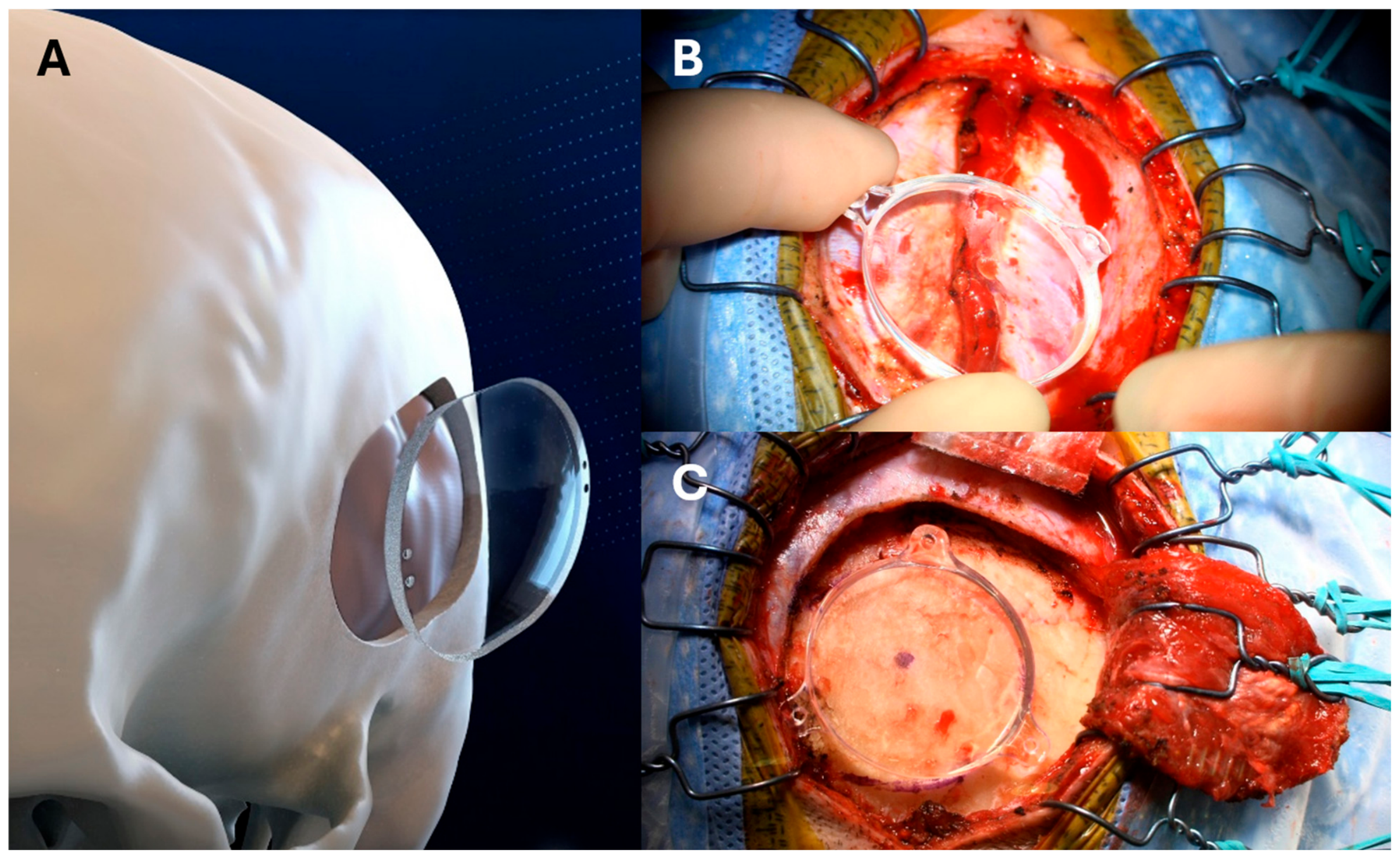
14]. TCUS resolves this constraint by substituting an acoustically transparent prosthetic implant for the autologous bone flap, creating a permanent, anatomically targeted acoustic window (
Figure 1) [
17,
58].
The conceptual and material science foundation was established by studies in 2019 demonstrating that polymethyl methacrylate (PMMA) cranioplasty implants produced ultrasound images with spatial fidelity comparable to CT and MRI, with negligible beam degradation or distortion across all imaging planes [
59]. Cadaveric work confirmed that both clear and opaque PMMA, as well as polyetheretherketone (PEEK), were sonolucent, while autologous bone and porous polyethylene were not [
44]. The first clinical application was reported in 2020 by Hadley et al., who described elective implantation of a sonolucent PMMA cranioplasty in a moyamoya patient undergoing STA-MCA bypass, enabling bedside confirmation of bypass patency postoperatively [
17]. In the same year, Flores et al. reported safety, feasibility, and patient-reported outcomes in a small series and estimated TCUS follow-up imaging costs at approximately
$900 per evaluation, compared to
$11,000 for conventional imaging modalities, a cost differential that, while based on estimated rather than formally validated cost-effectiveness data, suggests potential economic advantages for programs performing high volumes of bypass surgery [
58,
60].
5.2. Technical Aspects
The sonolucent PMMA implants currently in widest clinical use, most notably the ClearFit disc (Longeviti Neuro Solutions), are available in standardized dimensions: a 3 cm round implant for single-recipient anastomoses and a 4 × 5 cm disc for larger craniotomies with multiple potential recipient vessels [
16,
18]. Fixation techniques have evolved from early integral fixation wings to the current use of titanium plates and screws. When the proximity of the anastomosis to the overlying implant raises concern for graft compression, the inner surface can be thinned by drilling intraoperatively to create adequate clearance [
16,
61].
TCUS examinations can be performed using handheld or conventional ultrasound devices with a linear transducer placed directly over the implant [
58,
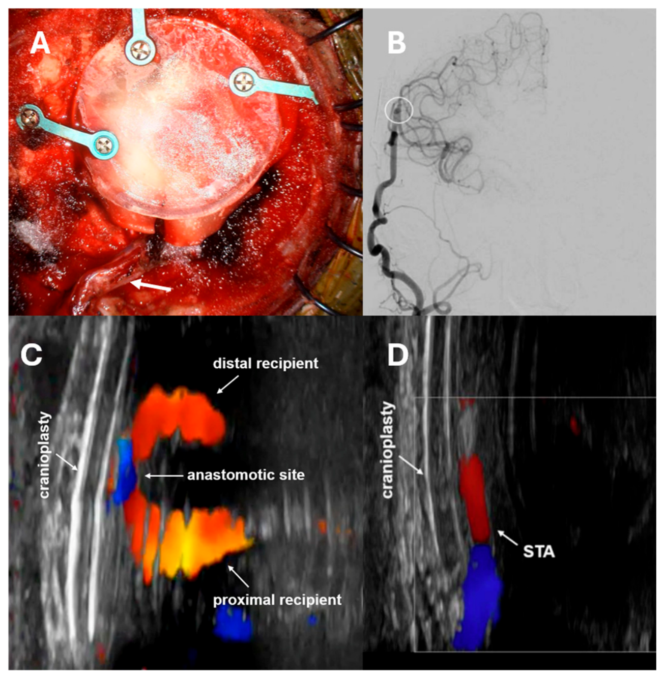
62]. Initial scanning establishes visualization of key anatomical landmarks: the implant itself, the extracranial donor STA passing beneath it, the anastomotic site, and the MCA proximal and distal to the anastomosis (
Figure 2) [
17]. Color Doppler permits direct real-time visualization of flow through the anastomosis, while spectral Doppler with angle correction enables waveform analysis and quantitative velocity measurement [
48]. Robust patency is characterized by strong, continuous, well-defined arterial waveforms in the STA with preserved diastolic flow and clear color signal through the anastomosis and into the recipient MCA territory [
16,
18,
48,
62].
Several practical and temporal constraints govern image acquisition. Because ultrasound does not penetrate air or fluid collections, the implant manufacturer recommends that the first postoperative ultrasound be deferred until at least 24 to 48 h after surgery in order to allow resorption of subgaleal and intracranial air and epithelialization of the surgical wound. TCUS is therefore generally not suitable for immediate intraoperative-to-early-postoperative monitoring in the first 24 h. Topical hemostatic agents, collagen-based overlays, and certain dural substitutes may further interfere with ultrasound penetration, and the fresh incision itself can be difficult to scan across, sometimes requiring additional gel as a standoff.
Since ultrasound does not transmit through bone, accurate probe alignment over the implant footprint is essential; acoustic shadowing at the implant margins can be used to confirm probe position. A low-frequency linear transducer (e.g., 9 MHz) with a “carotid” preset is often best suited to the shallow (1–3 cm) depth of EC-IC bypass imaging, whereas a sector (phased-array) probe is preferable for deeper neurovascular structures. Point-of-care and handheld systems can provide immediate bedside results when symptoms change or for routine surveillance, while higher-resolution radiology-console systems may be reserved for situations requiring superior image quality.
The primary technical challenge is prosthesis curvature, which can impair optimal transducer-scalp contact and image acquisition quality [
63]. Additional sources of artifact include early postoperative pneumocephalus, metallic plating systems, and dural sealants [
58,
62]. Despite these limitations, measurements of intracranial structures through PMMA cranioplasty have demonstrated excellent correlation with CT imaging in validation studies, and images acquired through the implant show spatial relationships superimposable to those obtained through direct dural contact [
59,
63].
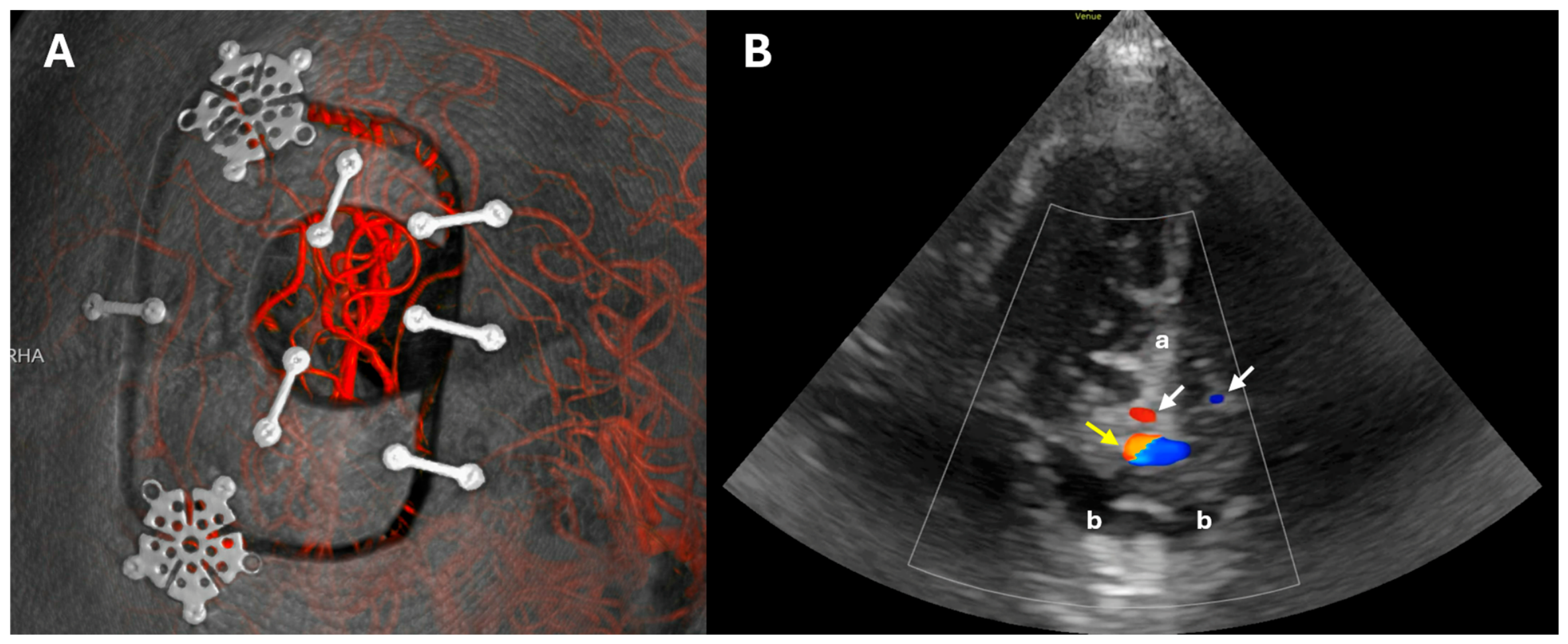
A distinction of clinical importance separates TCUS from conventional bedside Doppler without a sonolucent window. Standard bedside Doppler can detect an audible signal in the proximal STA but cannot differentiate between flow in the bypass limb and flow in non-anastomosed STA branches that may remain intact. A patent, non-bypassed STA branch will produce a reassuring Doppler signal even in the setting of complete bypass occlusion, potentially generating a dangerous false-positive assessment of graft patency [
17,
58]. TCUS, by directly imaging the anastomosis and the surrounding intracranial vasculature, resolves this issue (
Figure 3).
5.3. Multicenter Evaluation
The first systematic multicenter validation of TCUS for EC-IC bypass monitoring was provided by Salem et al. in 2023 [
18]. Aggregating data from five United States centers over a three-year period (2019–2022), this study enrolled 44 patients undergoing direct EC-IC bypass (the largest published cohort at that time) and was the first to establish generalizability of the technique across different institutions, surgeons, and patient populations.
Qualitative TCUS assessment of graft patency was technically feasible in 100% of cases, a finding that stands in favorable contrast to conventional TCD through native bone, which fails in 8–20% of patients due to inadequate acoustic windows depending on age, sex, and ethnicity. Intraoperative bypass confirmation employed multimodal assessment: microvascular Doppler in 90.9%, indocyanine green (ICG) videoangiography in 86.4%, and catheter angiography in 61.4% of cases, reflecting the real-world practice of complementary rather than exclusive reliance on any single modality [
18].
Postoperatively, 56.8% of patients received inpatient TCUS confirmation and 47.7% received outpatient surveillance, proportions representing early institutional adoption rather than an upper limit on feasible utilization. The absence of bypass failures in the cohort produced excellent clinical outcomes but also meant the study could not assess TCUS’ ability to detect a failing graft, the most clinically critical question for any surveillance modality.
The safety profile of the implant was reassuring: the overall complication rate was low, there were no implant-related adverse events, and the single surgical site infection was considered unrelated to the prosthetic material [
18]. This safety profile is comparable to or better than autologous bone flap replacement, addressing the theoretical concern that a synthetic implant would increase infection risk.
5.4. Longitudinal Performance and Correlation with Angiography
It should be emphasized that, in contrast to the comparatively well-established extracranial duplex and TCD literature, direct evidence for TCUS remains limited to a small number of single-center series, retrospective cohorts, and early multicenter experience, none of which is prospective or randomized. However, the most substantive evidence to date for long-term TCUS reliability and its concordance with formal angiography was published in 2025. This retrospective cohort study included 46 consecutive direct STA-MCA anastomoses in 40 patients at a single high-volume center (March 2021–May 2024) [
16]. All surgeries employed a PMMA sonolucent cranioplasty. This study addressed the specific gap left by prior work: the demonstration of TCUS performance not just in the immediate postoperative period, but longitudinally over years of follow-up, and with systematic comparison against formal angiography.
The study population was predominantly female (57.5%), with a mean age of 52.0 years; 55% underwent bypass for ischemic moyamoya disease and 35% for occlusive cerebrovascular disease. There were no cases of postoperative infection, and median clinical follow-up per anastomosis was 1.6 years.
Of the 40 patients, 32 (80%) underwent outpatient TCUS; the lower proportion in the overall cohort reflected evolving institutional practice and inconsistent documentation early in the study period rather than systematic exclusion. The first TCUS was performed at a median of 28.5 days (range 5–117 days), demonstrating patent bypass with robust flow in all 32 cases. A second TCUS was performed in 19 patients (41.3%) at a median of 8.4 months, with patency confirmed in all cases and robust flow demonstrated in 94.7%. Smaller subsets underwent third assessments (n = 5, median 1.2 years) and a fourth assessment (n = 1, 1.4 years), with patency and generally robust flow in all cases. The single case without robust flow at third assessment was the same patient who had demonstrated concerning findings at second assessment, described below.
Follow-up angiography was obtained in 41 of 46 anastomoses (89.1%) at a median of 1.1 years, demonstrating patency in 40 of 41 (97.6%). Among the 31 patients who underwent both TCUS and formal angiography, concordance was 100%, with no false-positive assessments of patency by TCUS. The single bypass occlusion in the cohort, which was identified by DSA after an ischemic stroke 42 days postoperatively, occurred before the patient’s first scheduled TCUS examination, and no TCUS was performed in this case [
16]. Thus, similar to the Salem et al. study [
18], the absence of bypass failures in the cohort again precluded the chance to test TCUS’ ability to detect a failing graft and remains a major gap in the literature.
Two cases illustrated TCUS’ capacity for hemodynamic characterization beyond binary patency assessment. In one, a second TCUS at 377 days demonstrated a qualitatively attenuated waveform with diminished color Doppler signal; DSA performed the same day confirmed a significantly narrowed anastomosis, and the patient remained asymptomatic on serial follow-up. In the other, a second TCUS identified a markedly reduced STA caliber, subsequently confirmed angiographically. Both cases demonstrate TCUS’ ability to detect evolving hemodynamic changes that correspond to angiographic findings and to prompt timely formal investigation, with both patients remaining clinically stable throughout.
5.5. Comparison with Conventional Imaging Modalities
TCUS offers several potential advantages relative to established bypass surveillance modalities. DSA provides unmatched spatial resolution and remains the reference standard for detailed anatomical characterization of anastomotic caliber, suture line integrity, and graft maturation. However, it is invasive, requires arterial catheterization, involves ionizing radiation and iodinated contrast, and carries procedural risks including stroke, arterial injury, and allergic reactions, attributes that limit its suitability for the serial surveillance that bypass patients require, often over many years or decades (
Table 1) [
16].
CTA offers a less invasive alternative but still involves radiation and contrast, restricting repeated use, particularly in younger patients and those with renal disease [
9,
48]. MRA avoids radiation, and advanced techniques such as arterial spin labeling-based vessel-selective 4D MRA have shown strong sensitivity and specificity for bypass visualization; however, MRA involves lengthy acquisition times, requires patient transport, and may have difficulty resolving smaller-caliber anastomoses or distinguishing subtle degrees of stenosis with the resolution of DSA [
11].
TCUS is noninvasive, radiation-free, requires no contrast, can be performed at the bedside or in the outpatient clinic, and can be repeated without cumulative risk or cost escalation [
16,
58]. The elimination of acoustic window dependency means that TCUS is technically feasible in virtually all patients who have received a sonolucent implant [
64,
65,
66]. The direct visualization of the anastomotic site provides anatomically specific information unavailable from extracranial duplex or TCD alone [
16]. The economic argument for TCUS appears favorable, particularly for programs with high bypass volumes requiring long-term serial follow-up; however, this rests on estimated imaging-cost comparisons rather than formal cost-effectiveness analysis, which has not yet been performed [
60].
The multicenter consistency of TCUS performance, showing 100% technical success across five centers, suggests the technique is robust and broadly generalizable rather than dependent on unique expertise at a specialized center [
18]. This is an important characteristic for any modality being considered for wider adoption. While prospective data are awaited, the available evidence supports a pragmatic, complementary role for TCUS within existing surveillance pathways.
Table 2 outlines clinical scenarios in which TCUS may reasonably serve as a first-line or interval surveillance tool, alongside those in which formal angiographic imaging remains necessary.
5.6. Limitations
An honest assessment of TCUS must acknowledge its current limitations. The most fundamental is spatial resolution: TCUS cannot provide the submillimeter anatomical detail of DSA, and it cannot characterize anastomotic caliber, suture line integrity, or the precise degree of stenosis at the level of resolution achievable with catheter angiography [
17,
58]. The two clinically important cases in the 2025 single-center series in which hemodynamic changes were detected qualitatively by TCUS both ultimately required DSA for definitive anatomical characterization [
16]. Thus, TCUS identified the problem but could not fully define it.
Further, the technique is operator-dependent, requiring training in both acquisition technique and image interpretation [
16]. Formal interobserver reliability data, standardized training curricula, and objective competency assessment metrics do not yet exist in the published literature. Current TCUS practice is predominantly qualitative, providing near-binary patency assessment supplemented by subjective waveform interpretation. The theoretical potential for quantitative TCUS flow measurement, including vessel diameter, peak systolic velocity, end-diastolic velocity, pulsatility index, and volumetric flow, has not yet been systematically realized in clinical practice, limiting TCUS to a more restricted diagnostic role than is in principle achievable [
64].
The additional cost of the PMMA implant relative to autologous bone flap replacement is a barrier to universal adoption. While a comprehensive cost analysis incorporating long-term surveillance imaging costs over the patient’s lifetime might favor the sonolucent implant given the repeated imaging avoided, this analysis has not yet been formally validated [
58,
60]. The implant is available only in standardized sizes, and anatomical constraints in some patients may limit optimal placement. For high-flow bypasses with long interposition graft courses, TCUS through a single acoustic window can assess only the intracranial anastomotic site, not the full length of the conduit. However, the more proximal segments can be assessed by ultrasound within the neck and deep to soft tissue.
Additionally, the published TCUS evidence base is largely drawn from moyamoya disease and occlusive cerebrovascular disease populations; systematic evaluation in the aneurysm bypass population, where hemodynamic profiles differ, has not yet been specifically reported [
16,
18,
58]. The total body of TCUS-specific literature remains small and is composed predominantly of retrospective single-center series and early multicenter experience, without prospective or comparative validation. Critically, the existing TCUS cohorts reported few or no graft failures; consequently, the sensitivity and specificity of TCUS for detecting true perioperative graft failure, arguably the most clinically important performance characteristic of any surveillance tool, remain essentially unestablished. Reported diagnostic metrics for sonographic bypass assessment (
Table 3) overwhelmingly describe prediction of patency or grading of bypass function rather than detection of failure, a distinction that should temper interpretation of the apparent concordance between TCUS and angiography. Lastly, as a narrative rather than systematic review, this article is also subject to selection bias in the studies cited and did not employ a formal risk-of-bias appraisal.
6. Future Directions
6.1. Quantitative Assessment and Protocol Standardization
The transition from qualitative to quantitative TCUS assessment represents the most important next step. Volumetric flow measurement, anastomotic diameter quantification, and standardized velocity metrics, comparable to those established for extracranial STA duplex, would transform TCUS from a patency surveillance tool into a comprehensive hemodynamic monitoring instrument. Correlation of quantitative Doppler-derived metrics with intraoperative flow data and long-term angiographic findings is an immediate research priority. If successful, TCUS quantitative thresholds analogous to those established for high-flow (>200 mL/min for hyperperfusion risk) and low-flow (Matsushima-grade discriminating thresholds) bypass monitoring by extracranial duplex could be validated for intracranial direct measurement.
Standardization of imaging acquisition protocols is a complementary requirement. Variability in transducer selection, frequency, Doppler angle correction technique, and measurement conventions limits inter-institutional comparability and prevents the establishment of universal reference ranges. Formal training programs and competency standards, comparable to those that exist for diagnostic carotid duplex ultrasonography, are needed as TCUS transitions from early adoption to routine practice. Three-dimensional reconstruction of sonographic images would additionally improve spatial assessment.
6.2. Artificial Intelligence Integration
Preliminary work has explored AI-assisted intracranial sonographic image analysis, with early studies demonstrating good concordance between clinician and algorithm-based image quality assessment (intraclass correlation coefficients of 0.829–0.839) [
67,
68]. The potential applications are broad: automated assessment of image acquisition adequacy, AI-assisted anatomical landmark identification, automated extraction of quantitative flow parameters, and longitudinal comparison of serial examinations to detect subtle interval changes [
69]. These capabilities would directly address operator dependency, the most important current limitation of TCUS, and could expand access to high-quality bypass surveillance beyond centers with specialized expertise.
7. Conclusions
Sonographic imaging of EC-IC bypass has evolved over four decades from indirect extracranial Doppler waveform assessment to TCUS. Each advance has expanded the temporal and anatomical reach of ultrasound monitoring and reduced dependence on invasive, radiation-exposing angiographic surveillance.
TCUS represents the most significant contemporary advance in this domain. By creating a permanent, surgically implanted acoustic window at the site of the anastomosis, it enables direct real-time visualization of bypass patency and hemodynamics throughout the postoperative course, from the immediate perioperative period through years of long-term follow-up, without radiation, contrast, or patient transport. The multicenter Salem et al. (2023) series demonstrated technical feasibility across institutions, and the longitudinal Grin et al. (2025) cohort established complete concordance with formal angiography over a median follow-up exceeding one year [
16,
18].
These data support TCUS as a reliable, cost-effective complement to formal angiography for serial bypass surveillance, with formal DSA reserved for cases requiring anatomical detail beyond TCUS resolution or for investigation of concerning ultrasound findings. The high hemodynamic stakes that characterize aneurysm bypass, where the graft may represent the sole source of perfusion for a dependent territory, amplify the clinical value of a reliable, repeatable, noninvasive monitoring tool.
However, key limitations remain. Current assessment is predominantly qualitative, the technique is operator-dependent, and systematic evidence from high-flow and aneurysm-specific bypass populations is limited. These limitations define the next steps in sonographic research: development and validation of standardized quantitative protocols, assessment of interobserver reliability and learning curves, AI-assisted image analysis, and prospective evaluation in the full spectrum of EC-IC bypass indications. As this evidence matures, TCUS appears well-positioned to assume a larger role in routine bypass surveillance, pending prospective validation, with angiographic imaging remaining the reference standard and the appropriate investigation for anatomically complex, hemodynamically equivocal, or symptomatic cases.
Author Contributions
E.A.G. and E.N.: Conceptualization, Investigation, Visualization, Writing (original draft), Writing (review & editing). All authors have read and agreed to the published version of the manuscript.
Funding
This research received no external funding.
Data Availability Statement
Due to this manuscript’s nature as a review article, no new data were created.
Conflicts of Interest
Erez Nossek is a consultant for Rapid Medical and Longeviti Neuro Solutions, serves on the medical board of Longeviti, and holds stock equity or options in Rapid Medical and Longeviti. Eric Grin reports no conflicts of interest. The authors have made deliberate effort to present the limitations, evidence gaps, and cost considerations of the discussed technology in a balanced manner.
Abbreviations
ACA, anterior cerebral artery; CTA, computed tomography angiography; DSA, digital subtraction angiography; EC-IC, extracranial–intracranial; ICA, internal carotid artery; MCA, middle cerebral artery; MRA, magnetic resonance angiography; PCA, posterior cerebral artery; PICA, posterior inferior cerebellar artery; PMMA, polymethyl methacrylate; PSV, peak systolic velocity; SCA, superior cerebellar artery; STA, superficial temporal artery; TCD, transcranial Doppler; TCUS, trans-sonolucent cranioplasty ultrasonography.
References
- Wessels, L.; Hecht, N.; Vajkoczy, P. Bypass in neurosurgery-indications and techniques. Neurosurg. Rev. 2019, 42, 389–393. [Google Scholar] [PubMed]
- Gonzalez, N.R.; Amin-Hanjani, S.; Bang, O.Y.; Coffey, C.; Du, R.; Fierstra, J.; Fraser, J.F.; Kuroda, S.; Tietjen, G.E.; Yaghi, S.; et al. Adult moyamoya disease and syndrome: Current perspectives and future directions: A scientific statement from the american heart association/american stroke association. Stroke 2023, 54, e465–e479. [Google Scholar] [CrossRef] [PubMed]
- Yoon, S.; Burkhardt, J.K.; Lawton, M.T. Long-term patency in cerebral revascularization surgery: An analysis of a consecutive series of 430 bypasses. J. Neurosurg. 2019, 131, 80–87. [Google Scholar] [CrossRef] [PubMed]
- Nussbaum, E.S.; Kallmes, K.M.; Lassig, J.P.; Goddard, J.K.; Madison, M.T.; Nussbaum, L.A. Cerebral revascularization for the management of complex intracranial aneurysms: A single-center experience. J. Neurosurg. 2019, 131, 1297–1307. [Google Scholar] [CrossRef] [PubMed]
- Kocaeli, H.; Andaluz, N.; Choutka, O.; Zuccarello, M. Use of radial artery grafts in extracranial-intracranial revascularization procedures. Neurosurg. Focus 2008, 24, E5. [Google Scholar] [PubMed]
- Dodier, P.; Wang, W.-T.; Hosmann, A.; Hirschmann, D.; Marik, W.; Frischer, J.M.; Gruber, A.; Rössler, K.; Bavinzski, G. Combined standard bypass and parent artery occlusion for management of giant and complex internal carotid artery aneurysms. J. NeuroInterv. Surg. 2022, 14, 593–598. [Google Scholar] [PubMed]
- Khan, N.R.; Elarjani, T.; Jamshidi, A.M.; Lu, V.M.; Silva, M.A.; Richardson, A.; Harrington, T.; Valdes, T.; Campo, N.; Krementz, N.; et al. Direct bypass surgery for moyamoya and steno-occlusive vasculopathy: Clinical outcomes, intraoperative blood flow analysis, long-term follow-up, and long-term bypass patency in a single surgeon case series of 162 procedures. World Neurosurg. 2022, 168, e500–e517. [Google Scholar] [PubMed]
- Hurth, H.; Hauser, T.-K.; Haas, P.; Wang, S.; Mengel, A.; Tatagiba, M.; Ernemann, U.; Khan, N.; Roder, C. Early post-operative ct-angiography imaging after ec-ic bypass surgery in moyamoya patients. Front. Neurol. 2021, 12, 655943. [Google Scholar] [PubMed]
- Langner, S.; Fleck, S.; Seipel, R.; Schroeder, H.W.; Hosten, N.; Kirsch, M. Perfusion ct scanning and ct angiography in the evaluation of extracranial-intracranial bypass grafts. J. Neurosurg. 2011, 114, 978–983. [Google Scholar] [CrossRef] [PubMed]
- Thines, L.; Agid, R.; Dehdashti, A.R.; da Costa, L.; Wallace, M.C.; Terbrugge, K.G.; Tymianski, M. Assessment of extracranial-intracranial bypass patency with 64-slice multidetector computerized tomography angiography. Neuroradiology 2009, 51, 505–515. [Google Scholar] [PubMed]
- Wang, M.; Yang, Y.; Wang, Y.; Li, M.; Zhang, J.; Zhang, B. Vessel-selective 4d mra based on asl might potentially show better performance than 3d tof mra for treatment evaluation in patients with intra-extracranial bypass surgery: A prospective study. Eur. Radiol. 2021, 31, 5263–5271, Correction in Eur. Radiol. 2021, 31, 7214. [Google Scholar] [PubMed]
- Chen, J.Y.; Ding, Y.H.; Li, Y.; Shi, S.S.; Chen, J.; Tu, X.K. Assessment of bypass patency using transcranial doppler sonography: Correlations with computerized tomography angiography findings in patients with moyamoya disease. Neurosurg. Rev. 2023, 46, 64. [Google Scholar] [CrossRef] [PubMed]
- Wang, G.; Zhang, X.; Wang, B.; Wen, Y.; Chen, S.; Liu, J.; Ke, B.; Wu, H.; Zhang, G.; Li, M.; et al. Flow evaluation of sta-mca bypass using quantitative ultrasonography: An alternative to standard angiography for follow up of bypass graft. J. Stroke Cerebrovasc. Dis. 2020, 29, 105000. [Google Scholar] [PubMed]
- Morton, R.P.; Abecassis, I.J.; Moore, A.E.; Kelly, C.M.; Levitt, M.R.; Kim, L.J.; Sekhar, L.N. The use of ultrasound for postoperative monitoring of cerebral bypass grafts: A technical report. J. Clin. Neurosci. 2017, 40, 169–174. [Google Scholar] [CrossRef] [PubMed]
- Lupetin, A.R.; Davis, D.A.; Beckman, I.; Dash, N. Transcranial doppler sonography. Part 1. Principles, technique, and normal appearances. Radiographics 1995, 15, 179–191. [Google Scholar] [CrossRef] [PubMed]
- Grin, E.A.; Sangwon, K.L.; Wiggan, D.D.; Negash, B.; Sharashidze, V.; Shapiro, M.; Raz, E.; Chung, C.; Baranoski, J.; Riina, H.A.; et al. Trans-sonolucent cranioplasty ultrasonography for long-term superficial temporal artery-middle cerebral artery bypass patency surveillance. Oper. Neurosurg. 2025. [Google Scholar] [CrossRef] [PubMed]
- Hadley, C.; North, R.; Srinivasan, V.; Kan, P.; Burkhardt, J.K. Elective sonolucent cranioplasty for real-time ultrasound monitoring of flow and patency of an extra- to intracranial bypass. J. Craniofac. Surg. 2020, 31, 622–624. [Google Scholar] [CrossRef] [PubMed]
- Salem, M.M.; Ravindran, K.; Hoang, A.N.; Doron, O.; Esparza, R.; Raper, D.; Jankowitz, B.T.; Tanweer, O.; Lopes, D.; Langer, D.; et al. Sonolucent cranioplasty in extracranial to intracranial bypass surgery: Early multicenter experience of 44 cases. Oper. Neurosurg. 2023, 25, 20–27. [Google Scholar] [CrossRef] [PubMed]
- Mehdorn, H.M.; Spira, G.; Jamjoom, Z.A.; John, V. Noninvasive evaluation of extra-/intracranial anastomoses by dynamic ct and doppler sonography. AJNR Am. J. Neuroradiol. 1983, 4, 857–860. [Google Scholar] [PubMed]
- Hyodo, A.; Mizukami, M.; Kawase, T.; Nagata, K.; Yunoki, K.; Yamaguchi, K. Postoperative evaluation of extracranial-intracranial arterial bypass by means of ultrasonic quantitative flow measurement and computed mapping of the electroencephalogram. Neurosurgery 1984, 15, 381–386. [Google Scholar] [CrossRef] [PubMed]
- Aaslid, R.; Markwalder, T.M.; Nornes, H. Noninvasive transcranial doppler ultrasound recording of flow velocity in basal cerebral arteries. J. Neurosurg. 1982, 57, 769–774. [Google Scholar] [CrossRef] [PubMed]
- Harders, A.; Gilsbach, J. Transcranial doppler sonography and its application in extracranial-intracranial bypass surgery. Neurol. Res. 1985, 7, 129–141. [Google Scholar] [PubMed]
- Harders, A.; Gilsbach, J. Transcranial doppler sonography in neurosurgery. Ultraschall Med. 1984, 5, 237–245. [Google Scholar] [PubMed]
- Bonow, R.H.; Young, C.C.; Bass, D.I.; Moore, A.; Levitt, M.R. Transcranial doppler ultrasonography in neurological surgery and neurocritical care. Neurosurg. Focus 2019, 47, E2. [Google Scholar] [CrossRef] [PubMed]
- Marinoni, M.; Ginanneschi, A.; Forleo, P.; Amaducci, L. Technical limits in transcranial doppler recording: Inadequate acoustic windows. Ultrasound Med. Biol. 1997, 23, 1275–1277. [Google Scholar] [PubMed]
- Lee, C.H.; Jeon, S.H.; Wang, S.J.; Shin, B.S.; Kang, H.G. Factors associated with temporal window failure in transcranial doppler sonography. Neurol. Sci. 2020, 41, 3293–3299. [Google Scholar] [CrossRef] [PubMed]
- Sloan, M.A.; Alexandrov, A.V.; Tegeler, C.H.; Spencer, M.P.; Caplan, L.R.; Feldmann, E.; Wechsler, L.R.; Newell, D.W.; Gomez, C.R.; Babikian, V.L.; et al. Assessment: Transcranial doppler ultrasonography: Report of the therapeutics and technology assessment subcommittee of the american academy of neurology. Neurology 2004, 62, 1468–1481. [Google Scholar] [PubMed]
- Saqqur, M.; Khan, K.; Derksen, C.; Alexandrov, A.; Shuaib, A. Transcranial doppler and transcranial color duplex in defining collateral cerebral blood flow. J. Neuroimaging 2018, 28, 455–476. [Google Scholar] [CrossRef] [PubMed]
- Nogueira, R.C.; Beishon, L.C.; Castro, P.; Claassen, J.; Minhas, J.S. Transcranial doppler in the era of personalized medicine: An important tool for the assessment of cerebrovascular function. Cerebrovasc. Dis. 2025, 55, 427–435. [Google Scholar] [CrossRef] [PubMed]
- Stendel, R.; Pietila, T.; Al Hassan, A.A.; Schilling, A.; Brock, M. Intraoperative microvascular doppler ultrasonography in cerebral aneurysm surgery. J. Neurol. Neurosurg. Psychiatry 2000, 68, 29–35. [Google Scholar] [CrossRef] [PubMed]
- Bailes, J.E.; Tantuwaya, L.S.; Fukushima, T.; Schurman, G.W.; Davis, D. Intraoperative microvascular doppler sonography in aneurysm surgery. Neurosurgery 1997, 40, 965–970; discussion 970–972. [Google Scholar] [CrossRef]
- Hui, P.-J.; Yan, Y.-H.; Zhang, S.-M.; Wang, Z.; Yu, Z.-Q.; Zhou, Y.-X.; Li, X.-D.; Cui, G.; Zhou, D.; Hui, G.-Z.; et al. Intraoperative microvascular doppler monitoring in intracranial aneurysm surgery. Chin. Med. J. (Engl.) 2013, 126, 2424–2429. [Google Scholar] [CrossRef] [PubMed]
- Badie, B.; Lee, F.T., Jr.; Pozniak, M.A.; Strother, C.M. Intraoperative sonographic assessment of graft patency during extracranial-intracranial bypass. AJNR Am. J. Neuroradiol. 2000, 21, 1457–1459. [Google Scholar] [PubMed]
- Charbel, F.T.; Hoffman, W.E.; Misra, M.; Ostergren, L. Ultrasonic perivascular flow probe: Technique and application in neurosurgery. Neurol. Res. 1998, 20, 439–442. [Google Scholar] [CrossRef] [PubMed]
- Friedman, J.A.; Anderson, R.E.; Meyer, F.B. Techniques of intraoperative cerebral blood flow measurement. Neurosurg. Focus 2000, 9, e4. [Google Scholar] [CrossRef] [PubMed]
- Amin-Hanjani, S.; Alaraj, A.; Charbel, F.T. Flow replacement bypass for aneurysms: Decision-making using intraoperative blood flow measurements. Acta Neurochir. 2010, 152, 1021–1032; discussion 1032. [Google Scholar] [CrossRef] [PubMed]
- Rustemi, O.; Amin-Hanjani, S.; Shakur, S.F.; Du, X.; Charbel, F.T. Donor selection in flow replacement bypass surgery for cerebral aneurysms: Quantitative analysis of long-term native donor flow sufficiency. Neurosurgery 2016, 78, 332–341; discussion 341–342. [Google Scholar] [CrossRef]
- Sekhar, L.N.; Shenoy, V.S.; Sen, R.D.; Miller, C.A.; Montoure, A.; Ghodke, B.V.; Kim, L.J. Current indications, trends, and long-term results of 233 bypasses to treat complex intracranial aneurysms: A location-specific analysis. Neurosurgery 2024, 95, 1106–1116. [Google Scholar] [PubMed]
- Amin-Hanjani, S.; Du, X.; Mlinarevich, N.; Meglio, G.; Zhao, M.; Charbel, F.T. The cut flow index: An intraoperative predictor of the success of extracranial-intracranial bypass for occlusive cerebrovascular disease. Neurosurgery 2005, 56, 75–85. [Google Scholar] [CrossRef]
- Goren, O.; Monteith, S.J.; Hadani, M.; Bakon, M.; Harnof, S. Modern intraoperative imaging modalities for the vascular neurosurgeon treating intracerebral hemorrhage. Neurosurg. Focus 2013, 34, E2. [Google Scholar] [CrossRef] [PubMed]
- McGah, P.M.; Nerva, J.D.; Morton, R.P.; Barbour, M.C.; Levitt, M.R.; Mourad, P.D.; Kim, L.J.; Aliseda, A. In vitro validation of endovascular doppler-derived flow rates in models of the cerebral circulation. Physiol. Meas. 2015, 36, 2301–2317. [Google Scholar] [PubMed]
- Haaland, K.; Karlsson, B.; Skovlund, E.; Thoresen, M. Simultaneous measurements of cerebral circulation with electromagnetic flowmetry and doppler ultrasound velocity in the newborn pig. Pediatr. Res. 1994, 36, 601–606. [Google Scholar] [CrossRef] [PubMed][Green Version]
- Olislagers-De Slegte, R.G.; Smeets, R.W.; Valk, J.; Crezee, F. Ultrasound in follow-up of the postoperative brain. Preliminary report on six cases. Neuroradiology 1984, 26, 267–272. [Google Scholar] [PubMed]
- Belzberg, M.; Ben Shalom, N.; Yuhanna, E.; Manbachi, A.; Tekes, A.; Huang, J.; Brem, H.; Gordon, C.R. Sonolucent cranial implants: Cadaveric study and clinical findings supporting diagnostic and therapeutic transcranioplasty ultrasound. J. Craniofac. Surg. 2019, 30, 1456–1461. [Google Scholar] [CrossRef] [PubMed]
- Morton, R.P.; Moore, A.E.; Barber, J.; Tariq, F.; Hare, K.; Ghodke, B.; Kim, L.J.; Sekhar, L.N. Monitoring flow in extracranial-intracranial bypass grafts using duplex ultrasonography: A single-center experience in 80 grafts over 8 years. Neurosurgery 2014, 74, 62–70. [Google Scholar] [PubMed]
- Yamaguchi, K.; Kawamata, T.; Kawashima, A.; Hori, T.; Okada, Y. Incidence and predictive factors of cerebral hyperperfusion after extracranial-intracranial bypass for occlusive cerebrovascular diseases. Neurosurgery 2010, 67, 1548–1554; discussion 1554. [Google Scholar] [CrossRef] [PubMed]
- Teo, K.; Choy, D.K.; Lwin, S.; Ning, C.; Yeo, T.T.; Shen, L.; Chong, V.F.; Teoh, H.L.; Seet, R.C.; Chan, B.P.; et al. Cerebral hyperperfusion syndrome after superficial temporal artery-middle cerebral artery bypass for severe intracranial steno-occlusive disease: A case control study. Neurosurgery 2013, 72, 936–942; discussion 942–943. [Google Scholar] [PubMed]
- Kim, J.H.; Ha, S.-K.; Jin, S.-W.; Lee, H.-B.; Kim, S.-D.; Kim, S.-H.; Lim, D.-J. Color doppler ultrasonography for predicting the patency of anastomosis after superficial temporal to middle cerebral artery bypass surgery. Acta Neurochir. 2021, 163, 1503–1513. [Google Scholar] [CrossRef] [PubMed]
- Nakamizo, A.; Inoue, T.; Kikkawa, Y.; Uda, K.; Hirata, Y.; Okamura, K.; Yasaka, M.; Okada, Y. Postoperative evaluation of changes in extracranial-intracranial bypass graft using superficial temporal artery duplex ultrasonography. AJNR Am. J. Neuroradiol. 2009, 30, 900–905. [Google Scholar] [CrossRef] [PubMed]
- Wu, M.; Huang, Z.; Zhang, D.; Wang, L.; Sun, J.; Wang, S.; Zhao, Y.; Zhao, J. Color doppler hemodynamic study of the superficial temporal arteries in superficial temporal artery-middle cerebral artery (sta-mca) bypass surgery for moyamoya disease. World Neurosurg. 2011, 75, 258–263. [Google Scholar] [PubMed]
- Umemura, A.; Yamada, K.; Masago, A.; Kanda, Y.; Matsumoto, T.; Shimazu, N. Hemodynamic flow patterns evaluated by transcranial color-coded duplex sonography after sta-mca bypass for internal carotid artery occlusion. Cerebrovasc. Dis. 2002, 14, 143–147. [Google Scholar] [PubMed]
- Otsuka, M.; Kikkawa, Y.; Kamide, T.; Suzuki, K.; Shibata, A.; Ikeda, T.; Kurita, H. Factors related to improvement of cerebrovascular reserve after superficial temporal artery to middle cerebral artery anastomosis for patients with atherosclerotic steno-occlusive disease. Acta Neurochir. 2019, 161, 799–805. [Google Scholar] [CrossRef] [PubMed]
- Low, S.W.; Teo, K.; Lwin, S.; Yeo, L.L.L.; Paliwal, P.R.; Ahmad, A.; Sinha, A.K.; Teoh, H.L.; Wong, L.Y.H.; Chong, V.F.; et al. Improvement in cerebral hemodynamic parameters and outcomes after superficial temporal artery-middle cerebral artery bypass in patients with severe stenoocclusive disease of the intracranial internal carotid or middle cerebral arteries. J. Neurosurg. 2015, 123, 662–669. [Google Scholar] [CrossRef] [PubMed]
- Gerhard-Herman, M.; Gardin, J.M.; Jaff, M.; Mohler, E.; Roman, M.; Naqvi, T.Z. Guidelines for noninvasive vascular laboratory testing: A report from the american society of echocardiography and the society for vascular medicine and biology. Vasc. Med. 2006, 11, 183–200. [Google Scholar] [CrossRef] [PubMed]
- Grin, E.A.; Wiggan, D.D.; Sangwon, K.L.; Baranoski, J.; Sharashidze, V.; Shapiro, M.; Raz, E.; Chung, C.; Nelson, P.K.; Riina, H.A.; et al. Interrupted versus running sutures for superficial temporal artery to middle cerebral artery cranial bypass. Oper. Neurosurg. 2024, 29, 367–373. [Google Scholar] [CrossRef] [PubMed]
- Matsukawa, H.; Tanikawa, R.; Kamiyama, H.; Tsuboi, T.; Noda, K.; Ota, N.; Miyata, S.; Takeda, R.; Tokuda, S. Graft occlusion and graft size changes in complex internal carotid artery aneurysm treated by extracranial to intracranial bypass using high-flow grafts with therapeutic internal carotid artery occlusion. Neurosurgery 2017, 81, 672–679. [Google Scholar] [CrossRef] [PubMed]
- Sebök, M.; Höbner, L.M.; Grob, A.; Fierstra, J.; Schubert, T.; Wegener, S.; Luft, A.R.; Kulcsár, Z.; Regli, L.; Esposito, G. Flow capacity of a superficial temporal artery as a donor in a consecutive series of 100 patients with superficial temporal artery-middle cerebral artery bypass. J. Neurosurg. 2025, 142, 62–69. [Google Scholar] [PubMed]
- Flores, A.R.; Srinivasan, V.M.; Seeley, J.; Huggins, C.; Kan, P.; Burkhardt, J.K. Safety, feasibility, and patient-rated outcome of sonolucent cranioplasty in extracranial-intracranial bypass surgery to allow for transcranioplasty ultrasound assessment. World Neurosurg. 2020, 144, e277–e284. [Google Scholar] [PubMed]
- Belzberg, M.; Ben Shalom, N.; Lu, A.; Yuhanna, E.; Manbachi, A.; Tekes, A.; Huang, J.; Brem, H.; Gordon, C. Transcranioplasty ultrasound through a sonolucent cranial implant made of polymethyl methacrylate: Phantom study comparing ultrasound, computed tomography, and magnetic resonance imaging. J. Craniofac. Surg. 2019, 30, e626–e629. [Google Scholar] [CrossRef] [PubMed]
- O’Malley, G.R., Jr.; Cassimatis, N.D.; Maggio, J.; Patel, P.; Patel, N.V. Sonolucent cranial implants: A window into the future of management of neurosurgical patients? A systematic review and cost analysis. World Neurosurg. 2024, 181, e848–e855. [Google Scholar] [PubMed]
- Grin, E.A.; Shapiro, M.; Raz, E.; Sharashidze, V.; Chung, C.; Rutledge, C.; Baranoski, J.; Riina, H.A.; Pacione, D.; Nossek, E. Neurovascular pathology in intracranial mucormycosis: Treatment by cranial bypass and literature review. Oper. Neurosurg. 2026, 30, 147–152. [Google Scholar] [PubMed]
- Rossitto, C.P.; Devarajan, A.; Zhang, J.Y.; Benowitz, R.; Price, G.; Ali, M.; Tabani, H.; Hardigan, T.; Liang, J.W.; Dangayach, N.S.; et al. Transcranioplasty ultrasonography through a sonolucent prosthesis: A review of feasibility, safety, and benefits. World Neurosurg. 2023, 178, 152–161.e1. [Google Scholar] [CrossRef] [PubMed]
- Spena, G.; Guerrini, F.; Grimod, G.; Salmaggi, A.; Mazzeo, L.A. Polymethyl methacrylate cranioplasty is an effective ultrasound window to explore intracranial structures: Preliminary experience and future perspectives. World Neurosurg. 2019, 127, e1013–e1019. [Google Scholar] [CrossRef] [PubMed]
- Esmeraldo, M.A.; de Melo, E.R.; Fritzen, M.; Braga, K.; Brunetto, M.F.; Ferreira, P.S.C.; Chiovatto, E.D.; Chiovatto, R.D.; Freeman, C.W.; Gomes, A.C.; et al. Updates on adult transcranial doppler, gray-scale, and contrast-enhanced us techniques. Radiographics 2026, 46, e250182. [Google Scholar] [CrossRef] [PubMed]
- Brisson, R.T.; Santos, R.d.S.A.; Stefano, L.H.S.S.; Barreira, C.M.A.; Arruda, J.F.d.L.; Dias, F.A.; Camilo, M.R.; Pontes-Neto, O.M. Association between tomographic characteristics of the temporal bone and transtemporal window quality on transcranial color doppler ultrasound in patients with stroke or transient ischemic attack. Ultrasound Med. Biol. 2021, 47, 511–516. [Google Scholar] [CrossRef] [PubMed]
- Grin, E.A.; Baranoski, J.; Rutledge, C.; Wiggan, D.D.; Chung, C.; Raz, E.; Sharashidze, V.; Shapiro, M.; Riina, H.A.; Zhang, C.; et al. Cranial bypass for occlusive carotid dissection in osteogenesis imperfecta: Illustrative case. J. Neurosurg. Case Lessons 2025, 10, CASE25378. [Google Scholar] [CrossRef] [PubMed]
- Mozzini, C.; Lucchini, G.; Bertogna, M.; Casadei, A.; Pagani, M. Artificial intelligence in heart and brain ultrasound: The least you need to know. Intern. Emerg. Med. 2025, 21, 749–758. [Google Scholar] [CrossRef] [PubMed]
- Hsu, K.-C.; Lin, C.-H.; Johnson, K.R.; Liu, C.-H.; Chang, T.-Y.; Huang, K.-L.; Fann, Y.-C.; Lee, T.-H. Autodetect extracranial and intracranial artery stenosis by machine learning using ultrasound. Comput. Biol. Med. 2020, 116, 103569. [Google Scholar] [CrossRef] [PubMed]
- Du, B.; Wang, J.; Zheng, H.; Xiao, C.; Fang, S.; Lu, M.; Mao, R. A novel transcranial ultrasound imaging method with diverging wave transmission and deep learning approach. Comput. Methods Programs Biomed. 2020, 186, 105308. [Google Scholar] [CrossRef] [PubMed]
| Disclaimer/Publisher’s Note: The statements, opinions and data contained in all publications are solely those of the individual author(s) and contributor(s) and not of MDPI and/or the editor(s). MDPI and/or the editor(s) disclaim responsibility for any injury to people or property resulting from any ideas, methods, instructions or products referred to in the content. |