Senescent Astrocytes: A New Player in Brain Aging and Cognitive Decline

 , , ,
, , ,
Abstract
1. Introduction
2. Molecular Mechanisms of Cellular Senescence
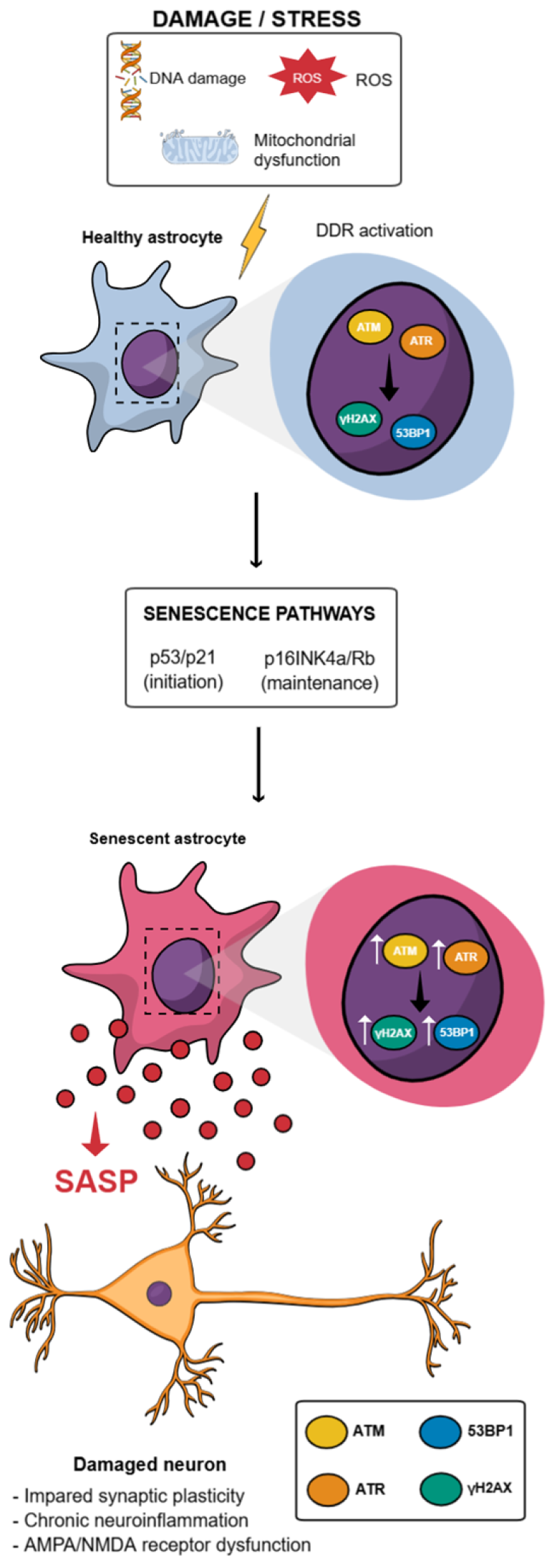
3. DNA Damage Response-Driven Cell Cycle Arrest in Senescence: The p53/p21 and p16INK4a/Rb Pathways
4. Chronic NF-κB Activation Contributes to the SASP
5. mTOR Signaling and Metabolic Reprogramming in Senescence
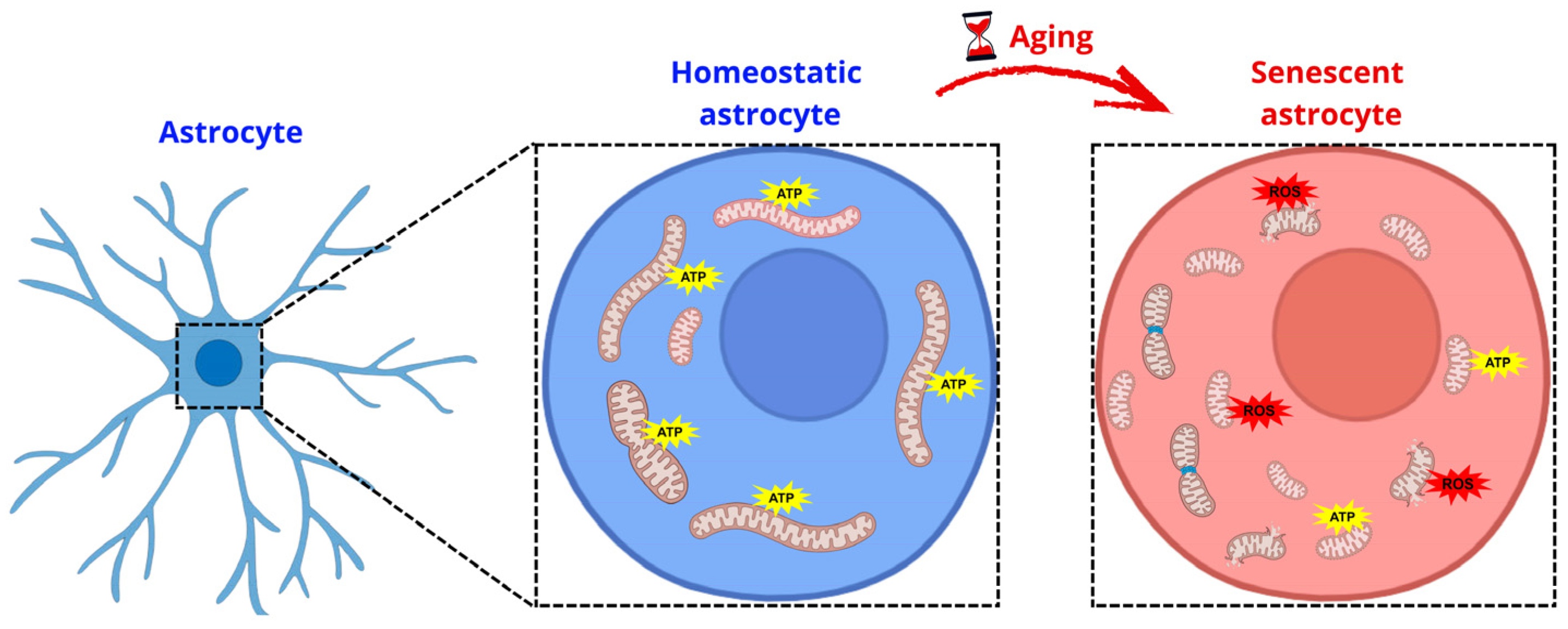
6. Astrocytic Mitochondrial Dysfunction in Aging and Cellular Senescence
7. Astrocyte Senescence: Impaired Synaptic Function
8. Conclusions
Author Contributions
Funding
Institutional Review Board Statement
Informed Consent Statement
Data Availability Statement
Acknowledgments
Conflicts of Interest
Abbreviations
| Aβ | Amyloid-beta |
| ATM | Ataxia Telangiectasia Mutated |
| ATR | Ataxia Telangiectasia and Rad3-related |
| BBB | Blood–Brain Barrier |
| CDK | Cyclin-dependent kinase |
| CNS | Central Nervous System |
| DDR | DNA Damage Response |
| DRP1 | Dynamin-related protein 1 |
| EAAT1 | Excitatory Amino Acid Transporter 1 (GLAST) |
| EAAT2 | Excitatory Amino Acid Transporter 2 (GLT-1) |
| IKK | IκB kinase |
| IL-1α | Interleukin-1 alpha |
| IL-1β | Interleukin-1 beta |
| IL-6 | Interleukin-6 |
| IL-8 | Interleukin-8 |
| LTP | Long-term potentiation |
| MFN1 | Mitofusin 1 |
| MFN2 | Mitofusin 2 |
| MMP | Matrix metalloproteinase |
| mTOR | mechanistic Target of Rapamycin |
| mTORC1 | mechanistic Target of Rapamycin Complex 1 |
| mTORC2 | mechanistic Target of Rapamycin Complex 2 |
| PDGF-AA | Platelet-Derived Growth Factor-AA |
| Rb | Retinoblastoma protein |
| ROS | Reactive Oxygen Species |
| SA-β-gal | Senescence-associated β-galactosidase |
| SASP | Senescence-Associated Secretory Phenotype |
| TGF-β | Transforming Growth Factor-β |
| TIMP-1 | Tissue Inhibitor of Metalloproteinases-1 |
| TNF-α | Tumor Necrosis Factor-α |
| 53BP1 | p53-binding protein 1 |
| γ-H2AX | gamma-H2AX (phosphorylated H2A.X) |
References
- Verkhratsky, A.; Nedergaard, M. Physiology of Astroglia. Physiol. Rev. 2018, 98, 239–389. [Google Scholar] [CrossRef] [Scilit] [PubMed]
- Bolanos, J.P.; Magistretti, P.J. The neuron-astrocyte metabolic unit as a cornerstone of brain energy metabolism in health and disease. Nat. Metab. 2025, 7, 2414–2423. [Google Scholar] [CrossRef] [Scilit]
- Manu, D.R.; Slevin, M.; Barcutean, L.; Forro, T.; Boghitoiu, T.; Balasa, R. Astrocyte Involvement in Blood-Brain Barrier Function: A Critical Update Highlighting Novel, Complex, Neurovascular Interactions. Int. J. Mol. Sci. 2023, 24, 17146. [Google Scholar] [CrossRef] [Scilit]
- Lee, S.Y.; Chung, W.S. Astrocytic crosstalk with brain and immune cells in healthy and diseased conditions. Curr. Opin. Neurobiol. 2024, 84, 102840. [Google Scholar] [CrossRef] [Scilit]
- Bouvier, D.S.; Fixemer, S.; Heurtaux, T.; Jeannelle, F.; Frauenknecht, K.B.M.; Mittelbronn, M. The Multifaceted Neurotoxicity of Astrocytes in Ageing and Age-Related Neurodegenerative Diseases: A Translational Perspective. Front. Physiol. 2022, 13, 814889. [Google Scholar] [CrossRef] [Scilit] [PubMed]
- Boisvert, M.M.; Erikson, G.A.; Shokhirev, M.N.; Allen, N.J. The Aging Astrocyte Transcriptome from Multiple Regions of the Mouse Brain. Cell Rep. 2018, 22, 269–285. [Google Scholar] [CrossRef] [Scilit]
- Matias, I.; Morgado, J.; Gomes, F.C.A. Astrocyte Heterogeneity: Impact to Brain Aging and Disease. Front. Aging Neurosci. 2019, 11, 59. [Google Scholar] [CrossRef] [Scilit]
- Roger, L.; Tomas, F.; Gire, V. Mechanisms and Regulation of Cellular Senescence. Int. J. Mol. Sci. 2021, 22, 13173. [Google Scholar] [CrossRef] [Scilit]
- Huang, W.; Hickson, L.J.; Eirin, A.; Kirkland, J.L.; Lerman, L.O. Cellular senescence: The good, the bad and the unknown. Nat. Rev. Nephrol. 2022, 18, 611–627. [Google Scholar] [CrossRef] [Scilit]
- Melo Dos Santos, L.S.; Trombetta-Lima, M.; Eggen, B.; Demaria, M. Cellular senescence in brain aging and neurodegeneration. Ageing Res. Rev. 2024, 93, 102141. [Google Scholar] [CrossRef] [Scilit] [PubMed]
- Ogrodnik, M. Cellular aging beyond cellular senescence: Markers of senescence prior to cell cycle arrest in vitro and in vivo. Aging Cell 2021, 20, e13338. [Google Scholar] [CrossRef] [Scilit] [PubMed]
- Salminen, A.; Kauppinen, A.; Kaarniranta, K. Emerging role of NF-kappaB signaling in the induction of senescence-associated secretory phenotype (SASP). Cell. Signal. 2012, 24, 835–845. [Google Scholar] [CrossRef] [Scilit] [PubMed]
- Coppe, J.P.; Desprez, P.Y.; Krtolica, A.; Campisi, J. The senescence-associated secretory phenotype: The dark side of tumor suppression. Annu. Rev. Pathol. 2010, 5, 99–118. [Google Scholar] [CrossRef] [Scilit]
- Di Micco, R.; Krizhanovsky, V.; Baker, D.; d’Adda di Fagagna, F. Cellular senescence in ageing: From mechanisms to therapeutic opportunities. Nat. Rev. Mol. Cell Biol. 2021, 22, 75–95. [Google Scholar] [CrossRef] [Scilit]
- Zhu, J.; Wu, C.; Yang, L. Cellular senescence in Alzheimer’s disease: From physiology to pathology. Transl. Neurodegener. 2024, 13, 55. [Google Scholar] [CrossRef] [Scilit] [PubMed]
- Mohamad Kamal, N.S.; Safuan, S.; Shamsuddin, S.; Foroozandeh, P. Aging of the cells: Insight into cellular senescence and detection Methods. Eur. J. Cell Biol. 2020, 99, 151108. [Google Scholar] [CrossRef] [Scilit]
- Wang, B.; Han, J.; Elisseeff, J.H.; Demaria, M. The senescence-associated secretory phenotype and its physiological and pathological implications. Nat. Rev. Mol. Cell Biol. 2024, 25, 958–978. [Google Scholar] [CrossRef] [Scilit]
- Correia-Melo, C.; Marques, F.D.; Anderson, R.; Hewitt, G.; Hewitt, R.; Cole, J.; Carroll, B.M.; Miwa, S.; Birch, J.; Merz, A.; et al. Mitochondria are required for pro-ageing features of the senescent phenotype. EMBO J. 2016, 35, 724–742. [Google Scholar] [CrossRef] [Scilit]
- Victorelli, S.; Salmonowicz, H.; Chapman, J.; Martini, H.; Vizioli, M.G.; Riley, J.S.; Cloix, C.; Hall-Younger, E.; Machado Espindola-Netto, J.; Jurk, D.; et al. Apoptotic stress causes mtDNA release during senescence and drives the SASP. Nature 2023, 622, 627–636. [Google Scholar] [CrossRef] [Scilit]
- Bitto, A.; Sell, C.; Crowe, E.; Lorenzini, A.; Malaguti, M.; Hrelia, S.; Torres, C. Stress-induced senescence in human and rodent astrocytes. Exp. Cell Res. 2010, 316, 2961–2968. [Google Scholar] [CrossRef] [Scilit]
- Willis, C.M.; Nicaise, A.M.; Bongarzone, E.R.; Givogri, M.; Reiter, C.R.; Heintz, O.; Jellison, E.R.; Sutter, P.A.; TeHennepe, G.; Ananda, G.; et al. Astrocyte Support for Oligodendrocyte Differentiation can be Conveyed via Extracellular Vesicles but Diminishes with Age. Sci. Rep. 2020, 10, 828. [Google Scholar] [CrossRef] [Scilit]
- Preininger, M.K.; Zaytseva, D.; Lin, J.M.; Kaufer, D. Blood-brain barrier dysfunction promotes astrocyte senescence through albumin-induced TGFbeta signaling activation. Aging Cell 2023, 22, e13747. [Google Scholar] [CrossRef] [Scilit]
- Marques, M.; Hayashide, L.S.; Amorim, P.; Fernandes, B.M.; Araujo, A.P.B.; Messor, D.F.; Leocadio, V.E.; Pessoa, B.; Correa, J.; Villablanca, C.; et al. Doxorubicin Induces a Senescent Phenotype in Murine and Human Astrocytes. J. Neurochem. 2025, 169, e70177. [Google Scholar] [CrossRef] [Scilit]
- Matias, I.; Diniz, L.P.; Damico, I.V.; Araujo, A.P.B.; Neves, L.D.S.; Vargas, G.; Leite, R.E.P.; Suemoto, C.K.; Nitrini, R.; Jacob-Filho, W.; et al. Loss of lamin-B1 and defective nuclear morphology are hallmarks of astrocyte senescence in vitro and in the aging human hippocampus. Aging Cell 2022, 21, e13521. [Google Scholar] [CrossRef] [Scilit]
- Diniz, L.P.; Araujo, A.P.B.; Carvalho, C.F.; Matias, I.; de Sa Hayashide, L.; Marques, M.; Pessoa, B.; Andrade, C.B.V.; Vargas, G.; Queiroz, D.D.; et al. Accumulation of damaged mitochondria in aging astrocytes due to mitophagy dysfunction: Implications for susceptibility to mitochondrial stress. Biochim. Biophys. Acta Mol. Basis Dis. 2024, 1870, 167470. [Google Scholar] [CrossRef] [Scilit]
- Simmnacher, K.; Krach, F.; Schneider, Y.; Alecu, J.E.; Mautner, L.; Klein, P.; Roybon, L.; Prots, I.; Xiang, W.; Winner, B. Unique signatures of stress-induced senescent human astrocytes. Exp. Neurol. 2020, 334, 113466. [Google Scholar] [CrossRef] [Scilit]
- Turnquist, C.; Beck, J.A.; Horikawa, I.; Obiorah, I.E.; Von Muhlinen, N.; Vojtesek, B.; Lane, D.P.; Grunseich, C.; Chahine, J.J.; Ames, H.M.; et al. Radiation-induced astrocyte senescence is rescued by Delta133p53. Neuro-oncology 2019, 21, 474–485. [Google Scholar] [CrossRef] [Scilit]
- Hou, J.; Cui, C.; Kim, S.; Sung, C.; Choi, C. Ginsenoside F1 suppresses astrocytic senescence-associated secretory phenotype. Chem.-Biol. Interact. 2018, 283, 75–83. [Google Scholar] [CrossRef] [Scilit] [PubMed]
- Limbad, C.; Oron, T.R.; Alimirah, F.; Davalos, A.R.; Tracy, T.E.; Gan, L.; Desprez, P.Y.; Campisi, J. Astrocyte senescence promotes glutamate toxicity in cortical neurons. PLoS ONE 2020, 15, e0227887. [Google Scholar] [CrossRef] [Scilit] [PubMed]
- Kim, D.; Yoo, S.H.; Yeon, G.B.; Oh, S.S.; Shin, W.H.; Kang, H.C.; Lee, C.K.; Kim, H.W.; Kim, D.S. Senescent Astrocytes Derived from Human Pluripotent Stem Cells Reveal Age-Related Changes and Implications for Neurodegeneration. Aging Dis. 2024, 16, 1709–1731. [Google Scholar] [CrossRef] [Scilit] [PubMed]
- Bang, M.; Kim, D.G.; Gonzales, E.L.; Kwon, K.J.; Shin, C.Y. Etoposide Induces Mitochondrial Dysfunction and Cellular Senescence in Primary Cultured Rat Astrocytes. Biomol. Ther. 2019, 27, 530–539. [Google Scholar] [CrossRef] [Scilit]
- Bang, M.; Yang, S.J.; Ahn, T.; Han, S.H.; Shin, C.Y.; Kwon, K.J. Minoxidil Regulates Aging-Like Phenotypes in Rat Cortical Astrocytes In Vitro. Biomol. Ther. 2023, 31, 116–126. [Google Scholar] [CrossRef] [Scilit]
- Crowe, E.P.; Tuzer, F.; Gregory, B.D.; Donahue, G.; Gosai, S.J.; Cohen, J.; Leung, Y.Y.; Yetkin, E.; Nativio, R.; Wang, L.S.; et al. Changes in the Transcriptome of Human Astrocytes Accompanying Oxidative Stress-Induced Senescence. Front. Aging Neurosci. 2016, 8, 208. [Google Scholar] [CrossRef] [Scilit]
- Araujo, A.P.B.; Vargas, G.; Hayashide, L.S.; Matias, I.; Andrade, C.B.V.; de Carvalho, J.J.; Gomes, F.C.A.; Diniz, L.P. Aging promotes an increase in mitochondrial fragmentation in astrocytes. Front. Cell. Neurosci. 2024, 18, 1496163. [Google Scholar] [CrossRef] [Scilit] [PubMed]
- Anderson, M.A.; Ao, Y.; Sofroniew, M.V. Heterogeneity of reactive astrocytes. Neurosci. Lett. 2014, 565, 23–29. [Google Scholar] [CrossRef] [Scilit]
- Liddelow, S.A.; Barres, B.A. Reactive Astrocytes: Production, Function, and Therapeutic Potential. Immunity 2017, 46, 957–967. [Google Scholar] [CrossRef] [Scilit]
- Clayton, B.L.L.; Liddelow, S.A. Heterogeneity of Astrocyte Reactivity. Annu. Rev. Neurosci. 2025, 48, 231–249. [Google Scholar] [CrossRef] [Scilit]
- Suda, Y.; Nakashima, T.; Matsumoto, H.; Sato, D.; Nagano, S.; Mikata, H.; Yoshida, S.; Tanaka, K.; Hamada, Y.; Kuzumaki, N.; et al. Normal aging induces PD-1-enriched exhausted microglia and A1-like reactive astrocytes in the hypothalamus. Biochem. Biophys. Res. Commun. 2021, 541, 22–29. [Google Scholar] [CrossRef] [Scilit]
- Clarke, L.E.; Liddelow, S.A.; Chakraborty, C.; Munch, A.E.; Heiman, M.; Barres, B.A. Normal aging induces A1-like astrocyte reactivity. Proc. Natl. Acad. Sci. USA 2018, 115, E1896–E1905. [Google Scholar] [CrossRef] [Scilit]
- Cohen, J.; Torres, C. Astrocyte senescence: Evidence and significance. Aging Cell 2019, 18, e12937. [Google Scholar] [CrossRef] [Scilit] [PubMed]
- Muwanigwa, M.N.; Modamio-Chamarro, J.; Antony, P.M.A.; Gomez-Giro, G.; Kruger, R.; Bolognin, S.; Schwamborn, J.C. Alpha-synuclein pathology is associated with astrocyte senescence in a midbrain organoid model of familial Parkinson’s disease. Mol. Cell. Neurosci. 2024, 128, 103919. [Google Scholar] [CrossRef] [Scilit]
- Verma, D.K.; Seo, B.A.; Ghosh, A.; Ma, S.X.; Hernandez-Quijada, K.; Andersen, J.K.; Ko, H.S.; Kim, Y.H. Alpha-Synuclein Preformed Fibrils Induce Cellular Senescence in Parkinson’s Disease Models. Cells 2021, 10, 1694. [Google Scholar] [CrossRef] [Scilit] [PubMed]
- Gaikwad, S.; Puangmalai, N.; Bittar, A.; Montalbano, M.; Garcia, S.; McAllen, S.; Bhatt, N.; Sonawane, M.; Sengupta, U.; Kayed, R. Tau oligomer induced HMGB1 release contributes to cellular senescence and neuropathology linked to Alzheimer’s disease and frontotemporal dementia. Cell Rep. 2021, 36, 109419. [Google Scholar] [CrossRef] [Scilit]
- Ungerleider, K.; Beck, J.; Lissa, D.; Turnquist, C.; Horikawa, I.; Harris, B.T.; Harris, C.C. Astrocyte senescence and SASP in neurodegeneration: Tau joins the loop. Cell Cycle 2021, 20, 752–764. [Google Scholar] [CrossRef] [Scilit] [PubMed]
- Ungerleider, K.; Beck, J.A.; Lissa, D.; Joruiz, S.; Horikawa, I.; Harris, C.C. Delta133p53alpha Protects Human Astrocytes from Amyloid-beta Induced Senescence and Neurotoxicity. Neuroscience 2022, 498, 190–202. [Google Scholar] [CrossRef] [Scilit]
- Bhat, R.; Crowe, E.P.; Bitto, A.; Moh, M.; Katsetos, C.D.; Garcia, F.U.; Johnson, F.B.; Trojanowski, J.Q.; Sell, C.; Torres, C. Astrocyte senescence as a component of Alzheimer’s disease. PLoS ONE 2012, 7, e45069. [Google Scholar] [CrossRef] [Scilit]
- Yan, X.; Zeng, R.; Cao, Y. Astragaloside IV confronts amyloid-beta-induced astrocyte senescence via hsp90aa1. J. Appl. Biomed. 2024, 22, 129–135. [Google Scholar] [CrossRef] [Scilit]
- Shang, D.; Hong, Y.; Xie, W.; Tu, Z.; Xu, J. Interleukin-1beta Drives Cellular Senescence of Rat Astrocytes Induced by Oligomerized Amyloid beta Peptide and Oxidative Stress. Front. Neurol. 2020, 11, 929. [Google Scholar] [CrossRef] [Scilit]
- Dworak, H.; Rozmaric, T.; Grillari, J.; Ogrodnik, M. Cells of all trades-on the importance of spatial positioning of senescent cells in development, healing and aging. FEBS Lett. 2025, 599, 2087–2106. [Google Scholar] [CrossRef] [Scilit] [PubMed]
- Storer, M.; Mas, A.; Robert-Moreno, A.; Pecoraro, M.; Ortells, M.C.; Di Giacomo, V.; Yosef, R.; Pilpel, N.; Krizhanovsky, V.; Sharpe, J.; et al. Senescence is a developmental mechanism that contributes to embryonic growth and patterning. Cell 2013, 155, 1119–1130. [Google Scholar] [CrossRef] [Scilit] [PubMed]
- Demaria, M.; Ohtani, N.; Youssef, S.A.; Rodier, F.; Toussaint, W.; Mitchell, J.R.; Laberge, R.M.; Vijg, J.; Van Steeg, H.; Dolle, M.E.; et al. An essential role for senescent cells in optimal wound healing through secretion of PDGF-AA. Dev. Cell 2014, 31, 722–733. [Google Scholar] [CrossRef] [Scilit]
- Rhinn, M.; Ritschka, B.; Keyes, W.M. Cellular senescence in development, regeneration and disease. Development 2019, 146, dev151837. [Google Scholar] [CrossRef] [Scilit]
- Ruis, P.; Boulton, S.J. The end protection problem-an unexpected twist in the tail. Genes Dev. 2021, 35, 1–21. [Google Scholar] [CrossRef] [Scilit]
- d’Adda di Fagagna, F. Living on a break: Cellular senescence as a DNA-damage response. Nat. Rev. Cancer 2008, 8, 512–522. [Google Scholar] [CrossRef] [Scilit]
- Volonte, D.; Liu, Z.; Musille, P.M.; Stoppani, E.; Wakabayashi, N.; Di, Y.P.; Lisanti, M.P.; Kensler, T.W.; Galbiati, F. Inhibition of nuclear factor-erythroid 2-related factor (Nrf2) by caveolin-1 promotes stress-induced premature senescence. Mol. Biol. Cell 2013, 24, 1852–1862. [Google Scholar] [CrossRef] [Scilit] [PubMed]
- Passos, J.F.; Nelson, G.; Wang, C.; Richter, T.; Simillion, C.; Proctor, C.J.; Miwa, S.; Olijslagers, S.; Hallinan, J.; Wipat, A.; et al. Feedback between p21 and reactive oxygen production is necessary for cell senescence. Mol. Syst. Biol. 2010, 6, 347. [Google Scholar] [CrossRef] [Scilit]
- Le, O.N.; Rodier, F.; Fontaine, F.; Coppe, J.P.; Campisi, J.; DeGregori, J.; Laverdiere, C.; Kokta, V.; Haddad, E.; Beausejour, C.M. Ionizing radiation-induced long-term expression of senescence markers in mice is independent of p53 and immune status. Aging Cell 2010, 9, 398–409. [Google Scholar] [CrossRef] [Scilit]
- Gewirtz, D.A.; Holt, S.E.; Elmore, L.W. Accelerated senescence: An emerging role in tumor cell response to chemotherapy and radiation. Biochem. Pharmacol 2008, 76, 947–957. [Google Scholar] [CrossRef] [Scilit]
- Serrano, M.; Lin, A.W.; McCurrach, M.E.; Beach, D.; Lowe, S.W. Oncogenic ras provokes premature cell senescence associated with accumulation of p53 and p16INK4a. Cell 1997, 88, 593–602. [Google Scholar] [CrossRef] [Scilit] [PubMed]
- Dulic, V.; Beney, G.E.; Frebourg, G.; Drullinger, L.F.; Stein, G.H. Uncoupling between phenotypic senescence and cell cycle arrest in aging p21-deficient fibroblasts. Mol. Cell. Biol. 2000, 20, 6741–6754. [Google Scholar] [CrossRef] [Scilit]
- Campisi, J. Cellular senescence: Putting the paradoxes in perspective. Curr. Opin. Genet. Dev. 2011, 21, 107–112. [Google Scholar] [CrossRef] [Scilit]
- Palanivel, C.; Madduri, L.S.V.; Hein, A.L.; Jenkins, C.B.; Graff, B.T.; Camero, A.L.; Zhou, S.; Enke, C.A.; Ouellette, M.M.; Yan, Y. PR55alpha-controlled protein phosphatase 2A inhibits p16 expression and blocks cellular senescence induction by gamma-irradiation. Aging 2024, 16, 4116–4137. [Google Scholar] [CrossRef] [Scilit]
- Campisi, J.; d’Adda di Fagagna, F. Cellular senescence: When bad things happen to good cells. Nat. Rev. Mol. Cell Biol. 2007, 8, 729–740. [Google Scholar] [CrossRef] [Scilit]
- Hernandez-Segura, A.; Nehme, J.; Demaria, M. Hallmarks of Cellular Senescence. Trends Cell Biol. 2018, 28, 436–453. [Google Scholar] [CrossRef] [Scilit] [PubMed]
- Narasimha, A.M.; Kaulich, M.; Shapiro, G.S.; Choi, Y.J.; Sicinski, P.; Dowdy, S.F. Cyclin D activates the Rb tumor suppressor by mono-phosphorylation. eLife 2014, 3, e02872. [Google Scholar] [CrossRef] [Scilit] [PubMed]
- Di Leonardo, A.; Linke, S.P.; Clarkin, K.; Wahl, G.M. DNA damage triggers a prolonged p53-dependent G1 arrest and long-term induction of Cip1 in normal human fibroblasts. Genes Dev. 1994, 8, 2540–2551. [Google Scholar] [CrossRef] [Scilit]
- Herbig, U.; Jobling, W.A.; Chen, B.P.; Chen, D.J.; Sedivy, J.M. Telomere shortening triggers senescence of human cells through a pathway involving ATM, p53, and p21(CIP1), but not p16(INK4a). Mol. Cell 2004, 14, 501–513. [Google Scholar] [CrossRef] [Scilit]
- Engeland, K. Cell cycle regulation: p53-p21-RB signaling. Cell Death Differ. 2022, 29, 946–960. [Google Scholar] [CrossRef] [Scilit]
- Beausejour, C.M.; Krtolica, A.; Galimi, F.; Narita, M.; Lowe, S.W.; Yaswen, P.; Campisi, J. Reversal of human cellular senescence: Roles of the p53 and p16 pathways. EMBO J. 2003, 22, 4212–4222. [Google Scholar] [CrossRef] [Scilit]
- Kumari, R.; Jat, P. Mechanisms of Cellular Senescence: Cell Cycle Arrest and Senescence Associated Secretory Phenotype. Front. Cell Dev. Biol. 2021, 9, 645593. [Google Scholar] [CrossRef] [Scilit] [PubMed]
- Kritsilis, M.; Rizou, S.V.; Koutsoudaki, P.N.; Evangelou, K.; Gorgoulis, V.G.; Papadopoulos, D. Ageing, Cellular Senescence and Neurodegenerative Disease. Int. J. Mol. Sci. 2018, 19, 2937. [Google Scholar] [CrossRef] [Scilit]
- Dirac, A.M.; Bernards, R. Reversal of senescence in mouse fibroblasts through lentiviral suppression of p53. J. Biol. Chem. 2003, 278, 11731–11734. [Google Scholar] [CrossRef] [Scilit]
- Wagner, K.D.; Wagner, N. The Senescence Markers p16INK4A, p14ARF/p19ARF, and p21 in Organ Development and Homeostasis. Cells 2022, 11, 1966. [Google Scholar] [CrossRef] [Scilit] [PubMed]
- Hoare, M.; Ito, Y.; Kang, T.W.; Weekes, M.P.; Matheson, N.J.; Patten, D.A.; Shetty, S.; Parry, A.J.; Menon, S.; Salama, R.; et al. NOTCH1 mediates a switch between two distinct secretomes during senescence. Nat. Cell Biol. 2016, 18, 979–992. [Google Scholar] [CrossRef] [Scilit]
- Herranz, N.; Gil, J. Mechanisms and functions of cellular senescence. J. Clin. Investig. 2018, 128, 1238–1246. [Google Scholar] [CrossRef] [Scilit] [PubMed]
- Lopes-Paciencia, S.; Saint-Germain, E.; Rowell, M.C.; Ruiz, A.F.; Kalegari, P.; Ferbeyre, G. The senescence-associated secretory phenotype and its regulation. Cytokine 2019, 117, 15–22. [Google Scholar] [CrossRef] [Scilit]
- Kolesnichenko, M.; Mikuda, N.; Hopken, U.E.; Kargel, E.; Uyar, B.; Tufan, A.B.; Milanovic, M.; Sun, W.; Krahn, I.; Schleich, K.; et al. Transcriptional repression of NFKBIA triggers constitutive IKK- and proteasome-independent p65/RelA activation in senescence. EMBO J. 2021, 40, e104296. [Google Scholar] [CrossRef] [Scilit]
- Ohanna, M.; Giuliano, S.; Bonet, C.; Imbert, V.; Hofman, V.; Zangari, J.; Bille, K.; Robert, C.; Bressac-de Paillerets, B.; Hofman, P.; et al. Senescent cells develop a PARP-1 and nuclear factor-kappaB-associated secretome (PNAS). Genes Dev. 2011, 25, 1245–1261. [Google Scholar] [CrossRef] [Scilit]
- Freund, A.; Patil, C.K.; Campisi, J. p38MAPK is a novel DNA damage response-independent regulator of the senescence-associated secretory phenotype. EMBO J. 2011, 30, 1536–1548. [Google Scholar] [CrossRef] [Scilit] [PubMed]
- Haga, M.; Okada, M. Systems approaches to investigate the role of NF-kappaB signaling in aging. Biochem. J. 2022, 479, 161–183. [Google Scholar] [CrossRef] [Scilit] [PubMed]
- Fang, R.; Wang, C.; Jiang, Q.; Lv, M.; Gao, P.; Yu, X.; Mu, P.; Zhang, R.; Bi, S.; Feng, J.M.; et al. NEMO-IKKbeta Are Essential for IRF3 and NF-kappaB Activation in the cGAS-STING Pathway. J. Immunol. 2017, 199, 3222–3233. [Google Scholar] [CrossRef] [Scilit]
- Ageeva, T.; Rizvanov, A.; Mukhamedshina, Y. NF-kappaB and JAK/STAT Signaling Pathways as Crucial Regulators of Neuroinflammation and Astrocyte Modulation in Spinal Cord Injury. Cells 2024, 13, 581. [Google Scholar] [CrossRef] [Scilit] [PubMed]
- Jong Huat, T.; Camats-Perna, J.; Newcombe, E.A.; Onraet, T.; Campbell, D.; Sucic, J.T.; Martini, A.; Forner, S.; Mirzaei, M.; Poon, W.; et al. The impact of astrocytic NF-kappaB on healthy and Alzheimer’s disease brains. Sci. Rep. 2024, 14, 14305. [Google Scholar] [CrossRef] [Scilit]
- Tilstra, J.S.; Clauson, C.L.; Niedernhofer, L.J.; Robbins, P.D. NF-kappaB in Aging and Disease. Aging Dis. 2011, 2, 449–465. [Google Scholar]
- Wiley, C.D.; Campisi, J. From Ancient Pathways to Aging Cells-Connecting Metabolism and Cellular Senescence. Cell Metab. 2016, 23, 1013–1021. [Google Scholar] [CrossRef] [Scilit]
- Yang, M.; Lu, Y.; Piao, W.; Jin, H. The Translational Regulation in mTOR Pathway. Biomolecules 2022, 12, 802. [Google Scholar] [CrossRef] [Scilit]
- Sharpless, N.E.; Sherr, C.J. Forging a signature of in vivo senescence. Nat. Rev. Cancer 2015, 15, 397–408. [Google Scholar] [CrossRef] [Scilit]
- Zhang, H.; Stallock, J.P.; Ng, J.C.; Reinhard, C.; Neufeld, T.P. Regulation of cellular growth by the Drosophila target of rapamycin dTOR. Genes Dev. 2000, 14, 2712–2724. [Google Scholar] [CrossRef] [Scilit]
- Herranz, N.; Gallage, S.; Mellone, M.; Wuestefeld, T.; Klotz, S.; Hanley, C.J.; Raguz, S.; Acosta, J.C.; Innes, A.J.; Banito, A.; et al. mTOR regulates MAPKAPK2 translation to control the senescence-associated secretory phenotype. Nat. Cell Biol. 2015, 17, 1205–1217. [Google Scholar] [CrossRef] [Scilit]
- Laberge, R.M.; Sun, Y.; Orjalo, A.V.; Patil, C.K.; Freund, A.; Zhou, L.; Curran, S.C.; Davalos, A.R.; Wilson-Edell, K.A.; Liu, S.; et al. MTOR regulates the pro-tumorigenic senescence-associated secretory phenotype by promoting IL1A translation. Nat. Cell Biol. 2015, 17, 1049–1061. [Google Scholar] [CrossRef] [Scilit]
- Joseph, G.A.; Wang, S.X.; Jacobs, C.E.; Zhou, W.; Kimble, G.C.; Tse, H.W.; Eash, J.K.; Shavlakadze, T.; Glass, D.J. Partial Inhibition of mTORC1 in Aged Rats Counteracts the Decline in Muscle Mass and Reverses Molecular Signaling Associated with Sarcopenia. Mol. Cell. Biol. 2019, 39, e00141-19. [Google Scholar] [CrossRef] [Scilit]
- Zhang, L.; Pitcher, L.E.; Prahalad, V.; Niedernhofer, L.J.; Robbins, P.D. Targeting cellular senescence with senotherapeutics: Senolytics and senomorphics. FEBS J. 2023, 290, 1362–1383. [Google Scholar] [CrossRef] [Scilit]
- Li, C.Y.; Li, X.; Liu, S.F.; Qu, W.S.; Wang, W.; Tian, D.S. Inhibition of mTOR pathway restrains astrocyte proliferation, migration and production of inflammatory mediators after oxygen-glucose deprivation and reoxygenation. Neurochem. Int. 2015, 83–84, 9–18. [Google Scholar] [CrossRef] [Scilit] [PubMed]
- Hochmuth, L.; Hirrlinger, J. Physiological and Pathological Role of mTOR Signaling in Astrocytes. Neurochem. Res. 2024, 50, 53. [Google Scholar] [CrossRef] [Scilit]
- Li, M.; Yu, J.; Deng, H.; Xie, S.; Li, Q.; Zhao, Y.; Yin, S.; Ji, Y.F. Upregulation of glutamate transporter 1 by mTOR/Akt pathway in astrocyte culture during oxygen-glucose deprivation and reoxygenation. Exp. Brain Res. 2023, 241, 201–209. [Google Scholar] [CrossRef] [Scilit]
- Wong, M.; Ess, K.C.; Uhlmann, E.J.; Jansen, L.A.; Li, W.; Crino, P.B.; Mennerick, S.; Yamada, K.A.; Gutmann, D.H. Impaired glial glutamate transport in a mouse tuberous sclerosis epilepsy model. Ann. Neurol. 2003, 54, 251–256. [Google Scholar] [CrossRef] [Scilit] [PubMed]
- Latacz, A.; Russell, J.A.; Oclon, E.; Zubel-lojek, J.; Pierzchala-Koziec, K. mTOR Pathway-Novel Modulator of Astrocyte Activity. Folia Biol. 2015, 63, 95–105. [Google Scholar] [CrossRef] [Scilit] [PubMed]
- Nacarelli, T.; Azar, A.; Sell, C. Aberrant mTOR activation in senescence and aging: A mitochondrial stress response? Exp. Gerontol. 2015, 68, 66–70. [Google Scholar] [CrossRef] [Scilit]
- Leng, K.; Rooney, B.; McCarthy, F.; Xia, W.; Rose, I.V.L.; Bax, S.; Chin, M.; Fathi, S.; Herrington, K.A.; Leonetti, M.; et al. mTOR activation induces endolysosomal remodeling and nonclassical secretion of IL-32 via exosomes in inflammatory reactive astrocytes. J. Neuroinflamm. 2024, 21, 198. [Google Scholar] [CrossRef] [Scilit]
- Dong, B.; Li, D.; Song, S.; He, N.; Yue, S.; Yin, S. MTOR Promotes Astrocyte Activation and Participates in Neuropathic Pain through an Upregulation of RIP3. Neurochem. Res. 2025, 50, 93. [Google Scholar] [CrossRef] [Scilit]
- Bartman, S.; Coppotelli, G.; Ross, J.M. Mitochondrial Dysfunction: A Key Player in Brain Aging and Diseases. Curr. Issues Mol. Biol. 2024, 46, 1987–2026. [Google Scholar] [CrossRef] [Scilit] [PubMed]
- Popov, A.; Brazhe, N.; Morozova, K.; Yashin, K.; Bychkov, M.; Nosova, O.; Sutyagina, O.; Brazhe, A.; Parshina, E.; Li, L.; et al. Mitochondrial malfunction and atrophy of astrocytes in the aged human cerebral cortex. Nat. Commun. 2023, 14, 8380. [Google Scholar] [CrossRef] [Scilit]
- Li, H.; Cai, R.; Zhou, Y.; Jiang, Y.; Tan, S. cGAS-STING signaling in brain aging and neurodegeneration: Molecular links and therapeutic perspectives. J. Neuroinflamm. 2025, 22, 235. [Google Scholar] [CrossRef] [Scilit] [PubMed]
- Morales-Rosales, S.L.; Santin-Marquez, R.; Posadas-Rodriguez, P.; Rincon-Heredia, R.; Montiel, T.; Librado-Osorio, R.; Luna-Lopez, A.; Rivero-Segura, N.A.; Torres, C.; Cano-Martinez, A.; et al. Senescence in Primary Rat Astrocytes Induces Loss of the Mitochondrial Membrane Potential and Alters Mitochondrial Dynamics in Cortical Neurons. Front. Aging Neurosci. 2021, 13, 766306. [Google Scholar] [CrossRef] [Scilit]
- Matias, I.; Diniz, L.P.; Araujo, A.P.B.; Damico, I.V.; de Moura, P.; Cabral-Miranda, F.; Diniz, F.; Parmeggiani, B.; de Mello Coelho, V.; Leite, R.E.P.; et al. Age-Associated Upregulation of Glutamate Transporters and Glutamine Synthetase in Senescent Astrocytes In Vitro and in the Mouse and Human Hippocampus. ASN Neuro 2023, 15, 17590914231157974. [Google Scholar] [CrossRef] [Scilit]
- Diniz, L.P.; Tortelli, V.; Matias, I.; Morgado, J.; Bergamo Araujo, A.P.; Melo, H.M.; Seixas da Silva, G.S.; Alves-Leon, S.V.; de Souza, J.M.; Ferreira, S.T.; et al. Astrocyte Transforming Growth Factor Beta 1 Protects Synapses against Abeta Oligomers in Alzheimer’s Disease Model. J. Neurosci. Off. J. Soc. Neurosci. 2017, 37, 6797–6809. [Google Scholar] [CrossRef] [Scilit]
- Diniz, L.P.; Morgado, J.; Bergamo Araujo, A.P.; da Silva Antonio, L.M.; Mota-Araujo, H.P.; de Sena Murteira Pinheiro, P.; Sagrillo, F.S.; Cesar, G.V.; Ferreira, S.T.; Figueiredo, C.P.; et al. Histone deacetylase inhibition mitigates cognitive deficits and astrocyte dysfunction induced by amyloid-beta (Abeta) oligomers. Br. J. Pharmacol. 2024, 181, 4028–4049. [Google Scholar] [CrossRef] [Scilit]
- Wang, M.; Tian, T.; Zhou, H.; Jiang, S.Y.; Jiao, Y.Y.; Zhu, Z.; Xia, J.; Ma, J.H.; Du, R.H. Metformin normalizes mitochondrial function to delay astrocyte senescence in a mouse model of Parkinson’s disease through Mfn2-cGAS signaling. J. Neuroinflamm. 2024, 21, 81. [Google Scholar] [CrossRef] [Scilit]
- Palese, F.; Rakotobe, M.; Zurzolo, C. Transforming the concept of connectivity: Unveiling tunneling nanotube biology and their roles in brain development and neurodegeneration. Physiol. Rev. 2025, 105, 1823–1865. [Google Scholar] [CrossRef] [Scilit]
- Rostami, J.; Holmqvist, S.; Lindstrom, V.; Sigvardson, J.; Westermark, G.T.; Ingelsson, M.; Bergstrom, J.; Roybon, L.; Erlandsson, A. Human Astrocytes Transfer Aggregated Alpha-Synuclein via Tunneling Nanotubes. J. Neurosci. Off. J. Soc. Neurosci. 2017, 37, 11835–11853. [Google Scholar] [CrossRef] [Scilit]
- Wang, Y.; Cui, J.; Sun, X.; Zhang, Y. Tunneling-nanotube development in astrocytes depends on p53 activation. Cell Death Differ. 2011, 18, 732–742. [Google Scholar] [CrossRef] [Scilit]
- Zhou, C.; Huang, M.; Wang, S.; Chu, S.; Zhang, Z.; Chen, N. Tunneling nanotubes: The transport highway for astrocyte-neuron communication in the central nervous system. Brain Res. Bull. 2024, 209, 110921. [Google Scholar] [CrossRef] [Scilit]
- Walters, H.E.; Cox, L.S. Intercellular Transfer of Mitochondria between Senescent Cells through Cytoskeleton-Supported Intercellular Bridges Requires mTOR and CDC42 Signalling. Oxidative Med. Cell. Longev. 2021, 2021, 6697861. [Google Scholar] [CrossRef] [Scilit] [PubMed]
- Villablanca, C.; Vidal, R.; Gonzalez-Billault, C. Are cytoskeleton changes observed in astrocytes functionally linked to aging? Brain Res. Bull. 2023, 196, 59–67. [Google Scholar] [CrossRef] [Scilit] [PubMed]
- Chugh, P.; Paluch, E.K. The actin cortex at a glance. J. Cell Sci. 2018, 131, jcs186254. [Google Scholar] [CrossRef] [Scilit]
- Araque, A.; Parpura, V.; Sanzgiri, R.P.; Haydon, P.G. Tripartite synapses: Glia, the unacknowledged partner. Trends Neurosci. 1999, 22, 208–215. [Google Scholar] [CrossRef] [Scilit] [PubMed]
- Rothstein, J.D.; Dykes-Hoberg, M.; Pardo, C.A.; Bristol, L.A.; Jin, L.; Kuncl, R.W.; Kanai, Y.; Hediger, M.A.; Wang, Y.; Schielke, J.P.; et al. Knockout of glutamate transporters reveals a major role for astroglial transport in excitotoxicity and clearance of glutamate. Neuron 1996, 16, 675–686. [Google Scholar] [CrossRef] [Scilit] [PubMed]
- Harada, K.; Kamiya, T.; Tsuboi, T. Gliotransmitter Release from Astrocytes: Functional, Developmental, and Pathological Implications in the Brain. Front. Neurosci. 2015, 9, 499. [Google Scholar] [CrossRef] [Scilit]
- Pellerin, L.; Pellegri, G.; Bittar, P.G.; Charnay, Y.; Bouras, C.; Martin, J.L.; Stella, N.; Magistretti, P.J. Evidence supporting the existence of an activity-dependent astrocyte-neuron lactate shuttle. Dev. Neurosci. 1998, 20, 291–299. [Google Scholar] [CrossRef] [Scilit]
- Bonansco, C.; Couve, A.; Perea, G.; Ferradas, C.A.; Roncagliolo, M.; Fuenzalida, M. Glutamate released spontaneously from astrocytes sets the threshold for synaptic plasticity. Eur. J. Neurosci. 2011, 33, 1483–1492. [Google Scholar] [CrossRef] [Scilit]
- Allen, N.J.; Bennett, M.L.; Foo, L.C.; Wang, G.X.; Chakraborty, C.; Smith, S.J.; Barres, B.A. Astrocyte glypicans 4 and 6 promote formation of excitatory synapses via GluA1 AMPA receptors. Nature 2012, 486, 410–414. [Google Scholar] [CrossRef] [Scilit]
- Diniz, L.P.; Tortelli, V.; Garcia, M.N.; Araujo, A.P.; Melo, H.M.; Silva, G.S.; Felice, F.G.; Alves-Leon, S.V.; Souza, J.M.; Romao, L.F.; et al. Astrocyte transforming growth factor beta 1 promotes inhibitory synapse formation via CaM kinase II signaling. Glia 2014, 62, 1917–1931. [Google Scholar] [CrossRef] [Scilit]
- Popov, A.; Brazhe, A.; Denisov, P.; Sutyagina, O.; Li, L.; Lazareva, N.; Verkhratsky, A.; Semyanov, A. Astrocyte dystrophy in ageing brain parallels impaired synaptic plasticity. Aging Cell 2021, 20, e13334. [Google Scholar] [CrossRef] [Scilit]
- Jagadeesh, M.; Sivasubramanian, M.; Monteiro, R.; Balasubramanian, P.; Subramanian, M. Senescence-Associated Secretory Phenotype (SASP) factors down-regulate glutamate transporter expression in human brainstem astrocytes through a paracrine fashion. FASEB J. 2021, 35, 03934. [Google Scholar] [CrossRef] [Scilit]
- Murphy-Royal, C.; Johnston, A.D.; Boyce, A.K.J.; Diaz-Castro, B.; Institoris, A.; Peringod, G.; Zhang, O.; Stout, R.F.; Spray, D.C.; Thompson, R.J.; et al. Stress gates an astrocytic energy reservoir to impair synaptic plasticity. Nat. Commun. 2020, 11, 2014. [Google Scholar] [CrossRef] [Scilit] [PubMed]
- Lalo, U.; Rasooli-Nejad, S.; Pankratov, Y. Exocytosis of gliotransmitters from cortical astrocytes: Implications for synaptic plasticity and aging. Biochem. Soc. Trans. 2014, 42, 1275–1281. [Google Scholar] [CrossRef] [Scilit] [PubMed]
- Yirmiya, R.; Goshen, I. Immune modulation of learning, memory, neural plasticity and neurogenesis. Brain Behav. Immun. 2011, 25, 181–213. [Google Scholar] [CrossRef] [Scilit] [PubMed]
- Wang, D.S.; Zurek, A.A.; Lecker, I.; Yu, J.; Abramian, A.M.; Avramescu, S.; Davies, P.A.; Moss, S.J.; Lu, W.Y.; Orser, B.A. Memory deficits induced by inflammation are regulated by alpha5-subunit-containing GABAA receptors. Cell Rep. 2012, 2, 488–496. [Google Scholar] [CrossRef] [Scilit]
- Cibelli, M.; Fidalgo, A.R.; Terrando, N.; Ma, D.; Monaco, C.; Feldmann, M.; Takata, M.; Lever, I.J.; Nanchahal, J.; Fanselow, M.S.; et al. Role of interleukin-1beta in postoperative cognitive dysfunction. Ann. Neurol. 2010, 68, 360–368. [Google Scholar] [CrossRef] [Scilit]
- Wei, H.; Chadman, K.K.; McCloskey, D.P.; Sheikh, A.M.; Malik, M.; Brown, W.T.; Li, X. Brain IL-6 elevation causes neuronal circuitry imbalances and mediates autism-like behaviors. Biochim. Biophys. Acta 2012, 1822, 831–842. [Google Scholar] [CrossRef] [Scilit] [PubMed]
- Donzis, E.J.; Tronson, N.C. Modulation of learning and memory by cytokines: Signaling mechanisms and long term consequences. Neurobiol. Learn. Mem. 2014, 115, 68–77. [Google Scholar] [CrossRef] [Scilit]
- Prieto, G.A.; Tong, L.; Smith, E.D.; Cotman, C.W. TNFalpha and IL-1beta but not IL-18 Suppresses Hippocampal Long-Term Potentiation Directly at the Synapse. Neurochem. Res. 2019, 44, 49–60. [Google Scholar] [CrossRef] [Scilit] [PubMed]
- Hein, A.M.; Stasko, M.R.; Matousek, S.B.; Scott-McKean, J.J.; Maier, S.F.; Olschowka, J.A.; Costa, A.C.; O’Banion, M.K. Sustained hippocampal IL-1beta overexpression impairs contextual and spatial memory in transgenic mice. Brain Behav. Immun. 2010, 24, 243–253. [Google Scholar] [CrossRef] [Scilit] [PubMed]
- Ohgidani, M.; Kato, T.A.; Sagata, N.; Hayakawa, K.; Shimokawa, N.; Sato-Kasai, M.; Kanba, S. TNF-alpha from hippocampal microglia induces working memory deficits by acute stress in mice. Brain Behav. Immun. 2016, 55, 17–24. [Google Scholar] [CrossRef] [Scilit]
- Wu, A.; Lee, D.; Xiong, W.C. Lactate Metabolism, Signaling, and Function in Brain Development, Synaptic Plasticity, Angiogenesis, and Neurodegenerative Diseases. Int. J. Mol. Sci. 2023, 24, 13398. [Google Scholar] [CrossRef] [Scilit]
- Gibbs, M.E.; Anderson, D.G.; Hertz, L. Inhibition of glycogenolysis in astrocytes interrupts memory consolidation in young chickens. Glia 2006, 54, 214–222. [Google Scholar] [CrossRef] [Scilit]
- Yang, X.; Chen, Y.H.; Liu, L.; Gu, Z.; You, Y.; Hao, J.R.; Sun, N.; Gao, C. Regulation of glycolysis-derived L-lactate production in astrocytes rescues the memory deficits and Abeta burden in early Alzheimer’s disease models. Pharmacol. Res. 2024, 208, 107357. [Google Scholar] [CrossRef] [Scilit]
- Henneberger, C.; Papouin, T.; Oliet, S.H.; Rusakov, D.A. Long-term potentiation depends on release of D-serine from astrocytes. Nature 2010, 463, 232–236. [Google Scholar] [CrossRef] [Scilit]
- Fossat, P.; Turpin, F.R.; Sacchi, S.; Dulong, J.; Shi, T.; Rivet, J.M.; Sweedler, J.V.; Pollegioni, L.; Millan, M.J.; Oliet, S.H.; et al. Glial D-serine gates NMDA receptors at excitatory synapses in prefrontal cortex. Cereb. Cortex 2012, 22, 595–606. [Google Scholar] [CrossRef] [Scilit] [PubMed]
- Bussian, T.J.; Aziz, A.; Meyer, C.F.; Swenson, B.L.; van Deursen, J.M.; Baker, D.J. Clearance of senescent glial cells prevents tau-dependent pathology and cognitive decline. Nature 2018, 562, 578–582. [Google Scholar] [CrossRef] [Scilit] [PubMed]
- Aguado, J.; Amarilla, A.A.; Taherian Fard, A.; Albornoz, E.A.; Tyshkovskiy, A.; Schwabenland, M.; Chaggar, H.K.; Modhiran, N.; Gomez-Inclan, C.; Javed, I.; et al. Senolytic therapy alleviates physiological human brain aging and COVID-19 neuropathology. Nat. Aging 2023, 3, 1561–1575. [Google Scholar] [CrossRef] [Scilit] [PubMed]



| Category | Marker | Characteristics | References |
|---|---|---|---|
| Classical Markers | SA-β-gal | Senescence-associated β-galactosidase activity; detectable at pH 6 due to increased lysosomal number and size | [20,23,26,27,28,29,30,31,32,24,25] |
| p16INK4a | Increased expression; cell cycle regulatory protein | [20,27,29,30,32,24] | |
| p21 | Increased expression; tumor suppressor protein involved in cell cycle arrest | [20,23,26,25,33] | |
| p53 | Increased expression; tumor suppressor protein | [20,23] | |
| SASP (Secreted Factors) | IL-6, IL-1β and TNF-α | Elevated pro-inflammatory cytokine | [23,26,28,29,30,31,32,24] |
| MMP-3 | Increased matrix metalloproteinase | [23,26,27,30,24] | |
| TIMP-1 | Elevated astrocyte activation marker | [21] | |
| Nuclear Structure | Lamin B1 (reduced) | Structural protein of the nuclear lamina; its loss is considered a robust marker of astrocytic senescence | [23,29,30,24,25] |
| Nuclear deformities | Loss of nuclear circularity and increased nuclear area resulting from Lamin B1 reduction. | [23,30,31,32,24] | |
| DNA Damage | γ-H2AX | Increased recruitment; indicative of DNA damage response activation | [23,27,30] |
| 53BP1 | Increased recruitment; involved in the DNA damage response | [20,23,27,28] | |
| Mitochondrial Dysfunction | Morphological alterations | Changes in mitochondrial morphology and respiratory function | [30,32,25,34] |
| Organelle fragmentation | Increased mitochondrial fragmentation | [25,34] | |
| Impaired mitophagy | Blockade of the autophagic pathway leading to accumulation of damaged mitochondria | [25] |
| Feature | Senescent Astrocytes | Reactive Astrocytes |
|---|---|---|
| Trigger | Persistent genotoxic, oxidative or metabolic stress; chronic DDR | Acute injury, inflammation, infection, or neurodegeneration |
| Cell-cycle status | Stable and irreversible cell-cycle arrest | No stable cell-cycle arrest; context-dependent proliferative capacity |
| Reversibility | Irreversible | Largely reversible and context-dependent |
| Core markers | p16INK4a, p21, p53, loss of lamin B1, SA-β-Gal | GFAP upregulation and STAT3 activation, often accompanied by vimentin and nestin expression |
| DDR activation | Sustained and unresolved | Transient or limited |
| SASP/secretory profile | Chronic SASP with IL-6, IL-1β, TNFα, MMPs | Inflammatory or protective secretome, not a canonical SASP |
| NF-κB activity | Chronic and self-amplifying | Context-dependent and often transient |
| Functional impact | Long-term loss of homeostatic support; neurotoxicity | Can be protective or detrimental depending on stimulus |
| Role in aging | Accumulates with age | Observed in aging and disease; not uniquely age-driven |
| Therapeutic implication | Target for senolytic or senomorphic strategies | Modulation rather than elimination |
Disclaimer/Publisher’s Note: The statements, opinions and data contained in all publications are solely those of the individual author(s) and contributor(s) and not of MDPI and/or the editor(s). MDPI and/or the editor(s) disclaim responsibility for any injury to people or property resulting from any ideas, methods, instructions or products referred to in the content. |
© 2026 by the authors. Licensee MDPI, Basel, Switzerland. This article is an open access article distributed under the terms and conditions of the Creative Commons Attribution (CC BY) license.
Share and Cite
Pessoa, B.; Hayashide, L.d.S.; Dias, G.; Pontes, B.; Pinto, R.S.; Diniz, L.P. Senescent Astrocytes: A New Player in Brain Aging and Cognitive Decline. Brain Sci. 2026, 16, 76. https://doi.org/10.3390/brainsci16010076
Pessoa B, Hayashide LdS, Dias G, Pontes B, Pinto RS, Diniz LP. Senescent Astrocytes: A New Player in Brain Aging and Cognitive Decline. Brain Sciences. 2026; 16(1):76. https://doi.org/10.3390/brainsci16010076
Chicago/Turabian StylePessoa, Bruna, Lívia de Sá Hayashide, Gustavo Dias, Bruno Pontes, Rafael Serafim Pinto, and Luan Pereira Diniz. 2026. "Senescent Astrocytes: A New Player in Brain Aging and Cognitive Decline" Brain Sciences 16, no. 1: 76. https://doi.org/10.3390/brainsci16010076
APA StylePessoa, B., Hayashide, L. d. S., Dias, G., Pontes, B., Pinto, R. S., & Diniz, L. P. (2026). Senescent Astrocytes: A New Player in Brain Aging and Cognitive Decline. Brain Sciences, 16(1), 76. https://doi.org/10.3390/brainsci16010076