Review

The APOE–Microglia Axis in Alzheimer’s Disease: Functional Divergence and Therapeutic Perspectives—A Narrative Review

1
School of Basic Medical Sciences, Qingdao University, Qingdao 266071, China
2
Department of Physiology, Binzhou Medical University, Yantai 264003, China
3
Department of Physiology and Pathophysiology, School of Basic Medical Sciences, Qingdao University, Qingdao 266071, China
4
Institute of Brain Sciences and Related Disorders, Qingdao University, Qingdao 266071, China
5
Department of Life Sciences and Health, University of Health and Rehabilitation Sciences, Qingdao 266000, China
*
Authors to whom correspondence should be addressed.
These authors contributed equally to this work.
Brain Sci. 2025, 15(7), 675; https://doi.org/10.3390/brainsci15070675 (registering DOI)
Submission received: 12 May 2025 / Revised: 11 June 2025 / Accepted: 17 June 2025 / Published: 23 June 2025
(This article belongs to the Section Neurodegenerative Diseases)

Abstract

Apolipoprotein E (APOE) alleles play distinct roles in the pathogenesis of Alzheimer’s disease (AD), with APOEε4 being the strongest genetic risk factor for late-onset AD, while APOEε2 appears protective. Despite extensive research, the precise mechanisms by which APOE alleles contribute to AD pathology remain incompletely understood. Recent advances in multi-omics technologies and single-cell analyses have revealed that APOE alleles shape microglial phenotypes, thereby affecting amyloid clearance, inflammatory responses, tau pathology, and lipid metabolism. In this review, we provide a detailed overview of how APOE alleles differentially regulate microglial activation, inflammatory signaling, phagocytic activity, and lipid metabolism in the context of AD, with a particular focus on the APOEε4-mediated disruption of microglial homeostasis via pathways such as TREM2 signaling, NF-κB/NLRP3 activation, ACSL1 upregulation, and HIF-1α induction. These insights not only advance our understanding of APOE allele-specific contributions to AD pathology, but also highlight novel therapeutic strategies targeting the APOE–microglia axis.

1. Introduction

Alzheimer’s disease (AD), the most prevalent neurodegenerative disorder and primary cause of dementia, is characterized by progressive memory loss, cognitive dysfunction, behavioral alterations, and mood disturbances. As the disease advances, patients gradually lose the ability to perform daily activities, ultimately becoming fully dependent on caregivers [1]. The prevalence of AD increases significantly with age, rising from approximately 10% in individuals over 65 to nearly 30% in those over 85. By 2050, it is projected that 152 million people will be affected by AD [2]. Beyond its profound impact on individuals, AD places a considerable strain on public health systems worldwide. As the global population ages, the morbidity and mortality rates associated with AD continue to rise, making it one of the top concerns for global healthcare. Despite extensive research, the exact cause of AD remains unclear [2,3]. Consequently, unraveling the pathogenesis of AD and developing novel diagnostic and treatment strategies are urgently needed. AD is primarily characterized by the extracellular accumulation of amyloid-β (Aβ) plaques, composed of Aβ peptides, along with the formation of neurofibrillary tangles (NFTs) resulting from hyperphosphorylated microtubule-associated protein tau [4]. Neuroinflammation also plays a significant role in AD pathology [5,6,7]. These pathological changes are closely associated with the severity of clinical symptoms and are influenced by various genetic factors [4,8].
Genetic factors play a crucial role in the pathogenesis of AD, with the APOE alleles being the most significant genetic risk factor for late-onset AD [9]. In peripheral tissues, APOE is mainly produced by the liver and macrophages. In the central nervous system (CNS), astrocytes serve as the primary cellular source of APOE under physiological conditions, where the protein is synthesized in the endoplasmic reticulum and Golgi apparatus before being secreted as lipoprotein particles [10]. Microglia, though not major producers under homeostasis, markedly upregulate APOE expression upon activation—particularly during aging or in response to AD pathology [11,12]. APOE participates in lipid and cholesterol transport within both the CNS and peripheral circulation through interactions with receptors (e.g., LDLR, LRP1, and HSPG) [13]. Beyond astrocytes and microglia, low-level APOE expression has been documented in other CNS cell types, including vascular pericytes, choroid plexus cells, and stress-activated neurons (e.g., following brain injury or during AD progression) [14].
In the human genome, the APOE gene comprises three main alleles—APOEε2, APOEε3, and APOEε—which encode APOE2, APOE3, and APOE4, respectively. The differences between APOE isoforms lie in the amino acids at positions 112 and 158. Specifically, APOE2 has cysteine at both positions, APOE3 has cysteine at position 112 and arginine at position 158, and APOE4 has arginine at both positions [15]. There are significant differences in lipid affinity and binding efficiency with the LDLR family among different APOE subtypes. APOE4 has lower lipid affinity but higher binding affinity with LDLR family receptors; APOE3 shows moderate levels in both lipid affinity and receptor binding efficiency; APOE2 has the highest lipid affinity, yet the lowest binding efficiency with LDLR family receptors [16]. Genetic susceptibility to AD follows a clear hierarchy among APOE alleles, with APOEε2 demonstrating protective effects, APOEε3 representing neutral risk, and APOEε4 conferring progressively stronger disease vulnerability. Approximately 40% of AD patients possess at least one APOEε4 [17]. Carriers of one APOEε4 copy exhibit a 3- to 4-fold increased risk of developing late-onset AD, typically with disease onset 2 to 5 years earlier. In contrast, individuals homozygous for APOEε4 face a 9- to 15-fold higher risk and experience disease onset 5 to 10 years earlier than non-carriers [18,19]. Compared to APOEε3/ε3 homozygotes, APOEε2 carriers show an approximately 50% lower AD risk [17,20]. From an evolutionary perspective, APOEε4 represents the ancestral allele conserved in primates and most mammals. During evolution, APOEε3 first emerged, followed by APOEε2. The later an isoform appears during evolution, the stronger its protective function seems to be [21]. The differential AD susceptibility associated with distinct APOE alleles reflects their isoform-specific modulation of AD pathogenesis. APOEε4 drives AD progression through multifactorial mechanisms, including enhanced Aβ aggregation, exacerbated tau pathology, amplified neuroinflammation, and accelerated neuronal degeneration [22,23,24,25,26]. Conversely, APOEε2 exerts neuroprotection by suppressing Aβ deposition, attenuating neuroinflammatory cascades, and resisting tau hyperphosphorylation [27,28,29,30,31]. While APOEε3 typically maintains lipid homeostasis without significantly affecting AD progression [32], some rare variants (e.g., APOE3-Jac, APOE3ch) may paradoxically reduce AD risk by enhancing microglial clearance of tau pathology, suppressing neuroinflammation, and mitigating amyloid β toxicity [33,34].
Microglia play a complex role in AD pathology, exhibiting dual effects that depend on the pathological environment and their activation state. In the early stages of the disease, microglia become activated upon recognizing pathological substances such as Aβ plaques, releasing pro-inflammatory cytokines and chemokines to enhance the local immune responses. This activation facilitates microglial migration to sites of damage, where they phagocytose Aβ plaques, tau protein aggregates, and damaged neurons, thereby restricting the spread of pathological factors and preserving neural homeostasis [35,36,37,38,39]. However, as AD progresses, the continuous accumulation of Aβ and tau protein deposits persistently stimulates microglia by binding to multiple receptors, leading to their chronic activation. In this state, microglia release excessive inflammatory mediators and harmful substances [40]. This sustained inflammatory response not only exacerbates neuroinflammation but also accelerates Aβ deposition and promotes tau protein propagation, thereby driving further neurodegenerative changes [40,41,42,43]. Moreover, the accumulation of extensive NFTs pushes microglia into a detrimental inflammatory state, resulting in the phagocytosis of synapses and the secretion of neurotoxic factors. This harmful process ultimately causes neuronal damage and perpetuates a vicious cycle of neurodegeneration [35].
The APOE–microglia axis—a conceptual framework highlighting how APOE protein isoforms differentially modulate microglial phenotypes—has emerged as a key regulator of AD pathogenesis. As resident immune cells in the brain, microglia exhibit functions that are specifically regulated by APOE genotypes, which shape their functional attributes in neuroinflammation, phagocytosis, and metabolic homeostasis. Recent studies on this dynamic interplay have significantly advanced our understanding of AD, revealing how allele-specific microglial behaviors critically influence disease progression. Notably, translational research in this field relies on appropriate animal models. Wild-type mice possess only a single endogenous Apoe gene, fundamentally differing from the polymorphic APOE variants in humans. The advent of CRISPR/Cas9 gene editing has enabled the precise introduction of human APOEε2/ε3/ε4 alleles into the murine genome, permitting mechanistic investigations into how these variants differentially modulate microglial function. Research has demonstrated that microglia carrying APOEε4 (hereinafter referred to as APOEε4 microglia) exhibit an exacerbated inflammatory response upon activation, resulting in aggravated neuronal damage [44,45]. Their phagocytic capacity is significantly impaired, most notably manifested through the deficient clearance of Aβ plaques [44,46,47,48]. Additionally, the migratory function of APOEε4 microglia is impaired, causing delays in their arrival at injury sites in response to pathological changes [49,50]. These combined functional deficits synergistically contribute to the progression of AD [39]. In contrast, APOEε2 microglia may demonstrate enhanced Aβ clearance and reduced release of pro-inflammatory factors, effectively mitigating neuroinflammation and promoting neuroprotection [27,46,51,52,53,54,55]. APOEε3 maintains normal lipid metabolism, balanced inflammatory responses, and efficient Aβ phagocytic capacity in microglia [46,49,56] (Table 1). Based on these findings, this review aims to comprehensively evaluate the latest research on the influence of distinct APOE alleles on microglial function in AD and explore future research directions and potential clinical applications.

2. The Role of APOEε4 in Microglia Function Associated with AD

2.1. APOEε4 Regulates Microglial Activation States in AD Pathogenesis

Microglia, the primary immune cells of the CNS, play a pivotal role in neuroinflammation and neurodegenerative diseases [57]. Their activation state, which ranges from a ramified surveillance morphology to an amoeboid, reactive phenotype, is closely linked to disease progression [58]. In multiple microglial models, including those derived from AD transgenic mice, human-induced pluripotent stem cell (hiPSC), and mouse N9 microglia transfected with human APOE genes, APOEε4 microglia exhibit reduced process branching, a shortened process length, and enlarged cell bodies, presenting an “activated” morphology [59,60,61]. In human brain tissue, researchers observed that the number and area occupied by activated microglia in the frontal and temporal cortices increased progressively with the number of APOEε4 alleles [62]. Furthermore, using positron emission tomography (PET) and linear regression analysis, researchers demonstrated that the APOEε4 genotype is significantly associated with microglial activation in medial temporal lobe regions (e.g., transentorhinal cortex and hippocampus) at early Braak stages (I-II), independent of Aβ plaques and tau pathology [63]. These results demonstrate that APOEε4 promotes microglial activation. In addition, APOEε4 affects the activation of microglia towards specific phenotypes in the AD pathological environment.
Traditionally, microglial activation has been classified into two categories: the pro-inflammatory M1 state and the anti-inflammatory M2 state [58]. However, in recent years, single-cell sequencing has identified APOE allele-associated microglial subpopulations in AD patients or mouse models (Table 2). In 5XFAD mice (a transgenic AD mouse model), researchers identified disease-associated microglia (DAM) [64], also known as neurodegenerative microglia (MGnD) [65], a distinct subpopulation induced by apoptotic neurons, β-amyloid plaques, or tau pathology [10,64]. DAM exhibit a distinct gene expression profile characterized by the upregulation of key markers such as TREM2, APOE, and LPL, alongside the downregulation of homeostatic genes like CX3CR1, P2RY12, and TMEM119 [64]. These cells are predominantly localized to pathological regions, particularly around amyloid plaques, where they play dual roles in both neuroprotection and neurodegeneration [64,66,67]. On the one hand, DAM/MGnD microglia delay disease progression by clearing pathological aggregates (e.g., Aβ) through TREM2-dependent phagocytosis [65,68]. On the other hand, their chronic activation can lead to detrimental outcomes, such as excessive synaptic pruning mediated by the complement pathway [69,70] and the release of reactive oxygen species (ROS) [71] and pro-inflammatory cytokines like TNF-α, which exacerbate neuronal damage [72].
The effects of APOEε4 on the DAM/MGnD phenotype exhibit significant context-dependence, with its regulatory direction varying across disease models and pathological stages. In the human microglial cell line HMC3, APOE4 (but not APOE2/3) specifically upregulates the expression of TREM2 and its downstream effector Clec7a, suggesting that APOEε4 may enhance DAM/MGnD-related pathways under certain conditions [54]. Consistent with this, studies in APOE-targeted replacement (APOE-TR) mice demonstrate that aging alone can induce upregulation of DAM/MGnD genes and enrichment of DAM-like microglia against an APOEε4 background, independent of AD pathology [77]. However, in APOE-knockout mice with microglia/central nervous system-associated macrophages (CAMs)-specific inducible expression of APOEε4, APOEε4 downregulates key disease-associated microglia (DAM)/microglial neurodegenerative phenotype (MGnD) genes (such as TREM2), thereby impairing the transition of microglia to the activated reactive state [46]. Similarly, data from APOE4 knock-in mice and AD patients further confirm that APOEε4 negatively regulates the DAM/MGnD phenotype, suppressing the expression of critical DAM/MGnD genes, including Clec7a, Lgals3, and Spp1. Moreover, the selective removal of APOEε4 in microglia rescues MGnD dysfunction in both APP/PS1 and MAPT P301S (PS19) transgenic mice [56], further supporting its inhibitory role. However, in MAPT P301S (PS19) transgenic mice, APOEε4 drives pro-inflammatory gene expression, lysosomal dysfunction, and lipid metabolic abnormalities in microglia, inducing a neurotoxic phenotype that overlaps with DAM phenotype but has unique transcriptional characteristics [78,79].
The TREM2-APOE signaling axis plays a crucial role in regulating the DAM/MGnD phenotype of microglia in neurodegenerative disease models. TREM2, a transmembrane receptor expressed on microglia, can detect phosphatidylserine exposed by apoptotic cells or damaged neurons. In various mouse models of neurodegenerative diseases and human brain samples, researchers have discovered that TREM2 activation induced by apoptotic neurons triggers the APOE signaling pathway. The subsequent activation of this pathway mediates the transformation of microglia from a homeostatic phenotype to a neurodegeneration-associated DAM/MGnD phenotype [65]. TREM2 associates with the transmembrane adaptor DAP12(TYROBP); the TREM2-DAP12 receptor complex transmits intracellular signals through the protein tyrosine kinase SYK. This process is crucial for the activation of the DAM/MGnD phenotype [80]. Notably, the TREM2-APOE axis also activates miR-155 in DAM/MGnD microglia [65]. Paradoxically, as a pro-inflammatory microRNA, miR-155 suppresses the downstream IFNγ signaling pathway, thereby blocking the DAM/MGnD response [81]. This may be due to the TREM2-APOE axis preventing excessive inflammatory response during the early activation of MGnD phenotype. Moreover, through multi-dimensional studies on mouse genetic models (including gene knockout/knock-in) and human pathological samples, it has been confirmed that APOEε4 inhibits the MGnD phenotype through the PU.1-ITGB8-TGFβ pathway, where the interaction between TGFβ and the TREM2-APOE axis serves as the core mechanism. Specifically, the ITGB8-TGFβ signaling pathway is vital for microglial homeostasis. APOEε4 upregulates Spi-1 (PU.1), potentiates the ITGB8-TGFβ pathway, and suppresses the DAM/MGnD response. Meanwhile, TGFβ and the TREM2-APOE axis mutually inhibit each other to co-regulate DAM/MGnD microglial formation [56]. In addition, the IL-17F—IL-17RA axis also plays a role in the transition of microglia into the DAM/MGnD phenotype. In AD mouse models, the deletion of APOEε4 in neutrophils (APOE4NTKO mice) reduced IL-17F expression in neutrophils, restored the microglial response to neurodegeneration, and limited plaque pathology. Blocking the 3IL-17F-IL-17RA axis with anti-IL-17F antibodies in AD mice promoted the transition of microglia to the DAM/MGnD phenotype, improved microglial phagocytic function, and alleviated cognitive impairment [82]. Furthermore, in IL-33-treated APP/PS1 mice, microglia upregulate the chemotactic protein VCAM1 (Vascular Cell Adhesion Molecule 1), promoting their migration toward Aβ plaques enriched with APOE. The interaction between VCAM1 and APOE drives microglial transition into a DAM/MGnD phenotype, characterized by enhanced phagocytic capacity and inflammatory signaling [83] (Figure 1). The investigation of these signaling pathways has uncovered a complex regulatory network underlying APOEε4-mediated microglial dysfunction. Future research is needed to deeply elucidate the precise molecular mechanisms of each signaling pathway, particularly the cascade of upstream and downstream molecules within the TREM2-APOE axis and the regulatory details thereof. Additionally, a systematic analysis of the interactions between different pathways—including but not limited to the synergistic or antagonistic relationships between the TREM2-APOE axis and the IL-17F-IL-17RA axis, the ITGB8-TGFβ pathway, and others—is essential. By constructing integrated network models of these signaling pathways, we will gain a comprehensive understanding of the molecular basis of APOEε4-induced microglial dysfunction, providing a theoretical foundation for the development of precision therapeutic strategies.

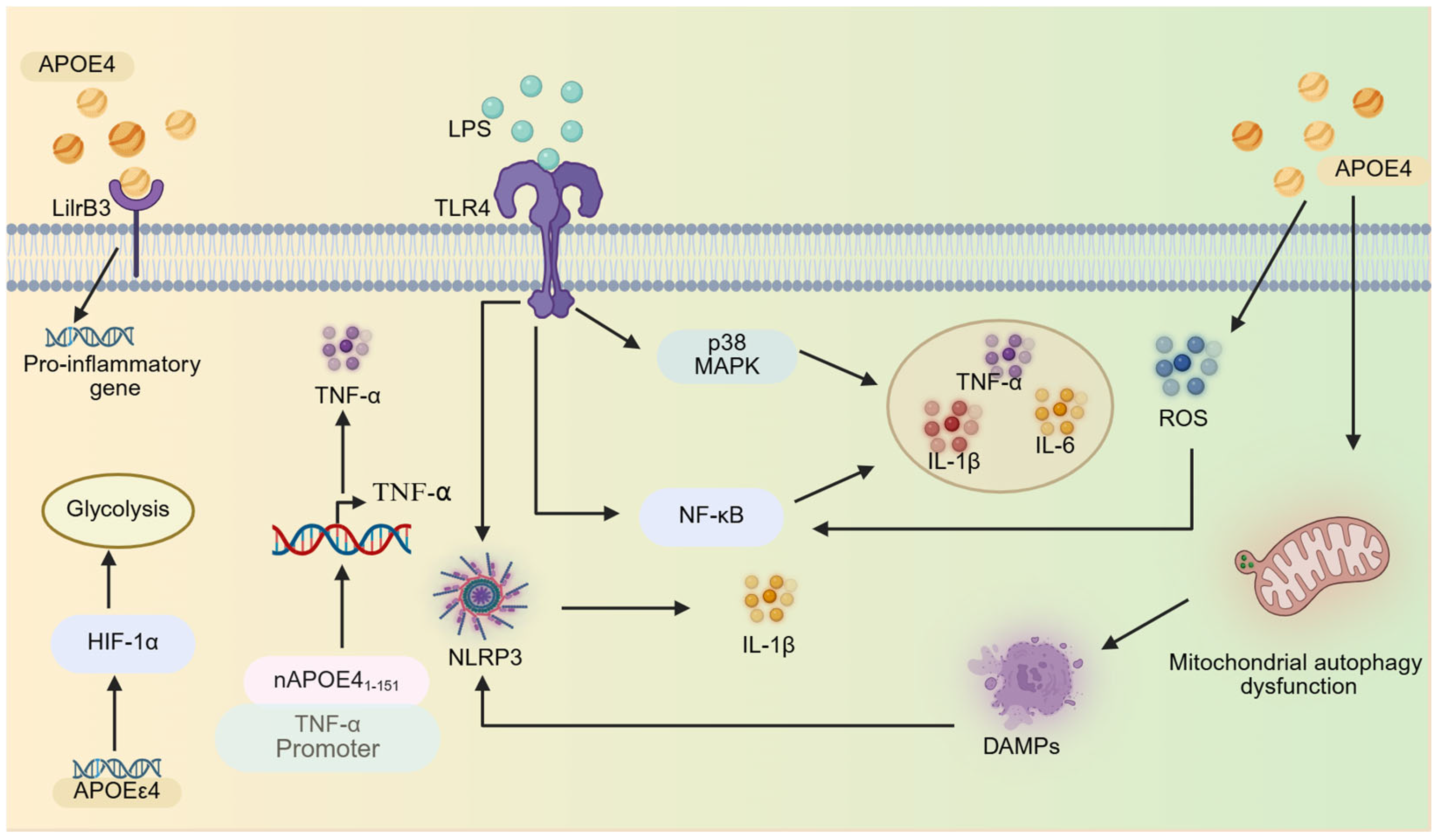
2.2. APOΕε4 Regulates Microglial Inflammatory Responses

APOEε4 acts as a molecular amplifier of microglial reactivity. In the absence of inflammatory stimuli, compared to APOEε2 and APOEε3 microglia, APOEε4 microglia exhibit elevated baseline secretion of tumor necrosis factor-α (TNF-α), thus indicating a primed pro-inflammatory state [84,85,86]. Following lipopolysaccharide (LPS) stimulation, APOEε4 microglia further amplify their inflammatory response, secreting significantly higher levels of pro-inflammatory mediators, including TNF-α, interleukin-1β (IL-1β), and interleukin-6 (IL-6) [44,45,52,87]. Notably, this hyperreactivity is gene-dose-dependent, with homozygous APOEε4 carriers showing more pronounced inflammatory responses than heterozygous individuals [52]. Interestingly, in APOE4-targeted replacement mice, basal pro-inflammatory gene expression (such as IL-1β, TNF-α, and NOS2) in female microglia is higher than that in male microglia, and the levels of secreted cytokines such as IL-1β, TNF-α, and IL-6 are significantly higher. This difference exists in the resting state without stimulation, and it further increases after inflammatory stimuli such as LPS and IFN-γ. Estrogen may amplify APOEε4-induced pro-inflammatory gene transcription by enhancing the binding ability of NF-κB to DNA [85].
A study found that in the microglia of human brain tissues from APOEε4 carriers, gene sets related to pro-inflammatory responses (such as T cell-mediated immune regulation, interferon-γ (IFN-γ) production, immune effector process regulation, etc.) were significantly upregulated, indicating that APOEε4 microglia are in a state of continuously activated pro-inflammation [88]. In the normal aging human brain without neuritic plaques, researchers identified a set of “microglia-APOE cluster genes”, primarily associated with phagocytosis, pro-inflammatory responses, antigen presentation, opsonization, cytoskeletal regulation, and cellular motility. These genes exhibited coordinated upregulation in APOEε4 carriers and downregulation in APOEε2 carriers. Notably, in AD patients with neuritic plaques, the APOEε4 carriers showed significant upregulation of these microglia-APOE cluster genes, yet no linear correlation with disease severity (Braak staging) was observed. These results suggest that APOEε4 may prime microglia into a pro-inflammatory state through innate genetic factors rather than relying on pathological stimuli [89]. In patients with primary tauopathies (such as corticobasal degeneration [CBD], Pick’s disease, and progressive supranuclear palsy [PSP]) carrying APOEε4, the pro-inflammatory gene sets in brain microglia—including those related to the TLR family, IL-1β, and IFN-γ signaling pathways—are significantly upregulated, with notably higher enrichment levels than those in APOEε3 carriers [45].
However, a study has found that the inflammatory response of human APOEε4 brains to AD pathology is significantly weakened, with the inflammatory gene expression profiles being highly similar to those of non-AD control (APOEε3–control) brains. Moreover, microglia activation-related pathways (such as the SALL1-regulated quiescent-to-activated transition) and immune response pathways (such as cytokine signaling) are not significantly activated, suggesting an inability to clear Aβ plaques and tau tangles through inflammatory responses. In contrast, APOEε3 brains exhibit a pronounced inflammatory response to AD pathology, in which microglia transition from a quiescent state to an activated state to combat AD pathology. This study is the first to reveal molecular evidence in human brain tissue that APOEε4 leads to defects in AD-related inflammatory pathways [90]. Similarly, in both human brains (including those of AD patients) and AD mice (5 × FAD mice, a common mouse model of AD progression, crossed with mice carrying human APOEε2, APOEε3, or APOEε4), researchers identified a special cell population called terminally inflammatory microglia (TIMs), which are characterized by ineffective inflammatory responses and impaired responses to chronic stimuli. The frequency of TIMs reaches 69% in 96-week-old AD*APOEε4 mice, while the frequency of TIMs in the brains of human AD patients increases with the progression of Braak stage and the carriage of APOEε4, suggesting that their accumulation is driven by aging and APOEε4 [73]. Based on these observations, we hypothesize that APOEε4 drives a biphasic microglial response: early hyperactivation followed by late exhaustion. However, most animal models capture only the early hyperactivation phase, while human autopsy samples reflect the late exhaustion phase. This methodological gap obscures the dynamic regulatory role of APOEε4 in AD pathogenesis. Collectively, these findings suggest that APOEε4 creates a hyperresponsive but exhaustion-prone inflammatory system. The transition from hyperactivation to exhaustion raises important questions about the temporal dynamics of APOEε4-mediated neuroinflammation. Are TIM a cause or consequence of disease progression? Furthermore, the reliance on post-mortem samples introduces potential artifacts, such as post-mortem interval effects, which may disproportionately impact APOEε4 microglial transcriptomes. Future studies should prioritize longitudinal analyses, fresh-tissue validation, and advanced imaging techniques to unravel the spatiotemporal dynamics of APOEε4 in AD pathogenesis.
APOEε4 amplifies neuroinflammatory responses through two primary mechanisms: inflammatory pathway activation and metabolic reprogramming. These mechanisms collectively drive microglial hyperactivation and sustain chronic inflammation in AD.

2.2.1. Inflammatory Pathway Activation

APOEε4 enhances microglial inflammatory responses by hyperactivating key signaling pathways. Following LPS stimulation, APOEε4 microglia exhibit stronger nuclear factor-κB (NF-κB) activation compared to APOEε3 microglia [85]. APOE4 stimulation induces NF-κB activation and concurrently triggers NLRP3 inflammasome assembly, leading to the significant secretion of pro-inflammatory cytokines (TNF-α, IL-1β, and IL-6) from microglia. Additionally, APOE4 stimulation induces the generation of ROS through oxidative stress [91], which further amplifies NF-κB and NLRP3 inflammasome activation, creating a vicious cycle of inflammation [92,93]. Moreover, APOE4 stimulation impairs microglial mitophagy, leading to the accumulation of damaged mitochondria and the release of damage-associated molecular patterns (DAMPs). These DAMPs activate inflammasomes, further exacerbating NLRP3 neuroinflammation [91].
The TLR4-p38 MAPK pathway also plays a critical role. APOEε4 microglia exhibit enhanced pro-inflammatory cytokine secretion through hyperactivation of the p38 MAPK pathway. Notably, treatment with the p38 MAPK-specific inhibitor SB203580 significantly suppressed cytokine production in APOEε4 microglia compared to APOEε2 microglia, suggesting genotype-dependent p38 mitogen-activated protein kinase (p38 MAPK) signaling sensitivity [52]. Similarly, the stimulation of microglia with LPS or oligomeric amyloid-beta 42 (oAβ42) activates the TLR4-p38 MAPK signaling pathway, triggering the release of pro-inflammatory cytokines. These effects are more pronounced in APOEε4 microglia than in APOEε3 microglia. Both TLR4 antagonists (e.g., LPS-RS and IAXO series) and p38α inhibitors (downstream effectors of TLR4) effectively suppress oAβ42-induced TNF-α secretion [94]. Notably, gender differences significantly influence TLR4-mediated neuroinflammation, with female APOEε4 carriers exhibiting heightened sensitivity to inflammatory stimuli. The mechanism may be related to the higher TLR4 expression in females or more active downstream signaling (e.g., NF-κB) [95]. While these findings underscore APOEε4-mediated regulation in driving neuroinflammation, most studies rely on LPS or Aβ stimulation models, which may not fully recapitulate the chronic, low-grade inflammation observed in human AD. Additionally, the gender-specific effects of APOEε4 warrant further investigation. Future research is required to clarify how X-chromosome genes (such as TLR4 and CD14) or sex hormones (such as estrogen fluctuations) regulate the function of APOEε4 in microglia; for example, through epigenetic or transcription factor regulation [85,95].
Moreover, a unique aspect of APOE4-mediated pro-inflammatory is its amino-terminal fragment, nAPOE41–151, which binds to the TNF-α promoter, upregulating its expression and enhancing IL-1β secretion. This fragment also downregulates CXORF56, a homeostatic checkpoint gene, promoting microglial morphological activation and creating a positive feedback loop that sustains inflammation [96,97,98]. Paradoxically, full-length APOE4 may exert anti-inflammatory effects under certain conditions [99]. Notably, a seminal 2016 study employing multimodal experimental approaches (including molecular interaction assays, genomic analyses, and animal models) provided the first definitive evidence that APOE, particularly the APOE4 isoform, can function as a transcription factor by directly binding DNA to modulate gene expression [100]. Although nAPOE41–151 exhibits characteristics resembling those of a transcriptional regulator, these experiments do not confirm its identity as a bona fide transcription factor [96,97,98,99]. These findings elucidate previously underappreciated, non-canonical functions of APOE that extend beyond its well-characterized role in lipid metabolism. Furthermore, recent studies have revealed that LilrB3, an immune checkpoint receptor protein expressed on the surface of microglia, serves as a specific receptor for APOE4, exhibiting minimal binding affinity for APOE2. The specific interaction between APOE4 and LilrB3 activates microglia, driving their transition into a pro-inflammatory state [101]. Additionally, LPS activates cholesterol 25-Hydroxylase (CH25H) expression in microglia through the TLR4 pathway, catalyzing the conversion of cholesterol to 25-hydroxycholesterol (25-HC) and significantly increasing its production. During this process, the APOEε4 genotype further enhances the induction efficiency of CH25H, causing APOEε4 microglia to produce significantly higher levels of 25-HC than APOEε2/3 microglia after LPS stimulation. The generated 25-HC promotes IL-1β maturation and secretion by activating the NLRP3/caspase-1 inflammasome. APOEε4 microglia are more sensitive to this process. The above mechanisms form a positive feedback loop in the APOEε4 background: APOEε4 not only promotes 25-HC production by enhancing CH25H expression but also improves the sensitivity of microglia to 25-HC-mediated inflammatory responses through unknown mechanisms, ultimately leading to exponentially exacerbated IL-1β-mediated neuroinflammation [102].

2.2.2. Metabolic Reprogramming

The second major mechanism involves the APOEε4-mediated regulation of microglial energy metabolism, which creates an energy landscape favoring sustained cytokine production [77]. Specifically, under steady-state conditions, microglia primarily rely on oxidative phosphorylation for energy generation [103,104]. However, compared to APOEε3, APOEε4 triggers a metabolic shift toward glycolysis during inflammation. This shift is marked by upregulated glycolysis-related genes and suppressed oxidative phosphorylation pathways. This metabolic shift provides an energetic foundation for sustained cytokine production, as glycolysis enables rapid ATP generation to meet the heightened energy demands of cells during inflammatory responses [77]. Mechanistically, this shift is driven by the enhanced activity of hypoxia-inducible factor 1α (HIF-1α), which promotes glycolytic flux and suppresses mitochondrial respiration [45,77,105] (Figure 2). However, the causal relationship between glycolytic predominance and microglial hyperactivation remains unresolved, with current evidence being insufficient to determine whether this metabolic reprogramming initiates or results from excessive immune activation [105].

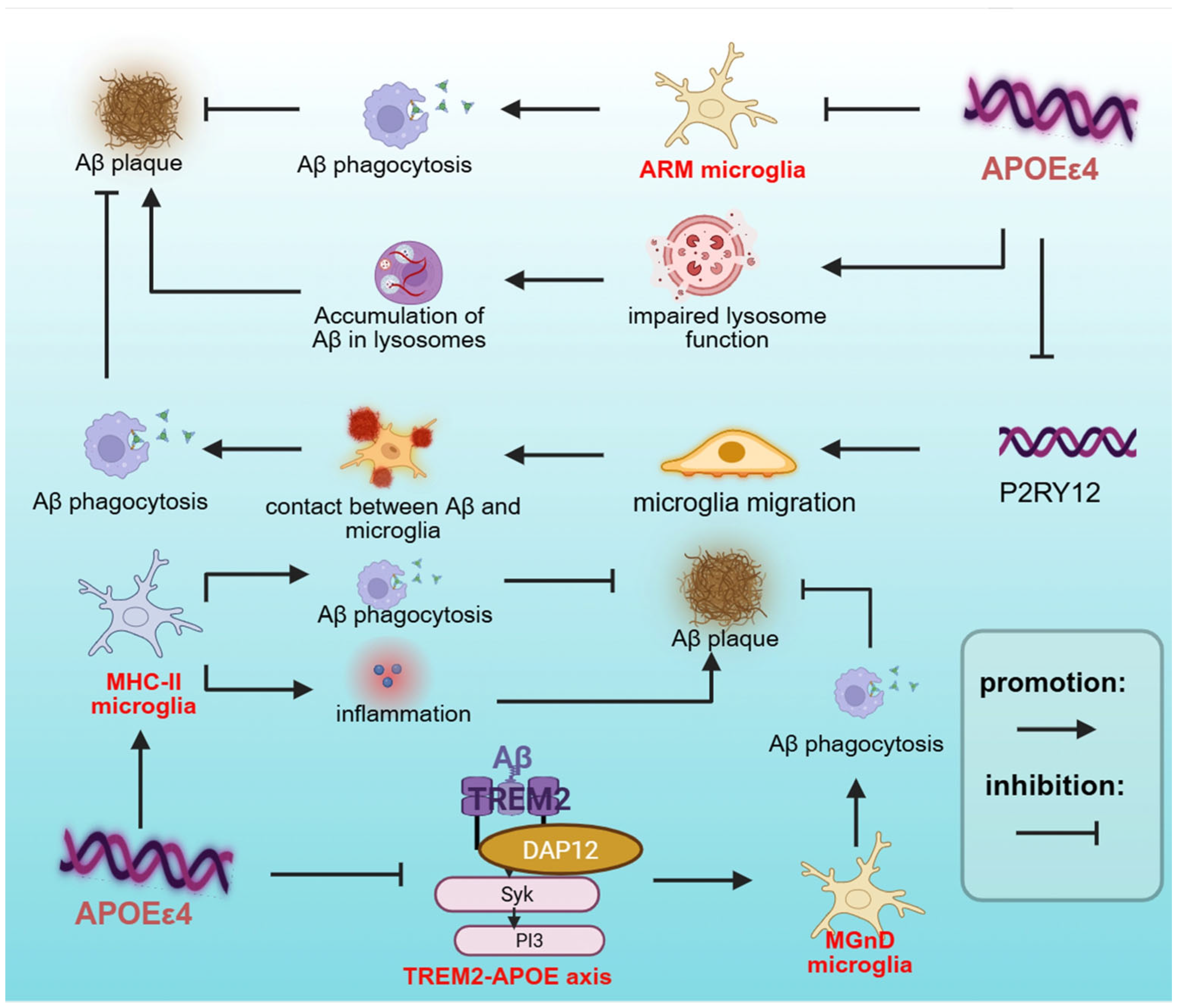
2.3. APOEε4 Regulates Microglial Phagocytosis in AD

In various AD models, APOEε4 microglia demonstrate significant impairments in phagocytosing Aβ aggregates, leading to their pathological accumulation in the brain [46,48,60]. Notably, in human APOEε4 neuron-engrafted chimeric mice, microglial depletion resulted in an approximately 50% reduction in the average number of Aβ aggregates, demonstrating a synergistic interaction between APOEε4 and microglia in promoting Aβ aggregate formation [47].
In both AD mouse models and human brains, microglia associated with Aβ plaques typically exhibit a DAM/MGnD phenotype, characterized by the upregulated expression of phagocytosis-related genes and the presence of engulfed Aβ particles [64,65]. Research demonstrates that the TREM2-APOE signaling axis plays a pivotal regulatory role in this process—Aβ binding to TREM2 receptors triggers downstream signaling cascades that promote microglial transition to the DAM/MGnD phenotype, thereby enhancing Aβ phagocytic clearance and effectively mitigating its neurotoxic effects [46,64,65,106]. However, this protective mechanism is significantly impaired in the APOEε4 context. Under Aβ plaque conditions, APOEε4 microglia show a markedly compromised transition to the DAM/MGnD phenotype, likely due to the disrupted TREM2-APOE signaling axis [46,56]. Although TREM2 and its signaling adaptor TYROBP expression positively correlates with APOEε4 in microglial-like cells [60], APOEε4 downregulates TREM2 expression in E4FAD mice. This impairment compromises microglial recognition and the binding of Aβ deposits, consequently disrupting protective barrier formation and plaque compaction [107]. TREM2 deficiency further exacerbates microglial barrier dysfunction, reducing both plaque-associated microglial numbers and their Aβ-encapsulating capacity [108,109]. Notably, the impact of TREM2 deficiency exhibits allele-specific effects, with APOEε4 microglia demonstrating more pronounced phagocytic deficits compared to APOEε3 microglia [49,108]. This genotype-dependent phenomenon underscores the critical importance of considering genetic background when developing TREM2-targeted therapeutic strategies.
Additionally, researchers have identified two distinct microglial subpopulations associated with amyloid plaques: ARM and MHC-II microglia. Amyloid-responsive microglia (ARM) are a specific subpopulation identified in post-mortem human brain tissue, particularly in the middle frontal neocortex. ARM counteract Aβ deposition through protective barrier formation and enhanced phagocytic capacity, characterized by high expression of Cluster of Differentiation 163 (CD163), a scavenger receptor that facilitates the receptor-mediated endocytosis of hemoglobin–haptoglobin complexes following hemorrhage [74]. In the brain tissue of Alzheimer’s patients, CD163-positive microglia are often found in close proximity to Aβ plaques, especially neuritic plaques, and exhibit increased expression of CD68, a lysosomal marker [110]. ARM are more abundant in AD patients carrying the APOEε3/ε3 genotype, while their numbers are significantly reduced in APOEε4 carriers and individuals with the TREM2-R47H variant [74]. Using an in vivo chimeric AD model (hiPSC-derived neurons transplanted into the hippocampus of APOE knock-in mice), researchers identified another microglial subpopulation, MHC-II microglia, which is characterized by high expression of MHC-II molecules and pro-inflammatory genes. Notably, neuronal APOEε4 drives the enrichment of microglial subsets with elevated MHC-II expression. Strikingly, even in mice with an APOEε4 genetic background, the proportion of MHC-II+ microglial subsets is significantly decreased when neurons lack APOEε4 (hEKO-E4KI group) [47]. Previous studies indicate that MHC II-positive microglia accumulate around amyloid plaques and NFTs in both AD mouse models and human AD brains [111,112]. Researchers found that the intracerebroventricular (ICV) injection of amyloid β (Aβ)-specific T helper 1 (Aβ-Th1) cells into 5XFAD mice induces the differentiation of MHCII+ microglia. MHCII+ microglia exhibit a stronger capacity to clear Aβ plaques compared with MHCII− microglia [113]. In a recent study, hiPSC-derived neurons carrying APOEε4 (hE4) were transplanted into the hippocampus of APOE4 knock-in (E4KI) mice to establish a human–mouse chimeric model. The results revealed a novel mechanism by which APOEε4 promotes the progression of Aβ deposition and tau pathology through remodeling microglial phenotypes (such as inducing high-expression MHC-II subsets) and forming “toxic interactions” with neurons [114].
APOEε4 significantly influences microglial activation, driving their differentiation into specific phenotypes, such as DAM/MGnD, ARM, and MHC-II microglia. However, the diminished phagocytic capacity of APOEε4 microglia is not solely attributable to reduced activation potential. There exist additional mechanisms underlying APOEε4-mediated deficits in phagocytic function. In E4FAD mice, although reactive microglia accumulate around Aβ plaques, they display a dystrophic morphology characterized by swollen somata and thickened processes, significantly compromising phagocytic function [115]. In addition, in vivo and in vitro, APOEε4 microglia exhibit significant lysosomal impairment, characterized by aberrant lysosomal trafficking and impaired endosome-lysosome activity [78,84,116]. Under normal conditions, microglia internalize Aβ via endocytosis, followed by the fusion of endosomes with lysosomes, where Aβ is efficiently degraded by hydrolases [117,118]. However, lysosomal impairment severely compromises this clearance process, leading to the accumulation of Aβ within lysosomes [55]. The Aβ accumulated in lysosomes exhibits a “seeding effect,” which can induce further Aβ aggregation in vitro, suggesting that lysosomal dysfunction may trigger Aβ aggregation and deposition [119]. Despite these insights, the precise molecular mechanisms linking APOEε4 to lysosomal impairment remain incompletely understood. Notably, both APOE3 and APOE4 facilitate Aβ binding to the cell surface and promote its lysosomal trafficking and degradation, with APOE3 exhibiting a more pronounced effect [119]. In the co-culture model of brain sections from 5XFAD transgenic mice and N9 microglia stably transfected with human APOEε4, as well as in the in vitro functional validation model of APOEε4/N9 microglia, a recent study found that APOEε4 impairs the clearance of Aβ by microglia by inhibiting autophagic flux and mitochondrial function. Autophagy inducers such as rapamycin can reverse this defect by enhancing autophagy and mitophagy. This study reveals a novel mechanism by which APOEε4 promotes pathological progression in AD through the autophagy–mitochondria axis [114].
Furthermore, APOEε4 compromises microglial chemotaxis toward Aβ plaque due to the decreased expression of the P2RY12, which is crucial for microglial protrusion extension and plaque coverage. As an ATP receptor, P2RY12 is crucial for microglial protrusion migration in response to Aβ [49,50] (Figure 3). Notably, isoform-specific lipidation states critically regulate Aβ clearance efficiency. Lipidated APOE2 enhances Aβ clearance, whereas the reduced lipidation efficiency of APOE4 impairs this process, conferring a functional disadvantage [55]. In addition, APOEε4 also has a cross-cellular regulatory effect. In the APP/PS1 mouse model, APOEε4 expression attenuates LGALS3-dependent microglia–astrocyte crosstalk, leading to defective astrocytic plaque encapsulation and clearance. Strikingly, microglia-specific APOEε4 knockout rescues LGALS3 signaling, reactivating astrocytic phagocytic function and improving plaque clearance [56].
APOEε4 not only limits the ability of microglia to engulf and clear Aβ, but also regulates their response to tau protein. In chimeric mice transplanted with human APOEε4 neurons, the impact of microglia on tau pathology is dependent on APOE genotype. In the presence of APOEε4, microglia play a significant role in promoting tau pathology, as evidenced by the significantly reduced p-tau deposition in the hippocampus following microglial depletion in APOEε4 chimeric mice. By contrast, APOEε3 does not exhibit this effect [47]. Additionally, APOEε4 microglia exhibit pronounced lysosomal abnormalities, including increased lysosomal mass and abnormally reduced baseline pH (hyperacidification). Notably, upon myelin stimulation, these cells fail to exhibit the expected further acidification, instead showing minimal pH changes or even slight alkalinization [84]. Given that lysosomal hydrolytic enzymes require an acidic environment (pH 4.5–5.0) for optimal activity, this dysregulated acidification directly compromises microglial tau degradation capacity, leading to intracellular tau accumulation [120]. If lysosomes fail to degrade the engulfed tau, the undigested tau is then released into exosomes, which are secreted by microglia. These exosomes transfer pathological tau to neurons, where the exogenous tau interacts with endogenous tau, promoting its aggregation and misfolding [121,122]. Additionally, in the APOEε4 context, TREM2 deficiency exacerbates tau-mediated neurodegeneration and tau pathology. While TREM2 knockout attenuates certain TREM2-dependent reactive gene expression in microglia, it preserves the elevated expression of TREM2-independent genes (e.g., lysosomal functional markers CTSB and CTSD). These findings demonstrate that, under APOEε4 conditions, tau pathology drives a TREM2-independent microglial proliferative response that actively promotes tau-mediated neurodegeneration [123].
APOEε4 also affects the ability of microglia to phagocytose and degrade other substrates, thereby influencing the progression of AD. In APOE-TR mice, during cuprizone (CPZ)-induced demyelination, microglia in APOEε4 mice exhibited reduced efficiency in clearing the myelin debris that are abundant in lipids compared to APOEε2 mice [53]. Although AD is not typically considered a demyelinating disorder, focal demyelination has been observed in both AD patients and mouse models, and it is associated with Aβ and neurofibrillary pathology [124,125]. Additionally, the phagocytic ability of microglia in APOE4 knock-in mice against apoptotic neurons is significantly lower than that of microglia in APOE3 knock-in mice, and they fail to upregulate the expression of genes related to engulfment when encountering apoptotic neurons [56]. Moreover, microglia in APOE4 knock-in mice exhibit significantly enhanced phagocytosis of synaptic materials compared to those in APOE3-KI mice. Mechanistically, APOE4 induces upregulation of MHC-I molecules on the surface of neurons. This abnormal expression is recognized by microglia as an “eat-me” signal, thereby inducing excessive synaptic phagocytosis by microglia [126]. Notably, compared with APOE4 knock-in mice, microglia in the hippocampal tissue of the P301S/APOE4 knock-in mouse model exhibit significantly enhanced synaptic phagocytosis, and their excessive synaptic phagocytosis is an important factor leading to synaptic loss in AD [78]. Furthermore, astrocyte-derived APOE4 may promote microglial synaptic clearance in tauopathy. In the tau pathology mice model, the study found that the deletion of astrocytic APOEε4 reduced the expression of disease-associated microglia (DAM/MGnD) genes (such as Clec7a, H2-D1, and Spp1), increased the expression of homeostatic genes (such as P2ry12), and decreased the phagocytosis of synaptic elements by microglia [10].

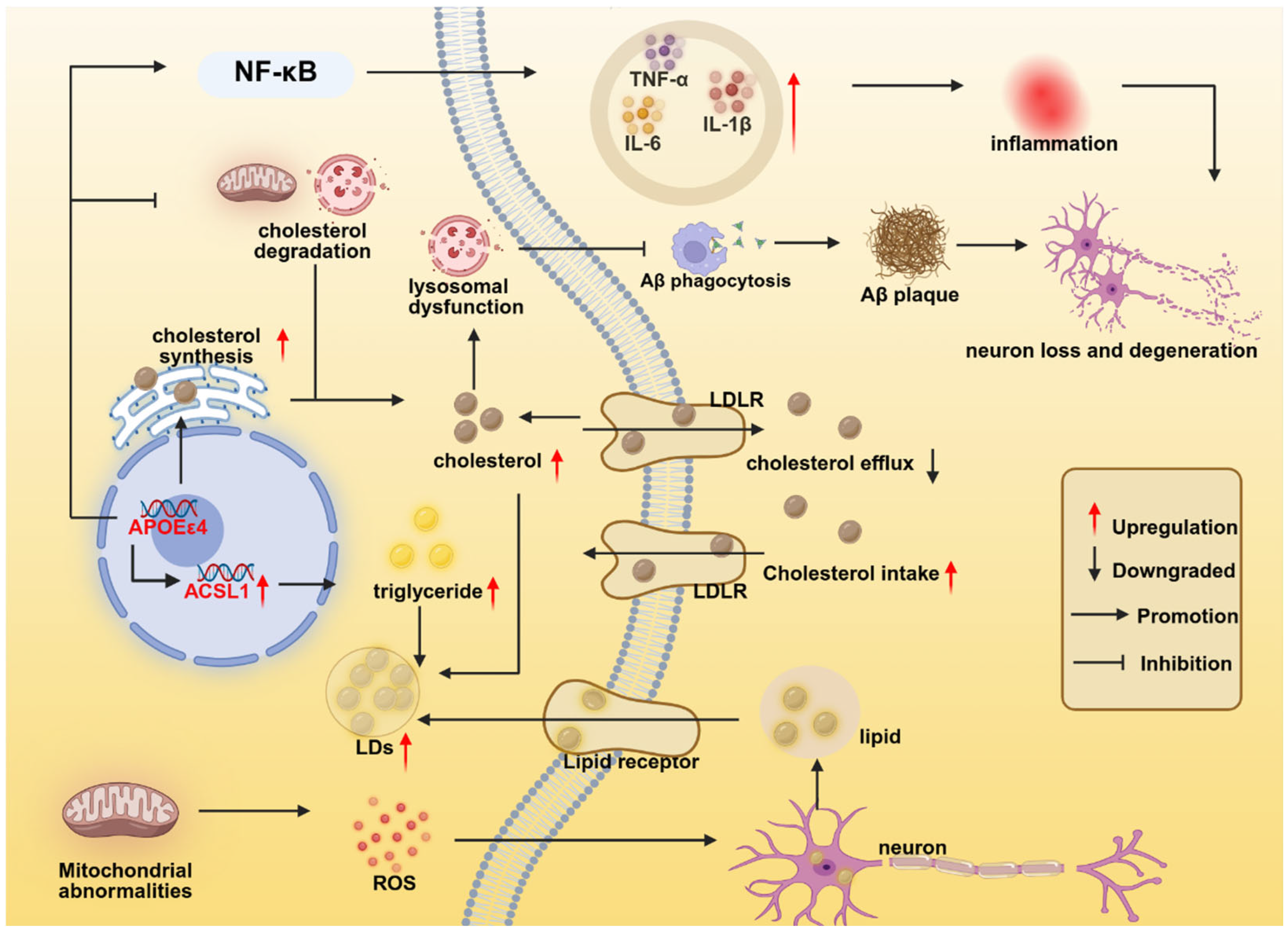
2.4. APOEε4 Regulates Microglial Lipid Metabolism

APOE plays a central role in lipid metabolism within microglia, primarily through its ability to bind lipids and form lipoprotein particles, which facilitate intercellular lipid transport [127]. APOE regulates cholesterol metabolism in microglia through interacting with receptors such as the low-density lipoprotein receptor (LDLR), low-density lipoprotein receptor-related protein 1 (LRP1), and TREM2. These interactions mediate cholesterol uptake and clearance, maintaining intracellular lipid homeostasis [128]. Extensive research has demonstrated that APOEε4 disrupts microglial lipid homeostasis and exacerbates the accumulation of lipid droplets (LDs) in microglia. During aging and AD, microglia accumulate LDs, which are primarily composed of cholesterol, triglycerides, and their esters [76,105,129]. However, APOEε4 microglia exhibit significantly greater LDs accumulation compared to APOEε3 microglia [46,56,76,126]. This phenomenon is observed even in the absence of aging or AD pathology, suggesting an intrinsic lipid dysregulation in APOEε4 microglia [46,84,105,129]. Notably, transient receptor potential vanilloid 1 (TRPV1) is a ligand-gated ion channel capable of reversing APOE4-induced LDs accumulation in microglia [126].
The mechanisms underlying APOEε4-mediated LDs accumulation are multifaceted. In hiPSC-derived APOEε4 microglia, genes associated with cholesterol biosynthesis (including HMGCR, SREBF2, and SCAP) were significantly upregulated, while the expression of cholesterol efflux-related genes (such as ABCA1 and ABCG1) was downregulated. Additionally, reduced expression of lysosomal-related genes (e.g., LIPA) indicates impaired lysosomal function and compromised cholesterol and lipid degradation [130]. Furthermore, in induced microglia-like cells (iMGLs) expressing APOEε4, genes involved in lipid synthesis (e.g., ACSL1) are upregulated, whereas those related to fatty acid β-oxidation (e.g., ACOX1, CPT1A) and lipid catabolism (e.g., LIPA, SMPD1) are downregulated, accompanied by reduced lysosomal acid lipase activity. This results in the inefficient degradation of LDs, exacerbating lipid accumulation [105]. In APOEε4 microglia, the accumulation of LDs is associated with increased lysosomal mass and acidic pH, which may impair lysosomal lipid degradation efficiency, thereby forming a vicious cycle of lipid accumulation—lysosomal dysfunction—further lipid accumulation [84]. Moreover, APOE exhibits isoform-dependent cholesterol efflux capacity, with the following order: APOE2 > APOE3 > APOE4. APOE mediates cholesterol efflux through ATP-binding cassette transporter A1 (ABCA1), facilitating the formation of high-density lipoprotein (HDL) particles and consequently reducing intracellular cholesterol levels in microglia. Due to its having the lowest cholesterol efflux efficiency, APOE4 results in the most pronounced intracellular cholesterol accumulation in microglia [131]. Additionally, APOE4 and tau pathology synergistically disrupt cholesterol metabolism in microglia through dual mechanisms involving enhanced synthesis and impaired lysosomal degradation. This interaction upregulates cholesterol biosynthesis genes (e.g., HMGCR) via SREBP2 activation, thereby promoting endogenous cholesterol production. Simultaneously, it suppresses the LXR pathway, disrupting its negative feedback regulation of SREBP2. Furthermore, the combined effects of APOE4 and tau significantly reduce lysosomal acid lipase (LIPA) activity, impairing the degradation of both cholesteryl esters and phosphatidylcholines [78].
In aged mice, GRN knockout mice (a model for frontotemporal dementia, FTD), and brain tissues from normal human aging, researchers have identified a special subpopulation of microglia: lipid-droplet-accumulating microglia (LDAM). These cells exhibit massive intracellular accumulation of LDs. Moreover, these cells also show severe functional defects, such as decreased phagocytic ability, elevated ROS production, and a pro-inflammatory phenotype [75]. Subsequently, researchers identified LDAM in the brain tissues of AD patients, particularly in those with the APOE4/4 genotype, and proposed that the formation of LDAM is closely associated with acyl-CoA synthetase long-chain family member 1 (ACSL1) expression in microglia. ACSL1 is a key enzyme in lipid droplet biogenesis, and the overexpression of ACSL1 is sufficient to induce the formation of triglyceride-specific lipid droplets in multiple cell types. In iPSC-derived microglia, fibrillar Aβ (fAβ) induces ACSL1 expression, triglyceride synthesis, and LDs accumulation, with this induction being more pronounced against the APOE4 background. Notably, LDAM exhibits defective phagocytic function, lysosomal accumulation, and secretion of inflammation-related chemokines [76].
Growing evidence suggests that the APOEε4-mediated dysregulation of microglial lipid metabolism is linked to its inflammatory response and impaired phagocytic function. Accumulating evidence demonstrates an interplay between Aβ and microglial lipid metabolism, which is markedly enhanced in the APOEε4 context. In the presence of Aβ, microglial LDs accumulation is exacerbated, particularly in APOEε4 microglia [46,56]. In the brains of AD patients, especially those carrying the APOEε4/ε4 genotype, lipid bodies resembling LDs have been observed. These lipid bodies are typically located near Aβ plaques and may exist within or around microglia. The number of lipid bodies is positively correlated with the number of Aβ plaques, the level of tau pathology, and the expression of the ACSL1 gene [76]. In addition, in vivo and in vitro models, Aβ stimulation leads to the accumulation of LDs in microglia, accompanied by an upregulation of ACSL1 expression, which is more pronounced against the APOEε4/ε4 background. Mechanistically, in APOEε4 microglia, NF-κB hyperactivation enhances ACSL1 transcription via direct binding to its promoter [46,76]. Based on the above findings, we conclude that Aβ pathology promotes the accumulation of LDs in microglia, particularly in APOEε4 microglia, and the ACSL1 may be a potential mediator of this process. Intriguingly, dysregulated microglial lipid metabolism, particularly cholesterol metabolism, can in turn influence Aβ pathology. Microglial intracellular cholesterol levels regulate Aβ trafficking to late endosomes/lysosomes for degradation. Reduced intracellular cholesterol enhances late endosomal/lysosomal transport efficiency by promoting Rab7 recycling via guanine nucleotide dissociation inhibitor (GDI)-mediated mechanisms. Specifically, cholesterol depletion facilitates Rab7 membrane dissociation and subsequent recycling, thereby accelerating the transport of Aβ-containing vesicles to lysosomes [131]. As previously discussed, APOEε4 microglia exhibit more pronounced LDs accumulation and elevated intracellular cholesterol levels, which consequently impair Aβ lysosomal trafficking and degradation. Recent studies have revealed a novel cholesterol-induced mechanism of Aβ pathology. Lipidated APOE (cholesterol-bound form) is efficiently endocytosed by microglia via the LDLR and subsequently enters the endolysosomal system. Within the acidic environment of lysosomes, APOE undergoes delipidation and conformational changes, forming β-sheet-rich fibrillar aggregates (amyloid-like fibrils). This process is directly linked to lysosomal dysfunction, as delipidated APOE cannot be effectively cleared. Intracellular cholesterol depletion exacerbates lysosomal impairment, significantly promoting APOE aggregation. These APOE aggregates directly induce Aβ fibrillization and deposition within microglia. Furthermore, through microglial cell death or extracellular vesicle release, APOE aggregates can be transmitted to neighboring cells, thereby propagating Aβ pathology [132].
LDs accumulation promotes microglia to exhibit enhanced neuroinflammatory effects, and this effect is amplified in the APOEε4 background. Studies have found that LDAM rich in LDs produce high levels of ROS and release pro-inflammatory cytokines [75]. In human-induced pluripotent stem cell-derived microglia (iMG) with APOEε4/4 and APOEε3/3 genotypes, APOEε4/4 iMG enriched in LDs exhibit more pronounced inflammatory responses. Transcriptome analysis shows that compared with LDs–low iMG, LDs–high iMG of APOEε4/4 have higher expression of NF-κB-related pro-inflammatory cytokines (such as TNFA and IL1B), and the NF-κB pathway is significantly activated [76]. In APOE-TR mice and microglial cell line BV2, LDs accumulation activates microglia through the NF-κB pathway, inducing the expression of MHC-II and the secretion of pro-inflammatory factors (such as IL-6 and TNF-α) by microglia, and promoting T cell infiltration and neuroinflammation. This mechanism is particularly significant in the context of APOEε4. The TRPV1 activator capsaicin can inhibit the activity of the NF-κB pathway in APOEε4 microglia [126]. Additionally, inflammatory stimuli can trigger the formation of LDs in microglia. In the mouse microglial cell line BV2 model, lipopolysaccharide (LPS) activates the NF-κB pathway, upregulates lipid synthesis-related genes (such as ACLY and PLIN3), and inhibits lipid catabolism genes (such as ADRB1/2), leading to abnormal fatty acid β-oxidation and LDs accumulation [75]. In APOEε4/iPSC-derived microglia, the proportion of lipid droplet-positive cells is significantly higher than that in APOEε3 cells. After interferon-γ (IFN-γ) stimulation, the lipid droplet positivity rate in APOEε4 microglia further increases, with a higher amplitude than that in APOEε3 cells [129]. Research has found that LPS promotes the transcription and translation of LDLR in microglia by activating the TLR4/NF-κB pathway. APOEε4 synergistically enhances this process. Increased LDLR expression enhances cellular cholesteryl ester uptake, ultimately leading to lysosomal dysfunction and the exacerbation of neuroinflammation. This mechanism exhibits a clear genotypic dependency (APOEε4 > ε3 > ε2) [133]. Moreover, studies demonstrate that LPS treatment upregulates ACSL1 expression in iPSC-derived microglia, an effect potentiated by the APOEε4 genotype. Mechanistically, APOEε4 synergizes with LPS to enhance the transcriptional activation of the ACSL1 gene by promoting the activation of the NF-κB pathway [76] (Figure 4).
LDs accumulation in microglia also detrimentally affects neurons. On one hand, LDs accumulation in microglia, such as the high LDs burden state induced by APOEε4, significantly inhibits its purinergic signaling pathway (e.g., P2RY12 receptor-mediated Ca2+ response), resulting in microglia being unable to effectively sense soluble factors such the as ATP and glutamate released by neurons, thereby losing the ability to dynamically monitor neuronal network activity. On the other hand, APOEε4 exacerbates lipid metabolic disorders in microglia, prompting abnormal cholesterol efflux into the extracellular environment. Excessive cholesterol enhances the activity of G protein-gated inwardly rectifying potassium channels (GIRK3) on the neuronal cell membrane, leading to hyperpolarization of the neuronal resting membrane potential and a decrease in the frequency of action potential firing. Additionally, cholesterol enrichment promotes abnormal aggregation of neuronal lipid rafts, further interfering with synaptic transmission efficiency [105]. Furthermore, microglia with abundant LDs induce tau phosphorylation and apoptosis in neurons by secreting lipid droplet-associated neurotoxic factors (e.g., pro-inflammatory cytokines, lipid metabolites), and this process is dependent on the presence of APOE [76].

3. The Role of APOEε2 in Microglia Function Associated with AD

The existing literature shows that, compared to APOEε3 and APOEε4 microglia, APOEε2 microglia demonstrate stronger protective functions in the pathological environment of AD, particularly in terms of maintaining brain homeostasis, regulating neuroinflammation, and effectively clearing Aβ deposits. APOEε2 may regulate microglial functional activities (including motility, phagocytosis, and immunomodulation) through modulating the expression of microglia-associated proteins. Specifically, APOEε2 is significantly associated with high expression of ionized calcium-binding adapter molecule 1 (Iba1, a marker of microglial motility) and macrophage scavenger receptor-A (MSR-A, a plaque-associated phagocytic receptor), while it is significantly associated with low expression of cluster of differentiation 68 (CD68, a marker of phagocytic function) and human leukocyte antigen–DR (HLA-DR, an antigen-presenting molecule). These results suggest that APOEε2 may exert neuroprotective effects by preserving microglial motility (associated with Iba1 expression) and alleviating neuronal damage induced by elevated CD68 and HLA-DR expression [51]. Moreover, compared to APOEε4 microglia, APOEε2 microglia exhibit lower activation levels upon inflammatory stimulation, secrete fewer pro-inflammatory cytokines, and induce less neuronal damage [87]. Following LPS stimulation, APOEε2 microglia exhibit attenuated activation of the p38 MAPK signaling pathway [52]. Furthermore, in microglial predominant gene clusters rich in biological processes such as engulfment, inflammatory response, cell–cell interaction, and metabolism, the expression levels of specific genes within this cluster are lower in APOEε2 carriers than in APOEε3 homozygotes, regardless of the presence of neuritic amyloid plaques [89]. Similarly, APOE2 stimulation reduces the expression levels of microglial activation markers TREM2 and Clec7a, suggesting its ability to suppress the transition of microglia into disease-associated activation states (e.g., DAM/MGnD phenotype) [54]. These findings demonstrate that APOEε2 plays a critical role in maintaining microglial immune surveillance function while sustaining a relatively low inflammatory activation state, thereby reducing potential neurotoxic damage caused by excessive inflammatory activation.
Compared to APOE3 or APOE4, APOE2 significantly enhances the microglial degradation of soluble Aβ. This enhanced degradative capacity may be attributed to the structural and functional properties of APOE2, which potentially accelerates Aβ clearance by interacting with Aβ itself or related degradative enzymes, thereby reducing Aβ accumulation in the brain [55]. A study on macrophages supports this hypothesis. Bone marrow-derived macrophages from APOE2-TR mice demonstrated a significantly enhanced degradation capacity for both soluble and insoluble Aβ species compared to those from APOE3-TR and APOE4-TR mice. Mechanistically, this superior degradative performance may be attributed to APOE2’s ability to potentiate matrix metalloproteinase-9 (MMP-9) enzymatic activity, thereby facilitating more efficient Aβ proteolysis [134]. However, studies examining microglial phagocytic function toward apoptotic neuronal cells and Aβ have shown no significant differences between APOEε2 microglia and wild-type or APOEε3 microglia in terms of their ability to engulf apoptotic neurons or Aβ [61]. This may be due to differences in experimental models and detection methods. Notably, studies have demonstrated that the APOE can directly interact with Aβ by binding to residues 12–28 of the Aβ peptide, an interaction that promotes both Aβ aggregation and cerebral deposition. In both APP/APOEε4 and APP/APOEε2 transgenic mouse models, the co-deposition of APOE with Aβ plaques has been consistently observed. Compared to APOE4, APOE2 does not provide complete protection against Aβ pathology, but rather exerts a more moderate promotive effect on Aβ-related pathological processes [135].
Several lines of evidence indirectly support the role of APOEε2 in modulating microglial function to ameliorate AD pathology. APOEε2 gene therapy significantly reduces the activation of APOEε4 microglia, accompanied by a decrease in Aβ deposition and improvements in neuroinflammation and neurodegenerative changes [27]. These findings provide new insights into the potential clinical applications of APOEε2 gene therapy as a treatment option for AD. In the CPZ-induced demyelination model, APOEε2 microglia demonstrated a robust capacity to clear myelin debris, effectively responding to demyelination injury and thereby facilitating tissue repair [53]. Additionally, researchers have identified genes that are preferentially expressed in aged human microglia, which are significantly enriched in AD susceptibility genes and increase with age. However, APOEε2 reduces the expression of these genes, suggesting that it may help decrease the incidence of the aged microglial phenotype [136].

4. The Role of APOEε3 in Microglia Function Associated with AD

APOEε3 microglia demonstrate a unique equilibrium between homeostatic maintenance and pathological response in AD. Using APOE-knockout mice with microglia/central nervous system-associated macrophages (CAMs)-specific inducible expression of APOEε3, researchers discovered that APOEε3 microglia can efficiently perform immunosurveillance in the brain and directionally migrate toward injury sites to form a potential barrier preventing further damage. This rapid-response mechanism is crucial for neuroprotection—it not only contains the spread of injury-induced excessive inflammatory responses but also minimizes damage to surrounding healthy neurons, thereby maintaining homeostasis of the brain microenvironment [46]. Compared to nAPOE41–151, the genes upregulated in microglia following stimulation with nAPOE31–151 (the amino-terminal fragment of APOE3) are associated with various cellular processes, including mitosis, cytoskeleton regulation, and cell signaling. These gene functions are more closely related to the physiological regulation of microglia, supporting their normal functions in maintaining brain homeostasis [98].
APOEε3 expression in microglia has significant potential to improve amyloid deposition and related pathological conditions, increasing the microglial coverage of amyloid plaques and the expression of TREM2 [46,49,107]. The intracellular signaling response triggered by TREM2 activation is considered essential for the effective phagocytosis of Aβ by microglia [109]. In the presence of Aβ, APOEε3 regulates the expression of genes related to antigen presentation, immune response, inflammatory pathways, and interferon response in microglia, leading to a shift toward specific phenotypes (ARM and MGnD microglia). This enhances their positive response to Aβ, increases their phagocytic activity, and modulates the inflammatory response [46,49,56]. During the progression of neurodegenerative lesions, APOEε3 microglia upregulate key DAM/MGnD genes while downregulating TGFβ signaling molecules, thereby reducing the levels of hyperphosphorylated tau protein and ultimately decreasing neuronal loss [56]. Notably, there are notable sex differences in the interactions of APOEε3 microglia with amyloid plaques; compared with age-matched female E3 FAD mice, male E3 FAD mice exhibit greater microglial plaque coverage, plaque compaction, and TREM2 expression, along with a reduced plaque burden [107]. Furthermore, DAM/MGnD microglia expressing APOEε3 can secrete specific factors, such as lectin galactoside-binding soluble 3 (LGALS3), to activate astrocytes, promoting their recruitment to Aβ plaques and the encapsulation of the plaques, thereby limiting the diffusion and toxicity of Aβ [56].
In recent years, researchers have discovered a negative correlation between certain mutations of APOEε3 and the risk of AD. The R136S mutation in APOEε3, known as the APOE3 Christchurch variant (APOE3ch) or APOE3 R136S, enhances the ability of microglia to handle plaque-associated NFTs and tau seeds, thereby reducing tau accumulation in synapses. Additionally, it modulates the uptake of Aβ and tau by microglia, suppresses their inflammatory response, and decreases the toxicity of the exosomes they secrete, providing protective effects against AD [34,137,138,139]. Another extremely rare APOEε3 missense mutation, the APOEε3-Jacksonville variant (APOEε3-Jac, also known as APOE p.V236E), can significantly reduce amyloid plaque burden and plaque-associated neurotoxicity, thereby substantially lowering the risk of AD [33,140]. The discovery of these protective APOEε3 variants and elucidation of their neuroprotective mechanisms have identified novel molecular targets and therapeutic approaches for developing AD treatment strategies targeting the APOE–microglial pathway.

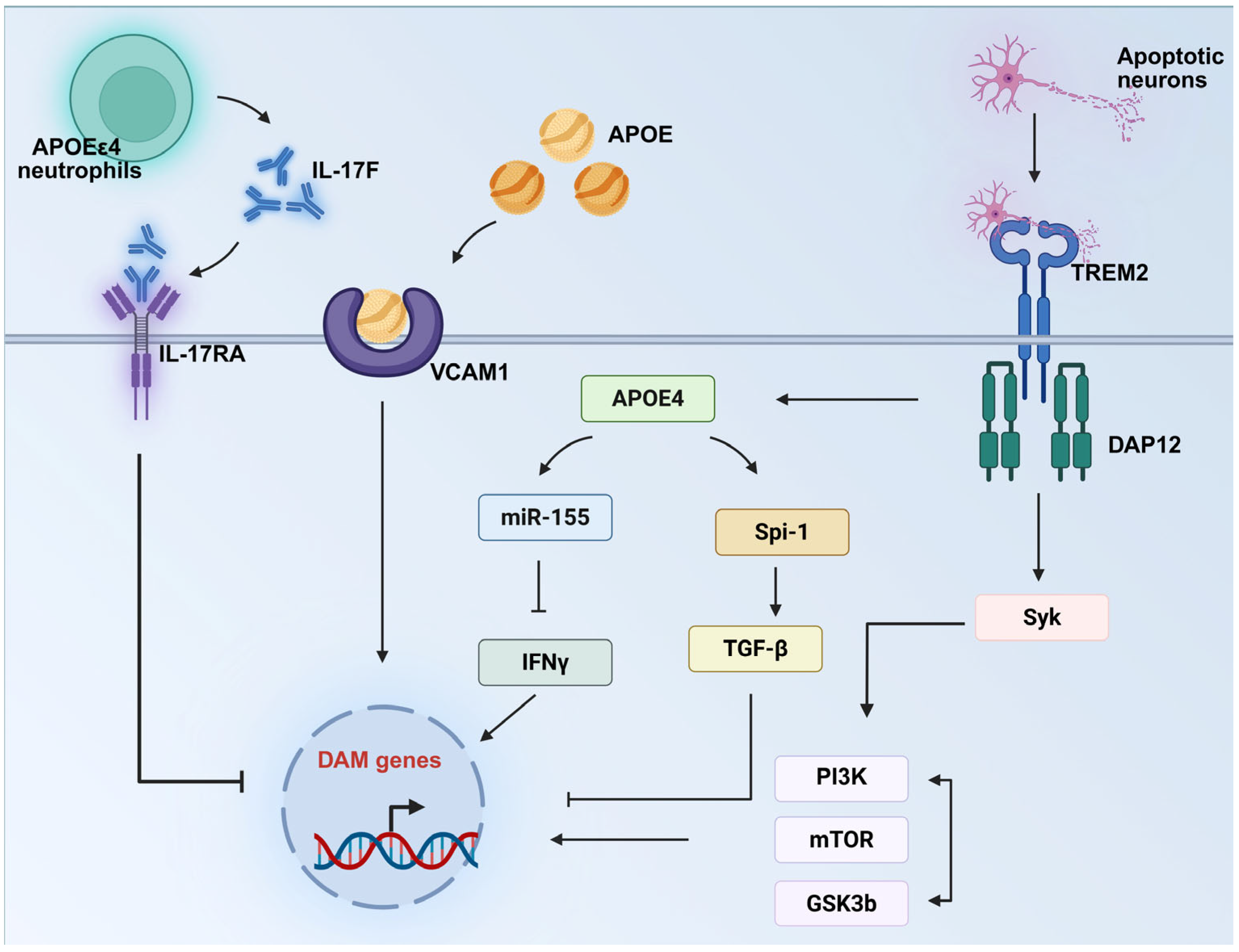
5. The APOE–Microglia Axis: A Novel Concept in Alzheimer’s Disease Pathogenesis and Therapy

The body of evidence presented in the previous section highlights that APOE alleles influence microglial phenotypes, inflammatory responses, phagocytic function, and lipid metabolism, forming a closely interconnected signaling axis—the APOE–microglia axis. This axis reveals complex interactions among genetics, immunity, and metabolism in AD and links the genetic risk of APOE alleles to the dynamic functions of microglia. At its core, the APOE–microglia axis involves bidirectional regulation between APOE proteins and microglia: microglia upregulate APOE expression in the AD pathological environment, while APOE proteins in turn shape microglial functional states, thereby influencing key AD pathological processes such as amyloid pathology, tau pathology, and neuroinflammation. The three major APOE alleles drive functional divergence in microglia by differentially regulating their phenotypes, states, and molecular pathways. Elucidating these mechanisms would both uncover the cellular basis of AD genetic susceptibility and identify critical targets for APOE genotype-based personalized therapies.

6. Clinical Therapeutic Prospects

The APOE–microglia axis represents a pivotal target for AD therapeutics. Promising clinical applications include APOEε2 gene therapy, which has shown potential in ameliorating AD pathology [27]. Similarly, gene-editing techniques aimed at modifying the APOE genotype in microglia could restore normal phagocytic function, enhancing Aβ plaques clearance while mitigating excessive neuroinflammatory responses [29,141]. These approaches aim to reduce APOE4 levels and increase APOE2 levels, which have been demonstrated to exhibit therapeutic efficacy [142]. For APOEε4 carriers, reducing APOE levels has emerged as a viable therapeutic strategy. However, precise control of this reduction is critical to avoid off-target effects [143]. Furthermore, therapeutic strategies targeting APOE-regulated specific sites hold significant promise (Table 3). However, given that AD pathogenesis involves a multifaceted interplay of genetic, epigenetic, and environmental factors, the APOE–microglia axis represents only one component of this intricate network. Thus, targeting it alone is unlikely to fully halt AD progression, underscoring the need for integrated, comprehensive therapeutic strategies.
However, APOE–microglia axis-related therapies also face challenges. It should be noted that APOE is expressed throughout the body. Therefore, when applying APOE-targeted drugs in vivo, the greater challenge may lie in off-target effects, necessitating the prioritization of tissue and cell specificity in drug design [147]. Regarding reducing the systemic effects of APOE-targeted drugs, there are multiple research directions worthy of in-depth exploration in the future. From the perspective of drug delivery systems, the development of brain-targeted delivery vectors represents one of the key directions. For instance, by leveraging specific transporters on the blood–brain barrier (BBB), such as transferrin receptors and insulin receptors, drugs targeting APOE can be conjugated to ligands that bind to these transporters. This enables receptor-mediated transcytosis across the BBB, facilitating the preferential enrichment of drugs in the brain and reducing their distribution in other peripheral tissues [148,149]. In terms of gene therapy, viral vectors can be used to achieve brain-specific gene editing or regulation. Adeno-associated virus (AAV) has good safety and low immunogenicity, and there are multiple serotypes, some of which have a high tropism for brain tissues. For example, by modifying the AAV vector to carry therapeutic genes for APOE (such as genes regulating APOE expression or correcting the abnormal structure of APOE4) and using brain-specific promoters in promoter selection, the therapeutic genes can only function in the brain, avoiding unnecessary interference with the function of APOE in other tissues throughout the body [27,150].

7. Future Research Directions

While the APOE–microglia interplay offers crucial insights into AD mechanisms and therapeutic opportunities, key aspects of this regulatory axis remain elusive. Emerging evidence indicates that microglia harbor multiple signaling pathways that are either regulated by APOE alleles or involved in APOE interactions. Under pathological AD conditions, APOE may dysregulate these pathways, triggering microglial dysfunction that ultimately drives AD progression [56,65,76,80,81,91]. Therefore, systematically elucidating the precise molecular mechanisms of APOE-mediated pathway regulation, identifying novel APOE-regulated pathways, and deciphering their inter-pathway crosstalk are critical for establishing a comprehensive APOE–microglial functional regulatory network in AD. Notably, distinct APOE alleles (ε2/ε3/ε4) may exert differential regulatory effects on these pathways [56,76,91]. In-depth investigation of these allele-specific regulatory mechanisms will provide crucial theoretical insights into AD pathogenesis.
Another crucial area is to dissect the heterogeneous effects of APOE alleles on microglial function. Microglial cells exhibit remarkable heterogeneity, enabling them to differentially respond to diverse stimuli (e.g., infection, injury, or degeneration) and thereby enhance the adaptive capacity of the CNS [151]. Current research utilizing single-cell multi-omics technologies can precisely identify distinct phenotypic and functional states of microglia regulated by APOE alleles, such as DAM/MGnD microglia [64], LDAM [75], and TIM [73]. The identification of APOE-regulated microglial phenotypes not only provides critical therapeutic targets for developing precision interventions against specific microglial states, but also offers novel biomarkers for monitoring disease progression and evaluating prognosis [64,73,74,75].
Furthermore, microglia do not drive AD pathogenesis in isolation—rather, multiple CNS cell types, including astrocytes and neurons, collectively contribute to disease progression through extensive cross-talk with microglia [152,153]. It is therefore imperative to investigate how APOEε4 modulates these intercellular interactions, rather than focusing solely on its cell-autonomous effects on microglial function [82,154]. Similarly, the inflammatory response, lipid metabolism, and phagocytic functions of microglia are not isolated processes, but rather interconnected and integrated through intricate intracellular signaling pathways and molecular mechanisms. These functional interactions collectively form a comprehensive regulatory network that governs microglia’s physiological and pathological roles in the central nervous system [155]. As previously discussed, APOE exerts holistic regulation over microglial functions. APOE (particularly the APOE4 isoform)-mediated lipid metabolic dysregulation is significantly associated with aberrant inflammatory responses and phagocytic dysfunction, forming an interconnected pathological network. However, the precise molecular mechanisms underlying this multidimensional regulation remain to be elucidated.
Further research into the neuroprotective mechanisms of APOEε2 and APOEε3 is also critical. Current studies demonstrate a pronounced “APOEε4-centric” bias, with severely inadequate exploration of APOEε2 and APOEε3. This imbalance significantly hinders our ability to develop a systematic understanding of the APOE family’s comprehensive mechanistic role in AD. It should be noted that APOEε4 represents the ancestral allele conserved in non-human primates and all examined mammalian species. The more pertinent question is why and how APOEε2 and APOEε3 alleles evolved to modify microglial (and other cellular) functions, rather than why APOEε4 exhibits detrimental effects [21]. Moreover, exploring how APOEε2 and APOEε3 modulate microglial activity and their functional differences compared to APOEε4 may reveal new therapeutic targets. Additionally, studying rare APOEε3 variants, such as APOE3ch and APOE3-Jac, may provide insights into protective mechanisms against AD [33,34,137].

8. Conclusions

In the pathogenesis of AD, the APOE–microglia axis serves as a central regulatory hub. Different APOE alleles (APOEε2, APOEε3, and APOEε4) profoundly shape AD pathological progression by governing microglial phenotypes, inflammatory signaling, phagocytic capacity, and lipid metabolic homeostasis. Specifically, APOEε4 drives AD progression through a cascade of dysfunctions, including aberrant microglial hyperactivation, impaired Aβ clearance, exacerbated neuroinflammation, and disrupted lipid homeostasis. In contrast, APOEε2 confers neuroprotection by suppressing excessive microglial activation, enhancing Aβ phagocytosis, and attenuating inflammatory responses, thereby reducing AD susceptibility. APOEε3 maintains basal microglial lipid metabolism and inflammatory balance, while rare protective variants (e.g., APOE3ch and APOE3-Jac) further mitigate AD risk by fine-tuning microglial functional profiles. Future investigations should prioritize dissecting the precise molecular mechanisms of the APOE–microglia axis, with a focus on the regulatory roles of APOEε2 and APOEε3. Additionally, elucidating the intercellular crosstalk between microglia and other CNS) cell types mediated by this axis is essential. Such mechanistic insights will provide a robust theoretical foundation for developing genotype-specific therapeutic strategies targeting the APOE-microglia axis in AD.

Author Contributions

Writing—original draft preparation, A.L. and T.W.; writing—review and editing, L.Y. and Y.Z.; visualization, A.L. All authors have read and agreed to the published version of the manuscript.

Funding

This work was supported by the National Natural Science Foundation of China (Grant no. 32371211 to Yu Zhou).

Acknowledgments

All figures were created using BioRender.com (under a paid license).

Conflicts of Interest

The authors declare no conflicts of interest.

References

  1. Rogus-Pulia, N.; Foundas, A.L.; Mueller, K.D. Alzheimer’s Disease. In Neurologic and Neurodegenerative Diseases of the Larynx; Weissbrod, P.A., Francis, D.O., Eds.; Springer International Publishing: Cham, Switzerland, 2020; pp. 177–189. [Google Scholar] [CrossRef]
  2. Tahami Monfared, A.A.; Byrnes, M.J.; White, L.A.; Zhang, Q. Alzheimer’s Disease: Epidemiology and Clinical Progression. Neurol. Ther. 2022, 11, 553–569. [Google Scholar] [CrossRef] [PubMed]
  3. Dumurgier, J.; Sabia, S. Epidemiology of Alzheimer’s disease: Latest trends. Rev. Prat. 2020, 70, 149–151. [Google Scholar] [PubMed]
  4. Serrano-Pozo, A.; Frosch, M.P.; Masliah, E.; Hyman, B.T. Neuropathological alterations in Alzheimer disease. Cold Spring Harb. Perspect. Med. 2011, 1, a006189. [Google Scholar] [CrossRef] [PubMed]
  5. AmeliMojarad, M.; AmeliMojarad, M. The neuroinflammatory role of microglia in Alzheimer’s disease and their associated therapeutic targets. CNS Neurosci. Ther. 2024, 30, e14856. [Google Scholar] [CrossRef]
  6. Al-Ghraiybah, N.F.; Wang, J.; Alkhalifa, A.E.; Roberts, A.B.; Raj, R.; Yang, E.; Kaddoumi, A. Glial Cell-Mediated Neuroinflammation in Alzheimer’s Disease. Int. J. Mol. Sci. 2022, 23, 10572. [Google Scholar] [CrossRef]
  7. Li, Y.; Chen, X.; Zhou, M.; Feng, S.; Peng, X.; Wang, Y. Microglial TLR4/NLRP3 Inflammasome Signaling in Alzheimer’s Disease. J. Alzheimer’s Dis. 2024, 97, 75–88. [Google Scholar] [CrossRef]
  8. Bird, T.D. Genetic aspects of Alzheimer disease. Genet. Med. 2008, 10, 231–239. [Google Scholar] [CrossRef]
  9. Laws, S.M.; Hone, E.; Gandy, S.; Martins, R.N. Expanding the association between the APOE gene and the risk of Alzheimer’s disease: Possible roles for APOE promoter polymorphisms and alterations in APOE transcription. J. Neurochem. 2003, 84, 1215–1236. [Google Scholar] [CrossRef]
  10. Wang, C.; Xiong, M.; Gratuze, M.; Bao, X.; Shi, Y.; Andhey, P.S.; Manis, M.; Schroeder, C.; Yin, Z.; Madore, C.; et al. Selective removal of astrocytic APOE4 strongly protects against tau-mediated neurodegeneration and decreases synaptic phagocytosis by microglia. Neuron 2021, 109, 1657–1674.e1657. [Google Scholar] [CrossRef]
  11. Sala Frigerio, C.; Wolfs, L.; Fattorelli, N.; Thrupp, N.; Voytyuk, I.; Schmidt, I.; Mancuso, R.; Chen, W.T.; Woodbury, M.E.; Srivastava, G.; et al. The Major Risk Factors for Alzheimer’s Disease: Age, Sex, and Genes Modulate the Microglia Response to Aβ Plaques. Cell Rep. 2019, 27, 1293–1306.e1296. [Google Scholar] [CrossRef]
  12. Kang, S.S.; Ebbert, M.T.W.; Baker, K.E.; Cook, C.; Wang, X.; Sens, J.P.; Kocher, J.P.; Petrucelli, L.; Fryer, J.D. Microglial translational profiling reveals a convergent APOE pathway from aging, amyloid, and tau. J. Exp. Med. 2018, 215, 2235–2245. [Google Scholar] [CrossRef] [PubMed]
  13. Zhao, N.; Liu, C.C.; Qiao, W.; Bu, G. Apolipoprotein E, Receptors, and Modulation of Alzheimer’s Disease. Biol. Psychiatry 2018, 83, 347–357. [Google Scholar] [CrossRef] [PubMed]
  14. Raulin, A.C.; Doss, S.V.; Trottier, Z.A.; Ikezu, T.C.; Bu, G.; Liu, C.C. ApoE in Alzheimer’s disease: Pathophysiology and therapeutic strategies. Mol. Neurodegener. 2022, 17, 72. [Google Scholar] [CrossRef] [PubMed]
  15. Weisgraber, K.H.; Rall, S.C., Jr.; Mahley, R.W. Human E apoprotein heterogeneity. Cysteine-arginine interchanges in the amino acid sequence of the apo-E isoforms. J. Biol. Chem. 1981, 256, 9077–9083. [Google Scholar] [CrossRef]
  16. Liu, C.C.; Liu, C.C.; Kanekiyo, T.; Xu, H.; Bu, G. Apolipoprotein E and Alzheimer disease: Risk, mechanisms and therapy. Nat. Rev. Neurol. 2013, 9, 106–118. [Google Scholar] [CrossRef]
  17. Farrer, L.A.; Cupples, L.A.; Haines, J.L.; Hyman, B.; Kukull, W.A.; Mayeux, R.; Myers, R.H.; Pericak-Vance, M.A.; Risch, N.; van Duijn, C.M. Effects of age, sex, and ethnicity on the association between apolipoprotein E genotype and Alzheimer disease. A meta-analysis. APOE and Alzheimer Disease Meta Analysis Consortium. J. Am. Med. Assoc. 1997, 278, 1349–1356. [Google Scholar] [CrossRef]
  18. Yamazaki, Y.; Zhao, N.; Caulfield, T.R.; Liu, C.C.; Bu, G. Apolipoprotein E and Alzheimer disease: Pathobiology and targeting strategies. Nat. Rev. Neurol. 2019, 15, 501–518. [Google Scholar] [CrossRef]
  19. Corder, E.H.; Saunders, A.M.; Risch, N.J.; Strittmatter, W.J.; Schmechel, D.E.; Gaskell, P.C.; Rimmler, J.B., Jr.; Locke, P.A.; Conneally, P.M.; Schmader, K.E.; et al. Protective effect of apolipoprotein E type 2 allele for late onset Alzheimer disease. Nat. Genet. 1994, 7, 180–184. [Google Scholar] [CrossRef]
  20. Genin, E.; Hannequin, D.; Wallon, D.; Sleegers, K.; Hiltunen, M.; Combarros, O.; Bullido, M.J.; Engelborghs, S.; De Deyn, P.; Berr, C.; et al. APOE and Alzheimer disease: A major gene with semi-dominant inheritance. Mol. Psychiatry 2011, 16, 903–907. [Google Scholar] [CrossRef]
  21. Huebbe, P.; Rimbach, G. Evolution of human apolipoprotein E (APOE) isoforms: Gene structure, protein function and interaction with dietary factors. Ageing Res. Rev. 2017, 37, 146–161. [Google Scholar] [CrossRef]
  22. Therriault, J.; Benedet, A.L.; Pascoal, T.A.; Mathotaarachchi, S.; Savard, M.; Chamoun, M.; Thomas, E.; Kang, M.S.; Lussier, F.; Tissot, C.; et al. APOEε4 potentiates the relationship between amyloid-β and tau pathologies. Mol. Psychiatry 2021, 26, 5977–5988. [Google Scholar] [CrossRef] [PubMed]
  23. Kloske, C.M.; Dugan, A.; Woolums, A.E.; Lee, T.; Anderson, S.; Patel, E.; Abner, E.L.; Nelson, P.T.; Fardo, D.W.; Wilcock, D.M. Impaired neuroinflammatory response of ApoE4 in Alzheimer’s disease patients. Alzheimer’s Dement. 2020, 16, e041052. [Google Scholar] [CrossRef]
  24. Friedberg, J.S.; Aytan, N.; Cherry, J.D.; Xia, W.; Standring, O.J.; Alvarez, V.E.; Nicks, R.; Svirsky, S.; Meng, G.; Jun, G.; et al. Associations between brain inflammatory profiles and human neuropathology are altered based on apolipoprotein E ε4 genotype. Sci. Rep. 2020, 10, 2924. [Google Scholar] [CrossRef]
  25. Lopresti, B.J.; Campbell, E.M.; Yu, Z.; Anderson, S.J.; Cohen, A.D.; Minhas, D.S.; Snitz, B.E.; Royse, S.K.; Becker, C.R.; Aizenstein, H.J.; et al. Influence of apolipoprotein-E genotype on brain amyloid load and longitudinal trajectories. Neurobiol. Aging 2020, 94, 111–120. [Google Scholar] [CrossRef]
  26. Wang, N.; Cai, L.; Pei, X.; Lin, Z.; Huang, L.; Liang, C.; Wei, M.; Shao, L.; Guo, T.; Huang, F.; et al. Microglial apolipoprotein E particles contribute to neuronal senescence and synaptotoxicity. iScience 2024, 27, 110006. [Google Scholar] [CrossRef]
  27. Jackson, R.J.; Keiser, M.S.; Meltzer, J.C.; Fykstra, D.P.; Dierksmeier, S.E.; Hajizadeh, S.; Kreuzer, J.; Morris, R.; Melloni, A.; Nakajima, T.; et al. APOE2 gene therapy reduces amyloid deposition and improves markers of neuroinflammation and neurodegeneration in a mouse model of Alzheimer disease. Mol. Ther. 2024, 32, 1373–1386. [Google Scholar] [CrossRef]
  28. Groot, C. Multimodal imaging correlates of the protective APOE2 allele. Alzheimer’s Dement. 2020, 16, e039421. [Google Scholar] [CrossRef]
  29. Li, Z.; Shue, F.; Zhao, N.; Shinohara, M.; Bu, G. APOE2: Protective mechanism and therapeutic implications for Alzheimer’s disease. Mol. Neurodegener. 2020, 15, 63. [Google Scholar] [CrossRef]
  30. Serrano-Pozo, A.; Qian, J.; Monsell, S.E.; Betensky, R.A.; Hyman, B.T. APOEε2 is associated with milder clinical and pathological Alzheimer disease. Ann. Neurol. 2015, 77, 917–929. [Google Scholar] [CrossRef]
  31. Goldberg, T.E.; Huey, E.D.; Devanand, D.P. Association of APOE e2 genotype with Alzheimer’s and non-Alzheimer’s neurodegenerative pathologies. Nat. Commun. 2020, 11, 4727. [Google Scholar] [CrossRef]
  32. Roda, A.R.; Montoliu-Gaya, L.; Villegas, S. The Role of Apolipoprotein E Isoforms in Alzheimer’s Disease. J. Alzheimer’s Dis. 2019, 68, 459–471. [Google Scholar] [CrossRef] [PubMed]
  33. Medway, C.W.; Abdul-Hay, S.; Mims, T.; Ma, L.; Bisceglio, G.; Zou, F.; Pankratz, S.; Sando, S.B.; Aasly, J.O.; Barcikowska, M.; et al. ApoE variant p.V236E is associated with markedly reduced risk of Alzheimer’s disease. Mol. Neurodegener. 2014, 9, 11. [Google Scholar] [CrossRef] [PubMed]
  34. Arboleda-Velasquez, J.F.; Lopera, F.; O’Hare, M.; Delgado-Tirado, S.; Marino, C.; Chmielewska, N.; Saez-Torres, K.L.; Amarnani, D.; Schultz, A.P.; Sperling, R.A.; et al. Resistance to autosomal dominant Alzheimer’s disease in an APOE3 Christchurch homozygote: A case report. Nat. Med. 2019, 25, 1680–1683. [Google Scholar] [CrossRef] [PubMed]
  35. Hansen, D.V.; Hanson, J.E.; Sheng, M. Microglia in Alzheimer’s disease. J. Cell Biol. 2018, 217, 459–472. [Google Scholar] [CrossRef]
  36. Uddin, M.S.; Lim, L.W. Glial cells in Alzheimer’s disease: From neuropathological changes to therapeutic implications. Ageing Res. Rev. 2022, 78, 101622. [Google Scholar] [CrossRef]
  37. Bolós, M.; Llorens-Martín, M.; Jurado-Arjona, J.; Hernández, F.; Rábano, A.; Avila, J. Direct Evidence of Internalization of Tau by Microglia In Vitro and In Vivo. J. Alzheimer’s Dis. 2016, 50, 77–87. [Google Scholar] [CrossRef]
  38. Skuljec, J.; Sun, H.; Pul, R.; Bénardais, K.; Ragancokova, D.; Moharregh-Khiabani, D.; Kotsiari, A.; Trebst, C.; Stangel, M. CCL5 induces a pro-inflammatory profile in microglia in vitro. Cell. Immunol. 2011, 270, 164–171. [Google Scholar] [CrossRef]
  39. Doens, D.; Fernández, P.L. Microglia receptors and their implications in the response to amyloid β for Alzheimer’s disease pathogenesis. J. Neuroinflammation 2014, 11, 48. [Google Scholar] [CrossRef]
  40. Xie, Z.; Meng, J.; Wu, Z.; Nakanishi, H.; Hayashi, Y.; Kong, W.; Lan, F.; Narengaowa Yang, Q.; Qing, H.; Ni, J. The Dual Nature of Microglia in Alzheimer’s Disease: A Microglia-Neuron Crosstalk Perspective. Neuroscientist 2023, 29, 616–638. [Google Scholar] [CrossRef]
  41. Maphis, N.; Xu, G.; Kokiko-Cochran, O.N.; Jiang, S.; Cardona, A.; Ransohoff, R.M.; Lamb, B.T.; Bhaskar, K. Reactive microglia drive tau pathology and contribute to the spreading of pathological tau in the brain. Brain 2015, 138 Pt 6, 1738–1755. [Google Scholar] [CrossRef]
  42. Miao, J.; Ma, H.; Yang, Y.; Liao, Y.; Lin, C.; Zheng, J.; Yu, M.; Lan, J. Microglia in Alzheimer’s disease: Pathogenesis, mechanisms, and therapeutic potentials. Front. Aging Neurosci. 2023, 15, 1201982. [Google Scholar] [CrossRef] [PubMed]
  43. Botella Lucena, P.; Heneka, M.T. Inflammatory aspects of Alzheimer’s disease. Acta Neuropathol. 2024, 148, 31. [Google Scholar] [CrossRef] [PubMed]
  44. Wang, N.; Wei, M.; Li, X.; Jia, L.; Li, J.; Xu, H.; Zhang, Y.-w.; Zheng, H. Apolipoprotein E4 expressed by microglia impairs microglial functions and enhances neurotoxicity. Res. Sq. 2020. [Google Scholar] [CrossRef]
  45. Shi, Y.; Yamada, K.; Liddelow, S.A.; Smith, S.T.; Zhao, L.; Luo, W.; Tsai, R.M.; Spina, S.; Grinberg, L.T.; Rojas, J.C.; et al. ApoE4 markedly exacerbates tau-mediated neurodegeneration in a mouse model of tauopathy. Nature 2017, 549, 523–527. [Google Scholar] [CrossRef]
  46. Liu, C.C.; Wang, N.; Chen, Y.; Inoue, Y.; Shue, F.; Ren, Y.; Wang, M.; Qiao, W.; Ikezu, T.C.; Li, Z.; et al. Cell-autonomous effects of APOE4 in restricting microglial response in brain homeostasis and Alzheimer’s disease. Nat. Immunol. 2023, 24, 1854–1866. [Google Scholar] [CrossRef]
  47. Rao, A.; Chen, N.; Kim, M.J.; Blumenfeld, J.; Yip, O.; Liang, Z.; Shostak, D.; Hao, Y.; Nelson, M.R.; Koutsodendris, N. Microglia depletion reduces human neuronal APOE4-related pathologies in a chimeric Alzheimer’s disease model. Cell Stem Cell 2025, 32, 86–104.e107. [Google Scholar] [CrossRef]
  48. Najm, R.; Zalocusky, K.A.; Zilberter, M.; Yoon, S.Y.; Hao, Y.; Koutsodendris, N.; Nelson, M.; Rao, A.; Taubes, A.; Jones, E.A.; et al. In Vivo Chimeric Alzheimer’s Disease Modeling of Apolipoprotein E4 Toxicity in Human Neurons. Cell Rep. 2020, 32, 107962. [Google Scholar] [CrossRef]
  49. Fitz, N.F.; Nam, K.N.; Wolfe, C.M.; Letronne, F.; Playso, B.E.; Iordanova, B.E.; Kozai, T.D.Y.; Biedrzycki, R.J.; Kagan, V.E.; Tyurina, Y.Y.; et al. Phospholipids of APOE lipoproteins activate microglia in an isoform-specific manner in preclinical models of Alzheimer’s disease. Nat. Commun. 2021, 12, 3416. [Google Scholar] [CrossRef]
  50. Sepulveda, J.; Kim, J.Y.; Binder, J.; Vicini, S.; Rebeck, G.W. APOE4 genotype and aging impair injury-induced microglial behavior in brain slices, including toward Aβ, through P2RY12. Mol. Neurodegener. 2024, 19, 24. [Google Scholar] [CrossRef]
  51. Minett, T.; Classey, J.; Matthews, F.E.; Fahrenhold, M.; Taga, M.; Brayne, C.; Ince, P.G.; Nicoll, J.A.; Boche, D. Microglial immunophenotype in dementia with Alzheimer’s pathology. J. Neuroinflammation 2016, 13, 135. [Google Scholar] [CrossRef]
  52. Maezawa, I.; Nivison, M.; Montine, K.S.; Maeda, N.; Montine, T.J. Neurotoxicity from innate immune response is greatest with targeted replacement of E4 allele of apolipoprotein E gene and is mediated by microglial p38MAPK. Faseb J. 2006, 20, 797–799. [Google Scholar] [CrossRef] [PubMed]
  53. Wang, N.; Wang, M.; Jeevaratnam, S.; Rosenberg, C.; Ikezu, T.C.; Shue, F.; Doss, S.V.; Alnobani, A.; Martens, Y.A.; Wren, M.; et al. Opposing effects of apoE2 and apoE4 on microglial activation and lipid metabolism in response to demyelination. Mol. Neurodegener. 2022, 17, 75. [Google Scholar] [CrossRef] [PubMed]
  54. Iannucci, J.; Sen, A.; Grammas, P. Isoform-Specific Effects of Apolipoprotein E on Markers of Inflammation and Toxicity in Brain Glia and Neuronal Cells In Vitro. Curr Issues Mol. Biol. 2021, 43, 215–225. [Google Scholar] [CrossRef] [PubMed]
  55. Jiang, Q.; Lee, C.Y.; Mandrekar, S.; Wilkinson, B.; Cramer, P.; Zelcer, N.; Mann, K.; Lamb, B.; Willson, T.M.; Collins, J.L.; et al. ApoE promotes the proteolytic degradation of Abeta. Neuron 2008, 58, 681–693. [Google Scholar] [CrossRef]
  56. Yin, Z.; Rosenzweig, N.; Kleemann, K.L.; Zhang, X.; Brandão, W.; Margeta, M.A.; Schroeder, C.; Sivanathan, K.N.; Silveira, S.; Gauthier, C.; et al. APOE4 impairs the microglial response in Alzheimer’s disease by inducing TGFβ-mediated checkpoints. Nat. Immunol. 2023, 24, 1839–1853. [Google Scholar] [CrossRef]
  57. Bohlen, C.J.; Friedman, B.A.; Dejanovic, B.; Sheng, M. Microglia in Brain Development, Homeostasis, and Neurodegeneration. Annu. Rev. Genet. 2019, 53, 263–288. [Google Scholar] [CrossRef]
  58. Benarroch, E.E. Microglia: Multiple roles in surveillance, circuit shaping, and response to injury. Neurology 2013, 81, 1079–1088. [Google Scholar] [CrossRef]
  59. Moser, V.A.; Workman, M.J.; Hurwitz, S.J.; Lipman, R.M.; Pike, C.J.; Svendsen, C.N. Microglial transcription profiles in mouse and human are driven by APOE4 and sex. iScience 2021, 24, 103238. [Google Scholar] [CrossRef]
  60. Lin, Y.T.; Seo, J.; Gao, F.; Feldman, H.M.; Wen, H.L.; Penney, J.; Cam, H.P.; Gjoneska, E.; Raja, W.K.; Cheng, J.; et al. APOE4 Causes Widespread Molecular and Cellular Alterations Associated with Alzheimer’s Disease Phenotypes in Human iPSC-Derived Brain Cell Types. Neuron 2018, 98, 1141–1154.e1147. [Google Scholar] [CrossRef]
  61. Muth, C.; Hartmann, A.; Sepulveda-Falla, D.; Glatzel, M.; Krasemann, S. Phagocytosis of Apoptotic Cells Is Specifically Upregulated in ApoE4 Expressing Microglia in vitro. Front. Cell. Neurosci. 2019, 13, 181. [Google Scholar] [CrossRef]
  62. Egensperger, R.; Kösel, S.; von Eitzen, U.; Graeber, M.B. Microglial activation in Alzheimer disease: Association with APOE genotype. Brain Pathol. 1998, 8, 439–447. [Google Scholar] [CrossRef] [PubMed]
  63. Ferrari-Souza, J.P.; Lussier, F.Z.; Leffa, D.T.; Therriault, J.; Tissot, C.; Bellaver, B.; Ferreira, P.C.L.; Malpetti, M.; Wang, Y.T.; Povala, G.; et al. APOEε4 associates with microglial activation independently of Aβ plaques and tau tangles. Sci. Adv. 2023, 9, eade1474. [Google Scholar] [CrossRef]
  64. Keren-Shaul, H.; Spinrad, A.; Weiner, A.; Matcovitch-Natan, O.; Dvir-Szternfeld, R.; Ulland, T.K.; David, E.; Baruch, K.; Lara-Astaiso, D.; Toth, B.; et al. A Unique Microglia Type Associated with Restricting Development of Alzheimer’s Disease. Cell 2017, 169, 1276–1290.e1217. [Google Scholar] [CrossRef] [PubMed]
  65. Krasemann, S.; Madore, C.; Cialic, R.; Baufeld, C.; Calcagno, N.; El Fatimy, R.; Beckers, L.; O’Loughlin, E.; Xu, Y.; Fanek, Z.; et al. The TREM2-APOE Pathway Drives the Transcriptional Phenotype of Dysfunctional Microglia in Neurodegenerative Diseases. Immunity 2017, 47, 566–581.e569. [Google Scholar] [CrossRef]
  66. Prinz, M.; Masuda, T.; Wheeler, M.A.; Quintana, F.J. Microglia and Central Nervous System-Associated Macrophages-From Origin to Disease Modulation. Annu. Rev. Immunol. 2021, 39, 251–277. [Google Scholar] [CrossRef]
  67. Shi, Y.; Holtzman, D.M. Interplay between innate immunity and Alzheimer disease: APOE and TREM2 in the spotlight. Nat. Rev. Immunol. 2018, 18, 759–772. [Google Scholar] [CrossRef]
  68. Yuan, P.; Condello, C.; Keene, C.D.; Wang, Y.; Bird, T.D.; Paul, S.M.; Luo, W.; Colonna, M.; Baddeley, D.; Grutzendler, J. TREM2 Haplodeficiency in Mice and Humans Impairs the Microglia Barrier Function Leading to Decreased Amyloid Compaction and Severe Axonal Dystrophy. Neuron 2016, 90, 724–739. [Google Scholar] [CrossRef]
  69. Hong, S.; Beja-Glasser, V.F.; Nfonoyim, B.M.; Frouin, A.; Li, S.; Ramakrishnan, S.; Merry, K.M.; Shi, Q.; Rosenthal, A.; Barres, B.A.; et al. Complement and microglia mediate early synapse loss in Alzheimer mouse models. Science 2016, 352, 712–716. [Google Scholar] [CrossRef]
  70. Lui, H.; Zhang, J.; Makinson, S.R.; Cahill, M.K.; Kelley, K.W.; Huang, H.Y.; Shang, Y.; Oldham, M.C.; Martens, L.H.; Gao, F.; et al. Progranulin Deficiency Promotes Circuit-Specific Synaptic Pruning by Microglia via Complement Activation. Cell 2016, 165, 921–935. [Google Scholar] [CrossRef]
  71. Simpson, D.S.A.; Oliver, P.L. ROS Generation in Microglia: Understanding Oxidative Stress and Inflammation in Neurodegenerative Disease. Antioxidants 2020, 9, 743. [Google Scholar] [CrossRef]
  72. Leng, F.; Edison, P. Neuroinflammation and microglial activation in Alzheimer disease: Where do we go from here? Nat. Rev. Neurol. 2021, 17, 157–172. [Google Scholar] [CrossRef] [PubMed]
  73. Millet, A.; Ledo, J.H.; Tavazoie, S.F. An exhausted-like microglial population accumulates in aged and APOE4 genotype Alzheimer’s brains. Immunity 2024, 57, 153–170.e156. [Google Scholar] [CrossRef] [PubMed]
  74. Nguyen, A.T.; Wang, K.; Hu, G.; Wang, X.; Miao, Z.; Azevedo, J.A.; Suh, E.; Van Deerlin, V.M.; Choi, D.; Roeder, K.; et al. APOE and TREM2 regulate amyloid-responsive microglia in Alzheimer’s disease. Acta Neuropathol. 2020, 140, 477–493. [Google Scholar] [CrossRef] [PubMed]
  75. Marschallinger, J.; Iram, T.; Zardeneta, M.; Lee, S.E.; Lehallier, B.; Haney, M.S.; Pluvinage, J.V.; Mathur, V.; Hahn, O.; Morgens, D.W.; et al. Lipid-droplet-accumulating microglia represent a dysfunctional and proinflammatory state in the aging brain. Nat. Neurosci. 2020, 23, 194–208. [Google Scholar] [CrossRef]
  76. Haney, M.S.; Pálovics, R.; Munson, C.N.; Long, C.; Johansson, P.K.; Yip, O.; Dong, W.; Rawat, E.; West, E.; Schlachetzki, J.C.M.; et al. APOE4/4 is linked to damaging lipid droplets in Alzheimer’s disease microglia. Nature 2024, 628, 154–161. [Google Scholar] [CrossRef]
  77. Lee, S.; Devanney, N.A.; Golden, L.R.; Smith, C.T.; Schwartz, J.L.; Walsh, A.E.; Clarke, H.A.; Goulding, D.S.; Allenger, E.J.; Morillo-Segovia, G.; et al. APOE modulates microglial immunometabolism in response to age, amyloid pathology, and inflammatory challenge. Cell Rep. 2023, 42, 112196. [Google Scholar] [CrossRef]
  78. Litvinchuk, A.; Suh, J.H.; Guo, J.L.; Lin, K.; Davis, S.S.; Bien-Ly, N.; Tycksen, E.; Tabor, G.T.; Remolina Serrano, J.; Manis, M.; et al. Amelioration of Tau and ApoE4-linked glial lipid accumulation and neurodegeneration with an LXR agonist. Neuron 2024, 112, 384–403.e388. [Google Scholar] [CrossRef]
  79. Koutsodendris, N.; Blumenfeld, J.; Agrawal, A.; Traglia, M.; Grone, B.; Zilberter, M.; Yip, O.; Rao, A.; Nelson, M.R.; Hao, Y.; et al. Neuronal APOE4 removal protects against tau-mediated gliosis, neurodegeneration and myelin deficits. Nat. Aging 2023, 3, 275–296. [Google Scholar] [CrossRef]
  80. Wang, S.; Sudan, R.; Peng, V.; Zhou, Y.; Du, S.; Yuede, C.M.; Lei, T.; Hou, J.; Cai, Z.; Cella, M.; et al. TREM2 drives microglia response to amyloid-β via SYK-dependent and -independent pathways. Cell 2022, 185, 4153–4169.e4119. [Google Scholar] [CrossRef]
  81. Yin, Z.; Herron, S.; Silveira, S.; Kleemann, K.; Gauthier, C.; Mallah, D.; Cheng, Y.; Margeta, M.A.; Pitts, K.M.; Barry, J.L.; et al. Identification of a protective microglial state mediated by miR-155 and interferon-γ signaling in a mouse model of Alzheimer’s disease. Nat. Neurosci. 2023, 26, 1196–1207. [Google Scholar] [CrossRef]
  82. Rosenzweig, N.; Kleemann, K.L.; Rust, T.; Carpenter, M.; Grucci, M.; Aronchik, M.; Brouwer, N.; Valenbreder, I.; Cooper-Hohn, J.; Iyer, M.; et al. Sex-dependent APOE4 neutrophil-microglia interactions drive cognitive impairment in Alzheimer’s disease. Nat. Med. 2024, 30, 2990–3003. [Google Scholar] [CrossRef] [PubMed]
  83. Lau, S.F.; Wu, W.; Wong, H.Y.; Ouyang, L.; Qiao, Y.; Xu, J.; Lau, J.H.; Wong, C.; Jiang, Y.; Holtzman, D.M.; et al. The VCAM1-ApoE pathway directs microglial chemotaxis and alleviates Alzheimer’s disease pathology. Nat. Aging 2023, 3, 1219–1236. [Google Scholar] [CrossRef] [PubMed]
  84. Machlovi, S.I.; Neuner, S.M.; Hemmer, B.M.; Khan, R.; Liu, Y.; Huang, M.; Zhu, J.D.; Castellano, J.M.; Cai, D.; Marcora, E.; et al. APOE4 confers transcriptomic and functional alterations to primary mouse microglia. Neurobiol. Dis. 2022, 164, 105615. [Google Scholar] [CrossRef] [PubMed]
  85. Mhatre-Winters, I.; Eid, A.; Han, Y.; Tieu, K.; Richardson, J.R. Sex and APOE Genotype Alter the Basal and Induced Inflammatory States of Primary Microglia from APOE Targeted Replacement Mice. Int. J. Mol. Sci. 2022, 23, 9829. [Google Scholar] [CrossRef]
  86. Lanfranco, M.F.; Sepulveda, J.; Kopetsky, G.; Rebeck, G.W. Expression and secretion of apoE isoforms in astrocytes and microglia during inflammation. Glia 2021, 69, 1478–1493. [Google Scholar] [CrossRef]
  87. Zhu, Y.; Nwabuisi-Heath, E.; Dumanis, S.B.; Tai, L.M.; Yu, C.; Rebeck, G.W.; LaDu, M.J. APOE genotype alters glial activation and loss of synaptic markers in mice. Glia 2012, 60, 559–569. [Google Scholar] [CrossRef]
  88. Li, Z.; Martens, Y.A.; Ren, Y.; Jin, Y.; Sekiya, H.; Doss, S.V.; Kouri, N.; Castanedes-Casey, M.; Christensen, T.A.; Miller Nevalainen, L.B.; et al. APOE genotype determines cell-type-specific pathological landscape of Alzheimer’s disease. Neuron 2025, 113, 1380–1397. [Google Scholar] [CrossRef]
  89. Serrano-Pozo, A.; Li, Z.; Noori, A.; Nguyen, H.N.; Mezlini, A.; Li, L.; Hudry, E.; Jackson, R.J.; Hyman, B.T.; Das, S. Effect of APOE alleles on the glial transcriptome in normal aging and Alzheimer’s disease. Nat. Aging 2021, 1, 919–931. [Google Scholar] [CrossRef]
  90. Kloske, C.M.; Dugan, A.J.; Weekman, E.M.; Winder, Z.; Patel, E.; Nelson, P.T.; Fardo, D.W.; Wilcock, D.M. Inflammatory Pathways Are Impaired in Alzheimer Disease and Differentially Associated With Apolipoprotein E Status. J. Neuropathol. Exp. Neurol. 2021, 80, 922–932. [Google Scholar] [CrossRef]
  91. Liu, X.T.; Chen, X.; Zhao, N.; Geng, F.; Zhu, M.M.; Ren, Q.G. Synergism of ApoE4 and systemic infectious burden is mediated by the APOE-NLRP3 axis in Alzheimer’s disease. Psychiatry Clin. Neurosci. 2024, 78, 517–526. [Google Scholar] [CrossRef]
  92. Yao, Z.; Liu, N.; Zhu, X.; Wang, L.; Zhao, Y.; Liu, Q.; Gao, C.; Li, J. Subanesthetic isoflurane abates ROS-activated MAPK/NF-κB signaling to repress ischemia-induced microglia inflammation and brain injury. Aging 2020, 12, 26121–26139. [Google Scholar] [CrossRef] [PubMed]
  93. Schoonbroodt, S.; Piette, J. Oxidative stress interference with the nuclear factor-kappa B activation pathways. Biochem. Pharmacol. 2000, 60, 1075–1083. [Google Scholar] [CrossRef] [PubMed]
  94. LaDu, M.; Wohlgenant, S.; Van Eldik, L.; Tai, L. TLR4-p38 pathway as a mechanism of APOE-modulated neuroinflammation in Alzheimer disease. FASEB J. 2014, 28, 1056–1060. [Google Scholar] [CrossRef]
  95. Balu, D.; Valencia-Olvera, A.C.; Nguyen, A.; Patnam, M.; York, J.; Peri, F.; Neumann, F.; LaDu, M.J.; Tai, L.M. A small-molecule TLR4 antagonist reduced neuroinflammation in female E4FAD mice. Alzheimers Res. Ther. 2023, 15, 181. [Google Scholar] [CrossRef]
  96. Love, J.E.; Day, R.J.; Gause, J.W.; Brown, R.J.; Pu, X.; Theis, D.I.; Caraway, C.A.; Poon, W.W.; Rahman, A.A.; Morrison, B.E.; et al. Nuclear uptake of an amino-terminal fragment of apolipoprotein E4 promotes cell death and localizes within microglia of the Alzheimer’s disease brain. Int. J. Physiol. Pathophysiol. Pharmacol. 2017, 9, 40–57. [Google Scholar]
  97. Pollock, T.B.; Mack, J.M.; Day, R.J.; Isho, N.F.; Brown, R.J.; Oxford, A.E.; Morrison, B.E.; Hayden, E.J.; Rohn, T.T. A Fragment of Apolipoprotein E4 Leads to the Downregulation of a CXorf56 Homologue, a Novel ER-Associated Protein, and Activation of BV2 Microglial Cells. Oxidative Med. Cell. Longev. 2019, 2019, 5123565. [Google Scholar] [CrossRef]
  98. Pollock, T.B.; Cholico, G.N.; Isho, N.F.; Day, R.J.; Suresh, T.; Stewart, E.S.; McCarthy, M.M.; Rohn, T.T. Transcriptome Analyses in BV2 Microglial Cells Following Treatment with Amino-Terminal Fragments of Apolipoprotein E. Front. Aging Neurosci. 2020, 12, 256. [Google Scholar] [CrossRef]
  99. Rohn, T.T.; Beck, J.D.; Galla, S.J.; Isho, N.F.; Pollock, T.B.; Suresh, T.; Kulkarni, A.; Sanghal, T.; Hayden, E.J. Fragmentation of Apolipoprotein E4 is Required for Differential Expression of Inflammation and Activation Related Genes in Microglia Cells. Int. J. Neurodegener. Dis. 2021, 4, 20. [Google Scholar] [CrossRef]
  100. Theendakara, V.; Peters-Libeu, C.A.; Spilman, P.; Poksay, K.S.; Bredesen, D.E.; Rao, R.V. Direct Transcriptional Effects of Apolipoprotein E. J. Neurosci. 2016, 36, 685–700. [Google Scholar] [CrossRef]
  101. Zhou, J.; Wang, Y.; Huang, G.; Yang, M.; Zhu, Y.; Jin, C.; Jing, D.; Ji, K.; Shi, Y. LilrB3 is a putative cell surface receptor of APOE4. Cell Res. 2023, 33, 116–130. [Google Scholar] [CrossRef]
  102. Wong, M.Y.; Lewis, M.; Doherty, J.J.; Shi, Y.; Cashikar, A.G.; Amelianchik, A.; Tymchuk, S.; Sullivan, P.M.; Qian, M.; Covey, D.F.; et al. 25-Hydroxycholesterol amplifies microglial IL-1β production in an apoE isoform-dependent manner. J. Neuroinflammation 2020, 17, 192. [Google Scholar] [CrossRef] [PubMed]
  103. Hu, Y.; Mai, W.; Chen, L.; Cao, K.; Zhang, B.; Zhang, Z.; Liu, Y.; Lou, H.; Duan, S.; Gao, Z. mTOR-mediated metabolic reprogramming shapes distinct microglia functions in response to lipopolysaccharide and ATP. Glia 2020, 68, 1031–1045. [Google Scholar] [CrossRef] [PubMed]
  104. Nair, S.; Sobotka, K.S.; Joshi, P.; Gressens, P.; Fleiss, B.; Thornton, C.; Mallard, C.; Hagberg, H. Lipopolysaccharide-induced alteration of mitochondrial morphology induces a metabolic shift in microglia modulating the inflammatory response in vitro and in vivo. Glia 2019, 67, 1047–1061. [Google Scholar] [CrossRef]
  105. Victor, M.B.; Leary, N.; Luna, X.; Meharena, H.S.; Scannail, A.N.; Bozzelli, P.L.; Samaan, G.; Murdock, M.H.; von Maydell, D.; Effenberger, A.H.; et al. Lipid accumulation induced by APOE4 impairs microglial surveillance of neuronal-network activity. Cell Stem Cell 2022, 29, 1197–1212.e1198. [Google Scholar] [CrossRef]
  106. Gao, C.; Jiang, J.; Tan, Y.; Chen, S. Microglia in neurodegenerative diseases: Mechanism and potential therapeutic targets. Signal Transduct. Target. Ther. 2023, 8, 359. [Google Scholar] [CrossRef]
  107. Stephen, T.L.; Cacciottolo, M.; Balu, D.; Morgan, T.E.; LaDu, M.J.; Finch, C.E.; Pike, C.J. APOE genotype and sex affect microglial interactions with plaques in Alzheimer’s disease mice. Acta Neuropathol. Commun. 2019, 7, 82. [Google Scholar] [CrossRef]
  108. Fitz, N.F.; Wolfe, C.M.; Playso, B.E.; Biedrzycki, R.J.; Lu, Y.; Nam, K.N.; Lefterov, I.; Koldamova, R. Trem2 deficiency differentially affects phenotype and transcriptome of human APOE3 and APOE4 mice. Mol. Neurodegener. 2020, 15, 41. [Google Scholar] [CrossRef]
  109. McQuade, A.; Kang, Y.J.; Hasselmann, J.; Jairaman, A.; Sotelo, A.; Coburn, M.; Shabestari, S.K.; Chadarevian, J.P.; Fote, G.; Tu, C.H.; et al. Gene expression and functional deficits underlie TREM2-knockout microglia responses in human models of Alzheimer’s disease. Nat. Commun. 2020, 11, 5370. [Google Scholar] [CrossRef]
  110. Pey, P.; Pearce, R.K.; Kalaitzakis, M.E.; Griffin, W.S.; Gentleman, S.M. Phenotypic profile of alternative activation marker CD163 is different in Alzheimer’s and Parkinson’s disease. Acta Neuropathol. Commun. 2014, 2, 21. [Google Scholar] [CrossRef]
  111. Serrano-Pozo, A.; Gómez-Isla, T.; Growdon, J.H.; Frosch, M.P.; Hyman, B.T. A phenotypic change but not proliferation underlies glial responses in Alzheimer disease. Am. J. Pathol. 2013, 182, 2332–2344. [Google Scholar] [CrossRef]
  112. Perlmutter, L.S.; Scott, S.A.; Barrón, E.; Chui, H.C. MHC class II-positive microglia in human brain: Association with Alzheimer lesions. J. Neurosci. Res. 1992, 33, 549–558. [Google Scholar] [CrossRef] [PubMed]
  113. Mittal, K.; Eremenko, E.; Berner, O.; Elyahu, Y.; Strominger, I.; Apelblat, D.; Nemirovsky, A.; Spiegel, I.; Monsonego, A. CD4 T Cells Induce A Subset of MHCII-Expressing Microglia that Attenuates Alzheimer Pathology. iScience 2019, 16, 298–311. [Google Scholar] [CrossRef] [PubMed]
  114. Bassal, R.; Rivkin-Natan, M.; Rabinovich, A.; Michaelson, D.M.; Frenkel, D.; Pinkas-Kramarski, R. APOE4 impairs autophagy and Aβ clearance by microglial cells. Inflamm. Res. 2025, 74, 61. [Google Scholar] [CrossRef] [PubMed]
  115. Rodriguez, G.A.; Tai, L.M.; LaDu, M.J.; Rebeck, G.W. Human APOE4 increases microglia reactivity at Aβ plaques in a mouse model of Aβ deposition. J. Neuroinflammation 2014, 11, 111. [Google Scholar] [CrossRef]
  116. Fote, G.M.; Geller, N.R.; Efstathiou, N.E.; Hendricks, N.; Vavvas, D.G.; Reidling, J.C.; Thompson, L.M.; Steffan, J.S. Isoform-dependent lysosomal degradation and internalization of apolipoprotein E requires autophagy proteins. J. Cell Sci. 2022, 135, jcs258687. [Google Scholar] [CrossRef]
  117. Solé-Domènech, S.; Cruz, D.L.; Capetillo-Zarate, E.; Maxfield, F.R. The endocytic pathway in microglia during health, aging and Alzheimer’s disease. Ageing Res. Rev. 2016, 32, 89–103. [Google Scholar] [CrossRef]
  118. Hao, Y.; Su, C.; Liu, X.; Sui, H.; Shi, Y.; Zhao, L. Bioengineered microglia-targeted exosomes facilitate Aβ clearance via enhancing activity of microglial lysosome for promoting cognitive recovery in Alzheimer’s disease. Biomater. Adv. 2022, 136, 212770. [Google Scholar] [CrossRef] [PubMed]
  119. Li, J.; Kanekiyo, T.; Shinohara, M.; Zhang, Y.; LaDu, M.J.; Xu, H.; Bu, G. Differential regulation of amyloid-β endocytic trafficking and lysosomal degradation by apolipoprotein E isoforms. J. Biol. Chem. 2012, 287, 44593–44601. [Google Scholar] [CrossRef]
  120. Kim, Y.; Ha, T.Y.; Lee, M.S.; Chang, K.A. Regulatory Mechanisms and Therapeutic Implications of Lysosomal Dysfunction in Alzheimer’s Disease. Int. J. Biol. Sci. 2025, 21, 1014–1031. [Google Scholar] [CrossRef]
  121. Hopp, S.C.; Lin, Y.; Oakley, D.; Roe, A.D.; DeVos, S.L.; Hanlon, D.; Hyman, B.T. The role of microglia in processing and spreading of bioactive tau seeds in Alzheimer’s disease. J. Neuroinflammation 2018, 15, 269. [Google Scholar] [CrossRef]
  122. Asai, H.; Ikezu, S.; Tsunoda, S.; Medalla, M.; Luebke, J.; Haydar, T.; Wolozin, B.; Butovsky, O.; Kügler, S.; Ikezu, T. Depletion of microglia and inhibition of exosome synthesis halt tau propagation. Nat. Neurosci. 2015, 18, 1584–1593. [Google Scholar] [CrossRef] [PubMed]
  123. Gratuze, M.; Schlachetzki, J.C.M.; D’Oliveira Albanus, R.; Jain, N.; Novotny, B.; Brase, L.; Rodriguez, L.; Mansel, C.; Kipnis, M.; O’Brien, S.; et al. TREM2-independent microgliosis promotes tau-mediated neurodegeneration in the presence of ApoE4. Neuron 2023, 111, 202–219.e207. [Google Scholar] [CrossRef] [PubMed]
  124. Lau, S.F.; Cao, H.; Fu, A.K.Y.; Ip, N.Y. Single-nucleus transcriptome analysis reveals dysregulation of angiogenic endothelial cells and neuroprotective glia in Alzheimer’s disease. Proc. Natl. Acad. Sci. USA 2020, 117, 25800–25809. [Google Scholar] [CrossRef] [PubMed]
  125. Carmeli, C.; Donati, A.; Antille, V.; Viceic, D.; Ghika, J.; von Gunten, A.; Clarke, S.; Meuli, R.; Frackowiak, R.S.; Knyazeva, M.G. Demyelination in mild cognitive impairment suggests progression path to Alzheimer’s disease. PLoS ONE 2013, 8, e72759. [Google Scholar] [CrossRef]
  126. Wang, C.; Lu, J.; Sha, X.; Qiu, Y.; Chen, H.; Yu, Z. TRPV1 regulates ApoE4-disrupted intracellular lipid homeostasis and decreases synaptic phagocytosis by microglia. Exp. Mol. Med. 2023, 55, 347–363. [Google Scholar] [CrossRef]
  127. Martenka, J.; Soszyńska, K. Predictive value of apolipoproteine E genotypes. Wiad. Lek. 2016, 69, 569–575. [Google Scholar]
  128. Yen, J.J.; Yu, I.I. The role of ApoE-mediated microglial lipid metabolism in brain aging and disease. Immunometabolism 2023, 5, e00018. [Google Scholar] [CrossRef]
  129. Sienski, G.; Narayan, P.; Bonner, J.M.; Kory, N.; Boland, S.; Arczewska, A.A.; Ralvenius, W.T.; Akay, L.; Lockshin, E.; He, L.; et al. APOE4 disrupts intracellular lipid homeostasis in human iPSC-derived glia. Sci. Transl. Med. 2021, 13, eaaz4564. [Google Scholar] [CrossRef]
  130. Tcw, J.; Qian, L.; Pipalia, N.H.; Chao, M.J.; Liang, S.A.; Shi, Y.; Jain, B.R.; Bertelsen, S.E.; Kapoor, M.; Marcora, E.; et al. Cholesterol and matrisome pathways dysregulated in astrocytes and microglia. Cell 2022, 185, 2213–2233.e2225. [Google Scholar] [CrossRef]
  131. Lee, C.Y.; Tse, W.; Smith, J.D.; Landreth, G.E. Apolipoprotein E promotes β-amyloid trafficking and degradation by modulating microglial cholesterol levels. J. Biol. Chem. 2012, 287, 2032–2044. [Google Scholar] [CrossRef]
  132. Kaji, S.; Berghoff, S.A.; Spieth, L.; Schlaphoff, L.; Sasmita, A.O.; Vitale, S.; Büschgens, L.; Kedia, S.; Zirngibl, M.; Nazarenko, T.; et al. Apolipoprotein E aggregation in microglia initiates Alzheimer’s disease pathology by seeding β-amyloidosis. Immunity 2024, 57, 2651–2668.e12. [Google Scholar] [CrossRef] [PubMed]
  133. Guo, J.L.; Braun, D.; Fitzgerald, G.A.; Hsieh, Y.T.; Rougé, L.; Litvinchuk, A.; Steffek, M.; Propson, N.E.; Heffner, C.M.; Discenza, C.; et al. Decreased lipidated ApoE-receptor interactions confer protection against pathogenicity of ApoE and its lipid cargoes in lysosomes. Cell 2024, 188, 187–206.e26. [Google Scholar] [CrossRef] [PubMed]
  134. Zhao, L.; Lin, S.; Bales, K.R.; Gelfanova, V.; Koger, D.; Delong, C.; Hale, J.; Liu, F.; Hunter, J.M.; Paul, S.M. Macrophage-mediated degradation of beta-amyloid via an apolipoprotein E isoform-dependent mechanism. J. Neurosci. 2009, 29, 3603–3612. [Google Scholar] [CrossRef]
  135. Pankiewicz, J.E.; Guridi, M.; Kim, J.; Asuni, A.A.; Sanchez, S.; Sullivan, P.M.; Holtzman, D.M.; Sadowski, M.J. Blocking the apoE/Aβ interaction ameliorates Aβ-related pathology in APOE ε2 and ε4 targeted replacement Alzheimer model mice. Acta Neuropathol. Commun. 2014, 2, 75. [Google Scholar] [CrossRef]
  136. Olah, M.; Patrick, E.; Villani, A.C.; Xu, J.; White, C.C.; Ryan, K.J.; Piehowski, P.; Kapasi, A.; Nejad, P.; Cimpean, M.; et al. A transcriptomic atlas of aged human microglia. Nat. Commun. 2018, 9, 539. [Google Scholar] [CrossRef]
  137. Chen, Y.; Song, S.; Parhizkar, S.; Lord, J.; Zhu, Y.; Strickland, M.R.; Wang, C.; Park, J.; Tabor, G.T.; Jiang, H.; et al. APOE3ch alters microglial response and suppresses Aβ-induced tau seeding and spread. Cell 2024, 187, 428–445.e420. [Google Scholar] [CrossRef]
  138. Chen, G.; Wang, M.; Zhang, Z.; Hong, D.K.; Ahn, E.H.; Liu, X.; Kang, S.S.; Ye, K. ApoE3 R136S binds to Tau and blocks its propagation, suppressing neurodegeneration in mice with Alzheimer’s disease. Neuron 2025, 113, 719–736.e5. [Google Scholar] [CrossRef]
  139. Nelson, M.R.; Liu, P.; Agrawal, A.; Yip, O.; Blumenfeld, J.; Traglia, M.; Kim, M.J.; Koutsodendris, N.; Rao, A.; Grone, B.; et al. The APOE-R136S mutation protects against APOE4-driven Tau pathology, neurodegeneration and neuroinflammation. Nat. Neurosci. 2023, 26, 2104–2121. [Google Scholar] [CrossRef]
  140. Liu, C.C.; Murray, M.E.; Li, X.; Zhao, N.; Wang, N.; Heckman, M.G.; Shue, F.; Martens, Y.; Li, Y.; Raulin, A.C.; et al. APOE3-Jacksonville (V236E) variant reduces self-aggregation and risk of dementia. Sci. Transl. Med. 2021, 13, eabc9375. [Google Scholar] [CrossRef]
  141. Reiman, E.M.; Arboleda-Velasquez, J.F.; Quiroz, Y.T.; Huentelman, M.J.; Beach, T.G.; Caselli, R.J.; Chen, Y.; Su, Y.; Myers, A.J.; Hardy, J.; et al. Exceptionally low likelihood of Alzheimer’s dementia in APOE2 homozygotes from a 5000-person neuropathological study. Nat. Commun. 2020, 11, 667. [Google Scholar] [CrossRef]
  142. Jackson, R.J.; Hyman, B.T.; Serrano-Pozo, A. Multifaceted roles of APOE in Alzheimer disease. Nat. Rev. Neurol. 2024, 20, 457–474. [Google Scholar] [CrossRef] [PubMed]
  143. Li, Y.; Macyczko, J.R.; Liu, C.C.; Bu, G. ApoE4 reduction: An emerging and promising therapeutic strategy for Alzheimer’s disease. Neurobiol. Aging 2022, 115, 20–28. [Google Scholar] [CrossRef] [PubMed]
  144. Mouzat, K.; Chudinova, A.; Polge, A.; Kantar, J.; Camu, W.; Raoul, C.; Lumbroso, S. Regulation of Brain Cholesterol: What Role Do Liver X Receptors Play in Neurodegenerative Diseases? Int. J. Mol. Sci. 2019, 20, 3858. [Google Scholar] [CrossRef]
  145. Zhang, R.; Wuerch, E.; Yong, V.W.; Xue, M. LXR agonism for CNS diseases: Promises and challenges. J. Neuroinflamm. 2024, 21, 97. [Google Scholar] [CrossRef]
  146. Wang, Y.Y.; Huang, Z.T.; Yuan, M.H.; Jing, F.; Cai, R.L.; Zou, Q.; Pu, Y.S.; Wang, S.Y.; Chen, F.; Yi, W.M.; et al. Role of Hypoxia Inducible Factor-1α in Alzheimer’s Disease. J. Alzheimer’s Dis. 2021, 80, 949–961. [Google Scholar] [CrossRef] [PubMed]
  147. Belaidi, A.A.; Bush, A.I.; Ayton, S. Apolipoprotein E in Alzheimer’s disease: Molecular insights and therapeutic opportunities. Mol. Neurodegener. 2025, 20, 47. [Google Scholar] [CrossRef]
  148. Tashima, T. Smart Strategies for Therapeutic Agent Delivery into Brain across the Blood-Brain Barrier Using Receptor-Mediated Transcytosis. Chem. Pharm. Bull. 2020, 68, 316–325. [Google Scholar] [CrossRef]
  149. Jones, A.R.; Shusta, E.V. Blood-brain barrier transport of therapeutics via receptor-mediation. Pharm. Res. 2007, 24, 1759–1771. [Google Scholar] [CrossRef]
  150. Kimura, S.; Harashima, H. Non-invasive gene delivery across the blood-brain barrier: Present and future perspectives. Neural Regen. Res. 2022, 17, 785–787. [Google Scholar] [CrossRef]
  151. Masuda, T.; Sankowski, R.; Staszewski, O.; Prinz, M. Microglia Heterogeneity in the Single-Cell Era. Cell Rep. 2020, 30, 1271–1281. [Google Scholar] [CrossRef]
  152. Wu, Y.; Eisel, U.L.M. Microglia-Astrocyte Communication in Alzheimer’s Disease. J. Alzheimer’s Dis. 2023, 95, 785–803. [Google Scholar] [CrossRef] [PubMed]
  153. Merighi, S.; Nigro, M.; Travagli, A.; Gessi, S. Microglia and Alzheimer’s Disease. Int. J. Mol. Sci. 2022, 23, 12990. [Google Scholar] [CrossRef] [PubMed]
  154. Lee, H.; Aylward, A.J.; Pearse, R.V., 2nd; Lish, A.M.; Hsieh, Y.C.; Augur, Z.M.; Benoit, C.R.; Chou, V.; Knupp, A.; Pan, C.; et al. Cell-type-specific regulation of APOE and CLU levels in human neurons by the Alzheimer’s disease risk gene SORL1. Cell Rep. 2023, 42, 112994. [Google Scholar] [CrossRef] [PubMed]
  155. Chausse, B.; Kakimoto, P.A.; Kann, O. Microglia and lipids: How metabolism controls brain innate immunity. Semin. Cell Dev. Biol. 2021, 112, 137–144. [Google Scholar] [CrossRef]
Figure 1. Molecular mechanisms by which APOE4 modulates the DAM/MGnD phenotype in microglia. In neurodegenerative conditions, phosphatidylserine exposed by apoptotic neurons activates the TREM2 receptor on microglia, initiating downstream signaling cascades through its interaction with DAP12. This triggers two key pathways: (1) SYK kinase phosphorylation directly upregulates DAM/MGnD-related genes and (2) collaboration with the APOE signaling pathway promotes DAM/MGnD phenotype acquisition. However, APOE4 disrupts this process via multiple mechanisms: (1) upregulating miR-155 to suppress IFNγ signaling, thereby impairing DAM activation; (2) increasing the expression of the transcription factor Spi-1 (PU.1), which activates the ITGB8-TGF-β pathway to antagonize the pro-DAM effects of TREM2-APOE. Notably, APOEε4 neutrophils exacerbate this suppression by releasing IL-17F, which engages microglial IL-17RA receptors. Separately, the APOE-VCAM1 interaction has been shown to promote DAM/MGnD phenotype formation.
Brainsci 15 00675 g001
Figure 2. Inflammatory signaling pathways involved in microglial regulation by APOEε4. In microglia, LPS binding to TLR4 activates NF-κB and p38 MAPK signaling pathways, driving pro-inflammatory cytokine release (TNF-α, IL-1β, IL-6) and NLRP3 inflammasome-dependent IL-1β secretion—processes significantly exacerbated by APOEε4. Additionally, APOE4 disrupts mitochondrial autophagy, leading to ROS accumulation, which amplifies inflammatory signaling through NF-κB positive feedback, while mitochondrial damage releases DAMPs that further activate inflammasomes. Furthermore, the APOE4 proteolytic fragment nAPOE41–151 directly binds the TNF-α-promoter to enhance its expression, and APOE4-LilrB3 interaction promotes microglial activation and pro-inflammatory polarization. Notably, APOEε4 also enhances HIF-1α activity, inducing glycolytic reprogramming that sustains microglial inflammatory responses.
Brainsci 15 00675 g002
Figure 3. APOEε4 disrupts microglial Aβ clearance through multiple convergent mechanisms. APOEε4 promotes the formation of MHC-II microglia, which exhibit enhanced inflammatory responses and Aβ phagocytosis—the former promotes, while the latter suppresses, Aβ plaque accumulation. Additionally, APOEε4 inhibits the TREM2-APOE axis, impairing the formation of MGnD and ARM, both of which enhance Aβ clearance. APOEε4 also disrupts microglial lysosomal function, leading to intracellular Aβ accumulation and promoting plaque formation. Furthermore, APOEε4 downregulates P2RY12, impairing microglial chemotaxis and the ability of microglia to form protective barriers around plaques.
Brainsci 15 00675 g003
Figure 4. APOEε4 disrupts microglial lipid metabolism, linking it to inflammation and phagocytic deficits. APOEε4 disrupts microglial lipid metabolism by increasing ACSL1-mediated triglyceride synthesis while simultaneously promoting cholesterol uptake and biosynthesis but suppressing cholesterol degradation and efflux, ultimately leading to intracellular cholesterol accumulation and lipid droplets (LDs) formation. The resulting lipid-laden microglia exhibit elevated ROS production, which induces neuronal lipid release, thereby further exacerbating microglial LDs accumulation, establishing a vicious cycle. Moreover, LDs accumulation promotes NF-κB pathway activation with consequent pro-inflammatory cytokine release, whereas cholesterol accumulation impairs lysosomal function and compromises Aβ clearance. These pathological changes collectively drive sustained neuroinflammation, Aβ plaque deposition, and, ultimately, neuronal degeneration and death.
Brainsci 15 00675 g004
Table 1. Comparative effects of different APOE alleles on microglial function.
Microglia FunctionAPOEε2APOEε3APOEε4
Activation statesMild activationHomeostaticHyperactive
InflammationSuppressedNeutralElevated
Aβ clearanceEfficientModerateImpaired
Tau pathologyProtectiveMilddeteriorating
Lipid metabolismBalancedBalancedDisrupted
Migration abilityNormalNormalImpaired
Table 2. Distinct microglial subpopulations modulated by APOEε4.
TypePromoting
Factors
Inhibiting
Factors
Related GenesFunctional FeaturesRefs.
DAM
(MGnD)
APOE-TREM2 axis, Aβ
plaques, apoptotic neurons
——TREM2,
APOE, TYROBP, LPL
Increased phagocytosis[64,65]
TIMsNeuroinflammation, aging, APOEε4, cellular stress——NF-κB,
C/EBP,
AP-1
Impaired phagocytosis/
inflammation
[73]
ARMAβ plaques, tau pathologyAPOEε4
TREM2-R47H
CD163Increased phagocytosis[74]
MHC-II microgliaAβ plaques, tau pathology, Neuronal APOE4——MHC-IIIncreased inflammation/
phagocytosis
[47]
LDAMInflammation, APOEε4,
Aβ plaques
——ACSL1Increased inflammation,
impaired phagocytosis
[75,76]
Abbreviations: Aβ: amyloid-β; ACSL1: Acyl-CoA synthetase long-chain family member 1; AP-1: activator protein 1; ARM: amyloid-responsive microglia; C/EBP: CCAAT/enhancer-binding protein; CD163: cluster of differentiation 163; DAM: disease-associated microglia; LDAM: lipid-droplet accumulating microglia; LPL: lipoprotein lipase; MHC-II: major histocompatibility complex class II; MGnD: neurodegenerative microglia; NF-κB: nuclear factor kappa-light-chain-enhancer of activated B cells; TIMs: terminally inflammatory microglia; TREM2: triggering receptor expressed on myeloid cells 2; TYROBP: TYRO protein tyrosine kinase binding protein.
Table 3. Potential AD therapeutic strategies targeting APOE-microglia axis.
AxisTherapeutic TargetInterventionMechanismsRefs.
LXR-APOE axisLXRLXR agonists (GW3965)Activation of LXR promotes APOE upregulation, enhancing cholesterol transport and metabolism, and preventing intracellular cholesterol accumulation.[144,145]
APOE—HIF-1α axisHIF-1αHIF-1α inhibitorsAPOEε4 upregulates HIF-1α, driving microglia toward a phenotype resembling DAM/MGnD with pro-inflammatory and glycolytic metabolic shifts.[77,146]
VCAM1-APOE axisVCAM1VCAM1 agonistsMicroglial VCAM1 expression promoting migration toward APOE-containing Aβ plaques. VCAM1-APOE interaction induces DAM/MGnD transformation, enhancing Aβ clearance.[83]
APOE4—ITGB8-TGFβ axisTGFβTGFβ inhibitorThe microglial APOE4-ITGB8-TGFβ pathway serves as a negative regulator of microglial response to AD pathology, and restoring the MGnD phenotype via blocking ITGB8-TGFβ signaling provides a promising therapeutic intervention for AD.[56,65]
TREM2-APOE axisTREM2, SYK, miR155TREM2/SYK agonists,
miR-15 inhibitor
TREM2-SYK signaling axis activation is essential for the DAM/MGnD phenotype.
miR-155 suppresses the downstream IFNγ signaling pathway, thereby blocking the DAM/MGnD response.
[65,80,81]
IL-17F—IL-17RA axisIL-17FIL-17F inhibitorAPOEε4-associated neutrophils exhibit elevated IL-17F expression, which engages microglial IL-17RA to inhibit the DAM/MGnD phenotype. Disrupting this IL-17F/IL-17RA axis improved cognitive function in a mouse model of AD.[82]
APOE—NF-κB/NLRP3 axisNF-κB, NLRP3NF-κB/NLRP3 inhibitor,APOE4 can significantly induce the activation of NF-κB and more effectively activate the NLRP3 inflammasome, enhancing the neuroinflammatory response of microglia.[91]
APOE—NF-κB ACSL1 axisACSL1ACSL1 inhibitor (Triacin C)APOEε4 promotes microglial lipid droplet accumulation (LDAM phenotype) through NF-κB-mediated transcriptional activation of ACSL1.[76]
APOE4- LilrB3 axisLilrB3LilrB3 antagonistThe specific interaction between APOE4 protein and LilrB3(an immune checkpoint receptor protein expressed on the surface of microglia) activates microglia, driving their transition into a pro-inflammatory state.[101]
Abbreviations: Aβ: amyloid-β; ACSL1: Acyl-CoA synthetase long-chain family member 1; AD: Alzheimer’s disease, APOE: apolipoprotein E, DAM: disease-associated microglia; HIF-1α: hypoxia-inducible factor 1-alpha; IFNγ: interferon gamma; IL-17F: interleukin 17F; IL-17RA: interleukin 17 receptor A; ITGB8: integrin subunit beta 8; LDAM: lipid-droplet accumulating microglia; LilrB3: leukocyte immunoglobulin-like receptor B3; LPL: lipoprotein lipase; LXR: liver X receptor; miR-155: microRNA-155; miR-15: microRNA-15; MGnD: neurodegenerative microglia; NF-κB: nuclear factor kappa-light-chain-enhancer of activated B cells; NLRP3: NLR family pyrin domain containing 3; SYK: spleen-associated tyrosine kinase; TGFβ: transforming growth factor beta; TIMs: terminally inflammatory microglia; TREM2: triggering receptor expressed on myeloid cells 2; VCAM1: vascular cell adhesion molecule 1.
Disclaimer/Publisher’s Note: The statements, opinions and data contained in all publications are solely those of the individual author(s) and contributor(s) and not of MDPI and/or the editor(s). MDPI and/or the editor(s) disclaim responsibility for any injury to people or property resulting from any ideas, methods, instructions or products referred to in the content.

