Systematic Review

A Meta-Analysis of Task-Based fMRI Studies on Alcohol Use Disorder

by
Maxime Roberge
1,2,†,
Mélanie Boisvert
1,2,† and
Stéphane Potvin
1,2,*
1
Centre de Recherche de l’Institut Universitaire en Santé Mentale de Montréal, Montreal, QC H1N 3V2, Canada
2
Department of Psychiatry and Addiction, University of Montreal, Montreal, QC H3T 1J4, Canada
*
Author to whom correspondence should be addressed.
These authors contributed equally to this work.
Brain Sci. 2025, 15(7), 665; https://doi.org/10.3390/brainsci15070665
Submission received: 13 May 2025 / Revised: 12 June 2025 / Accepted: 19 June 2025 / Published: 20 June 2025
(This article belongs to the Section Neuropsychiatry)

Abstract

:
Background: Previous syntheses on the neural effects of alcohol have been restricted to tasks assessing craving, cognitive control, and reward processing. Despite extensive research, a comprehensive synthesis of functional magnetic resonance imaging (fMRI) findings on alcohol use disorder (AUD) remains lacking. This study aimed to identify consistent brain activation alterations across all cognitive and emotional tasks administered to individuals with AUD while distinguishing between short-term and long-term abstinence and using activation likelihood estimation meta-analysis. Sub-analyses on task types were performed. Methods: A systematic review identified 67 fMRI studies on participants with an AUD. Results: The meta-analysis revealed significant alterations in brain activity, including both hypo- and hyperactivation in the left putamen across all AUD participants. These alterations were observed more frequently during decision-making and reward tasks. Short-term abstinent individuals exhibited hypoactivation in the right middle frontal gyrus (MFG), corresponding to the dorsolateral prefrontal cortex. In contrast, long-term abstinent individuals displayed hypoactivation in the right superior frontal gyrus (SFG) and dorsal anterior cingulate cortex (dACC). This meta-analysis highlights critical neural alterations in AUD, particularly in regions associated with reward processing (putamen), executive functions (MFG and SFG), and attentional salience (dACC). Putamen changes were predominantly observed during short-term abstinence and in decision-making, as well as reward processing tasks. dACC and SFG hypoactivation were specific to long-term abstinence, while MFG hypoactivation was specific to short-term abstinence. Conclusions: These findings support prior research indicating a motivational imbalance and persistent executive dysfunctions in AUD. Standardizing consumption metrics and expanding task diversity in future research is essential to further refine our understanding of the neural effects of AUD.

1. Introduction

Alcohol is one of the most consumed psychoactive substances in the world [1]. Nearly 400 million people live with an alcohol-related disorder around the world, of which 209 million suffer from dependence, resulting in approximately 2.6 million annual deaths [2]. In the United States of America, the costs associated with alcohol, including accidents, lost productivity, and healthcare utilization, have reached USD 249 billion over the last decade [3,4,5]. Heavy and chronic alcohol use raises the probability of experiencing anxiety, depression, and bulimia nervosa and adopting violent or suicidal behaviors [6,7]. Finally, chronic alcohol use increases the risk of neurodegenerative diseases, such as Alzheimer’s disease [8]. Due to the addiction liability of alcohol and its psychiatric and cognitive effects, several neuroimaging studies have been pursued to understand the effects of chronic alcohol use on the brain.
Structural neuroimaging research has demonstrated that chronic alcohol use is associated with brain tissue shrinkage, evidenced by reduced cortical thickness, particularly in the frontal and temporal lobes, as well as in the cingulate gyrus [1,9,10]. Voxel-based morphometry studies have revealed that key regions of the mesolimbic system, namely, the putamen and the nucleus accumbens, are particularly impaired in chronic alcohol users [9,11,12]. Diffusion neuroimaging has shown that chronic alcohol use is associated with alterations to the integrity of white matter fibers, notably in the corpus callosum, cingulum, and fornix [13]. On the other hand, the involvement of corticospinal and thalamic tracts remains debated [14]. Chronic alcohol use impairs gray matter in (meso)-limbic and frontotemporal circuits, involved in reward and memory, and white matter fibers, known to be involved in cognition, emotion, and self-processing [10]. While some of these alterations may be mitigated by abstinence, the variability of results across studies complicates conclusions about the persistency of the observed changes [14,15].
As for functional neuroimaging, most task-based research using functional magnetic resonance imaging (fMRI) has focused on experimental paradigms assessing craving (with alcohol-related cues) [16,17,18,19], executive functions [17,18,20], and reward processing [17,21]. While meta-analyses on cravings have revealed that alcohol is associated with dysfunctions in the brain reward system, the default-mode network, and parieto-occipital regions, meta-analyses on executive functions (e.g., cognitive control) have revealed alterations in frontal and cingulate regions, and meta-analyses on reward processing have revealed expected alterations in brain reward regions [17,18,19,20,21]. Echoing the results of the task-based fMRI literature, research syntheses on the functional connectivity literature have shown that alcohol is associated with impaired functional connectivity between the ventral striatum and the ventromedial prefrontal cortex (vmPFC), which are core regions of the brain reward system [22]. Despite the strengths of these meta-analytic findings, results have been heterogeneous and sometimes inconsistent across meta-analyses. The heterogeneity of findings may be explained by the heterogeneity of the included populations. While some syntheses have included only studies on alcohol use disorder (AUD) [20], others have also included studies on binge or heavy drinking [16,18,19,21]. In addition, in most previous syntheses, studies with no group of healthy controls have been included [16,17,18], making it difficult to determine if the observed patterns of activations are abnormal or not. Another issue of previous syntheses is that most of them have included studies using predefined regions of interest (ROIs) [16,17,19,20,21], although this is not recommended in the fMRI meta-analytic guidelines [23], as this approach may skew results toward a priori hypotheses. Furthermore, some meta-analyses have included studies on substances other than alcohol [16,17], making it difficult to determine if the results are related to alcohol per se or not. A final limitation of previous syntheses is that sub-analyses on abstinence have been performed in only a minority of cases [19], leaving the question of the potential reversibility of alcohol effects unanswered.
In addition to tasks assessing cravings, executive functions, and reward processing, several fMRI studies have examined alternative experimental paradigms, including tasks assessing negative emotion processing, decision-making, language, memory, auditory functions, and social–cognitive processes [24,25,26,27,28]. This interest stems from the high co-occurrence between chronic alcohol use and anxio-depressive symptoms [29], as well as the fact that AUD is associated with cognitive deficits that are not restricted to executive functions. Indeed, two large meta-analyses have shown that AUD is associated with deficits in attention, decision-making, psychomotor speed, social cognition, and verbal fluency (language) [30,31]. Unfortunately, previous meta-analyses of task-based fMRI on chronic alcohol use have not included studies examining emotion processing, decision-making, language, memory, auditory functions, or social cognitive processes in the scanner. This has prevented a comprehensive synthesis of the neural alterations observed in chronic alcohol users in the entirety of the task-based fMRI literature. In theory, the addition of the full range of tasks that have been used in the field may help unravel widespread neural alterations, extending the alterations that have been observed in brain reward and executive regions in previous syntheses [17,18,19,20,21]. This assumption is consistent with the fact that alcohol produces its psychoactive effects primarily via glutamatergic and GABAergic receptors that are found in high concentrations across the brain [1].
In view of the state of knowledge, we propose to perform a meta-analysis of task-based fMRI studies on alcohol, regardless of the task used in the scanner. This across-task approach has already been successfully applied in the context of several mental disorders, including borderline personality disorder, major depressive disorder, bipolar disorder, posttraumatic stress disorder, anxiety disorders, and neurodevelopmental disorders [32,33,34,35,36,37]. Secondary analyses of task types will be performed. To reduce the heterogeneity of the population, we will include studies involving participants with AUD, compared to a control group of healthy volunteers, or studies adopting dimensional analyses of AUD severity levels (from recreational use to disorder). Also, we will only include studies having used a whole-brain analysis approach, excluding predefined ROIs, to provide the most comprehensive overview of the neural effects of alcohol. By doing so, we expect to observe neural alterations that are more widespread than the alterations reported in previous fMRI syntheses on alcohol.

2. Methods

2.1. Literature Search and Selection Criteria

An exhaustive search of PubMed, Web of Science, Google Scholar, and Embase was performed independently by two authors (MR and MB) with the following keywords: ‘Alcohol Use Disorder’ and ‘AUD’ and ‘fMRI’ and ‘Task’. In addition, cross-referencing of the most recent and large-scale meta-analyses in the field was conducted [16,17,18,19] to ensure the inclusion of all relevant studies.
The selected articles were then reviewed by all authors (SP, MB, and MR) to affirm the validity of the studies in the present research. To ensure the quality of the selected articles, rigorous exclusion criteria were chosen. These criteria included binge drinking; heavy drinking; or any other type of alcohol consumption that was not supported by a DSM or AUDIT diagnosis, acute administration, or any other mental health or substance use disorder that was primary. Articles in a language other than English (or French) or which were incomplete were also excluded from our analysis. All these criteria were motives for exclusion. For studies originating from the same cohort (e.g., the Boystown program), we only included studies reporting results from distinct experimental tasks.

2.2. Data Extraction

Data extraction was performed by one author (MR) and was verified by the other authors (MB and SP). Data from the selected articles were extracted manually and included sociodemographic variables (the number of participants, the substance consumed and the age of each group, the duration of abstinence, and the diagnostic criterion), as well as the MRI analysis variables (power in tesla, smoothing [mm], voxel size [mm], repetition time [TR, ms], task, contrast used, direction of activation [hyper/hypo], the coordinates [x, y, z], and the stereotaxic atlas used [mainly MNI and TAL]).

2.3. Activation Likelihood Estimation

The activation likelihood estimation (ALE) method was employed for the coordinate-based meta-analysis using GingerALE version 3.0.2 (https://www.brainmap.org/ale/, accessed on 12 February 2024). This technique assesses spatial convergence across studies and requires that peak foci be provided in stereotactic coordinates [38]. Consequently, coordinates in the ‘x, y, z’ format were extracted from each article. These coordinates were then converted from Talairach to Montreal Neurological Institute (MNI) coordinates using the icbm2tal transform [39].
The main meta-analysis was conducted by combining long-term abstinent (more than 4 weeks abstinent) and short-term abstinent participants with AUD (less than four weeks abstinent). As in previous studies on the neurocognitive effects of alcohol, 4 weeks (28 days) seems to be the best variable to discriminate notable differences in abstinence [40]. Sub-analyses were also conducted for the short-term and long-term abstinence studies. The ALE approach does not account for the direction of effects. Therefore, for each group of experiments (short- and long-term abstinence, total), three main separate analyses were conducted, one for the hyperactivated group of foci, one for the hypoactivated group of foci, and one combining both the hypoactivated and hyperactivated foci groups, as in several past meta-analyses using ALE [16,17,21]. For each analysis, activation maps were modeled for each foci group using a mask, the foci, and a Gaussian blur. The full width at half-maximum (FWHM) of the Gaussian filter was determined based on the sample size of each study, incorporating spatial uncertainty associated with each foci group [41]. An unthresholded ALE image was generated from the union of the modeled activation maps. ALE scores were then compared against a null distribution using a histogram, and a table of p-values was produced. This unthresholded ALE image and p-value table were used to create a 3D p-value image, which was then thresholded for statistical significance using family-wise error (FWE) correction [41]. The uncorrected p-value threshold was set at p < 0.001, with cluster-level FWE correction applied at p < 0.05 using 1000 permutations. Significant results were functionally characterized using NeuroSynth meta-analytic term-based decoding, and the 5 most strongly correlated terms were identified.
Since meta-regression analyses are not supported by GingerALE, alternative steps were taken to examine the moderating effects of age, sex ratio, MRI magnetic field strength (1.5T vs. 3T), MRI parameters (voxel size, time repetition, smoothing level, and number of tesla), and task types [craving, executive functions (e.g., attention, cognitive control, and working memory), emotion processing, decision-making, reward processing, and other functions assessed in fewer than 5 studies (language, audition, etc.)] on the probabilities of activation of each significant cluster. To accomplish this, probabilities of activation for each study were extracted and analyzed using SPSS Version 28. Binary logistic regressions were conducted for each confounding variable to assess its impact on the probabilities of activation (0 or 1) of significant clusters. These analyses were performed individually for each predictor and each significant cluster.

3. Results

3.1. Included Studies

Out of 3090 studies identified (after duplicates were removed), 2883 were excluded based on their abstracts. Of the remaining 257 articles, 190 were further excluded for the following reasons: (1) the analyses focused on predetermined regions of interest, (2) incomplete data, (3) a lack of between-group comparisons or results, (4) duplicate cohorts, (5) a reliance on connectivity results, (6) the absence of a control group, (7) the use of acute administration, (8) conference summaries, (9) involvement of binge/heavy drinkers, or (10) alcohol not being the primary diagnosis (Supplementary Figure S1 and Table S1). A total of 67 studies with a sample size of 2421 subjects met the final inclusion criteria (Table 1). Studies used tasks assessing craving, reward processing, executive functions, emotion processing, decision-making, language, memory, auditory functions, and social cognitive processes (Supplementary Table S2). The 67 studies were subdivided into studies involving short-term and long-term abstinent samples, apart from 4 studies that could not categorized considering that they involved mixed samples, with some patients with short-term and others with long-term abstinence.

3.1.1. Hypoactivation and Hyperactivation Pooled Together in the Whole Sample (Short-Term and Long-Term Abstinence)

This analysis included 730 foci from 67 experiments on 2421 subjects with an alcohol use disorder. Aberrant activity of the left putamen (68.1%), caudate body (22.5%), and caudate head (9.3%) was found to be significant at a corrected threshold of p < 0.05 (Table 2). Associations with terms like delay, gains, monetary, incentive, and losses were found in Neurosynth.

3.1.2. Hypoactivated Foci Group in the Whole Sample

Analysis of the hypoactivated foci group included 345 foci from 44 experiments on 1245 subjects with an alcohol use disorder. No clusters of significant decrease activation were found at a corrected threshold.

3.1.3. Hyperactivated Foci Group in the Whole Sample

For the hyperactivated foci group analysis, 385 foci were included, comprising 47 experiments on 1759 subjects with an alcohol use disorder. No clusters of significant increase activation were found at a corrected threshold.

3.1.4. Hypoactivation and Hyperactivation Pooled Together in the Short-Term Abstinent Sample

This analysis included 349 foci from 37 experiments on 1097 subjects with an alcohol use disorder. Aberrant activity of the left putamen was found to be significant at a corrected threshold of p < 0.05 (Table 2; Figure 1). Since this cluster was the same as the one found in the hyper–hypoactivated foci analysis in the combined sample, the Neurosynth terms were the same, as mentioned previously.

3.1.5. Hypoactivated Foci Group in the Short-Term Abstinent Sample

The analysis of hypoactivated foci included 125 foci from 25 experiments on 631 subjects with an AUD. Decreased activation of a cluster located in the right middle frontal gyrus (MFG) (56.7%), the superior frontal gyrus (SFG) (40%), and the sub-gyral (3.3%) was found in participants with alcohol use disorder compared to controls (Table 2; Figure 1). This cluster was associated with Neurosynth terms like response times, task difficulty, instructions, difficulty, and signal tasks.

3.1.6. Hyperactivated Foci Group in the Short-Term Abstinent Sample

For the hyperactivated foci group analysis, 224 foci were included, comprising 29 experiments on 893 subjects with an AUD. No clusters of significant increase activation were found at a corrected threshold.

3.1.7. Hypoactivation and Hyperactivation Pooled Together in the Long-Term Abstinent Sample

Included are 348 foci from 26 experiments on 1001 subjects with an AUD. This analysis yielded no clusters.

3.1.8. Hypoactivated Foci Group in the Long-Term Abstinent Sample

Analysis of the hypoactivated foci group included 212 foci from 17 experiments on 532 subjects with an AUD. This analysis yielded two significant clusters: (1) a cluster located in the right SFG (63.6%) and the MFG (36.4%); (2) a bilateral cluster in the cingulate gyrus (87.1%) and the medial frontal gyrus (12.9%) (Table 2; Figure 2). The first cluster was linked to Neurosynth terms like noxious, executive, working, working memory, and abilities. The second cluster was associated with terms like monitoring, task, error, learning task, and painful.

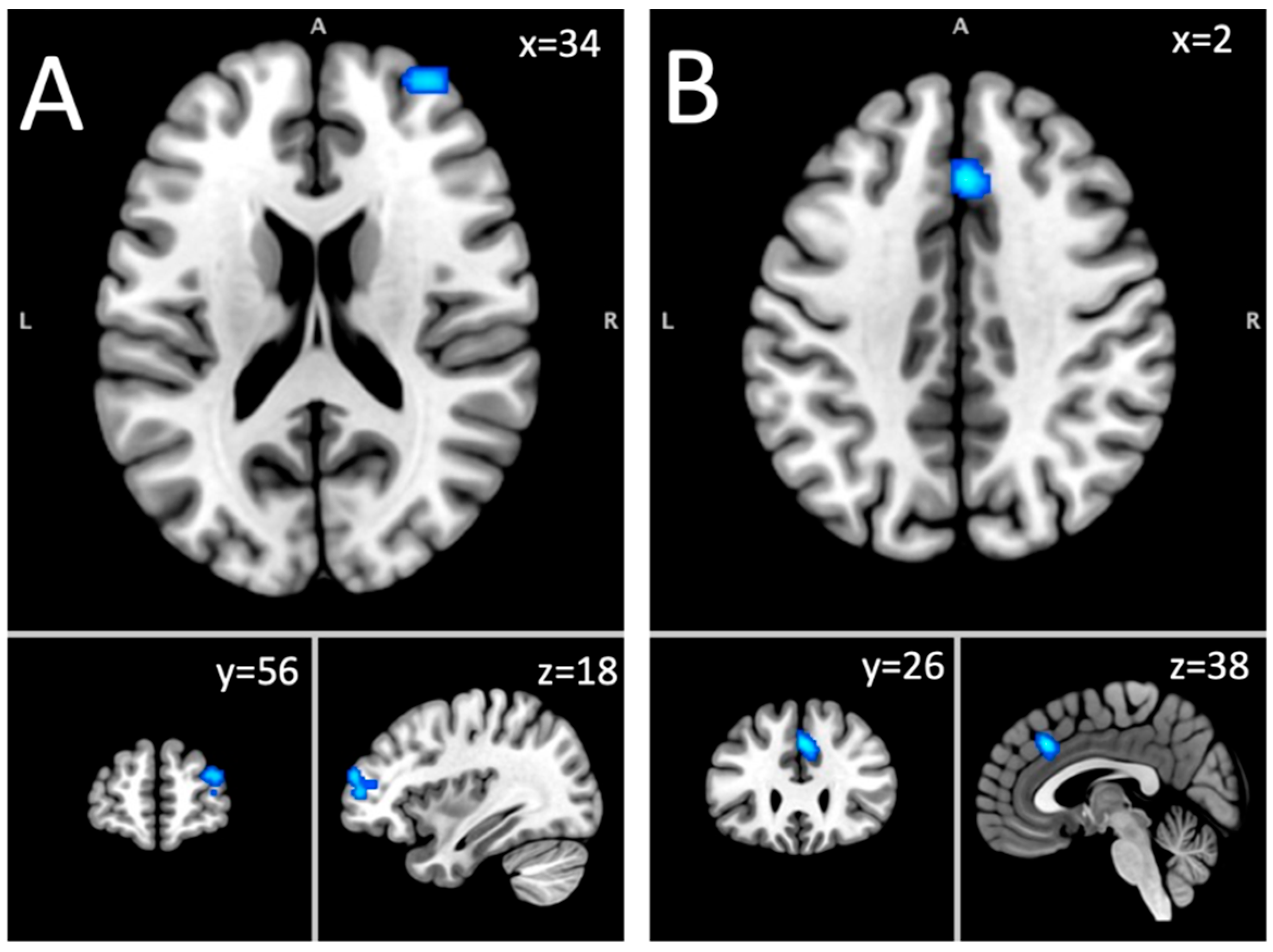
3.1.9. Hyperactivated Foci Group in the Long-Term Abstinent Sample

For the hyperactivated foci analysis, 136 foci were included, comprising 16 experiments on 615 subjects with an AUD. No clusters of significant increase activation were found at a corrected threshold.

3.2. Sub-Analyses on Sociodemographic Variables, MRI Parameters, and Task Types

No associations were found between sociodemographic variables, MRI parameters, and probabilities of activation in each significant cluster (Table 3). Relationships with task types were identified (Table 3). In the short-term sample, significant positive associations were observed between the putamen and decision-making tasks (OR = 9.33, 95% CI [1.27–68.60], p = 0.028), as well as reward tasks (OR = 30.00, 95% CI [2.33–386.33], p < 0.009). No significant relationships were found between the MFG and any task type. In the long-term abstinent sample, a significant positive association was identified between the SFG and reward tasks (OR = 9.00, 95% CI [1.03–78.57], p < 0.047). However, no significant associations were observed in the case of the cingulate gyrus.

4. Discussion

The objective of this meta-analysis was to provide a comprehensive overview of the neural effects of AUD through whole-brain fMRI analyses, regardless of the task used in the scanner, while distinguishing effects based on the duration of abstinence. The activity of the left putamen emerged as being disrupted in the analysis combining both hypo- and hyperactivations. A positive association was found between the aberrant activity of the putamen and decision-making and reward tasks. In short-term abstinent AUD participants, hypoactivation was observed in the right MFG, corresponding to the dorsolateral prefrontal cortex (dlPFC). For long-term abstinent AUD participants, hypoactivation was observed in the right SFG encompassing the MFG, in the lateral part of the prefrontal cortex (PFC), and in the dorsal anterior cingulate cortex (dACC). In the whole sample of both short- and long-term AUD participants, the putamen was also found to be altered. Lastly, sub-analyses revealed no association between neural alterations and sociodemographic and MRI parameters.
Our results demonstrated that the activation of the left putamen is bidirectionally altered in AUD and that these alterations are mostly driven by short-term abstinence studies. As such, this result is consistent with previous structural neuroimaging studies that have reliably shown that gray matter volumes/concentrations are decreased in AUD patients [9,12,13,42]. Our findings are also consistent with previous fMRI meta-analyses, which have shown a similar pattern of complex alterations in the putamen in AUD [17,21]. In healthy volunteers, fMRI studies have demonstrated that this dopamine-rich structure is involved in reward processing and associative learning [43,44]. In AUD, previous fMRI reward studies have determined that the putamen shows decreased activation during the Monetary Incentive Delay Task [21] and hyperactivation in response to alcohol cues [17,18]. In the sub-analyses of task types, we observed associations between the putamen and reward processing and reward-based decision-making, which are consistent with the previous literature in the field. Taken together, the results from the current meta-analysis and previous meta-analyses strongly suggest that AUD is associated with a motivational imbalance, whereby the brain reward system becomes hyperactivated in response to alcohol cues while displaying a blunted response to rewarding stimuli or cues that are not alcohol-related (example: money) [45,46,47]. The fact that significant putamen alterations were only observed during short-term abstinence is consistent with the clinical literature having shown that dysphoric symptoms and alcohol cravings tend to stabilize during the first 3–4 weeks of alcohol withdrawal [48,49].
Two clusters in the right MFG and SFG were found to be hypo-activated in AUD, corresponding to the right dlPFC and the right lateral PFC. These results are consistent with past structural neuroimaging studies, which have demonstrated a reduction in gray matter in the MFG and SFG in AUD, with the dlPFC appearing to be predominantly affected [11,12]. In the healthy population, large-scale fMRI evidence has shown that the right MFG and SFG play critical roles in core executive functions, including response inhibition and working memory [50,51,52]. In agreement with these findings, we performed functional decoding analyses, which revealed that the right dlPFC cluster is associated with functions like response inhibition, whereas the right lateral PFC cluster is associated with functions like working memory. Some authors have argued that executive dysfunctions are central to the development and maintenance of AUD [53,54] since these deficits weaken the ability to plan and execute effective strategies, resulting in social and occupational problems, as well as impaired impulse regulation and a loss of behavioral control [55]. Supporting the proposed interaction between executive and reward regions in AUD, the sub-analyses on task types showed an association between the SFG and reward tasks. As synthesized by Stavro and colleagues (2013) and Crowe and colleagues (2019) [30,31], a large body of cognitive studies has shown that working memory and response inhibition are significantly impaired in AUD and that these deficits clearly persist after the first weeks of alcohol withdrawal, with residual effects sometimes lasting several months. The fact that the lateral PFC was found to be impaired in long-term abstinence studies is consistent with the cognitive literature on AUD.
Hypoactivation was observed in long-term abstinence studies in AUD in the dACC, which is one of the core hubs of the (attentional) salience network [56]. The alteration of the dACC is supported by the structural neuroimaging literature, which has consistently shown gray matter reductions in this region in substance use disorders, including AUD [11,12,13,57]. Our result is also consistent with the results of previous fMRI meta-analyses, which have consistently shown alterations in ACC activation in AUD in separate analyses of reward processing, craving, and executive functions [16,17,18,19,20,21]. In the healthy population, the dACC has been shown to be involved in top-down attention control and the detection of salient changes in the interoceptive and external environment [58,59]. In view of the well-known roles of the dACC, the reduced activity observed in this region in AUD patients may signal attentional biases toward alcohol-related stimuli and/or an inability to re-allocate attentional resources to non-alcohol stimuli [60]. The fact that dACC hypo-activation was only observed in long-term abstinence studies suggests that the dACC may play a key role in alcohol relapse, as recently proposed by some investigators [61].
This meta-analysis has significant strengths compared to previous fMRI meta-analyses in the field. We conducted a meta-analysis including only whole-brain fMRI studies involving participants with AUD specifically, as compared to healthy control participants. This approach allowed us to include a more homogeneous study population and to produce a more comprehensive overview of alcohol’s effects on the AUD population while reducing biases associated with the selection of regions of interest. Moreover, our meta-analysis is the first to quantitatively review all tasks available, including task domains that were excluded from previous syntheses (audition, language, social cognition, etc.). This approach allowed us to include 27 fMRI studies that would have been excluded had we limited the analyses to tasks assessing executive functions, reward processing, and craving (as in previous meta-analyses). Despite the variety of the tasks reviewed, our findings converge on alterations in brain regions involved in reward processing, executive functions, and attentional salience, suggesting that the neural alterations observed in AUD may be restricted to these psychological domains.
Despite these strengths, a few limitations need to be acknowledged. Although the selection of tasks was comprehensive, the number of studies per task domain was rather small in several cases, namely in the case of tasks assessing language, audition, motor functions, social cognition, and episodic memory. For these domains, the number of studies was lower than the minimal number of studies that has been estimated to provide sufficient statistical power for fMRI meta-analyses (e.g., 17 studies) [62,63]. Therefore, statistical power was most probably insufficient for all these domains. As such, this could explain why we failed to observe neural impairments in the brain regions/networks involved in these functions. Likewise, the lack of effect observed in nearly all sub-analyses must be taken cautiously, considering that they may be explained by a lack of power. For instance, we observed no differences between studies using 1.5T vs. 3T scanners, potentially suggesting that future studies may interchangeably resort to 1.5T scanners. It must be noted, however, that only 15 studies in the current meta-analysis used 1.5T scanners. Moreover, although sub-analyses were performed on several variables that may have influenced results (age, sex, MRI parameters, etc.), we were unable to examine the influence on the results of the quantities of alcohol consumed by AUD patients. Indeed, the included studies often lacked clear data on consumption in terms of standardized units and precise temporality, and some relied on subjective measures influenced by national and political standards. This is unfortunate considering that alcohol is presumed to be more neurotoxic for those who have consumed larger quantities in their lifetime, although results on this topic are more complex than previously thought [64,65,66].
The current study sought to identify the neural alterations associated with AUD by performing a meta-analysis of whole-brain fMRI studies including all tasks available. We found that AUD was associated with neural impairments in brain regions involved in reward processing (the putamen), executive functions (the MFG), and attention salience (dACC). The length of abstinence influenced results, with striatal changes being driven by short-term abstinence studies and dACC changes being observed only in long-term abstinence studies. These findings underscore the importance of broadening the array of task domains in future fMRI research on AUD. Diversifying these domains may help move beyond the central triad of networks identified and more thoroughly examine the potential diffuse effects of AUD on brain functioning. Finally, it will be crucial to standardize, in future fMRI research, alcohol consumption units to facilitate data conversion and comparison and, thus, refine our understanding of the neural effects of AUD.

Supplementary Materials

The following supporting information can be downloaded at https://www.mdpi.com/article/10.3390/brainsci15070665/s1: Figure S1: PRISMA flowchart; Table S1: PRISMA 2020 Checklist; Table S2: Characteristics of the included studies.

Author Contributions

Conceptualization, M.B. and S.P.; methodology, S.P. and M.B.; validation, M.B. and S.P.; formal analysis, M.B. and M.R.; investigation, M.B. and M.R.; data curation, M.B. and M.R.; writing—original draft preparation, M.B. and M.R.; writing—review and editing, S.P. and M.B.; visualization, M.B. and M.R.; supervision, M.B. and S.P.; project administration, M.B. and S.P. All authors have read and agreed to the published version of the manuscript.

Funding

This research received no external funding.

Data Availability Statement

All data used are available in the manuscript. No new data were generated.

Acknowledgments

S.P. is the holder of the Eli Lilly Canada Chair on schizophrenia research; M.B. is the holder of a doctoral scholarship from the Canadian Institutes of Health Research (FBD-193359).

Conflicts of Interest

The authors declare no conflicts of interest.

References

  1. Meyer, J.; Farrar, A.M.; Bienzonski, D.; Yates, J.R. Psychopharmacology, Drugs, the Brain, and Behavior, 4th ed.; Oxford University Press: Oxford, UK, 2022. [Google Scholar]
  2. WHO. Over 3 Million Annual Deaths Due to Alcohol and Drug Use, Majority Among Men; WHO: Geneva, Switzerland, 25 June 2024. [Google Scholar]
  3. Chikritzhs, T.; Livingston, M. Alcohol and the Risk of Injury. Nutrients 2021, 13, 2777. [Google Scholar] [CrossRef] [PubMed]
  4. Sacks, J.J.; Gonzales, K.R.; Bouchery, E.E.; Tomedi, L.E.; Brewer, R.D. 2010 National and State Costs of Excessive Alcohol Consumption. Am. J. Prev. Med. 2015, 49, e73–e79. [Google Scholar] [CrossRef]
  5. Thorrisen, M.M.; Bonsaksen, T.; Hashemi, N.; Kjeken, I.; van Mechelen, W.; Aas, R.W. Association between alcohol consumption and impaired work performance (presenteeism): A systematic review. BMJ Open 2019, 9, e029184. [Google Scholar] [CrossRef]
  6. Hendriks, H.F.J. Alcohol and Human Health: What Is the Evidence? Annu. Rev. Food Sci. Technol. 2020, 11, 1–21. [Google Scholar] [CrossRef]
  7. Palzes, V.A.; Parthasarathy, S.; Chi, F.W.; Kline-Simon, A.H.; Lu, Y.; Weisner, C.; Ross, T.B.; Elson, J.; Sterling, S.A. Associations Between Psychiatric Disorders and Alcohol Consumption Levels in an Adult Primary Care Population. Alcohol Clin. Exp. Res. 2020, 44, 2536–2544. [Google Scholar] [CrossRef] [PubMed]
  8. Kamal, H.; Tan, G.C.; Ibrahim, S.F.; Shaikh, M.F.; Mohamed, I.N.; Mohamed, R.M.P.; Hamid, A.A.; Ugusman, A.; Kumar, J. Alcohol Use Disorder, Neurodegeneration, Alzheimer’s and Parkinson’s Disease: Interplay Between Oxidative Stress, Neuroimmune Response and Excitotoxicity. Front. Cell Neurosci. 2020, 14, 282. [Google Scholar] [CrossRef]
  9. Hahn, S.; Mackey, S.; Cousijn, J.; Foxe, J.J.; Heinz, A.; Hester, R.; Hutchinson, K.; Kiefer, F.; Korucuoglu, O.; Lett, T.; et al. Predicting alcohol dependence from multi-site brain structural measures. Hum. Brain Mapp. 2022, 43, 555–565. [Google Scholar] [CrossRef] [PubMed]
  10. Navarri, X.; Afzali, M.H.; Lavoie, J.; Sinha, R.; Stein, D.J.; Momenan, R.; Veltman, D.J.; Korucuoglu, O.; Sjoerds, Z.; van Holst, R.J.; et al. How do substance use disorders compare to other psychiatric conditions on structural brain abnormalities? A cross-disorder meta-analytic comparison using the ENIGMA consortium findings. Hum. Brain Mapp. 2022, 43, 399–413. [Google Scholar] [CrossRef]
  11. Li, L.; Yu, H.; Liu, Y.; Meng, Y.J.; Li, X.J.; Zhang, C.; Liang, S.; Li, M.L.; Guo, W.; Wang, Q.; et al. Lower regional grey matter in alcohol use disorders: Evidence from a voxel-based meta-analysis. BMC Psychiatry 2021, 21, 247. [Google Scholar] [CrossRef]
  12. Yang, X.; Tian, F.; Zhang, H.; Zeng, J.; Chen, T.; Wang, S.; Jia, Z.; Gong, Q. Cortical and subcortical gray matter shrinkage in alcohol-use disorders: A voxel-based meta-analysis. Neurosci. Biobehav. Rev. 2016, 66, 92–103. [Google Scholar] [CrossRef]
  13. Pando-Naude, V.; Toxto, S.; Fernandez-Lozano, S.; Parsons, C.E.; Alcauter, S.; Garza-Villarreal, E.A. Gray and white matter morphology in substance use disorders: A neuroimaging systematic review and meta-analysis. Transl. Psychiatry 2021, 11, 29. [Google Scholar] [CrossRef] [PubMed]
  14. Spindler, C.; Mallien, L.; Trautmann, S.; Alexander, N.; Muehlhan, M. A coordinate-based meta-analysis of white matter alterations in patients with alcohol use disorder. Transl. Psychiatry 2022, 12, 40. [Google Scholar] [CrossRef]
  15. Monnig, M.A.; Tonigan, J.S.; Yeo, R.A.; Thoma, R.J.; McCrady, B.S. White matter volume in alcohol use disorders: A meta-analysis. Addict. Biol. 2013, 18, 581–592. [Google Scholar] [CrossRef] [PubMed]
  16. Devoto, F.; Zapparoli, L.; Spinelli, G.; Scotti, G.; Paulesu, E. How the harm of drugs and their availability affect brain reactions to drug cues: A meta-analysis of 64 neuroimaging activation studies. Transl. Psychiatry 2020, 10, 429. [Google Scholar] [CrossRef]
  17. Klugah-Brown, B.; Di, X.; Zweerings, J.; Mathiak, K.; Becker, B.; Biswal, B. Common and separable neural alterations in substance use disorders: A coordinate-based meta-analyses of functional neuroimaging studies in humans. Hum. Brain Mapp. 2020, 41, 4459–4477. [Google Scholar] [CrossRef] [PubMed]
  18. Oberlin, B.G.; Shen, Y.I.; Kareken, D.A. Alcohol Use Disorder Interventions Targeting Brain Sites for Both Conditioned Reward and Delayed Gratification. Neurotherapeutics 2020, 17, 70–86. [Google Scholar] [CrossRef]
  19. Zeng, J.; Yu, S.; Cao, H.; Su, Y.; Dong, Z.; Yang, X. Neurobiological correlates of cue-reactivity in alcohol-use disorders: A voxel-wise meta-analysis of fMRI studies. Neurosci. Biobehav. Rev. 2021, 128, 294–310. [Google Scholar] [CrossRef]
  20. Cao, Y.; Tian, F.; Zeng, J.; Gong, Q.; Yang, X.; Jia, Z. The brain activity pattern in alcohol-use disorders under inhibition response Task. J. Psychiatr. Res. 2023, 163, 127–134. [Google Scholar] [CrossRef]
  21. Zeng, J.; You, L.; Yang, F.; Luo, Y.; Yu, S.; Yan, J.; Liu, M.; Yang, X. A meta-analysis of the neural substrates of monetary reward anticipation and outcome in alcohol use disorder. Hum. Brain Mapp. 2023, 44, 2841–2861. [Google Scholar] [CrossRef]
  22. Dugré, J.R.; Orban, P.; Potvin, S. Disrupted functional connectivity of the brain reward system in substance use problems: A meta-analysis of functional neuroimaging studies. Addict. Biol. 2023, 28, e13257. [Google Scholar] [CrossRef]
  23. Müller, V.I.; Cieslik, E.C.; Laird, A.R.; Fox, P.T.; Radua, J.; Mataix-Cols, D.; Tench, C.R.; Yarkoni, T.; Nichols, T.E.; Turkeltaub, P.E.; et al. Ten simple rules for neuroimaging meta-analysis. Neurosci. Biobehav. Rev. 2018, 84, 151–161. [Google Scholar] [CrossRef] [PubMed]
  24. Bagga, D.; Singh, N.; Modi, S.; Kumar, P.; Bhattacharya, D.; Garg, M.L.; Khushu, S. Assessment of lexical semantic judgment abilities in alcohol-dependent subjects: An fMRI study. J. Biosci. 2013, 38, 905–915. [Google Scholar] [CrossRef] [PubMed]
  25. Hermann, D.; Smolka, M.N.; Klein, S.; Heinz, A.; Mann, K.; Braus, D.F. Reduced fMRI activation of an occipital area in recently detoxified alcohol-dependent patients in a visual and acoustic stimulation paradigm. Addict. Biol. 2007, 12, 117–121. [Google Scholar] [CrossRef]
  26. Li, C.S.; Luo, X.; Yan, P.; Bergquist, K.; Sinha, R. Altered impulse control in alcohol dependence: Neural measures of stop signal performance. Alcohol Clin. Exp. Res. 2009, 33, 740–750. [Google Scholar] [CrossRef] [PubMed]
  27. Maurage, P.; Joassin, F.; Philippot, P.; Heeren, A.; Vermeulen, N.; Mahau, P.; Delperdange, C.; Corneille, O.; Luminet, O.; de Timary, P. Disrupted regulation of social exclusion in alcohol-dependence: An fMRI study. Neuropsychopharmacology 2012, 37, 2067–2075. [Google Scholar] [CrossRef]
  28. van Holst, R.J.; Clark, L.; Veltman, D.J.; van den Brink, W.; Goudriaan, A.E. Enhanced striatal responses during expectancy coding in alcohol dependence. Drug Alcohol. Depend. 2014, 142, 204–208. [Google Scholar] [CrossRef]
  29. McHugh, R.K.; Weiss, R.D. Alcohol Use Disorder and Depressive Disorders. Alcohol Res. Curr. Rev. 2019, 40, 8. [Google Scholar] [CrossRef]
  30. Crowe, S.F.; Cammisuli, D.M.; Stranks, E.K. Widespread cognitive deficits in alcoholism persistent following prolonged abstinence: An updated meta-analysis of studies that used standardized neuropsychological assessment tools. Arch. Clin. Neuropsychol. 2019, 35, 31–45. [Google Scholar] [CrossRef]
  31. Stavro, K.; Pelletier, J.; Potvin, S. Widespread and sustained cognitive deficits in alcoholism: A meta-analysis. Addict. Biol. 2013, 18, 203–213. [Google Scholar] [CrossRef]
  32. Degasperi, G.; Cristea, I.A.; Di Rosa, E.; Costa, C.; Gentili, C. Parsing variability in borderline personality disorder: A meta-analysis of neuroimaging studies. Transl. Psychiatry 2021, 11, 314. [Google Scholar] [CrossRef]
  33. Janiri, D.; Moser, D.A.; Doucet, G.E.; Luber, M.J.; Rasgon, A.; Lee, W.H.; Murrough, J.W.; Sani, G.; Eickhoff, S.B.; Frangou, S. Shared Neural Phenotypes for Mood and Anxiety Disorders A Meta-Analysis of 226 Task-Related Functional Imaging Studies. Focus 2021, 19, 256–263. [Google Scholar] [CrossRef] [PubMed]
  34. Pico-Perez, M.; Vieira, R.; Fernandez-Rodriguez, M.; De Barros, M.A.P.; Radua, J.; Morgado, P. Multimodal meta-analysis of structural gray matter, neurocognitive and social cognitive fMRI findings in schizophrenia patients. Psychol. Med. 2022, 52, 614–624. [Google Scholar] [CrossRef] [PubMed]
  35. Solomonov, N.; Victoria, L.W.; Lyons, K.; Phan, D.K.; Alexopoulos, G.S.; Gunning, F.M.; Fluckiger, C. Social reward processing in depressed and healthy individuals across the lifespan: A systematic review and a preliminary coordinate-based meta-analysis of fMRI studies. Behav. Brain Res. 2023, 454, 114632. [Google Scholar] [CrossRef]
  36. Tamon, H.; Fujino, J.; Itahashi, T.; Frahm, L.; Parlatini, V.; Aoki, Y.Y.; Castellanos, F.X.; Eickhoff, S.B.; Cortese, S. Shared and Specific Neural Correlates of Attention Deficit Hyperactivity Disorder and Autism Spectrum Disorder: A Meta-Analysis of 243 Task-Based Functional MRI Studies. Am. J. Psychiatry 2024, 181, 541–552. [Google Scholar] [CrossRef] [PubMed]
  37. Wang, Z.; He, D.; Yang, L.; Wang, P.; Xiao, J.; Zou, Z.; Min, W.; He, Y.; Yuan, C.; Zhu, H.; et al. Similarities and differences between post-traumatic stress disorder and major depressive disorder: Evidence from task-evoked functional magnetic resonance imaging meta-analysis. J. Affect. Disord. 2024, 361, 712–719. [Google Scholar] [CrossRef]
  38. Eickhoff, S.B.; Bzdok, D.; Laird, A.R.; Kurth, F.; Fox, P.T. Activation likelihood estimation meta-analysis revisited. Neuroimage 2012, 59, 2349–2361. [Google Scholar] [CrossRef]
  39. Lancaster, J.L.; Tordesillas-Gutierrez, D.; Martinez, M.; Salinas, F.; Evans, A.; Zilles, K.; Mazziotta, J.C.; Fox, P.T. Bias between MNI and Talairach coordinates analyzed using the ICBM-152 brain template. Hum. Brain Mapp. 2007, 28, 1194–1205. [Google Scholar] [CrossRef]
  40. Wallach, J.D.; Gueorguieva, R.; Phan, H.; Witkiewitz, K.; Wu, R.; O’Malley, S.S. Predictors of abstinence, no heavy drinking days, and a 2-level reduction in World Health Organization drinking levels during treatment for alcohol use disorder in the COMBINE study. Alcohol Clin. Exp. Res. 2022, 46, 1331–1339. [Google Scholar] [CrossRef]
  41. Eickhoff, S.B.; Laird, A.R.; Grefkes, C.; Wang, L.E.; Zilles, K.; Fox, P.T. Coordinate-based activation likelihood estimation meta-analysis of neuroimaging data: A random-effects approach based on empirical estimates of spatial uncertainty. Hum. Brain Mapp. 2009, 30, 2907–2926. [Google Scholar] [CrossRef]
  42. Fein, G.; Greenstein, D.; Cardenas, V.A.; Cuzen, N.L.; Fouche, J.P.; Ferrett, H.; Thomas, K.; Stein, D.J. Cortical and subcortical volumes in adolescents with alcohol dependence but without substance or psychiatric comorbidities. Psychiatry Res. 2013, 214, 1–8. [Google Scholar] [CrossRef]
  43. Ghandili, M.; Munakomi, S. Neuroanatomy, Putamen. In StatPearls [Internet]; StatPearls Publishing: Treasure Island, FL, USA, 2023. [Google Scholar]
  44. Hu, W.; Zhao, X.; Liu, Y.; Ren, Y.; Wei, Z.; Tang, Z.; Tian, Y.; Sun, Y.; Yang, J. Reward sensitivity modulates the brain reward pathway in stress resilience via the inherent neuroendocrine system. Neurobiol. Stress. 2022, 20, 100485. [Google Scholar] [CrossRef] [PubMed]
  45. Kim, S.I. Neuroscientific model of motivational process. Front. Psychol. 2013, 4, 98. [Google Scholar] [CrossRef]
  46. Poulton, A.; Hester, R. Transition to substance use disorders: Impulsivity for reward and learning from reward. Soc. Cogn. Affect. Neurosci. 2020, 15, 1182–1191. [Google Scholar] [CrossRef]
  47. Weinstein, A.M. Reward, motivation and brain imaging in human healthy participants—A narrative review. Front. Behav. Neurosci. 2023, 17, 1123733. [Google Scholar] [CrossRef]
  48. Petit, G.; Deschietere, G.; Loas, G.; Luminet, O.; de Timary, P. Link Between Anhedonia and Depression During Early Alcohol Abstinence: Gender Matters. Alcohol Alcohol. 2020, 55, 71–77. [Google Scholar] [CrossRef] [PubMed]
  49. Seo, D.; Sinha, R. Neuroplasticity and predictors of alcohol recovery. Alcohol Res. Curr. Rev. 2015, 37, 143. [Google Scholar]
  50. Depue, B.E.; Banich, M.T. Increased inhibition and enhancement of memory retrieval are associated with reduced hippocampal volume. Hippocampus 2012, 22, 651–655. [Google Scholar] [CrossRef]
  51. Miller, J.A.; Tambini, A.; Kiyonaga, A.; D’Esposito, M. Long-term learning transforms prefrontal cortex representations during working memory. Neuron 2022, 110, 3805–3819 e3806. [Google Scholar] [CrossRef] [PubMed]
  52. Oldrati, V.; Patricelli, J.; Colombo, B.; Antonietti, A. The role of dorsolateral prefrontal cortex in inhibition mechanism: A study on cognitive reflection test and similar tasks through neuromodulation. Neuropsychologia 2016, 91, 499–508. [Google Scholar] [CrossRef]
  53. Lopez-Caneda, E.; Rodriguez Holguin, S.; Cadaveira, F.; Corral, M.; Doallo, S. Impact of alcohol use on inhibitory control (and vice versa) during adolescence and young adulthood: A review. Alcohol Alcohol. 2014, 49, 173–181. [Google Scholar] [CrossRef]
  54. Si, Y.; Wang, L.; Zhao, M. Anti-saccade as a Tool to Evaluate Neurocognitive Impairment in Alcohol Use Disorder. Front. Psychiatry 2022, 13, 823848. [Google Scholar] [CrossRef]
  55. Rure, D.; Shakya, M.; Singhal, A.; Varma, A.; Mishra, N.; Pathak, U. A Study of the association of neurocognition with relapse and quality of life in patients of alcohol dependence. Ind. Psychiatry J. 2024, 33, 133–140. [Google Scholar] [CrossRef]
  56. Ham, T.; Leff, A.; de Boissezon, X.; Joffe, A.; Sharp, D.J. Cognitive control and the salience network: An investigation of error processing and effective connectivity. J. Neurosci. 2013, 33, 7091–7098. [Google Scholar] [CrossRef]
  57. Xiao, P.; Dai, Z.; Zhong, J.; Zhu, Y.; Shi, H.; Pan, P. Regional gray matter deficits in alcohol dependence: A meta-analysis of voxel-based morphometry studies. Drug Alcohol. Depend. 2015, 153, 22–28. [Google Scholar] [CrossRef] [PubMed]
  58. Hardee, J.E.; Weigard, A.S.; Heitzeg, M.M.; Martz, M.E.; Cope, L.M. Sex differences in distributed error-related neural activation in problem-drinking young adults. Drug Alcohol. Depend. 2024, 263, 112421. [Google Scholar] [CrossRef]
  59. Luo, Q.; Mitchell, D.; Jones, M.; Mondillo, K.; Vythilingam, M.; Blair, R.J. Common regions of dorsal anterior cingulate and prefrontal–parietal cortices provide attentional control of distracters varying in emotionality and visibility. NeuroImage 2007, 38, 631–639. [Google Scholar] [CrossRef] [PubMed]
  60. Murray, L.; Welsh, J.C.; Johnson, C.G.; Kaiser, R.H.; Farchione, T.J.; Janes, A.C. Alcohol- and non-alcohol-related interference: An fMRI study of treatment-seeking adults with alcohol use disorder. Drug Alcohol Depend. 2022, 235, 109462. [Google Scholar] [CrossRef] [PubMed]
  61. Padula, C.B.; Tenekedjieva, L.T.; McCalley, D.M.; Al-Dasouqi, H.; Hanlon, C.A.; Williams, L.M.; Kozel, F.A.; Knutson, B.; Durazzo, T.C.; Yesavage, J.A.; et al. Targeting the Salience Network: A Mini-Review on a Novel Neuromodulation Approach for Treating Alcohol Use Disorder. Front. Psychiatry 2022, 13, 893833. [Google Scholar] [CrossRef]
  62. Eickhoff, S.B.; Nichols, T.E.; Laird, A.R.; Hoffstaedter, F.; Amunts, K.; Fox, P.T.; Bzdok, D.; Eickhoff, C.R. Behavior, sensitivity, and power of activation likelihood estimation characterized by massive empirical simulation. Neuroimage 2016, 137, 70–85. [Google Scholar] [CrossRef]
  63. Garrison, J.R.; Done, J.; Simons, J.S. Interpretation of published meta-analytical studies affected by implementation errors in the GingerALE software. Neurosci. Biobehav. Rev. 2019, 102, 424–426. [Google Scholar] [CrossRef]
  64. Jeon, K.H.; Han, K.; Jeong, S.M.; Park, J.; Yoo, J.E.; Yoo, J.; Lee, J.; Kim, S.; Shin, D.W. Changes in Alcohol Consumption and Risk of Dementia in a Nationwide Cohort in South Korea. JAMA Netw. Open 2023, 6, e2254771. [Google Scholar] [CrossRef] [PubMed]
  65. Xu, W.; Wang, H.; Wan, Y.; Tan, C.; Li, J.; Tan, L.; Yu, J.T. Alcohol consumption and dementia risk: A dose-response meta-analysis of prospective studies. Eur. J. Epidemiol. 2017, 32, 31–42. [Google Scholar] [CrossRef] [PubMed]
  66. Zarezadeh, M.; Mahmoudinezhad, M.; Faghfouri, A.H.; Mohammadzadeh Honarvar, N.; Regestein, Q.R.; Papatheodorou, S.I.; Mekary, R.A.; Willett, W.C. Alcohol consumption in relation to cognitive dysfunction and dementia: A systematic review and dose-response meta-analysis of comparative longitudinal studies. Ageing Res. Rev. 2024, 100, 102419. [Google Scholar] [CrossRef] [PubMed]
Figure 1. Short-term abstinence fMRI results. Note. (A) = putamen (hyper- and hypoactivation); (B) = middle frontal gyrus (hypoactivation).
Brainsci 15 00665 g001
Figure 2. Long-term abstinence fMRI results. Note. (A) = superior frontal gyrus (hypoactivation); (B) = dorsal anterior cingulate cortex (hypoactivation).
Brainsci 15 00665 g002
Table 1. Demographics.
n Casesn ControlsMean Age CasesMean Age Controls% of Males for Cases% of Males for ControlsMean Days of Abstinence (Range)
Whole sample
2421145836.9437.0776.7373.26189.54 (5–2994) a
Short-term abstinent sample
109765335.7134.4676.3270.7116.10 (5–25.30) b
Long-term abstinent sample
99148837.4240.1577.5678.35416.47 (34–2994) c
Note. a = information available only for 44/67 studies; b = information available only for 23/37 studies; c = information available only for 19/26 studies.
Table 2. Results of the ALE meta-analysis.
RegionsL/RCluster Size (mm3)ALE ValueZ-ScoreCoordinates (MNI)
Hyper–hypoactivation combined: whole sample
Putamen, caudate body, caudate headL18400.03675.66−20, 12, −2
Hyperactivation: whole sample
No clusters found
Hypoactivation: whole sample
No clusters found
Hyper–hypoactivation combined: short-term abstinent sample
PutamenL10960.02364.88−20, 12, −2
Hyperactivation: short-term abstinent sample
No clusters found
Hypoactivation: short-term abstinent sample
Middle frontal gyrus, superior frontal gyrus, sub-gyralR8560.0164.4824, 8, 66
Hyper–hypoactivation combined: long-term abstinent sample
No clusters found
Hyperactivation: long-term abstinent sample
No clusters found
Hypoactivation: long-term abstinent sample
Superior frontal gyrus, middle frontal gyrusR8560.01473.8534, 56, 18
Cingulate gyrus, medial frontal gyrusR & L8000.0174.262, 26, 38
Note. L = left; R = right; MNI = Montreal Neurologic Institute.
Table 3. Binary logistic regressions for confounding variables and probabilities of activation of each cluster.
95 C.I. for Odds Ratio
PredictorsOdds RatioLowerHigherp-Value
Hyper–hypoactivation in the short-term abstinent sample: putamen
Age0.9850.9001.0770.735
Sex ratio (% male)0.9810.9511.0130.245
Days of abstinence0.9390.7461.1820.590
MRI field strength0.7370.1104.9550.753
Smoothing level0.7010.4221.1640.170
Voxel size1.0470.9931.1030.088
Time repetition0.9980.9961.0000.115
Craving studies0.4890.0504.7930.539
Decision-making studies9.3331.27068.5970.028 *
Emotion studies1.3500.12414.7340.806
Executive function studies0.3640.0383.5180.382
Reward processing studies30.0002.330386.3250.009 *
Other task studies3.3750.45924.8370.232
Hypoactivation in the short-term abstinent sample: middle frontal gyrus
Age0.9420.8541.0400.234
Sex ratio (% male)0.9850.9511.0210.417
Days of abstinence0.8590.6641.1130.251
MRI field strength0.7620.0609.6110.833
Smoothing level1.0680.6481.7600.797
Voxel size1.0540.9891.1230.108
Time repetition1.0000.9991.0010.924
Craving studies0.0000.0000.0000.999
Decision-making studies7.2500.78666.8420.080
Emotion studies0.0000.0000.0000.999
Executive function studies2.3000.28318.7050.436
Reward processing studies3.3330.25942.9250.356
Other task studies1.8670.16021.7420.618
Hypoactivation in the long-term abstinent sample: superior frontal gyrus
Age0.9630.8941.0380.323
Sex ratio (% male)0.9930.9501.0370.746
Days of abstinence1.0020.9991.0040.153
MRI field strength0.5000.0663.7700.501
Smoothing level0.5860.2781.2330.159
Voxel size0.9800.9391.0220.337
Time repetition1.0010.9991.0040.267
Craving studies0.0000.0000.0000.999
Decision-making studies0.0000.0000.0001.000
Emotion studies3.8000.20172.0000.374
Executive functions studies1.1670.1668.1860.877
Reward processing studies9.0001.03178.5740.047 *
Other task studies1.5000.20810.8230.688
Hypoactivation in the long-term abstinent sample: cingulate gyrus
Age1.0250.9311.1280.617
Sex ratio (% male)1.0220.9641.0840.456
Days of abstinence0.9970.9901.0040.387
MRI field strength1.1250.09713.0360.925
Smoothing level0.9560.4502.0310.906
Voxel size0.9990.9481.0520.955
Time repetition1.0000.9971.0020.708
Craving studies1.5000.12218.4410.751
Decision-making studies0.0000.0000.0001.000
Emotion studies0.0000.0000.0000.999
Executive functions studies0.0000.0000.0000.999
Reward processing studies6.3330.63063.6390.117
Other task studies3.4000.37730.6550.275
Note. MRI = magnetic resonance imaging. *, significant result (p < 0.05).
Disclaimer/Publisher’s Note: The statements, opinions and data contained in all publications are solely those of the individual author(s) and contributor(s) and not of MDPI and/or the editor(s). MDPI and/or the editor(s) disclaim responsibility for any injury to people or property resulting from any ideas, methods, instructions or products referred to in the content.

