Review

Hypoxia’s Impact on Hippocampal Functional Connectivity: Insights from Resting-State fMRI Studies

by
Julia Micaux
1,2,†,
Abir Troudi Habibi
1,2,†,
Franck Mauconduit
3 and
Marion Noulhiane
1,2,*
1
University Paris-Saclay, CEA, Joliot Institute, NeuroSpin, Clinical and Translational Applied NeuroImaging Research Unit (UNIACT), 91191 Gif-sur-Yvette, France
2
University of Paris Cité, Inserm, U1141 NeuroDiderot, InDev Team, 75019 Paris, France
3
CEA, NeuroSpin, CNRS, University of Paris-Saclay, 91191 Gif-sur-Yvette, France
*
Author to whom correspondence should be addressed.
These authors contributed equally to this work.
Brain Sci. 2025, 15(6), 643; https://doi.org/10.3390/brainsci15060643 (registering DOI)
Submission received: 30 April 2025 / Revised: 6 June 2025 / Accepted: 13 June 2025 / Published: 14 June 2025
(This article belongs to the Special Issue Brain Network Connectivity Analysis in Neuroscience)

Abstract

:
The hippocampus is one of the brain’s most vulnerable structures to hypoxia, playing a crucial role in memory and spatial navigation. This sensitivity makes it a key region for understanding the effects of hypoxia on brain connectivity. This review examines the effects of both acute and chronic hypoxia on resting-state networks (RSNs) that contribute to hippocampal functional connectivity (FC). Hypoxia, characterized by a reduced oxygen supply to the brain, can result from environmental factors (such as high-altitude exposure) or hypoxia-induced pathological conditions (including obstructive sleep apnea and hypoxic–ischemic encephalopathy). The hippocampus’s susceptibility to hypoxic damage significantly impairs brain connectivity. This review examines through rs-fMRI studies how hypoxia alters hippocampal FC, focusing on its effects on RSNs involved in hippocampal functions, and compares acute and chronic hypoxic states. We seek to determine whether distinct or shared patterns of FC changes exist between acute and chronic hypoxia, and how hypoxia indirectly changes hippocampal FC, given the challenges of studying it in isolation. By addressing these questions, this review aims to deepen our understanding of hypoxia-induced changes in hippocampal FC and provide insights into potential therapeutic strategies to mitigate its effects on cognitive functions.

1. Introduction

1.1. Background

A rapid decline in oxygen levels marks the transition from normoxia to hypoxia. Hypoxia can be classified into two main etiology types, acute and chronic, which can arise in both environmental and pathological contexts. Environmental hypoxia, such as high-altitude exposure or voluntary breath-holding in freediving, can either induce transient (acute) or sustained (chronic) oxygen deprivation. Similarly, hypoxia can cause pathological conditions in both acute and chronic forms, including obstructive sleep apnea and hypoxic–ischemic encephalopathy. While these forms differ in etiology, both environmental and pathological hypoxia can lead to significant cognitive and physiological consequences [1,2,3]. Acute hypoxia involves sudden oxygen deprivation, while chronic hypoxia is marked by prolonged exposure to reduced oxygen levels. Both forms can profoundly disrupt hippocampal structure and function, a brain structure involved in memory and spatial navigation [4,5]. The hippocampus is susceptible to oxygen deprivation, making it highly vulnerable to hypoxic injury [6,7,8,9,10]. This vulnerability positions the hippocampus as a critical region for understanding how hypoxia affects neuronal integrity and brain connectivity. Structural MRI studies have shown that both acute and chronic hypoxia can lead to changes in hippocampal volume and neural network disruptions [7,11,12]. Also, disrupted hippocampal function is well documented, leading to cognitive impairments, including memory deficits and executive function decline [4]. These changes likely result from impaired neuronal communication, reduced neurogenesis, and decreased metabolic adaptation [13,14]. Given the hippocampus’s pivotal role in cognitive function, understanding how hypoxia alters its FC is crucial for understanding long-term cognitive effects.
Resting-state functional MRI (rs-fMRI) is a non-invasive imaging technique used to investigate spontaneous brain activity in the absence of task performance. During rs-fMRI acquisition, the subject lies quietly (eyes open or closed) and the scanner captures low-frequency (<0.1 Hz) fluctuations in the blood-oxygen-level-dependent (BOLD) signal across the whole brain. These fluctuations reflect changes in the relative concentrations of oxyhemoglobin and deoxyhemoglobin, serving as an indirect marker of neuronal activity and neurovascular coupling [15,16,17,18,19,20].
Data are typically acquired using gradient-echo echo-planar imaging (EPI) sequences over 5–10 min, providing full-brain coverage with high sensitivity to neurovascular coupling [21]. By using the rs-fMRI technique, researchers can investigate and explore neural and vascular responses to oxygen deprivation, as it provides valuable insights into intrinsic brain connectivity. This method helps identify disruptions in networks, such as the hippocampus and its default mode network (DMN), which are particularly sensitive to hypoxic conditions [22,23,24,25]. Measures such as seed-based connectivity, independent component analysis (ICA), and amplitude of low-frequency fluctuations (ALFF) provide complementary insights into FC and regional spontaneous neural activity [26,27,28]. Advanced tools, including functional connectivity density (FCD) and dynamic causal modeling (DCM), further enhance our understanding of the brain’s functional organization under hypoxic conditions [29,30,31].
However, direct assessments of hippocampal FC during hypoxia have been limited, with most studies focusing on RSN FC and reporting memory loss as a cognitive outcome. This review aims to fill this gap by specifically examining changes in hippocampal FC through RSN alterations under hypoxic conditions.

1.2. Scope of Review

This narrative review focuses on environmental hypoxia and hypoxia-induced pathologies, with an emphasis on understanding hippocampal FC changes during such hypoxic conditions. The review aims to achieve the following:
  • Identify the RSNs involved in the hippocampal networks during rest.
  • Explore how rs-fMRI reveals changes in hippocampal FC during hypoxia.
  • Compare the effects of acute and chronic hypoxia on brain FC, examining whether they produce distinct or shared patterns of network changes.
By addressing these questions, this review seeks to provide an integrative perspective on hippocampal adaptability to hypoxic conditions and inform the development of potential interventions to enhance cognitive resilience in individuals exposed to hypoxia. Our objective is not to exhaustively catalogue all published studies on the topic, but rather to synthesize key findings, identify common methodological approaches and limitations, and highlight emerging patterns that may inform future research.

2. Physiological Basis of Hypoxia in the Context of Resting-State fMRI

Hypoxia, defined as insufficient oxygen availability to meet metabolic demands, has profound effects on brain physiology and FC [32,33]. Oxygen is critical for ATP production via oxidative phosphorylation. When oxygen levels drop, either suddenly (acute hypoxia) or over prolonged periods (chronic hypoxia), the brain undergoes a cascade of compensatory and pathological processes that can alter neuronal activity and disrupt BOLD signal dynamics [34].
Acute hypoxia triggers rapid oxygen deprivation (e.g., high-altitude or apnea) and elicits immediate systemic responses such as hyperventilation and increased cardiac output. In the brain, it induces oxidative stress and mitochondrial dysfunction, particularly in regions like the hippocampus and prefrontal cortex [35,36]. These changes impair synaptic function and may transiently disrupt resting-state networks, which are sensitive to metabolic alterations. Although some oxidative markers normalize within 24 h, apoptotic pathways may remain active, potentially affecting rs-fMRI readouts beyond the hypoxic exposure window [33,35,37,38].
Chronic hypoxia, characterized by prolonged oxygen deprivation over weeks to years, is commonly associated with conditions such as high-altitude exposure or obstructive sleep apnea, which leads to sustained metabolic stress [34,39]. Adaptations like erythropoiesis and vascular remodeling help maintain oxygen delivery but are often accompanied by white matter changes, hippocampal atrophy, and altered FC patterns in rs-fMRI [40,41]. The structural and functional alterations are also implicated in the cognitive decline observed in chronic hypoxia and may predispose individuals to neurodegenerative diseases such as Alzheimer’s [42]. Moreover, chronic hypoxia impacts hormonal responses, particularly in the context of exercise. For example, chronic resistance training under normobaric hypoxia has been shown to suppress the growth hormone response to exercise in older adults [43]. These findings highlight the complex interplay between chronic hypoxia, systemic physiological responses, and its long-term impact on brain health and cognitive function. Understanding these mechanisms is essential for developing interventions to mitigate the detrimental effects of chronic hypoxia.
The transition from normoxia to hypoxia and then to reoxygenation (e.g., during intermittent hypoxia or following apnea) initiates dynamic oxidative and metabolic changes that modulate the BOLD signal. As illustrated conceptually in Figure 1, the acute reoxygenation phase is often accompanied by a surge in reactive oxygen species (ROS), which may transiently disrupt local neuronal signaling and induce short-range functional disconnection or altered network synchrony detectable by rs-fMRI [25,37]. If oxidative stress persists, it may evolve into a chronic state. This may progressively affect baseline neuronal activity and contribute to disruptions in long-range connectivity [35]. These alterations may reflect broader shifts in brain network organization associated with sustained metabolic stress and neurovascular adaptations.
By understanding how oxygen availability shapes brain physiology, rs-fMRI can serve as a valuable tool for capturing both transient and lasting effects of hypoxia on neural networks.

3. Hippocampal Functional Networks at Rest

Rs-fMRI reveals intrinsic brain organization through resting-state networks (RSNs), which reflect patterns of spontaneous, coordinated activity across different brain regions [25,44]. The hippocampus, a key hub for memory, spatial navigation, and future thinking, is functionally integrated into multiple RSNs, several of which are particularly relevant for understanding how hypoxia may impact brain function.
The DMN, first identified by Greicius et al. [45], is closely linked to the hippocampus, is active during self-referential and memory-related processes, and is especially sensitive to metabolic and vascular changes, making it a primary target of hypoxia-related dysfunction [46,47,48,49,50]. The fronto-parietal network (FPN), involved in executive control, attention, and cognitive flexibility, modulates hippocampal activity during memory encoding and retrieval, facilitating the dynamic balance between internal and goal-directed cognition [51].
The salience network supports the detection of emotionally or contextually significant stimuli, as its interactions with the hippocampus help prioritize memories based on relevance and emotional salience [52,53,54].
The central executive network (CEN) engages in higher-order cognitive functions, such as working memory, decision-making, and problem-solving. It enables cognitive control and is activated during tasks requiring sustained attention or complex reasoning. The hippocampus interacts with the CEN to ensure smooth transitions between memory processes, cognitive control, and emotional regulation [55,56].
Attention-related networks, the dorsal (DAN) and ventral VAN attention networks, coordinate goal-directed and stimulus-driven attention, respectively [57,58]. The hippocampus contributes contextual and spatial information essential for sustaining attention and guiding behavior [59,60,61,62]. Sensory–motor networks (SMNs), while primarily responsible for sensory processing and motor control, also interact with hippocampal subregions such as the dentate gyrus and CA3 during spatial navigation and sensorimotor integration [49,63].
Lastly, the visual network, encompassing primary and extra-striate visual areas, engages with the hippocampus during visuo-spatial tasks, supporting memory-guided perception and navigation [50,52,64].
Together, these interactions highlight the hippocampus’s central role in a broad range of functional networks, as presented in Figure 2. As such, any hypoxia-induced disruption in these RSNs, whether acute or chronic, is likely to alter hippocampal FC. This hypothesis will be further explored in the following sections.

4. Rs-fMRI in Acute Hypoxia

4.1. Cerebral Adaptations to Hypoxic Environments

Hypoxic environments, particularly those causing acute hypoxia, significantly challenge brain function by limiting oxygen supply to neuronal tissues. Such conditions can arise in high-altitude environments, during intense physical activity, breath-holding, aviation, carbon monoxide poisoning, or in confined spaces with low oxygen levels.
Short-term exposure leads to functional changes in the hippocampus due to its high sensitivity to oxygen levels. Functional recordings showed a significant increase in the frequency of slow oscillations (2.1–2.2 Hz) under hypoxic conditions. Additionally, the firing frequency of interneurons in the hilus and CA3 region decreased, while the activity of pyramidal cells in the CA1 and CA3 regions increased. Notably, hypoxia also disrupted the regularity of neuronal firing patterns in these regions. The findings indicate that mild, short-term hypoxia can induce significant alterations in hippocampal network activity, which may affect information processing [12].
At high altitude, the body engages compensatory mechanisms such as increased ventilation, elevated heart rate, and augmented erythropoiesis [65]. Yet these physiological responses are often insufficient to safeguard optimal hippocampal function.
Recent studies using rs-fMRI have demonstrated that acute high-altitude exposure leads to decreased FC between the hippocampus and critical cortical regions, including the prefrontal cortex and the DMN. This reduced FC correlates with deficits in sustained attention and memory retrieval, highlighting the hippocampus as a central hub impaired during hypoxic stress [66].
Additionally, simulated high-altitude hypoxia has shown decreased FC between important networks linked to the hippocampus, such as the FPN and visual network, as well as the DAN and VAN, leading to perceptual deficits across sensory modalities, notably auditory and visual processing [22]. These connectivity disruptions manifest as longer reaction times, reduced response accuracy, impaired memory, and overall cognitive slowing, particularly in prefrontal and parietal cortices [66,67].
Interestingly, controlled breathing techniques have emerged as potential modulators of these effects by improving cerebral oxygenation and supporting hippocampal activity under acute hypoxia [68]. In aviation, pilots are exposed to rapid altitude ascents, which have been associated with hippocampal volume alterations and shifts in DMN connectivity, possibly reflecting adaptive changes [69,70]. However, exposure to aircraft noise has been linked to hippocampal dysfunction and related cognitive impairments, especially in working memory and emotional regulation, with changes observed in connectivity involving the amygdala, thalamus, and frontal gyrus [71,72,73].
In contrast, hyperoxia conditions have been shown to enhance connectivity within the DMN and AN, reinforcing the hippocampus’s sensitivity to oxygen level fluctuations [5,74].
Despite the detrimental effects of acute hypoxia, the brain demonstrates a remarkable capacity for adaptation. With repeated exposure or acclimatization, cognitive functions such as attention and executive control may be preserved or even improved [75]. This is attributed to neuroplastic mechanisms, where the brain compensates for reduced connectivity by enhancing activity in alternative regions and responses in specific regions [76,77,78]. Yet, although such compensatory strategies help mitigate some effects, they cannot fully restore hippocampal-dependent processes like flexible memory encoding and retrieval [79].
Lastly, interventions such as intermittent hypoxic training and cognitive stimulation were shown to promote neuroplasticity, enhance hippocampal resilience, and activate neuroprotective pathways, offering promising strategies to counteract the cognitive challenges associated with hypoxia [77]. Figure 3 illustrates hippocampal FC changes under environmental acute hypoxia.

4.2. Cerebral Adaptations to Hypoxia-Induced Pathological Conditions

Acute hypoxia, as seen in conditions like hypoxic–ischemic encephalopathy (HIE) and acute mountain sickness (AMS), induces significant cerebral changes, particularly affecting the hippocampus. Despite compensatory neurophysiological mechanisms, these adaptations often fail to fully maintain homeostasis under low-oxygen conditions, contributing to persistent altitude-related health issues such as AMS [80].
In AMS, rapid ascent to HA leads to brain-centered symptoms, with rs-fMRI having identified the SMN as a key predictor of AMS severity and its cognitive [81]. Importantly, hypoxia-related disturbances prominently affect the hippocampus, a region susceptible to oxygen deprivation, resulting in disruptions across connected networks such as the DMN and FPN. These network alterations impair attentional performance under hypobaric hypoxia, exacerbated by additional environmental stressors such as low temperatures, dehydration, and sleep deprivation [82].
Similarly, in the HIE, a leading cause of long-term neurodevelopmental deficits, the hippocampus is particularly vulnerable to hypoxic injury. Severe HIE cases show decreased local network efficiency and altered connectivity, notably involving the left rolandic operculum and superior temporal gyrus [83]. FC reductions within sensory–motor and cognitive networks have been reported in moderate to severe HIE [84], while compensatory increases between motor areas and frontal, temporal, and parietal cortices suggest attempts at functional reorganization [85].
The hippocampus, through its integrative role between memory and sensorimotor processes, appears central in these adaptive mechanisms. Its compromised function not only underlies cognitive impairments but also predicts long-term neurodevelopmental outcomes. Indeed, network degradation, particularly affecting hippocampal connections, correlates with poorer motor and developmental scores [86].
Despite structural vulnerabilities, evidence of neuroplasticity offers promising perspectives: cooled children with HIE without severe disabilities exhibit functional recovery, hinting at the hippocampus’s capacity for adaptive reorganization [87]. Similarly, animal models confirm spontaneous hippocampal plasticity supporting motor recovery following neonatal hypoxic injury [88].
Moreover, studies focusing on full-term infants with varying severity of HIE show preserved small-world network organization, but severe cases present reduced local efficiency and fewer hub regions, notably involving hippocampal structures, which could serve as early biomarkers for targeted interventions [83].
As shown in Figure 4, the hippocampus emerges as a central node mediating neuroplastic responses and predicting cognitive and motor outcomes, making it a critical target for therapeutic strategies such as intermittent hypoxic training, neuroprotective interventions, and rehabilitation aimed at enhancing functional recovery.

5. Rs-fMRI in Chronic Hypoxia

5.1. Cerebral Adaptations to Chronic Hypoxic Environments

Prolonged exposure to high-altitude environments, where oxygen levels are significantly reduced, has profound effects on cognitive functions, particularly affecting the hippocampus. Chronic hypoxia leads to reduced FC between the hippocampus and other brain regions involved in memory processes [89], contributing to lasting cognitive deficits, especially in memory formation and spatial navigation [90].
Adaptations to high-altitude environments are evident in populations like Tibetans, who show structural changes in the hippocampus, including the bilateral CA3, right CA4, and dentate gyrus. These adaptations are potentially linked to genetic factors influencing hemoglobin and hematocrit levels, which support oxygen transport [91]. In contrast, Han Chinese individuals living at high altitudes showed smaller hippocampal volumes compared to those living at sea level, though the differences were not always statistically significant [92]. The effect of high-altitude exposure on cognitive abilities varies based on altitude and duration of exposure, with psychomotor skills and long-term memory showing the most pronounced decline, while perceptual processes, inhibitory control, and problem-solving abilities often remain unaffected [93,94].
Additionally, high-altitude exposure impairs verbal working memory and brain functionality [95], with decreased FC post exposure strongly correlated with declines in memory and reaction time. These FC changes reflect alterations in the topological properties of brain functional networks, particularly in regions involved in attention, perception, memory, and motor modulation, ultimately impairing cognitive performance. In particular, volume changes in the left hippocampus and right caudate were positively associated with improvements in delayed verbal memory performance [96,97]. As the hippocampus plays a critical role in recollection, damage to this medial temporal structure leads to selective deficits in this function. Specifically, the left hippocampus is more involved than the right in cognitive processes such as temporal sequence memory [98], associative memory for match–mismatch tasks [99], and event memory [100].
Chronic hypoxia further disrupts neuronal activity and FC in the visual network, leading to increased activity in areas such as the right calcarine gyrus and supplementary motor cortex, while reducing connectivity between the lingual gyrus and postcentral gyrus, indicating SMN disruption [101], which could be a crucial predictor for ACM [81]. Executive function is similarly impaired, with decreased accuracy and slower response times in cognitive tests linked to reduced gray matter density in the olfactory cortex, insula, and temporal lobes [102]. Despite these adaptive brain activity changes in response to low oxygen, long-term exposure may have detrimental effects, with FC alterations correlating with cognitive impairments in the FPN and SMN [103]. Patients with CMS and soldiers exposed to high altitudes also exhibit significant brain structure and functional changes, with CMS patients showing memory decline and soldiers demonstrating increased FC as a compensatory mechanism [104,105]. These findings suggest that chronic hypoxia induces brain reorganization and neural compensation [89,106,107,108]. Animal models further support these effects, with rats exposed to prolonged hypobaric hypoxia showing impaired cognitive performance [109], structural abnormalities, neural apoptosis, and mitochondrial damage in the hippocampus and frontal cortex [110]. Furthermore, cognitive deficits have been observed in mice exposed to a simulated high altitude, particularly in learning and memory tasks [111].
Overall, chronic exposure to high-altitude environments significantly alters hippocampal functional integrity and structure, as illustrated in Figure 5, leading to cognitive impairments. These insights emphasize the need for strategies to mitigate the long-term effects of hypoxia on brain health [112].

5.2. Cerebral Adaptations to Hypoxia Induced by Pathological Conditions

Obstructive sleep apnea (OSA) is characterized by repeated breathing interruptions during sleep, leading to fragmented sleep and intermittent hypoxia, which has been associated with cognitive deficits, particularly in attention, memory, and executive function [113]. Studies suggest that OSA can affect crucial brain structures such as the hippocampus and frontal cortex, which are integral for cognitive performance [114].
FC in the DMN has been significantly associated with hypoxemia measures, such as the apnea–hypopnea index (AHI) and oxygen desaturation index (ODI) [115], suggesting that disrupted connectivity in brain regions, especially in the hippocampus, correlates with poorer cognitive outcomes. Patients with OSA exhibit altered brain connectivity, particularly involving the hippocampus. These changes have been linked to intermittent nocturnal hypoxia and sleep fragmentation, both of which are hallmark features of OSA. While some studies suggest a compensatory mechanism in certain populations, such as Tibetans with OSA [116], the cumulative impact of repeated hypoxic episodes and disrupted sleep architecture may impair hippocampal function through complementary pathways, including neuroinflammation, oxidative stress, and reduced neurogenesis [117].
Further research has shown functional differences under high-altitude conditions in the hippocampus, reporting that hypoxia and disrupted sleep could contribute to neuronal loss in the hippocampus [118]. Further animal studies have shown that prolonged sleep deprivation significantly suppresses cell proliferation in the dentate gyrus of the hippocampus, with no evidence of recovery after a short period of sleep. Both sleep-deprived rats and those given a brief recovery sleep showed a marked reduction in new cell production compared to controls, with the effect being more pronounced in the posterior hippocampus. These findings suggest that extended sleep loss has a lasting negative impact on hippocampal neurogenesis that is not quickly reversed [119].
Dynamic FC (dFC) studies have also highlighted changes in FC related to sleep indicators and cognitive performance, with altered FC in networks such as the DMN, DAN, VAN, and SN contributing to neuropsychological deficits [120,121,122].
Abnormal dynamic regional spontaneous brain activity has been observed in patients with OSA, especially in the frontal gyrus, cingulate gyrus, and precuneus [121].
OSA patients with MCI showed altered FC in various brain regions such as the temporal gyrus, the frontal gyrus, and the bilateral posterior cingulate/calcarine/cerebellar anterior lobe [122]. Given that these regions, especially the frontal gyrus, are functionally connected with the hippocampus, it is plausible that FC changes in OSA could indirectly impact hippocampal connectivity. However, this hypothesis has not yet been directly confirmed by the available studies.
Additionally, topological analysis in severe, untreated OSA patients revealed disrupted small-world network properties, with decreased global efficiency and increased local efficiency, further suggesting a potential mechanism for cognitive impairment in OSA [123]. Spontaneous brain activity analysis has shown that OSA patients exhibit lower FC in regions associated with the DMN, while showing higher FC in regions such as the cerebellum, cingulate gyrus, and lentiform nucleus, with these changes correlating negatively with sleep stages [124]. In older adults with OSA, increased FC between hippocampal regions, such as the para-hippocampal cortex, correlates with severe nocturnal hypoxemia and poorer working memory performance [125]. Similar cognitive impairments are seen in children with OSA, where changes in brain activity and FC measures highlight the impact of hypoxia on hippocampal function [126].
Chronic environmental hypoxia, as seen in chronic mountain sickness (CMS), similarly affects brain function [80], with individuals suffering from CMS exhibiting abnormal brain activity across multiple regions, particularly the para-hippocampal gyrus [127]. This suggests that prolonged exposure to chronic hypoxia enhances neuronal activity and enlarges hippocampal volume, which in turn disrupts the normal physiological regulation of hyperventilation, ultimately exacerbating the hypoxic condition as reported by other studies [128,129,130].
Given the heterogeneity of the available findings and the limited number of studies specifically investigating the effects of chronic hypoxia on the hippocampus, we summarized in Figure 6 the main cerebral changes reported in chronic hypoxia-induced pathologies. These changes involve altered FC between the SMN, FPN, and DMN with the hippocampus, which may contribute both to cognitive impairments and to disrupted neuroplasticity necessary for functional recovery.

6. Acute and Chronic Hypoxia Patterns

As summarized in Table 1, acute and chronic environmental hypoxia both impact brain FC, particularly in networks involving the hippocampus, which plays a crucial role in memory and cognitive control. In both conditions, studies consistently report reduced hippocampal FC with regions such as the prefrontal cortex and the DMN, which correlates with impairments in memory retrieval and executive functions [66,71].
However, the patterns of disruption and adaptation differ between acute and chronic hypoxia. Acute hypoxia elicits immediate physiological responses (e.g., increased ventilation and heart rate) aimed at enhancing oxygen delivery. The responses often fall short, leading to short-term cognitive deficits and transient FC alterations [78]. Conversely, chronic environmental hypoxia induces long-term structural and functional brain changes, including persistent reductions in hippocampal FC. These adaptations involve complex genetic and environmental mechanisms, such as increased hemoglobin, which may partially compensate for oxygen deprivation, but do not fully prevent long-term cognitive decline [77,101].
Similarly, acute and chronic pathological hypoxia both affect brain FC with similar network targets but distinct trajectories. Acute pathological hypoxia leads to immediate, severe disruptions in brain activity, resulting in rapid cognitive impairment. Yet there is potential for recovery through neuroplasticity, particularly with therapeutic interventions [85,87]. Chronic pathological hypoxia results in long-term, intermittent, and cumulative FC disruptions, particularly in the hippocampus and attentional networks. While some compensatory increases in FC have been observed, they are often insufficient to fully mitigate the sustained cognitive impairments [89,110].
In summary, both acute and chronic pathological hypoxia impair cognitive function and brain connectivity, especially in the hippocampus. Acute hypoxia tends to cause immediate but potentially reversible effects, whereas chronic hypoxia produces more durable impairments with limited compensatory recovery.

7. Discussion

Despite growing interest in the impact of hypoxia on brain function, hippocampal FC under hypoxic conditions remains insufficiently characterized, even though the hippocampus is known to be highly vulnerable to oxygen deprivation [8,131]. This narrative review aims to shed light on the functional changes within the hippocampal network by examining FC changes in RSNs associated with the hippocampus, focusing on both environmental and pathological hypoxia. We highlighted (1) RSNs associated with the hippocampal network during rest, (2) how rs-fMRI uncovers changes in hippocampal connectivity under hypoxic conditions, and (3) the effects of acute and chronic hypoxia on brain connectivity, analyzing whether these conditions yield distinct or overlapping patterns of network changes.
At rest, the hippocampus is primarily embedded with the DMN, but it also interacts with networks involved in memory, executive function, and attention. Several rs-fMRI studies have shown that acute hypoxia, marked by a sudden drop in oxygen availability, leads to transient disruptions in FC between the hippocampus and cortical areas, notably in the DMN and attention network, with accompanying cognitive impairments such as slower reaction times and memory deficits [22]. In contrast, chronic hypoxia, often due to sustained environmental or induced pathological conditions such as high-altitude exposure or OSA, induces more persistent and widespread FC alterations. These include long-term reductions in FC between the hippocampus and regions critical for memory and spatial processing, along with potential but often insufficient compensatory increases in FC elsewhere [132]. Overall, chronic hypoxia, in both environmental hypoxia and induced pathologies, may lead to adaptive neuroplastic changes, such as compensatory increases in connectivity in certain brain areas, although these compensatory mechanisms are often insufficient to prevent cognitive decline.
Despite these differences in temporal profiles between acute and chronic hypoxia, both forms consistently disrupt FC patterns, particularly involving hippocampal interactions with memory and executive-related brain networks. However, comparing findings across studies remains challenging due to significant methodological heterogeneity. Differences in hypoxia-induced protocols (e.g., simulated altitude, clinical OSA), subject populations (e.g., age, comorbidities), rs-fMRI acquisition parameters (e.g., scan length, TR), and analytic strategies (e.g., seed-based vs. data-driven approaches) can lead to inconsistent results and hinder metanalytic comparisons [133,134,135].
A promising methodological advance in recent years is the integration of rs-fMRI with arterial spin labeling (ASL) perfusion imaging, allowing for simultaneous investigation of both functional and physiological changes. Unlike rs-fMRI alone, which is sensitive to neurovascular fluctuations without direct metabolic quantification, ASL provides quantitative measures of cerebral blood flow (CBF) and, in more advanced implementations, oxygen extraction and metabolism. Combined ASL-rs-fMRI approaches have been used to fit biophysical models estimating the cerebral metabolic rate of oxygen consumption (CMRO2), thereby offering a more complete picture of tissue oxygenation and neurovascular coupling under hypoxic stress [136,137,138].
One notable technique includes dual-echo ASL acquisitions, which allows researchers to obtain both perfusion and BOLD data without prolonging scan duration. These data can be fitted to flow metabolism matching models to estimate voxel-wise CMRO2 [139]. Additionally, early attempts at single-sequence acquisitions, such as the EPI-ASL-PAIR sequence, have demonstrated feasibility for integrating ASL and BOLD contrasts in a resting-state context within a short 5 min scan per slice [140]. However, most CMRO2 quantification methods were originally developed for task-based studies and are yet to be validated robustly in the resting-state paradigm [141].
Despite their promise, these multimodal techniques introduce additional complexity, require careful calibration, and often demand specialized MRI hardware. To date, there is no widely accepted standard for integrating ASL and rs-fMRI within a single, harmonized acquisition and analysis pipeline [142]. While both ASL and rs-fMRI rely on BOLD contrast mechanisms, they probe different components of the hemodynamic response: ASL primarily captures CBF and perfusion dynamics, while rs-fMRI reflects low-frequency oscillations related to neural synchrony. Their partial complementarity offers a compelling rationale for joint acquisition, but interpretation requires careful modeling of neurovascular coupling [143].
To fully realize the clinical potential of these advanced imaging techniques in hypoxia research, several critical steps are needed: (1) standardization of multimodal imaging protocols; (2) extending CMRO2 quantification to resting-state paradigms (current methods for voxel-wise CMRO2 estimation need to be adapted and validated for resting-state data, especially under spontaneous physiological fluctuations induced by hypoxia); (3) integration with cognitive and behavioral metrics; and finally (4) validation in prospective clinical models for monitoring hypoxia-related neurological disorders.

8. Conclusions

In conclusion, this review highlights the critical yet underexplored area of hippocampal FC changes under hypoxic conditions, emphasizing both acute and chronic environmental and pathological hypoxia. The hippocampus, due to its high metabolic demand, emerges as a key structure vulnerable to oxygen deprivation, with acute hypoxia associated with transient but potentially reversible FC disruptions, and chronic hypoxia linked to enduring structural and functional impairments.
Beyond identifying these connectivity patterns, we underscore the added value of advanced multimodal approaches, particularly the integration of rs-fMRI with ASL perfusion imaging. These techniques offer complementary insights into both neural and vascular dimensions of brain function and hold promise for improving the specificity of hypoxia-related biomarkers.
Nonetheless, to translate these developments into clinically useful tools, several challenges remain. Chief among them is the lack of standardization across imaging protocols, analysis pipelines, and outcome metrics. We therefore advocate coordinated efforts to harmonize acquisition parameters, validate multimodal biomarkers in prospective cohorts, and link imaging findings to cognitive and behavioral outcomes. Addressing these methodological and translational gaps will be essential for advancing our understanding of hypoxia-induced hippocampal dysfunction and for informing targeted therapeutic interventions.

Author Contributions

A.T.H. and J.M. contributed equally to this work and share first authorship. A.T.H., J.M., and M.N. were involved in the conceptualization, methodology, and literature investigation. A.T.H. led the original draft preparation, review, editing, and preparation of the final version, while J.M. focused on critical review and editing. A.T.H. and J.M. contributed to the creation of visual materials for the manuscript. M.N. provided overall supervision, guidance, and critical feedback throughout the writing process. F.M. was responsible for the design of the MRI sequences of the HippoXia Project. All authors have read and agreed to the published version of the manuscript.

Funding

Julia Micaux received a PhD fellowship from the CEA. This study was funded by the Agence Nationale de la Recherche (ANR-21-CE37-0022 HippoXia).

Institutional Review Board Statement

The research protocol adheres to ethical guidelines for research involving humans (Declaration of Helsinki) and was approved by the ethics committee (Ethical Approval Number: 2022-A01030-43, approval date: 1 September 2022).

Informed Consent Statement

Informed consent was obtained from all subjects involved in this study.

Data Availability Statement

The datasets referenced in this article are not publicly available, as participants consented to their data being used solely under the supervision of the principal investigator. Requests to access the datasets should be directed to M.N.

Conflicts of Interest

Abir Troudi Habibi and Julia Micaux share first-author contributions. The authors declare no conflicts of interest.

Abbreviations

The following abbreviations are used in this manuscript:
AHIApnea–Hypopnea Index
ALFFAmplitude of Low-Frequency Fluctuations
AMSAcute Mountain Sickness
ASLArterial Spin Labeling
ATPAdenosine Triphosphate
BHBreath Holding
BOLDBlood-Oxygen-Level Dependent
CENCentral Executive Network
CMRO2Cerebral Metabolic Rate of Oxygen
CMSChronic Mountain Sickness
DANDorsal Attention Network
DCMDynamic Causal Modeling
dFCDynamic Functional Connectivity
DMNDefault Mode Network
EPIEcho-Planar Imaging
fALFFFractional Amplitude of Low-Frequency Fluctuation
FCFunctional Connectivity
FCDFunctional Connectivity Density
FPNFronto-Parietal Network
HAHigh Altitude
HIEHypoxic–Ischemic Encephalopathy
ICAIndependent Component Analysis
MCIMild Cognitive Impairment
ODIOxygen Desaturation Index
OSAObstructive Sleep Apnea
PAIRPresaturation with Inversion Recovery
ReHoRegional Homogeneity
ROSReactive Oxygen Species
rs-fMRIResting-State Dunctional Magnetic Resonance Imaging
RSNResting-State Network
SMNSensory–Motor Network
SNSalience Network
VANVentral Attention Network
VNVisual Network

References

  1. West, J.B. Respiratory Physiology: The Essentials; Lippincott Williams & Wilkins: Philadelphia, PA, USA, 2012. [Google Scholar]
  2. Davis, C.; Hackett, P. Advances in the prevention and treatment of High-Altitude Illness. Emerg. Med. Clin. 2017, 35, 241–260. [Google Scholar] [CrossRef] [PubMed]
  3. Kumar, V.; Abbas, A.K.; Aster, J.C. Robbins Basic Pathology E-Book; Elsevier Health Sciences: Amsterdam, The Netherlands, 2017. [Google Scholar]
  4. Choudhry, H.; Harris, A.L. Advances in Hypoxia-Inducible Factors Biology. Cell Metab. 2018, 27, 281–298. [Google Scholar] [CrossRef]
  5. Wu, Y.W.; Tang, C.Y.; Ng, J.; Wong, E.; Carpenter, D.; Tao, X. Effects of hyperoxia on resting state functional magnetic resonance imaging. NeuroReport 2014, 25, 1186–1190. [Google Scholar] [CrossRef]
  6. Kawasaki, K.; Traynelis, S.F.; Dingledine, R. Different responses of CA1 and CA3 regions to hypoxia in rat hippocampal slice. J. Neurophysiol 1990, 63, 385–394. [Google Scholar] [CrossRef] [PubMed]
  7. Hencz, A.J.; Magony, A.; Thomas, C.; Kovacs, K.; Szilagy, G.; Pal, J.; Sik, A.B. Sort-term hyperoxia-induced functional and morphological changes in rat hippocampus. Front. Cell. Neurosci. 2024, 18, 1376577. [Google Scholar] [CrossRef]
  8. Lana, D.; Ugolini, F.; Giovannini, M.G. An Overview on the Differential Interplay Among Neurons–Astrocytes–Microglia in CA1 and CA3 Hippocampus in Hypoxia/Ischemia. Front. Cell. Neurosci. 2020, 14, 585833. [Google Scholar] [CrossRef] [PubMed]
  9. McClelland, J.L. Role of the hippocampus in Learning and memory: A Computational Analysis. In Brain and Values, 1st ed.; Pribram, K.H., Ed.; Psychology Press: New York, NY, USA, 2018. [Google Scholar] [CrossRef]
  10. Zhu, Y. Emotion Regulation of Hippocampus Using Real-Time fMRI Neurofeedback in Healthy Human. Front. Hum. Neurosci. 2019, 13, 242. [Google Scholar] [CrossRef]
  11. Wang, Z.-X. Changes in Hippocampus and Amygdala Volume with Hypoxic Stress Related to Cardiorespiratory Fitness under a High-Altitude Environment. Brain Sci. 2022, 12, 359. [Google Scholar] [CrossRef] [PubMed]
  12. Hencz, A.; Magony, A.; Thomas, C.; Kovacs, K.; Szilagyi, G.; Pal, J.; Sik, A. Mild hypoxia-induced structural and functional changes of the hippocampal network. Front. Cell. Neurosci. 2023, 17, 1277375. [Google Scholar] [CrossRef]
  13. Aboouf, M.A.; Thiersch, M.; Soliz, J.; Gassmann, M.; Schneider Gasser, E.M. The Brain at High Altitude: From Molecular Signaling to Cognitive Performance. Int. J. Mol. Sci. 2023, 24, 10179. [Google Scholar] [CrossRef]
  14. Cui, C. Cerebral Hypoxia-Induced Molecular Alterations and Their Impact on the Physiology of Neurons and Dendritic Spines: A Comprehensive Review. Cell. Mol. Neurobiol. 2024, 44, 58. [Google Scholar] [CrossRef]
  15. Drew, P.J. Vascular and neural basis of the BOLD signal. Curr. Opin. Neurobiol. 2019, 58, 61–69. [Google Scholar] [CrossRef] [PubMed]
  16. Kim, S.-G.; Ogawa, S. Biophysical and Physiological Origins of Blood Oxygenation Level-Dependent fMRI Signals. J. Cereb. Blood Flow Metab. 2012, 32, 1188–1206. [Google Scholar] [CrossRef] [PubMed]
  17. Uludag, K. Physiology and Physics of the fMRI Signal. In fMRI: From Nuclear Spins to Brain Functions; Uludag, K., Ugurbil, K., Berliner, L., Eds.; Biological Magnetic Resonance; Springer: Boston, MA, USA, 2015. [Google Scholar] [CrossRef]
  18. Bandettini, P.A. Twenty years of functional MRI: The science and the stories. NeuroImage 2012, 62, 575–588. [Google Scholar] [CrossRef]
  19. Yousaf, T.; Dervenoulas, G.; Politis, M. Chapter Two—Advances in MRI Methodology. In Imaging in Movement Disorders: Imaging Methodology and Applications in Parkinson’s Disease; Politis, M., Ed.; Academic Press: Cambridge, MA, USA, 2018; Volume 141, pp. 31–76. [Google Scholar] [CrossRef]
  20. Bijsterbosch, J.; Smith, S.M.; Beckmann, C. An Introduction to Resting State FMRI Functional Connectivity; Oxford University Press: Oxford, UK, 2017. [Google Scholar]
  21. Lv, H. Resting-State Functional MRI: Everything That Nonexperts Have Always Wanted to Know. Am. J. Neuroradiol. 2018, 39, 1390–1399. [Google Scholar] [CrossRef]
  22. Liu, J. Impaired brain networks functional connectivity after acute mild hypoxia. Medicine 2022, 101, e30485. [Google Scholar] [CrossRef] [PubMed]
  23. Fox, M.D.; Raichle, M.E. Spontaneous fluctuations in brain activity observed with functional magnetic resonance imaging. Nat. Rev. Neurosci. 2007, 8, 700–711. [Google Scholar] [CrossRef]
  24. Zuo, X.-N.; Xing, X.-X. Test-retest reliabilities of resting-state FMRI measurements in human brain functional connectomics: A systems neuroscience perspective. Neurosci. Biobehav. Rev. 2014, 45, 100–118. [Google Scholar] [CrossRef]
  25. Smitha, K.A.; Akhil Raja, K.; Arun, K.M.; Rajesh, P.G.; Thomas, B.; Kapilamoorthy, T.R.; Kesavadas, C. Resting state fMRI: A review on methods in resting state connectivity analysis and resting state networks. Neuroradiol. J. 2017, 30, 305–317. [Google Scholar] [CrossRef]
  26. Lurie, D.J.; Kessler, D.; Basset, D.S.; Betzel, R.F.; Breakspear, M.; Kheilholz, S.; Kucyi, A.; Liégeois, R.; Lindquist, M.A.; McIntosh, A.R.; et al. Questions and controversies in the study of time-varying functional connectivity in resting fMRI. Netw. Neurosci. 2020, 4, 30–69. [Google Scholar] [CrossRef]
  27. Lee, T.-W. Independent Component Analysis. In Independent Component Analysis: Theory and Applications; Lee, T.-W., Ed.; Springer: Boston, MA, USA, 1998; pp. 27–66. [Google Scholar] [CrossRef]
  28. Yang, H. Amplitude of low frequency fluctuation within visual areas revealed by resting-state functional MRI. NeuroImage 2007, 36, 144–152. [Google Scholar] [CrossRef] [PubMed]
  29. Tomasi, D.; Volkow, N.D. Functional connectivity density mapping. Proc. Natl. Acad. Sci. USA 2010, 107, 9885–9890. [Google Scholar] [CrossRef]
  30. Friston, K.J.; Kahan, J.; Biswal, B.; Razi, A. A DCM for resting state fMRI. NeuroImage 2014, 94, 396–407. [Google Scholar] [CrossRef]
  31. Medaglia, J.D. Graph Theoretic Analysis of Resting Stae Functional MR Imaging. Neuroimaging Clin. N. Am. 2017, 27, 593–607. [Google Scholar] [CrossRef]
  32. Foltyn, J.; Ploszczyca, K.; Czuba, M.; Niemaszyk, A.; Langfort, J.; Gajda, R. Effects of Normobaric Hypoxia of Varying Severity on Metabolic and Hormonal Responses Following Resistance Exercise in Men and Women. J. Clin. Med. 2025, 14, 1514. [Google Scholar] [CrossRef] [PubMed]
  33. Zani, A.; Dishi, Y.; Proverbio, A.M. From oxygen shortage to neurocognitive challenges: Behavioral patterns and imaging insights. Front. Cogn. 2024, 3, 1468306. [Google Scholar] [CrossRef]
  34. Dmytriv, T.R.; Duve, K.V.; Storey, K.B.; Lushchak, V.I. Vicious cycle of oxidative stress and neuroinflammation in pathophysiology of chronic vascular encephalopathy. Front. Physiol. 2024, 15, 1443604. [Google Scholar] [CrossRef] [PubMed]
  35. Coimbra-Costa, D.; Alva, N.; Duran, M.; Carbonell, T.; Rama, R. Oxidative stress and apoptosis after acute respiratory hypoxia and reoxygenation in rat brain. Redox Biol. 2017, 12, 216–225. [Google Scholar] [CrossRef]
  36. Turner, C.E.; Barker-Collo, S.L.; Connell, C.J.W.; Gant, N. Acute hypoxic gas breathing severely impairs cognition and task learning in humans. Physiol. Behav. 2015, 142, 104–110. [Google Scholar] [CrossRef]
  37. Damgaard, V.; Mariegaard, J.; Lindhardsen, J.M.; Ehrenreich, H.; Miskowiak, K.W. Neuroprotective Effects of Moderate Hypoxia: A Systematic Review. Brain Sci. 2023, 13, 1648. [Google Scholar] [CrossRef]
  38. McMorris, T.; Hale, B.J.; Barwood, M.; Costello, J.; Corbett, J. Effect of acute hypoxia on cognition: A systematic review and meta-regression analysis. Neurosci. Biobehav. Rev. 2017, 74, 225–232. [Google Scholar] [CrossRef] [PubMed]
  39. Young, J.M.; Williams, D.R.; Thompson, A.A.R. Thin Air, Thick Vessels: Historical and Current Perspectives on Hypoxic Pulmonary Hypertension. Front. Med. 2019, 6, 93. [Google Scholar] [CrossRef]
  40. Chen, X. Combined fractional anisotropy and subcortical volumetric abnormalities in healthy immigrants to high altitude: A longitudinal study. Hum. Brain Mapp. 2019, 40, 4202–4212. [Google Scholar] [CrossRef] [PubMed]
  41. Li, G. Chronic hypoxia leads to cognitive impairment by promoting HIF-2α-mediated ceramide catabolism and alpha-synuclein hyperphosphorylation. Cell Death Discov. 2022, 8, 473. [Google Scholar] [CrossRef]
  42. Lei, L. HIF-1α Causes LCMT1/PP2A Deficiency and Mediates Tau Hyperphosphorylation and Cognitive Dysfunction during Chronic Hypoxia. Int. J. Mol. Sci. 2022, 23, 16140. [Google Scholar] [CrossRef]
  43. Allsopp, G.L.; Addinsall, A.B.; Hoffmann, S.M.; Russell, A.P.; Wright, C.R. Hormonal and metabolic responses of older adults to resistance training in normobaric hypoxia. Eur. J. Appl. Physiol. 2022, 122, 1007–1017. [Google Scholar] [CrossRef]
  44. Seitzman, B.A.; Snyder, A.Z.; Leuthardt, E.C.; Shimony, J.S. The State of Resting State Networks. Top. Magn. Reson. Imaging 2019, 28, 189–196. [Google Scholar] [CrossRef] [PubMed]
  45. Greicius, M.D.; Krasnow, B.; Reiss, A.L.; Menon, V. Functional connectivity in the resting brain: A network analysis of the default mode hypothesis. Proc. Natl. Acad. Sci. USA 2003, 100, 253–258. [Google Scholar] [CrossRef]
  46. Menon, V. 20 years of the default mode network: A review and synthesis. Neuron 2023, 111, 2469–2487. [Google Scholar] [CrossRef]
  47. Smallwood, J.; Bernhardt, B.C.; Leech, R.; Bzdok, D.; Jefferies, E.; Margulies, D.S. The default mode network in congnition: A topographical perspective. Nat. Rev. Neurosci. 2021, 22, 503–513. [Google Scholar] [CrossRef]
  48. Hughes, C.; Setton, R.; Mwilambwe-Tshilobo, L.; Baracchini, G.; Tuner, G.R.; Spreng, N. Precision mapping of the default mode network reveals common and distinct (inter) activity for autobiographical memory and theory mind. J. Neurophysiol. 2024, 132, 375–388. [Google Scholar] [CrossRef] [PubMed]
  49. Ezama, L.; Hernández-Cabrera, J.A.; Seoane, S.; Pereda, E.; Janssen, N. Functional connectivity of the hippocampus and its subfields in resting-state networks. Eur. J. Neurosci. 2021, 53, 3378–3393. [Google Scholar] [CrossRef] [PubMed]
  50. Barnett, A.J.; Reilly, W.; Dimsdale-Zucker, H.R.; Mizrak, E.; Reagh, Z.; Ranganath, C. Intrinsic connectivity reveals functionally distinct cortico-hippocampal networks in the human brain. PLoS Biol. 2021, 19, e3001275. [Google Scholar] [CrossRef]
  51. Danieli, K.; Guyon, A.; Bethus, I. Episodic Memory formation: A review of complex Hippocampus input pathways. Prog. Neuro-Psychopharmacol. Biol. Psychiatry 2023, 126, 110–757. [Google Scholar] [CrossRef]
  52. Gordon, E.M.; Laumann, T.O.; Gilmore, A.W.; Newbold, D.J.; Greene, D.J.; Berg, J.J.; Ortega, M.; Hoyt-Drazen, C.; Gratton, C.; Sun, H.; et al. Precision Functional Mapping of Individual Human Brains. Neuron 2017, 95, 791–807. [Google Scholar] [CrossRef]
  53. Uddin, L.Q. Salience Network of the Human Brain; Academic Press: Cambridge, MA, USA, 2016. [Google Scholar]
  54. Andreano, J.M.; Touroutoglou, A.; Dickerson, B.C.; Barrett, L.F. Resting connectivity between salience nodes predicts recognition memory. Soc. Cogn. Affect. Neurosci. 2017, 12, 948–955. [Google Scholar] [CrossRef] [PubMed]
  55. Liang, X.; Zou, Q.; He, Y.; Yang, Y. Topologically Reorganized Connectivity Architecture of Default-Mode, Executive-Control, and Salience Networks across Working Memory Task Loads. Cereb. Cortex 2016, 26, 1501–1511. [Google Scholar] [CrossRef]
  56. Fang, X. Resting-State Coupling between Core Regions within the Central-Executive and Salience Networks Contributes to Working Memory Performance. Front. Behav. Neurosci. 2016, 10, 27. [Google Scholar] [CrossRef]
  57. Machner, B. Resting-State Functional Connectivity in the Dorsal Attention Network Relates to Behavioral Performance in Spatial Attention Tasks and May Show Task-Related Adaptation. Front. Hum. Neurosci. 2022, 15, 757128. [Google Scholar] [CrossRef]
  58. Bernard, F.; Lemee, J.-M.; Mazerand, E.; Leiber, L.-M.; Menei, P.; Ter Minassian, A. The ventral attention network: The mirror of the language network in the right brain hemisphere. J. Anat. 2020, 237, 632–642. [Google Scholar] [CrossRef]
  59. Vossel, S.; Geng, J.J.; Fink, G.R. Dorsal and Ventral Attention Systems: Distinct Neural Circuits but Collaborative Roles. Neuroscientist 2014, 20, 150–159. [Google Scholar] [CrossRef] [PubMed]
  60. Dunne, L.; Opitz, B. Attention control processes that prioritise task execution may come at the expense of incidental memory encoding. Brain Cogn. 2020, 144, 105–602. [Google Scholar] [CrossRef] [PubMed]
  61. Aly, M.; Turk-Browne, N.B. Attention Stabilizes Representations in the Human Hippocampus. Cereb. Cortex 2016, 26, 783–796. [Google Scholar] [CrossRef]
  62. van Ede, F.; Board, A.G.; Nobre, A.C. Goal-directed and stimulus-driven selection of internal representations. Proc. Natl. Acad. Sci. USA 2020, 117, 24590–24598. [Google Scholar] [CrossRef]
  63. Lee, M.H.; Smyser, C.D.; Shimony, J.S. Resting-State fMRI: A Review of Methods and Clinical Applications. Am. J. Neuroradiol. 2013, 34, 1866–1872. [Google Scholar] [CrossRef]
  64. Kassab, R.; Alexandre, F. Pattern separation in the hippocampus: Distinct circuits under different conditions. Brain Struct. Funct. 2018, 223, 2785–2805. [Google Scholar] [CrossRef]
  65. Pena, E.; El Alam, S.; Siques, P.; Brito, J. Oxidative Stress and Diseases Associated with High-Altitude Exposure. Antioxidants 2022, 11, 267. [Google Scholar] [CrossRef]
  66. Ramírez-delaCruz, M.; Bravo-Sánchez, A.; Sánchez-Infante, J.; Abián, P.; Abián-Vicén, J. Effects of Acute Hypoxic Exposure in Simulated Altitude in Healthy Adults on Cognitive Performance: A Systematic Review and Meta-Analysis. Biology 2024, 13, 835. [Google Scholar] [CrossRef] [PubMed]
  67. Davranche, K.; Casini, L.; Arnal, P.J.; Rupp, T.; Perrey, S.; Verges, S. Cognitive functions and cerebral oxygenation changes during acute and prolonged hypoxic exposure. Physiol. Behav. 2016, 164, 189–197. [Google Scholar] [CrossRef]
  68. Critchley, H.D. Slow Breathing and Hypoxic Challenge: Cardiorespiratory Consequences and Their Central Neural Substrates. PLoS ONE 2015, 10, e0127082. [Google Scholar] [CrossRef]
  69. ADAMSON, M.M. Pilot Expertise and Hippocampal Size: Associations with Longitudinal Flight Simulator Performance. Aviat. Space Environ. Med. 2012, 83, 850–857. [Google Scholar] [CrossRef] [PubMed]
  70. Chen, X. Altered Default Mode Network Dynamics in Civil Aviation Pilots. Front. Neurosci. 2020, 13, 1406. [Google Scholar] [CrossRef]
  71. Cheng, H.; Sun, G.; Li, M.; Yin, M.; Chen, H. Neuron loss and dysfunctionality in hippocampus explain aircraft noise induced working memory impairment: A resting-state fMRI study on military pilots. Biosci. Trends 2019, 13, 430–440. [Google Scholar] [CrossRef]
  72. Liu, J. Changes in resting-state brain function of pilots after hypoxic exposure based on methods for fALFF and ReHo analysis. Med. J. Chin. Peoples Lib. Army 2015, 40, 507–512. [Google Scholar]
  73. Xu, K. Research on brain functions related to visual information processing and body coordination function of pilots based on the low-frequency amplitude method. Front. Hum. Neurosci. 2023, 17, 796526. [Google Scholar] [CrossRef] [PubMed]
  74. Annen, J. Mapping the functional brain state of a world champion freediver in static dry apnea. Brain Struct. Funct. 2021, 226, 2675–2688. [Google Scholar] [CrossRef]
  75. Song, R.; Tao, G.; Guo, F.; Ma, H.; Zhang, J.; Wang, Y. The change of attention network functions and physiological adaptation during high-altitude hypoxia and reoxygenation. Physiol. Behav. 2023, 268, 114240. [Google Scholar] [CrossRef] [PubMed]
  76. Miskowiak, K.W. Effects of cognitive training under hypoxia on cognitive proficiency and neuroplasticity in remitted patients with mood disorders and healthy individuals: ALTIBRAIN study protocol for a randomized controlled trial. Trials 2024, 25, 648. [Google Scholar] [CrossRef]
  77. Zhang, G. Intermittent hypoxia training effectively protects against cognitive decline caused by acute hypoxia exposure. Pflüg. Arch.—Eur. J. Physiol. 2024, 476, 197–210. [Google Scholar] [CrossRef]
  78. Wang, L.; Sang, L.Q.; Cui, Y.; Li, P.Y.; Qiao, L.; Wang, Q.N.; Zhao, W.Q.; Hu, Q.; Zhang, N.J.; Zhang, Y.; et al. Effects of acute high-altitude exposure on working memory: A functional near-infrared spectroscopy study. Brain Behav. 2022, 12, e2776. [Google Scholar] [CrossRef]
  79. Zhang, X. Neuroplasticity of visual brain network induced by hypoxia. Cereb. Cortex 2024, 34, bhae198. [Google Scholar] [CrossRef]
  80. Sharma, P.; Pandey, P.; Kumari, P.; Sharma, N.K. Introduction to High Altitude and Hypoxia. In High Altitude Sickness—Solutions from Genomics, Proteomics and Antioxidant Interventions; Sharma, N.K., Arya, A., Eds.; Springer Nature: Singapore, 2022; pp. 1–17. [Google Scholar] [CrossRef]
  81. Zhang, W. Investigating Sea-Level Brain Predictors for Acute Mountain Sickness: A Multimodal MRI Study before and after High-Altitude Exposure. Am. J. Neuroradiol. 2024, 45, 809–818. [Google Scholar] [CrossRef]
  82. Limmer, M.; Platen, P. The influence of hypoxia and prolonged exercise on attentional performance at high and extreme altitudes: A pilot study. PLoS ONE 2018, 13, 0205285. [Google Scholar] [CrossRef]
  83. Li, H.-X. Resting-state network complexity and magnitude changes in neonates with severe hypoxic ischemic encephalopathy. Neural Regen. Res. 2019, 14, 642–648. [Google Scholar] [CrossRef] [PubMed]
  84. Wang, Y. Changes of Functional Brain Network in Neonates with Different Degrees of Hypoxic-Ischemic Encephalopathy. Brain Connect. 2023, 13, 427–435. [Google Scholar] [CrossRef]
  85. Jiang, L. Alterations in motor functional connectivity in Neonatal Hypoxic Ischemic Encephalopathy. Brain Inj. 2022, 36, 287–294. [Google Scholar] [CrossRef]
  86. Boerwinkle, V.L. Association of network connectivity via resting state functional MRI with consciousness, mortality, and outcomes in neonatal acute brain injury. NeuroImage Clin. 2022, 34, 102962. [Google Scholar] [CrossRef] [PubMed]
  87. Spencer, A.P.C.; Goodfellow, M.; Chakkarapani, E.; Brooks, J.C.W. Resting-state functional connectivity in children cooled for neonatal encephalopathy. Brain Commun. 2024, 6, fcae154. [Google Scholar] [CrossRef] [PubMed]
  88. Jung, W.-B. Neuroplasticity for spontaneous functional recovery after neonatal hypoxic ischemic brain injury in rats observed by functional MRI and diffusion tensor imaging. NeuroImage 2016, 126, 140–150. [Google Scholar] [CrossRef]
  89. Yuan, H.; Wang, Y.; Liu, P.-F.; Yue, Y.-L.; Guo, J.-S.; Wang, Z.-C. Abnormal brain activity in rats with sustained hypobaric hypoxia exposure: A resting-state functional magnetic resonance imaging study. Chin. Med. J. 2019, 132, 2621–2627. [Google Scholar] [CrossRef]
  90. Miller, J. Lateralized hippocampal oscillations underlie distinct aspects of human spatial memory and navigation. Nat. Commun. 2018, 9, 2423. [Google Scholar] [CrossRef] [PubMed]
  91. Zhang, X. Consistent differences in brain structure and functional connectivity in high-altitude native Tibetans and immigrants. Brain Imaging Behav. 2023, 17, 271–281. [Google Scholar] [CrossRef] [PubMed]
  92. Zhang, L. Hippocampal adaptation to high altitude: A neuroanatomic profile of hippocampal subfields in Tibetans and acclimatized Han Chinese residents. Front. Neuroanat. 2022, 16, 999033. [Google Scholar] [CrossRef] [PubMed]
  93. Li, Y.; Wang, Y. Effects of Long-Term Exposure to High Altitude Hypoxia on Cognitive Function and Its Mechanism: A Narrative Review. Brain Sci. 2022, 12, 808. [Google Scholar] [CrossRef]
  94. Su, R. The effects of long-term high-altitude exposure on cognition: A meta-analysis. Neurosci. Biobehav. Rev. 2024, 161, 105682. [Google Scholar] [CrossRef]
  95. Yan, X.; Zhang, J.; Gong, Q.; Weng, X. Prolonged high-altitude residence impacts verbal working memory: An fMRI study. Exp. Brain Res. 2011, 208, 437–445. [Google Scholar] [CrossRef] [PubMed]
  96. Xin, Z. Alteration in topological properties of brain functional network after 2-year high altitude exposure: A panel study. Brain Behav. 2020, 10, 01656. [Google Scholar] [CrossRef]
  97. Chen, X. Cognitive and neuroimaging changes in healthy immigrants upon relocation to a high altitude: A panel study. Hum. Brain Mapp. 2017, 38, 3865–3877. [Google Scholar] [CrossRef]
  98. Lehn, H.; Steffenach, H.-A.; van Strien, N.M.; Veltman, D.J.; Witter, M.P.; Håberg, A.K. A Specific Role of the Human Hippocampus in Recall of Temporal Sequences. J. Neurosci. 2009, 29, 3475–3484. [Google Scholar] [CrossRef]
  99. Kumaran, D.; Maguire, E.A. Match–Mismatch Processes Underlie Human Hippocampal Responses to Associative Novelty. J. Neurosci. 2007, 27, 8517–8524. [Google Scholar] [CrossRef]
  100. Maguire, E.A.; Frith, C.D. Lateral Asymmetry in the Hippocampal Response to the Remoteness of Autobiographical Memories. J. Neurosci. 2003, 23, 5302–5307. [Google Scholar] [CrossRef] [PubMed]
  101. Zhang, X. Resting-State Neuronal Activity and Functional Connectivity Changes in the Visual Cortex after High Altitude Exposure: A Longitudinal Study. Brain Sci. 2022, 12, 724. [Google Scholar] [CrossRef] [PubMed]
  102. Chen, X. Altered resting-state networks may explain the executive impairment in young health immigrants into high-altitude area. Brain Imaging Behav. 2021, 15, 147–156. [Google Scholar] [CrossRef] [PubMed]
  103. Zhang, Y.Q.; Zhang, W.; Liu, J.; Ji, W. Effects of Chronic Hypoxic Environment on Cognitive Function and Neuroimaging Measures in a High-Altitude Population. Front. Aging Neurosci. 2022, 14, 788322. [Google Scholar] [CrossRef]
  104. Zhang, J. Adaptive Modulation of Adult Brain Gray and White Matter to High Altitude: Structural MRI Studies. PLoS ONE 2013, 8, 68621. [Google Scholar] [CrossRef]
  105. Zhang, J. Alteration of Spontaneous Brain Activity After Hypoxia-Reoxygenation: A Resting-State fMRI Study. High Alt. Med. Biol. 2017, 18, 20–26. [Google Scholar] [CrossRef]
  106. Zhong, M.; Zeng, H.; Wang, D.; Li, J.; Duan, X.; Li, Y. Structure and activity alteration in adult highland residents’ cerebrum: Voxel-based morphometry and amplitude of low-frequency fluctuation study. Front. Neurosci. 2022, 16, 1035308. [Google Scholar] [CrossRef]
  107. Zhang, X. Brain Structural and Functional Alterations in Native Tibetans Living at High Altitude. Neuroscience 2023, 520, 134–143. [Google Scholar] [CrossRef]
  108. Chen, J. Increased Intraregional Synchronized Neural Activity in Adult Brain after Prolonged Adaptation to High-Altitude Hypoxia: A Resting-State fMRI Study. High Alt. Med. Biol. 2016, 17, 16–24. [Google Scholar] [CrossRef]
  109. Cao, Y.; Cao, S.; Ge, R.-L.; Bao, H.; Mou, Y.; Ji, W. Brain-aging related protein expression and imaging characteristics of mice exposed to chronic hypoxia at high altitude. Front. Aging Neurosci. 2023, 15, 1268230. [Google Scholar] [CrossRef]
  110. Cramer, N.P. Neuronal and vascular deficits following chronic adaptation to high altitude. Exp. Neurol. 2019, 311, 293–304. [Google Scholar] [CrossRef]
  111. Luo, Q.; Zhang, J.-X.; Huang, S.; Hu, Y.-H.; Wang, H.; Chen, X. Effects of long-term exposure to high altitude on brain structure in healthy people: An MRI-based systematic review and meta-analysis. Front. Psychiatry. 2023, 14, 1196113. [Google Scholar] [CrossRef] [PubMed]
  112. Veasey, S.C.; Rosen, I.M. Obstructive Sleep Apnea in Adults. N. Engl. J. Med. 2019, 380, 1442–1449. [Google Scholar] [CrossRef] [PubMed]
  113. Bao, J. Elucidating the association of obstructive sleep apnea with brain structure and cognitive performance. BMC Psychiatry 2024, 24, 338. [Google Scholar] [CrossRef] [PubMed]
  114. Chang, Y.-T. Functional connectivity in default mode network correlates with severity of hypoxemia in obstructive sleep apnea. Brain Behav. 2020, 10, 01889. [Google Scholar] [CrossRef]
  115. Kang, D. Brain functional changes in tibetan with obstructive sleep apnea hypopnea syndrome: A resting state fMRI study. Medicine 2020, 99, 18957. [Google Scholar] [CrossRef]
  116. Qin, Z.; Kang, D.; Feng, X.; Kong, D.; Wang, F.; Bao, H. Resting-state functional magnetic resonance imaging of high altitude patients with obstructive sleep apnoea hypopnoea syndrome. Sci. Rep. 2020, 10, 15546. [Google Scholar] [CrossRef]
  117. Tung, A.; Takase, L.; Fornal, C.; Jacobs, B. Effects of sleep deprivation and recovery sleep upon cell proliferation in adult rat dentate gyrus. Neuroscience 2005, 134, 721–723. [Google Scholar] [CrossRef]
  118. Li, K. Dynamic regional homogeneity alterations and cognitive impairment in patients with moderate and severe obstructive sleep apnea. Front. Neurosci. 2022, 16, 940721. [Google Scholar] [CrossRef]
  119. Huang, L. Abnormal dynamic functional connectivity in the hippocampal subregions of patients with untreated moderate-to-severe obstructive sleep apnea. Sleep Med. 2023, 112, 273–281. [Google Scholar] [CrossRef]
  120. He, Y.; Shen, J.; Wang, X.; Wu, Q.; Liu, J.; Ji, Y. Preliminary study on brain resting-state networks and cognitive impairments of patients with obstructive sleep apnea–hypopnea syndrome. BMC Neurol. 2022, 22, 456. [Google Scholar] [CrossRef] [PubMed]
  121. Park, H.R.; Cha, J.; Joo, E.Y.; Kim, H. Altered cerebrocerebellar functional connectivity in patients with obstructive sleep apnea and its association with cognitive function. Sleep 2022, 45, zsab209. [Google Scholar] [CrossRef] [PubMed]
  122. Shu, Y. Inherent regional brain activity changes in male obstructive sleep apnea with mild cognitive impairment: A resting-state magnetic resonance study. Front. Aging Neurosci. 2022, 14, 1022628. [Google Scholar] [CrossRef]
  123. Chen, L.-T. Disrupted small-world brain functional network topology in male patients with severe obstructive sleep apnea revealed by resting-state fMRI. Neuropsychiatr. Dis. Treat. 2017, 13, 1471–1482. [Google Scholar] [CrossRef]
  124. Peng, D.-C.; Dai, X.-J.; Gong, H.-H.; Li, H.-J.; Nie, X.; Zhang, W. Altered intrinsic regional brain activity in male patients with severe obstructive sleep apnea: A resting-state functional magnetic resonance imaging study. Neuropsychiatr. Dis. Treat. 2014, 10, 1819–1829. [Google Scholar]
  125. Naismith, S.L. Nocturnal Hypoxemia Is Associated with Altered Parahippocampal Functional Brain Connectivity in Older Adults at Risk for Dementia. J. Alzheimer’s Dis. 2020, 73, 571–584. [Google Scholar] [CrossRef]
  126. Bai, J. Altered Spontaneous Brain Activity Related to Neurologic and Sleep Dysfunction in Children With Obstructive Sleep Apnea Syndrome. Front. Neurosci. 2021, 15, 595412. [Google Scholar] [CrossRef]
  127. Bao, H.; He, X.; Wang, F.; Kang, D. Study of Brain Structure and Function in Chronic Mountain Sickness Based on fMRI. Front. Neurol. 2021, 12, 763835. [Google Scholar] [CrossRef] [PubMed]
  128. Davenport, P.W.; Vovk, A. Cortical and subcortical central neural pathways in respiratory sensations. Respir. Physiol. Neurobiol. 2009, 167, 72–86. [Google Scholar] [CrossRef]
  129. Harper, R.M. fMRI responses to cold pressor challenges in control and obstructive sleep apnea subjects. J. Appl. Physiol. 2003, 94, 1583–1595. [Google Scholar] [CrossRef]
  130. Macey, K.E. fMRI signal changes in response to forced expiratory loading in congenital central hypoventilation syndrome. J. Appl. Physiol. 2004, 97, 1897–1907. [Google Scholar] [CrossRef]
  131. Guan, L.; Ge, R.; Ma, S. Impact of hypoxia on the hippocampus: A review. Medicine 2025, 104, e41479. [Google Scholar] [CrossRef]
  132. Yu, J.J.; Non, A.L.; Heinrich, E.C.; Gu, W.; Alcock, J.; Moya, E.A.; Lawrence, E.S.; Tift, M.S.; O’Brien, K.A.; Storz, J.F.; et al. Time Domains of Hypoxia Responses and -Omics Insights. Front. Physiol. 2022, 13. [Google Scholar] [CrossRef] [PubMed]
  133. Howard, R.S.; Holmes, P.A.; Koutroumanidis, M.A. Hypoxic-ischaemic brain injury. Pract. Neurol. 2011, 11, 4–18. [Google Scholar] [CrossRef]
  134. Kolisnyk, M. Predicting neurologic recovery after severe acute brain injury using resting-state networks. J. Neurol. 2023, 270, 6071–6080. [Google Scholar] [CrossRef] [PubMed]
  135. Cole, D.M.; Smith, S.M.; Beckmann, C.F. Advances and pitfalls in the analysis and interpretation of resting-state FMRI data. Front. Syst. Neurosci. 2010, 4, 1459. [Google Scholar] [CrossRef]
  136. Duncan, N.W.; Northoff, G. Overview of potential procedural and participant-related confounds for neuroimaging of the resting state. J. Psychiatry Neurosci. 2013, 38, 84–96. [Google Scholar] [CrossRef] [PubMed]
  137. Le, L.N.N. Cortical oxygen extraction fraction using quantitative BOLD MRI and cerebral blood flow during vasodilation. Front. Physiol. 2023, 14, 1231793. [Google Scholar] [CrossRef]
  138. Deckers, P.T. Hemodynamic and metabolic changes during hypercapnia with normoxia and hyperoxia using pCASL and TRUST MRI in healthy adults. J. Cereb. Blood Flow Metab. 2022, 42, 861–875. [Google Scholar] [CrossRef]
  139. Chen, J.J.; Gauthier, C.J. The Role of Cerebrovascular-Reactivity Mapping in Functional MRI: Calibrated fMRI and Resting-State fMRI. Front. Physiol. 2021, 12, 657362. [Google Scholar] [CrossRef]
  140. Kurban, D.; Ivanov, D.; Kashyap, S.; Huber, L.; Liberman, G.; Poser, B.A. Concurrent CBF and BOLD FMRI with dual-echo spiral simultaneous multi-slice acquisitions at 7T. NeuroImage 2022, 247, 118820. [Google Scholar] [CrossRef] [PubMed]
  141. Cohen, A.D.; Nencka, A.S.; Lebel, R.M.; et Wang, Y. Multiband multi-echo imaging of simultaneous oxygenation and flow timeseries for resting state connectivity. PLoS ONE 2017, 12, 0169253. [Google Scholar] [CrossRef] [PubMed]
  142. Cohen, A.D.; Nencka, A.S.; Wang, Y. Multiband multi-echo simultaneous ASL/BOLD for task-induced functional MRI. PLoS ONE 2018, 13, 0190427. [Google Scholar] [CrossRef] [PubMed]
  143. Kang, D.; In, M.H.; Jo, H.J.; Halverson, M.; Meyer, N.K. Improved Resting-State Functional MRI Using Multi-Echo Echo-Planar Imaging on a Compact 3T MRI Scanner with High-Performance Gradients. Sensors 2023, 23, 4329. [Google Scholar] [CrossRef]
Figure 1. A schematic representation of the physiological phases associated with acute and chronic hypoxia. The transition from normoxia to acute hypoxia triggers a rapid drop in oxygen availability (“acute hypoxia zone”), followed by a reoxygenation phase during which oxidative stress peaks due to a surge in reactive oxygen species (ROS). This phase may transiently alter resting-state network activity through metabolic and neurovascular disruption. If hypoxia persists or if reoxygenation remains incomplete, a state of chronic hypoxia may develop, leading to sustained oxidative stress, neuroinflammation, and structural brain changes. These chronic alterations can disrupt long-range connectivity and modulate baseline BOLD signal fluctuations, as often observed in rs-fMRI studies of patients with chronic hypoxic exposure.
Brainsci 15 00643 g001
Figure 2. Surface plots of the FC of the hippocampal network at rest with other RSNs identified using a seed-based connectivity analysis on a sample of 20 healthy adult males from the HipoXia Project (ANR-21-CE37-0022 HippoXia, Ethic Approval Number: 2022-A01030-43). DMN: default mode network; SMN: sensory–motor network; SN: salience network; CEN: central executive network; VN: visual network; AN: attention network; FPN: fronto-parietal network.
Brainsci 15 00643 g002
Figure 3. Cerebral adaptations to environmental acute hypoxia. The red dashed edges illustrate altered FC. DMN: default mode network; SMN: sensory–motor network; VN: visual network; AN: attention network; FPN: fronto-parietal network.
Brainsci 15 00643 g003
Figure 4. Cerebral adaptations to acute hypoxia-induced pathologies. Dashed edges illustrate altered FC. DMN: default mode network; SMN: sensory–motor network; FPN: fronto-parietal network; AMS: acute mountain sickness.
Brainsci 15 00643 g004
Figure 5. Cerebral adaptations to environmental chronic hypoxia. The red dashed edges illustrate altered FC. DMN: default mode network; SMN: sensory–motor network; FPN: fronto-parietal network.
Brainsci 15 00643 g005
Figure 6. Cerebral adaptations to pathological conditions induced by chronic hypoxia. The red dashed edges illustrate altered FC. DMN: default mode network; SMN: sensory–motor networks; FPN: fronto-parietal network.
Brainsci 15 00643 g006
Table 1. Summary of key findings on effects of acute and chronic hypoxia on hippocampal functional connectivity.
Hypoxia’s TypeKey FindingsAffected Networks
Acute Hypoxia (e.g., High Altitude, Aviation)Decreased FC between the hippocampus and prefrontal cortex/DMN: impaired attention and memory.DMN, FPN, DAN, VAN, Visual, Prefrontal
Disrupted FC in DAN, VAN, and FPN: longer reaction times.
Controlled breathing improves cerebral oxygenation.
Aviation-related hypoxia alters hippocampal volume and DMN connectivity.
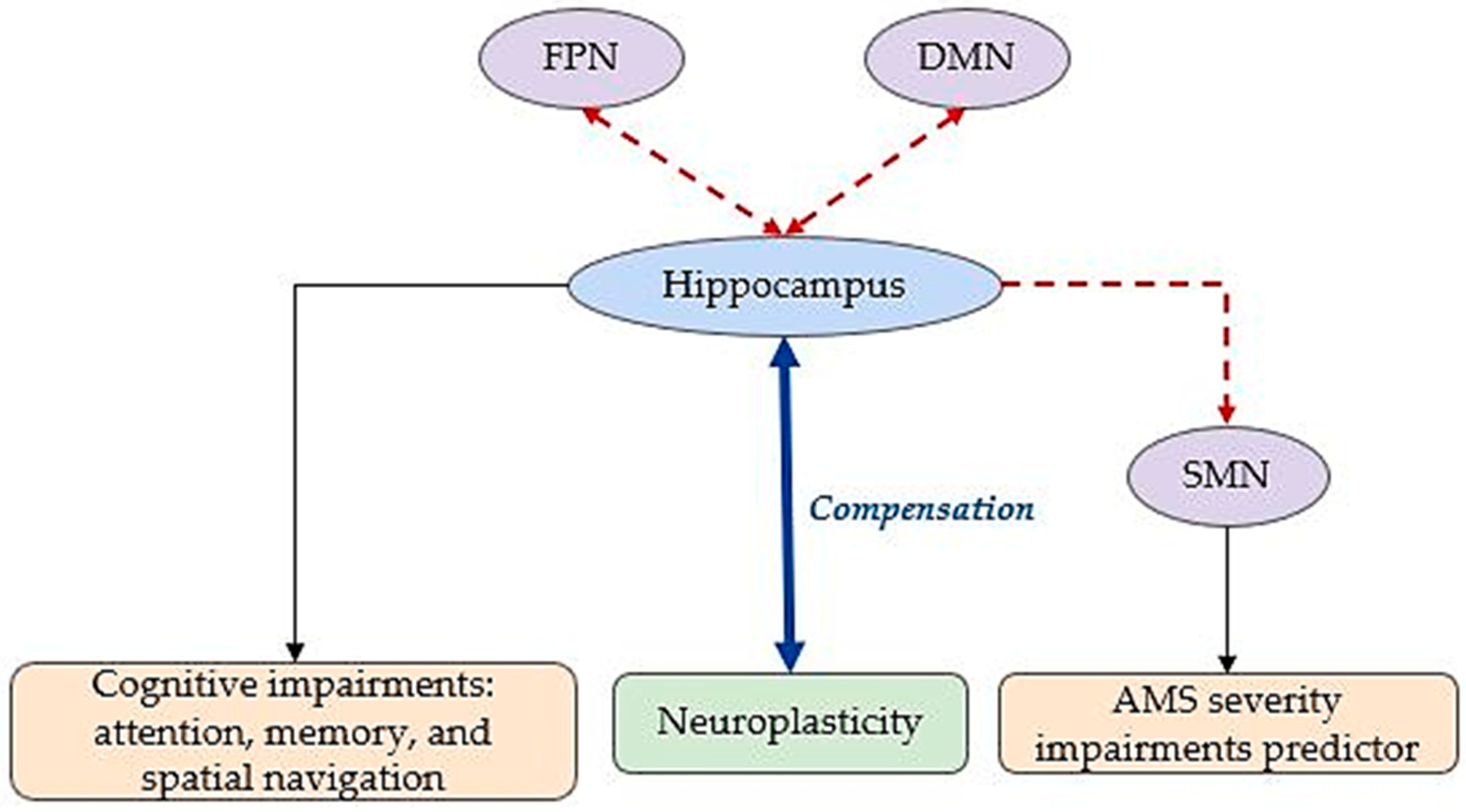
Pathological Acute Hypoxia (e.g., HIE, AMS)FC impairments in DMN, SMN, and FPN, with SMN as a key predictor of AMS severity.DMN, SMN, FPN, Motor, Temporal
HIE shows decreased local efficiency and hippocampal connectivity.
Compensatory increases in FC among motor, frontal, and parietal areas: neuroplasticity observed in recovery cases.
Chronic Environmental Hypoxia (e.g., High Altitude)Reduced hippocampal FC with memory networks: cognitive decline in memory and spatial navigation.DMN, FPN, SMN, Visual, Memory Networks
FC disruptions in visual network and SMN: adaptation varies across populations.
Animal models show hippocampal damage and mitochondrial impairment.
Chronic Pathological Hypoxia (e.g., OSA, CMS)Altered FC in DMN, DAN, VAN, and SN; hippocampal dysfunction linked to intermittent hypoxia and sleep fragmentation: cognitive impairments in attention, memory, and executive functions.DMN, VAN, DAN, SN, Frontal, Para-Hippocampal Gyrus
Topological disruptions in network efficiency.
Para-hippocampal activity changes in CMS; observed effects in adults, children, and animal models.
Disclaimer/Publisher’s Note: The statements, opinions and data contained in all publications are solely those of the individual author(s) and contributor(s) and not of MDPI and/or the editor(s). MDPI and/or the editor(s) disclaim responsibility for any injury to people or property resulting from any ideas, methods, instructions or products referred to in the content.

