Review

The Application of Gamma-Range Auditory Steady-State Responses in Animal Models: A Semi-Structured Literature Review

by
Urte Jasinskyte
1,
Cestmir Vejmola
2,3,
Robertas Guzulaitis
1 and
Inga Griskova-Bulanova
1,3,4,*
1
Institute of Bioscience, Life Sciences Center, Vilnius University, 10257 Vilnius, Lithuania
2
Psychedelic Research Center, National Institute of Mental Health, 250 67 Klecany, Czech Republic
3
Third Faculty of Medicine, Charles University, 100 00 Prague, Czech Republic
4
Translational Health Research Institute, Faculty of Medicine, Vilnius University, 08406 Vilnius, Lithuania
*
Author to whom correspondence should be addressed.
Brain Sci. 2025, 15(11), 1159; https://doi.org/10.3390/brainsci15111159
Submission received: 27 September 2025 / Revised: 20 October 2025 / Accepted: 23 October 2025 / Published: 28 October 2025

Abstract

Background: Gamma-range auditory steady-state responses (ASSRs) are emerging as promising translational biomarkers of neural network function. While extensively studied in human neuropsychiatric and neurodevelopmental research, their application in animal models has expanded in recent years, providing mechanistic insights into disease-related neural dynamics. However, methodological approaches vary widely, findings remain fragmented, and outcomes are not easily generalized. Methods: A literature search was conducted in March 2025 across PubMed and Scopus to identify studies investigating gamma-range ASSRs (30–100 Hz) in animal models with relevance to psychiatric and developmental conditions. Results: Most studies employed rodents, with a smaller number involving non-human primates, and used pharmacological, genetic, lesion-based, or developmental manipulations relevant to schizophrenia, autism spectrum disorder, and related conditions. ASSRs were highly sensitive to NMDA receptor antagonism, state- and trait-related factors, and exhibited region- and layer-specific generation patterns centered on the auditory cortex. Less common paradigms, such as chirps and gap-in-noise, also demonstrated translational potential. Conclusions: Animal research confirms that gamma-range ASSRs provide a sensitive, cross-species readout of circuit dysfunctions observed in psychiatric and neurodevelopmental disorders. To maximize their translational utility, future work should prioritize methodological harmonization, systematic inclusion of sex and behavioral state factors, and replication across laboratories. Strengthening these aspects will enhance the value of ASSRs as biomarkers for early detection, patient stratification, and treatment monitoring in clinical psychiatry.

1. Introduction

Gamma-range auditory steady-state responses (ASSRs) have been extensively studied in humans as potential neurophysiological markers for psychiatric and neurodevelopmental disorders [1]. These responses reflect neural entrainment elicited by periodic auditory stimulation, typically in the form of click trains or amplitude-modulated (AM) tones within the gamma frequency range (30–100 Hz) [2]. It should be noted, however, that synchronization within this range likely reflects contributions from distinct neural generators: lower gamma frequencies (30–50 Hz) are predominantly cortical, whereas higher frequencies (≥80 Hz) may involve more subcortical and brainstem sources, as the frequency range of phase-locked responses decreases along the ascending auditory pathway [3,4]. Because gamma ASSRs depend on the integrity of excitatory–inhibitory balance [5], gamma ASSRs are consistently altered in disorders such as schizophrenia (SZ), where deficits at 40 Hz have been reported in patients [6], in ultra-high-risk individuals [7], and even proposed as predictors of treatment outcomes [6,8]. Comparable alterations have also been described in autism spectrum disorders (ASD), where reductions in gamma synchrony are associated with impaired cortical network development [9]. While influential reviews such as O’Donnell et al. (2013) [1] established the promise of ASSRs as psychiatric biomarkers, they did not provide a structured synthesis of animal studies or systematically evaluate how different disease models reproduce alterations observed in patients. The present review addresses this gap.
Animal models allow controlled manipulation of neural circuits, neurotransmitter systems, and developmental trajectories, enabling mechanistic insights that cannot be achieved in humans. Moreover, the comparability of auditory stimulation paradigms across species makes ASSRs particularly suitable for translational research. Early experimental work in species such as cats [10], guinea pigs [11], gerbils [12], and rabbits [3] laid the foundation for understanding ASSR physiology, but recent efforts have shifted mainly to work on mice, rats, and monkeys to align more closely with translational psychiatry and disease modeling. Rodent models provide valuable mechanistic insight owing to their genetic manipulability and experimental accessibility, though their translatability to human neurophysiology remains limited across many domains [13,14,15,16]. Over the past decade, pharmacological [17,18,19], genetic [20,21,22], lesion-based [23,24], and developmental [25,26] models have been applied to study ASSRs, with the goal of reproducing patient-relevant alterations and probing underlying mechanisms.
Importantly, gamma ASSRs are not only descriptive readouts but also provide mechanistic information, as they index the capacity of neural circuits to sustain synchronized oscillations and reflect the balance of excitation and inhibition in cortical networks [5]. These mechanisms are directly implicated in psychiatric and neurodevelopmental disorders [25,27], where disrupted gamma synchrony has been linked to cognitive impairments in attention [28], working memory [29], and sensory processing [5]. Developmental animal models further allow the investigation of how ASSRs emerge and change across maturation, providing a window into altered trajectories relevant for ASD [30,31] and Fragile X syndrome (FXS) [25,32].
Despite their promise, findings from animal studies remain fragmented. Variability in stimulation protocols, recording methods, experimental states, and modeling strategies complicates cross-study synthesis and limits the integration of preclinical data into clinically meaningful frameworks. Addressing this challenge is essential, given the urgent need for objective biomarkers that can support early detection, patient stratification, and treatment monitoring in psychiatry. ASSRs are particularly promising in this regard because they are non-invasive, repeatable, and measurable across species using analogous paradigms.
Taken together, these considerations highlight the need for a structured synthesis of animal research on gamma-range ASSRs. This review addresses how gamma-range ASSRs are elicited, manipulated, and interpreted in animal models, and whether they reproduce alterations observed in patients. By focusing on disease-relevant models and excluding studies limited to basic auditory physiology without translational value, we aim to clarify the role of animal ASSRs as a cross-species biomarker, identify key methodological challenges, and outline future directions to strengthen their translational impact in developing clinically relevant biomarkers for diagnosis, prognosis, and treatment monitoring.

2. Materials and Methods

We conducted a semi-structured literature review to provide a focused overview of how gamma-range ASSRs are used in animal research. This approach enabled a systematic mapping of relevant studies while maintaining flexibility in selection, allowing us to emphasize works with translational relevance without being limited by overly rigid criteria. Unlike fully systematic reviews, our goal was not exhaustive coverage but a representative synthesis of current research.
To identify relevant studies, we conducted database searches in PubMed and Scopus (March 2025) using combinations of keywords related to auditory steady-state responses, animal models, and electrophysiological methods. Search strings included terms such as “auditory steady-state response” OR “ASSR” OR “steady-state auditory evoked potential” combined with “animal model,” “rat,” “mouse,” “rodent,” or “monkey” and with “EEG,” “electrophysiology,” “evoked potential,” or “local field potential”. The full keyword lists are provided in Appendix A.
The selection process followed predefined inclusion and exclusion criteria to ensure relevance to the study’s aim. We focused on original research articles presenting empirical findings; studies conducted in rodent models with additional inclusion of nonhuman primate studies relevant to neuropsychiatric disorders; research specifically assessing gamma-range ASSRs (30–100 Hz); studies utilizing electroencephalography (EEG), electrocorticography (ECoG), or local field potentials (LFPs) as core measurement techniques; and articles published in English. Only peer-reviewed publications were considered; preprints, unpublished data, and conference abstracts were excluded. We did not include non-original research (e.g., reviews, theoretical papers, meta-analyses, case reports); studies not published in English; research focused primarily on hearing mechanisms rather than on neurophysiological or neuropsychiatric implications of ASSR; and studies utilizing irrelevant auditory paradigms, such as transient evoked potentials instead of steady-state responses. The study selection process was independently conducted by two researchers, who screened article titles, abstracts, and full texts where necessary to ensure relevance. In cases where the abstract did not provide sufficient information, the full text was reviewed to confirm eligibility. Discrepancies between reviewers were resolved through discussion and consensus.
Because our aim was not the exhaustive coverage of all animal ASSR research but rather a synthesis with translational relevance, we applied an additional filter beyond database-based inclusion criteria. Specifically, studies were retained only if their design, outcomes, or manipulations allowed meaningful comparison to human neuropsychiatric or neurophysiological research. For example, reports focusing exclusively on auditory physiology (e.g., cochlear or brainstem responses without cortical readouts), or those limited to methodological validation without psychiatric or network-level interpretation, were excluded. Similarly, works using transient auditory evoked potentials rather than steady-state paradigms were not considered. This approach was chosen to maintain focus on findings with clear cross-species value for understanding psychiatric disorders, while acknowledging that it necessarily omits some technically relevant but less translationally oriented studies.

3. Results

The initial search identified 132 records (PubMed: 36; Scopus: 96) (Figure 1). After removing duplicates (n = 32), 100 unique articles remained (Figure 1). Following screening and the application of predefined inclusion criteria, 56 studies were included in this review (Figure 1, Table S1).
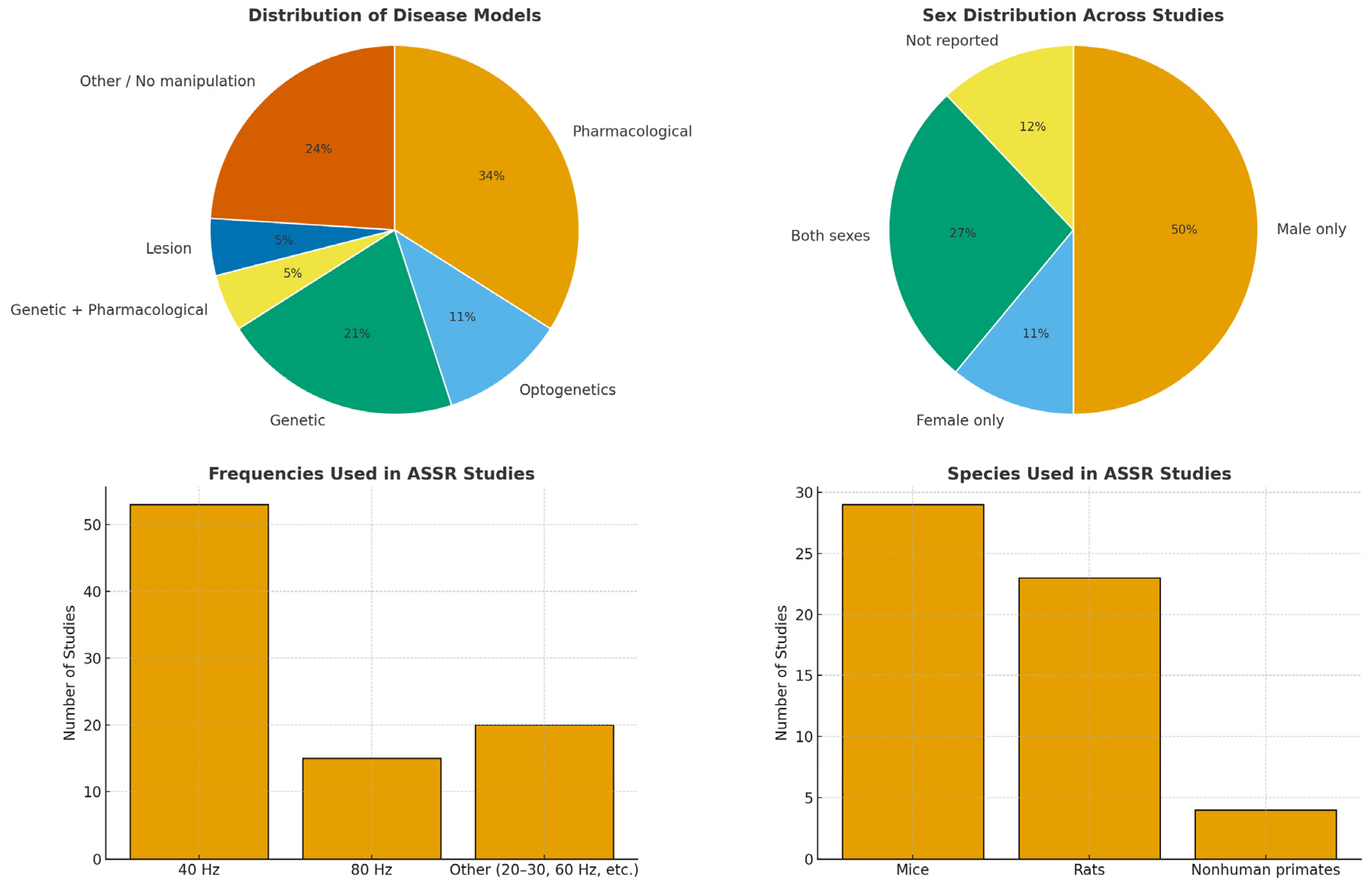
Given the semi-structured nature of the review, some relevant studies may have been missed due to database limitations, keyword filtering, or the exclusion of gray literature. Nonetheless, this review provides a comprehensive and representative synthesis of current ASSR research in animal models relevant to neuropsychiatric disorders. Figure 2 summarizes the main findings after generalizing the included works.

3.1. Disease Models

Most studies employed mice (29) and rats (23), emphasizing their central role in ASSR-related translational research. A smaller number used nonhuman primates, including macaques (2), common marmosets (2), and rhesus monkeys (1). Mouse models included both wild-type (WT) and genetically modified strains (e.g., SRKO, Fmr1 KO, GRIN2A KO) to model ASD (13 studies), while rat studies predominantly utilized pharmacological agents to model SZ (16 studies), with fewer studies addressing other psychiatric or neurological conditions. Among the pharmacological models, MK-801 was the most commonly used drug (9 studies), followed by ketamine (7) and phencyclidine (PCP) (2) (all being non-competitive antagonists of the N-methyl-D-aspartate (NMDA) receptor complex), indicating a research preference for NMDA receptor antagonists.

3.2. Group Characteristics and Experimental Contexts

Regarding experimental design, 28 studies used only male subjects, 6 studies used only females, 15 used both sexes, and sex was not reported in 7 studies (Figure 1). The number of animals per study ranged from 5 to 164 in rodent models, and from 2 to 5 in nonhuman primate studies. Three studies did not report animal numbers. Regarding experimental conditions, freely moving animals were used in 40 studies, head-fixed setups in 13, and anesthetized conditions in 3.

3.3. Stimulation Protocols

Stimulation protocols covered a wide frequency range (1–480 Hz), focusing strongly on the gamma band (30–80 Hz). Stimuli were primarily click trains; however, some studies also employed AM tones, chirps, or gap-in-noise paradigms. Studies often used multiple stimulation frequencies, where 40 Hz was the most frequently used stimulation frequency (53 studies), followed by 80 Hz (15 studies), reflecting their relevance for gamma-range ASSR investigations (Figure 1).

4. Discussion

This section synthesizes key methodological patterns and findings across the reviewed studies, emphasizing their implications for understanding and modeling neuropsychiatric disorders. We examine how neurochemical manipulations and disease-specific models have been used to probe the mechanisms underlying gamma-range ASSRs, explore the neural circuitry and spatial organization of ASSR generation, and consider how brain traits and states, and methodological settings shape ASSR outcomes. These aspects are critical for improving the translational value of animal models and advancing the utility of ASSRs as neurophysiological markers of psychiatric and neurodevelopmental conditions.
We first consider how different disease models (pharmacological, genetic, lesion-based) utilize gamma-range ASSRs to capture circuit dysfunctions relevant to psychiatric and neurodevelopmental disorders. Next, we examine the neural circuits that generate ASSRs, highlighting cortical and subcortical contributions and their implications for understanding disorder-related alterations. We then explore how transient brain states (e.g., arousal, anesthesia, attention) and stable biological traits (e.g., sex, hormonal status) shape ASSR outcomes and influence translational relevance. Finally, we discuss methodological factors, including stimulation paradigms, recording approaches, and experimental designs that affect comparability across studies and limit cross-species translation

4.1. Disease Modeling

A central advantage of animal research lies in the ability to establish models that reproduce core features of neuropsychiatric and neurodevelopmental disorders. Gamma-range ASSRs have proven particularly useful in this context, as they provide a sensitive readout for circuit-level dysfunctions that parallel those observed in patients. SZ, where alterations of ASSRs have been consistently observed in clinical populations [33], has been modeled most extensively, using multiple approaches that converge on impaired gamma synchronization. Pharmacological induction of NMDA receptor hypofunction is widely accepted as a rodent model of SZ-like pathophysiology and consistently produces reductions in 40 Hz ASSRs, albeit with some variability across dose, brain region, and protocol (e.g., [17,34,35,36]). Notably, pharmacological NMDA receptor blockade evokes similar decreases in healthy humans [37,38], underscoring the translational validity of this approach. Complementary non-pharmacological models such as the neonatal ventral hippocampal lesion (NVHL) have also demonstrated persistent reductions in gamma-range ASSRs [23,39], supporting their relevance for developmental aspects of SZ.
Genetic models further expand this repertoire, including PLC-β1 knock-out (KO) mice [40], Grin1 mutants with glycogen synthase kinase-3 beta (GSK3β) modulation [20,41], and serine racemase (SRKO) mice modeling D-serine metabolism disruption [42], again mimicking ASSR reductions observed in humans.
Beyond SZ, ASD has been investigated in relation to gamma-range ASSRs [9], particularly in Fmr1 and PTEN mutants, where reductions in phase-locking and altered developmental trajectories [31,43,44] mirror findings of altered responses in human cohorts. Several studies have also extended ASSR applications to other conditions: systemic lupus erythematosus (anti-P antibody model, [45]), FXS with potential pharmacological rescue using minocycline [44], and inflammation-related changes induced by interferon-alpha (IFN-α) treatment [46].
Taken together, these diverse models indicate that gamma-range ASSRs constitute a unifying translational tool for probing circuit-level dysfunctions across neuropsychiatric and neurodevelopmental disorders. Notably, many alterations observed in animal studies closely parallel those reported in patients, underscoring the validity of ASSRs as cross-species biomarkers that bridge mechanistic investigations in animal models with clinically relevant phenomena in humans.

4.2. Localization of ASSRs and Neural Circuitry

While disease models demonstrate that gamma-range ASSRs are sensitive to neuropsychiatric and developmental pathophysiology, understanding the precise neural circuits underlying these alterations requires localization studies that animal models are uniquely positioned to provide.
Across models, the auditory cortex (AC) emerges as the dominant generator of these responses. For instance, Li et al. (2018) [24] demonstrated that ASSR deficits in the NVHL model were localized specifically to the primary AC, with non-primary auditory regions remaining unaffected. Consistent with this, Gautam et al. (2024) [47] showed that gamma synchrony emerged rapidly in the AC, highlighting its role as the earliest cortical locus of entrainment.
The prefrontal cortex (PFC) appears to play a more variable role. While some studies reported robust gamma entrainment (e.g., [44,48]), others failed to detect clear responses (e.g., [49]), suggesting that PFC involvement may be weaker, more state-dependent, or reliant on cross-regional interactions. Further structural specificity has been revealed by laminar recordings. Li et al. (2021) [50] found that 40 Hz responses were present across all cortical layers of the AC, with the strongest activity in the thalamorecipient granular layer. Johnson et al. (2024) [51] emphasized that superficial and deep layers are critical for temporal precision, supporting coordinated entrainment across networks. Optogenetic activation studies add to this picture: stimulating basal forebrain parvalbumin-positive neurons was shown to enhance AC–PFC coupling [52,53], providing causal evidence for top-down modulation in sustaining gamma synchrony [54,55].
In addition to cortical regions, subcortical structures also contribute to ASSR generation. The hippocampus and amygdala have been implicated in supporting gamma synchronization [49], and the thalamus is particularly critical as a relay structure. Wang et al. (2020) showed that NMDA receptor blockade in the medial geniculate body (MGB) significantly suppressed 40 Hz ASSRs in the AC but not in the PFC, reinforcing its role as a key driver of auditory gamma entrainment [45,46,56]. Methodological advances are beginning to bridge these findings across species. For example, Jiricek et al. (2021) [57] used multichannel EEG and source localization in rats to identify generators not only in auditory but also in motor cortices, with motor activity interpreted as reflecting behavioral freezing in response to stimulation. This highlights the importance of monitoring behavioral state when interpreting ASSR dynamics. They also highlight the variability that may arise due to methodological differences, behavioral or brain state fluctuations, and intrinsic network properties that influence the capacity to sustain gamma synchrony.
These neuroanatomical insights are not only mechanistically informative but also align with the circuits implicated in neuropsychiatric and neurodevelopmental disorders [58,59,60]. By linking AC dysfunction with distributed cortical–subcortical networks, animal models provide a translational framework for understanding how gamma synchrony disruptions manifest in conditions such as SZ and ASD.

4.3. State and Trait Modulation of ASSRs

Gamma-range ASSRs are sensitive to both state-dependent and trait-related influences, including arousal [61], consciousness levels [62], and biological sex [63]. Animal models provide unique opportunities to examine how these factors modulate neural synchrony, offering insights that are difficult to obtain in human studies and directly relevant to the variability observed in psychiatric populations.
Most experiments have been conducted in freely moving animals, allowing for naturalistic behavior but introducing variability in motor activity and internal brain state. Behavior was rarely tracked systematically, leaving distinctions between spontaneous movement and arousal-related effects underexplored. Li et al. (2020) [64], for example, reported that movement did not affect late-latency ASSRs and only influenced early-latency components, yet the influence of spontaneous versus evoked arousal remains unclear. Head-fixed preparations offer tighter experimental control but raise concerns about stress and altered arousal states, while anesthetized designs provide stability at the cost of suppressing neural synchronization. Wang et al. (2018) [49] observed significant reductions in 40 Hz ASSR power and phase-locking under anesthesia, mirroring reductions seen in humans during decreased consciousness [62]. In the same study, a mild arousing stimulus (foot shock) enhanced 40 Hz ASSRs and strengthened functional connectivity from auditory to medial PFC, pointing to a circuit mechanism by which arousal boosts sensory processing. These findings resonate with clinical reports showing altered arousal regulation in SZ [65] and mood disorders [66], where state fluctuations may exacerbate gamma synchrony deficits [67]. Importantly, no study has systematically compared different experimental states under matched protocols, underscoring the need for standardized or at least well-reported conditions to improve comparability across studies.
Experimental manipulations provide further evidence of state-dependent modulation. Optogenetic activation of basal forebrain parvalbumin-positive neurons altered cortical gamma activity in a state-dependent fashion (Kim et al., 2015 [48]), likely by synchronizing inhibitory networks [40]. Hwang et al. (2019) [52] showed that synchronized stimulation of these neurons enhanced ASSR magnitude and reorganized cortical topography in a manner consistent with increased attention, a finding directly relevant to attention deficits in SZ and ADHD [68]. In line, McNally et al. (2020) [69] reported that disruption of the ascending arousal system suppressed gamma ASSRs, resembling functional impairments observed in SZ [70]. Taken together, these findings suggest that arousal-related circuits exert powerful control over cortical gamma synchrony and provide mechanistic links to psychiatric symptom domains.
In addition to dynamic states, stable biological traits also influence ASSRs. Most reviewed studies used only male animals, limiting the understanding of sex-related differences. However, studies that included both sexes reported differences in amplitude, phase-locking, and developmental trajectories, particularly in models of FXS (Fmr1) and ASD (PTEN mutants) [31,43]. Male model mice showed more persistent deficits in 40 Hz phase-locking, whereas female mice exhibited earlier maturation of temporal processing. One study further suggested that the estrus cycle stage may modulate cortical oscillations [71], a finding consistent with prior human evidence [72]. Such sex and hormonal influences are clinically relevant, as women and men show different prevalence rates and symptom profiles in disorders such as SZ [73], ASD [74], and depression [75,76]. Furthermore, hormonal fluctuations are increasingly recognized as modulators of psychiatric symptom expression [77,78].
Taken together, evidence from animal models shows that both transient brain states (e.g., arousal, anesthesia, attention) and intrinsic traits (e.g., sex, hormone status) substantially shape ASSR dynamics. These influences parallel patient observations, where altered arousal regulation, attention deficits, and sex-related variability are hallmarks of neuropsychiatric and neurodevelopmental conditions. In addition, sample sizes varied widely, from very small groups of two to five nonhuman primates to over one hundred rodents, raising concerns about statistical power and reproducibility in the smaller studies. Accounting for brain states and ensuring adequately powered designs are essential for both experimental planning and data interpretation; these are prerequisites for establishing the translational validity of animal ASSR models.

4.4. Evidence from Non-Human Primates

Although rodent models yield important mechanistic insights, their relevance to human neurophysiology is often restricted. Research in non-human primates provides an essential translational link between rodent and human studies [79,80] of auditory steady-state responses. Although relatively few, these studies demonstrate that gamma-range ASSRs are conserved across primate species, with broadly comparable topography and frequency tuning to those observed in humans [81,82,83]. Moreover, pharmacological manipulations [83] and intracranial recordings [84] in awake animals confirm that cortical network entrainment can be reliably assessed in these models using EEG and ECoG techniques. Taken together, these findings highlight the translational value of primate models for validating neural mechanisms underlying ASSR generation and for bridging preclinical and clinical research.

4.5. Methodological Settings

Based on the reviewed studies, there is a notable degree of methodological consistency in the use of stimulation (e.g., 40–50 Hz clicks) to elicit responses, reflecting its established relevance for probing cortical synchronization. However, some also included other frequencies (e.g., 20, 25, 30, 60, 80 Hz), which may impact response profiles and limit direct comparability across studies. Importantly, less common paradigms such as chirps and gap-in-noise, though rarely used in human research, also demonstrated translational validity in animal models. All studies targeted auditory and frontal cortical areas, yet there was considerable variability in electrode placement (spanning primary auditory, frontal, and parietal regions) and in recording techniques (ranging from surface EEG, ECoG to LFP). These methodological differences may influence signal characteristics and comparability, and should be considered when interpreting findings, particularly in cross-study comparisons of signal strength, localization, and spectral characteristics.
From a translational perspective, reliance on a limited set of paradigms (primarily 40 Hz clicks) has the advantage of aligning with clinical protocols used in SZ and ASD research. However, expanding to more diverse paradigms may help uncover additional circuit properties relevant to disorders characterized by abnormal sensory processing, such as Fragile X syndrome or attention-deficit conditions. Likewise, greater methodological standardization, particularly in electrode placement, behavioral state tracking, and reporting practices, would improve reproducibility and enable clearer mapping between animal and human findings.

4.6. Limitation

It should be noted that our semi-structured approach deliberately prioritized studies with translational relevance to neuropsychiatric research, which means that some technically relevant but less clinically oriented reports (e.g., those focused solely on auditory physiology or methodological validation) were not included. While this narrows the scope, it strengthens the review’s focus on findings most directly comparable to human studies.

5. Conclusions

This review highlights the methodological diversity and translational potential of gamma-range ASSRs in animal models of neuropsychiatric and neurodevelopmental disorders. Across diverse models, ASSRs consistently emerged as a sensitive marker of cortical network function. In particular, their disruption in models of SZ and ASD supports their value for probing circuit-level dysfunctions that parallel human pathology. The use of pharmacological, genetic, lesion-based, and developmental models demonstrates that ASSRs can capture convergent deficits across distinct experimental approaches, reinforcing their potential as a cross-species biomarker. At the same time, methodological variability, including differences in stimulation paradigms, recording techniques, subject characteristics, and experimental states, remains a barrier to comparability and standardization. Taken together, animal ASSR studies reproduce key alterations consistently reported in SZ and ASD, while extending insights to other models (Fragile X, lupus, inflammation). Future research should prioritize methodological harmonization, systematic inclusion of female animals, and broader application of ASSRs across a wider range of disease models to strengthen their translational impact on understanding, diagnosing, and monitoring neuropsychiatric and neurodevelopmental disorders.

Supplementary Materials

The following supporting information can be downloaded at https://www.mdpi.com/article/10.3390/brainsci15111159/s1. Table S1: Summary of studies included in the review. Refs. [50,85,86,87,88,89,90,91,92,93,94,95,96,97,98,99,100,101] are cited in Supplementary Materials file.

Author Contributions

Conceptualization, I.G.-B.; methodology, U.J., C.V. and I.G.-B.; validation, R.G., C.V. and I.G.-B.; formal analysis, U.J. and I.G.-B.; data curation, I.G.-B. and R.G.; writing—original draft preparation, U.J. and I.G.-B.; writing—review and editing, C.V. and R.G.; supervision, I.G.-B. All authors have read and agreed to the published version of the manuscript.

Funding

C.V. and I.G.-B. were supported by the ERDF-Project Brain Dynamics (No. CZ.02.01.01/00/22_008/0004643).

Institutional Review Board Statement

Not applicable.

Informed Consent Statement

Not applicable.

Data Availability Statement

Not applicable.

Conflicts of Interest

The authors declare no conflicts of interest.

Abbreviations

The following abbreviations are used in this manuscript:
ACAuditory cortex
AMAmplitude-modulated
ASDAutism spectrum disorders
ASSRsAuditory steady state responses
BF-PVBasal forebrain parvalbumin
ECoGElectrocorticography
EEGElectroencephalography
EGFEpidermal growth factor
FXSFragile X Syndrome
GABAGamma-aminobutyric acid
GlyT1Glycine transporter 1
GSK3 βGlycogen synthase kinase-3
IFN-αInterferon-alpha
KOKnock-out
LFPLocal field potentials
MGBMedial geniculate body
NMDAN-methyl D-aspartate
NVHLNeonatal ventral hippocampal lesion
Pcdh10Protocadherin 10
PCPPhencyclidine
PFCPrefrontal cortex
PVParvalbumin
SRKOSerine racemase knock-out
SZSchizophrenia
TRNThalamic reticular nucleus
WTWild-type

Appendix A

The keyword combination used for search in PubMed database is following: (“auditory steady-state response”[Title/Abstract] OR “ASSR”[Title/Abstract] OR “steady-state auditory evoked potential”[Title/Abstract]) AND (“animal model”[Title/Abstract] OR “rat”[Title/Abstract] OR “mouse”[Title/Abstract] OR “mice”[Title/Abstract] OR “rodent”[Title/Abstract] OR “monkey”[Title/Abstract]) AND (“EEG”[Title/Abstract] OR “electrophysiology”[Title/Abstract] OR “evoked potential”[Title/Abstract] OR “local field potential”[Title/Abstract]).
The keyword combinations used for search in Scopus database are following: “auditory steady-state response*” OR “ASSR” OR “steady-state auditory evoked potential*” OR “SSAEP” AND “animal model*” OR “rat” OR “mouse” OR “mice” OR “rodent*” OR “monkey” AND “EEG” OR “electrophysiology” OR “evoked potential*” OR “local field potential*” OR “LFP”.

References

  1. O’Donnell, B.F.; Vohs, J.L.; Krishnan, G.P.; Rass, O.; Hetrick, W.P.; Morzorati, S.L. The Auditory Steady-State Response (ASSR). In Supplements to Clinical Neurophysiology; Elsevier: Amsterdam, The Netherlands, 2013; Volume 62, pp. 101–112. ISBN 978-0-7020-5307-8. [Google Scholar]
  2. Brenner, C.A.; Krishnan, G.P.; Vohs, J.L.; Ahn, W.-Y.; Hetrick, W.P.; Morzorati, S.L.; O’Donnell, B.F. Steady State Responses: Electrophysiological Assessment of Sensory Function in Schizophrenia. Schizophr. Bull. 2009, 35, 1065–1077. [Google Scholar] [CrossRef]
  3. Kuwada, S.; Anderson, J.S.; Batra, R.; Fitzpatrick, D.C.; Teissier, N.; D’Angelo, W.R. Sources of the Scalp-Recorded Amplitude-Modulation Following Response. J. Am. Acad. Audiol. 2002, 13, 188–204. [Google Scholar] [CrossRef]
  4. Joris, P.X.; Schreiner, C.E.; Rees, A. Neural Processing of Amplitude-Modulated Sounds. Physiol. Rev. 2004, 84, 541–577. [Google Scholar] [CrossRef]
  5. Sugiyama, S.; Ohi, K.; Kuramitsu, A.; Takai, K.; Muto, Y.; Taniguchi, T.; Kinukawa, T.; Takeuchi, N.; Motomura, E.; Nishihara, M.; et al. The Auditory Steady-State Response: Electrophysiological Index for Sensory Processing Dysfunction in Psychiatric Disorders. Front. Psychiatry 2021, 12, 644541. [Google Scholar] [CrossRef]
  6. Grent, T.; Gajwani, R.; Gross, J.; Gumley, A.I.; Krishnadas, R.; Lawrie, S.M.; Schwannauer, M.; Schultze-Lutter, F.; Uhlhaas, P.J. 40-Hz Auditory Steady-State Responses Characterize Circuit Dysfunctions and Predict Clinical Outcomes in Clinical High-Risk for Psychosis Participants: A Magnetoencephalography Study. Biol. Psychiatry 2021, 90, 419–429. [Google Scholar] [CrossRef]
  7. Tada, M.; Nagai, T.; Kirihara, K.; Koike, S.; Suga, M.; Araki, T.; Kobayashi, T.; Kasai, K. Differential Alterations of Auditory Gamma Oscillatory Responses Between Pre-Onset High-Risk Individuals and First-Episode Schizophrenia. Cereb. Cortex 2016, 26, 1027–1035. [Google Scholar] [CrossRef]
  8. Koshiyama, D.; Kirihara, K.; Tada, M.; Nagai, T.; Fujioka, M.; Ichikawa, E.; Ohta, K.; Tani, M.; Tsuchiya, M.; Kanehara, A.; et al. Auditory Gamma Oscillations Predict Global Symptomatic Outcome in the Early Stages of Psychosis: A Longitudinal Investigation. Clin. Neurophysiol. 2018, 129, 2268–2275. [Google Scholar] [CrossRef]
  9. Seymour, R.A.; Rippon, G.; Gooding-Williams, G.; Sowman, P.F.; Kessler, K. Reduced Auditory Steady State Responses in Autism Spectrum Disorder. Mol. Autism 2020, 11, 56. [Google Scholar] [CrossRef]
  10. Mäkelä, J.P.; Karmos, G.; Molnár, M.; Csépe, V.; Winkler, I. Steady-State Responses from the Cat Auditory Cortex. Hear. Res. 1990, 45, 41–50. [Google Scholar] [CrossRef]
  11. Jeng, F.-C.; Abbas, P.J.; Brown, C.J.; Miller, C.A.; Nourski, K.V.; Robinson, B.K. Electrically Evoked Auditory Steady-State Responses in Guinea Pigs. Audiol. Neurotol. 2007, 12, 101–112. [Google Scholar] [CrossRef]
  12. Dolphin, W.F.; Chertoff, M.E.; Burkard, R. Comparison of the Envelope Following Response in the Mongolian Gerbil Using Two-Tone and Sinusoidally Amplitude-Modulated Tones. J. Acoust. Soc. Am. 1994, 96, 2225–2234. [Google Scholar] [CrossRef]
  13. Azkona, G.; Sanchez-Pernaute, R. Mice in Translational Neuroscience: What R We Doing? Prog. Neurobiol. 2022, 217, 102330. [Google Scholar] [CrossRef]
  14. Leenaars, C.H.C.; Kouwenaar, C.; Stafleu, F.R.; Bleich, A.; Ritskes-Hoitinga, M.; De Vries, R.B.M.; Meijboom, F.L.B. Animal to Human Translation: A Systematic Scoping Review of Reported Concordance Rates. J. Transl. Med. 2019, 17, 223. [Google Scholar] [CrossRef]
  15. Granzotto, A.; Vissel, B.; Sensi, S.L. Lost in Translation: Inconvenient Truths on the Utility of Mouse Models in Alzheimer’s Disease Research. eLife 2024, 13, e90633. [Google Scholar] [CrossRef]
  16. Brynildsen, J.K.; Rajan, K.; Henderson, M.X.; Bassett, D.S. Network Models to Enhance the Translational Impact of Cross-Species Studies. Nat. Rev. Neurosci. 2023, 24, 575–588. [Google Scholar] [CrossRef]
  17. Schuelert, N.; Dorner-Ciossek, C.; Brendel, M.; Rosenbrock, H. A Comprehensive Analysis of Auditory Event-Related Potentials and Network Oscillations in an NMDA Receptor Antagonist Mouse Model Using a Novel Wireless Recording Technology. Physiol. Rep. 2018, 6, e13782. [Google Scholar] [CrossRef]
  18. Rosenbrock, H.; Dorner-Ciossek, C.; Giovannini, R.; Schmid, B.; Schuelert, N. Effects of the Glycine Transporter-1 Inhibitor Iclepertin (BI 425809) on Sensory Processing, Neural Network Function, and Cognition in Animal Models Related to Schizophrenia. J. Pharmacol. Exp. Ther. 2022, 382, 223–232. [Google Scholar] [CrossRef]
  19. Adraoui, F.W.; Hettak, K.; Viardot, G.; Alix, M.; Guiffard, S.; Meot, B.; L’Hostis, P.; Maurin, A.; Delpy, E.; Drieu La Rochelle, C.; et al. Differential Effects of Aripiprazole on Electroencephalography-Recorded Gamma-Band Auditory Steady-State Response, Spontaneous Gamma Oscillations and Behavior in a Schizophrenia Rat Model. Int. J. Mol. Sci. 2024, 25, 1035. [Google Scholar] [CrossRef]
  20. Nakao, K.; Nakazawa, K. Brain State-Dependent Abnormal LFP Activity in the Auditory Cortex of a Schizophrenia Mouse Model. Front. Neurosci. 2014, 8, 168. [Google Scholar] [CrossRef]
  21. Kozono, N.; Okamura, A.; Honda, S.; Matsumoto, M.; Mihara, T. Gamma Power Abnormalities in a Fmr1-Targeted Transgenic Rat Model of Fragile X Syndrome. Sci. Rep. 2020, 10, 18799. [Google Scholar] [CrossRef]
  22. Herzog, L.E.; Wang, L.; Yu, E.; Choi, S.; Farsi, Z.; Song, B.J.; Pan, J.Q.; Sheng, M. Mouse Mutants in Schizophrenia Risk Genes GRIN2A and AKAP11 Show EEG Abnormalities in Common with Schizophrenia Patients. Transl. Psychiatry 2023, 13, 92. [Google Scholar] [CrossRef]
  23. Vohs, J.L.; Andrew Chambers, R.; Krishnan, G.P.; O’Donnell, B.F.; Berg, S.; Morzorati, S.L. GABAergic Modulation of the 40 Hz Auditory Steady-State Response in a Rat Model of Schizophrenia. Int. J. Neuropsychopharm. 2010, 13, 487. [Google Scholar] [CrossRef]
  24. Li, S.; Ma, L.; Wang, Y.; Wang, X.; Li, Y.; Qin, L. Auditory Steady-State Responses in Primary and Non-Primary Regions of the Auditory Cortex in Neonatal Ventral Hippocampal Lesion Rats. PLoS ONE 2018, 13, e0192103. [Google Scholar] [CrossRef]
  25. Croom, K.; Rumschlag, J.A.; Erickson, M.A.; Binder, D.K.; Razak, K.A. Developmental Delays in Cortical Auditory Temporal Processing in a Mouse Model of Fragile X Syndrome. J. Neurodev. Disord. 2023, 15, 23. [Google Scholar] [CrossRef]
  26. Jonak, C.R.; Assad, S.A.; Garcia, T.A.; Sandhu, M.S.; Rumschlag, J.A.; Razak, K.A.; Binder, D.K. Phenotypic Analysis of Multielectrode Array EEG Biomarkers in Developing and Adult Male Fmr1 KO Mice. Neurobiol. Dis. 2024, 195, 106496. [Google Scholar] [CrossRef] [PubMed]
  27. Sohal, V.S.; Rubenstein, J.L.R. Excitation-Inhibition Balance as a Framework for Investigating Mechanisms in Neuropsychiatric Disorders. Mol. Psychiatry 2019, 24, 1248–1257. [Google Scholar] [CrossRef] [PubMed]
  28. Minzenberg, M.J.; Firl, A.J.; Yoon, J.H.; Gomes, G.C.; Reinking, C.; Carter, C.S. Gamma Oscillatory Power Is Impaired During Cognitive Control Independent of Medication Status in First-Episode Schizophrenia. Neuropsychopharmacology 2010, 35, 2590–2599. [Google Scholar] [CrossRef]
  29. Chen, C.-M.A.; Stanford, A.D.; Mao, X.; Abi-Dargham, A.; Shungu, D.C.; Lisanby, S.H.; Schroeder, C.E.; Kegeles, L.S. GABA Level, Gamma Oscillation, and Working Memory Performance in Schizophrenia. NeuroImage Clin. 2014, 4, 531–539. [Google Scholar] [CrossRef] [PubMed]
  30. Port, R.G.; Gajewski, C.; Krizman, E.; Dow, H.C.; Hirano, S.; Brodkin, E.S.; Carlson, G.C.; Robinson, M.B.; Roberts, T.P.L.; Siegel, S.J. Protocadherin 10 Alters γ Oscillations, Amino Acid Levels, and Their Coupling; Baclofen Partially Restores These Oscillatory Deficits. Neurobiol. Dis. 2017, 108, 324–338. [Google Scholar] [CrossRef]
  31. Croom, K.; Rumschlag, J.A.; Erickson, M.A.; Binder, D.; Razak, K.A. Sex Differences during Development in Cortical Temporal Processing and Event Related Potentials in Wild-Type and Fragile X Syndrome Model Mice. J. Neurodev. Disord. 2024, 16, 24. [Google Scholar] [CrossRef]
  32. Tao, X.; Croom, K.; Newman-Tancredi, A.; Varney, M.; Razak, K.A. Acute Administration of NLX-101, a Serotonin 1A Receptor Agonist, Improves Auditory Temporal Processing during Development in a Mouse Model of Fragile X Syndrome. J. Neurodev. Disord. 2025, 17, 1. [Google Scholar] [CrossRef]
  33. Thuné, H.; Recasens, M.; Uhlhaas, P.J. The 40-Hz Auditory Steady-State Response in Patients With Schizophrenia: A Meta-Analysis. JAMA Psychiatry 2016, 73, 1145. [Google Scholar] [CrossRef]
  34. Raza, M.U.; Sivarao, D.V. Test-Retest Reliability of Tone- and 40 Hz Train-Evoked Gamma Oscillations in Female Rats and Their Sensitivity to Low-Dose NMDA Channel Blockade. Psychopharmacology 2021, 238, 2325–2334. [Google Scholar] [CrossRef]
  35. Sivarao, D.V.; Frenkel, M.; Chen, P.; Healy, F.L.; Lodge, N.J.; Zaczek, R. MK-801 Disrupts and Nicotine Augments 40 Hz Auditory Steady State Responses in the Auditory Cortex of the Urethane-Anesthetized Rat. Neuropharmacology 2013, 73, 1–9. [Google Scholar] [CrossRef] [PubMed]
  36. Sivarao, D.V.; Chen, P.; Senapati, A.; Yang, Y.; Fernandes, A.; Benitex, Y.; Whiterock, V.; Li, Y.-W.; Ahlijanian, M.K. 40 Hz Auditory Steady-State Response Is a Pharmacodynamic Biomarker for Cortical NMDA Receptors. Neuropsychopharmacology 2016, 41, 2232–2240. [Google Scholar] [CrossRef] [PubMed]
  37. Light, G.A.; Zhang, W.; Joshi, Y.B.; Bhakta, S.; Talledo, J.A.; Swerdlow, N.R. Single-Dose Memantine Improves Cortical Oscillatory Response Dynamics in Patients with Schizophrenia. Neuropsychopharmacology 2017, 42, 2633–2639. [Google Scholar] [CrossRef]
  38. Murphy, N.; Ramakrishnan, N.; Vo-Le, B.; Vo-Le, B.; Smith, M.A.; Iqbal, T.; Swann, A.C.; Mathew, S.J.; Lijffijt, M. A Randomized Cross-over Trial to Define Neurophysiological Correlates of AV-101 N-Methyl-d-Aspartate Receptor Blockade in Healthy Veterans. Neuropsychopharmacology 2021, 46, 820–827. [Google Scholar] [CrossRef]
  39. Vohs, J.L.; Chambers, R.A.; O’Donnell, B.F.; Krishnan, G.P.; Morzorati, S.L. Auditory Steady State Responses in a Schizophrenia Rat Model Probed by Excitatory/Inhibitory Receptor Manipulation. Int. J. Psychophysiol. 2012, 86, 136–142. [Google Scholar] [CrossRef] [PubMed]
  40. Shahriari, Y.; Krusienski, D.; Dadi, Y.S.; Seo, M.; Shin, H.-S.; Choi, J.H. Impaired Auditory Evoked Potentials and Oscillations in Frontal and Auditory Cortex of a Schizophrenia Mouse Model. World J. Biol. Psychiatry 2016, 17, 439–448. [Google Scholar] [CrossRef]
  41. Nakao, K.; Singh, M.; Sapkota, K.; Hagler, B.C.; Hunter, R.N.; Raman, C.; Hablitz, J.J.; Nakazawa, K. GSK3β Inhibition Restores Cortical Gamma Oscillation and Cognitive Behavior in a Mouse Model of NMDA Receptor Hypofunction Relevant to Schizophrenia. Neuropsychopharmacology 2020, 45, 2207–2218. [Google Scholar] [CrossRef]
  42. Balla, A.; Ginsberg, S.D.; Abbas, A.I.; Sershen, H.; Javitt, D.C. Translational Neurophysiological Biomarkers of N-Methyl-d-Aspartate Receptor Dysfunction in Serine Racemase Knockout Mice. Biomark. Neuropsychiatry 2020, 2, 100019. [Google Scholar] [CrossRef]
  43. Croom, K.; Rumschlag, J.A.; Molinaro, G.; Erickson, M.A.; Binder, D.K.; Huber, K.M.; Razak, K.A. Developmental Trajectory and Sex Differences in Auditory Processing in a PTEN-Deletion Model of Autism Spectrum Disorders. Neurobiol. Dis. 2024, 200, 106628. [Google Scholar] [CrossRef]
  44. Lovelace, J.W.; Ethell, I.M.; Binder, D.K.; Razak, K.A. Minocycline Treatment Reverses Sound Evoked EEG Abnormalities in a Mouse Model of Fragile X Syndrome. Front. Neurosci. 2020, 14, 771. [Google Scholar] [CrossRef]
  45. Wang, X.; Li, Y.; Li, Z.; Li, J.; Xu, J.; Yang, P.; Qin, L. Neuroprotective Effect of Microglia against Impairments of Auditory Steady-State Response Induced by Anti-P IgG from SLE Patients in Naïve Mice. J. Neuroinflammation 2020, 17, 31. [Google Scholar] [CrossRef]
  46. Li, Y.; Wang, X.; Chen, J.; Li, Z.; Yang, P.; Qin, L. Aberrant Auditory Steady-State Response of Awake Mice Induced by Chronic Interferon-α Treatment. Front. Pharmacol. 2021, 11, 584425. [Google Scholar] [CrossRef]
  47. Gautam, D.; Shields, A.; Krepps, E.; Ummear Raza, M.; Sivarao, D.V. Click Train Elicited Local Gamma Synchrony: Differing Performance and Pharmacological Responsivity of Primary Auditory and Prefrontal Cortices. Brain Res. 2024, 1841, 149091. [Google Scholar] [CrossRef] [PubMed]
  48. Kim, T.; Thankachan, S.; McKenna, J.T.; McNally, J.M.; Yang, C.; Choi, J.H.; Chen, L.; Kocsis, B.; Deisseroth, K.; Strecker, R.E.; et al. Cortically Projecting Basal Forebrain Parvalbumin Neurons Regulate Cortical Gamma Band Oscillations. Proc. Natl. Acad. Sci. USA 2015, 112, 3535–3540. [Google Scholar] [CrossRef]
  49. Wang, Y.; Ma, L.; Wang, X.; Qin, L. Differential Modulation of the Auditory Steady State Response and Inhibitory Gating by Chloral Hydrate Anesthesia. Sci. Rep. 2018, 8, 3683. [Google Scholar] [CrossRef] [PubMed]
  50. Li, Z.; Li, J.; Wang, S.; Wang, X.; Chen, J.; Qin, L. Laminar Profile of Auditory Steady-State Response in the Auditory Cortex of Awake Mice. Front. Syst. Neurosci. 2021, 15, 636395. [Google Scholar] [CrossRef] [PubMed]
  51. Johnson, T.D.; Gallagher, A.J.; Coulson, S.; Rangel, L.M. Network Resonance and the Auditory Steady State Response. Sci. Rep. 2024, 14, 16799. [Google Scholar] [CrossRef]
  52. Hwang, E.; Brown, R.E.; Kocsis, B.; Kim, T.; McKenna, J.T.; McNally, J.M.; Han, H.-B.; Choi, J.H. Optogenetic Stimulation of Basal Forebrain Parvalbumin Neurons Modulates the Cortical Topography of Auditory Steady-State Responses. Brain Struct. Funct. 2019, 224, 1505–1518. [Google Scholar] [CrossRef]
  53. Toader, O.; von Heimendahl, M.; Schuelert, N.; Nissen, W.; Rosenbrock, H. Suppression of Parvalbumin Interneuron Activity in the Prefrontal Cortex Recapitulates Features of Impaired Excitatory/Inhibitory Balance and Sensory Processing in Schizophrenia. Schizophr. Bull. 2020, 46, 981–989. [Google Scholar] [CrossRef]
  54. Muller, L.; Chavane, F.; Reynolds, J.; Sejnowski, T.J. Cortical Travelling Waves: Mechanisms and Computational Principles. Nat. Rev. Neurosci. 2018, 19, 255–268. [Google Scholar] [CrossRef] [PubMed]
  55. Sugiyama, S.; Inui, K.; Ohi, K.; Shioiri, T. The Influence of Novelty Detection on the 40-Hz Auditory Steady-State Response in Schizophrenia: A Novel Hypothesis from Meta-Analysis. Prog. Neuro-Psychopharmacol. Biol. Psychiatry 2024, 135, 111096. [Google Scholar] [CrossRef] [PubMed]
  56. Farahani, E.D.; Wouters, J.; van Wieringen, A. Improving Source Modeling of Auditory Steady-State Responses with Frequency-Specific Brain Maps. Neuroscience, 2019; in press. [Google Scholar]
  57. Jiricek, S.; Koudelka, V.; Lacik, J.; Vejmola, C.; Kuratko, D.; Wójcik, D.K.; Raida, Z.; Hlinka, J.; Palenicek, T. Electrical Source Imaging in Freely Moving Rats: Evaluation of a 12-Electrode Cortical Electroencephalography System. Front. Neuroinform. 2021, 14, 589228. [Google Scholar] [CrossRef]
  58. Price, J.L.; Drevets, W.C. Neurocircuitry of Mood Disorders. Neuropsychopharmacology 2010, 35, 192–216. [Google Scholar] [CrossRef]
  59. Paul, T.; See, J.W.; Vijayakumar, V.; Njideaka-Kevin, T.; Loh, H.; Lee, V.J.Q.; Dogrul, B.N. Neurostructural Changes in Schizophrenia and Treatment-Resistance: A Narrative Review. Psychoradiology 2024, 4, kkae015. [Google Scholar] [CrossRef]
  60. Eilam-Stock, T.; Wu, T.; Spagna, A.; Egan, L.J.; Fan, J. Neuroanatomical Alterations in High-Functioning Adults with Autism Spectrum Disorder. Front. Neurosci. 2016, 10, 237. [Google Scholar] [CrossRef]
  61. Griskova, I.; Morup, M.; Parnas, J.; Ruksenas, O.; Arnfred, S.M. The Amplitude and Phase Precision of 40 Hz Auditory Steady-State Response Depend on the Level of Arousal. Exp. Brain Res. 2007, 183, 133–138. [Google Scholar] [CrossRef]
  62. Binder, M.; Górska, U.; Griskova-Bulanova, I. 40 Hz Auditory Steady-State Responses in Patients with Disorders of Consciousness: Correlation between Phase-Locking Index and Coma Recovery Scale-Revised Score. Clin. Neurophysiol. 2017, 128, 799–806. [Google Scholar] [CrossRef]
  63. Melynyte, S.; Pipinis, E.; Genyte, V.; Voicikas, A.; Rihs, T.; Griskova-Bulanova, I. 40 Hz Auditory Steady-State Response: The Impact of Handedness and Gender. Brain Topogr. 2018, 31, 419–429. [Google Scholar] [CrossRef]
  64. Li, Y.; Wang, X.; Li, Z.; Chen, J.; Qin, L. Effect of Locomotion on the Auditory Steady State Response of Head-Fixed Mice. World J. Biol. Psychiatry 2020, 22, 362–372. [Google Scholar] [CrossRef]
  65. Liu, H.-H.; Liu, C.-M.; Hsieh, M.H.; Chien, Y.-L.; Hsu, Y.-F.; Lai, W.-S. Dysregulated Affective Arousal Regulates Reward-Based Decision Making in Patients with Schizophrenia: An Integrated Study. Schizophrenia 2022, 8, 26. [Google Scholar] [CrossRef]
  66. Hegerl, U.; Wilk, K.; Olbrich, S.; Schoenknecht, P.; Sander, C. Hyperstable Regulation of Vigilance in Patients with Major Depressive Disorder. World J. Biol. Psychiatry 2012, 13, 436–446. [Google Scholar] [CrossRef]
  67. Palmisano, A.; Pandit, S.; Smeralda, C.L.; Demchenko, I.; Rossi, S.; Battelli, L.; Rivolta, D.; Bhat, V.; Santarnecchi, E. The Pathophysiological Underpinnings of Gamma-Band Alterations in Psychiatric Disorders. Life 2024, 14, 578. [Google Scholar] [CrossRef] [PubMed]
  68. Hwang, H.; Kim, S.M.; Kim, H.J.; Han, D.H. Comparison of Attention and Brain Functional Connectivity between Patient Groups with Schizophrenia and Attention Deficit Hyperactivity Disorder. Psychiatry Res. 2025, 345, 116376. [Google Scholar] [CrossRef]
  69. McNally, J.M.; Aguilar, D.D.; Katsuki, F.; Radzik, L.K.; Schiffino, F.L.; Uygun, D.S.; McKenna, J.T.; Strecker, R.E.; Deisseroth, K.; Spencer, K.M.; et al. Optogenetic Manipulation of an Ascending Arousal System Tunes Cortical Broadband Gamma Power and Reveals Functional Deficits Relevant to Schizophrenia. Mol. Psychiatry 2020, 26, 3461–3475. [Google Scholar] [CrossRef] [PubMed]
  70. Hirano, Y.; Nakamura, I.; Tamura, S.; Onitsuka, T. Long-Term Test-Retest Reliability of Auditory Gamma Oscillations Between Different Clinical EEG Systems. Front. Psychiatry 2020, 11, 876. [Google Scholar] [CrossRef] [PubMed]
  71. Jasinskyte, U.; Buisas, R.; Griskova-Bulanova, I.; Guzulaitis, R. Auditory Steady-State Responses in the Auditory Cortex of Mice during Estrus Cycle. Brain Res. 2023, 1810, 148376. [Google Scholar] [CrossRef]
  72. Griskova-Bulanova, I.; Griksiene, R.; Korostenskaja, M.; Ruksenas, O. 40 Hz Auditory Steady-State Response in Females: When Is It Better to Entrain? Acta Neurobiol. Exp. 2014, 74, 91–97. [Google Scholar] [CrossRef] [PubMed]
  73. Moniem, I.; Kafetzopoulos, V. Sex Differences in Schizophrenia: Symptomatology, Treatment Efficacy and Adverse Effects. Front. Psychiatry 2025, 16, 1594334. [Google Scholar] [CrossRef]
  74. Napolitano, A.; Schiavi, S.; La Rosa, P.; Rossi-Espagnet, M.C.; Petrillo, S.; Bottino, F.; Tagliente, E.; Longo, D.; Lupi, E.; Casula, L.; et al. Sex Differences in Autism Spectrum Disorder: Diagnostic, Neurobiological, and Behavioral Features. Front. Psychiatry 2022, 13, 889636. [Google Scholar] [CrossRef]
  75. Sloan, D.M.; Sandt, A.R. Gender Differences in Depression. Womens Health 2006, 2, 425–434. [Google Scholar] [CrossRef]
  76. Parker, G.; Fletcher, K.; Paterson, A.; Anderson, J.; Hong, M. Gender Differences in Depression Severity and Symptoms Across Depressive Sub-Types. J. Affect. Disord. 2014, 167, 351–357. [Google Scholar] [CrossRef]
  77. Handy, A.B.; Greenfield, S.F.; Yonkers, K.A.; Payne, L.A. Psychiatric Symptoms Across the Menstrual Cycle in Adult Women: A Comprehensive Review. Harv. Rev. Psychiatry 2022, 30, 100–117. [Google Scholar] [CrossRef]
  78. Mazza, M.; Marano, G. Unmasking the Cycle: Premenstrual and Menstrual Exacerbation of Psychiatric Disorders and Impact on Female Mental Health. World J. Psychiatry 2025, 15, 107132. [Google Scholar] [CrossRef] [PubMed]
  79. Lear, A.; Baker, S.N.; Clarke, H.F.; Roberts, A.C.; Schmid, M.C.; Jarrett, W. Understanding Them to Understand Ourselves: The Importance of NHP Research for Translational Neuroscience. Curr. Res. Neurobiol. 2022, 3, 100049. [Google Scholar] [CrossRef]
  80. Harding, J.D. Nonhuman Primates and Translational Research: Progress, Opportunities, and Challenges. ILAR J. 2017, 58, 141–150. [Google Scholar] [CrossRef] [PubMed]
  81. Konoike, N.; Iwaoki, H.; Miwa, M.; Sakata, H.; Itoh, K.; Nakamura, K. Comparison of Non-Invasive, Scalp-Recorded Auditory Steady-State Responses in Humans, Rhesus Monkeys, and Common Marmosets. Sci. Rep. 2022, 12, 9210. [Google Scholar] [CrossRef]
  82. Nakamura, T.; Dinh, T.H.; Asai, M.; Nishimaru, H.; Matsumoto, J.; Setogawa, T.; Ichijo, H.; Honda, S.; Yamada, H.; Mihara, T.; et al. Characteristics of Auditory Steady-State Responses to Different Click Frequencies in Awake Intact Macaques. BMC Neurosci. 2022, 23, 57. [Google Scholar] [CrossRef]
  83. Iwamura, Y.; Nakayama, T.; Matsumoto, A.; Ogi, Y.; Yamaguchi, M.; Kobayashi, A.; Matsumoto, K.; Katsura, Y.; Konoike, N.; Nakamura, K.; et al. Effect of Dopamine Receptor-Related Compounds on Naïve Common Marmosets for Auditory Steady-State Response. J. Neurophysiol. 2022, 128, 229–238. [Google Scholar] [CrossRef]
  84. Yan, T.; Suzuki, K.; Kameda, S.; Kuratomi, T.; Mihara, M.; Maeda, M.; Hirata, M. Intracranial EEG Recordings of High-Frequency Activity From a Wireless Implantable BMI Device in Awake Nonhuman Primates. IEEE Trans. Biomed. Eng. 2023, 70, 1107–1113. [Google Scholar] [CrossRef] [PubMed]
  85. Aguilar, D.D.; Radzik, L.K.; Schiffino, F.L.; Folorunso, O.O.; Zielinski, M.R.; Coyle, J.T.; Balu, D.T.; McNally, J.M. Altered Neural Oscillations and Behavior in a Genetic Mouse Model of NMDA Receptor Hypofunction. Sci. Rep. 2021, 11, 9031. [Google Scholar] [CrossRef]
  86. Cizus, E.; Jasinskyte, U.; Guzulaitis, R. Effects of Acute and Chronic Ketamine Administration on Spontaneous and Evoked Brain Activity. Brain Res. 2024, 1846, 149232. [Google Scholar] [CrossRef] [PubMed]
  87. Dejean, C.; Dupont, T.; Verpy, E.; Gonçalves, N.; Coqueran, S.; Michalski, N.; Pucheu, S.; Bourgeron, T.; Gourévitch, B. Detecting Central Auditory Processing Disorders in Awake Mice. Brain Sci. 2023, 13, 1539. [Google Scholar] [CrossRef]
  88. Gautam, D.; Raza, M.U.; Miyakoshi, M.; Molina, J.L.; Joshi, Y.B.; Clayson, P.E.; Light, G.A.; Swerdlow, N.R.; Sivarao, D.V. Click-Train Evoked Steady State Harmonic Response as a Novel Pharmacodynamic Biomarker of Cortical Oscillatory Synchrony. Neuropharmacology 2023, 240, 109707. [Google Scholar] [CrossRef]
  89. Inaba, H.; Kai, R.; Namba, H.; Sotoyama, H.; Jodo, E.; Nin, F.; Hibino, H.; Yabe, H.; Eifuku, S.; Horii, A.; et al. Perinatal Epidermal Growth Factor Signal Perturbation Results in the Series of Abnormal Auditory Oscillations and Responses Relevant to Schizophrenia. Schizophr. Bull. Open 2021, 2, sgaa070. [Google Scholar] [CrossRef]
  90. Kozono, N.; Honda, S.; Tada, M.; Kirihara, K.; Zhao, Z.; Jinde, S.; Uka, T.; Yamada, H.; Matsumoto, M.; Kasai, K.; et al. Auditory Steady State Response; Nature and Utility as a Translational Science Tool. Sci. Rep. 2019, 9, 8454. [Google Scholar] [CrossRef]
  91. Leishman, E.; O’Donnell, B.F.; Millward, J.B.; Vohs, J.L.; Rass, O.; Krishnan, G.P.; Bolbecker, A.R.; Morzorati, S.L. Phencyclidine Disrupts the Auditory Steady State Response in Rats. PLoS ONE 2015, 10, e0134979. [Google Scholar] [CrossRef] [PubMed]
  92. Li, J.; Li, Z.; Wang, X.; Liu, Y.; Wang, S.; Wang, X.; Li, Y.; Qin, L. The Thalamocortical Mechanism Underlying the Generation and Regulation of the Auditory Steady-State Responses in Awake Mice. J. Neurosci. 2024, 44, e1166232023. [Google Scholar] [CrossRef]
  93. Munch, A.S.; Amat-Foraster, M.; Agerskov, C.; Bastlund, J.F.; Herrik, K.F.; Richter, U. Sub-Anesthetic Doses of Ketamine Increase Single Cell Entrainment in the Rat Auditory Cortex during Auditory Steady-State Response. J. Psychopharmacol. 2023, 37, 822–835. [Google Scholar] [CrossRef]
  94. Prado-Gutierrez, P.; Martínez-Montes, E.; Weinstein, A.; Zañartu, M. Estimation of Auditory Steady-State Responses Based on the Averaging of Independent EEG Epochs. PLoS ONE 2019, 14, e0206018. [Google Scholar] [CrossRef]
  95. Ummear Raza, M.; Gautam, D.; Rorie, D.; Sivarao, D.V. Differential Effects of Clozapine and Haloperidol on the 40 Hz Auditory Steady State Response-Mediated Phase Resetting in the Prefrontal Cortex of the Female Sprague Dawley Rat. Schizophr. Bull. 2023, 49, 581–591. [Google Scholar] [CrossRef]
  96. Sullivan, E.M.; Timi, P.; Hong, L.E.; O’Donnell, P. Effects of NMDA and GABA-A Receptor Antagonism on Auditory Steady-State Synchronization in Awake Behaving Rats. Int. J. Neuropsychopharmacol. 2015, 18, pyu118. [Google Scholar] [CrossRef] [PubMed]
  97. Thankachan, S.; Katsuki, F.; McKenna, J.T.; Yang, C.; Shukla, C.; Deisseroth, K.; Uygun, D.S.; Strecker, R.E.; Brown, R.E.; McNally, J.M.; et al. Thalamic Reticular Nucleus Parvalbumin Neurons Regulate Sleep Spindles and Electrophysiological Aspects of Schizophrenia in Mice. Sci. Rep. 2019, 9, 3607. [Google Scholar] [CrossRef]
  98. Wang, Y.; Li, Z.; Tian, Z.; Wang, X.; Li, Y.; Qin, L. Emotional Arousal Modifies Auditory Steady State Response in the Auditory Cortex and Prefrontal Cortex of Rats. Stress 2019, 22, 492–500. [Google Scholar] [CrossRef]
  99. Wang, X.; Li, Y.; Chen, J.; Li, Z.; Li, J.; Qin, L. Aberrant Auditory Steady-State Response of Awake Mice After Single Application of the NMDA Receptor Antagonist MK-801 Into the Medial Geniculate Body. Int. J. Neuropsychopharmacol. 2020, 23, 459–468. [Google Scholar] [CrossRef] [PubMed]
  100. Yamazaki, M.; Honda, S.; Tamaki, K.; Irie, M.; Mihara, T. Effects of (+)-Bicuculline, a GABAa Receptor Antagonist, on Auditory Steady State Response in Free-Moving Rats. PLoS ONE 2020, 15, e0236363. [Google Scholar] [CrossRef]
  101. Zhang, J.; Ma, L.; Li, W.; Yang, P.; Qin, L. Cholinergic Modulation of Auditory Steady-State Response in the Auditory Cortex of the Freely Moving Rat. Neuroscience 2016, 324, 29–39. [Google Scholar] [CrossRef] [PubMed]
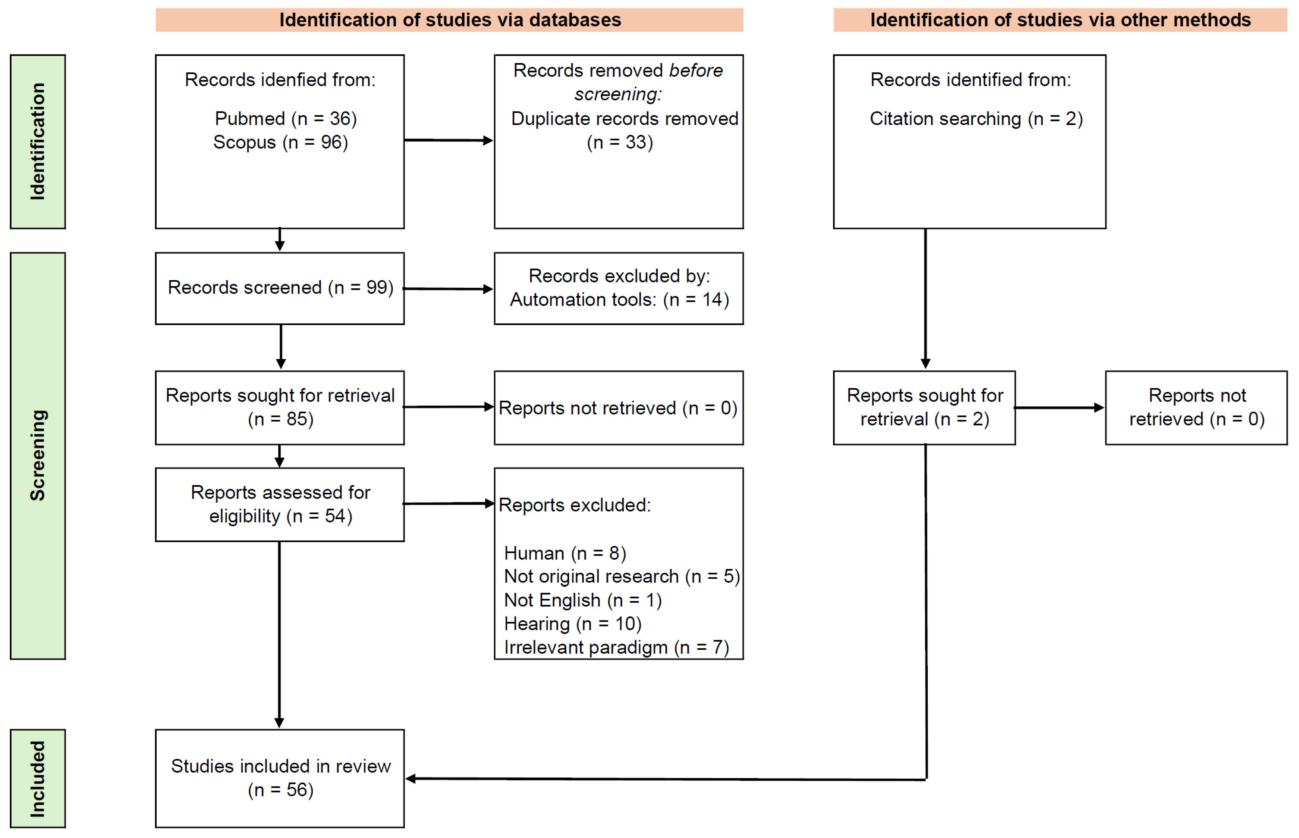
Figure 1. Scheme of studies inclusion process. Top-left: The records were identified in PubMed and Scopus databases. The duplicates were removed before screening. Top-right: The records identified from citation searching. Middle: The records were screened and excluded by predefined exclusion criteria. Bottom: 56 studies were included in the review.
Brainsci 15 01159 g001
Figure 2. Summary of gamma-range ASSR animal studies included in the review. Top-left: Distribution of model types, showing the proportion of pharmacological, optogenetic, genetic, combined genetic-pharmacological, lesion-based, and other/no-manipulation approaches. Top-right: Sex distribution, showing the predominance of male-only studies and limited inclusion of females. Bottom-left: Stimulation frequencies used, with 40 Hz as the most common, followed by 80 Hz and other gamma-band ranges. Bottom-right: Species distribution, highlighting mice and rats as the most frequent models, with fewer studies in nonhuman primates.
Brainsci 15 01159 g002
Disclaimer/Publisher’s Note: The statements, opinions and data contained in all publications are solely those of the individual author(s) and contributor(s) and not of MDPI and/or the editor(s). MDPI and/or the editor(s) disclaim responsibility for any injury to people or property resulting from any ideas, methods, instructions or products referred to in the content.