
Comparative Analysis of Brain Coping Mechanisms in Small Left-Hemisphere Lesions: Incidental vs. Symptomatic Gliomas

 , ,
, ,
Abstract
1. Introduction
2. Materials and Methods
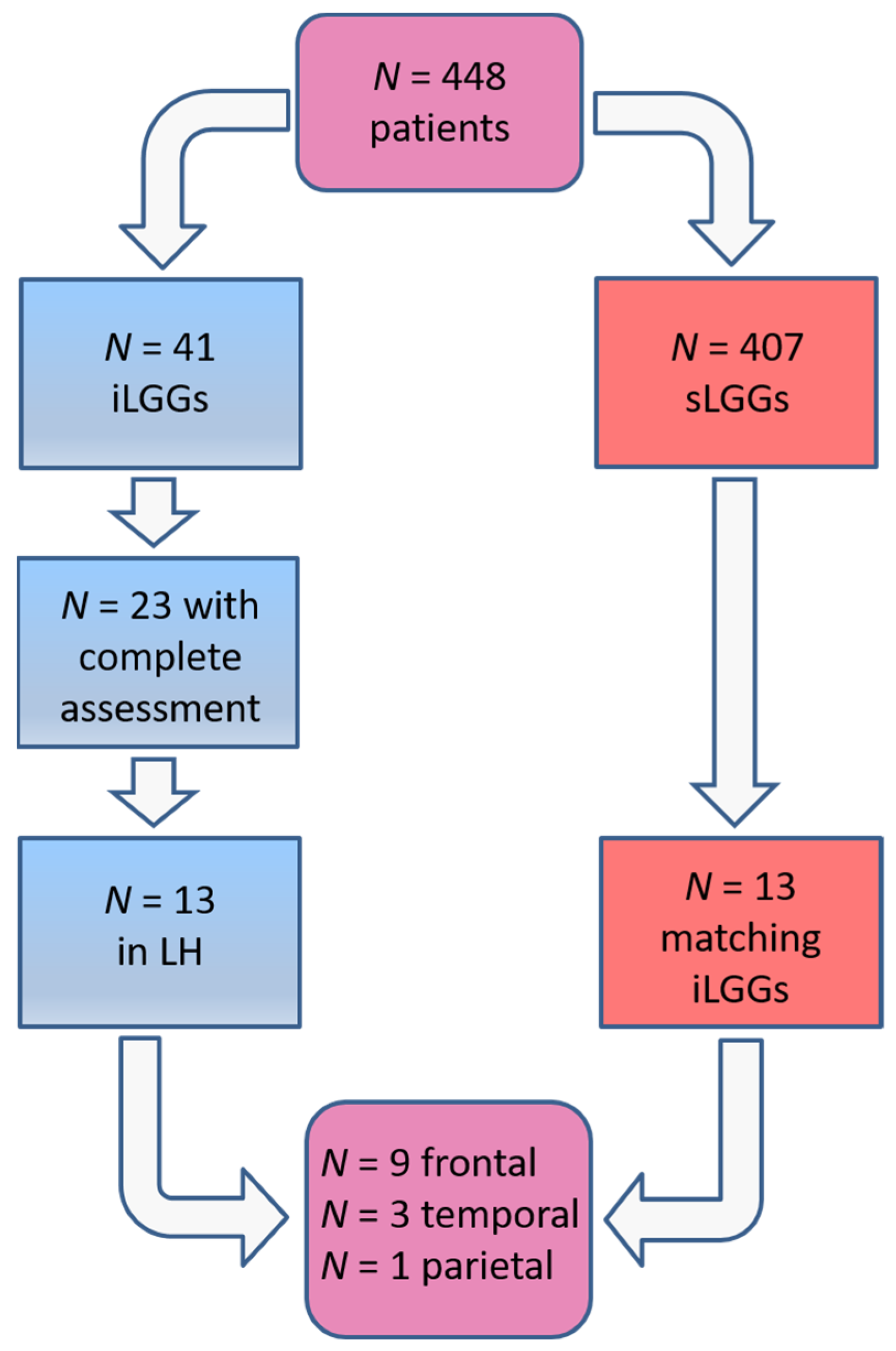
2.1. Participants

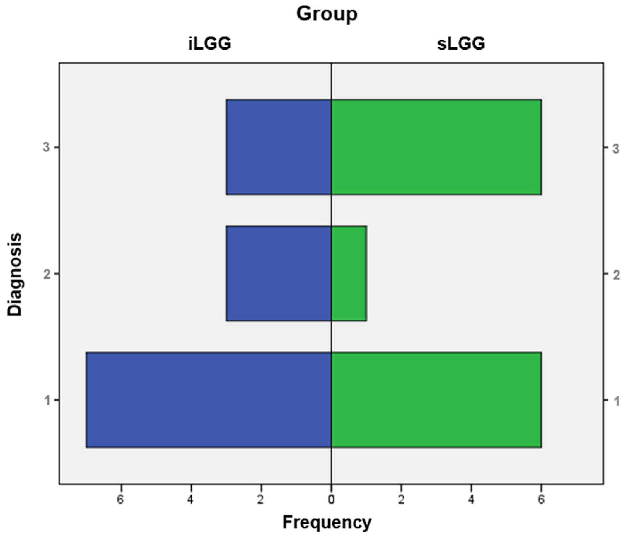
| Lesion Site | Vol (cm3) | Gender | Age | Edu | Hand | Naming (/30) | Symptomatology | |
|---|---|---|---|---|---|---|---|---|
| p1 | Frontal | 18 | F | 73 | 5 | 100 | 29 | Speech arrest, R arm myoclonus |
| p2 | Frontal | 13 | M | 28 | 13 | 100 | 30 | Epileptic attacks |
| p3 | Temporal | 30 | M | 43 | 11 | 100 | 28 | Auditory hallucinations |
| p4 | Temporal | 10 | F | 48 | 18 | 100 | 30 | Aphasia, anomia |
| p5 | Parietal | 15 | F | 46 | 18 | 100 | 28 | Epileptic attacks, paresthesia (lips) |
| p6 | Frontal | 33 | F | 36 | 13 | −50 | 30 | Epileptic attacks, fatigue, olfactory hallucinations |
| p7 | Frontal | 16 | M | 45 | 13 | 100 | 30 | Epileptic attacks, production aphasia, paresthesia (R arm) |
| p8 | Frontal | 32 | M | 40 | 5 | 100 | 30 | Epileptic attacks, brain fog |
| p9 | Frontal | 36 | M | 36 | 8 | 100 | 30 | Anomia, production aphasia |
| p10 | Frontal | 22 | M | 49 | 8 | 100 | 29 | Epileptic attacks, phonological dysarthria |
| p11 | Frontal | 23 | M | 44 | 13 | 100 | 30 | Epileptic attacks |
| p12 | Temporal | 15 | M | 42 | 13 | 96.67 | 30 | Epileptic attacks, anomia, phonological dysarthria |
| p13 | Frontal | 27 | F | 35 | 18 | 100 | 28 | Epileptic attacks |
2.2. Imaging Data
2.3. MRI Structural Analysis
2.4. fMRI Data Acquisition
fMRI Naming Task
2.5. Statistical Analyses
2.6. fMRI Data Processing
2.7. DTI Data Acquisition and Analysis
3. Results
3.1. Clinical Data
3.2. Functional Naming Network
3.3. DTI Parameters
4. Discussion
5. Conclusions
Supplementary Materials
Author Contributions
Funding
Institutional Review Board Statement
Informed Consent Statement
Data Availability Statement
Acknowledgments
Conflicts of Interest
References
- Eskandary, H.; Sabba, M.; Khajehpour, F.; Eskandari, M. Incidental findings in brain computed tomography scans of 3000 head trauma patients. Surg. Neurol. 2005, 63, 550–553. [Google Scholar] [CrossRef] [PubMed]
- Ius, T.; Cesselli, D.; Isola, M.; Pauletto, G.; Tomasino, B.; D’Auria, S.; Bagatto, D.; Pegolo, E.; Beltrami, A.P.; Loreto, C.; et al. Incidental low-grade gliomas: Single-institution management based on clinical, surgical, and molecular data. Neurosurgery 2020, 86, 391–399. [Google Scholar] [CrossRef]
- Opoku-Darko, M.; Lang, S.T.; Artindale, J.; Cairncross, J.G.; Sevick, R.J.; Kelly, J.J.P. Surgical management of incidentally discovered diffusely infiltrating low-grade glioma. J. Neurosurg. 2018, 129, 19–26. [Google Scholar] [CrossRef]
- Ius, T.; Ng, S.; Young, J.S.; Tomasino, B.; Polano, M.; Ben-Israel, D.; Kelly, J.J.P.; Skrap, M.; Duffau, H.; Berger, M.S. The benefit of early surgery on overall survival in incidental low-grade glioma patients: A multicenter study. Neuro. Oncol. 2022, 24, 624–638. [Google Scholar] [CrossRef] [PubMed]
- Potts, M.B.; Smith, J.S.; Molinaro, A.M.; Berger, M.S. Natural history and surgical management of incidentally discovered low-grade gliomas. J. Neurosurg. 2012, 116, 365–372. [Google Scholar] [CrossRef]
- De Oliveira Lima, G.L.; Zanello, M.; Mandonnet, E.; Taillandier, L.; Pallud, J.; Duffau, H. Incidental diffuse low-grade gliomas: From early detection to preventive neuro-oncological surgery. Neurosurg. Rev. 2016, 39, 377–384. [Google Scholar] [CrossRef]
- Gogos, A.J.; Young, J.S.; Pereira, M.P.; Morshed, R.A.; Potts, M.B.; Hervey-Jumper, S.L.; Berger, M.S. Surgical management of incidentally discovered low-grade gliomas. J. Neurosurg. 2020, 2, 480–487. [Google Scholar] [CrossRef] [PubMed]
- Noorani, I.; Sanai, N. Surgical Management of Incidental Gliomas. Neurosurg. Clin. N. Am. 2017, 28, 397–406. [Google Scholar] [CrossRef]
- Nakasu, S.; Nakasu, Y.; Tsuji, A.; Fukami, T.; Nitta, N.; Kawano, H.; Notsu, A.; Nozaki, K. Incidental diffuse low-grade gliomas: A systematic review and meta-analysis of treatment results with correction of lead-time and length-time biases. Neurooncol. Pract. 2022, 10, 113–125. [Google Scholar] [CrossRef]
- Duffau, H. Awake surgery for incidental WHO grade II gliomas involving eloquent areas. Acta Neurochir. 2012, 154, 575–584. [Google Scholar] [CrossRef] [PubMed]
- Zhang, Z.Y.; Chan, A.K.; Ng, H.K.; Ding, X.J.; Li, Y.X.; Shi, Z.F.; Zhu, W.; Zhong, P.; Wang, Y.; Mao, Y.; et al. Surgically treated incidentally discovered low-grade gliomas are mostly IDH mutated and 1p19q co-deleted with favorable prognosis. Int. J. Clin. Exp. Pathol. 2014, 7, 8627–8636. [Google Scholar] [PubMed]
- Allison, C.M.; Shumon, S.; Stummer, W.; Holling, M.; Surash, S. A Cohort Analysis of Truly Incidental Low-Grade Gliomas. World Neurosurg. 2022, 159, e347–e355. [Google Scholar] [CrossRef] [PubMed]
- Zeng, L.; Mei, Q.; Li, H.; Ke, C.; Yu, J.; Chen, J. A survival analysis of surgically treated incidental low-grade glioma patients. Sci. Rep. 2021, 11, 8522. [Google Scholar] [CrossRef]
- Mandonnet, E.; de Witt Hamer, P.; Pallud, J.; Bauchet, L.; Whittle, I.; Duffau, H. Silent diffuse low-grade glioma: Toward screening and preventive treatment? Cancer 2014, 120, 1758–1762. [Google Scholar] [CrossRef] [PubMed]
- Pallud, J.; Fontaine, D.; Duffau, H.; Mandonnet, E.; Sanai, N.; Taillandier, L.; Peruzzi, P.; Guillevin, R.; Bauchet, L.; Bernier, V.; et al. Natural history of incidental World Health Organization grade II gliomas. Ann. Neurol. 2010, 68, 727–733. [Google Scholar] [CrossRef]
- Cargnelutti, E.; Ius, T.; Skrap, M.; Tomasino, B. What do we know about pre- and postoperative plasticity in patients with glioma? A review of neuroimaging and intraoperative mapping studies. Neuroimage Clin. 2020, 28, 102435. [Google Scholar] [CrossRef]
- Cirillo, S.; Caulo, M.; Pieri, V.; Falini, A.; Castellano, A. Role of functional imaging techniques to assess motor and language cortical plasticity in glioma patients: A systematic review. Neural Plast. 2019, 2019, 4056436. [Google Scholar] [CrossRef]
- Duffau, H. Diffuse low-grade gliomas and neuroplasticity. Diagn. Interv. Imaging 2014, 95, 945–955. [Google Scholar] [CrossRef]
- Ng, S.; Duffau, H. Brain plasticity profiling as a key support to therapeutic decision-making in low-grade glioma oncological strategies. Cancers 2023, 15, 3698. [Google Scholar] [CrossRef]
- Guarracino, I.; Ius, T.; Pegolo, E.; Cesselli, D.; Skrap, M.; Tomasino, B. Multimodal assessment shows a mostly preserved cognitive status in incidentally discovered low grade gliomas: A single institution study. Cancers 2020, 12, 156. [Google Scholar] [CrossRef]
- Ng, S.; Herbet, G.; Lemaitre, A.L.; Cochereau, J.; Moritz-Gasser, S.; Duffau, H. Neuropsychological assessments before and after awake surgery for incidental low-grade gliomas. J. Neurosurg. 2020, 135, 871–880. [Google Scholar] [CrossRef] [PubMed]
- Cochereau, J.; Herbet, G.; Duffau, H. Patients with incidental WHO grade II glioma frequently suffer from neuropsychological disturbances. Acta Neurochir. 2016, 158, 305–312. [Google Scholar] [CrossRef]
- Cargnelutti, E.; Maieron, M.; Ius, T.; Skrap, M.; Tomasino, B. Normal-appearing white matter structural integrity in incidentally discovered low-grade gliomas: A single institution study. J. Neurosurg. Sci. 2023, 67, 200–205. [Google Scholar] [CrossRef] [PubMed]
- Cargnelutti, E.; Ius, T.; Skrap, M.; Tomasino, B. Normal-appearing naming-related functional activation in incidentally discovered lowgrade gliomas: A single institution study. J. Neurosurg. Sci. 2021, 68, 270–277. [Google Scholar] [CrossRef]
- Oldfield, R.C. The assessment and analysis of handedness: The Edinburgh inventory. Neuropsychologia 1971, 9, 97–113. [Google Scholar] [CrossRef]
- Miceli, G.; Laudanna, A.; Burani, C.; Capasso, R. Batteria per l’Analisi dei Deficit Afasici: BADA [BADA: A Battery for the Assessment of Aphasic Disorders]; CEPSAG: Roma, Italy, 1994. [Google Scholar]
- Ius, T.; Isola, M.; Budai, R.; Pauletto, G.; Tomasino, B.; Fadiga, L.; Skrap, M. Low-grade glioma surgery in eloquent areas: Volumetric analysis of extent of resection and its impact on overall survival. A single-institution experience in 190 patients: Clinical article. J. Neurosurg. 2012, 117, 1039–1052. [Google Scholar] [CrossRef]
- Snodgrass, J.G.; Vanderwart, M.A. standardized set of 260 pictures: Norms for name agreement, image agreement, familiarity, and visual complexity. J. Exp. Psychol. Hum. Learn. 1980, 6, 174–215. [Google Scholar] [CrossRef] [PubMed]
- Friston, K.J.; Frith, C.D.; Turner, R.; Frackowiak, R.S.J. Characterising evoked hemodynamics with fMRI. Neuroimage 1995, 2, 157–165. [Google Scholar] [CrossRef]
- Friston, K.J.; Holmes, A.P.; Worsley, K.J.; Poline, J.-B.; Frith, C.D.; Frackowiak, R.S.J. Statistical parametric maps in functional imaging: A general linear approach. Hum. Brain Mapp. 1995, 2, 189–210. [Google Scholar] [CrossRef]
- Eickhoff, S.; Stephan, K.E.; Mohlberg, H.; Grefkes, C.; Fink, G.R.; Amunts, K.; Zilles, K. A new SPM toolbox for combining probabilistic cytoarchitectonic maps and functional imaging data. Neuroimage 2005, 25, 1325–1335. [Google Scholar] [CrossRef]
- Stadlbauer, A.; Ganslandt, O.; Buslei, R.; Hammen, T.; Gruber, S.; Moser, E.; Buchfelder, M.; Salomonowitz, E.; Nimsky, C. Gliomas: Histopathologic evaluation of changes in directionality and magnitude of water diffusion at diffusion-tensor MR imaging. Radiology 2006, 240, 803–810. [Google Scholar] [CrossRef] [PubMed]
- Catani, M.; Thiebaut de Schotten, M. A diffusion tensor imaging tractography atlas for virtual in vivo dissections. Cortex 2008, 44, 1105–1132. [Google Scholar] [CrossRef]
- Catani, M.; Jones, D.K.; Ffytche, D.H. Perisylvian language networks of the human brain. Ann. Neurol. 2005, 57, 8–16. [Google Scholar] [CrossRef]
- Baldo, J.V.; Arévalo, A.; Patterson, J.P.; Dronkers, N.F. Grey and white matter correlates of picture naming: Evidence from a voxel-based lesion analysis of the Boston Naming Test. Cortex 2013, 49, 658–667. [Google Scholar] [CrossRef]
- Ekert, J.O.; Lorca-Puls, D.L.; Gajardo-Vidal, A.; Crinion, J.T.; Hope, T.M.H.; Green, D.W.; Price, C.J. A functional dissociation of the left frontal regions that contribute to single word production tasks. Neuroimage 2021, 245, 118734. [Google Scholar] [CrossRef] [PubMed]
- Harvey, D.Y.; Schnur, T.T. Distinct loci of lexical and semantic access deficits in aphasia: Evidence from voxel-based lesion-symptom mapping and diffusion tensor imaging. Cortex 2015, 67, 37–58. [Google Scholar] [CrossRef]
- Tomasino, B.; Tronchin, G.; Marin, D.; Maieron, M.; Fabbro, F.; Cubelli, R.; Skrap, M.; Luzzatti, C. Noun-verb naming dissociation in neurosurgical patients. Aphasiol 2019, 33, 1418–1440. [Google Scholar] [CrossRef]
- Duffau, H. Brain plasticity: From pathophysiological mechanisms to therapeutic applications. J. Clin. Neurosci. 2006, 13, 885–897. [Google Scholar] [CrossRef]
- Ius, T.; Mazzucchi, E.; Tomasino, B.; Pauletto, G.; Sabatino, G.; Della Pepa, G.M.; La Rocca, G.; Battistella, C.; Olivi, A.; Skrap, M. Multimodal integrated approaches in low grade glioma surgery. Sci. Rep. 2021, 11, 9964. [Google Scholar] [CrossRef]
- Novelli, G.; Papagno, C.; Capitani, E.; Laiacona, M. Tre test clinici di ricerca e produzione lessicale. Taratura su soggetti normali. Archivio di Psicologia, Neurol. Psichiatr. 1986, 47, 477–505. [Google Scholar]
- De Renzi, E.; Faglioni, P. Normative data and screening power of a shortened version of the Token Test. Cortex 1978, 14, 41–49. [Google Scholar] [CrossRef] [PubMed]
- Basso, A.; Capitani, E.; Laiacona, M. Raven’s coloured progressive matrices: Normative values on 305 adult normal controls. Funct. Neurol. 1987, 2, 189–194. [Google Scholar] [PubMed]
- Monaco, M.; Costa, A.; Caltagirone, C.; Carlesimo, G.A. Erratum to: Forward and backward span for verbal and visuo-spatial data: Standardization and normative data from an Italian adult population. Neurol. Sci. 2015, 36, 345–347. [Google Scholar] [CrossRef] [PubMed]
| sLGG | iLGG | Between-Group Difference | |
|---|---|---|---|
| Sex | 7 F, 6 M | 5 F, 8 M | χ2 = 0.62, df = 1, p = 0.43 (n.s.) |
| Age (years) | M = 43.46, SD = 12.54 | M = 37.46, SD = 12.54 | U = 61.50, p = 0.24 (n.s.) |
| Education (years) | M = 12.00, SD = 4.51 | M = 13.38, SD = 2.53 | U = 70.00, p = 0.43 (n.s.) |
| Handedness 1 | Right-handed = 12 Left-handed = 1 | Right-handed = 12 Left-handed = 1 | - |
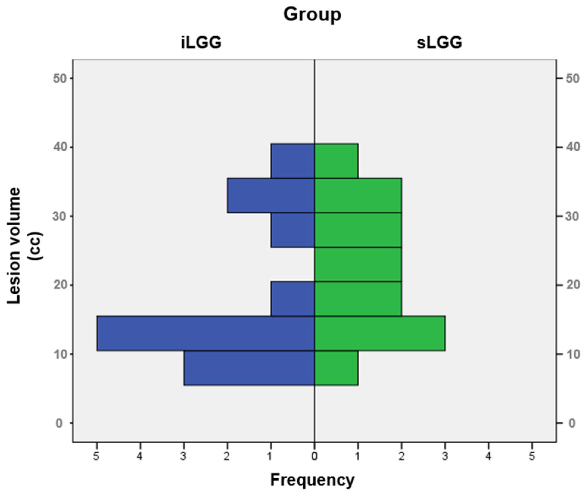
| Lesion volume | 22.31 cm3 (SD = 8.57 cm3; range = 10–36 cm3; median = 12 cm3) | 18.54 cm3 (SD = 10.71 cm3, range = 8–40 cm3; median = 18 cm3) | U = 57.00, p = 0.16 (n.s.) |
| Lesion growth pattern | Expansive for all patients but one (p9, see Table 2) | Expansive for all patients but one (p3 [24]) | - |
| Symptoms | Diverse symptoms and, particularly, epileptic seizures, which characterized all the patients in this group. 7 had language-related manifestations. | none | - |
| Object-naming abilities (BADA test [26]) | score ≥ 28/30 | score ≥ 28/30 | - |
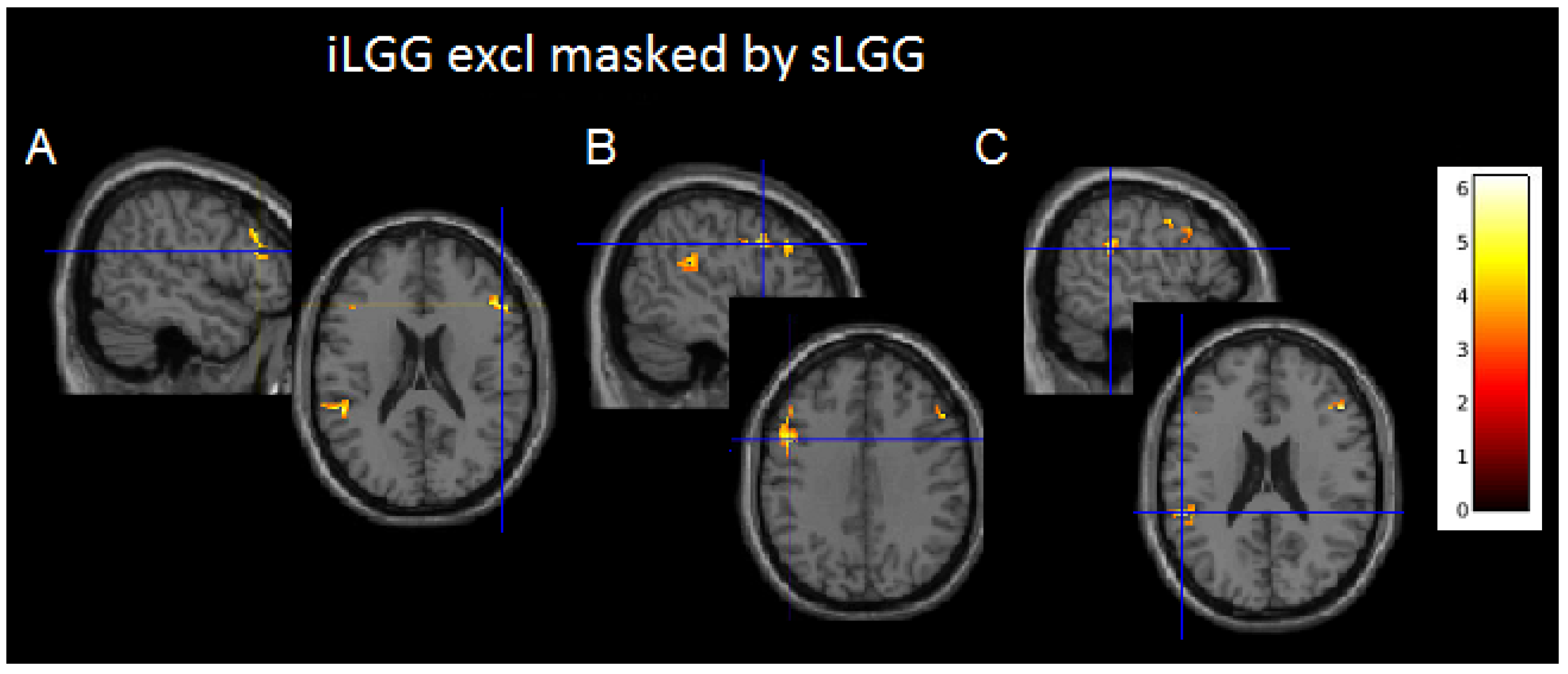
| Cluster | Cluster Size (Voxels) | T | Macroanatomical Area | Cytoarchitectonic Localization | MNI Coordinates | ||
|---|---|---|---|---|---|---|---|
| x | y | z | |||||
| iLGG masked exclusively by sLGG | |||||||
| 1. | 188 | ||||||
| 4.73 | L precentral gyrus | Area 44 | −48 | 8 | 34 | ||
| 5.14 | L middle frontal gyrus | - | −50 | 24 | 32 | ||
| 4.13 | L inferior frontal gyrus | - | −46 | 26 | 24 | ||
| 2. | 149 | ||||||
| 6.83 | L superior temporal gyrus | Area PFcm (IPL) | −54 | −38 | 22 | ||
| 4.95 | L supramarginal gyrus | Area PFcm (IPL) | −56 | −36 | 26 | ||
| 3. | 150 | ||||||
| 6.23 | R inferior frontal gyrus | Area 45 | 52 | 28 | 20 | ||
| 5.43 | R middle frontal gyrus | - | 52 | 26 | 32 | ||
| sLGG masked exclusively by iLGG | |||||||
| No suprathresholded clusters | |||||||
| Cluster | Cluster Size (Voxels) | T | Macroanatomical Area | Cytoarchitectonic Localization | MNI Coordinates | ||
|---|---|---|---|---|---|---|---|
| x | y | z | |||||
| iLGG masked inclusively by sLGG | |||||||
| 1. | 7.975 | ||||||
| 12.85 | R cuneus | Area hOc2 [V2] | 15 | −98 | 10 | ||
| 11.74 | L superior occipital gyrus | Area hOc3d [V3] | −16 | −96 | 10 | ||
| 9.73 | L middle occipital gyrus | Area hOc4lp | −30 | −86 | 4 | ||
| 9.59 | L fusiform gyrus | - | −36 | −58 | −10 | ||
| 9.58 | R middle occipital gyrus | Area hOc4lp | 38 | −84 | 10 | ||
| 9.56 | L middle occipital gyrus | Area hOc4d [V3A] | −24 | −88 | 14 | ||
| 9.31 | R superior occipital gyrus | Area hOc3d [V3d] | 20 | −94 | 20 | ||
| 8.94 | R fusiform gyrus | Area FG3 | 30 | −60 | −6 | ||
| 2. | 830 | ||||||
| 7.62 | L precentral gyrus | - | −48 | −4 | 42 | ||
| 6.19 | L precentral gyrus | Area 44 | −48 | 8 | 32 | ||
| 4.98 | L inferior frontal gyrus | Area 45 | −52 | 26 | 24 | ||
| 4.81 | L inferior frontal gyrus | Area 44 | −52 | 12 | 26 | ||
| 3. | 686 | ||||||
| 8.12 | R posterior-medial frontal gyrus | - | 2 | 14 | 56 | ||
| 7.05 | L posterior-medial frontal gyrus | - | −4 | 0 | 64 | ||
| 4. | 646 | ||||||
| 7.77 | R precentral gyrus | - | 52 | −2 | 48 | ||
| 6.06 | R inferior frontal gyrus | - | 40 | 6 | 30 | ||
| 5.87 | R middle frontal gyrus | - | 46 | −2 | 54 | ||
| 5. | 406 | ||||||
| 6.14 | L thalamus | Thalamus: Visual | −24 | −28 | −4 | ||
| 5.71 | L hippocampus | - | −28 | −28 | −8 | ||
| 6. | 317 | ||||||
| 6.60 | R inferior frontal gyrus | Area 45 | 48 | 20 | 6 | ||
| 6.39 | R insula | - | 32 | 28 | 4 | ||
| 4.93 | R inferior frontal gyrus | Area 44 | 52 | 14 | 2 | ||
| 7. | 254 | ||||||
| 6.73 | N/A | - | 26 | −32 | 8 | ||
| 6.70 | R hippocampus | - | 24 | −24 | −10 | ||
| 8. | 95 | 6.45 | N/A | Thalamus: Temporal | 24 | −28 | −2 |
| 6.89 | L inferior frontal gyrus | - | −34 | 32 | 0 | ||
| Cluster | Cluster Size (Voxels) | T | Macroanatomical Area | Cytoarchitectonic Localization | MNI Coordinates | ||
|---|---|---|---|---|---|---|---|
| x | y | z | |||||
| sLGG masked inclusively by iLGG | |||||||
| 1. | 8149 | ||||||
| 11.31 | R cuneus | Area hOc2 [V2] | 16 | −98 | 12 | ||
| 10.12 | N/A | Area FG3 | −35 | −55 | −8 | ||
| 10.07 | L middle occipital gyrus | Area hOc4d | −24 | −88 | 14 | ||
| 8.47 | L middle occipital gyrus | Area hOc3d [V3] | −18 | −96 | 10 | ||
| 8.46 | L inferior temporal gyrus | - | −44 | −62 | −6 | ||
| 8.20 | L inferior occipital gyrus | Area hOc4la | −46 | −70 | −4 | ||
| 7.46 | R angular gyrus | Area hIP3 (IPS) | 28 | −56 | 46 | ||
| 7.36 | R middle occipital gyrus | Area hOc4la | 38 | −80 | 8 | ||
| 2. | 354 | ||||||
| 6.29 | R precentral gyrus | - | 44 | 8 | 32 | ||
| 4.61 | R inferior frontal gyrus | Area 45 | 54 | 20 | 25 | ||
| 4.18 | R middle frontal gyrus | - | 44 | −2 | 54 | ||
| 3. | 310 | ||||||
| 5.88 | L inferior frontal gyrus | - | −52 | 36 | 0 | ||
| 5.64 | L insula | - | −30 | 28 | −2 | ||
| 4.44 | L inferior frontal gyrus | Area 44 | −52 | 12 | 4 | ||
| 4. | 283 | ||||||
| 6.65 | N/A | - | 26 | −30 | 8 | ||
| 6.24 | L thalamus | Thalamus: Temporal | 24 | −28 | −2 | ||
| 5. | 265 | ||||||
| 5.70 | L posterior-medial frontal gyrus | - | −4 | 0 | 64 | ||
| 5.50 | R posterior-medial frontal gyrus | - | 8 | 12 | 48 | ||
| 6. | 261 | L inferior frontal gyrus | Area 44 | −44 | 10 | 24 | |
| L inferior frontal gyrus | Area 45 | −54 | 28 | 16 | |||
| 7. | 238 | ||||||
| 6.89 | L thalamus | Thalamus: Temporal | −18 | −32 | 2 | ||
| 8. | 228 | 6.41 | L thalamus | Thalamus: Visual | −26 | −25 | 4 |
| 6.25 | R insula | - | 34 | 20 | 6 | ||
| 5.41 | R inferior frontal gyrus | - | 40 | 24 | 6 | ||
| 4.31 | R inferior frontal gyrus | Area 45 | 52 | 22 | 2 | ||
| 9. | 222 | ||||||
| 5.68 | L precentral gyrus | - | −50 | 0 | 46 | ||
| ILF | IFOF | UF | Whole AF | Long AF | ||||||||||||||||
|---|---|---|---|---|---|---|---|---|---|---|---|---|---|---|---|---|---|---|---|---|
| LH | RH | LH | RH | LH | RH | LH | RH | LH | RH | |||||||||||
| Num | FA | Num | FA | Num | FA | Num | FA | Num | FA | Num | FA | Num | FA | Num | FA | Num | FA | Num | FA | |
| iLGG | 277.37 (191.59) | 0.47 (0.03) | 244.72 (140.78) | 0.45 (0.02) | 173.55 (96.76) | 0.49 (0.03) | 308.36 (191.45) | 0.49 (0.02) | 227.55 (122.39) | 0.42 (0.03) | 238.28 (127.73) | 0.42 (0.01) | 555.63 (273.21) | 0.45 (0.03) | 598.82 (144.54) | 0.46 (0.03) | 198.18 (131.97) | 0.47 (0.04) | 132.36 (112.63) | 0.46 (0.04) |
| sLGG | 228.60 (134.45) | 0.46 (0.04) | 235.80 (125.25) | 0.46 (0.03) | 155.30 (117.18) | 0.47 (0.05) | 237.00 (88.39) | 0.49 (0.03) | 175.801 (47.64) | 0.40 (0.03) | 159.60 (81.86) | 0.41 (0.03) | 435.50 (252.15) | 0.44 (0.04) | 588.90 (208.55) | 0.44 (0.03) | 163.50 (137.90) | 0.47 (0.03) | 115.20 (103.53) | 0.45 (0.03) |
Disclaimer/Publisher’s Note: The statements, opinions and data contained in all publications are solely those of the individual author(s) and contributor(s) and not of MDPI and/or the editor(s). MDPI and/or the editor(s) disclaim responsibility for any injury to people or property resulting from any ideas, methods, instructions or products referred to in the content. |
© 2024 by the authors. Licensee MDPI, Basel, Switzerland. This article is an open access article distributed under the terms and conditions of the Creative Commons Attribution (CC BY) license (https://creativecommons.org/licenses/by/4.0/).
Share and Cite
Cargnelutti, E.; Ius, T.; Maieron, M.; D’Agostini, S.; Skrap, M.; Tomasino, B. Comparative Analysis of Brain Coping Mechanisms in Small Left-Hemisphere Lesions: Incidental vs. Symptomatic Gliomas. Brain Sci. 2024, 14, 887. https://doi.org/10.3390/brainsci14090887
Cargnelutti E, Ius T, Maieron M, D’Agostini S, Skrap M, Tomasino B. Comparative Analysis of Brain Coping Mechanisms in Small Left-Hemisphere Lesions: Incidental vs. Symptomatic Gliomas. Brain Sciences. 2024; 14(9):887. https://doi.org/10.3390/brainsci14090887
Chicago/Turabian StyleCargnelutti, Elisa, Tamara Ius, Marta Maieron, Serena D’Agostini, Miran Skrap, and Barbara Tomasino. 2024. "Comparative Analysis of Brain Coping Mechanisms in Small Left-Hemisphere Lesions: Incidental vs. Symptomatic Gliomas" Brain Sciences 14, no. 9: 887. https://doi.org/10.3390/brainsci14090887
APA StyleCargnelutti, E., Ius, T., Maieron, M., D’Agostini, S., Skrap, M., & Tomasino, B. (2024). Comparative Analysis of Brain Coping Mechanisms in Small Left-Hemisphere Lesions: Incidental vs. Symptomatic Gliomas. Brain Sciences, 14(9), 887. https://doi.org/10.3390/brainsci14090887