
The Cortical Silent Period and Its Association with Fatigue in Multiple Sclerosis: The Need for Standardized Data Collection


Abstract
:1. Introduction
2. Materials and Methods
2.1. Participants
2.2. Transcranial Magnetic Stimulation
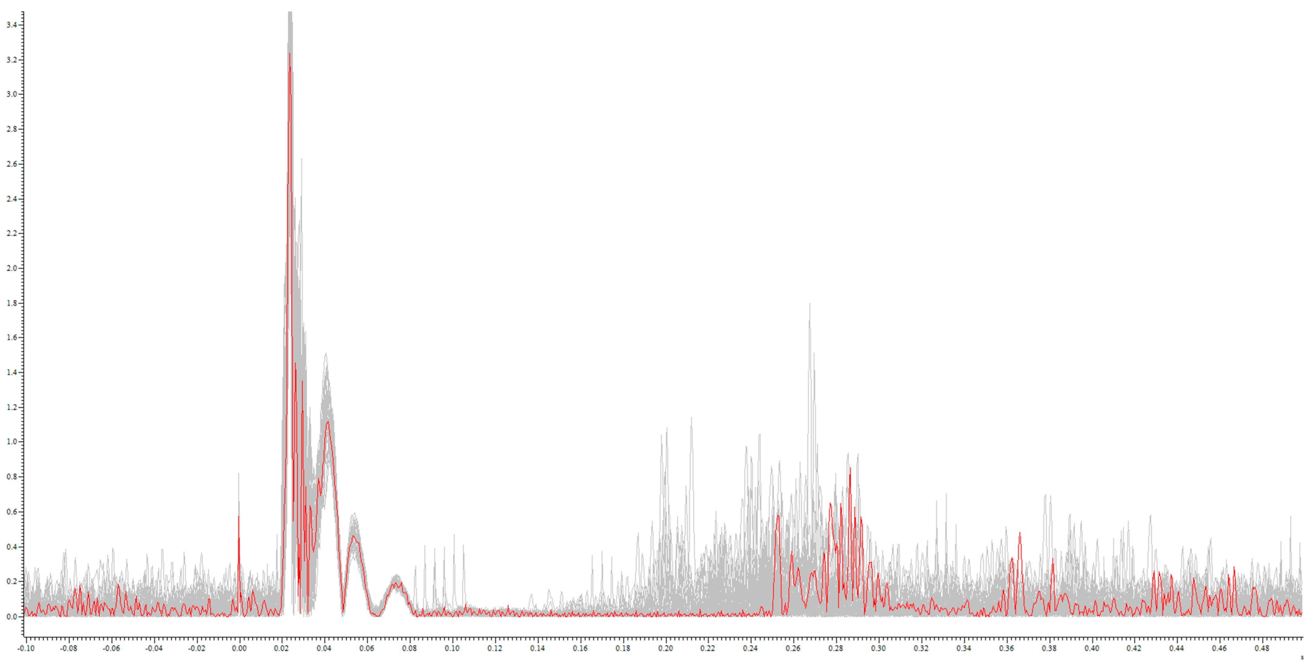
2.3. Data Evaluation
2.4. Assessment of Fatigue
2.5. Assessment of Depressive Symptoms
2.6. Statistics
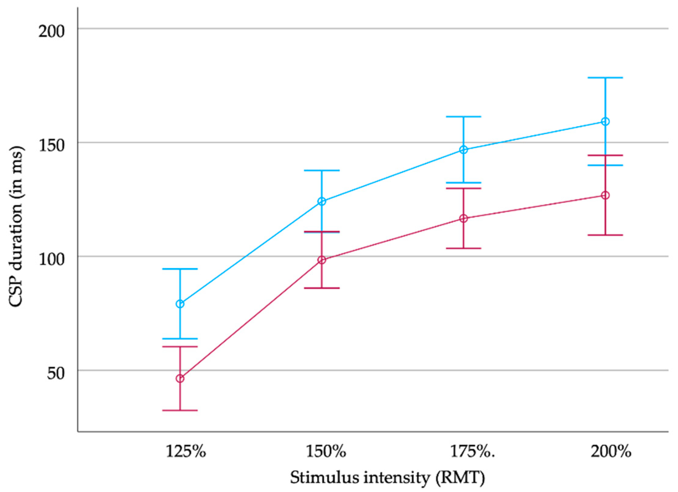
3. Results
4. Discussion
5. Conclusions
Author Contributions
Funding
Institutional Review Board Statement
Informed Consent Statement
Data Availability Statement
Conflicts of Interest
References
- Thompson, A.J.; Baranzini, S.E.; Geurts, J.; Hemmer, B.; Ciccarelli, O. Multiple sclerosis. Lancet 2018, 391, 1622–1636. [Google Scholar] [CrossRef] [PubMed]
- Reich, D.S.; Lucchinetti, C.F.; Calabresi, P.A. Multiple Sclerosis. N. Engl. J. Med. 2018, 378, 169–180. [Google Scholar] [CrossRef] [PubMed]
- Penner, I.-K.; Paul, F. Fatigue as a symptom or comorbidity of neurological diseases. Nat. Rev. Neurol. 2017, 13, 662–675. [Google Scholar] [CrossRef] [PubMed]
- Ramirez, A.O.; Keenan, A.; Kalau, O.; Worthington, E.; Cohen, L.; Singh, S. Prevalence and burden of multiple sclerosis-related fatigue: A systematic literature review. BMC Neurol. 2021, 21, 468. [Google Scholar]
- Penner, I.-K. Evaluation of cognition and fatigue in multiple sclerosis: Daily practice and future directions. Acta Neurol. Scand. 2016, 134 (Suppl. S200), 19–23. [Google Scholar] [CrossRef] [PubMed]
- Sparasci, D.; Gobbi, C.; Castelnovo, A.; Riccitelli, G.C.; Disanto, G.; Zecca, C.; Manconi, M. Fatigue, sleepiness and depression in multiple sclerosis: Defining the overlaps for a better phenotyping. J. Neurol. 2022, 269, 4961–4971. [Google Scholar] [CrossRef] [PubMed]
- Carta, M.G.; Paribello, P.; Anastasia, A.; De Berardis, D.; Nardi, A.E.; Fornaro, M. Pharmacological management of depression in patients with multiple sclerosis. Expert Opin. Pharmacother. 2018, 19, 1533–1540. [Google Scholar] [CrossRef]
- Penner, I.K.; Raselli, C.; Stöcklin, M.; Opwis, K.; Kappos, L.; Calabrese, P. The Fatigue Scale for Motor and Cognitive Functions (FSMC): Validation of a new instrument to assess multiple sclerosis-related fatigue. Mult. Scler. J. 2009, 15, 1509–1517. [Google Scholar] [CrossRef]
- Newland, P.; Starkweather, A.; Sorenson, M. Central fatigue in multiple sclerosis: A review of the literature. J. Spinal Cord Med. 2016, 39, 386–399. [Google Scholar] [CrossRef]
- Manjaly, Z.-M.; A Harrison, N.; Critchley, H.D.; Do, C.T.; Stefanics, G.; Wenderoth, N.; Lutterotti, A.; Müller, A.; Stephan, K.E. Pathophysiological and cognitive mechanisms of fatigue in multiple sclerosis. J. Neurol. Neurosurg. Psychiatry 2019, 90, 642–651. [Google Scholar] [CrossRef]
- Vucic, S.; Burke, D.; Kiernan, M.C. Fatigue in multiple sclerosis: Mechanisms and management. Clin. Neurophysiol. 2010, 121, 809–817. [Google Scholar] [CrossRef] [PubMed]
- Groppa, S.; Oliviero, A.; Eisen, A.; Quartarone, A.; Cohen, L.; Mall, V.; Kaelin-Lang, A.; Mima, T.; Rossi, S.; Thickbroom, G.; et al. A practical guide to diagnostic transcranial magnetic stimulation: Report of an IFCN committee. Clin. Neurophysiol. 2012, 123, 858–882. [Google Scholar] [CrossRef] [PubMed]
- Ziemann, U. Thirty years of transcranial magnetic stimulation: Where do we stand? Exp. Brain Res. 2017, 235, 973–984. [Google Scholar] [CrossRef] [PubMed]
- Vucic, S.; Chen, K.-H.S.; Kiernan, M.C.; Hallett, M.; Benninger, D.; Di Lazzaro, V.; Rossini, P.M.; Benussi, A.; Berardelli, A.; Currà, A.; et al. Clinical diagnostic utility of transcranial magnetic stimulation in neurological disorders. Updated report of an IFCN committee. Clin. Neurophysiol. 2023, 150, 131–175. [Google Scholar] [CrossRef] [PubMed]
- Valero-Cabré, A.; Amengual, J.L.; Stengel, C.; Pascual-Leone, A.; Coubard, O.A. Transcranial magnetic stimulation in basic and clinical neuroscience: A comprehensive review of fundamental principles and novel insights. Neurosci. Biobehav. Rev. 2017, 83, 381–404. [Google Scholar] [CrossRef]
- Ayache, S.S.; Chalah, M.A. Fatigue in multiple sclerosis—Insights into evaluation and management. Neurophysiol. Clin. Clin. Neurophysiol. 2017, 47, 139–171. [Google Scholar] [CrossRef] [PubMed]
- Snow, N.J.; Wadden, K.P.; Chaves, A.R.; Ploughman, M. Transcranial Magnetic Stimulation as a Potential Biomarker in Multiple Sclerosis: A Systematic Review with Recommendations for Future Research. Neural Plast. 2019, 2019, 6430596. [Google Scholar] [CrossRef]
- Ayache, S.S.; Serratrice, N.; Lahoud, G.N.A.; Chalah, M.A. Fatigue in Multiple Sclerosis: A Review of the Exploratory and Therapeutic Potential of Non-Invasive Brain Stimulation. Front. Neurol. 2022, 13, 813965. [Google Scholar] [CrossRef]
- Colombo, B.; Martinelli Boneschi, F.; Rossi, P.; Rovaris, M.; Maderna, L.; Filippi, M.; Comi, G. MRI and motor evoked potential findings in nondisabled multiple sclerosis patients with and without symptoms of fatigue. J. Neurol. 2000, 247, 506–509. [Google Scholar] [CrossRef]
- Petajan, J.H.; White, A.T. Motor-evoked potentials in response to fatiguing grip exercise in multiple sclerosis patients. Clin. Neurophysiol. 2000, 111, 2188–2195. [Google Scholar] [CrossRef]
- Conte, A.; Voti, P.L.; Pontecorvo, S.; Quartuccio, M.E.; Baione, V.; Rocchi, L.; Cortese, A.; Bologna, M.; Francia, A.; Berardelli, A. Attention-related changes in short-term cortical plasticity help to explain fatigue in multiple sclerosis. Mult. Scler. J. 2016, 22, 1359–1366. [Google Scholar] [CrossRef] [PubMed]
- Coates, K.D.; Aboodarda, S.J.; Krüger, R.L.; Martin, T.; Metz, L.M.; Jarvis, S.E.; Millet, G.Y. Multiple sclerosis-related fatigue: The role of impaired corticospinal responses and heightened exercise fatigability. J. Neurophysiol. 2020, 124, 1131–1143. [Google Scholar] [CrossRef] [PubMed]
- Liepert, J.; Mingers, D.; Heesen, C.; Bäumer, T.; Weiller, C. Motor cortex excitability and fatigue in multiple sclerosis: A transcranial magnetic stimulation study. Mult. Scler. J. 2005, 11, 316–321. [Google Scholar] [CrossRef] [PubMed]
- Chalah, M.A.; Kauv, P.; Créange, A.; Hodel, J.; Lefaucheur, J.-P.; Ayache, S.S. Neurophysiological, radiological and neuropsychological evaluation of fatigue in multiple sclerosis. Mult. Scler. Relat. Disord. 2019, 28, 145–152. [Google Scholar] [CrossRef] [PubMed]
- Santarnecchi, E.; Rossi, S.; Bartalini, S.; Cincotta, M.; Giovannelli, F.; Tatti, E.; Ulivelli, M. Neurophysiological Correlates of Central Fatigue in Healthy Subjects and Multiple Sclerosis Patients before and after Treatment with Amantadine. Neural Plast. 2015, 2015, 616242. [Google Scholar] [CrossRef]
- Chaves, A.R.; Kelly, L.P.; Moore, C.S.; Stefanelli, M.; Ploughman, M. Prolonged cortical silent period is related to poor fitness and fatigue, but not tumor necrosis factor, in Multiple Sclerosis. Clin. Neurophysiol. 2019, 130, 474–483. [Google Scholar] [CrossRef] [PubMed]
- Deriu, F.; Martinez, G.; Loi, N.; Ventura, L.; Ginatempo, F.; Dvir, Z.; Manca, A. Reporting quality of TMS studies in neurological conditions: A critical appraisal of the main gaps, challenges and clinical implications. J. Neurosci. Methods 2021, 362, 109293. [Google Scholar] [CrossRef]
- Hupfeld, K.; Swanson, C.; Fling, B.; Seidler, R. TMS-induced silent periods: A review of methods and call for consistency. J. Neurosci. Methods 2020, 346, 108950. [Google Scholar] [CrossRef]
- Werhahn, K.J.; Kunesch, E.; Noachtar, S.; Benecke, R.; Classen, J. Differential effects on motorcortical inhibition induced by blockade of GABA uptake in humans. J. Physiol. 1999, 517 Pt 2, 591–597. [Google Scholar] [CrossRef]
- Fuhr, P.; Agostino, R.; Hallett, M. Spinal motor neuron excitability during the silent period after cortical stimulation. Electroencephalogr. Clin. Neurophysiol. Potentials Sect. 1991, 81, 257–262. [Google Scholar] [CrossRef]
- Tataroglu, C.; Genc, A.; Idiman, E.; Cakmur, R.; Idiman, F. Cortical silent period and motor evoked potentials in patients with multiple sclerosis. Clin. Neurol. Neurosurg. 2003, 105, 105–110. [Google Scholar] [CrossRef] [PubMed]
- Leodori, G.; Mancuso, M.; Maccarrone, D.; Tartaglia, M.; Ianniello, A.; Certo, F.; Baione, V.; Ferrazzano, G.; Malimpensa, L.; Belvisi, D.; et al. Neural bases of motor fatigue in multiple sclerosis: A multimodal approach using neuromuscular assessment and TMS-EEG. Neurobiol. Dis. 2023, 180, 106073. [Google Scholar] [CrossRef] [PubMed]
- Nantes, J.C.; Zhong, J.; Holmes, S.A.; Whatley, B.; Narayanan, S.; Lapierre, Y.; Arnold, D.L.; Koski, L. Intracortical inhibition abnormality during the remission phase of multiple sclerosis is related to upper limb dexterity and lesions. Clin. Neurophysiol. 2016, 127, 1503–1511. [Google Scholar] [CrossRef] [PubMed]
- Nantes, J.C.; Zhong, J.; Holmes, S.A.; Narayanan, S.; Lapierre, Y.; Koski, L. Cortical Damage and Disability in Multiple Sclerosis: Relation to Intracortical Inhibition and Facilitation. Brain Stimul. 2016, 9, 566–573. [Google Scholar] [CrossRef]
- Rossi, S.; Antal, A.; Bestmann, S.; Bikson, M.; Brewer, C.; Brockmöller, J.; Carpenter, L.L.; Cincotta, M.; Chen, R.; Daskalakis, J.D.; et al. Safety and recommendations for TMS use in healthy subjects and patient populations, with updates on training, ethical and regulatory issues: Expert Guidelines. Clin. Neurophysiol. 2021, 132, 269–306. [Google Scholar] [CrossRef]
- Sommer, M.; Ciocca, M.; Chieffo, R.; Hammond, P.; Neef, A.; Paulus, W.; Rothwell, J.C.; Hannah, R. TMS of primary motor cortex with a biphasic pulse activates two independent sets of excitable neurones. Brain Stimul. 2018, 11, 558–565. [Google Scholar] [CrossRef]
- Oliviero, A.; Profice, P.; Tonali, P.A.; Pilato, F.; Saturno, E.; Dileone, M.; Ranieri, F.; Di Lazzaro, V. Effects of aging on motor cortex excitability. Neurosci. Res. 2006, 55, 74–77. [Google Scholar] [CrossRef]
- Orth, M.; Rothwell, J. The cortical silent period: Intrinsic variability and relation to the waveform of the transcranial magnetic stimulation pulse. Clin. Neurophysiol. 2004, 115, 1076–1082. [Google Scholar] [CrossRef]
- Beck, A.T.; Steer, R.A.; Brown, G.K. Beck Depression Inventory; Harcourt Brace Jovanovich: New York, NY, USA, 1987. [Google Scholar]
- Poenaru, S.; Abdallah, S.J.; Corrales-Medina, V.; Cowan, J. COVID-19 and post-infectious myalgic encephalomyelitis/chronic fatigue syndrome: A narrative review. Ther. Adv. Infect. Dis. 2021, 8, 20499361211009385. [Google Scholar] [CrossRef]
- Chaudhuri, A.; O Behan, P. Fatigue in neurological disorders. Lancet 2004, 363, 978–988. [Google Scholar] [CrossRef]
- Benedict, R.H.B.; Amato, M.P.; DeLuca, J.; Geurts, J.J.G. Cognitive impairment in multiple sclerosis: Clinical management, MRI, and therapeutic avenues. Lancet Neurol. 2020, 19, 860–871. [Google Scholar] [CrossRef]
- Flachenecker, P.; Kobelt, G.; Berg, J.; Capsa, D.; Gannedahl, M.; Platform, T.E.M.S. New insights into the burden and costs of multiple sclerosis in Europe: Results for Germany. Mult. Scler. J. 2017, 23 (Suppl. S2), 78–90. [Google Scholar] [CrossRef] [PubMed]
- Grothe, M.; Gross, S.; Süße, M.; Strauss, S.; Penner, I.K. The Seasonal Fluctuation of Fatigue in Multiple Sclerosis. Front. Neurol. 2022, 13, 900792. [Google Scholar] [CrossRef] [PubMed]
- Sanai, S.A.; Saini, V.; Benedict, R.H.; Zivadinov, R.; Teter, B.E.; Ramanathan, M.; Weinstock-Guttman, B. Aging and Multiple Sclerosis. Mult. Scler. 2016, 22, 717–725. [Google Scholar] [CrossRef] [PubMed]
- Jakimovski, D.; Weinstock-Guttman, B.; Roy, S.; Jaworski, M.; Hancock, L.; Nizinski, A.; Srinivasan, P.; Fuchs, T.A.; Szigeti, K.; Zivadinov, R.; et al. Cognitive Profiles of Aging in Multiple Sclerosis. Front. Aging Neurosci. 2019, 11, 105. [Google Scholar] [CrossRef]
- DeLuca, J.; Chiaravalloti, N.D.; Sandroff, B.M. Treatment and management of cognitive dysfunction in patients with multiple sclerosis. Nat. Rev. Neurol. 2020, 16, 319–332. [Google Scholar] [CrossRef]
- Kavaliunas, A.; Karrenbauer, V.D.; Binzer, S.; Hillert, J. Systematic Review of the Socioeconomic Consequences in Patients with Multiple Sclerosis with Different Levels of Disability and Cognitive Function. Front. Neurol. 2021, 12, 737211. [Google Scholar] [CrossRef]
- Bogaardt, H.; Golan, D.; Barrera, M.A.; Attrill, S.; Kaczmarek, O.; Zarif, M.; Bumstead, B.; Buhse, M.; Wilken, J.; Doniger, G.M.; et al. Cognitive impairment, fatigue and depression in multiple sclerosis: Is there a difference between benign and non-benign MS? Mult. Scler. Relat. Disord. 2023, 73, 104630. [Google Scholar] [CrossRef]
- Lysandropoulos, A.P.; Havrdova, E. ‘Hidden’ factors influencing quality of life in patients with multiple sclerosis. Eur. J. Neurol. 2015, 22 (Suppl. S2), 28–33. [Google Scholar] [CrossRef]
- Rodgers, S.; Manjaly, Z.M.; Calabrese, P.; Steinemann, N.; Kaufmann, M.; Salmen, A.; Chan, A.; Kesselring, J.; Kamm, C.P.; Kuhle, J.; et al. The Effect of Depression on Health-Related Quality of Life Is Mediated by Fatigue in Persons with Multiple Sclerosis. Brain Sci. 2021, 11, 751. [Google Scholar] [CrossRef]
- Pinarello, C.; Elmers, J.; Inojosa, H.; Beste, C.; Ziemssen, T. Management of multiple sclerosis fatigue in the digital age: From assessment to treatment. Front. Neurosci. 2023, 17, 1231321. [Google Scholar] [CrossRef] [PubMed]
- Hewlett, S.; Dures, E.; Almeida, C. Measures of fatigue: Bristol Rheumatoid Arthritis Fatigue Multi-Dimensional Questionnaire (BRAF MDQ), Bristol Rheumatoid Arthritis Fatigue Numerical Rating Scales (BRAF NRS) for severity, effect, and coping, Chalder Fatigue Questionnaire (CFQ), Checklist Individual Strength (CIS20R and CIS8R), Fatigue Severity Scale (FSS), Functional Assessment Chronic Illness Therapy (Fatigue) (FACIT-F), Multi-Dimensional Assessment of Fatigue (MAF), Multi-Dimensional Fatigue Inventory (MFI), Pediatric Quality Of Life (PedsQL) Multi-Dimensional Fatigue Scale, Profile of Fatigue (ProF), Short Form 36 Vitality Subscale (SF-36 VT), and Visual Analog Scales (VAS). Arthritis Care Res (Hoboken). Arthritis Care Res. 2011, 63 (Suppl. S11), S263–S286. [Google Scholar]
- Fleischer, V.; Ciolac, D.; Gonzalez-Escamilla, G.; Grothe, M.; Strauss, S.; Galindo, L.S.M.; Radetz, A.; Salmen, A.; Lukas, C.; Klotz, L.; et al. Subcortical Volumes as Early Predictors of Fatigue in Multiple Sclerosis. Ann. Neurol. 2022, 91, 192–202. [Google Scholar] [CrossRef] [PubMed]
- Patejdl, R.; Penner, I.K.; Noack, T.K.; Zettl, U.K. Multiple sclerosis and fatigue: A review on the contribution of inflammation and immune-mediated neurodegeneration. Autoimmun. Rev. 2016, 15, 210–220. [Google Scholar] [CrossRef] [PubMed]
- Stampanoni Bassi, M.; Mori, F.; Buttari, F.; Marfia, G.A.; Sancesario, A.; Centonze, D.; Iezzi, E. Neurophysiology of synaptic functioning in multiple sclerosis. Clin. Neurophysiol. 2017, 128, 1148–1157. [Google Scholar] [CrossRef] [PubMed]
- Siebner, H.R.; Funke, K.; Aberra, A.S.; Antal, A.; Bestmann, S.; Chen, R.; Classen, J.; Davare, M.; Di Lazzaro, V.; Fox, P.T.; et al. Transcranial magnetic stimulation of the brain: What is stimulated?—A consensus and critical position paper. Clin. Neurophysiol. 2022, 140, 59–97. [Google Scholar] [CrossRef]
- Bassi, M.S.; Buttari, F.; Gilio, L.; De Paolis, N.; Fresegna, D.; Centonze, D.; Iezzi, E. Inflammation and Corticospinal Functioning in Multiple Sclerosis: A TMS Perspective. Front. Neurol. 2020, 11, 566. [Google Scholar] [CrossRef]
- Nantes, J.C.; Proulx, S.; Zhong, J.; Holmes, S.A.; Narayanan, S.; Brown, R.A.; Hoge, R.D.; Koski, L. GABA and glutamate levels correlate with MTR and clinical disability: Insights from multiple sclerosis. NeuroImage 2017, 157, 705–715. [Google Scholar] [CrossRef]
- Uygur-Kucukseymen, E.; Pacheco-Barrios, K.; Yuksel, B.; Gonzalez-Mego, P.; Soysal, A.; Fregni, F. Non-invasive brain stimulation on clinical symptoms in multiple sclerosis patients: A systematic review and meta-analysis. Mult. Scler. Relat. Disord. 2023, 78, 104927. [Google Scholar] [CrossRef]
- Kan, R.L.D.; Padberg, F.; Giron, C.G.; Lin, T.T.Z.; Zhang, B.B.B.; Brunoni, A.R.; Kranz, G.S. Effects of repetitive transcranial magnetic stimulation of the left dorsolateral prefrontal cortex on symptom domains in neuropsychiatric disorders: A systematic review and cross-diagnostic meta-analysis. Lancet Psychiatry 2023, 10, 252–259. [Google Scholar] [CrossRef]
- Antonenko, D.; Schubert, F.; Bohm, F.; Ittermann, B.; Aydin, S.; Hayek, D.; Grittner, U.; Flöel, A. tDCS-Induced Modulation of GABA Levels and Resting-State Functional Connectivity in Older Adults. J. Neurosci. 2017, 37, 4065–4073. [Google Scholar] [CrossRef] [PubMed]
- Landi, D.; Vollaro, S.; Pellegrino, G.; Mulas, D.; Ghazaryan, A.; Falato, E.; Pasqualetti, P.; Rossini, P.; Filippi, M. Oral fingolimod reduces glutamate-mediated intracortical excitability in relapsing–remitting multiple sclerosis. Clin. Neurophysiol. 2015, 126, 165–169. [Google Scholar] [CrossRef] [PubMed]
- Kan, R.L.; Xu, G.X.; Shu, K.T.; Lai, F.H.; Kranz, G.; Kranz, G.S. Effects of non-invasive brain stimulation in multiple sclerosis: Systematic review and meta-analysis. Ther. Adv. Chronic Dis. 2022, 13, 20406223211069198. [Google Scholar] [CrossRef] [PubMed]
- Grothe, M.; Groppa, S.; Strauss, S.; Byblow, W.; Völzke, H.; Flöel, A. The importance of epidemiological data in motor neurophysiology. Clin. Neurophysiol. 2023, 154, 25–26. [Google Scholar] [CrossRef]

| Patients | Healthy Controls | |
|---|---|---|
| Age yrs (mean (SD)) | 44.0 (10.7) | 42.4 (11.8) |
| Gender male/female | 9 M/14 F | 9 M/14 F |
| Handedness right/left | 21 R/2 L | 21 R/2 L |
| EDSS (median (range)) 1 | 1.5 (0–6) | - |
| BDI (mean (SD)) 2 | 9.2 (6.9) | - |
| FSMC total (mean (SD)) 3 | 52.3 (20.8) | - |
| FSMC motor (mean (SD)) | 25.1 (10.8) | - |
| FSMC cognitive (mean (SD)) | 27.1 (10.8) | - |
| RMT (mean (SD)) 4 | 48.9 (7.3) | 49.5 (7.7) |
| MVC (mean (SD)) 5 | 37.6 (15.3) | 39.5 (17.4) |
| Correlation Matrix | FSMC | ||
|---|---|---|---|
| CSP duration | total | motor | cognitive |
| 125% RMT | 0.144 (p = 0.56) | 0.226 (p = 0.35) | 0.045 (p = 0.85) |
| 150% RMT | 0.109 (p = 0.65) | 0.131 (p = 0.58) | 0.080 (p = 0.74) |
| 175% RMT | −0.009 (p = 0.97) | 0.031 (p = 0.89) | −0.049 (p = 0.83) |
| 200% RMT | −0.236 (p = 0.39) | −0.300 (p = 0.28) | −0.158 (p = 0.56) |
| Overall RMT | −0.042 (p = 0.86) | −0.006 (p = 0.98) | −0.074 (p =0.75) |
| Ratio cMEPamp/CSPduration | 0.170 (p = 0.50) | −0.284 (p = 0.25) | −0.027 (p = 0.92) |
Disclaimer/Publisher’s Note: The statements, opinions and data contained in all publications are solely those of the individual author(s) and contributor(s) and not of MDPI and/or the editor(s). MDPI and/or the editor(s) disclaim responsibility for any injury to people or property resulting from any ideas, methods, instructions or products referred to in the content. |
© 2023 by the authors. Licensee MDPI, Basel, Switzerland. This article is an open access article distributed under the terms and conditions of the Creative Commons Attribution (CC BY) license (https://creativecommons.org/licenses/by/4.0/).
Share and Cite
Strauss, S.; Herr, T.; Nafz, C.; Seusing, N.; Grothe, M. The Cortical Silent Period and Its Association with Fatigue in Multiple Sclerosis: The Need for Standardized Data Collection. Brain Sci. 2024, 14, 28. https://doi.org/10.3390/brainsci14010028
Strauss S, Herr T, Nafz C, Seusing N, Grothe M. The Cortical Silent Period and Its Association with Fatigue in Multiple Sclerosis: The Need for Standardized Data Collection. Brain Sciences. 2024; 14(1):28. https://doi.org/10.3390/brainsci14010028
Chicago/Turabian StyleStrauss, Sebastian, Thorsten Herr, Christina Nafz, Nelly Seusing, and Matthias Grothe. 2024. "The Cortical Silent Period and Its Association with Fatigue in Multiple Sclerosis: The Need for Standardized Data Collection" Brain Sciences 14, no. 1: 28. https://doi.org/10.3390/brainsci14010028
APA StyleStrauss, S., Herr, T., Nafz, C., Seusing, N., & Grothe, M. (2024). The Cortical Silent Period and Its Association with Fatigue in Multiple Sclerosis: The Need for Standardized Data Collection. Brain Sciences, 14(1), 28. https://doi.org/10.3390/brainsci14010028