Abstract
Diabetic polyneuropathy (DPN) is the most frequent, although neglected, complication of long-term diabetes. Nearly 30% of hospitalized and 20% of community-dwelling patients with diabetes suffer from DPN; the incidence rate is approximately 2% annually. To date, there has been no curable therapy for DPN. Under these circumstances, cell therapy may be a vital candidate for the treatment of DPN. The epidemiology, classification, and treatment options for DPN are disclosed in the current review. Cell-based therapies using bone marrow-derived cells, embryonic stem cells, pluripotent stem cells, endothelial progenitor cells, mesenchymal stem cells, or dental pulp stem cells are our primary concern, which may be a useful treatment option to ease or to stop the progression of DPN. The importance of cryotherapies for treating DPN has been observed in several studies. These findings may help for the future researchers to establish more focused, accurate, effective, alternative, and safe therapy to reduce DPN. Cell-based therapy might be a permanent solution in the treatment and management of diabetes-induced neuropathy.
1. Introduction
In the modern world, the number of patients being affected by chronic metabolic disorders, including obesity, metabolic syndromes, dyslipidemia, and diabetes, is increasing. It is assumed that high-calorie intake, a sedentary lifestyle, and the consumption of fructose-containing beverages and foods lead to these metabolic disorders [1]. Diabetes occurs around the world and is more common in developed countries [2,3,4]. In the western world, diabetic polyneuropathy (DPN) (Box 1) is recognized as the most common complication related to diabetes. It has been estimated that 10–100% of patients with diabetes are affected by clinical or subclinical neuropathies [5]. Several studies have said that approximately 50% of the patients with diabetes would eventually develop polyneuropathy [6,7,8]. Additionally, DPN causes diabetic feet, including foot infections, ulcers, and limb amputations. It has been reported that at least 15% of diabetes will develop a foot ulcer [9].
Box 1. Definition and Classification of DPN.
Definition: According to a recent statement from the American Diabetes Association, DPN is defined as, “the presence of symptoms and/or signs of peripheral nerve dysfunction in people with diabetes after the exclusion of other cause” [10].
Classification of DPN [11]
1. Typical diabetic polyneuropathy or sensorimotor polyneuropathy (DSPN)
-Primary small fiber
-Subclinical DSPN
2. Atypical diabetic neuropathy
3. Painful DPN
Worldwide, tight blood glucose control is the only accepted therapeutic strategy to prevent the development of DPN, especially in type 1 diabetes [12,13,14]. In the pharmacological approach, an aldose reductase inhibitor is being used clinically in Japan and India. Alpha lipoic acid and benfotiamine are licensed for clinical use for treating DPN. However, there is no curable treatment in the progressive stage of DPN [15,16]. Two ideal strategies have been proposed for the curative treatment of DPN; one is gene therapy, and another is cell therapy [17,18].
For over a decade, stem cell therapy has been used as a therapeutic for different types of diseases due to its novel approach. The robust potential of stem cells to differentiate into specific types of cells and regenerate tissues and body organs has been proven in several studies [19]. Stem cell therapy has been believed to be a promising regenerative therapy for different neurological diseases including DPN because of its potency of regeneration and paracrine secretion of several factors such as angiogenic and neurotrophic factors (Refer to Table 1) [20]. In this article, we will discuss the possibility and future of cell therapy for the cure of progressive DPN.

Table 1.
A review of stem cell therapy and its effects on neurological diseases.
2. Clinical Manifestations of DPN
DPN is primarily distal symmetrical sensory polyneuropathy affecting the distal lower extremities. In most patients, symptoms of polyneuropathy can be described as “positive and negative symptoms.” Positive symptoms are superficial burning, paresthesia, deep aching pains, dysesthesia, contact-induced discomfort, and paroxysmal jabbing pains. These worsen at nighttime [51]. Negative symptoms are the loss of sensations to touch, vibration, pinprick, hot, and cold [51,52,53].
3. Etiology of DPN
To date, the exact mechanism of DPN remains unclear. However, hyperglycemia is generally known as the primary cause of DPN in type 1 and 2 diabetes. The recent literature suggests a number of potential pathways that may contribute to the development of DPN (Figure 1). Excess intracellular accumulation of glucose leads to reactive oxygen species (ROS) [54], damage to microvasculature [55], diminished neurotrophic factors, impaired nerve blood flow, reduced neuronal integrity [56,57], reduced nerve conduction velocity, and nerve energy failure [58]. Additionally, dyslipidemia occurs mainly in type 2 diabetes and is linked to DPN [59]. Growth factors such as vascular endothelial growth factor (VEGF), insulin-like growth factor (IGF), nerve growth factor (NGF), brain-derived neurotrophic factor (BDNF), and fibroblast growth factor-2 (bFGF) have both neurotrophic and angiogenic effects [60]. In diabetic mellitus (DM), all these components are diminished, causing functional atrophy and even nerve cell death, which concludes in the etiology of DPN [52,61]. Inflammatory mediators such as NF-κB, and TNF-α, TGF-β are produced as a result of the various glucose-induced pathways that induce oxidative stress and myelin damage [8,62,63]. Insulin receptors have been expressed in the dorsal root ganglion and found the neurotrophic effects of insulin in the peripheral nerves [64]. Insulin exerts its neurotrophic effects, the promotion of neuronal growth and survival, and insulin resistance leads to reduced neurotrophic signaling that contributes to the pathogenesis of peripheral nerves [65].
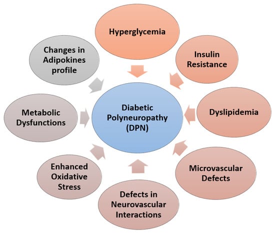
Figure 1.
Factors responsible for diabetic polyneuropathy (DPN).
Collectively, the biochemical damage induced by advanced glycation end products results in impaired nerve blood flow and diminished neurotrophic support, [15] and may play a role in disrupting neuronal integrity and repairing the mechexosamine pathway [66].
4. Current Management of the DPN
4.1. Prevention
According to the above statement, hyperglycemia and insulin deficiency played an essential role in the pathogenesis of DPN. Glycemic control and lifestyle modifications are the main focuses for preventing DPN [14,67].
4.2. Glucose Control
With type 1 diabetes, 78% of patients reduce the incidence of diabetes by enhancing tight glucose control [68]. However, 5–9% reduces the relative risk of DPN by controlling glycemic states [69,70]. Intensive insulin therapy can prevent or delay the development of DPN [71]. Lynn et al. observed that still today, tight glycemic control is the only content strategy that showed delay or prevention of DPN in type 1 patients with diabetes and slows the progressive development of neuropathy in some patients with type 2 diabetes [72]. Maintaining a stable blood glucose level reduces the symptoms of neuropathy and further nerve damage by 50% [73]. Anti-diabetic drugs such as Metformin and Thiazolidinediones (TZD) group can also play a role not only for controlling the glucose levels but also have beneficial vascular effects. Metformin prevents oxidative stress-induced cell death [74] and has neuroprotective effects via the inhabitation of oxidative stress-related neuronal cell death [75]. However, TZDs reduce inflammation and improve endothelial dysfunction [76] and oxidative stress [77]. Wiggin et al. observed that treatment with rosiglitazone, a TZD, reduces DN in streptozotocin (STZ)-treated DBA/2J mice. Rosiglitazone reduces oxidative stress and prevents the development of thermal hypoalgesia [78].
4.3. Pharmacological Approach
DPN can reduce the quality of life and enhances depression and social dysfunction [79]. A pharmacological approach is needed for DPN beyond lifestyle management or glycemic control [80,81]. There are four categories of drugs are available for DPN: 1. anticonvulsants (particularly alpha-2-delta ligand); 2. antidepressants (mainly TCAs); 3. serotonin-norepinephrine reuptake inhibitors (SNRIs); 4. opiate-receptor agonists and topical agents. Alpha-2-delta ligand: Pregabalin is one of the most used drugs for DPN. It received regulatory approval from the FDA, Health Canada, and the European medicine agency for treating DPN. It shows a 30–50% improvement in pain due to DPN [82,83,84,85,86,87]. However, not all trials with pregabalin show positive effects [82,83,88,89]. TCAs: Amitriptyline shows greater efficacy in painful DPN than other TCAs [90]. It needs a trial of 6–8 weeks to assess its effect [91]. However, FDA unapproved TCAs for treating DPN [92]. SNRIs: Duloxetine shows efficacy for treating painful DPN in multicenter randomized trials [82,83,87,88,89]. A small increase in HbA1c was observed in the patients treated with duloxetine. Tramadol, a topical capsaicin, may be useful and should be considered for treating painful DPN. Mohiuddin et al. reported that glucagon and glucagon-like peptides have some beneficial effects on the DPN in vitro model [93,94,95]. However, this is not yet accepted [82,83,96]. Despite the fact that all of these DPN medications may lessen the symptoms, none of the studies shows effectiveness to prevent the progression of DPN.
4.4. Angiogenic and Neurotrophic Factor Therapy
Neurotrophic factors such as NGF [52], IGF1 [97], and IGF2 [98] and ciliary neurotrophic factor (CNTF) [99] or glial cell line-derived neurotrophic factor (GDNF) have been shown to ameliorate DPN in animal models [100]. VEGF shows benefits to improve DPN at a certain level [101]. Intramuscular administration of FGF-2 increased blood flow in the sciatic nerve and improved nerve conduction velocity [102]. Moreover, prevention and minimization of metabolic disturbances, vascular damages, neuronal cell injuries, nerve perfusion, and ischemia may be approachable for DPN [52].
5. Stem Cell Therapy in DPN
In several studies, stem cell therapy appears very promising for the treatment of DPN. Stem cells can differentiate into the cells that are necessary to repair the damaged peripheral nerves and blood vessels. Adult stem cells and growth factors are injected into the damaged areas to reduce pain and improve blood flow to nerves. In our review, we have discussed various types of stem cells with their mechanism that is involved in the treatment of DPN [19].
5.1. Bone Marrow Mononuclear Cell Therapy
Bone marrow-derived cell therapies are the most accepted therapies because of their unique nature. An advantage of using circulating or BM-derived cells is that they can be harvested from a patient’s bone marrow and re-introduced back to the patient [103,104]. Thus, there is no chance of graft rejection. Bone marrow (BM) is a source of mononuclear cells (MNC). The BM-MNC is the term used to entitle all the cells in the bone marrow with unilobulated or rounded nuclei and inadequacy of granules in the cytoplasm [105]. Bone marrow-derived mononuclear cells (BM-MNCs) are a heterogeneous group of cells, which include mainly endothelial progenitor cells (EPCs), mesenchymal stromal cells (MSCs), and hematopoietic stem cells (HSCs) [106].
An advantage of using BM-MNCs as the source of cell therapy is that they are rather easy to acquire. They can be isolated from bone marrow by centrifugation and do not require the ex vivo culture system. Some studies have shown the beneficial effects of using BM-MNCs in the case of DPN. They improved neovascularization by increasing the levels of angiogenic factors such as VEGF, FGF-2, and angiopoietin-1 (Figure 2) [107,108]. In patients with ischemia, BM-MNCs transplantation has also been reported to be beneficial [108]. Because BM-MNCs transplantation has been shown to be an option in treating ischemic diseases, there was an interest in using a similar strategy in treating DPN [109]. A recent study showed that peripheral blood mononuclear cell implantation in rats with DPN partially recovered blood flow and improved the motor nerve conduction velocity (MNCV) of the sciatic nerve [110]. Kim, Park, Choi et al. reported that intramuscular transplantation of BM-MNCs enhances the expression of a number of angiogenic and neurotrophic factors, including VEGF, FGF-2, IGF-1, and NOS-3, in the vasa nervorum of DPN model rats. As a result, it improved nerve conduction velocity and promotes nerve vascularity. [111]. Shibata et al. reported that transplantation of BM-MNCs in STZ-induced 8-week age diabetic rat improves the transplantation of NCV, improves sciatic nerve blood flow, and increased the density of small vessels in the muscle. However, BM-MNCs taken from age-matched patients cannot show any beneficial effects in DPN [112]. Kondo et al. reported that transplantation of BM-MNCs derived from young rats ameliorated DPN, but BM-MNCs from mature or diabetic rats cannot show any efficacy [113]. Despite the beneficial effects of MSC transplantation in experimental DN shown previously, there appears to be a significant limitation in using MSCs for DPN therapy. A study showed that BM-derived MSCs might undergo chromosomal abnormalities and form malignant tumors after injection into mice with DPN. This study describes the careful monitoring of chromosomal status for transplantation of MSCs from in vitro expansion [114].
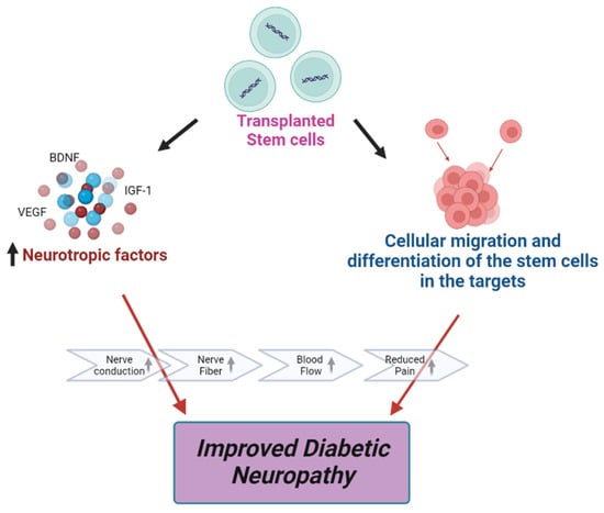
Figure 2.
Effects of stem cell therapy on DPN.
5.2. Pluripotent Stem Cell Therapy
Pluripotent stem cells are derived artificially from the mature somatic cell by insertion of (oct3/4, sox2, klf4, c-myc) using retrovirus as a vector. However, there is a chance of tumor formation because the c-myc is a potent oncogene [115,116]. Pluripotent stem cells are the new hope for regenerative medicine as they can produce every cell type in the body. Induced pluripotent stem cells (iPS) are used for organic synthesis, such as in the liver from human ‘liver buds’ iPSCs-LBs [117], and tissue repair, such as when iPSs are injected into the vitreous of damaged retina and the iPSCs engrafted into the retina [118]. Impaired vascularity and nerve degeneration are the most crucial pathophysiology of DPN. The neural crest-like cells (NCL) derived from iPS may have a therapeutic effect on DPN [119]. Angiogenesis occurs in STZ-induced diabetic mice by transplantation of NCL. It is due to the action of the angiogenic factor, vascular endothelial growth factor (VEGF) and basic fibroblast growth factor (bFGF), NGF, and Neurotrophin-3, which are secreted by NCL. Okawa et al. reported that transplantation of NCL derived from the aged mice into the 16 weeks STZ-induced mice. After 4 weeks, these transplanted cells produced growth factors such as NGF and Neurotrophin-3 and differentiated into vascular smooth muscle a cells which improve the impaired nerve and vascular functions [118]. However, it is questionable how long the locally grafted iPS cells will respond and in which amount there is no chance of tumor formation. Obstacles such as the chance of tumor formation, epigenetic memory, and new features obtained during remodeling are seen in iPSCs [120]. However, if these obstacles can be removed then, pluripotent stem cell therapy can be a good option for treating advanced stage DPN.
5.3. Endothelial Progenitor Cells (EPCs) Therapy
EPCs exhibit various therapeutic uses. These cells can also be isolated from umbilical cord blood and peripheral blood. EPCs can differentiate within endothelial cell intima of existing blood vessels. EPCs can be identified using several markers such as VE-Cadherin, CD31, CD34, CD45, CD45.1, CD45.2, CD117, CD133, CXCR4, ER71, CD146, Tie-2, VEGF-R2, and VEGF-R3. However, the majority of studies suggest CD 34+/KDR+/CD133+ markers for identifying EPCs [121,122]. Some studies have reported direct augmentation of neural neovascularization in the sciatic nerves of mice with DPN after local intramuscular injection of BM-derived EPCs [123]. The injected EPCs preferentially went to peripheral nerves; on the other hand, they went much less to the muscles. This shows that muscular neovascularization is not the mechanism at work. Additionally, the study showed that EPCs have durable engraftment into diabetic nerves [124]. Naruse et al. reported that intramuscular injection of EPCs in the hind limbs of STZ-induced diabetic nude rats increased the differentiation of endothelial cells in the hind limb. This results in the improvement of sciatic nerve conduction velocity (NCV) and blood flow [117]. However, this study is unable to demonstrate the exact mechanism through which EPCs enhance vascular health. Some of the recent studies did not agree about that EPCs differentiation as a major mechanism for neovascularization [125,126]. However, the main therapeutic effects are not through endothelial differentiation but are through angiogenesis; the overall evidence clearly suggests that BM-derived EPCs take part in blood vessel formation through vasculogenesis. Although the differentiation of EPCs plays a vital role in the recovery of damaged tissue, function is still controversial. Some studies have shown that the differentiation of EPCs into endothelial lineage cells, and they are incorporated into blood vessel formation [[122,123]. Naruse et al. reported that transplantation of cord blood-derived EPCs into the hind limb skeletal muscle increased the differentiation of EPCs into the endothelial cell in soleus muscle that leads to increase sciatic motor nerve conduction velocity and sciatic nerve blood flow in a rat DPN model [117]. However, this study does convey any clear idea about the mechanism by which the transplanted EPCs increase neovascularization or nerve conduction velocity. More recent studies have argued against the fact that EPCs do not differentiate into ECs [127,128].
5.4. Mesenchymal Stromal Cells Therapy
Mesenchymal stromal cells (MSCs) are the multipotent cells generally found in almost all post-natal organs and tissues [129] such as the umbilical cord [130], placenta [130], and dental pulp [131], and all of these have differentiation properties, which is multipotent. Meanwhile, MSCs are stupendous candidates for treating DPN. MSCs are identified by several markers such as CD54/CD102, CD166, CD73, CD90, CD44, and CD105 [20]. MSCs can differentiate into mesodermal cells in origin, such as bone, cartilage, and adipose tissue [132]. However, there is a controversy of whether MSCs are true stem cells or not, as they cannot fulfill the criteria of a stem cell. Their proliferation is self-limited, and self-renewal capacity of human MSCs still unproven [133,134,135]. H. J. Park et al. reported that transplantation of hMSCs into a Parkinson’s disease rat model exerted a neuroprotective effect [136]. Until January 2023, according to the statement of https://clinicaltrials.gov (accessed on 16 December 2022), almost 1158 clinical trials have been conducted on MSCs. However, most of these are the phase I or II that evaluate the effectiveness of MSCs in hepatic, nervous, renal, bone/cartilage, or autoimmune disorders. MSCs secrete anti-inflammatory, antiapoptotic molecules and trophic factors such as FGF, VEGF-A, and NGF, which support the growth of axons, remyelination, angiogenesis, and protection from apoptotic cell death through a paracrine effect [112,137]. Siniscalco et al. reported that after transplantation of MSCs in the cerebral ventricle, mechanical allodynia and thermal hyperalgesia are reduced in neuropathic mice [[138]. MSCs also improve the glycemic status by the regeneration of pancreatic beta cells in STZ-induced diabetic mice [139]. However, currently, MSCs have been used against acute graft-versus-host disease in Japan, New Zealand, and Canada.
5.5. Dental Pulp Stem Cell Therapy
Dental pulp stem cells (DPSCs) are mesenchymal stem cells located in the dental pulp cavity. DPSCs show high expression of CD29, CD90, and CD49d markers; these are common MSCs markers [140]. DPSCs have the natural capacity to proliferate, and they can differentiate into odontoblasts, adipocytes, osteoblasts, and neuronal cells [131,141]. However, DPSCs are an adorable candidate for cell therapy as they are easy to obtain from tooth extraction from early-age donors without any invasive procedure. DPSCs are highly expressed to neurotrophic and angiogenic factors such as NGF, NT-3, VEGF, and bFGF [41]. DPSC transplantation increases these factors and improves DPN [52,142,143]. Omi et al. reported that transplantation of DPSCs into the hind limb of the skeletal muscles of a rat model of DPN improved the sciatic motor/sensory nerve conduction velocity and sciatic nerve blood flow. They also suggested that transplantation of DPSCs eased the hypoalgesia in the DPN rats. Long-term diabetic nerves show reductions in the fiber area, occupancy rate, myelin area, and myelin thickness. The transplantation of DPSCs increases myelin thickness and myelin area, which indicates effective results for Schwann cells [140].
5.6. Embryonic Stem Cell Therapy
Embryonic stem cell therapy (ESC) is one of the possible candidates for treating DPN because it is infinitely renewable and amenable to molecular manipulation capacity [144]. Embryonic stem cells are derived from the pre-implantation of a blastocyst. It possesses an inner cell mass (ICM), which subsequently forms the ES cells, and the outer layer consists of cells collectively called trophoblasts, which give rise to the placenta. One of the most critical aspects of ES cell lines is that in response to appropriate stimuli, they can differentiate into multiple mature somatic cell types representing all three germ layers, both in vivo and in vitro. A group of studies shows that hESC can be differentiated into neural crest cells (NCCs) and cells with the morphological and molecular characteristics of myelinating Schwann cells [145,146,147]. ESCs show high expression of Nanog, GTCM 1, connexin 43(GJA1), Oct 4, and TDGF1 (crypto) markers [148]. Nakazawa et al. stated that differentiated NCCs increase several trophic factors, mainly BDNF, which is essential for neuronal survival and the elongation of the axon [149]. Jones et al. reported that transplantation of NCCs derived from ESC showed biologically active trophic factors and could stimulate neurite outgrowth in a rat sciatic nerve injury model [150]. However, no available research shows the use of ESC for treating DPN. Still, now there is no clinical trial for embryonic stem cell therapy in humans due to strong objections by certain religious communities [151,152] and due to the risk of the formation of tumors in vivo [153].
6. Challenges in Cell Therapy
Still today cell therapies have been applied only to animal models, and most of the reports have given a positive attitude for treating DN. However, there are particular challenges that have been observed during these studies. Before starting a human trial, these challenges must be faced: 1. risk of tumor formation, 2. graft rejections, 3. the optimal dose for cell survival and to reach the necessity, 4. route of transplantation, 5. the outcome of the transplantation, and 6. details of the mechanism of actions. Sarcoma has been reported in murine MSCs in vitro [154] as well as tumor development after allogeneic transplantation of MSCs [155] and BMSC transplantation in STZ-induced diabetic mice. [114].
7. The Route of Transplantation
For the efficacy and viability of the transplanted cell, it is essential to consider the mood of transplantation, whether it will be topical, intraocular, or systemic. Without a proper dose and route of transplantation, the efficacy will be reduced or not be observed. For treating diabetic foot ulcers, the administration of BM-MSCs as a nonvascular injection is mostly used currently. Systemic delivery may be one of the most provocative routes of administration. However, this will require a large number of cells to reach the target tissue, and the efficacy of the transplanted cells may be reduced [156].
8. Conclusions and Future Directions
As diabetes is a chronic metabolic disorder, it affects human health from head to toe [157,158,159,160,161]. Foot ulceration and limb amputations are the consequences of DPN if there is no effective clinical treatment. Conventional drug therapies are showing limited activities against DPN. Complimentary treatment often shows a potential role. However, its use is still limited. Pipelines do not suggest an active molecule against DPN. Cell therapy may not be a standard treatment option for all stages of DPN because of the different structural or functional changes marked in various stages of DPN. However, the costs and availabilities of these approaches are out of reach for ordinary people. Immunity has also been a problem with direct cell-derived therapies. Delivery approaches must be taken care with proper instrumentation. Though cell-based treatment approaches have been proven well in both animal and human subjects, appropriate clinical evaluations are necessary before making them for general use.
Author Contributions
Conceptualization, S.A. (Shamima Akter) and M.S.M.; validation, M.C.; resources, M.M.M. and S.A. (Shahida Arbee); data curation, M.A.T.S.; writing—M.S.M., S.A. (Shahida Arbee) and M.C.; super-vision, M.S.M. and M.C. All authors have read and agreed to the published version of the manuscript
Funding
This research received no external funding.
Institutional Review Board Statement
Not applicable.
Informed Consent Statement
Not applicable.
Data Availability Statement
Not applicable.
Conflicts of Interest
The authors declare that there are no conflict of interest regarding the publication of this paper.
References
- Guh, D.P.; Zhang, W.; Bansback, N.; Amarsi, Z.; Birmingham, C.L.; Anis, A.H. The incidence of co-morbidities related to obesity and overweight: A systematic review and meta-analysis. BMC Public Health 2009, 9, 88. [Google Scholar] [CrossRef] [PubMed]
- Diabetes-PAHO/WHO|Pan American Health Organization, Bulletin 2022. Available online: https://www.paho.org/en/topics/diabetes (accessed on 16 December 2022).
- Singh, A.; Choubey, M.; Bora, P.; Krishna, A. Adiponectin and Chemerin: Contrary Adipokines in Regulating Reproduction and Metabolic Disorders. Reprod. Sci. 2018, 25, 1462–1473. [Google Scholar] [CrossRef] [PubMed]
- Dai, W.; Choubey, M.; Patel, S.; Singer, H.A.; Ozcan, L. Adipocyte CAMK2 deficiency improves obesity-associated glucose intolerance. Mol. Metab. 2021, 53, 101300. [Google Scholar] [CrossRef] [PubMed]
- Feldman, E.L. Epidemiology and Classification of Diabetic Neuropathy-UpToDate. 2018. Available online: https://www.uptodate.com/contents/epidemiology-and-classification-of-diabetic-neuropathy (accessed on 16 December 2022).
- Dyck, P.J.; Litchy, W.J.; Lehman, K.A.; Hokanson, J.L.; Low, P.A.; O’Brien, P.C. Variables influencing neuropathic endpoints: The Rochester Diabetic Neuropathy Study of Healthy Subjects. Neurology 1995, 45, 1115–1121. [Google Scholar] [CrossRef] [PubMed]
- Dyck, P.J.; Kratz, K.M.; Karnes, J.L.; Litchy, W.J.; Klein, R.; Pach, J.M.; Wilson, D.M.; O’Brien, P.C.; Melton, L.J. The prevalence by staged severity of various types of diabetic neuropathy, retinopathy, and nephropathy in a population-based cohort: The Rochester Diabetic Neuropathy Study. Neurology 1993, 43, 817–824. [Google Scholar] [CrossRef]
- Edwards, J.L.; Vincent, A.M.; Cheng, H.T.; Feldman, E.L. Diabetic neuropathy: Mechanisms to management. Pharmacol. Ther. 2008, 120, 1–34. [Google Scholar]
- Boulton, A.J.; Kirsner, R.S.; Vileikyte, L. Neuropathic Diabetic Foot Ulcers. New Engl. J. Med. 2004, 351, 48–55. [Google Scholar] [CrossRef]
- Boulton, A.J.; Vinik, A.I.; Arezzo, J.C.; Bril, V.; Feldman, E.L.; Freeman, R.; Ziegler, D. Diabetic neuropathies: A statement by the American Diabetes Association. Diabetes Care 2005, 28, 956–962. [Google Scholar] [CrossRef]
- Tesfaye, S.; Boulton, A.J.M.; Dyck, P.J.; Freeman, R.; Horowitz, M.; Kempler, P.; Lauria, G.; Malik, R.A.; Spallone, V.; Vinik, A.; et al. Diabetic Neuropathies: Update on Definitions, Diagnostic Criteria, Estimation of Severity, and Treatments. Diabetes Care 2010, 33, 2285–2293. [Google Scholar] [CrossRef]
- UK Prospective Diabetes Study (UKPDS) Group. Intensive blood-glucose control with sulphonylureas or insulin compared with conventional treatment and risk of complications in patients with type 2 diabetes (UKPDS 33). Lancet 1998, 352, 837–853. [Google Scholar] [CrossRef]
- Martin, C.L.; Albers, J.; Herman, W.H.; Cleary, P.; Waberski, B.; Greene, D.A.; Stevens, M.J.; Feldman, E.L. Neuropathy Among the Diabetes Control and Complications Trial Cohort 8 Years After Trial Completion. Diabetes Care 2006, 29, 340–344. [Google Scholar] [CrossRef] [PubMed]
- Diabetes Control and Complications Trial Research Group. Effect of intensive diabetes treatment on the development and progression of long-term complications in adolescents with insulin-dependent diabetes mellitus: Diabetes Control and Complications Trial. J. Pediatr. 1994, 125, 177–188. [Google Scholar] [CrossRef] [PubMed]
- Stracke, H.; Gaus, W.; Achenbach, U.; Federlin, K.; Bretzel, R.G. Benfotiamine in diabetic polyneuropathy (BENDIP): Results of a randomised, double blind, placebo-controlled clinical study. Exp. Clin. Endocrinol. Diabetes Off. J. 2008, 116, 600–605. [Google Scholar] [CrossRef] [PubMed]
- Ang, C.D.; Alviar, M.J.M.; Dans, A.L.; Bautista-Velez GG, P.; Villaruz-Sulit MV, C.; Tan, J.J.; Roxas, A.A. Vitamin B for treating peripheral neuropathy. Cochrane Database Syst. Rev. 2008, 16, Cd004573. [Google Scholar] [CrossRef]
- Kato, N.; Nemoto, K.; Nakanishi, K.; Morishita, R.; Kaneda, Y.; Uenoyama, M.; Fujikawa, K. Nonviral gene transfer of human hepatocyte growth factor improves streptozotocin-induced diabetic neuropathy in rats. Diabetes 2005, 54, 846–854. [Google Scholar] [CrossRef]
- Mizukami, H.; Yagihashi, S. Exploring a new therapy for diabetic polyneuropathy—The application of stem cell transplantation. Front. Endocrinol. (Lausanne) 2014, 5, 45. [Google Scholar] [CrossRef]
- Ebrahimi, A.; Ahmadi, H.; Ghasrodashti, Z.P.; Tanideh, N.; Shahriarirad, R.; Erfani, A.; Ashkani-Esfahani, S. Therapeutic effects of stem cells in different body systems, a novel method that is yet to gain trust: A comprehensive review. Bosn. J. Basic Med. Sci. 2021, 21, 672. [Google Scholar] [CrossRef]
- Zhou, J.Y.; Zhang, Z.; Qian, G.S. Mesenchymal stem cells to treat diabetic neuropathy: A long and strenuous way from bench to the clinic. Cell Death Discov. 2016, 2, 16055. [Google Scholar] [CrossRef]
- Jin, K.; Zhu, Y.; Sun, Y.; Mao, X.O.; Xie, L.; Greenberg, D.A. Vascular endothelial growth factor (VEGF) stimulates neurogenesis in vitro and in vivo. Proc. Natl. Acad. Sci. USA 2002, 99, 11946–11950. [Google Scholar] [CrossRef]
- Donovan, M.H.; Yazdani, U.; Norris, R.D.; Games, D.; German, D.C.; Eisch, A.J. Decreased adult hippocampal neurogenesis in the PDAPP mouse model of Alzheimer’s disease. J. Comp. Neurol. 2006, 495, 70–83. [Google Scholar] [CrossRef]
- López-Toledano, M.A.; Shelanski, M.L. Increased neurogenesis in young transgenic mice overexpressing human APP(Sw, Ind). J. Alzheimer’S Dis. JAD 2007, 12, 229–240. [Google Scholar] [CrossRef] [PubMed]
- Blurton-Jones, M.; Kitazawa, M.; Martinez-Coria, H.; Castello, N.A.; Müller, F.J.; Loring, J.F.; LaFerla, F.M. Neural stem cells improve cognition via BDNF in a transgenic model of Alzheimer disease. Proc. Natl. Acad. Sci. USA 2009, 106, 13594–13599. [Google Scholar] [CrossRef] [PubMed]
- Kordower, J.H.; Winn, S.R.; Liu, Y.T.; Mufson, E.J.; Sladek, J.R.; Hammang, J.P.; E Baetge, E.; Emerich, D.F. The aged monkey basal forebrain: Rescue and sprouting of axotomized basal forebrain neurons after grafts of encapsulated cells secreting human nerve growth factor. Proc. Natl. Acad. Sci. USA 1994, 91, 10898–10902. [Google Scholar] [CrossRef] [PubMed]
- Park, D.; Yang, Y.H.; Bae, D.K.; Lee, S.H.; Yang, G.; Kyung, J.; Kim, Y.B. Improvement of cognitive function and physical activity of aging mice by human neural stem cells over-expressing choline acetyltransferase. Neurobiol. Aging 2013, 34, 2639–2646. [Google Scholar] [CrossRef] [PubMed]
- Lee, I.-S.; Jung, K.; Kim, I.-S.; Lee, H.; Kim, M.; Yun, S.; Hwang, K.; Shin, J.E.; Park, K.I. Human neural stem cells alleviate Alzheimer-like pathology in a mouse model. Mol. Neurodegener. 2015, 10, 38. [Google Scholar] [CrossRef]
- Zhang, Q.; Wu, H.H.; Wang, Y.; Gu, G.J.; Zhang, W.; Xia, R. Neural stem cell transplantation decreases neuroinflammation in a transgenic mouse model of Alzheimer’s disease. J. Neurochem. 2016, 136, 815–825. [Google Scholar] [CrossRef] [PubMed]
- Lilja, A.M.; Malmsten, L.; Röjdner, J.; Voytenko, L.; Verkhratsky, A.; Ögren, S.O.; Marutle, A. Neural Stem Cell Transplant-Induced Effect on Neurogenesis and Cognition in Alzheimer Tg2576 Mice Is Inhibited by Concomitant Treatment with Amyloid-Lowering or Cholinergic α7 Nicotinic Receptor Drugs. Neural Plast. 2015, 2015, 370432. [Google Scholar] [CrossRef]
- Xuan, A.G.; Luo, M.; Ji, W.D.; Long, D.H. Effects of engrafted neural stem cells in Alzheimer’s disease rats. Neurosci. Lett. 2009, 450, 167–171. [Google Scholar] [CrossRef]
- Bissonnette, C.J.; Lyass, L.; Bhattacharyya, B.J.; Belmadani, A.; Miller, R.J.; Kessler, J.A. The controlled generation of functional basal forebrain cholinergic neurons from human embryonic stem cells. Stem Cells 2011, 29, 802–811. [Google Scholar] [CrossRef]
- Park, D.; Yang, G.; Bae, D.K.; Lee, S.H.; Yang, Y.H.; Kyung, J.; Kim, Y.B. Human adipose tissue-derived mesenchymal stem cells improve cognitive function and physical activity in ageing mice. J. Neurosci. Res. 2013, 91, 660–670. [Google Scholar] [CrossRef]
- Zilka, N.; Zilkova, M.; Kazmerova, Z.; Sarissky, M.; Cigankova, V.; Novak, M. Mesenchymal stem cells rescue the Alzheimer’s disease cell model from cell death induced by misfolded truncated tau. Neuroscience 2011, 193, 330–337. [Google Scholar] [CrossRef] [PubMed]
- Lee, H.J.; Lee, J.K.; Lee, H.; Carter, J.E.; Chang, J.W.; Oh, W.; Bae, J.S. Human umbilical cord blood-derived mesenchymal stem cells improve neuropathology and cognitive impairment in an Alzheimer’s disease mouse model through modulation of neuroinflammation. Neurobiol. Aging 2012, 33, 588–602. [Google Scholar] [CrossRef] [PubMed]
- Thomsen, G.M.; Gowing, G.; Svendsen, S.; Svendsen, C.N. The past, present and future of stem cell clinical trials for ALS. Exp. Neurol. 2014, 262, 127–137. [Google Scholar] [CrossRef] [PubMed]
- Mazzini, L.; Gelati, M.; Profico, D.C.; Sgaravizzi, G.; Projetti Pensi, M.; Muzi, G.; Vescovi, A.L. Human neural stem cell transplantation in ALS: Initial results from a phase I trial. J. Transl. Med. 2015, 13, 17. [Google Scholar] [CrossRef]
- Human neural stem cell transplants improve motor function in a rat model of Huntington’s disease-McBride-2004. J. Comp. Neurol. 2004, 475, 211–219. [CrossRef] [PubMed]
- Lee, S.T.; Chu, K.; Park, J.E.; Lee, K.; Kang, L.; Kim, S.U.; Kim, M. Intravenous administration of human neural stem cells induces functional recovery in Huntington’s disease rat model. Neurosci. Res. 2005, 52, 243–249. [Google Scholar] [CrossRef]
- Vazey, E.M.; Chen, K.; Hughes, S.M.; Connor, B. Transplanted adult neural progenitor cells survive, differentiate and reduce motor function impairment in a rodent model of Huntington’s disease. Exp. Neurol. 2006, 199, 384–396. [Google Scholar] [CrossRef]
- Bachoud-Lévi, A.C.; Rémy, P.; Nǵuyen, J.P.; Brugières, P.; Lefaucheur, J.P.; Bourdet, C.; Peschanski, M. Motor and cognitive improvements in patients with Huntington’s disease after neural transplantation. Lancet 2000, 356, 1975–1979. [Google Scholar] [CrossRef]
- Bachoud-Lévi, A.C.; Gaura, V.; Brugières, P.; Lefaucheur, J.P.; Boissé, M.F.; Maison, P.; Peschanski, M. Effect of fetal neural transplants in patients with Huntington’s disease 6 years after surgery: A long-term follow-up study. Lancet Neurol. 2006, 5, 303–309. [Google Scholar] [CrossRef]
- Carstens, M.H.; Gómez, A.; Cortés, R.; Turner, E.; Pérez, C.; Ocon, M.; Correa, D. Non-reconstructable peripheral vascular disease of the lower extremity in ten patients treated with adipose-derived stromal vascular fraction cells. Stem Cell Res. 2017, 18, 14–21. [Google Scholar] [CrossRef]
- Bang, O.Y.; Lee, J.S.; Lee, P.H.; Lee, G. Autologous mesenchymal stem cell transplantation in stroke patients. Ann. Neurol. 2005, 57, 874–882. [Google Scholar] [CrossRef] [PubMed]
- Lee, J.S.; Hong, J.M.; Moon, G.J.; Lee, P.H.; Ahn, Y.H.; Bang, O.Y. A long-term follow-up study of intravenous autologous mesenchymal stem cell transplantation in patients with ischemic stroke. Stem Cells 2010, 28, 1099–1106. [Google Scholar] [CrossRef]
- Haas, S.; Weidner, N.; Winkler, J. Adult stem cell therapy in stroke. Curr. Opin. Neurol. 2005, 18, 59–64. [Google Scholar] [CrossRef] [PubMed]
- Einstein, O.; Karussis, D.; Grigoriadis, N.; Mizrachi-Kol, R.; Reinhartz, E.; Abramsky, O.; Ben-Hur, T. Intraventricular transplantation of neural precursor cell spheres attenuates acute experimental allergic encephalomyelitis. Mol. Cell. Neurosci. 2003, 24, 1074–1082. [Google Scholar] [CrossRef] [PubMed]
- Pluchino, S.; Quattrini, A.; Brambilla, E.; Gritti, A.; Salani, G.; Dina, G.; Martino, G. Injection of adult neurospheres induces recovery in a chronic model of multiple sclerosis. Nature 2003, 422, 688–694. [Google Scholar] [CrossRef] [PubMed]
- Bulte, J.W.; Ben-Hur, T.; Miller, B.R.; Mizrachi-Kol, R.; Einstein, O.; Reinhartz, E.; Frank, J.A. MR microscopy of magnetically labeled neurospheres transplanted into the Lewis EAE rat brain. Magn. Reson. Med. 2003, 50, 201–205. [Google Scholar] [CrossRef] [PubMed]
- Mothe, A.J.; Tator, C.H. Review of transplantation of neural stem/progenitor cells for spinal cord injury. Int. J. Dev. Neurosci. 2013, 31, 701–713. [Google Scholar] [CrossRef] [PubMed]
- Richter, M.W.; Roskams, A.J. Olfactory ensheathing cell transplantation following spinal cord injury: Hype or hope? Exp. Neurol. 2008, 209, 353–367. [Google Scholar] [CrossRef] [PubMed]
- Waxman, S.G.; Sabin, T.D. Diabetic truncal polyneuropathy. Arch. Neurol. 1981, 38, 46–47. [Google Scholar] [CrossRef]
- Apfel, S.C. Neurotrophic factors in the therapy of diabetic neuropathy. Am. J. Med. 1999, 107, 34–42. [Google Scholar] [CrossRef]
- Jensen, T.S.; Finnerup, N.B. Allodynia and hyperalgesia in neuropathic pain: Clinical manifestations and mechanisms. Lancet Neurol. 2014, 13, 924–935. [Google Scholar] [CrossRef] [PubMed]
- Vincent, A.M.; Mclean, L.L.; Backus, C.; Feldman, E.L. Short-term hyperglycemia produces oxidative damage and apoptosis in neurons. FASEB J. 2005, 19, 638–640. [Google Scholar] [CrossRef] [PubMed]
- Mahmood, D.; Singh, B.K.; Akhtar, M. Diabetic neuropathy: Therapies on the horizon. J. Pharm. Pharmacol. 2009, 61, 1137–1145. [Google Scholar] [CrossRef] [PubMed]
- Feldman, E.L.; Nave, K.A.; Jensen, T.S.; Bennett, D.L. New Horizons in Diabetic Neuropathy: Mechanisms, Bioenergetics, and Pain. Neuron 2017, 93, 1296–1313. [Google Scholar] [CrossRef] [PubMed]
- Jack, M.; Wright, D. Role of advanced glycation endproducts and glyoxalase I in diabetic peripheral sensory neuropathy. Transl. Res. 2012, 159, 355–365. [Google Scholar] [CrossRef]
- Obrosova, I.G.; Li, F.; Abatan, O.I.; Forsell, M.A.; Komjáti, K.; Pacher, P.; Szabó, C.; Stevens, M.J. Role of Poly(ADP-Ribose) Polymerase Activation in Diabetic Neuropathy. Diabetes 2004, 53, 711–720. [Google Scholar] [CrossRef]
- Vincent, A.M.; Callaghan, B.C.; Smith, A.L.; Feldman, E.L. Diabetic neuropathy: Cellular mechanisms as therapeutic targets. Nat. Rev. Neurol. 2011, 7, 573–583. [Google Scholar] [CrossRef]
- Zacchigna, S.; Lambrechts, D.; Carmeliet, P. Neurovascular signalling defects in neurodegeneration. Nat. Rev. Neurosci. 2008, 9, 169–181. [Google Scholar] [CrossRef]
- Pittenger, G.; Vinik, A. Nerve Growth Factor and Diabetic Neuropathy. Exp. Diabesity Res. 2003, 4, 271–285. [Google Scholar] [CrossRef]
- Liu, T.; Zhang, L.; Joo, D.; Sun, S.C. NF-κB signaling in inflammation. Signal Transduct. Target Ther. 2017, 2, 17023. [Google Scholar] [CrossRef]
- Yagihashi, S.; Mizukami, H.; Sugimoto, K. Mechanism of diabetic neuropathy: Where are we now and where to go? J. Diabetes Investig. 2010, 2, 18–32. [Google Scholar] [CrossRef] [PubMed]
- Guo, J.; Whittemore, R.; He, G.-P. The relationship between diabetes self-management and metabolic control in youth with type 1 diabetes: An integrative review. J. Adv. Nurs. 2011, 67, 2294–2310. [Google Scholar] [CrossRef] [PubMed]
- Kim, B.; Feldman, E.L. Insulin resistance in the nervous system. Trends Endocrinol. Metab. 2012, 23, 133–141. [Google Scholar] [CrossRef] [PubMed]
- Schleicher, E.D.; Weigert, C. Role of the hexosamine biosynthetic pathway in diabetic nephropathy. Kidney Int. 2000, 58, S13–S18. [Google Scholar] [CrossRef]
- Effect of intensive diabetes treatment on nerve conduction in the Diabetes Control and Complications Trial. Ann.Neurol. 1995, 38, 869–880. [CrossRef]
- Linn, T.; Ortac, K.; Laube, H.; Federlin, K. Intensive therapy in adult insulin-dependent diabetes mellitus is associated with improved insulin sensitivity and reserve: A randomized, controlled, prospective study over 5 years in newly diagnosed patients. Metabolism 1996, 45, 1508–1513. [Google Scholar] [CrossRef]
- Ismail-Beigi, F.; Craven, T.; Banerji, M.A.; Basile, J.; Calles, J.; Cohen, R.M.; Cuddihy, R.; Cushman, W.C.; Genuth, S.; Grimm, R.H.; et al. Effect of intensive treatment of hyperglycaemia on microvascular outcomes in type 2 diabetes: An analysis of the ACCORD randomised trial. Lancet 2010, 376, 419–430. [Google Scholar] [CrossRef]
- Callaghan, B.C.; Cheng, H.T.; Stables, C.L.; Smith, A.L.; Feldman, E.L. Diabetic neuropathy: Clinical manifestations and current treatments. Lancet. Neurol. 2012, 11, 521–534. [Google Scholar] [CrossRef]
- Ohkubo, Y.; Kishikawa, H.; Araki, E.; Miyata, T.; Isami, S.; Motoyoshi, S.; Kojima, Y.; Furuyoshi, N.; Shichiri, M. Intensive insulin therapy prevents the progression of diabetic microvascular complications in Japanese patients with non-insulin-dependent diabetes mellitus: A randomized prospective 6-year study. Diabetes Res. Clin. Pr. 1995, 28, 103–117. [Google Scholar] [CrossRef]
- Ang, L.; Jaiswal, M.; Martin, C.; Pop-Busui, R. Glucose Control and Diabetic Neuropathy: Lessons from Recent Large Clinical Trials. Curr. Diabetes Rep. 2014, 14, 528. [Google Scholar] [CrossRef]
- Diabetic Nerve Damage. 2022. Available online: https://www.joslin.org/patient-care/diabetes-education/diabetes-learning-center/diabetic-nerve-damage (accessed on 16 December 2022).
- Detaille, D.; Guigas, B.; Chauvin, C.; Batandier, C.; Fontaine, E.; Wiernsperger, N.; Leverve, X. Metformin Prevents High-Glucose–Induced Endothelial Cell Death Through a Mitochondrial Permeability Transition-Dependent Process. Diabetes 2005, 54, 2179–2187. [Google Scholar] [CrossRef] [PubMed]
- El-Mir, M.-Y.; Detaille, D.; R-Villanueva, G.; Delgado-Esteban, M.; Guigas, B.; Attia, S.; Fontaine, E.; Almeida, A.; Leverve, X. Neuroprotective Role of Antidiabetic Drug Metformin Against Apoptotic Cell Death in Primary Cortical Neurons. J. Mol. Neurosci. 2007, 34, 77–87. [Google Scholar] [CrossRef] [PubMed]
- Sjoholm, A.; Nystrom, T. Endothelial inflammation in insulin resistance. Lancet 2005, 365, 610–612. [Google Scholar] [CrossRef] [PubMed]
- Chen, K.; Chen, J.; Li, D.; Zhang, X.; Mehta, J.L. Angiotensin II regulation of collagen type I expression in cardiac fibroblasts: Modulation by PPAR-gamma ligand pioglitazone. Hypertension 2004, 44, 655–661. [Google Scholar] [CrossRef]
- Wiggin, T.D.; Kretzler, M.; Pennathur, S.; Sullivan, K.A.; Brosius, F.C.; Feldman, E.L. Rosiglitazone Treatment Reduces Diabetic Neuropathy in Streptozotocin-Treated DBA/2J Mice. Endocrinology 2008, 149, 4928–4937. [Google Scholar] [CrossRef]
- Sadosky, A.; Schaefer, C.; Mann, R.; Bergstrom, F.; Baik, R.; Parsons, B.; Nalamachu, S.; Nieshoff, E.; Stacey, B.R.; Tuchman, M.; et al. Burden of illness associated with painful diabetic peripheral neuropathy among adults seeking treatment in the US: Results from a retrospective chart review and cross-sectional survey. Diabetes Metab. Syndr. Obesity Targets Ther. 2013, 6, 79–92. [Google Scholar] [CrossRef]
- Smith, A.G.; Russell, J.; Feldman, E.L.; Goldstein, J.; Peltier, A.; Smith, S.; Hamwi, J.; Pollari, D.; Bixby, B.; Howard, J.; et al. Lifestyle Intervention for Pre-Diabetic Neuropathy. Diabetes Care 2006, 29, 1294–1299. [Google Scholar] [CrossRef]
- Oyibo, S.O.; Prasad, Y.D.M.; Jackson, N.J.; Jude, E.B.; Boulton, A.J.M. The relationship between blood glucose excursions and painful diabetic peripheral neuropathy: A pilot study. Diabet. Med. 2002, 19, 870–873. [Google Scholar] [CrossRef]
- Finnerup, N.B.; Attal, N.; Haroutounian, S.; McNicol, E.; Baron, R.; Dworkin, R.H.; Gilron, I.; Haanpää, M.; Hansson, P.; Jensen, T.S.; et al. Pharmacotherapy for neuropathic pain in adults: A systematic review and meta-analysis. Lancet Neurol. 2015, 14, 162–173. [Google Scholar] [CrossRef]
- Griebeler, M.L.; Morey-Vargas, O.L.; Brito, J.P.; Tsapas, A.; Wang, Z.; Carranza Leon, B.G.; Murad, M.H. Pharmacologic interventions for painful diabetic neuropathy: An umbrella systematic review and comparative effectiveness network meta-analysis. Ann. Intern. Med. 2014, 161, 639–649. [Google Scholar] [CrossRef]
- Freeman, R.; Durso-DeCruz, E.; Emir, B. Efficacy, safety, and tolerability of pregabalin treatment for painful diabetic peripheral neuropathy: Findings from seven randomized, controlled trials across a range of doses. Diabetes Care 2008, 31, 1448–1454. [Google Scholar] [CrossRef]
- Moore, R.A.; Straube, S.; Wiffen, P.J.; Derry, S.; McQuay, H.J. Pregabalin for acute and chronic pain in adults. Cochrane Database Syst. Rev. 2009, 8, CD007076. [Google Scholar] [CrossRef]
- Raskin, P.; Huffman, C.; Toth, C.; Asmus, M.J.; Messig, M.; Sanchez, R.J.; Pauer, L. Pregabalin in patients with inadequately treated painful diabetic peripheral neuropathy: A randomized withdrawal trial. Clin. J. Pain 2014, 30, 379–390. [Google Scholar] [CrossRef] [PubMed]
- Tesfaye, S.; Wilhelm, S.; Lledo, A.; Schacht, A.; Tölle, T.; Bouhassira, D.; Freynhagen, R. Duloxetine and pregabalin: High-dose monotherapy or their combination? The “COMBO-DN study”--a multinational, randomized, double-blind, parallel-group study in patients with diabetic peripheral neuropathic pain. Pain 2013, 154, 2616–2625. [Google Scholar] [CrossRef]
- Ziegler, D.; Duan, W.R.; An, G.; Thomas, J.W.; Nothaft, W. A randomized double-blind, placebo-, and active-controlled study of T-type calcium channel blocker ABT-639 in patients with diabetic peripheral neuropathic pain. Pain 2015, 156, 2013–2020. [Google Scholar] [CrossRef] [PubMed]
- Quilici, S.; Chancellor, J.; Löthgren, M.; Simon, D.; Said, G.; Le, T.K.; Garcia-Cebrian, A.; Monz, B. Meta-analysis of duloxetine vs. pregabalin and gabapentin in the treatment of diabetic peripheral neuropathic pain. BMC Neurol. 2009, 9, 6. [Google Scholar] [CrossRef] [PubMed]
- Bril, V.; England, J.; Franklin, G.M.; Backonja, M.; Cohen, J.; Del Toro, D.; Zochodne, D. Evidence-based guideline: Treatment of painful diabetic neuropathy: Report of the American Academy of Neurology, the American Association of Neuromuscular and Electrodiagnostic Medicine, and the American Academy of Physical Medicine and Rehabilitation. Pm&r 2011, 3, 345–352.e21. [Google Scholar]
- Dworkin, R.H.; O’Connor, A.B.; Backonja, M.; Farrar, J.T.; Finnerup, N.B.; Jensen, T.S.; Kalso, E.A.; Loeser, J.D.; Miaskowski, C.; Nurmikko, T.J.; et al. Pharmacologic management of neuropathic pain: Evidence-based recommendations. Pain 2007, 132, 237–251. [Google Scholar] [CrossRef]
- McQuay, H.J.; Tramér, M.; Nye, B.A.; Carroll, D.; Wiffen, P.J.; Moore, R.A. A systematic review of antidepressants in neuropathic pain. Pain 1996, 68, 217–227. [Google Scholar] [CrossRef]
- Mohiuddin, M.S.; Himeno, T.; Inoue, R.; Miura-Yura, E.; Yamada, Y.; Nakai-Shimoda, H.; Kamiya, H. Glucagon-Like Peptide-1 Receptor Agonist Protects Dorsal Root Ganglion Neurons against Oxidative Insult. J. Diabetes Res. 2019, 2019, 9426014. [Google Scholar] [CrossRef]
- Motegi, M.; Himeno, T.; Nakai-Shimoda, H.; Inoue, R.; Ozeki, N.; Hayashi, Y.; Kamiya, H. Deficiency of glucagon gene-derived peptides induces peripheral polyneuropathy in mice. Biochem. Biophys. Res. Commun. 2020, 532, 47–53. [Google Scholar] [CrossRef] [PubMed]
- Mohiuddin, M.S.; Himeno, T.; Yamada, Y.; Morishita, Y.; Kondo, M.; Tsunekawa, S.; Kamiya, H. Glucagon Prevents Cytotoxicity Induced by Methylglyoxal in a Rat Neuronal Cell Line Model. Biomolecules 2021, 11, 287. [Google Scholar] [CrossRef] [PubMed]
- Pop-Busui, R.; Boulton, A.J.; Feldman, E.L.; Bril, V.; Freeman, R.; Malik, R.A.; Sosenko, J.M.; Ziegler, D. Diabetic Neuropathy: A Position Statement by the American Diabetes Association. Diabetes Care 2016, 40, 136–154. [Google Scholar] [CrossRef]
- Schmidt, R.E.; Dorsey, D.A.; Beaudet, L.N.; Plurad, S.B.; Parvin, C.A.; Miller, M.S. Insulin-like Growth Factor I Reverses Experimental Diabetic Autonomic Neuropathy. Am. J. Pathol. 1999, 155, 1651–1660. [Google Scholar] [CrossRef] [PubMed]
- Zhuang, H.X.; Snyder, C.K.; Pu, S.F.; Ishii, D.N. Insulin-like Growth Factors Reverse or Arrest Diabetic Neuropathy: Effects on Hyperalgesia and Impaired Nerve Regeneration in Rats. Exp. Neurol. 1996, 140, 198–205. [Google Scholar] [CrossRef]
- Mizisin, A.P.; Vu, Y.; Shuff, M.; Calcutt, N.A. Ciliary Neurotrophic Factor Improves Nerve Conduction and Ameliorates Regeneration Deficits in Diabetic Rats. Diabetes 2004, 53, 1807–1812. [Google Scholar] [CrossRef] [PubMed]
- Anitha, M.; Gondha, C.; Sutliff, R.; Parsadanian, A.; Mwangi, S.; Sitaraman, S.V.; Srinivasan, S. GDNF rescues hyperglycemia-induced diabetic enteric neuropathy through activation of the PI3K/Akt pathway. J. Clin. Investig. 2006, 116, 344–356. [Google Scholar] [CrossRef]
- Ropper, A.H.; Gorson, K.C.; Gooch, C.L.; Weinberg, D.H.; Pieczek, A.; Ware, J.H.; Kershen, J.; Rogers, A.; Simovic, D.; Schratzberger, P.; et al. Vascular endothelial growth factor gene transfer for diabetic polyneuropathy: A randomized, double-blinded trial. Ann. Neurol. 2009, 65, 386–393. [Google Scholar] [CrossRef] [PubMed]
- Nakae, M.; Kamiya, H.; Naruse, K.; Horio, N.; Ito, Y.; Mizubayashi, R.; Hamada, Y.; Nakashima, E.; Akiyama, N.; Kobayashi, Y.; et al. Effects of Basic Fibroblast Growth Factor on Experimental Diabetic Neuropathy in Rats. Diabetes 2006, 55, 1470–1477. [Google Scholar] [CrossRef]
- Tacken, P.J.; De Vries, I.J.M.; Torensma, R.; Figdor, C. Dendritic-cell immunotherapy: From ex vivo loading to in vivo targeting. Nat. Rev. Immunol. 2007, 7, 790–802. [Google Scholar] [CrossRef]
- Prochazka, V.; Gumulec, J.; Chmelova, J.; Klement, P.; Klement, G.L.; Jonszta, T.; Krajca, J. Autologous bone marrow stem cell transplantation in patients with end-stage chronical critical limb ischemia and diabetic foot. Vnitr Lek 2009, 55, 173–178. [Google Scholar] [PubMed]
- Cuende, N.; Rico, L.; Herrera, C. Concise Review: Bone Marrow Mononuclear Cells for the Treatment of Ischemic Syndromes: Medicinal Product or Cell Transplantation? Stem Cells Transl. Med. 2012, 1, 403–408. [Google Scholar] [CrossRef] [PubMed]
- Ushio-Fukai, M.; Rehman, J. Redox and Metabolic Regulation of Stem/Progenitor Cells and Their Niche. Antioxidants Redox Signal. 2014, 21, 1587–1590. [Google Scholar] [CrossRef]
- Pittenger, M.F.; Mackay, A.M.; Beck, S.C.; Jaiswal, R.K.; Douglas, R.; Mosca, J.D.; Moorman, M.A.; Simonetti, D.W.; Craig, S.; Marshak, D.R. Multilineage Potential of Adult Human Mesenchymal Stem Cells. Science 1999, 284, 143–147. [Google Scholar] [CrossRef] [PubMed]
- Jackson, L.; Jones, D.R.; Scotting, P.; Sottile, V. Adult mesenchymal stem cells: Differentiation potential and therapeutic applications. J. Postgrad. Med. 2007, 53, 121–127. [Google Scholar] [PubMed]
- Kinnaird, T.; Stabile, E.; Burnett, M.S.; Shou, M.; Lee, C.W.; Barr, S.; Epstein, S.E. Local delivery of marrow-derived stromal cells augments collateral perfusion through paracrine mechanisms. Circulation 2004, 109, 1543–1549. [Google Scholar] [CrossRef]
- Hasegawa, T.; Kosaki, A.; Shimizu, K.; Matsubara, H.; Mori, Y.; Masaki, H.; Toyoda, N.; Inoue-Shibata, M.; Nishikawa, M.; Iwasaka, T. Amelioration of diabetic peripheral neuropathy by implantation of hematopoietic mononuclear cells in streptozotocin-induced diabetic rats. Exp. Neurol. 2006, 199, 274–280. [Google Scholar] [CrossRef]
- Kim, H.; Park, J.-S.; Choi, Y.J.; Kim, M.-O.; Huh, Y.H.; Kim, S.-W.; Han, J.W.; Lee, J.; Kim, S.; Houge, M.A.; et al. Bone Marrow Mononuclear Cells Have Neurovascular Tropism and Improve Diabetic Neuropathy. Stem Cells 2009, 27, 1686–1696. [Google Scholar] [CrossRef]
- Shibata, T.; Naruse, K.; Kamiya, H.; Kozakae, M.; Kondo, M.; Yasuda, Y.; Nakamura, N.; Ota, K.; Tosaki, T.; Matsuki, T.; et al. Transplantation of Bone Marrow–Derived Mesenchymal Stem Cells Improves Diabetic Polyneuropathy in Rats. Diabetes 2008, 57, 3099–3107. [Google Scholar] [CrossRef]
- Kondo, M.; Kamiya, H.; Himeno, T.; Naruse, K.; Nakashima, E.; Watarai, A.; Shibata, T.; Tosaki, T.; Kato, J.; Okawa, T.; et al. Therapeutic efficacy of bone marrow-derived mononuclear cells in diabetic polyneuropathy is impaired with aging or diabetes. J. Diabetes Investig. 2014, 6, 140–149. [Google Scholar] [CrossRef]
- Jeong, J.-O.; Han, J.W.; Kim, J.-M.; Cho, H.-J.; Park, C.; Lee, N.; Kim, D.-W.; Yoon, Y.-S. Malignant Tumor Formation After Transplantation of Short-Term Cultured Bone Marrow Mesenchymal Stem Cells in Experimental Myocardial Infarction and Diabetic Neuropathy. Circ. Res. 2011, 108, 1340–1347. [Google Scholar] [CrossRef] [PubMed]
- Nakagawa, M.; Takizawa, N.; Narita, M.; Ichisaka, T.; Yamanaka, S. Promotion of direct reprogramming by transformation-deficient Myc. Proc. Natl. Acad. Sci. 2010, 107, 14152–14157. [Google Scholar] [CrossRef] [PubMed]
- Maekawa, M.; Yamaguchi, K.; Nakamura, T.; Shibukawa, R.; Kodanaka, I.; Ichisaka, T.; Kawamura, Y.; Mochizuki, H.; Goshima, N.; Yamanaka, S. Direct reprogramming of somatic cells is promoted by maternal transcription factor Glis1. Nature 2011, 474, 225–229. [Google Scholar] [CrossRef] [PubMed]
- Naruse, K.; Hamada, Y.; Nakashima, E.; Kato, K.; Mizubayashi, R.; Kamiya, H.; Yuzawa, Y.; Matsuo, S.; Murohara, T.; Matsubara, T.; et al. Therapeutic Neovascularization Using Cord Blood–Derived Endothelial Progenitor Cells for Diabetic Neuropathy. Diabetes 2005, 54, 1823–1828. [Google Scholar] [CrossRef]
- Park, T.S.; Bhutto, I.; Zimmerlin, L.; Huo, J.S.; Nagaria, P.; Miller, D.; Zambidis, E.T. Vascular Progenitors from Cord Blood-Derived iPSC Possess Augmented Capacity for Regenerating Ischemic Retinal Vasculature. Circulation 2014, 129, 359–372. [Google Scholar] [CrossRef]
- Okawa, T.; Kamiya, H.; Himeno, T.; Kato, J.; Seino, Y.; Fujiya, A.; Kondo, M.; Tsunekawa, S.; Naruse, K.; Hamada, Y.; et al. Transplantation of Neural Crest-Like Cells Derived from Induced Pluripotent Stem Cells Improves Diabetic Polyneuropathy in Mice. Cell Transplant. 2013, 22, 1767–1783. [Google Scholar] [CrossRef]
- Ohnuki, M.; Takahashi, K. Present and future challenges of induced pluripotent stem cells. Philos. Trans. R. Soc. B Biol. Sci. 2015, 370, 20140367. [Google Scholar] [CrossRef]
- Timmermans, F.; Plum, J.; Yöder, M.C.; Ingram, D.A.; Vandekerckhove, B.; Case, J. Endothelial progenitor cells: Identity defined? J. Cell Mol. Med. 2009, 13, 87–102. [Google Scholar] [CrossRef]
- Basile, D.P.; Yoder, M.C. Circulating and tissue resident endothelial progenitor cells. J. Cell. Physiol. 2014, 229, 10–16. [Google Scholar] [CrossRef]
- Shi, Q.; Rafii, S.; Wu, M.H.; Wijelath, E.S.; Yu, C.; Ishida, A.; Fujita, Y.; Kothari, S.; Mohle, R.; Sauvage, L.R.; et al. Evidence for circulating bone marrow-derived endothelial cells. Blood 1998, 92, 362–367. [Google Scholar] [CrossRef]
- Jeong, J.-O.; Kim, M.-O.; Kim, H.; Lee, M.-Y.; Kim, S.-W.; Ii, M.; Lee, J.-U.; Lee, J.; Choi, Y.J.; Cho, H.-J.; et al. Dual Angiogenic and Neurotrophic Effects of Bone Marrow–Derived Endothelial Progenitor Cells on Diabetic Neuropathy. Circulation 2009, 119, 699–708. [Google Scholar] [CrossRef]
- Rehman, J.; Li, J.; Orschell, C.M.; March, K.L. Peripheral blood “endothelial progenitor cells” are derived from monocyte/macrophages and secrete angiogenic growth factors. Circulation 2003, 107, 1164–1169. [Google Scholar] [CrossRef] [PubMed]
- Cho, H.-J.; Lee, N.; Lee, J.Y.; Choi, Y.J.; Ii, M.; Wecker, A.; Jeong, J.-O.; Curry, C.; Qin, G.; Yoon, Y.-S. Role of host tissues for sustained humoral effects after endothelial progenitor cell transplantation into the ischemic heart. J. Exp. Med. 2007, 204, 3257–3269. [Google Scholar] [CrossRef] [PubMed]
- O’Neill, T.J., IV; Wamhoff, B.R.; Owens, G.K.; Skalak, T.C. Mobilization of bone marrow-derived cells enhances the angiogenic response to hypoxia without transdifferentiation into endothelial cells. Circ. Res. 2005, 97, 1027–1035. [Google Scholar] [CrossRef] [PubMed]
- Ziegelhoeffer, T.; Fernandez, B.; Kostin, S.; Heil, M.; Voswinckel, R.; Helisch, A.; Schaper, W. Bone Marrow-Derived Cells Do Not Incorporate Into the Adult Growing Vasculature. Circ. Res. 2004, 94, 230–238. [Google Scholar] [CrossRef]
- Porada, C.D.; Zanjani, E.D.; Almeida-Porad, G. Adult mesenchymal stem cells: A pluripotent population with multiple applications. Curr. Stem Cell Res. Ther. 2006, 1, 365–369. [Google Scholar] [CrossRef]
- Friedman, R.; Betancur, M.; Boissel, L.; Tuncer, H.; Cetrulo, C.; Klingemann, H. Umbilical Cord Mesenchymal Stem Cells: Adjuvants for Human Cell Transplantation. Biol. Blood Marrow Transplant. 2007, 13, 1477–1486. [Google Scholar] [CrossRef]
- Zhang, W.; Walboomers, X.F.; Van Kuppevelt, T.H.; Daamen, W.F.; Van Damme, P.A.; Bian, Z.; A Jansen, J. In vivo evaluation of human dental pulp stem cells differentiated towards multiple lineages. J. Tissue Eng. Regen. Med. 2008, 2, 117–125. [Google Scholar] [CrossRef]
- Pereira, R.F.; Halford, K.W.; O’Hara, M.D.; Leeper, D.B.; Sokolov, B.P.; Pollard, M.D.; Bagasra, O.; Prockop, D.J. Cultured adherent cells from marrow can serve as long-lasting precursor cells for bone, cartilage, and lung in irradiated mice. Proc. Natl. Acad. Sci. USA 1995, 92, 4857–4861. [Google Scholar] [CrossRef]
- Poloni, A.; Maurizi, G.; Babini, L.; Serrani, F.; Berardinelli, E.; Mancini, S.; Costantini, B.; Discepoli, G.; Leoni, P. Human Mesenchymal Stem Cells from Chorionic Villi and Amniotic Fluid are not Susceptible to Transformation after Extensive in Vitro Expansion. Cell Transplant. 2011, 20, 643–654. [Google Scholar] [CrossRef] [PubMed]
- Sacchetti, B.; Funari, A.; Remoli, C.; Giannicola, G.; Kogler, G.; Liedtke, S.; Cossu, G.; Serafini, M.; Sampaolesi, M.; Tagliafico, E.; et al. No Identical “Mesenchymal Stem Cells” at Different Times and Sites: Human Committed Progenitors of Distinct Origin and Differentiation Potential Are Incorporated as Adventitial Cells in Microvessels. Stem Cell Rep. 2016, 6, 897–913. [Google Scholar] [CrossRef] [PubMed]
- Sacchetti, B.; Funari, A.; Michienzi, S.; Di Cesare, S.; Piersanti, S.; Saggio, I.; Tagliafico, E.; Ferrari, S.; Robey, P.G.; Riminucci, M.; et al. Self-Renewing Osteoprogenitors in Bone Marrow Sinusoids Can Organize a Hematopoietic Microenvironment. Cell 2007, 131, 324–336. [Google Scholar] [CrossRef] [PubMed]
- Park, H.J.; Lee, P.H.; Bang, O.Y.; Lee, G.; Ahn, Y.H. Mesenchymal stem cells therapy exerts neuroprotection in a progressive animal model of Parkinson’s disease. J. Neurochem. 2008, 107, 141–151. [Google Scholar] [CrossRef] [PubMed]
- Quertainmont, R.; Cantinieaux, D.; Botman, O.; Sid, S.; Schoenen, J.; Franzen, R. Mesenchymal Stem Cell Graft Improves Recovery after Spinal Cord Injury in Adult Rats through Neurotrophic and Pro-Angiogenic Actions. PLoS ONE 2012, 7, e39500. [Google Scholar] [CrossRef] [PubMed]
- Siniscalco, D.; Giordano, C.; Galderisi, U.; Luongo, L.; Alessio, N.; Di Bernardo, G.; de Novellis, V.; Rossi, F.; Maione, S. Intra-brain microinjection of human mesenchymal stem cells decreases allodynia in neuropathic mice. Cell. Mol. Life Sci. 2009, 67, 655–669. [Google Scholar] [CrossRef]
- Ezquer, F.E.; Ezquer, M.E.; Parrau, D.B.; Carpio, D.; Yañez, A.J.; Conget, P.A. Systemic Administration of Multipotent Mesenchymal Stromal Cells Reverts Hyperglycemia and Prevents Nephropathy in Type 1 Diabetic Mice. Biol. Blood Marrow Transplant. 2008, 14, 631–640. [Google Scholar] [CrossRef] [PubMed]
- Omi, M.; Hata, M.; Nakamura, N.; Miyabe, M.; Ozawa, S.; Nukada, H.; Naruse, K. Transplantation of dental pulp stem cells improves long-term diabetic polyneuropathy together with improvement of nerve morphometrical evaluation. Stem Cell Res. Ther. 2017, 8, 279. [Google Scholar] [CrossRef]
- D’Aquino, R.; Graziano, A.; Sampaolesi, M.; Laino, G.; Pirozzi, G.; De Rosa, A.; Papaccio, G. Human postnatal dental pulp cells co-differentiate into osteoblasts and endotheliocytes: A pivotal synergy leading to adult bone tissue formation. Cell Death Differ. 2007, 14, 1162–1171. [Google Scholar] [CrossRef]
- Munson, J.B.; Shelton, D.L.; McMahon, S.B. Adult mammalian sensory and motor neurons: Roles of endogenous neurotrophins and rescue by exogenous neurotrophins after axotomy. J. Neurosci. 1997, 17, 470–476. [Google Scholar] [CrossRef]
- Seitz, M.; Grosheva, M.; Skouras, E.; Angelova, S.K.; Ankerne, J.; Jungnickel, J.; Angelov, D.N. Poor functional recovery and muscle polyinnervation after facial nerve injury in fibroblast growth factor-2-/- mice can be improved by manual stimulation of denervated vibrissal muscles. Neuroscience 2011, 182, 241–247. [Google Scholar] [CrossRef]
- Fairbairn, N.G. Augmenting peripheral nerve regeneration using stem cells: A review of current opinion. World J. Stem Cells 2015, 7, 11–26. [Google Scholar] [CrossRef] [PubMed]
- Pomp, O.; Brokhman, I.; Ben-Dor, I.; Reubinoff, B.; Goldstein, R.S. Generation of Peripheral Sensory and Sympathetic Neurons and Neural Crest Cells from Human Embryonic Stem Cells. Stem Cells 2005, 23, 923–930. [Google Scholar] [CrossRef] [PubMed]
- Lee, G.; Kim, H.; Elkabetz, Y.; Al Shamy, G.; Panagiotakos, G.; Barberi, T.; Tabar, V.; Studer, L. Isolation and directed differentiation of neural crest stem cells derived from human embryonic stem cells. Nat. Biotechnol. 2007, 25, 1468–1475. [Google Scholar] [CrossRef]
- Ziegler, L.; Grigoryan, S.; Yang, I.H.; Thakor, N.; Goldstein, R.S. Efficient Generation of Schwann Cells from Human Embryonic Stem Cell-Derived Neurospheres. Stem Cell Rev. Rep. 2010, 7, 394–403. [Google Scholar] [CrossRef] [PubMed]
- Bhattacharya, B.; Miura, T.; Brandenberger, R.; Mejido, J.; Luo, Y.; Yang, A.X.; Joshi, B.H.; Ginis, I.; Thies, R.S.; Amit, M.; et al. Gene expression in human embryonic stem cell lines: Unique molecular signature. Blood 2004, 103, 2956–2964. [Google Scholar] [CrossRef]
- Nakazawa, T.; Tamai, M.; Mori, N. Brain-derived neurotrophic factor prevents axotomized retinal ganglion cell death through MAPK and PI3K signaling pathways. Investig. Opthalmology Vis. Sci. 2002, 43, 3319–3326. [Google Scholar]
- Jones, I.; Novikova, L.N.; Novikov, L.N.; Renardy, M.; Ullrich, A.; Wiberg, M.; Carlsson, L.; Kingham, P.J. Regenerative effects of human embryonic stem cell-derived neural crest cells for treatment of peripheral nerve injury. J. Tissue Eng. Regen. Med. 2018, 12, e2099–e2109. [Google Scholar] [CrossRef]
- Annas, G.J.; Caplan, A.; Elias, S. Stem cell politics, ethics and medical progress. Nat. Med. 1999, 5, 1339–1341. [Google Scholar] [CrossRef]
- de Wert, G.; Mummery, C. Human embryonic stem cells: Research, ethics and policy. Hum. Reprod. 2003, 18, 672–682. [Google Scholar] [CrossRef]
- Blum, B.; Benvenisty, N. The Tumorigenicity of Human Embryonic Stem Cells. Adv. Cancer Res. 2008, 100, 133–158. [Google Scholar]
- Tolar, J.; Nauta, A.J.; Osborn, M.J.; Mortari, A.P.; McElmurry, R.T.; Bell, S.; Xia, L.; Zhou, N.; Riddle, M.; Schroeder, T.M.; et al. Sarcoma Derived from Cultured Mesenchymal Stem Cells. Stem Cells 2006, 25, 371–379. [Google Scholar] [CrossRef]
- Djouad, F.; Plence, P.; Bony, C.; Tropel, P.; Apparailly, F.; Sany, J.; Noёl, D.; Jorgensen, C. Immunosuppressive effect of mesenchymal stem cells favors tumor growth in allogeneic animals. Blood 2003, 102, 3837–3844. [Google Scholar] [CrossRef]
- Plock, J.A.; Schnider, J.T.; Schweizer, R.; Gorantla, V.S. Are cultured mesenchymal stromal cells an option for immunomodulation in transplantation? Front. Immunol. 2013, 4, 41. [Google Scholar] [CrossRef] [PubMed]
- Tasnim, S.D.; Auny, F.M.; Hassan, Y.; Yesmin, R.; Ara, I.; Mohiuddin, M.S.; Mamun, M.A. Antenatal depression among women with gestational diabetes mellitus: A pilot study. Reprod. Health 2022, 19, 71. [Google Scholar] [CrossRef]
- Mohib, M.; Rabby, S.F.; Paran, T.Z.; Hasan, M.; Ahmed, I.; Hasan, N.; Sagor, A.T.; Mohiuddin, S. Protective role of green tea on diabetic nephropathy—A review. Cogent Biol. 2016, 2, 1248166. [Google Scholar] [CrossRef]
- Choubey, M.; Ranjan, A.; Bora, P.S.; Krishna, A. Protective role of adiponectin against testicular impairment in high-fat diet/streptozotocin-induced type 2 diabetic mice. Biochimie 2020, 168, 41–52. [Google Scholar] [CrossRef]
- Choubey, M.; Ranjan, A.; Krishna, A. Adiponectin/AdipoRs signaling as a key player in testicular aging and associated metabolic disorders. Vitam. Horm. 2021, 115, 611–634. [Google Scholar]
- Choubey, M. Growth Hormone and Insulin-like Growth Factor-I: Novel Insights into the Male Reproductive Health. Growth Disord. Acromegaly 2020, 6, 113–128. [Google Scholar] [CrossRef]


Disclaimer/Publisher’s Note: The statements, opinions and data contained in all publications are solely those of the individual author(s) and contributor(s) and not of MDPI and/or the editor(s). MDPI and/or the editor(s) disclaim responsibility for any injury to people or property resulting from any ideas, methods, instructions or products referred to in the content. |
© 2023 by the authors. Licensee MDPI, Basel, Switzerland. This article is an open access article distributed under the terms and conditions of the Creative Commons Attribution (CC BY) license (https://creativecommons.org/licenses/by/4.0/).