Article

Prolonged Zaleplon Treatment Increases the Expression of Proteins Involved in GABAergic and Glutamatergic Signaling in the Rat Hippocampus

1
Department of Molecular Biology and Endocrinology, VINCA Institute of Nuclear Sciences, National Institute of the Republic of Serbia, University of Belgrade, P.O. Box 522-090, 11000 Belgrade, Serbia
2
Institute of Pharmacology, Clinical Pharmacology and Toxicology, Faculty of Medicine, University of Belgrade, 11000 Belgrade, Serbia
3
Department of General Physiology and Biophysics, Institute of Physiology and Biochemistry “Ivan Djaja”, Faculty of Biology, University of Belgrade, 11000 Belgrade, Serbia
4
Faculty of Pharmacy, University of Belgrade, 11000 Belgrade, Serbia
5
Laboratory for Molecular Neuropsychiatry, Division of Molecular Medicine, Rudjer Boskovic Institute, 10000 Zagreb, Croatia
*
Author to whom correspondence should be addressed.
These authors contributed equally to this work.
Brain Sci. 2023, 13(12), 1707; https://doi.org/10.3390/brainsci13121707
Submission received: 17 November 2023 / Revised: 4 December 2023 / Accepted: 7 December 2023 / Published: 12 December 2023
(This article belongs to the Special Issue New Insights in GABA Signaling)

Abstract

:
Zaleplon is a positive allosteric modulator of the γ-aminobutyric acid (GABA)A receptor approved for the short-term treatment of insomnia. Previous publications on zaleplon have not addressed the proteins involved in its mechanism of action but have mostly referred to behavioral or pharmacological studies. Since both GABAergic and glutamatergic signaling have been shown to regulate wakefulness and sleep, we examined the effects of prolonged zaleplon treatment (0.625 mg/kg for 5 days) on these systems in the hippocampus of male Wistar rats. Western blot and immunohistochemical analyses showed that the upregulated components of GABAergic signaling (glutamate decarboxylase, vesicular GABA transporter, GABA, and α1 subunit of the GABAA receptor) were accompanied by increased protein levels in the glutamatergic system (vesicular glutamate transporter 1 and NR1, NR2A, and NR2B subunits of N-methyl-d-aspartate receptor). Our results, showing that zaleplon enhances GABA neurotransmission in the hippocampus, were not surprising. However, we found that treatment also increased glutamatergic signaling. This could be the result of the downregulation of adenosine A1 receptors, important modulators of the glutamatergic system. Further studies are needed to investigate the effects of the zaleplon-induced increase in hippocampal glutamatergic neurotransmission and the possible involvement of the adenosine system in zaleplon’s mechanism of action.

Graphical Abstract

1. Introduction

Zaleplon treatment (5–20 mg, lasting from a few days to 2 weeks, with a maximum of 4 weeks) is currently indicated for the short-term management of symptoms in patients with initial insomnia [1]. This member of non-benzodiazepine hypnotics, collectively called the Z drugs, is pyrazolopyrimidine, which, due to the quick onset of action and ultrashort half-life (achieving peak plasma concentration and elimination half-life in approximately 1 h), is particularly effective for sleep induction rather than its maintenance.
Zaleplon binds at the interface of the α and γ subunits of the γ-aminobutyric acid (GABAA) receptor, acts as a positive allosteric modulator, and causes a conformational change in the receptor’s structure. This modification results in a higher affinity of the receptor binding site for GABA, thus increasing the frequency of the chloride ion channel opening and, consequently, inhibiting brain excitability [2]. To date, most of the literature data on the effects of zaleplon refer to pharmacological and behavioral studies. It was demonstrated that zaleplon has a much higher affinity for the α1-containing subtype than for the α2-, α3-, and α5-containing subtypes of GABAA receptors and that this α1 selectivity may underlie the predominant sedative–hypnotic actions of zaleplon [3]. Moreover, the binding selectivity and pharmacokinetic profile of zaleplon minimize the possibility of side effects such as those produced by benzodiazepines (next-day sedation, dependence, withdrawal). Still, although Z drugs appeared on the market as safe substitutes for benzodiazepines, cases of misuse, abuse, dependence, and death involving these medications have also been reported [1,4]. As for behavioral studies, zaleplon has been reported to have a preferential sedative effect in rats without impairing learning and memory [5]. In addition, Troy [6] and Verster [7], along with their colleagues, have shown, in studies of healthy volunteers receiving a single therapeutic dose of zaleplon, that it is a safe hypnotic that does not impair memory and learning. The lack of association between Z drugs and cognitive impairment may be due, in part, to their affinity for the α1 GABAA subunit rather than the α5 GABAA subunit [8], as it has been speculated that benzodiazepines may increase the risk of cognitive impairment through α5 GABAA in the hippocampus. Zaleplon has also been shown not to impair sleep-dependent synaptic plasticity [2].
A growing body of evidence suggests that the backbone of the sleep−wake regulatory system is largely dependent on the neurotransmitters glutamate and GABA [9]. Through their different distributions, proportions, and discharge profiles, glutamatergic and GABAergic neurons contribute to the generation and shifting of sleep−wake states. This fine-tuning can be achieved by controlling the presynaptic release of the neurotransmitters glutamate and GABA as a result of changes in their metabolism (synthesis or degradation), compartmentalization, release, and recycling. However, it is generally believed that plasticity mechanisms in the brain are primarily mediated by the regulation of glutamate and GABA receptor expression and function [10].
Given that research on zaleplon at the molecular level is sparse, the aim of our study was to examine the expression of synaptic proteins involved in GABAergic and glutamatergic signaling in the hippocampus of male Wistar rats treated with zaleplon for 5 days. The 67 kDa isoform of glutamate decarboxylase (GAD67), an enzyme responsible for the synthesis of GABA from glutamate, the vesicular GABA transporter (VGAT), which mediates the accumulation of GABA into synaptic vesicles, as well as the protein expression of the α1 subunit of the GABAA receptor, which is responsible for the sedative–hypnotic effect of zaleplon, were investigated using the Western blot technique. The GABA content in the hippocampus was analyzed using immunohistochemical staining. In addition, we investigated the zaleplon-induced changes in the protein levels of the vesicular glutamate transporter 1 (vGlut1), which mediates glutamate uptake into synaptic vesicles, and the NR1, NR2A, and NR2B subunits of the N-methyl-d-aspartate (NMDA) glutamate receptor.
Increased expression was found for all the parameters examined. While the results related to the components of the GABAergic signaling system are consistent with the proposed role and mechanism of action of zaleplon as an hypnotic [11], we sought the mechanism explaining the upregulation of the components of the glutamatergic signaling system.
Adenosine is a naturally occurring purine nucleoside that is an important modulator of neuronal activity. It is released by almost all cells and influences the release of neurotransmitters, receptors, and neuronal and glial transporters or controls the function of other neuronal modulators. Once released, adenosine can act via four different types of G protein-coupled receptors from the P1 receptor family, namely, adenosine receptors (A1R, A2AR, A2BR, A3) [12]. The A1R is widely distributed in the hippocampus and is heterogeneously expressed in neurons and interneurons in a pre- and postsynaptic density as well as in non-neuronal cells [13,14]. Presynaptically, A1Rs reduce neurotransmitter release by the G protein-coupled inhibition of voltage-dependent Ca2+ channels (VDCCs) or by inhibition of the Ca2+-independent spontaneous release of neurotransmitters. In addition, adenosine A1R inhibits the activity of postsynaptic VDCCs and NMDARs, leading to a reduction in neuronal excitability and representing the neuroprotective role of adenosine in excitotoxic events [15]. Considering the importance of adenosine and NMDA receptors in sleep processes [16,17] and the interrelationship between these two systems, we investigated whether there is a causal relationship between changes in A1R and synaptic NMDA receptors. The finding of significantly reduced levels of the A1R protein provides an explanation for the increased levels of proteins involved in glutamatergic signaling.
To our knowledge, this is the first study aimed at investigating the molecular changes in the components of the GABAergic and glutamatergic neurotransmitter systems in the hippocampus of adult male rats following zaleplon treatment within the therapeutic dose. Interestingly, the results have raised a new question regarding the involvement of the adenosine system in the mechanism of action of zaleplon.

2. Materials and Methods

All experimental procedures using laboratory animals were approved by the Ethics Committee for the Use of Laboratory Animals of the Vinca Institute of Nuclear Sciences—National Institute of the Republic of Serbia, University of Belgrade, Belgrade, Republic of Serbia (license number: 323-07-06,688/2020-05), according to the guidelines of the EU-registered Serbian Laboratory Animal Science Association (SLASA), a member of the Federation of the European Laboratory Animal Science Associations (FELASA). Care was taken to minimize pain and discomfort of the animals and all the experimental procedures were carried out in accordance with the UK Animals (Scientific Procedures) Act 1986, its associated guidelines, and the National Institutes of Health guide for the care and use of Laboratory animals (NIH Publications No. 80-23, revised 1996).
Adult male Wistar rats (2.5–3 months old) obtained from a local colony were housed under standard conditions: 12 h light/dark regime, ad libitum access to commercial rat pellets and tap water, and constant ambient temperature (21 ± 2 °C) and humidity. At the beginning of the experiment, the rats were randomly divided into two groups. In the first group, the rats were injected with the selected dose of zaleplon (Zal, n = 9 per group, 0.625 mg/kg i. p.), while the rats in the second group received the corresponding amount of saline (Con, n = 9 per group, vehicle i. p.) for five consecutive days. The zaleplon dose was chosen based on the acute dose-dependent effects, i.e., 0.625 mg/kg facilitated retrieval-based learning and produced anxiolytic-like effects without inducing sedation, as estimated in the active avoidance (AA) paradigm [18].
Upon completion of the zaleplon treatment, the rats were decapitated with a small animal guillotine (Harvard Apparatus, Holliston, MA, USA), and the brains were isolated for preparation of hippocampal synaptosomes (n = 6 per group) or immunohistochemical analysis (n = 3 per group). The hippocampus was selected as a target brain structure due to the following reasons: (1) high densities of GABAA receptors [19], (2) its critical role in sleep-related memory processes [20], and (3) its contribution to the misuse, dependence, and withdrawal syndrome in patients using GABAA receptor-positive modulators (although rare, as mentioned earlier) [21]. The isolated hippocampi were placed in an ice-cold isolation medium (0.32 M sucrose, 5 mM Tris-HCl, pH 7.4) for immediate preparation of synaptosomes, as described previously [22]. Briefly, after an initial short 10 min centrifugation at 1000× g, the resulting supernatant was transferred and centrifuged at 10,000× g for 20 min. The crude synaptosomal pellet was layered onto a Ficoll gradient, and the synaptosomes were collected after centrifugation at 65,000× g at 4 °C for 55 min. The protein content was determined with the modified Lowry method [23] using bovine serum albumin as a standard.
After determining the protein concentration, an equal amount of total proteins from each sample was diluted in Laemmli sample buffer (250 mM Tris–HCl, pH 6.8, 10% SDS, 30% glycerol, 5% β-mercaptoethanol, 0.02% bromophenol blue), separated on 8% or 10% of SDS-PAGE gel, depending on the molecular weight of the target protein, and transferred to PVDF (polyvinylidene difluoride) membranes (Imobilion-P membrane, Millipore, Billerica, MA, USA). The membranes were blocked in TBS with 5% of non-fat milk (Sigma-Aldrich, Burlington, MA, USA) and 0.1% of Tween 20 (Sigma-Aldrich, Burlington, MA, USA) for 1 h, incubated with a primary antibody overnight at 4 °C, followed by incubation with a horseradish peroxidase-conjugated secondary antibody for 1 h at room temperature (RT) (Table 1). After washing in TBST, the membranes were incubated with the enhanced chemiluminescence system (Immobilon Western Chemiluminescent HRP Substrate, Millipore, Billerica, MA, USA), and immunoreactive bands were detected on X-ray films in a dark chamber or using the ChemiDoc-It2 510 Imaging System. The signal intensity was evaluated using the Image J software (https://imagej.net/ij/index.html), accessed on 21 December 2022, with β-actin as a loading control. Representative Western blot images can be found in Figure 1.
The Western blot data were presented as the mean (% of control) ± SEM, and a two-tailed Student’s t test was used to compare the means between two groups. An alpha level of 0.05 was considered significant in the statistical test.
For an immunohistochemical analysis, the isolated brains were fixed in 4% paraformaldehyde (dissolved in 0.1 M PBS) for 24 h at 4 °C. After fixation, the brains were transferred into graded sucrose (10–30% in 0.2 M phosphate buffer) for cryoprotection at 4 °C. The dorsal hippocampus (−3.12 to −3.84 mm AP to Bregma) was cryosectioned in 25 µm thick coronal sections, air-dried for 2 h at RT, and stored at −20 °C until further use.
The sections were first hydrated at 37 °C for 30 min in 1× PBS and then permeabilized using Triton (0.5%) for 15 min. Furthermore, the sections were blocked in 1× PBST with 0.1% of Triton and 10% of goat serum for 1 h at RT and incubated overnight at 4 °C with a rabbit polyclonal anti-GABA (1:500, A2062 Sigma-Aldrich, Burlington, MA, United States) primary antibody. The sections were subsequently washed in PBST and incubated with an anti-rabbit secondary antibody (1:400 dilution in PBS, Alexa 488, Invitrogen, Waltham, MA, United States) for 2 h at RT. After washing in PBS, the slides were covered with a DAPI mounting medium (Sigma-Aldrich, Burlington, MA, USA) and evaluated using fluorescent microscopy. Micrographs were captured on an Axio Observer Microscope (Z1 AxioVision 4.6 software system, Carl Zeiss, Oberkochen, Baden-Wurttemberg, Germany) at 20× magnification. The presented microscopic images are representative findings obtained from two separate stainings for each group (n = 3). In each animal, five fields from two to four nonadjacent hippocampal sections were analyzed.

3. Results

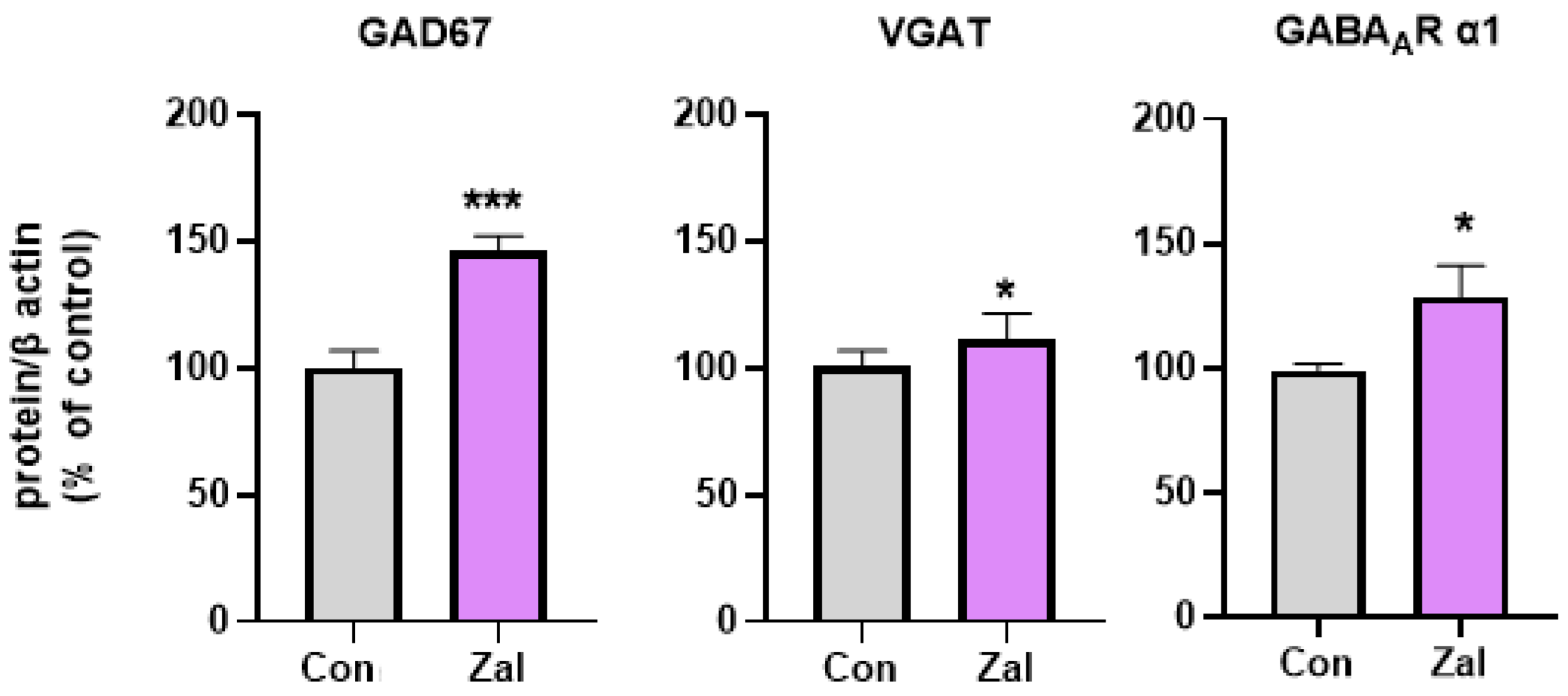
The statistical analysis revealed a significant increase or trend towards higher hippocampal protein levels in the components of the GABAergic system. A significantly higher expression of GAD67 (t (9) = 5.1140, p = 0.00063, Supplementary Figure S1) was observed in the zaleplon-treated rats compared to the control group. Concerning the α1 subunit of the GABAA receptor and the VGAT protein, the statistics revealed a trend toward increased levels of these proteins compared to the control rats ((t (10) = 2.2190, p = 0.050) and (t (8) = 2.2657, p = 0.053, respectively), Supplementary Figure S1). These findings suggest upregulatory effects of prolonged zaleplon treatment on several components of the GABAergic system in the rat hippocampus (Figure 2, Supplementary Figure S1).
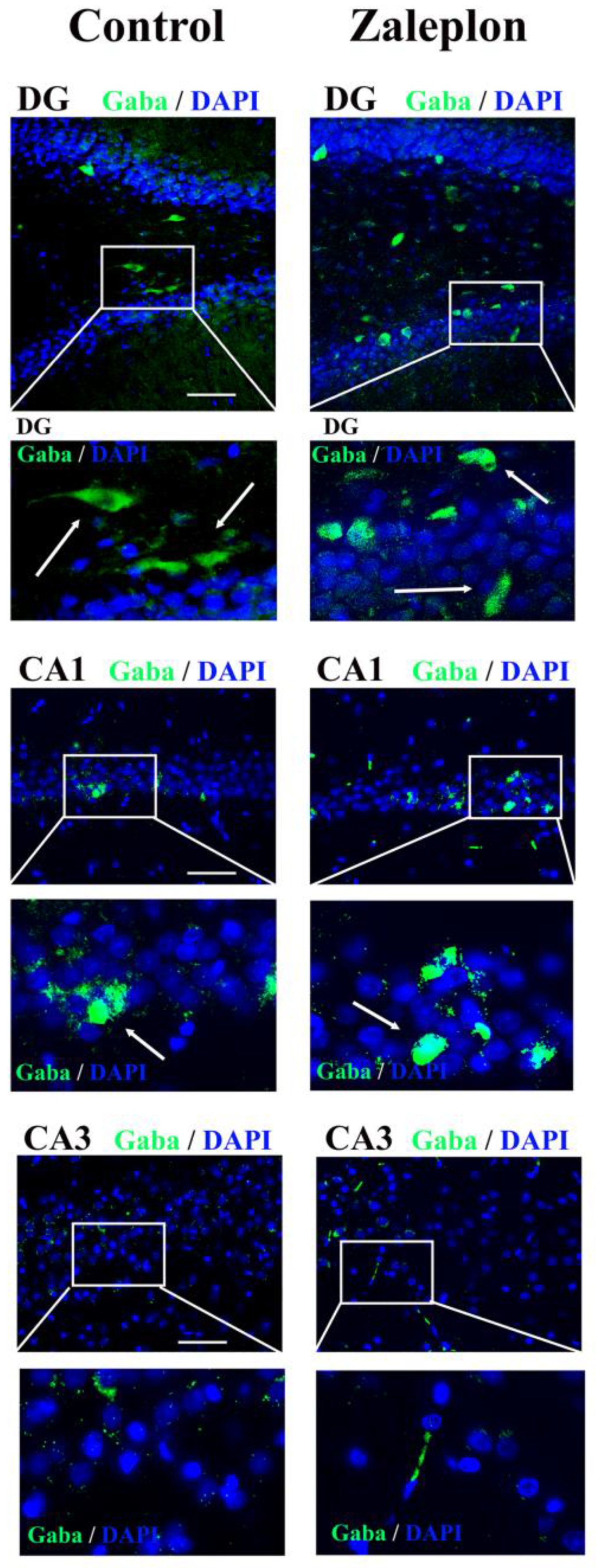
Since the Western blot analysis revealed the increased expression of GAD67 and VGAT proteins in the synaptosomal fraction, immunohistochemical staining was performed to investigate the localization of GABA+ cells in the dorsal hippocampus (involved in learning, memory, sleep, etc.) using an anti-GABA antibody. GABA+ cells (green) were observed in the DG, CA1, and CA3 regions. The treatment with zaleplon increased the expression of GABA in the DG and CA1 regions, while GABA expression in the CA3 region remained unchanged (Figure 3). However, despite the significant differences in GABA expression in the specific hippocampal regions between the control and zaleplon–treated rats, the statistical analysis could not confirm significance due to the small number of samples (n = 3).
The statistical analysis indicated a significant increase in all the studied components of the glutamatergic system in the zaleplon-treated rats compared to the control group: vGlut1 (t (10) = 3.6056, p = 0.0048), NR1 (t (9) = 3,9789, p = 0.0032), NR2A (t (10) = 2,4564, p = 0.034), and NR2B (t (10) = 2.3640, p = 0.0397) (Figure 4, Supplementary Figure S1). On the other hand, A1R protein expression was significantly decreased in the hippocampus of the rats treated with zaleplon compared to the control group (t (8) = 3.3703, p = 0.0098, Supplementary Figure S1).

4. Discussion

Although there are numerous studies on the pharmacokinetics and pharmacodynamics of Z drugs, relatively few studies have addressed the changes in the components underlying their mechanism of action. In this study, we report, for the first time, changes in the expression of synaptic proteins involved in GABAergic and glutamatergic signaling in the rat hippocampus after 5 days of treatment with zaleplon. Importantly, we point out the significant role of the adenosine receptor in the mechanism of action of zaleplon, reflecting the complexity of the interaction of neurotransmitter systems in the brain.
Zaleplon is primarily indicated for the treatment of acute and chronic insomnia, a disorder associated with low GABA levels or impaired GABAergic transmission [11,24,25]. Specifically, magnetic resonance spectroscopy revealed that the overall levels of GABA in the brain regions of patients with insomnia were lower than in the healthy controls [26,27,28]. A significant decrease in the GABA concentrations in the discrete brain regions was also associated with sleep deprivation in rats [29]. Consistent with the sedative–hypnotic mechanism of zaleplon action, the results of our study suggest increased GABA synthesis and its packaging into vesicles, as evidenced by higher levels of the GAD67 protein and GABA immunostaining and VGAT proteins, respectively. Previous studies reported increased levels of GABA in the whole brain and plasma of healthy rabbits after administration of both low and high doses of zaleplon [30], whereas the same effect was observed in the whole brain of rats at the high dose of zaleplon (4 mg/kg, p. o.) but not at the low dose (1 mg/kg, p. o.) [31]. In particular, our results distinguish the more intense immunohistochemical GABA staining in the DG and CA1 hippocampal regions of the zaleplon-treated rats. In addition, increased protein levels of the GABAA receptor α1 subunit, which is responsible for the sedative effect of zaleplon, were detected. This finding could provide an explanation for the safe pharmacological profile of zaleplon, i.e., the rare development of side effects (tolerance, abuse, dependence, etc.) compared to, e.g., benzodiazepines, where prolonged treatment leads to a significant decrease in the α1 subunit of the GABAA receptor, a key event for the occurrence of tolerance [19]. However, based on our results, it is not possible to draw a conclusion as to whether the increase in the number of the α1 subunit of GABAA receptors is a consequence of de novo synthesis or only its relocation from the extracellular space. More specifically, synaptic GABAA receptors are recruited directly from their extrasynaptic counterparts [32,33], revealing a dynamic mechanism by which neurons modulate the number of GABAA receptors located at inhibitory synapses. However, since significantly lower mRNA levels of the α1 and α2 subunits of GABAA receptors were found in the insomnia disorder group [11], we might speculate that zaleplon increases the expression of GABAA receptor subunits that are downregulated in insomnia.
Glutamate, a primary excitatory neurotransmitter and an important intermediate of cell metabolism in the brain, has a far-reaching influence on the sleep−wake regulatory system [34]. Sleep deprivation has been associated with a significant increase in glutamate levels in the hippocampus [35], prefrontal cortex, and thalamus of rats [29]. In addition, several pathologies associated with insomnia are characterized by increased glutamate levels in different brain regions [36]. Surprisingly, we observed increased protein levels of vGlut1 and the NR1, NR2A, and NR2B subunits of the NMDA receptor following treatment with zaleplon. It is known that the hippocampus is a crucial site for learning and memory processes [37] and that the induction of long-term potentiation (LTP), an important cellular mechanism responsible for learning and memory, is typically dependent on the activation of NMDA glutamate receptors. The resulting increase in Ca2+ influx into postsynaptic cells activates a number of biochemical processes in these neurons, including the formation of nitric oxide (NO) [38]. The aforementioned glutamate—NMDA receptor—nitric oxide synthase—NO system plays an essential role in the modulation of learning-related synaptic plasticity such as learning-dependent LTP [39]. A previous study by Samardzic and their co-authors aimed to investigate the behavioral profile of acutely administered different doses of zaleplon in rats using the active avoidance test and the elevated plus maze. Their results suggest that very low doses of zaleplon facilitate retrieval-based learning and produce anxiolytic-like effects without causing sedation [18]. Given that we have demonstrated enhanced components of glutamatergic signaling and in light of the aforementioned role of the NMDA, it will be of interest to further investigate the behavioral consequences of zaleplon treatment and test the intriguing hypothesis of the memory-enhancing effects of zaleplon.
In the search for the underlying mechanism for the detected increase in glutamatergic components, adenosine has emerged. Adenosine can be considered a central excitatory and inhibitory neurotransmitter and an important sleep-regulating substance [40,41]. Adenosine promotes sleep induction via A1 receptors, whereas prolonged sleep deprivation has been associated with an upregulation of the A1 receptors in different regions of the human [42,43] and rat brain [44]. In addition, glutamate released from the presynaptic membrane is known to activate astrocytes, which, in turn, release ATP and adenosine [34]. These molecules bind to purinergic P2Y and adenosine A1 receptors, respectively, activating them and triggering heterosynaptic suppression [45]. In particular, the activation of A1 receptors leads to a combined effect of presynaptic inhibition of glutamate release and reduced activation and surface expression of postsynaptic NMDA receptors [16,34], thereby suppressing neuronal hyperexcitability. We were able to demonstrate significantly reduced protein levels of A1 receptors in the hippocampus following prolonged zaleplon administration. This evidence of significantly reduced A1 receptor protein levels may provide an explanation for the increased levels of the proteins involved in glutamatergic signaling and has raised a new question to which we are seeking an answer in our ongoing study—the involvement of the adenosine system in the mechanism of action of zaleplon.

5. Conclusions and Future Prospects

To our knowledge, these are the first results reporting molecular changes in the neurotransmitter systems in the hippocampus of naive adult male rats following zaleplon treatment within the therapeutic dose. Considering that zaleplon belongs to the group of hypnotics, our results showing an enhancement of GABAergic signaling by zaleplon are not surprising, as decreased GABA neurotransmission is associated with the insomnia disorder. Unexpectedly, however, we observed increased protein levels of components of glutamatergic signaling in the zaleplon-treated rats. This phenomenon could be explained by a downregulation of the adenosine A1 receptors in the hippocampus, an important modulator of the glutamatergic system, which was also detected after prolonged zaleplon administration. In our ongoing study, we will investigate the effects of the observed zaleplon-induced increase in glutamatergic neurotransmission in the hippocampus and the role of the adenosine system in the mechanism of action of zaleplon as well.

6. Authors’ Considerations and Limitations of the Study

The authors would like to emphasize that there has been very little to no research on molecular studies of Z drugs in general. Therefore, the data presented provide valuable and, to our knowledge, the first information on components of GABAergic and glutamatergic neurotransmission following zaleplon treatment and highlight the adenosine system as an important modulator of zaleplon’s mechanism of action.
The main objective of the study was to investigate the mechanism of action of zaleplon, focusing on relevant neurotransmitter systems. As therapeutic outcomes in pathological conditions were not the focus of the study, animal models of sleep disorders were not used, although it would certainly be relevant to investigate the effect of zaleplon in these pathological conditions. The use of the synaptosomal fraction also limits the conclusions that can be drawn about the proteins studied. Therefore, the inclusion of other tissue fractions in the future would provide more detailed data on the overall process.

Supplementary Materials

The following supporting information can be downloaded at: https://www.mdpi.com/article/10.3390/brainsci13121707/s1, Figure S1: original (unprocessed) images of representative blots.

Author Contributions

Conceptualization, J.M. and J.S.; formal analysis, J.M.; investigation, J.M., M.Z.K., S.I. and S.D.; methodology, J.M. and J.S.; visualization, J.M. and S.I.; writing—original draft, J.M.; writing—review and editing, J.S., M.Z.K., S.I., S.D., T.M., M.R. and D.S.S. The results on A1R were part of the following master thesis, under the supervision of J.M. and S.D.: Mackic I. Effect of repeated zaleplon treatment on the expression of adenosine A1 and A2A receptors in the hippocampus of Wistar rats. Master thesis, Faculty of Biology, University of Belgrade, Belgrade, 2023. All authors have read and agreed to the published version of the manuscript.

Funding

This work was financially supported by the Ministry of Science, Technological Development and Innovation of the Serbia, with contract 451-03-47/2023-01/200017.

Institutional Review Board Statement

All the experimental procedures were approved by the Ethics Committee for the Use of Laboratory Animals of Vinca Institute of Nuclear Sciences—National Institute of the Serbia, University of Belgrade, Belgrade, Serbia (decision issued by the Ministry of Agriculture, Forestry and Water Management, number: 323-07-06688/2020-05, date: 15.06.2020.) following compliance with the European Communities Council Directive (2010/63/EU) for animal experiments, according to the guidelines of the EU-registered Serbian Laboratory Animal Science Association, a member of the Federation of the European Laboratory Animal Science Associations.

Informed Consent Statement

Not applicable.

Data Availability Statement

The data are contained within the article.

Acknowledgments

Zaleplon was kindly donated by Belupo d.d., Croatia. The figures were partly generated using Servier Medical Art, provided by Servier, licenced under a Creative Commons Attribution 3.0 un-ported license.

Conflicts of Interest

The authors declare no conflict of interest. The funders had no role in the following: in the design of the study; in the collection, analyses, or interpretation of the data; in the writing of the manuscript; or in the decision to publish the results.

References

  1. Schifano, F.; Chiappini, S.; Corkery, J.M.; Guirguis, A. An Insight into Z-Drug Abuse and Dependence: An Examination of Reports to the European Medicines Agency Database of Suspected Adverse Drug Reactions. Int. J. Neuropsychopharmacol. 2019, 22, 270–277. [Google Scholar] [CrossRef]
  2. Palagini, L.; Bianchini, C. Pharmacotherapeutic management of insomnia and effects on sleep processes, neural plasticity, and brain systems modulating stress: A narrative review. Front. Neurosci. 2022, 16, 893015. [Google Scholar] [CrossRef] [PubMed]
  3. Ghit, A.; Assal, D.; Al-Shami, A.S.; Hussein, D.E.E. GABAA receptors: Structure, function, pharmacology, and related disorders. J. Genet. Eng. Biotechnol. 2021, 19, 123. [Google Scholar] [CrossRef] [PubMed]
  4. Atkin, T.; Comai, S.; Gobbi, G. Drugs for Insomnia beyond Benzodiazepines: Pharmacology, Clinical Applications, and Discovery. Pharmacol. Rev. 2018, 70, 197–245. [Google Scholar] [CrossRef]
  5. Noguchi, H.; Kitazumi, K.; Mori, M.; Shiba, T. Effect of zaleplon on learning and memory in rats. Naunyn-Schmiedeberg’s Arch. Pharmacol. 2002, 366, 183–188. [Google Scholar] [CrossRef]
  6. Troy, S.M.; Lucki, I.; Unruh, M.A.; Cevallos, W.H.; Leister, C.A.; Martin, P.T.; Furlan, P.M.; Mangano, R. Comparison of the effects of zaleplon, zolpidem, and triazolam on memory, learning, and psychomotor performance. J. Clin. Psychopharmacol. 2000, 20, 328–337. [Google Scholar] [CrossRef] [PubMed]
  7. Verster, J.C.; Volkerts, E.R.; Schreuder, A.H.C.M.L.; Eijken, E.J.E.; van Heuckelum, J.H.G.; Veldhuijzen, D.S.; Verbaten, M.N.; Paty, I.; Darwish, M.; Danjou, P.; et al. Residual Effects of Middle-of-the-Night Administration of Zaleplon and Zolpidem on Driving Ability, Memory Functions, and Psychomotor Performance. J. Clin. Psychopharmacol. 2002, 22, 576. [Google Scholar] [CrossRef]
  8. Guo, F.; Yi, L.; Zhang, W.; Bian, Z.-J.; Zhang, Y.-B. Association between Z Drugs Use and Risk of Cognitive Impairment in Middle-Aged and Older Patients with Chronic Insomnia. Front. Hum. Neurosci. 2021, 15, 775144. [Google Scholar] [CrossRef]
  9. Saper, C.B.; Fuller, P.M. Wake–sleep circuitry: An overview. Curr. Opin. Neurobiol. 2017, 44, 186–192. [Google Scholar] [CrossRef]
  10. Samardzic, J.; Jadzic, D.; Hencic, B.; Jancic, J.; Strac, D.S. Introductory Chapter: GABA/Glutamate Balance: A Key for Normal Brain Functioning. In GABA and Glutamate—New Developments in Neurotransmission Research; IntechOpen: London, UK, 2018; Available online: https://www.intechopen.com/state.item.id (accessed on 21 December 2022).
  11. Xiang, T.; Liao, J.; Cai, Y.; Fan, M.; Li, C.; Zhang, X.; Li, H.; Chen, Y.; Pan, J. Impairment of GABA inhibition in insomnia disorders: Evidence from the peripheral blood system. Front. Psychiatry 2023, 14, 1134434. [Google Scholar] [CrossRef]
  12. Liu, Y.; Chen, J.; Li, X.; Zhou, X.; Hu, Y.; Chu, S.; Peng, Y.; Chen, N. Research progress on adenosine in central nervous system diseases. CNS Neurosci. Ther. 2019, 25, 899–910. [Google Scholar] [CrossRef]
  13. Rivkees, S.A.; Price, S.L.; Zhou, F.C. Immunohistochemical detection of A1 adenosine receptors in rat brain with emphasis on localization in the hippocampal formation, cerebral cortex, cerebellum, and basal ganglia. Brain Res. 1995, 677, 193–203. [Google Scholar] [CrossRef]
  14. Rebola, N.; Pinheiro, P.C.; Oliveira, C.R.; Malva, J.O.; Cunha, R.A. Subcellular localization of adenosine A(1) receptors in nerve terminals and synapses of the rat hippocampus. Brain Res. 2003, 987, 49–58. [Google Scholar] [CrossRef]
  15. Rombo, D.M.; Ribeiro, J.A.; Sebastião, A.M. Hippocampal GABAergic transmission: A new target for adenosine control of excitability. J. Neurochem. 2016, 139, 1056–1070. [Google Scholar] [CrossRef]
  16. Deng, Q.; Terunuma, M.; Fellin, T.; Moss, S.J.; Haydon, P.G. Astrocytic activation of A1 receptors regulates the surface expression of NMDA receptors through a src kinase dependent pathway. Glia 2011, 59, 1084–1093. [Google Scholar] [CrossRef]
  17. Nam, H.W.; McIver, S.R.; Hinton, D.J.; Thakkar, M.M.; Sari, Y.; Parkinson, F.E.; Haydon, P.G.; Choi, D.-S. Adenosine and Glutamate Signaling in Neuron-Glial interactions: Implications in Alcoholism and Sleep Disorders. Alcohol. Clin. Exp. Res. 2012, 36, 1117–1125. [Google Scholar] [CrossRef] [PubMed]
  18. Samardzic, J.; Zekovic, J.; Hencic, B.; Vucevic, D.; Jancic, J.; Švob Štrac, D. ACNP 58th Annual Meeting: Poster Session III. Neuropsychopharmacology 2019, 44, 385–538. [Google Scholar]
  19. Zaric Kontic, M.; Dragic, M.; Martinovic, J.; Mihajlovic, K.; Brkic, Z.; Mitrovic, N.; Grkovic, I. Prolonged Alprazolam Treatment Alters Components of Glutamatergic Neurotransmission in the Hippocampus of Male Wistar Rats-The Neuroadaptive Changes following Long-Term Benzodiazepine (Mis)Use. Pharmaceuticals 2023, 16, 331. [Google Scholar] [CrossRef] [PubMed]
  20. Spanò, G.; Weber, F.D.; Pizzamiglio, G.; McCormick, C.; Miller, T.D.; Rosenthal, C.R.; Edgin, J.O.; Maguire, E.A. Sleeping with Hippocampal Damage. Curr. Biol. 2020, 30, 523–529.e3. [Google Scholar] [CrossRef] [PubMed]
  21. Avchalumov, Y.; Mandyam, C.D. Plasticity in the Hippocampus, Neurogenesis and Drugs of Abuse. Brain Sci. 2021, 11, 404. [Google Scholar] [CrossRef] [PubMed]
  22. Zaric, M.; Drakulic, D.; Stojanovic, I.G.; Mitrovic, N.; Grkovic, I.; Martinovic, J. Regional-specific effects of cerebral ischemia/reperfusion and dehydroepiandrosterone on synaptic NMDAR/PSD-95 complex in male Wistar rats. Brain Res. 2018, 1688, 73–80. [Google Scholar] [CrossRef] [PubMed]
  23. Markwell, M.A.; Haas, S.M.; Bieber, L.L.; Tolbert, N.E. A modification of the Lowry procedure to simplify protein determination in membrane and lipoprotein samples. Anal. Biochem. 1978, 87, 206–210. [Google Scholar] [CrossRef] [PubMed]
  24. Hepsomali, P.; Groeger, J.A.; Nishihira, J.; Scholey, A. Effects of Oral Gamma-Aminobutyric Acid (GABA) Administration on Stress and Sleep in Humans: A Systematic Review. Front. Neurosci. 2020, 14, 923. [Google Scholar] [CrossRef]
  25. Gottesmann, C. GABA mechanisms and sleep. Neuroscience 2002, 111, 231–239. [Google Scholar] [CrossRef]
  26. Park, S.; Kang, I.; Edden, R.A.E.; Namgung, E.; Kim, J.; Kim, J. Shorter sleep duration is associated with lower GABA levels in the anterior cingulate cortex. Sleep Med. 2020, 71, 1–7. [Google Scholar] [CrossRef]
  27. Plante, D.T.; Jensen, J.E.; Schoerning, L.; Winkelman, J.W. Reduced γ-aminobutyric acid in occipital and anterior cingulate cortices in primary insomnia: A link to major depressive disorder? Neuropsychopharmacology 2012, 37, 1548–1557. [Google Scholar] [CrossRef] [PubMed]
  28. Winkelman, J.W.; Buxton, O.M.; Jensen, J.E.; Benson, K.L.; O’Connor, S.P.; Wang, W.; Renshaw, P.F. Reduced brain GABA in primary insomnia: Preliminary data from 4T proton magnetic resonance spectroscopy (1H-MRS). Sleep 2008, 31, 1499–1506. [Google Scholar] [CrossRef]
  29. Kamal, S.M. Pharmacological modulation of brain levels of glutamate and GABA in rats exposed to total sleep deprivation. J. Exp. Pharmacol. 2010, 2, 65–71. [Google Scholar]
  30. Abd-Elrasheed, E.; Nageeb El-Helaly, S.; EL-Ashmoony, M.M.; Salah, S. Brain-targeted intranasal zaleplon solid dispersion in hydrophilic carrier system; 23 full-factorial design and in vivo determination of GABA neurotransmitter. Drug Dev. Ind. Pharm. 2018, 44, 741–749. [Google Scholar] [CrossRef]
  31. El-Naggar, M.E.; El-Fayoumi, H.M.; El-Maraghy, N.N.; El-Eraky, W.I.; El-Shenawy, S.M. Toxicological study of Zaleplon (Anxiolytic drug) in experimental animals. Zagazig J. Pharm. Sci. 2014, 23, 16–33. [Google Scholar] [CrossRef]
  32. Bogdanov, Y.; Michels, G.; Armstrong-Gold, C.; Haydon, P.G.; Lindstrom, J.; Pangalos, M.; Moss, S.J. Synaptic GABAA receptors are directly recruited from their extrasynaptic counterparts. EMBO J. 2006, 25, 4381–4389. [Google Scholar] [CrossRef] [PubMed]
  33. Choi, C.; Smalley, J.L.; Lemons, A.H.S.; Ren, Q.; Bope, C.E.; Dengler, J.S.; Davies, P.A.; Moss, S.J. Analyzing the mechanisms that facilitate the subtype-specific assembly of γ-aminobutyric acid type A receptors. Front. Mol. Neurosci. 2022, 15, 1017404. [Google Scholar] [CrossRef]
  34. He, S.; Zhang, X.; Qu, S. Glutamate, Glutamate Transporters, and Circadian Rhythm Sleep Disorders in Neurodegenerative Diseases. ACS Chem. Neurosci. 2019, 10, 175–181. [Google Scholar] [CrossRef] [PubMed]
  35. Cortese, B.M.; Mitchell, T.R.; Galloway, M.P.; Prevost, K.E.; Fang, J.; Moore, G.J.; Uhde, T.W. Region-specific alteration in brain glutamate: Possible relationship to risk-taking behavior. Physiol. Behav. 2010, 99, 445–450. [Google Scholar] [CrossRef]
  36. Meyerhoff, D.J.; Mon, A.; Metzler, T.; Neylan, T.C. Cortical Gamma-Aminobutyric Acid and Glutamate in Posttraumatic Stress Disorder and Their Relationships to Self-Reported Sleep Quality. Sleep 2014, 37, 893–900. [Google Scholar] [CrossRef]
  37. Wang, S.; Pan, D.-X.; Wang, D.; Wan, P.; Qiu, D.-L.; Jin, Q.-H. Nitric oxide facilitates active avoidance learning via enhancement of glutamate levels in the hippocampal dentate gyrus. Behav. Brain Res. 2014, 271, 177–183. [Google Scholar] [CrossRef] [PubMed]
  38. Andrabi, S.M.; Sharma, N.S.; Karan, A.; Shahriar, S.M.S.; Cordon, B.; Ma, B.; Xie, J. Nitric Oxide: Physiological Functions, Delivery, and Biomedical Applications. Adv. Sci. 2023, 10, 2303259. [Google Scholar] [CrossRef]
  39. Cossenza, M.; Socodato, R.; Portugal, C.C.; Domith, I.C.L.; Gladulich, L.F.H.; Encarnação, T.G.; Calaza, K.C.; Mendonça, H.R.; Campello-Costa, P.; Paes-de-Carvalho, R. Chapter Five—Nitric Oxide in the Nervous System: Biochemical, Developmental, and Neurobiological Aspects. In Vitamins & Hormones; Litwack, G., Ed.; Nitric Oxide; Academic Press: Cambridge, MA, USA, 2014; Volume 96, pp. 79–125. Available online: https://www.sciencedirect.com/science/article/pii/B9780128002544000052 (accessed on 4 December 2023).
  40. Reichert, C.F.; Deboer, T.; Landolt, H.-P. Adenosine, caffeine, and sleep–wake regulation: State of the science and perspectives. J. Sleep Res. 2022, 31, e13597. [Google Scholar] [CrossRef]
  41. Lazarus, M.; Oishi, Y.; Bjorness, T.E.; Greene, R.W. Gating and the Need for Sleep: Dissociable Effects of Adenosine A1 and A2A Receptors. Front. Neurosci. 2019, 13, 740. [Google Scholar] [CrossRef]
  42. Elmenhorst, D.; Meyer, P.T.; Winz, O.H.; Matusch, A.; Ermert, J.; Coenen, H.H.; Basheer, R.; Haas, H.L.; Zilles, K.; Bauer, A. Sleep Deprivation Increases A1 Adenosine Receptor Binding in the Human Brain: A Positron Emission Tomography Study. J. Neurosci. 2007, 27, 2410–2415. [Google Scholar] [CrossRef]
  43. Elmenhorst, D.; Elmenhorst, E.-M.; Hennecke, E.; Kroll, T.; Matusch, A.; Aeschbach, D.; Bauer, A. Recovery sleep after extended wakefulness restores elevated A1 adenosine receptor availability in the human brain. Proc. Natl. Acad. Sci. USA 2017, 114, 4243–4248. [Google Scholar] [CrossRef] [PubMed]
  44. Elmenhorst, D.; Basheer, R.; McCarley, R.W.; Bauer, A. Sleep deprivation increases A1 adenosine receptor density in the rat brain. Brain Res. 2009, 1258, 53–58. [Google Scholar] [CrossRef] [PubMed]
  45. Zhang, J.; Wang, H.; Ye, C.; Ge, W.; Chen, Y.; Jiang, Z.; Wu, C.; Poo, M.; Duan, S. ATP released by astrocytes mediates glutamatergic activity-dependent heterosynaptic suppression. Neuron 2003, 40, 971–982. [Google Scholar] [CrossRef] [PubMed]
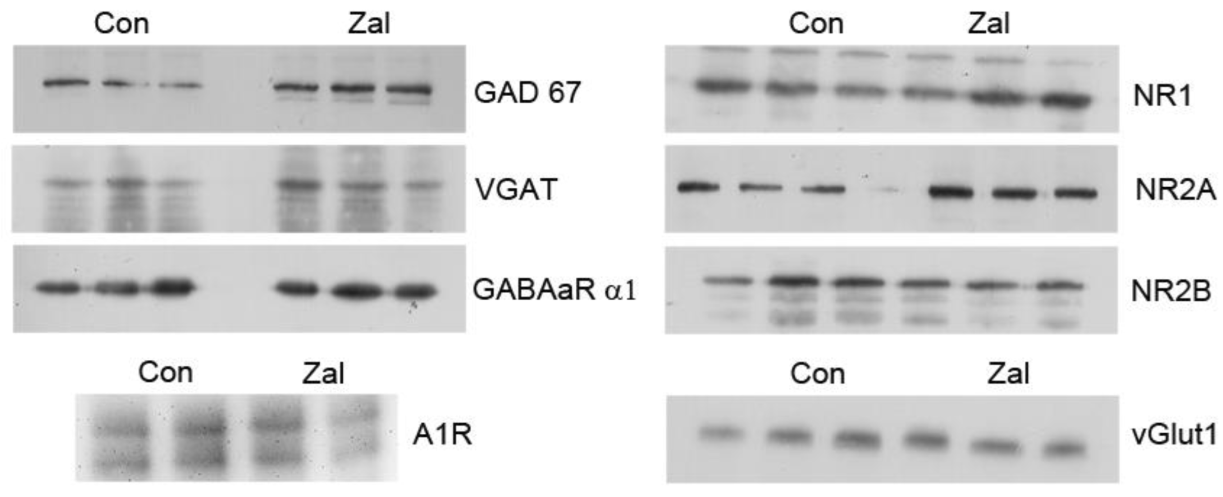
Figure 1. Representative Western blot analysis for the synaptosomal components of GABAergic and glutamatergic transmission and the adenosine A1R receptor. GABAAR α1—α1 subunit of the GABAA receptor; vGlut1—vesicular glutamate transporter 1; NR1, NR2A, NR2B—subunits of the N-methyl-d-aspartate (NMDA) glutamate receptor; GAD67—67 kDa isoform of glutamate decarboxylase; VGAT—vesicular GABA transporter; A1R—adenosine receptor; and β-actin—beta actin.
Brainsci 13 01707 g001
Figure 2. Protein levels of components of the GABAergic systems after prolonged treatment with 0.625 mg/kg zaleplon (Zal). Asterisks indicate a significant increase in the studied parameters in the Zal group compared to the Con group (* p < 0.05, *** p < 0.001). GABAAR α1—α1 subunit of the GABAA receptor; vGlut1—vesicular glutamate transporter 1; GAD67—67 kDa isoform of glutamate decarboxylase; VGAT—vesicular GABA transporter; and β-actin—beta actin.
Brainsci 13 01707 g002
Figure 3. Immunohistochemical staining of the cryosectioned brains of rats treated with saline and 0.625 mg/kg zaleplon with anti-GABA antibody (green) in the dentate gyrus (DG) region and the CA1 and CA3 regions of the hippocampus. The cell nuclei were stained with DAPI (blue). GABA+ cells are marked with arrows.
Brainsci 13 01707 g003
Figure 4. Protein levels of the components of the glutamatergic and adenosine systems after prolonged treatment with 0.625 mg/kg zaleplon (Zal). Asterisks indicate a significant increase in the studied parameters in the Zal group compared to the Con group (* p < 0.05, ** p < 0.01). vGlut1—vesicular glutamate transporter 1; NR1, NR2A, NR2B—subunits of the N-methyl-d-aspartate (NMDA) glutamate receptor; A1R—adenosine receptor; and β-actin—beta actin.
Brainsci 13 01707 g004
Table 1. List of primary and secondary antibodies used for the Western blot analysis.
AntigenManufacturer/Catalog No.Species/Dilution
GABAAR α1Sigma Aldrich, Burlington, MA, USA, G4416rabbit polyclonal, 1:5000
vGlut1Abcam, Cambridge, UK, 134283mouse monoclonal, 1:4000
NR1Cell Signaling Technology, Danvers, MA, USA, # 5704rabbit polyclonal, 1:1000
NR2AMerck Millipore, Burlington, MA, USA, # 07-632rabbit polyclonal, 1:1000
NR2BAbcam, Cambridge, UK, 93610mouse monoclonal, 1:4000
GAD67Millipore, Burlington, MA, MAB5406mouse monoclonal, 1:1000
VGATSanta Cruz Biotechnology, Dallas, TX, USA, sc-393373mouse monoclonal, 1:1000
A1RAlomone labs, Jerusalem, Israelrabbit polyclonal, 1:1000
β-actinThermo Fisher Scientific, Waltham, MA, USA; PA1-21167rabbit polyclonal, 1:5000
mouse IgGR&D systems, bio-techne, Minneapolis, MN, USA; HAF007goat polyclonal, 1:10,000
rabbit IgGInvitrogen, Waltham, MA, USA; # 31460goat polyclonal, 1:10,000
Note: GABAAR α1—α1 subunit of the GABAA receptor; vGlut1—vesicular glutamate transporter 1; NR1, NR2A, NR2B—subunits of the N-methyl-d-aspartate (NMDA) glutamate receptor; GAD67—67 kDa isoform of glutamate decarboxylase; VGAT—vesicular GABA transporter; A1R—adenosine receptor; and β-actin—beta actin.
Disclaimer/Publisher’s Note: The statements, opinions and data contained in all publications are solely those of the individual author(s) and contributor(s) and not of MDPI and/or the editor(s). MDPI and/or the editor(s) disclaim responsibility for any injury to people or property resulting from any ideas, methods, instructions or products referred to in the content.