Communication

Hippocampal Resting State Functional Connectivity Associated with Physical Activity in Periadolescent Children

1
Department of Neurological Sciences, University of Nebraska Medical Center, Omaha, NE 68198, USA
2
Department of Psychology, University of Nebraska Omaha, Omaha, NE 68182, USA
*
Author to whom correspondence should be addressed.
Brain Sci. 2023, 13(11), 1558; https://doi.org/10.3390/brainsci13111558
Submission received: 22 September 2023 / Revised: 31 October 2023 / Accepted: 1 November 2023 / Published: 7 November 2023
(This article belongs to the Section Developmental Neuroscience)

Abstract

:
Periadolescence is a neurodevelopmental period characterized by structural and functional brain changes that are associated with cognitive maturation. The development of the functional connectivity of the hippocampus contributes to cognitive maturation, especially memory processes. Notably, hippocampal development is influenced by lifestyle factors, including physical activity. Physical activity has been associated with individual variability in hippocampal functional connectivity. However, this relationship has not been characterized in a developmental cohort. In this study, we aimed to fill this gap by investigating the relationship between physical activity and the functional connectivity of the hippocampus in a cohort of periadolescents aged 8–13 years (N = 117). The participants completed a physical activity questionnaire, reporting the number of days per week they performed 60 min of physical activity; then, they completed a resting-state functional MRI scan. We observed that greater physical activity was significantly associated with differences in hippocampal functional connectivity in frontal and temporal regions. Greater physical activity was associated with decreased connectivity between the hippocampus and the right superior frontal gyrus and increased connectivity between the hippocampus and the left superior temporal sulcus. Capturing changes in hippocampal functional connectivity during key developmental periods may elucidate how lifestyle factors including physical activity influence brain network connectivity trajectories, cognitive development, and future disease risk.

1. Introduction

Throughout childhood and into early adulthood, the brain undergoes significant structural and functional changes, including altered connectivity within and between brain networks. Resting-state functional connectivity (rs-FC), generally characterized using functional magnetic resonance imaging (fMRI), provides valuable insights regarding brain function, organization, and associated cognitive processes and behaviors [1,2,3,4,5,6,7]. Maturation of functional brain networks is important for the development of more complex cognitive abilities that improve between childhood and adolescence, or the periadolescent epoch [8]. For example, the development of cognitive abilities, including memory and attention, is related to changes in rs-FC throughout periadolescence. Brain regions such as the hippocampus and extended brain networks important for memory and attention demonstrate increased within-network connectivity and decreased between-network connectivity that support better memory and improved attentional abilities in periadolescence [9,10]. It is well established that the brain undergoes significant alterations in rs-FC with development and aging, and an emerging research theme involves studying how the trajectory of these changes in healthy development and disease is influenced by modifiable lifestyle factors, such as physical activity and fitness.
The structure and function of the hippocampus, a brain region necessary for normal memory, is particularly influenced by physical activity and fitness levels during periadolescence [11,12,13,14,15]. Fitness, the cardiorespiratory conditioning that occurs as a result of exercise, is associated with hippocampal volume [16,17,18,19,20] in addition to functional connectivity between the hippocampus and other brain networks in periadolescent children [21,22]. Daily physical activity, a key contributor to fitness, is also associated with changes in interhemispheric functional connectivity in periadolescent children [23,24,25]. However, less is understood about how physical activity is associated with the functional connectivity of the hippocampus and the rest of the brain throughout periadolescence, as these relationships have not previously been tested in a healthy, typically developing cohort.
In order to fill this gap in the literature, we measured the rs-FC of the hippocampus, then tested its association with self-reported daily physical activity levels in a cohort of periadolescent children. Other techniques, including volumetric MRI and task-based fMRI as well as EEG, have been utilized to investigate similar questions. However, rs-FC offers unique insight into the brain’s intrinsic functional organization [16,24,26,27,28,29]. While a previous study investigated the relationship between hippocampal rs-FC and fitness levels, physical activity is a distinct construct that has not been investigated in relation to hippocampal rs-FC [21]. In the current study, rs-FC and self-reported physical activity data were measured in a new sample of periadolescent children (aged 8–13 years) with the aim of investigating how hippocampal rs-FC varies as a function of physical activity. We hypothesized that greater levels of physical activity would be associated with hippocampal rs-FC patterns that reflect increased within-network and decreased between-network connectivity, that is, patterns typically associated with more efficient brain networks [1,2,3,6,7,8]. Investigating this relationship may lead to insights into how physical activity influences functional brain networks throughout development. Advances in this area could have widespread effects spanning from public policy changes to disease prevention recommendations that could influence brain health not only during development but throughout the entire lifespan.

2. Materials and Methods

2.1. Participants

The participants in the current study came from a cohort of periadolescent children enrolled in the Polygenic Risk for Alzheimer’s disease in Nebraska Kids (PRANK) protocol, which is an observational study on brain and cognitive development. Healthy, typically developing periadolescent children aged 8–13 years (M = 10.92, SD = 1.61) from the Omaha, Nebraska, area assented to participate in the study after the research team obtained parent/guardian informed consent according to the University of Nebraska Medical Center’s Institutional Review Board guidelines. Participants were included if they had normal or corrected-to-normal vision and the ability to assent to and complete assessments in English. Participants were excluded if they had a history of drug or alcohol abuse, psychiatric disease, or neurological or developmental disease or had contraindications for neuroimaging (presence of an implant, orthodonture, etc.). The sample included 117 periadolescent children with sufficient low-motion MRI data that allowed for data processing and analysis (details below) [30]. The cross-sectional sample analyzed here contained 117 children (57F, 60M). The average age for girls (M = 11.37 years) was significantly greater than the average age for boys (M = 10.49 years) in this sample (t(115) = 3.09; p = 0.002). Age was calculated with respect to the day the MRI scan was completed. Demographic information is included in Table 1.

2.2. Physical Activity

Physical activity was assessed with a self-report measure adapted from the Adolescent Brain and Cognitive Development (ABCD) Study’s Youth Risk Behavior survey (modeled after the Center for Disease Control’s survey) [31,32]. Specifically, questions related to physical activity behavior were included in the questionnaire. The first item on this questionnaire asked child participants “During the past 7 days, on how many days were you physically active for a total of at least 60 min per day? (time you spent in any kind of physical activity that increased your heart rate and made you breathe hard part of the time)”. Each child’s response to this question was utilized as the variable of interest in the current analysis. Responses ranged from “0 days” up to “7 days” per week. Scalar values based this response were mean-centered and entered as the primary covariate of interest in the neuroimaging analysis.

2.3. MRI

2.3.1. Procedures

All data were collected from participants in Omaha, Nebraska, at the University of Nebraska Medical Center Core for Advanced Magnetic Resonance Imaging (CAMRI, RRID: SCR_022468). After receiving consent from parents and children, parents provided screening information for MRI compatibility. Data were collected with a Siemens 3T Prisma MRI scanner using a 32-channel head coil. The protocol for this scan was adapted from the Lifespan Human Connectome Project in Development protocol [33]. All participants were comfortably positioned in a supine position in the scanner and instructed to hold as still as possible.

2.3.2. Scanning Parameters

Structural and functional MRI data were collected using the following parameters: T1 MPRAGE [TE = 2.2 ms, TR = 2400 ms, 0.8 mm isotropic voxel, FOV = 256 mm, slice thickness = 0.8 mm, slices = 208, flip angle = 8°, acquisition time = 6 min 38 s], T2w SPACE [TE = 563 ms, TR = 3200, 0.8 mm isotropic voxel, FOV = 256 mm, slice thickness = 0.8 mm, slices = 208, acquisition time = 5 min 57 s] and T2* BOLD Resting State [2D multiband gradient-recalled echo (GRE) echo-planar image (EPI) sequence, TR = 800 ms, TE = 37 ms, flip angle = 52°, 2.0 mm isotropic voxel, acquisition time = 15 min 20 s, multiband acceleration factor = 8]. During the resting state scan, participants were instructed to stay awake, remain still, keep their eyes on a fixation cross, and keep their minds clear.

2.3.3. Data Processing and Analysis

MRI data were preprocessed using a minimal preprocessing pipeline developed by the Human Connectome Project (HCP) [34]. Briefly, the original T1 and T2 structural images were processed via the following steps: (1) distortion was corrected; (2) images were aligned and averaged between repeated runs; (3) images were aligned with the MNI space template using a rigid body, 6-degree-of-freedom transform; (4) readout distortion was removed; (5) images were corrected for inhomogeneity; (6) the bias field was estimated and corrected; and (7) FreeSurfer’s recon-all pipeline was applied to the data (RRID:SCR_001847; Version 6.0.0). Then, an internally developed pipeline automatically generated a bilateral hippocampal region of interest seed composed of left and right hippocampal masks. The output from this processing was then utilized in the fMRISurface pipeline to process the volume timeseries in MNI space in order to generate a CIFTI dense timeseries. The resulting resting state MRI data were processed to address well-characterized confounds related to motion. Realignment parameters from co-registration of rs-fMRI volumes were first low-pass-filtered to exclude motion artifacts related to respiration [30], and a threshold of 0.2 mm was used to identify rs-fMRI volumes with a relatively high level of motion for censoring during subsequent processing. Then, a single-step regression procedure implemented in AFNI software (RRID:SCR_005927; version 21.1.07) was used to simultaneously censor high-motion volumes identified as described above, apply a band pass filter (0.01–0.2 Hz), and remove effects related to timeseries covariates of no interest, including motion regressors and derivatives, core white matter, CSF, and global signal. Volumetric timeseries data and surface timeseries data were smoothed using a 4 mm FWHM kernel (3D- and 2D surface-constrained, respectively). This CIFTI timeseries was then statistically analyzed with the covariate of interest, physical activity.

2.3.4. Statistical Analysis
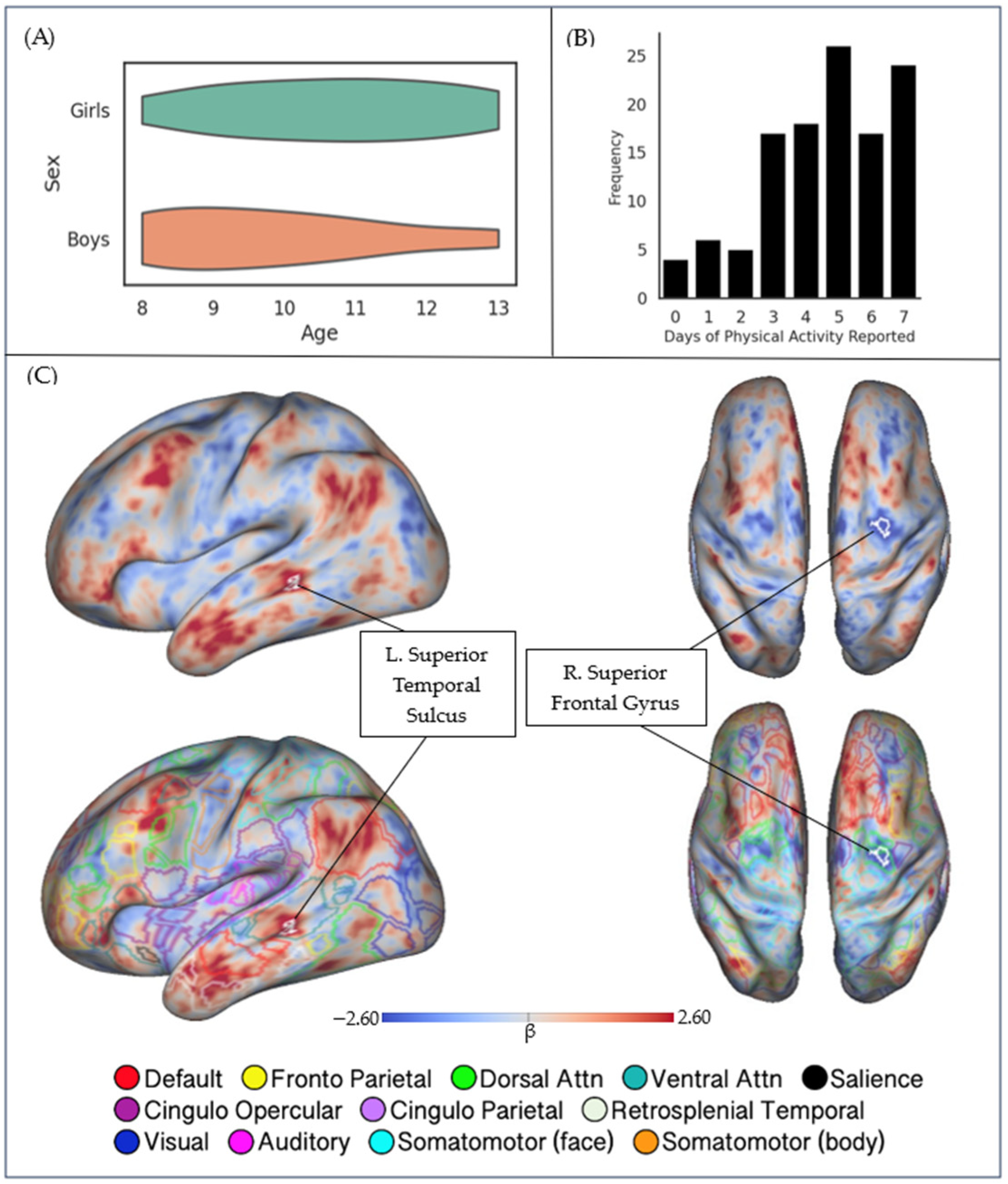
Statistical tests of voxelwise resting-state functional connectivity and covariate values were conducted. Voxelwise tests using one-sample, two-sided t-tests with linear modeling for covariates were implemented using AFNI’s 3dttest++ utility [35]. The primary covariate of interest was the number of reported days of physical activity. Additional covariates included age, sex, an age × sex interaction term, and median framewise displacement for resting state EPI (estimated from realignment parameters generated during preprocessing). Field-standard, two-step thresholding procedures were followed to ensure the rigor and reproducibility of our analysis [36]. Specifically, a voxelwise threshold was applied (p < 0.001) in order to identify significant clusters that covaried with the bilateral hippocampus seed as a function of physical activity, and then a spatial clusterwise threshold was utilized to identify statistically significant results, with alpha = 0.05. The analysis was restricted to surface targets, as this has been shown to increase the specificity of cortical activation patterns and connectivity results [37]. The covariate maps and significant clusters generated by this analysis were visualized using Connectome Workbench. An overlay of Gordon’s 333 cortical network parcellation map allowed for localization of significant clusters within networks [38,39].

3. Results

3.1. Behavioral Data

The participants, on average, reported having 4.61 days per week with 60 or more minutes of physical activity (SD = 1.91). The amount of reported physical activity was not significantly associated with age (r = −0.09, p = 0.36), and no differences in physical activity were found between males and females (t(114) = 1.03, p = 0.31). Distribution of age by sex and frequency of days of physical activity reported can be seen in Figure 1A,B.

3.2. Neuroimaging Data

Our analysis of covariance between physical activity and hippocampal rs-FC indicated that increased physical activity was associated with regional differences in hippocampal rs-FC. Specifically, statistically significant clusters were identified in two brain regions. The right superior frontal gyrus (MNI X = +30.7, Y = −3.8, Z = +50.1; area = 48.79 mm2) covaried negatively with physical activity; that is, a greater number of days of reported physical activity was associated with decreased functional connectivity between the hippocampus and the right superior frontal gyrus (rSFG). Meanwhile, the left superior temporal sulcus (MNI X = −57.3. Y = −35.4, Z = −3.2; area = 52.04 mm2) covaried positively with physical activity; that is, a greater number of days of reported physical activity was associated with increased functional connectivity between the hippocampus and the left superior temporal sulcus (lSTS). The locations of significant clusters are detailed in Table 2 and Figure 1C. The age–sex interaction term did not show any statistically significant covariance with hippocampal rs-FC, indicating that any age-sex differences did not influence the outcome of interest.
To provide additional context for these findings, the focal clusters of covariance were interpreted using a well-established functional parcellation of the human brain. An overlay of Gordon’s 333 network parcellation map revealed that the negatively covarying rSFG cluster fell within the territory of the dorsal attention network (DAN), and the positively covarying lSTS cluster fell in the territory of the ventral attention network (VAN). Bilateral, whole-brain maps of hippocampal rs-FC are displayed in Supplemental Figures S1 and S2.

4. Discussion

This study aimed to investigate the association between physical activity and resting state functional connectivity of the hippocampus in a developmental cohort of periadolescent children. The rationale for studying this relationship is based on the significant age-related change in hippocampal functional connectivity during development, a period that is especially sensitive to the influence of modifiable factors including physical activity and fitness [9,10,11]. In the current study, hippocampal rs-FC was significantly negatively associated with regions of the frontal lobe as a function of greater self-reported physical activity and positively associated with regions within the temporal lobe. Our hypothesis, that greater levels of physical activity are associated with increased within-network and decreased between-network functional connectivity in children, was partially supported by the pattern reported here.
Previous studies that have investigated associations between hippocampal rs-FC, physical activity, and fitness have reported strikingly similar patterns of covariation. A study involving Spanish adolescents reported regional similarities in hippocampal rs-FC but with cardiorespiratory fitness as a covariate [21]. Additional evidence suggests physical activity and fitness play a significant role in attentional network functional connectivity variation in this age group [22]. Research on young adults has also established a significant relationship between hippocampal rs-FC and physical activity and fitness [40,41,42,43,44]. The current study observed hippocampal rs-FC patterns similar to those reported in previous studies on young adults, and it adds to the growing body of evidence that hippocampal rs-FC may be influenced by physical activity during development. Furthermore, future studies expanding upon the 8–13-year-old age range of the current study will be necessary to fully characterize the associations between physical activity, fitness, and functional brain development.
The right frontal and temporal lobe clusters observed in this study are parts of brain regions often associated with the dorsal (DAN) and ventral (VAN) attention networks, respectively [38]. The DAN plays a key role in alerting and attending to relevant visuospatial stimuli, while the VAN is recruited during stimulus-driven tasks in association with short-term memory. Differences in these functional networks are important because attention and memory abilities improve significantly throughout childhood [45]. Increased within-network and decreased between-network connectivity of these networks is associated with greater attentional abilities and increasing age in children [1,46,47,48,49,50]. Furthermore, the phenomenon of a decrease in functional connectivity between the hippocampus and regions strongly associated with the DAN is consistent with a more segregated brain network system, which is characteristic of this developmental period [1,51,52]. Evidence suggests that during development, abundant short-range connections between regions are slowly pruned and replaced by long-range connections that, with progression through childhood, begin to more closely resemble the functional networks in the adult brain [6,7,53,54]. Physical activity may influence this developmental process: we observed that periadolescent children reporting greater levels of physical activity displayed a pattern of decreased functional connectivity between the hippocampus (hippocampal memory network) and a significant cluster in the DAN and increased functional connectivity between the hippocampus and the VAN. Based on both the current and prior work, we posit that physical activity plays an important role in the development of hippocampal rs-FC, particularly attention network connectivity with the hippocampus.
Importantly, there are methodological differences between the current study and prior work. For example, much of the current published literature on hippocampal volume and/or functional connectivity focuses on children with high BMI values (body mass index) or lower fitness levels [21,24,44], whereas the current study includes children with a wide range of BMIs (12.75–32.81 kg/m2) and does not include BMI as a variable of interest. While there is evidence that BMI may play a role in hippocampal function [55], there is scientific merit in understanding how these relationships apply to children across the spectrum of BMI and fitness throughout development. Exercise and physical activity influence the hippocampus regardless of BMI in the adult population [15,18], and it is important to understand whether this relationship is robust across there lifespan, beginning in childhood.
The current study is not without its limitations. First, self-reported physical activity is a coarse measure of how much movement and exercise periadolescent children are participating in daily, and quantitative measurements may be more robust. Several studies have used responses to the same questionnaire to test hypotheses associated with physical activity and other brain variables [23,56,57]. Another limitation of our data was that the distribution of reported days of physical activity was negatively skewed such that sedentary children were less well represented in the sample. However, there were four or more children (and associated rs-fMRI datasets) at every response level, and this provided adequate representation for our analysis. Also, our rs-fMRI data-processing approach incorporated global signal regression, a widely used approach intended to address methodological confounds inherent to rs-fMRI data; global signal regression has itself been subject to critique, and an active debate continues in the literature [58,59]. The high-quality brain imaging data and robust statistical thresholds utilized in the current study provide reassurance that our findings were most likely related to true variation in functional connectivity as a function of physical activity. In the future, quantitative measures of physical activity (accelerometry) should be analyzed to replicate these results and elucidate other patterns of rs-FC that could be influenced by these variables. Finally, the current study utilizes an observational design, while much of the currently published literature describes studies using interventional designs. However, our results fit well with the currently published findings within this body of research despite the observational nature of this study.
In summary, this study provides evidence that hippocampal rs-FC is significantly associated with physical activity in periadolescent children. The significant differences in the superior frontal gyrus and the superior temporal sulcus extend to developmental populations the previous findings of an association between physical activity and hippocampal rs-FC. These findings suggest that physical activity may be associated with greater segregation of functional networks. Future studies on rs-FC in periadolescent children employing quantitative measurements of physical activity could further elucidate how these factors influence brain development. Differences in these functional connectivity patterns as a function of physical activity could be related to underlying cognitive functions that depend on hippocampal contributions such as declarative or relational memory abilities. Understanding how modifiable lifestyle factors, including physical activity, influence the trajectory of development in brain regions that are especially vulnerable to neurological disease may have great importance for developing preventative interventional strategies with which to preserve these brain regions and networks. For example, changes in hippocampal rs-FC have been observed in association with a variety of diseases throughout the human lifespan, including childhood heart disease [60], temporal lobe epilepsy [61], and early changes in Alzheimer’s disease (AD) [62,63,64]. AD pathology initially affects the medial temporal lobes and the hippocampus, and, over time, AD affects multiple brain regions and networks, including attention networks; insight into how physical activity might strengthen and/or protect these regions and networks from an early age could be critical for disease prevention later in life.

Supplementary Materials

The following supporting information can be downloaded at https://www.mdpi.com/article/10.3390/brainsci13111558/s1, Figure S1: Bilateral, whole-brain map of mean hippocampal rs-FC; Figure S2: Bilateral, whole-brain map of rs-FC covarying with physical activity.

Author Contributions

Conceptualization, A.H.-W. and D.E.W.; methodology, A.H.-W. and D.E.W.; validation C.P., J.S. and M.R.; formal analysis, A.H.-W.; investigation, A.H.-W., C.P., J.S. and M.R.; resources, D.E.W.; data curation, A.H.-W., C.P., J.S. and M.R.; writing—original draft preparation, A.H.-W.; writing—review and editing, A.H.-W., C.P., J.S., M.R. and D.E.W.; visualization, A.H.-W.; supervision, D.E.W.; funding acquisition, D.E.W. All authors have read and agreed to the published version of the manuscript.

Funding

This research was funded by a grant from the National Institute on Aging, R01 AG064247, awarded to D.E.W., R01 AG064247-04S1 to M.R., and T32 AG076407 to the University of Nebraska Medical Center (A.H.-W., C.P.).

Institutional Review Board Statement

This study was conducted in accordance with the Declaration of Helsinki and approved by the Institutional Review Board (or Ethics Committee) of the University of Nebraska Medical Center (IRB# 0436-19-EP).

Informed Consent Statement

Written informed consent was obtained from all parents/guardians of child participants, and assent was obtained from child participants to publish this paper.

Data Availability Statement

The data presented in this study are available on request from the corresponding author. The data are not publicly available due to the ongoing, longitudinal nature of the study. The full dataset will be publicly available once all longitudinal data has been collected and deidentified.

Acknowledgments

We would like to acknowledge and thank Lillian Behm, Thomas DeCasare, Abigail Zatkalik, Kristen Nickolas, and Anna Wilhelm for managing data collection in this study.

Conflicts of Interest

The authors declare no conflict of interest.

References

  1. Fair, D.A.; Dosenbach, N.U.F.; Church, J.A.; Cohen, A.L.; Brahmbhatt, S.; Miezin, F.M.; Barch, D.M.; Raichle, M.E.; Petersen, S.E.; Schlaggar, B.L. Development of distinct control networks through segregation and integration. Proc. Natl. Acad. Sci. USA 2007, 104, 13507–13512. [Google Scholar] [CrossRef] [PubMed]
  2. Fair, D.A.; Cohen, A.L.; Dosenbach, N.U.F.; Church, J.A.; Miezin, F.M.; Barch, D.M.; Raichle, M.E.; Petersen, S.E.; Schlaggar, B.L. The maturing architecture of the brain’s default network. Proc. Natl. Acad. Sci. USA 2008, 105, 4028–4032. [Google Scholar] [CrossRef]
  3. Giedd, J.N.; Blumenthal, J.; Jeffries, N.O.; Castellanos, F.X.; Liu, H.; Zijdenbos, A.; Paus, T.; Evans, A.C.; Rapoport, J.L. Brain development during childhood and adolescence: A longitudinal MRI study. Nat. Neurosci. 1999, 2, 861–863. [Google Scholar] [CrossRef] [PubMed]
  4. Hoff, G.E.A.-J.; Van den Heuvel, M.P.; Benders, M.J.N.L.; Kersbergen, K.J.; De Vries, L.S. On development of functional brain connectivity in the young brain. Front. Hum. Neurosci. 2013, 7, 650. [Google Scholar] [CrossRef] [PubMed]
  5. Lenroot, R.K.; Giedd, J.N. Brain development in children and adolescents: Insights from anatomical magnetic resonance imaging. Neurosci. Biobehav. Rev. 2006, 30, 718–729. [Google Scholar] [CrossRef]
  6. Menon, V. Developmental pathways to functional brain networks: Emerging principles. Trends Cogn. Sci. 2013, 17, 627–640. [Google Scholar] [CrossRef]
  7. Stevens, M.C.; Pearlson, G.D.; Calhoun, V.D. Changes in the interaction of resting-state neural networks from adolescence to adulthood. Hum. Brain Mapp. 2009, 30, 2356–2366. [Google Scholar] [CrossRef]
  8. Stevens, W.D.; Spreng, R.N. Resting-state functional connectivity MRI reveals active processes central to cognition: Cognition and resting-state functional connectivity. WIREs Cogn. Sci. 2014, 5, 233–245. [Google Scholar] [CrossRef]
  9. Blankenship, S.L.; Redcay, E.; Dougherty, L.R.; Riggins, T. Development of hippocampal functional connectivity during childhood. Hum. Brain Mapp. 2017, 38, 182–201. [Google Scholar] [CrossRef]
  10. Warren, D.E.; Rangel, A.J.; Christopher-Hayes, N.J.; Eastman, J.A.; Frenzel, M.R.; Stephen, J.M.; Calhoun, V.D.; Wang, Y.; Wilson, T.W. Resting-state functional connectivity of the human hippocampus in periadolescent children: Associations with age and memory performance. Hum. Brain Mapp. 2021, 42, 3620–3642. [Google Scholar] [CrossRef] [PubMed]
  11. Clark, R.; Wendel, C.; Voss, M.W. Physical Activity and Cognitive Training: Impact on Hippocampal Structure and Function. In The Hippocampus from Cells to Systems; Hannula, D.E., Duff, M.C., Eds.; Springer International Publishing: Cham, Switzerland, 2017; pp. 209–243. ISBN 978-3-319-50405-6. [Google Scholar]
  12. Meijer, A.; Königs, M.; Vermeulen, G.T.; Visscher, C.; Bosker, R.J.; Hartman, E.; Oosterlaan, J. The effects of physical activity on brain structure and neurophysiological functioning in children: A systematic review and meta-analysis. Dev. Cogn. Neurosci. 2020, 45, 100828. [Google Scholar] [CrossRef] [PubMed]
  13. Valkenborghs, S.R.; Noetel, M.; Hillman, C.H.; Nilsson, M.; Smith, J.J.; Ortega, F.B.; Lubans, D.R. The Impact of Physical Activity on Brain Structure and Function in Youth: A Systematic Review. Pediatrics 2019, 144, e20184032. [Google Scholar] [CrossRef] [PubMed]
  14. Voelcker-Rehage, C.; Niemann, C. Structural and functional brain changes related to different types of physical activity across the life span. Neurosci. Biobehav. Rev. 2013, 37, 2268–2295. [Google Scholar] [CrossRef] [PubMed]
  15. Voss, M.W.; Soto, C.; Yoo, S.; Sodoma, M.; Vivar, C.; Van Praag, H. Exercise and Hippocampal Memory Systems. Trends Cogn. Sci. 2019, 23, 318–333. [Google Scholar] [CrossRef]
  16. Chaddock, L.; Erickson, K.I.; Prakash, R.S.; Kim, J.S.; Voss, M.W.; Vanpatter, M.; Pontifex, M.B.; Raine, L.B.; Konkel, A.; Hillman, C.H.; et al. A neuroimaging investigation of the association between aerobic fitness, hippocampal volume, and memory performance in preadolescent children. Brain Res. 2010, 1358, 172–183. [Google Scholar] [CrossRef] [PubMed]
  17. Erickson, K.I.; Voss, M.W.; Prakash, R.S.; Basak, C.; Szabo, A.; Chaddock, L.; Kim, J.S.; Heo, S.; Alves, H.; White, S.M.; et al. Exercise training increases size of hippocampus and improves memory. Proc. Natl. Acad. Sci. USA 2011, 108, 3017–3022. [Google Scholar] [CrossRef] [PubMed]
  18. Firth, J.; Stubbs, B.; Vancampfort, D.; Schuch, F.; Lagopoulos, J.; Rosenbaum, S.; Ward, P.B. Effect of aerobic exercise on hippocampal volume in humans: A systematic review and meta-analysis. NeuroImage 2018, 166, 230–238. [Google Scholar] [CrossRef]
  19. Herting, M.M.; Nagel, B.J. Aerobic fitness relates to learning on a virtual Morris Water Task and hippocampal volume in adolescents. Behav. Brain Res. 2012, 233, 517–525. [Google Scholar] [CrossRef]
  20. Zhang, Y.; Ji, W.; Jiang, F.; Wu, F.; Li, G.; Hu, Y.; Zhang, W.; Wang, J.; Fan, X.; Wei, X.; et al. Associations among body mass index, working memory performance, gray matter volume, and brain activation in healthy children. Cereb. Cortex 2023, 33, 6335–6344. [Google Scholar] [CrossRef]
  21. Esteban-Cornejo, I.; Stillman, C.M.; Rodriguez-Ayllon, M.; Kramer, A.F.; Hillman, C.H.; Catena, A.; Erickson, K.I.; Ortega, F.B. Physical fitness, hippocampal functional connectivity and academic performance in children with overweight/obesity: The ActiveBrains project. Brain Behav. Immun. 2021, 91, 284–295. [Google Scholar] [CrossRef]
  22. Voss, M.W.; Chaddock, L.; Kim, J.S.; VanPatter, M.; Pontifex, M.B.; Raine, L.B.; Cohen, N.J.; Hillman, C.H.; Kramer, A.F. Aerobic fitness is associated with greater efficiency of the network underlying cognitive control in preadolescent children. Neuroscience 2011, 199, 166–176. [Google Scholar] [CrossRef]
  23. Brooks, S.J.; Parks, S.M.; Stamoulis, C. Widespread Positive Direct and Indirect Effects of Regular Physical Activity on the Developing Functional Connectome in Early Adolescence. Cereb. Cortex 2021, 31, 4840–4852. [Google Scholar] [CrossRef] [PubMed]
  24. Krafft, C.E.; Pierce, J.E.; Schwarz, N.F.; Chi, L.; Weinberger, A.L.; Schaeffer, D.J.; Rodrigue, A.L.; Camchong, J.; Allison, J.D.; Yanasak, N.E.; et al. An eight month randomized controlled exercise intervention alters resting state synchrony in overweight children. Neuroscience 2014, 256, 445–455. [Google Scholar] [CrossRef] [PubMed]
  25. Ruotsalainen, I.; Glerean, E.; Karvanen, J.; Gorbach, T.; Renvall, V.; Syväoja, H.J.; Tammelin, T.H.; Parviainen, T. Physical activity and aerobic fitness in relation to local and interhemispheric functional connectivity in adolescents’ brains. Brain Behav. 2021, 11, e01941. [Google Scholar] [CrossRef] [PubMed]
  26. Fung, H.; Yeo, B.T.T.; Chen, C.; Lo, J.C.; Chee, M.W.L.; Ong, J.L. Adherence to 24-Hour Movement Recommendations and Health Indicators in Early Adolescence: Cross-Sectional and Longitudinal Associations in the Adolescent Brain Cognitive Development Study. J. Adolesc. Health 2023, 72, 460–470. [Google Scholar] [CrossRef] [PubMed]
  27. Kamijo, K.; Pontifex, M.B.; O’Leary, K.C.; Scudder, M.R.; Wu, C.-T.; Castelli, D.M.; Hillman, C.H. The effects of an afterschool physical activity program on working memory in preadolescent children: Fitness and working memory in children. Dev. Sci. 2011, 14, 1046–1058. [Google Scholar] [CrossRef] [PubMed]
  28. Hillman, C.H.; Pontifex, M.B.; Castelli, D.M.; Khan, N.A.; Raine, L.B.; Scudder, M.R.; Drollette, E.S.; Moore, R.D.; Wu, C.-T.; Kamijo, K. Effects of the FITKids Randomized Controlled Trial on Executive Control and Brain Function. Pediatrics 2014, 134, e1063–e1071. [Google Scholar] [CrossRef]
  29. Chaddock-Heyman, L.; Erickson, K.I.; Chappell, M.A.; Johnson, C.L.; Kienzler, C.; Knecht, A.; Drollette, E.S.; Raine, L.B.; Scudder, M.R.; Kao, S.-C.; et al. Aerobic fitness is associated with greater hippocampal cerebral blood flow in children. Dev. Cogn. Neurosci. 2016, 20, 52–58. [Google Scholar] [CrossRef]
  30. Fair, D.A.; Miranda-Dominguez, O.; Snyder, A.Z.; Perrone, A.; Earl, E.A.; Van, A.N.; Koller, J.M.; Feczko, E.; Tisdall, M.D.; van der Kouwe, A.; et al. Correction of respiratory artifacts in MRI head motion estimates. NeuroImage 2020, 208, 116400. [Google Scholar] [CrossRef]
  31. Barch, D.M.; Albaugh, M.D.; Avenevoli, S.; Chang, L.; Clark, D.B.; Glantz, M.D.; Hudziak, J.J.; Jernigan, T.L.; Tapert, S.F.; Yurgelun-Todd, D.; et al. Demographic, physical and mental health assessments in the adolescent brain and cognitive development study: Rationale and description. Dev. Cogn. Neurosci. 2018, 32, 55–66. [Google Scholar] [CrossRef]
  32. Casey, B.J.; Cannonier, T.; Conley, M.I.; Cohen, A.O.; Barch, D.M.; Heitzeg, M.M.; Soules, M.E.; Teslovich, T.; Dellarco, D.V.; Garavan, H.; et al. The Adolescent Brain Cognitive Development (ABCD) study: Imaging acquisition across 21 sites. Dev. Cogn. Neurosci. 2018, 32, 43–54. [Google Scholar] [CrossRef] [PubMed]
  33. Harms, M.P.; Somerville, L.H.; Ances, B.M.; Andersson, J.; Barch, D.M.; Bastiani, M.; Bookheimer, S.Y.; Brown, T.B.; Buckner, R.L.; Burgess, G.C.; et al. Extending the Human Connectome Project across ages: Imaging protocols for the Lifespan Development and Aging projects. NeuroImage 2018, 183, 972–984. [Google Scholar] [CrossRef]
  34. Glasser, M.F.; Sotiropoulos, S.N.; Wilson, J.A.; Coalson, T.S.; Fischl, B.; Andersson, J.L.; Xu, J.; Jbabdi, S.; Webster, M.; Polimeni, J.R.; et al. The minimal preprocessing pipelines for the Human Connectome Project. NeuroImage 2013, 80, 105–124. [Google Scholar] [CrossRef] [PubMed]
  35. Cox, R.W.; Chen, G.; Glen, D.R.; Reynolds, R.C.; Taylor, P.A. FMRI Clustering in AFNI: False-Positive Rates Redux. Brain Connect. 2017, 7, 152–171. [Google Scholar] [CrossRef] [PubMed]
  36. Eklund, A.; Knutsson, H.; Nichols, T.E. Cluster Failure Revisited: Impact of First Level Design and Data Quality on Cluster False Positive Rates. Hum Brain Mapp. 2019, 40, 2017–2032. [Google Scholar] [CrossRef] [PubMed]
  37. Brodoehl, S.; Gaser, C.; Dahnke, R.; Witte, O.W.; Klingner, C.M. Surface-based analysis increases the specificity of cortical activation patterns and connectivity results. Sci. Rep. 2020, 10, 5737. [Google Scholar] [CrossRef]
  38. Gordon, E.M.; Laumann, T.O.; Adeyemo, B.; Huckins, J.F.; Kelley, W.M.; Petersen, S.E. Generation and Evaluation of a Cortical Area Parcellation from Resting-State Correlations. Cereb. Cortex 2016, 26, 288–303. [Google Scholar] [CrossRef]
  39. Marcus, D.S.; Harwell, J.; Olsen, T.; Hodge, M.; Glasser, M.F.; Prior, F.; Jenkinson, M.; Laumann, T.; Curtiss, S.W.; Van Essen, D.C. Informatics and Data Mining Tools and Strategies for the Human Connectome Project. Front. Neuroinform. 2011, 5, 4. [Google Scholar] [CrossRef]
  40. Ikuta, T.; Loprinzi, P.D. Association of cardiorespiratory fitness on interhemispheric hippocampal and parahippocampal functional connectivity. Eur. J. Neurosci. 2019, 50, 1871–1877. [Google Scholar] [CrossRef]
  41. Kronman, C.A.; Kern, K.L.; Nauer, R.K.; Dunne, M.F.; Storer, T.W.; Schon, K. Cardiorespiratory fitness predicts effective connectivity between the hippocampus and default mode network nodes in young adults. Hippocampus 2020, 30, 526–541. [Google Scholar] [CrossRef]
  42. Li, M.; Huang, M.; Li, S.; Tao, J.; Zheng, G.; Chen, L. The effects of aerobic exercise on the structure and function of DMN-related brain regions: A systematic review. Int. J. Neurosci. 2017, 127, 634–649. [Google Scholar] [CrossRef] [PubMed]
  43. Stillman, C.M.; Uyar, F.; Huang, H.; Grove, G.A.; Watt, J.C.; Wollam, M.E.; Erickson, K.I. Cardiorespiratory fitness is associated with enhanced hippocampal functional connectivity in healthy young adults. Hippocampus 2018, 28, 239–247. [Google Scholar] [CrossRef] [PubMed]
  44. Voss, M.W.; Erickson, K.I.; Prakash, R.S.; Chaddock, L.; Malkowski, E.; Alves, H.; Kim, J.S.; Morris, K.S.; White, S.M.; Wójcicki, T.R.; et al. Functional connectivity: A source of variance in the association between cardiorespiratory fitness and cognition? Neuropsychologia 2010, 48, 1394–1406. [Google Scholar] [CrossRef]
  45. Vossel, S.; Geng, J.J.; Fink, G.R. Dorsal and Ventral Attention Systems: Distinct Neural Circuits but Collaborative Roles. Neuroscientist 2014, 20, 150–159. [Google Scholar] [CrossRef] [PubMed]
  46. Corbetta, M.; Kincade, J.M.; Shulman, G.L. Neural Systems for Visual Orienting and Their Relationships to Spatial Working Memory. J. Cogn. Neurosci. 2002, 14, 508–523. [Google Scholar] [CrossRef]
  47. Konrad, K.; Neufang, S.; Thiel, C.M.; Specht, K.; Hanisch, C.; Fan, J.; Herpertz-Dahlmann, B.; Fink, G.R. Development of attentional networks: An fMRI study with children and adults. NeuroImage 2005, 28, 429–439. [Google Scholar] [CrossRef] [PubMed]
  48. Rohr, C.S.; Vinette, S.A.; Parsons, K.A.L.; Cho, I.Y.K.; Dimond, D.; Benischek, A.; Lebel, C.; Dewey, D.; Bray, S. Functional Connectivity of the Dorsal Attention Network Predicts Selective Attention in 4–7 year-old Girls. Cereb. Cortex 2017, 27, 4350–4360. [Google Scholar] [CrossRef]
  49. Rohr, C.S.; Arora, A.; Cho, I.Y.K.; Katlariwala, P.; Dimond, D.; Dewey, D.; Bray, S. Functional network integration and attention skills in young children. Dev. Cogn. Neurosci. 2018, 30, 200–211. [Google Scholar] [CrossRef]
  50. Szczepanski, S.M.; Pinsk, M.A.; Douglas, M.M.; Kastner, S.; Saalmann, Y.B. Functional and structural architecture of the human dorsal frontoparietal attention network. Proc. Natl. Acad. Sci. USA 2013, 110, 15806–15811. [Google Scholar] [CrossRef] [PubMed]
  51. Fair, D.A.; Cohen, A.L.; Power, J.D.; Dosenbach, N.U.F.; Church, J.A.; Miezin, F.M.; Schlaggar, B.L.; Petersen, S.E. Functional Brain Networks Develop from a “Local to Distributed” Organization. PLoS Comput. Biol. 2009, 5, e1000381. [Google Scholar] [CrossRef]
  52. Supekar, K.; Musen, M.; Menon, V. Development of Large-Scale Functional Brain Networks in Children. PLoS Biol. 2009, 7, e1000157. [Google Scholar] [CrossRef] [PubMed]
  53. Faghiri, A.; Stephen, J.M.; Wang, Y.; Wilson, T.W.; Calhoun, V.D. Changing brain connectivity dynamics: From early childhood to adulthood. Hum. Brain Mapp. 2018, 39, 1108–1117. [Google Scholar] [CrossRef]
  54. Johnson, M.H. Functional brain development in humans. Nat. Rev. Neurosci. 2001, 2, 475–483. [Google Scholar] [CrossRef] [PubMed]
  55. Khan, N.A.; Baym, C.L.; Monti, J.M.; Raine, L.B.; Drollette, E.S.; Scudder, M.R.; Moore, R.D.; Kramer, A.F.; Hillman, C.H.; Cohen, N.J. Central Adiposity Is Negatively Associated with Hippocampal-Dependent Relational Memory among Overweight and Obese Children. J. Pediatr. 2015, 166, 302–308.e1. [Google Scholar] [CrossRef] [PubMed]
  56. Ma, J.; McGlade, E.C.; Huber, R.S.; Lyoo, I.K.; Renshaw, P.F.; Yurgelun-Todd, D.A. Overweight/Obesity-related microstructural alterations of the fimbria-fornix in the ABCD study: The role of aerobic physical activity. PLoS ONE 2023, 18, e0287682. [Google Scholar] [CrossRef]
  57. Petrican, R.; Fornito, A.; Boyland, E. Lifestyle Factors Counteract the Neurodevelopmental Impact of Genetic Risk for Accelerated Brain Aging in Adolescence. Biol. Psychiatry, 2023; Epub ahead of print. [Google Scholar] [CrossRef]
  58. Power, J.; Mitra, A.; Laumann, T.; Snyder, A.; Schlaggar, B.; Petersen, S. Methods to detect, charac-terize, and remove motion artifact in resting state Fmri. NeuroImage 2014, 84, 320–341. [Google Scholar] [CrossRef]
  59. Murphy, K.; Birn, R.; Handwerker, D.; Jones, T.; Bandettini, P. The impact of global signal regression on resting state correlations: Are anti-correlated networks introduced? NeuroImage 2009, 44, 893–905. [Google Scholar] [CrossRef]
  60. Enguix, V.; Easson, K.; Gilbert, G.; Saint-Martin, C.; Rohlicek, C.; Luck, D.; Lodygensky, G.A.; Brossard-Racine, M. Altered resting state functional connectivity in youth with congenital heart disease operated during infancy. PLoS ONE 2022, 17, e0264781. [Google Scholar] [CrossRef]
  61. Haneef, Z.; Lenartowicz, A.; Yeh, H.J.; Levin, H.S.; Engel, J.; Stern, J.M. Functional connectivity of hippocampal networks in temporal lobe epilepsy. Epilepsia 2014, 55, 137–145. [Google Scholar] [CrossRef]
  62. Schwab, S.; Afyouni, S.; Chen, Y.; Han, Z.; Guo, Q.; Dierks, T.; Wahlund, L.-O.; Grieder, M. Functional Connectivity Alterations of the Temporal Lobe and Hippocampus in Semantic Dementia and Alzheimer’s Disease. JAD 2020, 76, 1461–1475. [Google Scholar] [CrossRef] [PubMed]
  63. Wang, L.; Zang, Y.; He, Y.; Liang, M.; Zhang, X.; Tian, L.; Wu, T.; Jiang, T.; Li, K. Changes in hippocampal connectivity in the early stages of Alzheimer’s disease: Evidence from resting state fMRI. NeuroImage 2006, 31, 496–504. [Google Scholar] [CrossRef] [PubMed]
  64. Wang, J.; Wang, P.; Jiang, Y.; Wang, Z.; Zhang, H.; Li, H.; Biswal, B.B. Hippocampus-Based Dynamic Functional Connectivity Mapping in the Early Stages of Alzheimer’s Disease. JAD 2022, 85, 1795–1806. [Google Scholar] [CrossRef] [PubMed]
Figure 1. Participant demographics, physical activity, and hippocampal functional connectivity. (A) Age and sex distribution across the sample. Children ranged in age from 8–13 years old. (B) Frequency of days of physical activity reported. (C) (Top) rs-FC covariation with self-reported physical activity, with significant clusters outlined in white, and (bottom) overlayed with Gordon’s 333 parcellation. (Left) lateral perspective on the left hemisphere; (right) top-down perspective on both hemispheres; inflated cortical surfaces were selected to ensure visibility of sulcal values.
Brainsci 13 01558 g001
Table 1. Participant demographics.
CharacteristicM (SD)Range
Age (at time of MRI scan, years)10.92 (1.61)8.24–14.0
Pubertal stage1.48 (0.69)0.0–3.4
Average number of days reporting PA4.61(1.91)0–7
BMI (kg/m2)19.09 (4.25)12.75–32.81
Household income 8.74 (1.13)5.00–10.00
Note. N = 117, consisting of 57 F and 60 M. Pubertal stage was coded on a 4-point scale, where 1 = no development, 2 = beginning development, 3 = additional development, and 4 = post development. Household income was coded on an ordinal scale representing USD, where 1 USD 5000, 2 = USD 5000–USD 11,999, 3 = USD 12,000–USD 15,999, 4 = USD 16,000–USD 24,999, 5 = USD 25,000–USD 34,000, 6 = USD 35,000–USD 49,999, 7 = USD 50,000–USD 74,999, 8 = USD 75,000–USD 99,999, 9 = USD 100,000–USD 199,999, and 10 USD 200,000.
Table 2. Locations of significant clusters covarying with physical activity.
SeedNature of CorrelationCluster LocationPeak MNI Coordinate (X, Y, Z)Cluster Area (mm2)t Value
Bilateral HippocampusNegativeR. Superior Frontal Gyrus+30.7, −3.8, +50.148.794.724
Bilateral HippocampusPositiveL. Superior Temporal Sulcus−57.3, −35.4, −3.252.044.231
Note. All clusters in table withstood voxel-wise threshold of p < 0.001 and cluster threshold of p < 0.05.
Disclaimer/Publisher’s Note: The statements, opinions and data contained in all publications are solely those of the individual author(s) and contributor(s) and not of MDPI and/or the editor(s). MDPI and/or the editor(s) disclaim responsibility for any injury to people or property resulting from any ideas, methods, instructions or products referred to in the content.

