
Multi-Mode Imaging Scale for Endovascular Therapy in Patients with Acute Ischemic Stroke (META)

 ,
,
Abstract
:1. Introduction
2. Materials and Methods
2.1. Subject
2.2. Demographics, Variables and Measurements
2.3. Imaging Protocol and Image Analysis
2.4. Statistical Analysis
3. Results
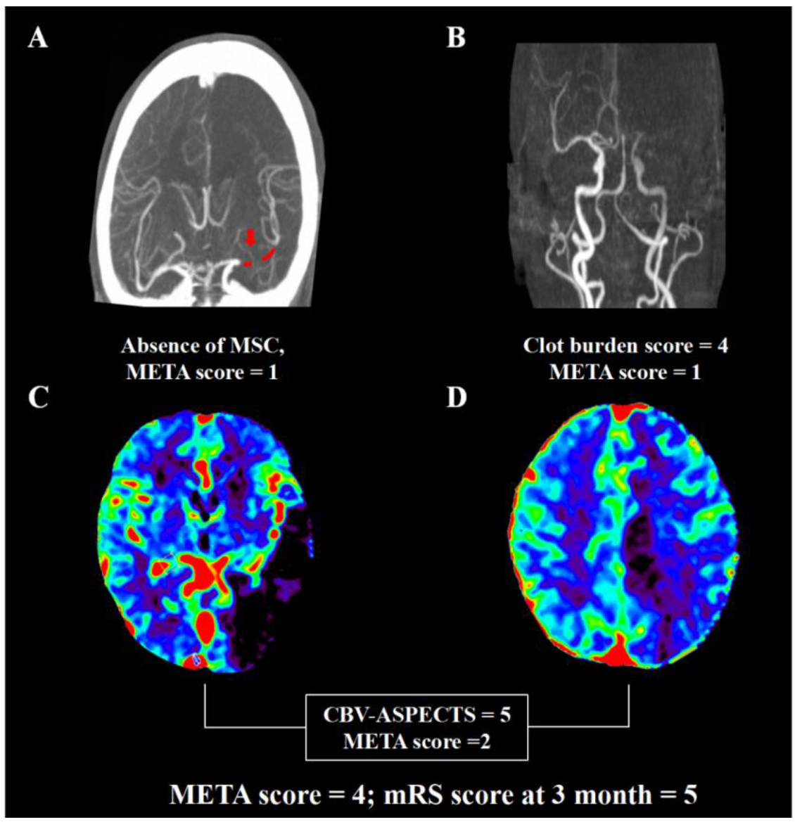
3.1. Development of Multi-Mode Prognostic Algorithm META
3.2. Association of META Score with Outcome
4. Discussion
5. Conclusions
Author Contributions
Funding
Institutional Review Board Statement
Informed Consent Statement
Data Availability Statement
Acknowledgments
Conflicts of Interest
References
- Berkhemer, O.A.; Fransen, P.S.; Beumer, D.; van den Berg, L.A.; Lingsma, H.F.; Yoo, A.J.; Schonewille, W.J.; Vos, J.A.; Nederkoorn, P.J.; Wermer, M.J.; et al. A randomized trial of intraarterial treatment for acute ischemic stroke. N. Engl. J. Med. 2015, 372, 11–20. [Google Scholar] [CrossRef] [PubMed] [Green Version]
- Campbell, B.C.; Mitchell, P.J.; Kleinig, T.J.; Dewey, H.M.; Churilov, L.; Yassi, N.; Yan, B.; Dowling, R.J.; Parsons, M.W.; Oxley, T.J.; et al. Endovascular therapy for ischemic stroke with perfusion-imaging selection. N. Engl. J. Med. 2015, 372, 1009–1018. [Google Scholar] [CrossRef] [Green Version]
- Goyal, M.; Demchuk, A.M.; Menon, B.K.; Eesa, M.; Rempel, J.L.; Thornton, J.; Roy, D.; Jovin, T.G.; Willinsky, R.A.; Sapkota, B.L.; et al. Randomized assessment of rapid endovascular treatment of ischemic stroke. N. Engl. J. Med. 2015, 372, 1019–1030. [Google Scholar] [CrossRef] [Green Version]
- Jovin, T.G.; Chamorro, A.; Cobo, E.; de Miquel, M.A.; Molina, C.A.; Rovira, A.; San Román, L.; Serena, J.; Abilleira, S.; Ribó, M.; et al. Thrombectomy within 8 hours after symptom onset in ischemic stroke. N. Engl. J. Med. 2015, 372, 2296–2306. [Google Scholar] [CrossRef] [PubMed] [Green Version]
- Saver, J.L.; Goyal, M.; Bonafe, A.; Diener, H.C.; Levy, E.I.; Pereira, V.M.; Albers, G.W.; Cognard, C.; Cohen, D.J.; Hacke, W.; et al. Stent-retriever thrombectomy after intravenous t-pa vs. T-pa alone in stroke. N. Engl. J. Med. 2015, 372, 2285–2295. [Google Scholar] [PubMed] [Green Version]
- Tsivgoulis, G.; Katsanos, A.H.; Schellinger, P.D.; Kohrmann, M.; Caso, V.; Palaiodimou, L.; Magoufis, G.; Arthur, A.; Fischer, U.; Alexandrov, A.V. Advanced neuroimaging in stroke patient selection for mechanical thrombectomy. Stroke 2018, 49, 3067–3070. [Google Scholar] [CrossRef]
- Ryu, W.H.A.; Avery, M.B.; Dharampal, N.; Allen, I.E.; Hetts, S.W. Utility of perfusion imaging in acute stroke treatment: A systematic review and meta-analysis. J. Neurointerv. Surg. 2017, 9, 1012–1016. [Google Scholar] [CrossRef]
- Santos, E.M.; Marquering, H.A.; den Blanken, M.D.; Berkhemer, O.A.; Boers, A.M.; Yoo, A.J.; Beenen, L.F.; Treurniet, K.M.; Wismans, C.; Van Noort, K.; et al. Thrombus permeability is associated with improved functional outcome and recanalization in patients with ischemic stroke. Stroke 2016, 47, 732–741. [Google Scholar] [CrossRef] [Green Version]
- Jolly, S.S.; Cairns, J.A.; Lavi, S.; Cantor, W.J.; Bernat, I.; Cheema, A.N.; Moreno, R.; Kedev, S.; Stankovic, G.; Rao, S.V.; et al. Thrombus aspiration in patients with high thrombus burden in the total trial. J. Am. Coll. Cardiol. 2018, 72, 1589–1596. [Google Scholar] [CrossRef]
- Zhang, S.; Lai, Y.; Ding, X.; Parsons, M.; Zhang, J.H.; Lou, M. Absent filling of ipsilateral superficial middle cerebral vein is associated with poor outcome after reperfusion therapy. Stroke 2017, 48, 907–914. [Google Scholar] [CrossRef] [Green Version]
- Liebeskind, D.S.; Jahan, R.; Nogueira, R.G.; Zaidat, O.O.; Saver, J.L.; Investigators, S. Impact of collaterals on successful revascularization in solitaire FR with the intention for thrombectomy. Stroke 2014, 45, 2036–2040. [Google Scholar] [CrossRef] [PubMed] [Green Version]
- Treurniet, K.M.; Yoo, A.J.; Berkhemer, O.A.; Lingsma, H.F.; Boers, A.M.; Fransen, P.S.; Beumer, D.; Van Den Berg, L.A.; Sprengers, M.E.; Jenniskens, S.F.; et al. Clot burden score on baseline computerized tomographic angiography and intra-arterial treatment effect in acute ischemic stroke. Stroke 2016, 47, 2972–2978. [Google Scholar] [CrossRef] [PubMed] [Green Version]
- Rangaraju, S.; Aghaebrahim, A.; Streib, C.; Sun, C.H.; Ribo, M.; Muchada, M.; Nogueira, R.; Frankel, M.; Gupta, R.; Jadhav, A.; et al. Pittsburgh response to endovascular therapy (pre) score: Optimizing patient selection for endovascular therapy for large vessel occlusion strokes. J. Neurointerv. Surg. 2015, 7, 783–788. [Google Scholar] [CrossRef] [PubMed] [Green Version]
- Flint, A.C.; Cullen, S.P.; Faigeles, B.S.; Rao, V.A. Predicting long-term outcome after endovascular stroke treatment: The totaled health risks in vascular events score. Am. J. Neuroradiol. 2010, 31, 1192–1196. [Google Scholar] [CrossRef] [Green Version]
- Campbell, B.C.; Christensen, S.; Levi, C.R.; Desmond, P.M.; Donnan, G.A.; Davis, S.M.; Parsons, M.W. Cerebral blood flow is the optimal CT perfusion parameter for assessing infarct core. Stroke 2011, 42, 3435–3440. [Google Scholar] [CrossRef]
- Zhou, Y.; Zhong, W.; Wang, A.; Huang, W.; Yan, S.; Zhang, R.; Liu, C.; Fu, J.; Jiaerken, Y.; Lou, M. Hypoperfusion in lenticulostriate arteries territory related to unexplained early neurological deterioration after intravenous thrombolysis. Int. J. Stroke 2019, 14, 306–309. [Google Scholar] [CrossRef]
- Barber, P.A.; Demchuk, A.M.; Zhang, J.; Buchan, A.M. Validity and reliability of a quantitative computed tomography score in predicting outcome of hyperacute stroke before thrombolytic therapy. Aspects study group. Alberta stroke programme early ct score. Lancet 2000, 355, 1670–1674. [Google Scholar] [CrossRef]
- Aviv, R.I.; Mandelcorn, J.; Chakraborty, S.; Gladstone, D.; Malham, S.; Tomlinson, G.; Fox, A.J.; Symons, S. Alberta stroke program early ct scoring of ct perfusion in early stroke visualization and assessment. Am. J. Neuroradiol. 2007, 28, 1975–1980. [Google Scholar] [CrossRef] [Green Version]
- Tan, I.Y.; Demchuk, A.M.; Hopyan, J.; Zhang, L.; Gladstone, D.; Wong, K.; Martin, M.; Symons, S.P.; Fox, A.J.; Aviv, R.I. CT angiography clot burden score and collateral score: Correlation with clinical and radiologic outcomes in acute middle cerebral artery infarct. Am. J. Neuroradiol. 2009, 30, 525–531. [Google Scholar] [CrossRef] [Green Version]
- Shi, F.; Chen, Z.; Gong, X.; Zhang, M.; Liebeskind, D.S.; Lou, M. Presence of multi-segment clot sign on dynamic ct angiography: A predictive imaging marker of recanalisation and good outcome in acute ischaemic stroke patients. Eur. Radiol. 2018, 28, 3413–3421. [Google Scholar] [CrossRef]
- Sarraj, A.; Albright, K.; Barreto, A.D.; Boehme, A.K.; Sitton, C.W.; Choi, J.; Lutzker, S.L.; Sun, C.H.J.; Bibars, W.; Nguyen, C.B.; et al. Optimizing prediction scores for poor outcome after intra-arterial therapy in anterior circulation acute ischemic stroke. Stroke 2013, 44, 3324–3330. [Google Scholar] [CrossRef] [PubMed] [Green Version]
- Goyal, M.; Menon, B.K.; van Zwam, W.H.; Dippel, D.W.; Mitchell, P.J.; Demchuk, A.M.; Dávalos, A.; Majoie, C.B.; van der Lugt, A.; De Miquel, M.A.; et al. Endovascular thrombectomy after large-vessel ischaemic stroke: A meta-analysis of individual patient data from five randomised trials. Lancet 2016, 387, 1723–1731. [Google Scholar] [CrossRef]
- Singh, B.; Parsaik, A.K.; Prokop, L.J.; Mittal, M.K. Endovascular therapy for acute ischemic stroke: A systematic review and meta-analysis. Mayo Clinic Proc. 2013, 88, 1056–1065. [Google Scholar] [CrossRef] [PubMed] [Green Version]
- Powers, W.J.; Rabinstein, A.A.; Ackerson, T.; Adeoye, O.M.; Bambakidis, N.C.; Becker, K.; Biller, J.; Brown, M.; Demaerschalk, B.M.; Hoh, B.; et al. 2018 guidelines for the early management of patients with acute ischemic stroke: A guideline for healthcare professionals from the american heart association/american stroke association. Stroke 2018, 49, e46–e110. [Google Scholar] [CrossRef] [PubMed]
- Bivard, A.; Parsons, M. Tissue is more important than time: Insights into acute ischemic stroke from modern brain imaging. Curr. Opin. Neurol. 2018, 31, 23–27. [Google Scholar] [CrossRef]
- Lum, C.; Ahmed, M.E.; Patro, S.; Thornhill, R.; Hogan, M.; Iancu, D.; Lesiuk, H.; Dos Santos, M.; Dowlatshahi, D. Computed tomographic angiography and cerebral blood volume can predict final infarct volume and outcome after recanalization. Stroke 2014, 45, 2683–2688. [Google Scholar] [CrossRef] [Green Version]
- Derraz, I.; Bourcier, R.; Soudant, M.; Soize, S.; Hassen, W.B.; Hossu, G.; Clarencon, F.; Derelle, A.L.; Tisserand, M.; Raoult, H.; et al. Does clot burden score on baseline t2*-mri impact clinical outcome in acute ischemic stroke treated with mechanical thrombectomy? J. Stroke 2019, 21, 91–100. [Google Scholar] [CrossRef] [Green Version]
- Nogueira, R.G.; Jadhav, A.P.; Haussen, D.C.; Bonafe, A.; Budzik, R.F.; Bhuva, P.; Yavagal, D.R.; Ribo, M.; Cognard, C.; Hanel, R.A.; et al. Thrombectomy 6 to 24 hours after stroke with a mismatch between deficit and infarct. N. Engl. J. Med. 2018, 378, 11–21. [Google Scholar] [CrossRef]
- Albers, G.W.; Marks, M.P.; Kemp, S.; Christensen, S.; Tsai, J.P.; Ortega-Gutierrez, S.; McTaggart, R.A.; Torbey, M.T.; Kim-Tenser, M.; Leslie-Mazwi, T.; et al. Thrombectomy for stroke at 6 to 16 hours with selection by perfusion imaging. N. Engl. J. Med. 2018, 378, 708–718. [Google Scholar] [CrossRef]
- Wollenweber, F.A.; Tiedt, S.; Alegiani, A.; Alber, B.; Bangard, C.; Berrouschot, J.; Bode, F.J.; Boeckh-Behrens, T.; Bohner, G.; Bormann, A.; et al. Functional outcome following stroke thrombectomy in clinical practice. Stroke 2019, 50, 2500–2506. [Google Scholar] [CrossRef]
- Lee, E.J.; Kim, Y.H.; Kim, N.; Kang, D.W. Deep into the brain: Artificial intelligence in stroke imaging. J. Stroke 2017, 19, 277–285. [Google Scholar] [CrossRef] [PubMed] [Green Version]
- Kuang, H.; Qiu, W.; Boers, A.M.; Brown, S.; Muir, K.; Majoie, C.B.; Dippel, D.W.; White, P.; Epstein, J.; Mitchell, P.J.; et al. Computed tomography perfusion-based machine learning model better predicts follow-up infarction in patients with acute ischemic stroke. Stroke 2021, 52, 223–231. [Google Scholar] [CrossRef] [PubMed]
- Hoelter, P.; Muehlen, I.; Goelitz, P.; Beuscher, V.; Schwab, S.; Doerfler, A. Automated aspect scoring in acute ischemic stroke: Comparison of three software tools. Neuroradiology 2020, 62, 1231–1238. [Google Scholar] [CrossRef] [PubMed]
- Nishi, H.; Oishi, N.; Ishii, A.; Ono, I.; Ogura, T.; Sunohara, T.; Chihara, H.; Fukumitsu, R.; Okawa, M.; Yamana, N.; et al. Predicting clinical outcomes of large vessel occlusion before mechanical thrombectomy using machine learning. Stroke 2019, 50, 2379–2388. [Google Scholar] [CrossRef]


| Variables | Poor Outcome (n = 153) | Good Outcome (n = 106) | p Value |
|---|---|---|---|
| Clinical characters | |||
| Age, years | 71 ± 12 | 65 ± 13 | <0.001 |
| Women, (%) | 58 (37.9) | 43 (40.6) | 0.699 |
| Baseline NIHSS, IQR | 16 (12–18) | 12 (9–16) | <0.001 |
| Onset to door time, min, IQR | 211 (110–316) | 178 (96–352) | 0.473 |
| Intravenous thrombosis, (%) | 106 (69.3) | 81 (76.4) | 0.259 |
| Baseline blood glucose, mg/dL, IQR | 126 (115–153) | 121 (108–139) | 0.008 |
| Radiological data | |||
| Clot burden score, IQR | 6 (4–8) | 7 (5–8) | 0.017 |
| CBV-ASPECTS, IQR | 8 (5–9) | 8 (8–9) | <0.001 |
| CBF-ASPECTS, IQR | 4 (2–6) | 5 (3–6) | <0.001 |
| Baseline core volume, mL, IQR | 40 (21–72) | 28 (17–46) | 0.001 |
| NECT-ASPECTS, IQR | 8 (6–9) | 9 (7–9) | 0.006 |
| Presence of LSA-, (%) | 112 (73.2) | 64 (60.4) | 0.031 |
| Poor collateral, (%) | 73 (47.7) | 29 (27.4) | 0.001 |
| Absence of MSC, (%) | 83 (54.2) | 41 (38.7) | 0.016 |
| Baseline occlusion location, (%) | 0.045 | ||
| Internal carotid artery | 69 (45.1) | 32 (30.2) | |
| Middle cerebral artery-M1 | 63 (41.2) | 58 (54.7) | |
| Middle cerebral artery-M2 | 21 (13.7) | 16 (15.1) | |
| Risk factors | |||
| Hypertension, (%) | 104 (68.0) | 58 (54.7) | 0.037 |
| Diabetes, (%) | 30 (19.6) | 11 (10.4) | 0.056 |
| Prior antiplatelet usage, (%) | 27 (17.6) | 20 (18.9) | 0.870 |
| History of stroke/TIA, (%) | 27 (17.6) | 18 (17.0) | 1.000 |
| Atrial fibrillation, (%) | 80 (52.3) | 50 (47.2) | 0.450 |
| Variables | OR | 95% CI | p Value |
|---|---|---|---|
| CBS ≤ 6 | 1.928 | 1.083–3.432 | 0.027 |
| CBV-ASPECTS ≤ 6 | 3.873 | 1.751–8.563 | 0.001 |
| CBF-ASPECTS ≤ 3 | 1.612 | 0.907–2.865 | 0.104 |
| NECT-ASPECTS | 2.395 | 1.180–4.861 | 0.016 |
| Baseline core volume ≥ 55 mL | 2.014 | 1.015–3.996 | 0.045 |
| Presence of LSA- | 1.400 | 0.773–2.535 | 0.267 |
| Poor collateral | 1.892 | 1.040–3.442 | 0.037 |
| Absence of MSC | 2.688 | 1.483–4.872 | 0.001 |
| Baseline occlusion location | 0.777 | 0.516–1.171 | 0.229 |
| Variables | Categories | Points |
|---|---|---|
| CBS | ≤6 | 1 |
| >6 | 0 | |
| CBV-ASPECTS | ≤6 | 2 |
| >6 | 0 | |
| Poor collateral | Yes | 1 |
| No | 0 | |
| Absence of MSC | Yes | 1 |
| No | 0 | |
| Total score | 0–5 |
Publisher’s Note: MDPI stays neutral with regard to jurisdictional claims in published maps and institutional affiliations. |
© 2022 by the authors. Licensee MDPI, Basel, Switzerland. This article is an open access article distributed under the terms and conditions of the Creative Commons Attribution (CC BY) license (https://creativecommons.org/licenses/by/4.0/).
Share and Cite
Zhong, W.; Chen, Z.; Yan, S.; Zhou, Y.; Zhang, R.; Luo, Z.; Yu, J.; Lou, M. Multi-Mode Imaging Scale for Endovascular Therapy in Patients with Acute Ischemic Stroke (META). Brain Sci. 2022, 12, 821. https://doi.org/10.3390/brainsci12070821
Zhong W, Chen Z, Yan S, Zhou Y, Zhang R, Luo Z, Yu J, Lou M. Multi-Mode Imaging Scale for Endovascular Therapy in Patients with Acute Ischemic Stroke (META). Brain Sciences. 2022; 12(7):821. https://doi.org/10.3390/brainsci12070821
Chicago/Turabian StyleZhong, Wansi, Zhicai Chen, Shenqiang Yan, Ying Zhou, Ruoxia Zhang, Zhongyu Luo, Jun Yu, and Min Lou. 2022. "Multi-Mode Imaging Scale for Endovascular Therapy in Patients with Acute Ischemic Stroke (META)" Brain Sciences 12, no. 7: 821. https://doi.org/10.3390/brainsci12070821
APA StyleZhong, W., Chen, Z., Yan, S., Zhou, Y., Zhang, R., Luo, Z., Yu, J., & Lou, M. (2022). Multi-Mode Imaging Scale for Endovascular Therapy in Patients with Acute Ischemic Stroke (META). Brain Sciences, 12(7), 821. https://doi.org/10.3390/brainsci12070821