Abstract
Neurological autoimmune diseases have various origins and pathogeneses. Specific antibodies are associated with paraneoplastic syndromes, other infectious agents, or inherited disorders. We aim to evaluate the relation between the autoantibodies, the chosen symptoms, demographic characteristics, and infection history. We retrospectively analysed 508 children during neurological diagnostics. We investigated serum antineuronal, IgG, IgM anti-ganglioside, and anti-aquaporin-4 in both the serum and cerebrospinal fluid (CSF) anti-cell surface and anti-synaptic protein antibodies in 463, 99, 44, 343, and 119 patients, respectively. The CSF polymerase chain reaction detection of Herpesviridae, enterovirus, B19 parvovirus, adenovirus, and parechovirus involved 261 patients. We included available clinical information and electroencephalographic, radiologic, and microbiological results. The IgM anti-ganglioside antibodies increased the risk of tics and positive symptoms (p = 0.0345, p = 0.0263, respectively), the anti-glutamic acid decarboxylase particle of paresis (p = 0.0074), and anti-neuroendothelium of mutism (p = 0.0361). Anti-neuroendothelium, IgM anti-ganglioside, and CSF anti-N-methyl-D-aspartate antibodies were more often associated with consciousness loss (p = 0.0496, p = 0.0044, p = 0.0463, respectively). Anti-myelin antibodies co-occured with Herpes simplex virus (HSV)-2 IgG (p = 0.0415), anti-CV2 with HSV-1 IgM (p = 0.0394), whereas anti-glial fibrillary acidic protein was linked with past Epstein-Barr virus infection. The anti-ganglioside IgM and anti-myelin particles were bilaterally correlated (p = 0.0472). The clinical pictures may overlap, requiring specialistic diagnostics. We noticed the links between the infection aetiology and the specific autoantibody’s positivity.
1. Introduction
Autoimmune diseases are intensively studied. The diversified aetiopathological mechanisms remain under continuous research. However, several hypotheses include, e.g., molecular bacterial and viral mimicry, individual susceptibility to autoagression, and other genetic predispositions [1,2].
Specific antibodies appear as a part of paraneoplastic neurological syndromes (PNS) triggered by malignancies [3]. Mentioned rare diseases do not directly occur because of local tumour infiltration or distant metastasising but due to immunisation to antigens common for healthy and cancerous tissues [3,4]. Amongst autoantibodies, the strongest correlation with neoplastic disorders was described for anti-Ma/Ta, antineuronal nuclear antibody (ANNA) type 1 (anti-Hu) and type 2 (anti-Ri), against Purkinje cell cytoplasmic antigen type 1 (anti-PCA1) (anti-Yo) and type 2 (anti-PCA2), amphiphysin, collapsin response mediator protein 5 (CRMP5) (anti-CV2), and anti-Tr antibodies, together named anti-onconeuronal antibodies. Although the clinical course may overleap, specific antibodies also co-occur with certain syndromes [4].
Non-paraneoplastic autoimmune neurological syndromes are a large group of diseases in which the immune system abnormally targets the proper central nervous system’s (CNS) antigens, but the underlying trigger remains outside the neoplastic transformation [5]. The particles causing the neurological impairment, aimed against neural antigens, involve anti-myelin, anti-myelin-associated glycoprotein (MAG), anti-neuroendothelium (NET), anti-glutamic acid decarboxylase (GAD), anti-non-myelinated fibres, and anti-glial fibrillary acidic protein (GFAP) antibodies.
The infective agents, such as Herpesviridae, Borrelia burgdorferi species (B. burgdorferi), and Mycoplasma pneumoniae (M. pneumoniae) [2,6], may induce the autoantibodies’ production [6] or facilitate their penetration into the intrathecal compartment [7], leading to clinical manifestations of postinfectious syndromes, probably due to molecular mimicry [1,2].
The study compares current or previous infection indicators, the analysed autoantibodies, and the systemic inflammatory markers in a paediatric population. We planned to observe the autoantibodies’ associations with age and the chosen symptoms incidence in the disease course.
2. Materials and Methods
We retrospectively analysed 508 paediatric patients between 2.5 months and 18 years of age suspected of autoimmune disease due to acute or persistent neurologic symptoms. The included participants were hospitalised in a tertiary centre between 3 January 2017 and 2 December 2019. Each child was diagnosed and treated with an individually suited plan.
The 463 patients’ sera were tested for nuclear and cytoplasmic antineuronal antibodies (anti-Hu, anti-Ri, anti-Yo, anti-Ma/Ta, anti-PCA2, anti-CV2, anti-Tr, against amphiphysin, GAD, MAG, myelin, NET, GFAP, and non-myelinated fibres), 99 patients for anti-ganglioside antibodies, 343 children were screened for antibodies against cell surface and synaptic proteins (anti-α-amino-3-hydroxy-5-methyl-4-isoxazolepropionic receptor (AMPAr), anti-γ-aminobutyric acid receptor-B (GABA-Br), anti-N-methyl-D-aspartate receptor (NMDAr), anti-1,3-dipropyl-8-phenylxanthine (DPPX), anti-contactin-associated protein-like-2 (CASPR2), anti-leucine-rich glioma-inactivated protein 1 (LGI1)), and 44 patients were tested for anti-aquaporin-4 (AQP4) antibodies. We screened 119 children’s cerebrospinal fluid (CSF) for antibodies against cell surface and synaptic proteins. All mentioned analyses used standardised commercial kits (EUROIMMUN, Luebeck, Germany). All children in whom the paraneoplastic antibody was detected underwent subsequent screening for underlying tumours. Figure 1 depicts the number of tested patients.
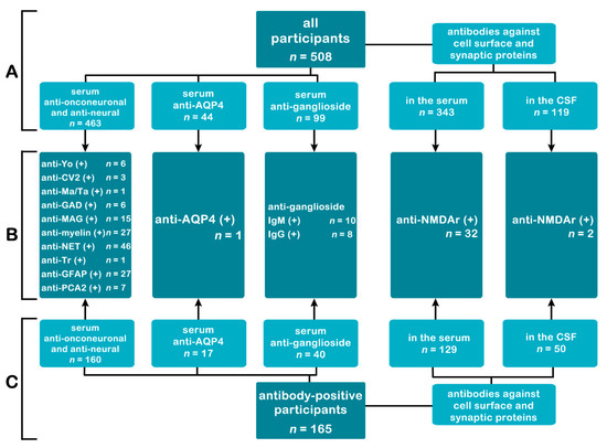
Figure 1.
The number of tested patients in subgroups. Part A—the number of patients tested for each group of antibodies in all research participants. Part C—the number of patients tested for each group of antibodies in the antibody-positive subgroup (patients with at least one autoantibody detected). Part B—common to A and C-number of detected antibodies. Abbreviations: anti-Yo—Purkinje cell cytoplasmic antibody type 1; AQP4—aquaporin 4; CSF—cerebrospinal fluid; CV2—collapsin response mediator protein 5; GAD—glutamic acid decarboxylase; GFAP—glial fibrillary acidic protein; Ig—immunoglobulin; MAG—myelin-associated glycoprotein; NET—neuroendothelium; NMDAr—N-Methyl-D-Aspartate receptor; PCA2—Purkinje cell cytoplasmic antigen type 2.
The CSF collected by lumbar puncture underwent a routine test with a standard laboratory analyser. The reference values (RV) were as follows: For CSF leukocyte count (CSF-L) RV ranged 0–5 cells/uL, and for CSF protein (CSF-P) concentration stayed within 15–45 mg/dL. The IgG index upper level of normal (ULN) equalled 0.75. The 261 patients underwent polymerase chain reaction panel detecting the genetic material of herpes simplex virus (HSV)-1, HSV-2, varicella-zoster virus (VZV), Epstein–Barr virus (EBV), cytomegalovirus (CMV), human herpesvirus (HHV)-6, HHV-7, B19 parvovirus, enterovirus (EV), adenovirus (ADV), and parechovirus (PV) (Fast Track Diagnostics, Junglinster, Luxembourg) in CSF. We included available antimicrobial results involving antibodies against HSV-1, HSV-2, EBV, CMV, EV, B. burgdorferi, M. pneumoniae, and tick-borne encephalitis virus (TBEV). The antistreptolysin O (ASO) ULN equalled 150 IU/mL. We analysed electroencephalography and radiological findings in magnetic resonance imaging (MRI) and computed tomography (CT). The participants’ final diagnoses stand in Table 1.

Table 1.
Clinical description of patients divided due to autoantibody presence.
We considered the frequency of consciousness loss, involuntary movements, paresis, focal symptoms, speech or sight aberrances, headaches, meningeal signs, and urination or defecation disorders. Moreover, we analysed symptoms’ association with the particular autoantibody.
We provided the statistical assessment with Statistica 13.3 (TIBCO Software Inc., 2017, Palo Alto, CA, USA). Depending on the data distribution and the number of groups, we used the χ2, the Mann–Whitney U (MW) test. A p-value of less than 0.05 we considered statistically significant.
The Institutional Ethical Committee stated that formal approval and informed consent is not required due to the retrospective character of the study, including anonymised data analysis from clinically justified procedures.
3. Results
Our study group included 508 participants with a median age of 8.9 years old. We compared the parameters separately in the whole population (p1) and among autoantibody-positive patients (p2). We found that patients with anti-CV2 (2.5 vs. 9.0 [y/a], p1 = 0.0328) and anti-PCA2 (4.7 vs. 9.1 [y/a], p1 = 0.0095) have significantly lower age than those without either particle; however, the anti-Yo-positive patients were older than those without mentioned particle (13.3 vs. 8.9 [y/a], p1 = 0.0208). The same conclusions appeared in analyses within the autoantibody-positive population (2.5 vs. 8.9 [y/a], p2 = 0.031; 4.7 vs. 9.0 [y/a], p2 = 0.0083; 13.3 vs. 8.7 [y/a], p2 = 0.0203, respectively). We noticed that anti-myelin-positive children prevail between 3 and 6 y/o (p1 = 0.0152; p2 = 0.0152).
In routine CSF examination, a significantly higher protein concentration occurred in a group with CSF anti-NMDAr antibodies than in children without them (p1 = 0.0499). What is more, both in whole and the autoantibody-positive populations, patients with anti-myelin antibodies had significantly lower glucose concentrations than children without the particles (p1 = 0.0054; p2 = 0.0094, respectively). The median and IQR of analysed values appear in Table 2.
Table 2.
Median and IQR values of analysed parameters with the division based on detected autoantibodies.
In the antineuronal autoantibody-positive group, patients suffered most frequently from consciousness alternation (25.6%), involuntary movements (24%), and headaches (22.5%). A large share presented focal signs (20.2%) and sight deterioration (17.1%). The symptoms’ frequency did not statistically differ from autoantibody-negative patients. Separately, patients with serum anti-NET, anti-ganglioside IgM, and CSF anti-NMDAr antibodies had a significantly higher consciousness loss incidence than children without mentioned immunoglobulins, both in the total research population (p1 = 0.0496, p1 = 0.0044, p1 = 0.0463, respectively) and in the autoantibody-positive patients’ group (p2 = 0.0185, p2 = 0.0258, p2 = 0.0367, respectively). Moreover, children with anti-MAG antibodies had a lower cognisance alternation rate (p1 = 0.0159; p2 = 0.0229). In addition, anti-ganglioside IgM positive patients statistically more often experienced involuntary movements (p1 = 0.0345), obsessive-compulsive disorder (OCD) (p1 = 0.0263), or positive symptoms (p1 = 0.0263). Similar observations did not apply to other autoantibodies. Our report suggests that the children with anti-GAD are more often diagnosed with varying severity paresis (p1 = 0.0074; p2 = 0.0078). Another result indicated that defecation problems might be significantly more frequent in the anti-GAD positive population (p2 = 0.0375); however, in our study, only one out of six anti-GAD positive children suffered from the mentioned ailment. We noticed mutism in 6.5% of patients with anti-NET antibodies. Compared to patients without such antibodies, it appeared to be a significant symptom (p1 = 0.0361). The CSF anti-NMDAr immunoglobulins’ presence was associated with headache in our population (p1 = 0.0271; p2 = 0.0294). The frequency of focal and meningeal signs, balance, gait, sight disturbances, seizures, and impaired urination between patients with and without specific autoantibody did not differ significantly. We placed the complete data in Table 3.
Table 3.
The incidence of analysed symptoms with the division based on detected autoantibodies.
The population with at least one autoantibody showed a higher anti-M. pneumoniae IgM positivity (p2 = 0.01; OR = 3.7623 CI95% (1.3575–10.4274))—especially anti-myelin antibodies (p2 < 0.01; OR = 5.3333 CI95% (1.5701–18.1165)), which is associated with HSV-2 IgG presence (p2 = 0.04; OR = 9.0952, CI95% (1.5505–53.3528)). The anti-CMV IgM appeared in all CSF anti-NMDAr positive patients. Further analyses confirmed the significance in the whole research group and the limited autoantibody-positive cohort (p1 = 0.01; p2 = 0.01). Previous EBV infection appears as an important anti-GFAP antibody production factor. In our cohort, anti-EBV viral-capsid antigen (VCA) IgG and anti-Epstein–Barr nuclear antigen (EBNA) antibodies’ prevalence resulted in the significant connection with anti-GFAP positivity (p1 < 0.01; OR = 6.6909, CI95% (1.5321–29.2197) and p1 = 0.02; OR = 3.4563 CI95% (1.1361–10.5155)), respectively). We placed the complete data in Table 4.
Table 4.
The frequency and positivity percentage in the entire group, in the autoantibody-positive population and with specific antibodies.
The paired autoantibodies’ analysis revealed the link between the anti-ganglioside IgM and anti-NET immunoglobulin coexistence (p1 = 0.047; OR = 6.0857 CI95% (1.1943–31.0118)). Other coappearing autoantibody pairs did not show statistical significance in our research.
The PCR CSF detection of neurotrophic viruses found specific HHV-7, EBV, EV, ADV, B19 parvovirus, HSV-1, HSV-2, and HHV-6 nucleic acids in 5 (1.9%), 4 (1.5%), 2 (0.8%), 2 (0.8%), 2 (0.8%), 1 (0.4%), 1 (0.4%), and 1 (0.4%) patient, respectively. Further statistical analyses with χ2 did not bring correlation with any autoantibody.
4. Discussion
4.1. Anti-NET Antibodies
Anti-NET antibodies appear in rheumatic diseases [8], demyelinating autoimmune disorders including multiple sclerosis (MS), peripheral polyneuropathies [9], and paraneoplastic syndromes [10]. The targets spread in cerebral and nerve-associated vessels of the peripheral (PNS) and CNS. Chronic diseases may induce autoantibodies, which later, due to injury to the blood–brain barrier (BBB), may penetrate the intrathecal compartment [9]. The neuroendothelial cells participate in haemostasis and inflammatory response [11,12]; in addition, the cognitive impairment correlates with antibodies titre [13]. Our results partially fit the data in other research, in which seizures were observed in 33% of patients, upper or lower motor neuron involvement in 27% and 22% of patients, respectively, cranial nerve impairment bothered half of all patients, and cerebellar syndrome occurred in 65% of them [14]. The positive psychiatric symptoms’ incidence was comparable to other autoantibodies and autoantibody-negative patients. In contrast, OCD exceeded the share amongst autoantibody-negative (6.5% vs. 3.8%) or participants with any autoantibodies detected (6.5% vs. 4.2%). However, the statistical significance was not found. In contrast to previous studies, we did not observe an increased cardiovascular incident rate in autoantibody-positive patients—ischaemic stroke appeared in one patient [13].
The pairwise tests revealed significant anti-NET and anti-ganglioside IgM coexistence. The publications describe the in vitro direct anti-monosialotetrahexosylganglioside antibodies’ cross-reaction on microvascular endothelial cells in neuropathy, leading to disruption of the barrier between vessels and nerves, causing the exposure of other antigens [15]. However, the hypothesis concerns the peripheral nervous system and does not reflect the clinical reports.
4.2. Anti-GFAP Antibodies
The antibodies to GFAP cause inflammations involving the brain, cerebellum, meninges and brain stem, resulting in encephalopathy [16]. The clinical picture contains severe headaches, consciousness deterioration, and involuntary movements and less frequently partial paresis, ataxia, and other walking difficulties. Patients show memory loss, psychotic episodes, or autonomic involvement such as urine and stool incontinence or retention [17]. Fang et al. estimate that every fifth anti-GFAP-positive patient requires prolonged immunosuppression to sustain the remission. In our population, the most significant differences concern consciousness deterioration (18.5% vs. 68.8%), headaches (22.2% vs. 81.3%), and involuntary movements (18.5% vs. 33.3%), whereas paresis frequency was similar in both reports (18.5% vs. 18.8%) [18]. The cited authors selectively analysed an adult population with autoimmune disorders, whereas we based our research on a heterogeneous paediatric cohort, including children with infective or unclear aetiology. In the case series, the authors described two children who presented gait impairment and nystagmus. What is more, the first diagnosed with meningoencephalitis showed dysautonomia, bladder dysfunction, and cerebellar syndrome, whereas the second suffered from headaches and consciousness alternation, followed by extrapyramidal syndrome [19]. Our four anti-GFAP-positive participants had other autoantibodies detectable. We did not observe children with positive psychiatric symptoms in our population, and only one patient experienced autonomic urination impairment. Our data revealed a significantly higher anti-GFAP incidence in children with detected anti-VCA EBV IgG or anti-EBNA IgG. However, the literature lacks data on the correlation between anti-GFAP and infectious agents.
4.3. Anti-Myelin Antibodies
Autoantibodies against myelin compounds participate in various clinical syndromes such as cerebellar dysfunction, extrapyramidal symptoms, motor neurons, and vision impairment. The involvement of cranial nerves differs between reports [14,20]. The anti-myelin particles appear in physiological protective reactions to CNS trauma but also occur in patients with a past oncological history or accompany primary autoimmune disorders and rheumatic diseases [14,21,22]. Our anti-myelin-positive patients suffered most frequently from involuntary movements, paresis, or sight deterioration and consciousness alternation.
Myelin oligodendrocyte glycoprotein (MOG) occurs exclusively in the CNS [23]. The antibodies against MOG predominantly affect an optic nerve and a spinal cord, accompanying difficulties with speech, swallowing, vision, and oculomotor function. The research in the paediatric population correlated the anti-MOG antibodies with neuromyelitis optica (NMO) and transverse myelitis (TM) in monofocal and polyfocal forms. The antibody frequently appears in acquired demyelinating diseases in childhood—especially in the younger population presenting acute disseminated encephalomyelitis (ADEM). The authors also concluded that anti-MOG-positivity during the acute phase in almost 50% is transient, as well as with non-MS disease course [24]. The positivity concerns 20% of patients with NMO and TM but twice more with ADEM [25,26]. Moreover, anti-MOG persistent positivity brings relapse risk 3–5 times greater [27]. The HSV may cause peripheral neuropathies, including facial nerve palsy [28]. We found a correlation between anti-myelin positivity and anti-HSV-2 IgG particles, but previous studies suggest anti-MOG antibodies’ peripheral presence preceding such infection. The viral disease impairs the BBB, enabling the circulating autoantibodies to conjugate with CNS and trigger symptoms [29]. Literature also confirms that influenza and EBV participate in the pathogenesis of anti-MOG ADEM [30], having a predictive value in the relapsing multiphasic course risk [27]. The viral infections preceding the NMO confirm that infectious diseases facilitate autoantibodies penetration to the intrathecal compartment [6]. The animal model independently proved the HSV-1 potential to trigger encephalitis and multifocal demyelinated CNS lesions [6]. Zheng et al. noticed the cross-reactivity between MOG and CMV in the rat experimental autoimmune encephalomyelitis (AIE) model [31]. However, postinfectious anti-MOG positive NMO appears after bacterial infections (e.g., B. burgdorferi and Mycobacterium tuberculosis (M. tuberculosis)) [7,32]. We found the association with M. pneumoniae IgM that fits previous reports hypothesising the molecular mimicry triggering anti-myelin antibodies production and immunocomplexes deposition causing perivenular inflammation [1,26]. Christie et al. confirmed the cross-reaction with galactocerebroside C in 38% of M. pneumoniae encephalitic patients [33].
4.4. Anti-MAG Antibodies
The anti-MAG antibodies are responsible for various neuropathies. Several kinds of research successfully confirmed the pathological role of the particles [34], e.g., the passive transfer of human-derived anti-MAG antibodies to chickens. The investigation revealed the development of a similar pathophysiological pattern of deposits in myelin sheets and structural change of the layers [35]. The most typical course begins with numbness and upcoming paraesthesia, leading to distal, symmetrical sensory impairment, mainly due to the injury of myelinated fibres. Motor involvement with muscle weakness and ataxia rarely happen [36]. In our population, patients most frequently experienced involuntary movements, sight deterioration, and gait impairment in 40%, 26.7%, and 20% of cases, respectively. Compared to the available report in which balance impairment involved 17.7% of adult patients, the incidence reached 13.3% in our population. We did not observe cranial nerve involvement or epileptic seizures, which bothered 50% and 66% of cited research patients. The same authors detected motor neurons involvement in over 34% and myopathy in 60% of participants [14]. Findings associated with anti-MAG antibodies more typical for the paediatric population include ataxia after chicken pox [37] and their presence in 70% of patients diagnosed with autism [38].
The detected link between anti-MAG positivity and antecedent CMV infection was described in a previous study in which a CMV DNA occurred in 88% of anti-MAG positive patients with chronic neuropathy. The mechanism remains unknown, but molecular mimicry was suspected [39]. However, several works undermine the correlation and suggest the latent CMV reactivation, but the acquirable data cannot justify anti-MAG production [40]. In addition, Lunn at. al. failed to confirm the hypothesis in patients with peripheral neuropathy and paraproteinemia [41].
4.5. Anti-PCA2 Antibodies
The anti-PCA2 is a partially characterised onconeuronal antibody associated with small-cell lung carcinoma (SCLC). In our research, the tics dominated amongst children’s symptoms; only one suffered from the walking impairment or experienced a consciousness decrease. However, the previous publications describe a wide range of symptoms potentially associated with the mentioned antibody due to encephalitis, myelitis, or neuropathy. Cognitive impairment appears as a short-term memory decrease, disorientation, and consciousness clouding. The paraneoplastic syndrome may manifest with cerebellar ataxia, regional paraesthesia, spastic paresis, or myasthenic disorder [42,43].
4.6. Anti-GAD Antibodies
The anti-GAD antibodies participate in autoimmune DM pathogenesis [44]. The neurological syndromes are rare, have marked female predominance, and appear in patients with anti-GAD detectable in CSF [45]. They involve cerebellar ataxia, stiff-person syndrome, AIE, and epileptic episodes resulting from impaired GABAergic transmission [46]. Paediatric data suggest that high anti-GAD titres provoke temporal-based focal seizures, psychiatric symptoms, and cognitive decrease, manifesting in remembrance difficulties and developmental slowdown [5]. On the other hand, up to 1.7% of healthy and 5% of neurological patients with different aetiology are anti-GAD positive [47]. We observed pareses and impaired defecation in our population in 66.7% and 16.7% of participants, respectively. The 16.7% of patients also experienced balance difficulties. Honnorat et al. described gait impairment in all patients; however, the study focused on patients with cerebellar involvement. The seizure and visual symptoms rates were lower, reaching 33.3% vs. 53% and 16.7% vs. up to 86%. We noticed a similar consciousness alternation frequency than previously described for anti-GAD-associated cerebellitis. We did not note memory deterioration, which appeared in 67% of patients in the cited research [44]. In research concerning children with AIE, authors described a change of behaviour and alternated consciousness in all three anti-GAD positive patients; two presented hallucinations and sleepiness, and one patient had an autonomic disorder or tremor [48].
4.7. Anti-Yo Antibodies
Anti-Yo belongs to well-described autoantibodies associated with ovarian and breast cancer, but 2–8.5% of non-oncologic patients occur positive [4,49]. The most typical clinical manifestation bothering 90% of cases includes cerebellar degeneration; however, brainstem encephalitis or peripheral neuropathy happens rarely [49]. In our non-oncologic population, we diagnosed paresis in two patients; in addition, two experienced consciousness deterioration and one patient had tics. In literature, varied ataxia and nystagmus concerned all patients; peripheral neuropathy appeared in 47% and cognitive impairment in 18% of paraneoplastic syndrome cases [50]. In the paediatric population, anti-Yo-related paraneoplastic syndrome with typical PCD, including ataxia, speech impairment, and vertigo, coappear with Hodgkin’s lymphoma; in children without malignancy, the anti-Yo positivity rate increases in patients diagnosed with attention deficit hyperactivity disorder [51].
4.8. Anti-CV2 Antibodies
The anti-CV2 antibody accompanies SCLC and, less often, thymoma, but 4–17.5% of non-oncologic patients show positivity [4,49]. Clinical manifestations include limbic encephalitis, cerebellar degeneration with ataxia and involuntary movements, encephalomyelitis, sensory and sensorimotor neuropathy, gastrointestinal pseudo-obstruction, and ocular inflammation [4,52]. Among our patients with anti-CV2 in serum, the one experienced headache and gait impairment; the other, with multi-antibody positivity, showed mutism, whereas the third with autism was asymptomatic. Psimaras et al. described peripheral neuropathy in 39%, cerebellitis in 13.4%, and limbic encephalitis in 4.3% of patients with anti-CV2 in CSF [49].
Interestingly, significant anti-CV2 positivity appeared amongst children with anti-HSV-1 IgM. A similar association was not previously described, nor with another infectious agent. The analysis requires further research because of the low positive patient quantity and potential statistical bias.
4.9. Anti-Tr Antibodies
Antibodies against Tr protein are partially characterised paraneoplastic molecules predominantly observed in Hodgkin lymphoma, but 11% of positive patients had no detectable neoplasm [4]. Amongst analysed children, we detected the anti-Tr antibodies in a single patient with autism and epilepsy hospitalised due to transient consciousness impairment. What is more, the boy also produced anti-GFAP antibodies. Typically, anti-Tr antibodies cause PCD or limbic encephalitis [49], but in children, paraneoplastic syndromes are casuistic, including vision impairment, ataxia, tremor, and muscular hypotonia [53]. Characteristic ataxia may appear as an isolated syndrome or combine with encephalopathy and peripheral neuropathy [42].
4.10. Anti-Ma/Ta Antibodies
This well-described onconeural antibody appears in germinal testicular tumours or non-SCLC and less frequently in other solid neoplasms. The main clinical presentations include limbic or brainstem encephalitis and PCD, with their frequencies assessed for 58%, 21%, and 16%, respectively [49]. Patients with underlying tumours constitute 58–96% of positive participants [4]. They experience consciousness alternation, cataplexy, sleep disorders and diencephalic disorders [54]; children are more prone to focal seizures, behaviour change, or speech and muscle tone impairment [5]. In paediatric non-paraneoplastic AIE typical symptoms include behavioural change and, speech and consciousness impairments, but sometimes positive psychiatric symptoms appear [48]. In our research, the boy with anti-Ma/Ta antibody suffered from blurred vision and headache associated with optic nerve inflammation, foretelling MS.
4.11. Anti-NMDAr Antibodies
Autoantibodies against NMDAr most frequently cause AIE. In the general population, 58% of patients are diagnosed with ovarian teratoma [55], but antecedent infection appears typically amongst children. The best-described infective anti-NMDAr encephalitis triggering factor is HSV-1; the other viruses’ roles are ambiguous [2,6,7,56]. Our research suggests the link between CSF anti-NMDAr and CMV IgM positivity. The clinical spectrum includes dyskinesis, choreoathetosis, dystonic posturing, and rigidity. Psychiatric symptoms such as insomnia, paranoia, anxiety disorders, and cognitive deterioration appear less frequently [3,42]. Our patients with CSF anti-NMDAr antibodies presented consciousness loss and severe headaches. Imbalance and motion problems probably resulted from generalised weakness. Amongst patients with those antibodies in serum, 13% experienced deterioration in cognitive functions. However, the most common complaint was vision impairment (50%), and 21% suffered from involuntary movements; psychiatric disorders appeared in 13% and paresis in 16% of cases. The results partially cover the previously observed clinical pictures of paediatric AIE; however, decreased consciousness occurred in less than 50% of cases. The report describes behavioural alternation in all patients, speech disorder in 73%, facial dyskinesias in 64%, and hallucinations in 18% of children [48].
4.12. Antiganglioside Antibodies
Gangliosides, crucial for signal transition, spread on neurons’ surfaces. The antibodies against them participate in chronic and acute neuropathies’ pathogeneses; the role in paediatric idiopathic epilepsy needs further research [57]. Antiganglioside antibodies may act as primary or secondary aetiological factors. Although specific targets characterise particular neuropathies, we analysed them collectively.
IgM autoantibodies appear in multifocal motor neuropathy, ataxic neuropathy, and ophthalmoplegia [36]. In our population, 50% experienced involuntary movements, 20% positive symptoms and OCD, whereas 40% experienced at least transient consciousness loss. We proved those symptoms’ statistical predominance in comparison to autoantibody-negative patients. Moreover, 20% experienced paresis, and 10% experienced gait impairment.
The children who presented IgG antibodies suffered more frequently from muscle weakness and gait impairment. Nevertheless, less often exhibited symptoms included consciousness alternation or positive psychiatric symptoms. In the literature, IgG anti-ganglioside antibodies link with specific symptomatic disorders or diseases with other known aetiology, e.g., Guillain–Barre syndrome (GBS), MS, and Alzheimer’s or Parkinson’s disease [57]. Research has widely described the association of GBS with previous CMV, EBV, and M. pneumoniae infections [58], but our analyses did not bring sufficient evidence to confirm the findings in our population.
4.13. Anti-Aquaporin-4 Antibodies
In our research, we detected one patient diagnosed with non-specific stress-associated vision impairment. She reported headache, sight deterioration, presented involuntary movements, and balance impairment. The NMO spectrum disorder most frequently appears as myelitis (84%) and optic neuritis (63%). A brain, brainstem, and area postrema are involved in around 15% of cases. The disease may occur idiopathically or after viral infection with a tendency to relapse [59,60]. Anti-AQP4 antibodies typically manifest with transient vision loss, peripheral sensory and motor impairment, and paroxysmal movements episodes in 20% of patients [42].
5. Conclusions
The paper presented the diseases’ manifestations and sought pathogenesis. The clinical pictures partially differed from courses described in adult patients. The main reasons may involve involve a lower incidence of malignancies and a higher prevalence of infectious diseases in children. The protocol involving autoantibodies’ assessment in non-specific neurologic diagnostics might contribute to the detection of pathogenic particles in the pre-clinical phase, enabling the close patient’s observation and prompt treatment in the early stage.
The main drawback resulting from a retrospective study is the difficulty in comparing symptoms between patients and low quantity in subgroups decreasing the tests’ strength. The other source of bias remains the inclusion of patients in different stages of diseases. This aspect concerns both infective and non-infective aetiologies; however, in the first case, the serological testing would be helpful, especially after the acute phase. The report’s objectivity could be improved by the standardised protocol used for patient anamneses and examinations.
Author Contributions
Conceptualization, A.M. and M.F.; data curation, K.L.; formal analysis, K.L.; investigation, S.M. and K.O.; methodology, A.M. and M.F.; project administration, A.M. and M.F.; resources, S.M. and K.O.; software, K.L.; supervision, A.M. and M.F.; validation, A.M. and M.F.; visualization, K.L.; writing—original draft, K.L.; writing—review and editing, K.L., A.M. and K.M.-M. All authors have read and agreed to the published version of the manuscript.
Funding
The study was supported by Poznan University of Medical Sciences funds.
Institutional Review Board Statement
The Institutional Ethical Committee stated that formal approval and informed consent is not required due to the retrospective character of the study, including anonymised data analysis from clinically justified procedures.
Informed Consent Statement
Patient consent was waived due to The Institutional Ethical Committee.
Data Availability Statement
Data sharing not applicable.
Conflicts of Interest
The authors declare no conflict of interest.
References
- D’Alonzo, R.; Mencaroni, E.; Di Genova, L.; Laino, D.; Principi, N.; Esposito, S. Pathogenesis and Treatment of Neurologic Diseases Associated With Mycoplasma Pneumoniae Infection. Front. Microbiol. 2018, 9, 2751. [Google Scholar] [CrossRef] [PubMed]
- Figlerowicz, M.; Mazur-Melewska, K.; Kemnitz, P.; Mania, A. Pediatric Postviral Autoimmune Disorders of the CNS. Future Virol. 2020, 15, 307–315. [Google Scholar] [CrossRef]
- Popławska-Domaszewicz, K.; Florczak-Wyspiańska, J.; Kozubski, W.; Michalak, S. Paraneoplastic Movement Disorders. Rev. Neurosci. 2018, 29, 745–755. [Google Scholar] [CrossRef] [PubMed]
- Graus, F. Recommended Diagnostic Criteria for Paraneoplastic Neurological Syndromes. J. Neurol. Neurosurg. Psychiatry 2004, 75, 1135–1140. [Google Scholar] [CrossRef] [Green Version]
- Barbagallo, M.; Vitaliti, G.; Pavone, P.; Romano, C.; Lubrano, R.; Falsaperla, R. Pediatric Autoimmune Encephalitis. J. Pediatr. Neurosci. 2017, 12, 130. [Google Scholar] [CrossRef]
- Boukhvalova, M.S.; Mortensen, E.; Mbaye, A.; Lopez, D.; Kastrukoff, L.; Blanco, J.C.G. Herpes Simplex Virus 1 Induces Brain Inflammation and Multifocal Demyelination in the Cotton Rat Sigmodon Hispidus. J. Virol. 2019, 94, e01161-19. [Google Scholar] [CrossRef] [Green Version]
- Koga, M.; Takahashi, T.; Kawai, M.; Fujihara, K.; Kanda, T. A Serological Analysis of Viral and Bacterial Infections Associated with Neuromyelitis Optica. J. Neurol. Sci. 2011, 300, 19–22. [Google Scholar] [CrossRef]
- Meroni, P.; Khamashta, M.; Youinou, P.; Shoenfeld, Y. Mosaic of Anti-Endothelial Antibodies: Review of the First International Workshop on Anti-Endothelial Antibodies: Clinical and Pathological Significance Milan, 9 November 1994. Lupus 1995, 4, 95–99. [Google Scholar] [CrossRef]
- Tanaka, Y.; Tsukada, N.; Koh, C.-S.; Yanagisawa, N. Anti-Endothelial Cell Antibodies and Circulating Immune Complexes in the Sera of Patients with Multiple Sclerosis. J. Neuroimmunol. 1987, 17, 49–59. [Google Scholar] [CrossRef]
- Koszewicz, M.; Michalak, S.; Bilinska, M.; Budrewicz, S.; Zaborowski, M.; Slotwinski, K.; Podemski, R.; Ejma, M. Is Peripheral Paraneoplastic Neurological Syndrome Possible in Primary Brain Tumors? Brain Behav. 2016, 6, e00465. [Google Scholar] [CrossRef] [Green Version]
- Meroni, P.L.; Tincani, A.; Sepp, N.; Raschi, E.; Testoni, C.; Corsini, E.; Cavazzana, I.; Pellegrini, S.; Salmaggi, A. Endothelium and the Brain in CNS Lupus. Lupus 2003, 12, 919–928. [Google Scholar] [CrossRef] [PubMed]
- Margari, F.; Petruzzelli, M.G.; Mianulli, R.; Toto, M.; Pastore, A.; Bizzaro, N.; Tampoia, M. Anti-Brain Autoantibodies in the Serum of Schizophrenic Patients: A Case-Control Study. Psychiatry Res. 2013, 210, 800–805. [Google Scholar] [CrossRef] [PubMed]
- Annunziata, P.; Cioni, C.; Moschini, F.; Riccucci, A.; Guazzi, G.C. Serum Anti-Brain Endothelium Antibodies and Cognitive Assessment in Patients with Binswanger’s Encephalopathy. J. Neurol. Sci. 1995, 128, 96–102. [Google Scholar] [CrossRef]
- Michalak, S.; Gruszczyńska, A.; Popławska, K.; Żurawicz, P.; Kozubski, W. Clinical significance of anti-MAG, anti-myelin and anti-neuroendothelium antibodies. Neuroskop 2011, 13, 100–105. [Google Scholar]
- Kanda, T.; Iwasaki, T.; Yamawaki, M.; Tai, T.; Mizusawa, H. Anti-GM1 Antibody Facilitates Leakage in an in Vitro Blood-Nerve Barrier Model. Neurology 2000, 55, 585–587. [Google Scholar] [CrossRef] [PubMed]
- Li, J.; Xu, Y.; Ren, H.; Zhu, Y.; Peng, B.; Cui, L. Autoimmune GFAP Astrocytopathy after Viral Encephalitis: A Case Report. Mult. Scler. Relat. Dis. 2018, 21, 84–87. [Google Scholar] [CrossRef]
- Troxell, R.M.; Christy, A. Atypical Pediatric Demyelinating Diseases of the Central Nervous System. Curr. Neurol. Neurosci. Rep. 2019, 19, 95. [Google Scholar] [CrossRef]
- Fang, B.; McKeon, A.; Hinson, S.R.; Kryzer, T.J.; Pittock, S.J.; Aksamit, A.J.; Lennon, V.A. Autoimmune Glial Fibrillary Acidic Protein Astrocytopathy: A Novel Meningoencephalomyelitis. JAMA Neurol. 2016, 73, 1297. [Google Scholar] [CrossRef]
- Oger, V.; Bost, C.; Salah, L.; Yazbeck, E.; Maurey, H.; Bellesme, C.; Sevin, C.; Adamsbaum, C.; Chrétien, P.; Benaiteau, M.; et al. Mild Encephalitis/Encephalopathy with Reversible Splenial Lesion Syndrome: An Unusual Presentation of Anti-GFAP Astrocytopathy. Eur. J. Paediatr. Neurol. 2020, 26, 89–91. [Google Scholar] [CrossRef]
- Salama, S.; Khan, M.; Pardo, S.; Izbudak, I.; Levy, M. MOG Antibody–Associated Encephalomyelitis/Encephalitis. Mult. Scler. 2019, 25, 1427–1433. [Google Scholar] [CrossRef]
- Yoles, E.; Hauben, E.; Palgi, O.; Agranov, E.; Gothilf, A.; Cohen, A.; Kuchroo, V.; Cohen, I.R.; Weiner, H.; Schwartz, M. Protective Autoimmunity Is a Physiological Response to CNS Trauma. J. Neurosci. 2001, 21, 3740–3748. [Google Scholar] [CrossRef] [PubMed]
- Berger, T.; Rubner, P.; Schautzer, F.; Egg, R.; Ulmer, H.; Mayringer, I.; Dilitz, E.; Deisenhammer, F.; Reindl, M. Antimyelin Antibodies as a Predictor of Clinically Definite Multiple Sclerosis after a First Demyelinating Event. N. Engl. J. Med. 2003, 349, 139–145. [Google Scholar] [CrossRef] [PubMed] [Green Version]
- Reindl, M.; Waters, P. Myelin Oligodendrocyte Glycoprotein Antibodies in Neurological Disease. Nat. Rev. Neurol. 2019, 15, 89–102. [Google Scholar] [CrossRef] [PubMed]
- Waters, P.; Fadda, G.; Woodhall, M.; O’Mahony, J.; Brown, R.A.; Castro, D.A.; Longoni, G.; Irani, S.R.; Sun, B.; Yeh, E.A.; et al. Serial Anti–Myelin Oligodendrocyte Glycoprotein Antibody Analyses and Outcomes in Children With Demyelinating Syndromes. JAMA Neurol. 2020, 77, 82. [Google Scholar] [CrossRef] [Green Version]
- Ambrosius, W.; Michalak, S.; Kozubski, W.; Kalinowska, A. Myelin Oligodendrocyte Glycoprotein Antibody-Associated Disease: Current Insights into the Disease Pathophysiology, Diagnosis and Management. Int. J. Mol. Sci. 2020, 22, 100. [Google Scholar] [CrossRef]
- Sato, D.K.; Callegaro, D.; Lana-Peixoto, M.A.; Waters, P.J.; de Haidar Jorge, F.M.; Takahashi, T.; Nakashima, I.; Apostolos-Pereira, S.L.; Talim, N.; Simm, R.F.; et al. Distinction between MOG Antibody-Positive and AQP4 Antibody-Positive NMO Spectrum Disorders. Neurology 2014, 82, 474–481. [Google Scholar] [CrossRef]
- López-Chiriboga, A.S.; Majed, M.; Fryer, J.; Dubey, D.; McKeon, A.; Flanagan, E.P.; Jitprapaikulsan, J.; Kothapalli, N.; Tillema, J.-M.; Chen, J.; et al. Association of MOG-IgG Serostatus With Relapse After Acute Disseminated Encephalomyelitis and Proposed Diagnostic Criteria for MOG-IgG–Associated Disorders. JAMA Neurol. 2018, 75, 1355. [Google Scholar] [CrossRef]
- McCormick, D. Herpes-Simplex Virus as Cause of Bell’s Palsy. Lancet 1972, 299, 937–939. [Google Scholar] [CrossRef]
- Sato, R.; Okanari, K.; Maeda, T.; Kaneko, K.; Takahashi, T.; Kenji, I. Postinfectious Acute Disseminated Encephalomyelitis Associated With Antimyelin Oligodendrocyte Glycoprotein Antibody. Child Neurol. Open 2020, 7, 2329048X2094244. [Google Scholar] [CrossRef]
- Nakamura, Y.; Nakajima, H.; Tani, H.; Hosokawa, T.; Ishida, S.; Kimura, F.; Kaneko, K.; Takahashi, T.; Nakashima, I. Anti-MOG Antibody-Positive ADEM Following Infectious Mononucleosis Due to a Primary EBV Infection: A Case Report. BMC Neurol. 2017, 17, 76. [Google Scholar] [CrossRef] [Green Version]
- Zheng, M.-M.; Zhang, X.-H. Cross-Reactivity between Human Cytomegalovirus Peptide 981-1003 and Myelin Oligodendroglia Glycoprotein Peptide 35-55 in Experimental Autoimmune Encephalomyelitis in Lewis Rats. Biochem. Biophys. Res. Commun. 2014, 443, 1118–1123. [Google Scholar] [CrossRef] [PubMed]
- Vieira, J.P.; Sequeira, J.; Brito, M.J. Postinfectious Anti–Myelin Oligodendrocyte Glycoprotein Antibody Positive Optic Neuritis and Myelitis. J. Child Neurol. 2017, 32, 996–999. [Google Scholar] [CrossRef] [PubMed] [Green Version]
- Christie, L.; Honarmand, S.; Yagi, S.; Ruiz, S.; Glaser, C. Anti-Galactocerebroside Testing in Mycoplasma Pneumoniae-Associated Encephalitis. J. Neuroimmunol. 2007, 189, 129–131. [Google Scholar] [CrossRef] [PubMed]
- Briani, C.; Ferrari, S.; Campagnolo, M.; Tagliapietra, M.; Castellani, F.; Salvalaggio, A.; Mariotto, S.; Visentin, A.; Cavallaro, T. Mechanisms of Nerve Damage in Neuropathies Associated with Hematological Diseases: Lesson from Nerve Biopsies. Brain Sci. 2021, 11, 132. [Google Scholar] [CrossRef] [PubMed]
- Tatum, A.H. Experimental Paraprotein Neuropathy, Demyelination by Passive Transfer of Human IgM Anti-Myelin-Associated Glycoprotein. Ann. Neurol. 1993, 33, 502–506. [Google Scholar] [CrossRef]
- Steck, A.; Yuki, N.; Graus, F. Antibody Testing in Peripheral Nerve Disorders. In Handbook of Clinical Neurology; Elsevier: Amsterdam, The Netherlands, 2013; Volume 115, pp. 189–212. ISBN 978-0-444-52902-2. [Google Scholar]
- Adams, C.; Diadori, P.; Schoenroth, L.; Fritzler, M. Autoantibodies in Childhood Post-Varicella Acute Cerebellar Ataxia. Can. J. Neurol. Sci. 2000, 27, 316–320. [Google Scholar] [CrossRef] [Green Version]
- Mostafa, G.A.; AL-Ayadhi, L.Y. Reduced Serum Concentrations of 25-Hydroxy Vitamin D in Children with Autism: Relation to Autoimmunity. J. Neuroinflamm. 2012, 9, 686. [Google Scholar] [CrossRef] [Green Version]
- Yuki, N.; Yamamoto, T.; Hirata, K. Correlation between Cytomegalovirus Infection and IgM Anti-MAG/SGPG Antibody-Associated Neuropathy. Ann. Neurol. 1998, 44, 408–410. [Google Scholar] [CrossRef]
- Irie, S.; Kanazawa, N.; Ogino, M.; Saito, T.; Funato, T. No Cytomegalovirus DNA in Sera from Patients with Anti-MAG/SGPG Antibody–Associated Neuropathy. Ann. Neurol. 2000, 47, 274. [Google Scholar] [CrossRef]
- Lunn, M.P.T.; Muir, P.; Brown, L.J.; MacMahon, E.M.E.; Gregson, N.A.; Hughes, R.A.C. Cytomegalovirus Is Not Associated with IgM Anti–Myelin-Associated Glycoprotein/Sulphate-3-Glucuronyl Paragloboside Antibody–Associated Neuropathy. Ann. Neurol. 1999, 46, 267–270. [Google Scholar] [CrossRef]
- Gövert, F.; Leypoldt, F.; Junker, R.; Wandinger, K.-P.; Deuschl, G.; Bhatia, K.P.; Balint, B. Antibody-Related Movement Disorders—A Comprehensive Review of Phenotype-Autoantibody Correlations and a Guide to Testing. Neurol. Res. Pract. 2020, 2, 6. [Google Scholar] [CrossRef] [PubMed] [Green Version]
- Kunstreich, M.; Kreth, J.H.; Oommen, P.T.; Schaper, J.; Karenfort, M.; Aktas, O.; Tibussek, D.; Distelmaier, F.; Borkhardt, A.; Kuhlen, M. Paraneoplastic Limbic Encephalitis with SOX1 and PCA2 Antibodies and Relapsing Neurological Symptoms in an Adolescent with Hodgkin Lymphoma. Eur. J. Paediatr. Neurol. 2017, 21, 661–665. [Google Scholar] [CrossRef] [PubMed]
- Honnorat, J.; Saiz, A.; Giometto, B.; Vincent, A.; Brieva, L.; de Andres, C.; Maestre, J.; Fabien, N.; Vighetto, A.; Casamitjana, R.; et al. Cerebellar Ataxia With Anti–Glutamic Acid Decarboxylase Antibodies: Study of 14 Patients. Arch. Neurol. 2001, 58, 225. [Google Scholar] [CrossRef] [PubMed] [Green Version]
- Dade, M.; Berzero, G.; Izquierdo, C.; Giry, M.; Benazra, M.; Delattre, J.-Y.; Psimaras, D.; Alentorn, A. Neurological Syndromes Associated with Anti-GAD Antibodies. Int. J. Mol. Sci. 2020, 21, 3701. [Google Scholar] [CrossRef] [PubMed]
- Kojima, G.; Inaba, M.; Bruno, M.K. PET-Positive Extralimbic Presentation of Anti-Glutamic Acid Decarboxylase Antibody-Associated Encephalitis. Epileptic Dis. 2014, 16, 358–361. [Google Scholar] [CrossRef] [Green Version]
- Meinck, H.-M. Antibodies against Glutamic Acid Decarboxylase: Prevalence in Neurological Diseases. J. Neurol. Neurosurg. Psychiatry 2001, 71, 100–103. [Google Scholar] [CrossRef] [Green Version]
- Douma, B.; Ben Younes, T.; Benrhouma, H.; Miladi, Z.; Zamali, I.; Rouissi, A.; Klaa, H.; Kraoua, I.; Ben Ahmed, M.; Ben Youssef Turki, I. Autoimmune Encephalitis in Tunisia: Report of a Pediatric Cohort. J. Immunol. Res. 2021, 2021, 6666117. [Google Scholar] [CrossRef]
- Psimaras, D.; Carpentier, A.F.; Rossi, C. The PNS Euronetwork Cerebrospinal Fluid Study in Paraneoplastic Syndromes. J. Neurol. Neurosurg. Psychiatry 2010, 81, 42–45. [Google Scholar] [CrossRef] [Green Version]
- Peterson, K.; Rosenblum, M.K.; Kotanides, H.; Posner, J.B. Paraneoplastic Cerebellar Degeneration. I. A Clinical Analysis of 55 Anti-Yo Antibody-Positive Patients. Neurology 1992, 42, 1931–1937. [Google Scholar] [CrossRef] [Green Version]
- Donfrancesco, R.; Nativio, P.; Di Benedetto, A.; Villa, M.P.; Andriola, E.; Melegari, M.G.; Cipriano, E.; Di Trani, M. Anti-Yo Antibodies in Children With ADHD: First Results About Serum Cytokines. J. Atten. Disord. 2020, 24, 1497–1502. [Google Scholar] [CrossRef] [Green Version]
- Grant, R.; Graus, F. Paraneoplastic Movement Disorders: Paraneoplastic Disorder. Mov. Disord. 2009, 24, 1715–1724. [Google Scholar] [CrossRef] [PubMed]
- Avramova, B.E.; Hristova, T.; Yordanova, M.; Vlahova, I.; Muchinova, A.; Bojinova, V.; Konstantinov, D. Cerebellar Degeneration as a Rare Paraneoplastic Syndrome in a Child With Hodgkin Lymphoma. J. Pediatr. Hematol. Oncol. 2016, 38, 470–472. [Google Scholar] [CrossRef] [PubMed]
- Dalmau, J. Clinical Analysis of Anti-Ma2-Associated Encephalitis. Brain 2004, 127, 1831–1844. [Google Scholar] [CrossRef] [PubMed] [Green Version]
- Dalmau, J.; Tüzün, E.; Wu, H.; Masjuan, J.; Rossi, J.E.; Voloschin, A.; Baehring, J.M.; Shimazaki, H.; Koide, R.; King, D.; et al. Paraneoplastic Anti-N-Methyl-D-Aspartate Receptor Encephalitis Associated with Ovarian Teratoma. Ann. Neurol. 2007, 61, 25–36. [Google Scholar] [CrossRef] [PubMed] [Green Version]
- Salovin, A.; Glanzman, J.; Roslin, K.; Armangue, T.; Lynch, D.R.; Panzer, J.A. Anti-NMDA Receptor Encephalitis and Nonencephalitic HSV-1 Infection. Neurol. Neuroimmunol. Neuroinflamm. 2018, 5, e458. [Google Scholar] [CrossRef] [Green Version]
- Wanleenuwat, P.; Iwanowski, P.; Kozubski, W. Antiganglioside Antibodies in Neurological Diseases. J. Neurol. Sci. 2020, 408, 116576. [Google Scholar] [CrossRef]
- Hughes, R.A.C.; Hadden, R.D.M.; Gregson, N.A.; Smith, K.J. Pathogenesis of Guillain–Barré Syndrome. J. Neuroimmunol. 1999, 100, 74–97. [Google Scholar] [CrossRef]
- Kim, S.-H.; Mealy, M.A.; Levy, M.; Schmidt, F.; Ruprecht, K.; Paul, F.; Ringelstein, M.; Aktas, O.; Hartung, H.-P.; Asgari, N.; et al. Racial Differences in Neuromyelitis Optica Spectrum Disorder. Neurology 2018, 91, e2089–e2099. [Google Scholar] [CrossRef] [Green Version]
- Tran, C.; Du Pasquier, R.A.; Cavassini, M.; Guex-Crosier, Y.; Meuli, R.; Ciuffreda, D.; Waeber, G. Neuromyelitis Optica Following CMV Primo-Infection. J. Intern. Med. 2007, 261, 500–503. [Google Scholar] [CrossRef]
Publisher’s Note: MDPI stays neutral with regard to jurisdictional claims in published maps and institutional affiliations. |
© 2022 by the authors. Licensee MDPI, Basel, Switzerland. This article is an open access article distributed under the terms and conditions of the Creative Commons Attribution (CC BY) license (https://creativecommons.org/licenses/by/4.0/).