Article

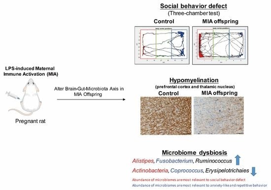
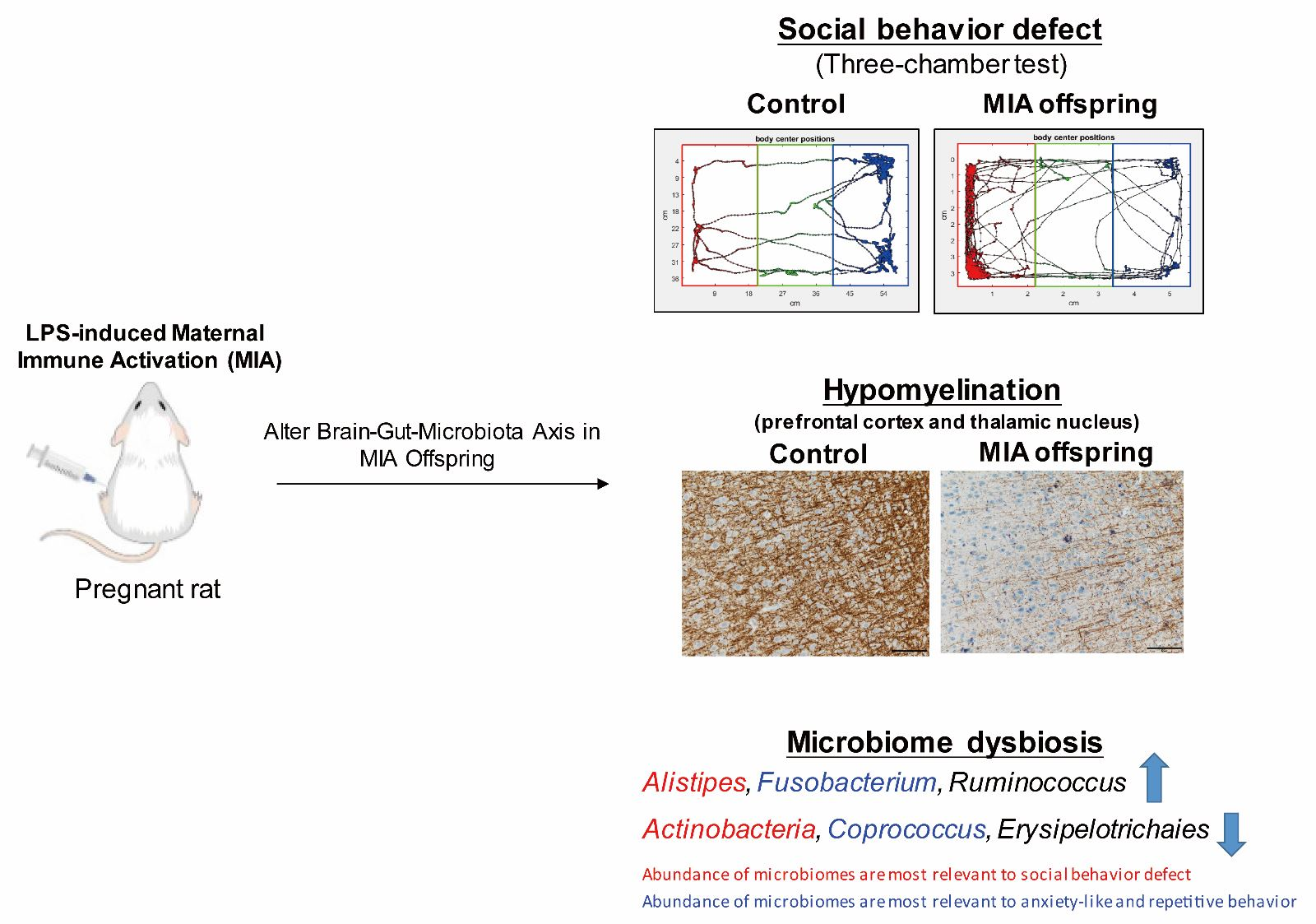
Maternal Immune Activation Causes Social Behavior Deficits and Hypomyelination in Male Rat Offspring with an Autism-Like Microbiota Profile

1
Department of Medical Research, Taipei Medical University Hospital, Taipei 110, Taiwan
2
Department of Microbiology and Immunology, School of Medicine, College of Medicine, Taipei Medical University, Taipei 110, Taiwan
3
Graduate Institute of Athletics and Coaching Science, National Taiwan Sport University, Taoyuan 333, Taiwan
4
Core Laboratory of Neuroscience, Office of R&D, Taipei Medical University, Taipei 110, Taiwan
5
Center for Neurotrauma and Neuroregeneration, Taipei Medical University, Taipei 110, Taiwan
6
PhD Program for Neural Regenerative Medicine, College of Medical Science and Technology, Taipei Medical University, Taipei 110, Taiwan
7
Joint Biobank, Office of Human Research, Taipei Medical University, Taipei 110, Taiwan
8
PhD Program for Clinical Drug Discovery of Herbal Medicine, College of Pharmacy, Taipei Medical University, Taipei 110, Taiwan
9
Graduate Institute of Pharmacognosy Science, College of Pharmacy, Taipei Medical University, Taipei 110, Taiwan
10
Department of Pharmaceutical Sciences, Taipei Medical University, Taipei 110, Taiwan
11
Department of Surgery, College of Medicine, Taipei Medical University, Taipei 110, Taiwan
12
Graduate Institute of Neural Regenerative Medicine, College of Medical Science and Technology, Taipei Medical University, Taipei 110, Taiwan
13
Department of Physical Medicine and Rehabilitation, School of Medicine, College of Medicine, Taipei Medical University, No. 250 Wu Hsing Street, Taipei 110, Taiwan
14
Department of Physical Medicine and Rehabilitation, Taipei Medical University Hospital, Taipei 110, Taiwan
*
Author to whom correspondence should be addressed.
Brain Sci. 2021, 11(8), 1085; https://doi.org/10.3390/brainsci11081085
Submission received: 16 July 2021 / Revised: 16 August 2021 / Accepted: 16 August 2021 / Published: 18 August 2021

Abstract

:
Maternal immune activation (MIA) increases the risk of autism spectrum disorder (ASD) in offspring. Microbial dysbiosis is associated with ASD symptoms. However, the alterations in the brain–gut–microbiota axis in lipopolysaccharide (LPS)-induced MIA offspring remain unclear. Here, we examined the social behavior, anxiety-like and repetitive behavior, microbiota profile, and myelination levels in LPS-induced MIA rat offspring. Compared with control offspring, MIA male rat offspring spent less time in an active social interaction with stranger rats, displayed more anxiety-like and repetitive behavior, and had more hypomyelination in the prefrontal cortex and thalamic nucleus. A fecal microbiota analysis revealed that MIA offspring had a higher abundance of Alistipes, Fusobacterium, and Ruminococcus and a lower abundance of Coprococcus, Erysipelotrichaies, and Actinobacteria than control offspring, which is consistent with that of humans with ASD. The least absolute shrinkage and selection operator (LASSO) method was applied to determine the relative importance of the microbiota, which indicated that the abundance of Alistipes and Actinobacteria was the most relevant for the profile of defective social behavior, whereas Fusobacterium and Coprococcus was associated with anxiety-like and repetitive behavior. In summary, LPS-induced MIA offspring showed an abnormal brain–gut–microbiota axis with social behavior deficits, anxiety-like and repetitive behavior, hypomyelination, and an ASD-like microbiota profile.

Graphical Abstract

1. Introduction

Maternal immune activation (MIA) has been linked to an increased risk of neurodevelopmental psychiatric disorders in offspring [1,2]. Animal models of MIA have been developed by activating the immune system with immunogens during pregnancy and then observing the development of defective behaviors in offspring with autism-like behavior [3]. MIA generates inflammatory cytokines to which the fetus is exposed during mid-gestation, possibly affecting fetal brain development [4]. Furthermore, the presence of inflammatory molecules and cytokines can affect central nervous system development [5] and adversely affect neuron survival [6].
Gut microbiota plays a critical role in regulating host physiology, metabolism, nutrition, and brain function [7]. Microbial dysbiosis is correlated with various adverse consequences, including behavioral abnormalities, neuropathology, immune dysfunction, and deficient gastrointestinal integrity [7]. The prenatal environment affects the microbiome of offspring [8]. Microbial dysbiosis is associated with the symptoms of autism spectrum disorder (ASD), including impaired social communication and repetitive behaviors [9]. However, how prenatal infection affects the brain–gut–microbiota axis, which regulates behavioral phenotypes, remains unclear.
Maternal lipopolysaccharide (LPS) exposure causes reproductive, behavioral, and neurochemical abnormalities in offspring [10]. LPS, an endotoxin, activates immune cells to release proinflammatory cytokines, which induce maternal cytokine responses and may increase the risk of atypical brain development [11]. Prenatal LPS treatment causes social behavior deficits in male offspring [9,12]. Pregnant rats injected with intraperitoneal LPS on gestation day (GD) 9.5 were demonstrated to induce the most relevant long-term neuropathological consequences in offspring [13,14]. However, the microbiota changes have not been quantified in these MIA offspring.
In this study, we investigated the relationship between microbiota and ASD-related behavior and demonstrated brain myelination changes in MIA offspring. Furthermore, we used the least absolute shrinkage and selection operator (LASSO) method to determine the most relevant microbiota genera associated with ASD-related behaviors. Taken together, we demonstrated that an abnormal brain–gut–microbiota axis with phenotypes includes a social behavior deficit, anxiety-like and repetitive behavior, hypomyelination, and dysbiosis microbiota in MIA offspring; thus, providing a link between maternal infection and the etiopathogenesis of ASD.

2. Materials and Methods

2.1. LPS-Induced MIA Rat Model

All animal procedures were approved by the Animal Care and Use Committees of Taipei Medical University. Eight-week-old Wistar female rats (BioLASCO, Taipei, Taiwan) in their first pregnancy were used in this study. Female rats were mated overnight with male rats with mating experience and were checked for the presence of a vaginal plug the subsequent morning to confirm mating. Pregnant rats were housed individually and allowed to raise their own litters until weaning. On GD 9.5, 500 μg/kg LPS (Escherichia coli O127:B8) or phosphate-buffered saline (PBS) was injected intraperitoneally into pregnant rats. Litters were left undisturbed until weaning at postnatal day 21. Offspring were housed in same-sex cages containing three rats until the end of the experiments. All animals were housed in temperature-controlled rooms under a 12 h light/dark cycle with ad libitum access to water and same food. All behavioral testing was performed during rats’ light cycle between 9 a.m. and 5 p.m. The experiments were performed in accordance with guidelines by the International Council for Laboratory Animal Science (ICLAS) for the care and use of laboratory animals for experiments.

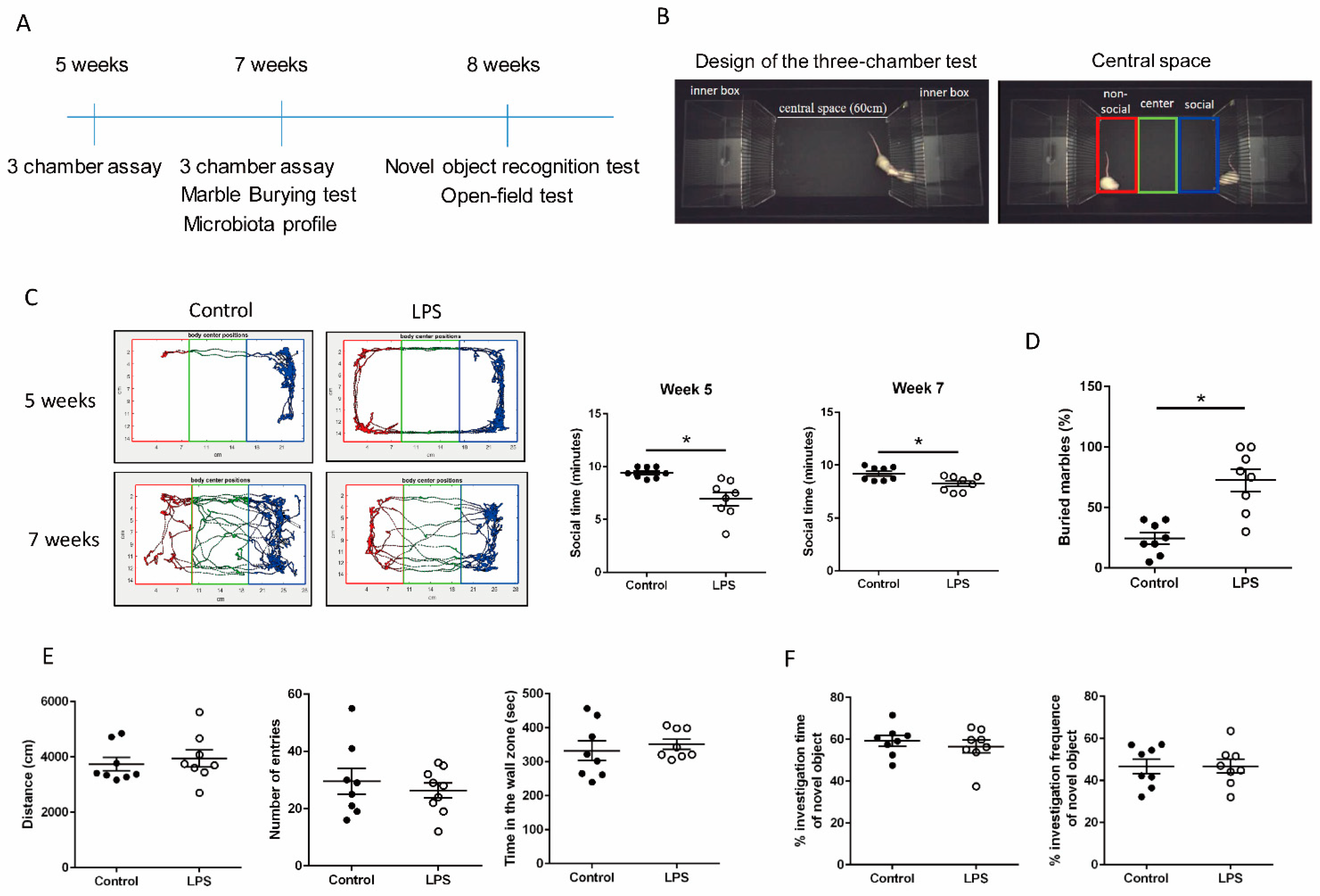
2.2. Three-Chamber Test

The three-chamber social interaction test was adapted from a previous study [15]. Stranger rats were age- and sex-matched with the testing rats. The body weights of stranger rats at 5 and 7 weeks were 130 and 195 g, respectively. Stranger rat was placed in the right-side chamber of three-chambered apparatus (Deep Brain Tech, Taiwan) to test the social interaction between stranger and the test rat during a 10 min test session (Figure 1B). The left chamber remained empty. The sociability score, used to measure the social preference of test rat was defined as the time spent in the social region of the central space near stranger rat during test session (Figure 1B). Test rat’s behavior was recorded using a camera (The Imaging Source, DFK 33UP1300, Bremen, Germany). OptiMouse software was used to calculate the total time spent by rats in three zones of the central space (nonsocial, center, and social regions) in a three-chambered apparatus (Figure 1B).

2.3. Marble-Burying Test

The marble-burying test was used to examine the test rat’s anxiety-like and repetitive behavior. A clean cage (22 × 45 × 20 cm3) was prepared with a 4 cm corncob bedding material containing 20 embedded marbles. After 30 min, the number of marbles that remained buried in the corncob bedding was recorded.

2.4. Novel Object Recognition Test

Recognition memory was evaluated using the novel object recognition (NOR) test, which was used to compare the amount of time a rat spent investigating a novel object versus a familiar object. The familiar object was a plastic square block, and the novel object was a plastic V-shaped block. First, rats were habituated to an open-field Plexiglas arena (60 × 60 × 100 cm3) for 10 min. After habituation, the rats were allowed to explore two identical familiar objects for 10 min during the familiarization phase. After a 24 h intertrial interval, one familiar object was replaced with a novel object, and the rats were returned to the arena and allowed to explore the two objects. A preference index, which measured the time spent by the test rat exploring the novel object over the familiar one, was calculated as a percentage using the following equation: ((Time N − Time F)/(Time N + Time F)) × 100, where F and N represent the time spent near the familiar and novel objects, respectively.

2.5. Open-Field Test

Using an open-field test, we evaluated the general motor activity and anxiety-related exploratory activity in the MIA and control rats. Rats were first habituated to an open-field Plexiglas arena (60 × 60 × 100 cm3) for 10 min. Their locomotor activity and anxiety-like behavior were monitored for 10 min using open-field tests and recorded using an EthoVision system. General locomotor activity was defined as the total distance traveled in the open field. Anxiety-like behavior was measured based on the number of entries and retention times in the wall zone of the open field. The center of the open field was defined as a 30 × 30-cm2 area in the geometric center of the arena. The wall zone was defined as a peripheral zone 5 cm from all four sides.

2.6. 16S rRNA Gene Sequencing and Next-Generation Sequencing
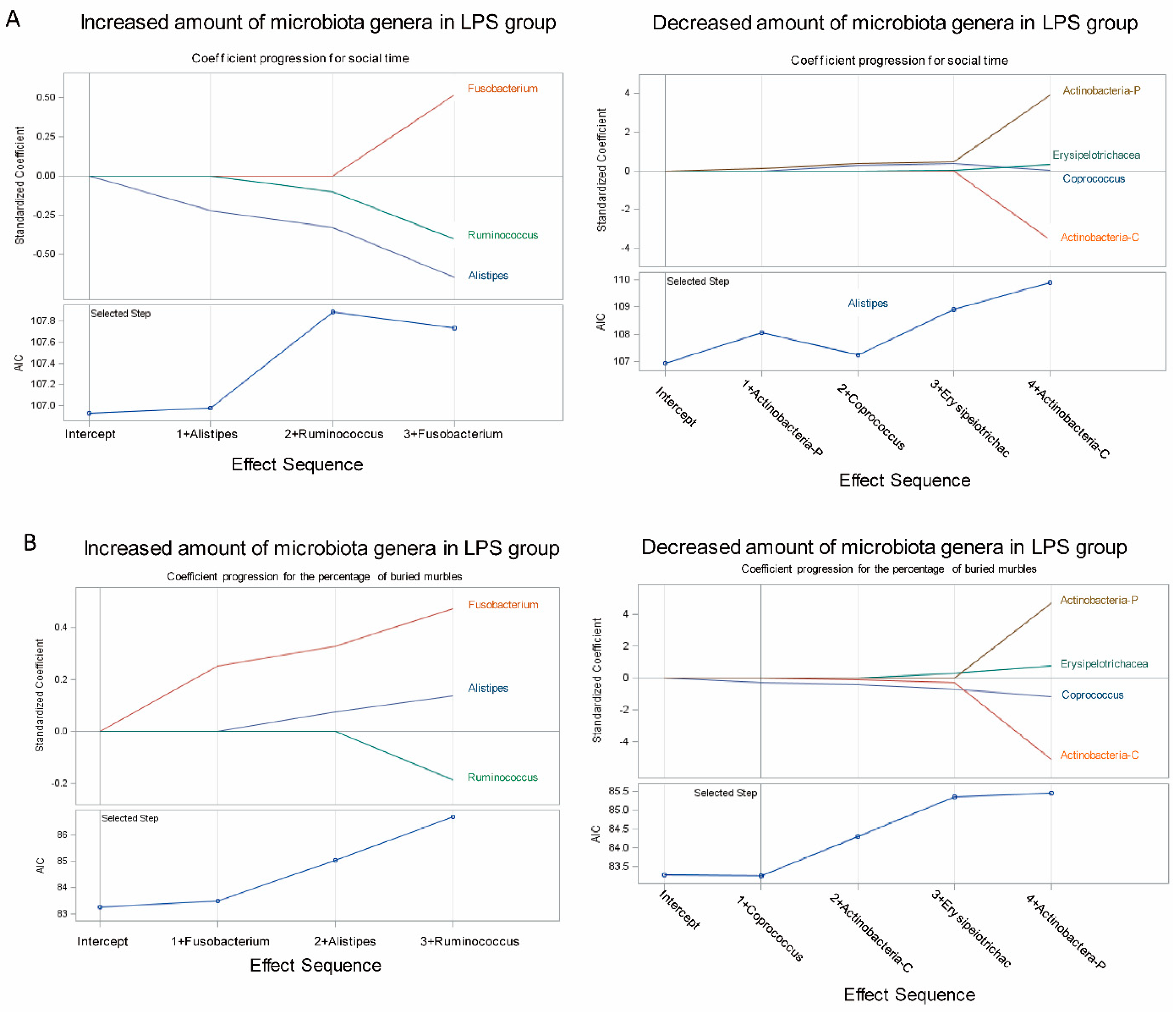
The stool samples of the rats were purified using the QIAamp Fast DNA Stool Mini Kit (QIAGEN, Germany). Library preparation was conducted following the protocol of 16S ribosomal RNA gene amplicons for the Illumina MiSeq system. Sequence reads were deposited in the European Nucleotide Archive (accession number: PRJEB28574). Universal primers (341F and 805R) were used to amplify the V3–V4 region of bacterial 16S rRNA genes; they were first removed from demultiplexed, paired reads using Cutadapt (v1.12; DOI:10.14806/ej.17.1.200). The filtered reads were then processed using the DADA2 package (v1.3.5) in R (v3.3.3) [16], following the workflow described by Callahan et al. [17], but without the rarefying procedure. Briefly, the forward and reverse reads were filtered and trimmed based on the read quality score and read length. Dereplication was then performed to merge identical reads, and the reads were then subjected to the denoising algorithm DADA2; the reads alternated between error rate estimation and sample composition inference until they converge into a consistent solution. Finally, paired reads with ≥20-bp overlap were merged, and chimeras were removed. A list of V3–V4 sequence variants found in the samples, which were inferred with DADA2, and the frequency of each sequence variant in each sample were obtained. Taxonomy assignment was performed using the SILVA database (v128) [18] for reference with a minimum bootstrap confidence of 80. Multiple sequence alignment of variants was performed using DECIPHER (v2.2.0) and the phylogenetic tree was constructed from the alignment using phangorn (v2.2.0) [19]. The count table, taxonomy assignment results, and phylogenetic tree were consolidated into a phyloseq object, and community analyses were performed using phyloseq (v1.19.1) [20]. The alpha-diversity indices were calculated using the estimated richness function of the phyloseq package. Data from the treatment and control groups were compared using the Wilcoxon–Mann–Whitney test (at α = 0.05). UniFrac distances were calculated using the GUniFrac package (v1.1) to assess community dissimilarity between the groups [21]. Principal coordinate analysis ordination on UniFrac distances was performed, and the adonis and betadisper functions from the vegan package (v2.4; https://CRAN.R-project.org/package=vegan, accessed on 24 August 2017) were used to analyze the dissimilarity of composition among the groups and the homogeneity of dispersion, respectively. Vegan is R package which provides tools for descriptive community ecology. It includes functions of diversity analysis, community ordination, and dissimilarity analysis. Adonis and Betadisper are the sister functions of Vegan package. Adonis analyzes and partitions the sum of squares using distance matrices. It can be seen as an ANOVA using distance matrices (analogous to MANOVA—multivariate analysis of variance). Therefore, it could test if two or more groups have similar compositions. Betadisper first calculates the average distance of group members to the group centroid in multivariate space (generated by a distance matrix). Then, an ANOVA is conducted to test if the dispersions (variances) of groups are different.

2.7. LASSO Method

In the LASSO algorithm, the feature importance was ranked in the framework of general linear models. Specifically, Tibshirani’s LASSO method was adopted (Tibshirani, 1996). LASSO arises from a constrained form of ordinary least squares regression, where the sum of the absolute values of the regression coefficients is constrained to be smaller than a specified parameter. The LASSO procedure offers extensive capabilities for customizing the selection with a wide variety of selection and stopping criteria, such as the Akaike information criterion (AIC).

2.8. Immunohistochemistry

The rats were euthanized and transcardially perfused with 25 mL of PBS and 4% paraformaldehyde. Whole brains were fixed with 4% paraformaldehyde for approximately 3 days. Paraformaldehyde-fixed 2 mm coronal slices were embedded in paraffin and cut into 5 µm thick sections, which were deparaffinized and rehydrated using a graded series of ethanol solutions. The sections then underwent an antigen retrieval process and were stained using horseradish peroxidase anti-myelin basic protein (MBP) antibody (BioLegend, catalog number 808405). Next, 3,3′-diaminobenzidine and hematoxylin staining was performed on sections by using the Chemicon IHC Select system (Millipore, catalog number DAB050). The sections were observed through microscopy (Olympus/Bx43). The MBP-positive area was calculated from sections by using HistoQuest tissue analysis software V2.0 (TissueGnostics, Vienna, Austria).

2.9. Statistical Analysis

Unpaired t-test were performed for behavioral data analyses using GraphPad Prism software. Error bars represent the standard error of mean. Microbiota enrichment analysis between the groups was conducted using the linear discriminant analysis (LDA) effect size (LEfSe) method. Data were compared using the Kruskal–Wallis and Wilcoxon tests; differences were considered significant at p ≤ 0.05 and a logarithmic LDA score of ≥ 2 [22]. Data were visualized as cladograms created using GraPhlAn [23].

3. Results

3.1. Maternal LPS Stimulation Causes Social Behavior Deficits and Anxiety-Like and Repetitive Behavior in MIA Male Offspring

No significant alterations in reproductive parameters were found in female rats exposed to prenatal LPS treatment (LPS-treated group) or PBS treatment (control group). The number of pups born in each litter, the parturition day, or individual bodyweight of offspring at 5 and 7 weeks showed no significant difference between prenatal LPS and PBS treatments (data not shown). To test whether MIA offspring began displaying social behavior deficits before or after sexual maturity, we examined their social behavior at 5 and 7 weeks. We used OptiMouse software to track the movement of rats in our three-chamber apparatus during the social behavior tests. Male offspring from the prenatal LPS-treated group spent less time in the social region of the central space in the three-chamber apparatus than those from the PBS-treated control group at both 5 and 7 weeks (Figure 1B,C), whereas female offspring had a normal social behavior (data not shown). Thus, male MIA offspring displayed social behavior deficits before and after sexual maturity. MIA male offspring also buried more marbles than the control male offspring (Figure 1D), which indicates anxiety-like and repetitive behavior. MIA and control male offspring had no differences in the total distance moved, number of entries, or time spent in the wall zone of the open-field assay (Figure 1E). Male offspring from the LPS group demonstrated a similar preference for the novel object compared with male offspring from the control group (Figure 1F). Taken together, the results revealed that locomotor activity and cognition were similar in LPS and control groups.

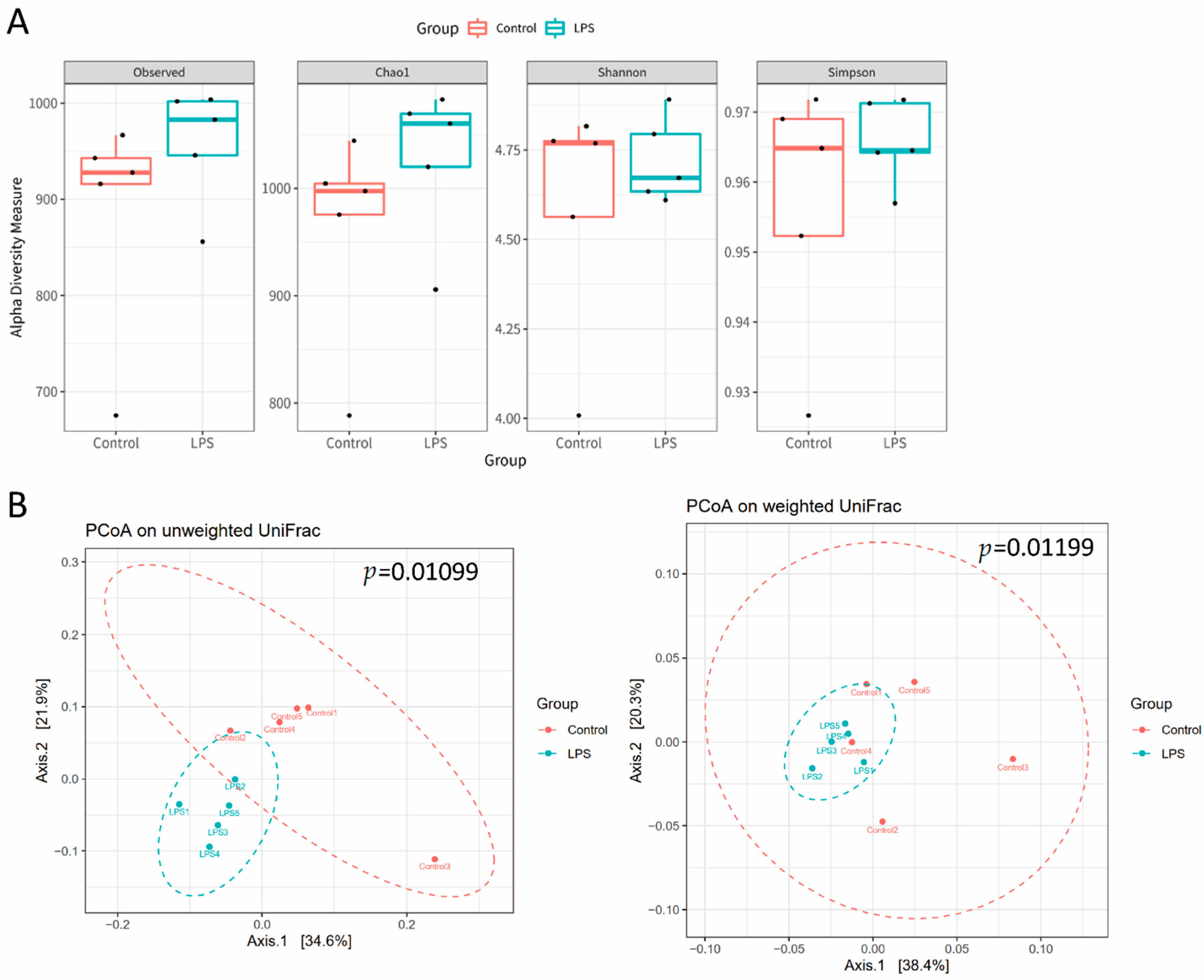
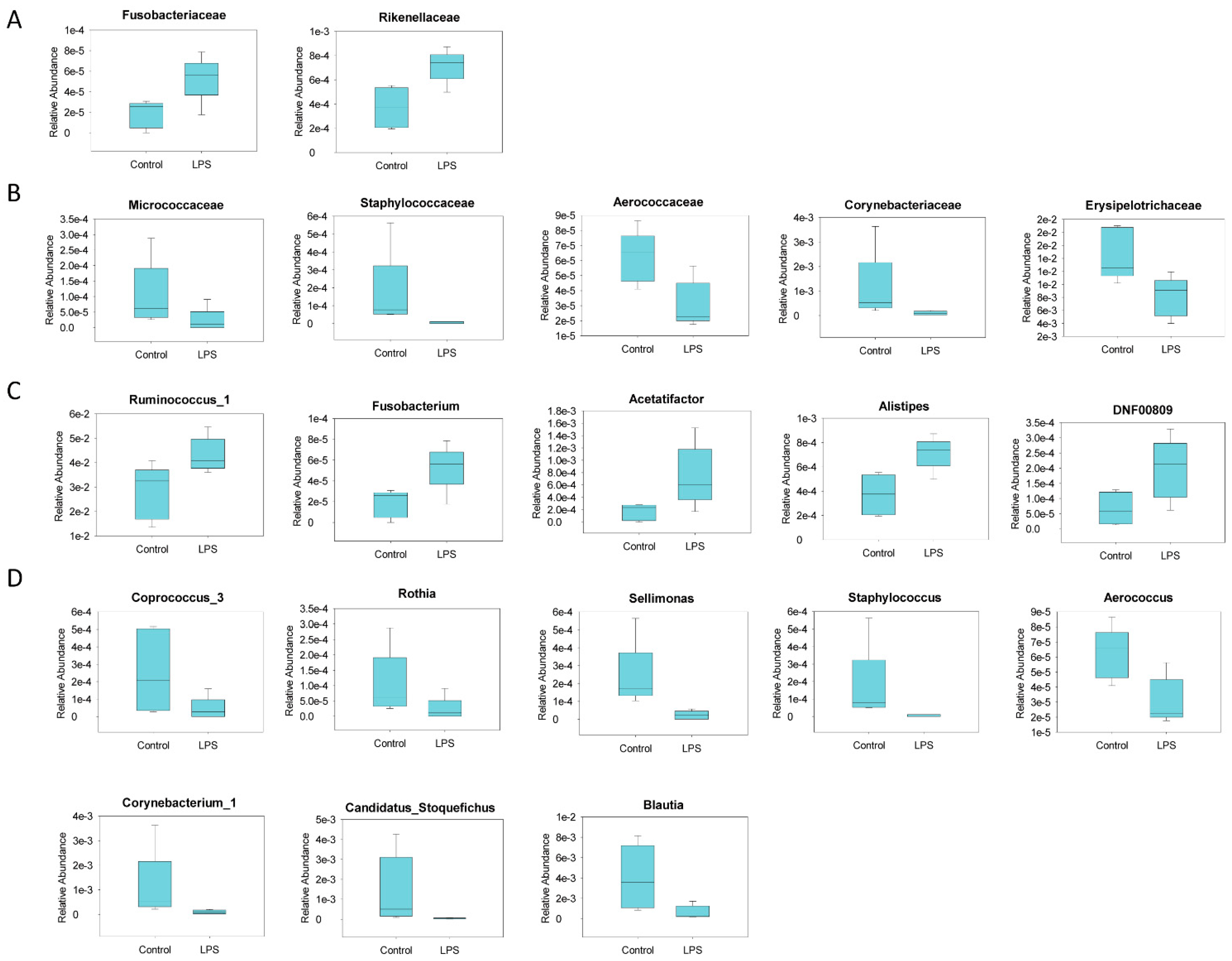
3.2. Fecal Microbiome Profile in Male MIA Offspring Is Similar to That of Patients with ASD

The gut microbiota profile in ASD was determined by identifying fecal microbiota through 16S rRNA gene sequencing and next-generation sequencing [24]. We observed that the fecal microbiota of prenatal LPS-treated male offspring had slightly, but not significantly, higher alpha diversity than that of the control offspring (Figure 2A). An unweighted and weighted UniFrac principal coordinate analysis indicated that the fecal microbiota profile of male MIA offspring was significantly different from that of PBS-treated male offspring (Figure 2B). A significant increase in Fusobacteria abundance and decrease in Actinobacteria abundance at the phylum level of microbial composition was observed in MIA offspring compared with that in control offspring (Figure 3 and Table 1). Compared with control offspring, MIA male offspring had a significantly increased abundance of Fusobacteriaceae and Rikenellaceae families (Figure 4A), and significantly decreased abundance of Micrococcaceae, Staphylococcaceae, Aerococcaceae, Corynebacteriaceae, and Erysipelotrichaceae families (Figure 4B). At the genus level, compared with control offspring, MIA male offspring had significantly increased Ruminococcus_1, Fusobacterium, Acetatifactor, Alistipes, and DNF00809 (Figure 4C), and significantly decreased Coprococcus_3, Rothia, Sellimonas, Staphylococcus, Aerococcus, Corynebacterium_1, Candidatus_Stoquefichus, and Blautia (Figure 4D).

3.3. Association of Fecal Microbiome Profile with Social Behavior and Anxiety-Like and Repetitive Behavior in MIA Male Offspring

The increased abundance of Alistipes, Fusobacterium, and Ruminococcus and decreased abundance of Coprococcus [25,26,27], Erysipelotrichaies, and Actinobacteria in LPS-induced MIA offspring were consistent with that of humans with ASD [28,29,30]. Next, we determined the association of ASD-related microbiota (increased or decreased amount of microbiota in the LPS group) with the level of the social behavior deficit and with anxiety-like and repetitive behavior by using the LASSO method. The LASSO procedure offers extensive capabilities to build a model that can determine the coefficient progression of selected microbiota with the profile of the indicated phenotypes (social behavior time and the percentage of buried marbles). The AIC method was applied to determine which microbiota profile was the best fit for the model that could reflect the profile of social behavior time and anxiety-like and repetitive behavior. Figure 5A indicates that the AIC values of Alistipes and phylum Actinobacteria were the smallest in terms of the information loss; thus, the abundance of Alistipes and phylum Actinobacteria had a higher association level with the profile of defective social behavior than the other microbiota, and Fusobacterium and Coprococcus had a higher association level with the profile of buried marble percentage based on AIC (Figure 5B).

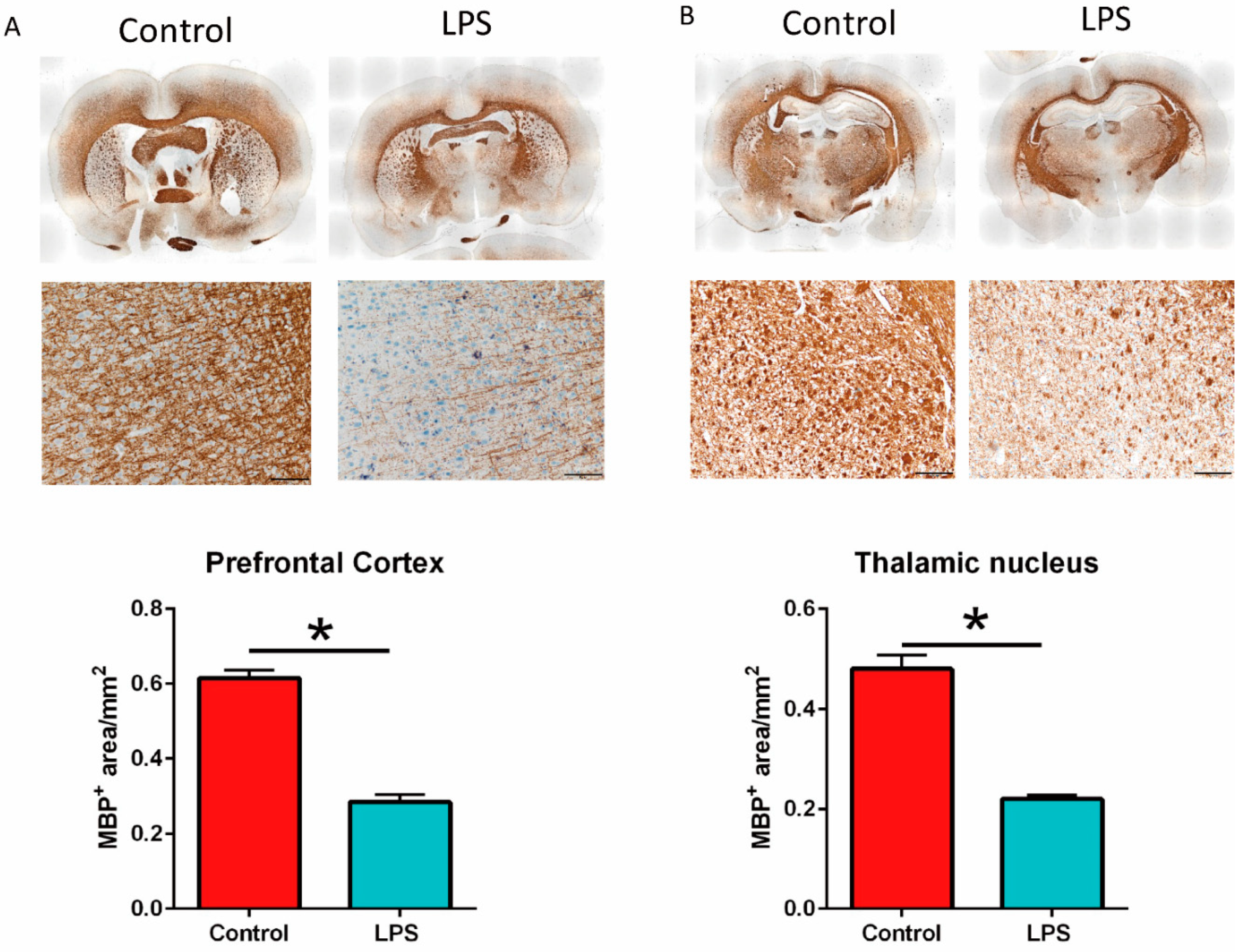
3.4. Maternal LPS Stimulation Causes Hypomyelination in the Prefrontal Cortex and Thalamus Nucleus in MIA Male Offspring

Prenatal LPS stimulation at gestation days 15 and 16 causes abnormal myelination in the cortical and limbic brain regions in offspring [31]. In our study, prenatal LPS stimulation at gestation day 9.5 resulted in a significantly decreased MBP+ area in the prefrontal cortex (Figure 6A) and thalamic nucleus (Figure 6B) of the male MIA offspring (LPS group) than in control offspring by immunohistochemistry staining. In other words, maternal LPS stimulation caused hypomyelination in the prefrontal cortex and thalamic nucleus of adult male offspring.

4. Discussion

MIA causes an altered brain–gut–microbiota profile in male offspring, with autism-like phenotypes. In this study, we demonstrated that prenatal LPS stimulation caused altered social behavior and anxiety-like and repetitive behavior in male offspring, as revealed by our three-chamber test and marble-burying test, respectively. The NOR test and open-field assay demonstrated that MIA male offspring had a behavior profile similar to that of control offspring, indicating normal recognition memory and locomotor activity. MIA-induced hypomyelination in the prefrontal cortex and thalamic nucleus and altered microbiota profile provided evidence to show that LPS stimulation in the gestation stage causes altered brain–gut–microbiota axis phenotypes in offspring.
Patients with ASD have alterations in the gut microbiota composition compared with individuals without ASD [7,9]. In the present study, the gut microbiota diversity in male MIA offspring with ASD phenotypes was similar to the fecal microbiota profiles of children with ASD, such as a significant increase in Ruminococcus, Fusobacterium, and Alistipes [25,26,27]. We also observed a significant reduction in Coprococcus, Erysipelotrichaies, and Actinobacteria, which is consistent with the results of previous studies [28,29,30]. Compared with children without ASD, multiple microbiota species are elevated or reduced in children with ASD who have complex neurodevelopmental disorders involving disruptions in language and social behavior, restricted interests, and repetitive behaviors [9]. Microbiota transfer therapies have reported an improvement in ASD behavioral symptoms [32,33], suggesting that correcting the altered microbiota profile is a promising treatment strategy for ASD. Among the various changed microbiota species in MIA offspring, we demonstrated that the abundance of Alistipes and Actinobacteria was associated with the profile of social behavior, and Fusobacterium and Coprococcus was associated with the profile of anxiety-like and repetitive behavior; this suggests that Alistipes, Actinobacteria, Fusobacterium, and Coprococcus may be microbiome biomarkers or treatment targets for ASD.
MIA animal models are unique experimental tools for overcoming the limitations of epidemiological studies, such as the longitudinal evaluation of neurobiological processes from gestation to adulthood. Maternal exposure to LPS induces hypomyelination in the internal capsule and in newborn rats [34]. Maternal valproic acid stimulation causes hypomyelination of the prefrontal cortex and has been associated with social behavior deficit [35]. Poly I:C-induced MIA stimulation also causes a disruption in the myelin structure and a weakened thalamocortical connection in offspring [36]. In the present study, prenatal LPS-induced MIA caused the following changes in adult offspring: social behavior deficit, anxiety-like and repetitive behavior, ASD-related microbiota, and hypomyelination in the prefrontal cortex and thalamic nucleus. Prenatal LPS stimulation causes the inflammatory responses in brain of MIA offspring. The elevated levels of inflammatory cytokine (TNF-α and IL-1β) in the gestational stage are associated with behavioral impairment and hypomyelination [34,37]. The activation of an inflammatory reaction, including pro-inflammatory and anti-inflammatory cytokines, is associated with ASD [38,39,40,41]. In addition, poly I:C-induced MIA increases TNF-α and IL-1β expression in the colon of MIA offspring with an altered microbiota profile [42]. These results suggest that maternal LPS stimulation might impart an inflammatory reaction that is associated with an altered brain–gut–microbiota axis. Together, these findings suggest that MIA stress alters the brain–gut–microbiota axis leading to an inflammatory reaction, defective myelination and abnormal microbiota in offspring in ASD-related behavior deficits.
The most commonly used MIA animal models are prenatal poly I:C and LPS stimulation in pregnant animals. Poly I:C is a synthetic analogue of the viral double-strand RNA which activates Toll-like receptors (TLR)-3, whereas LPS is an endotoxin from Gram-negative bacteria which activates TLR-4 [43]. Prenatal poly I:C stimulation can alter the MIA microbiota profile in MIA offspring with a behavior impairment [29,42,44,45]. However, the link of an altered microbiota and social behavior impairment remains unclear in the LPS-induced MIA animal model. A previous study demonstrated several different effects of poly I:C and LPS prenatal stimulation on the behavior, development, and inflammatory response in pregnant mice and their offspring [46]. For example, prenatal LPS stimulation caused a decrease in the astrocytic marker (GFAP) and neuronal marker (NeuN) expression level in offspring at GD18, whereas GFAP and NeuN expression levels were not altered in the poly I:C-induced MIA model [46]. In addition, plasma cytokine IL-2, IL-5, IL-6, and inflammatory marker mGLuR5 were significantly increased in the brain of poly I:C-induced MIA offspring, but not in LPS-induced MIA offspring [46]. In this study, we found that human ASD-related Ruminococcus are increased in the LPS-induced MIA model, whereas Ruminococcus are decreased in the poly I:C-induced MIA model [29]. Taken together, different prenatal stimulation sources (virus or bacteria) may induce a differential profile of brain cell markers and microbiota profile in MIA offspring.
The prevalence of ASD is higher in males, with an approximate male-to-female ratio of 3:1 [47]. In this study, our MIA female offspring had normal social behavior when compared to the control group. In addition, sex may affect the microbiota profile [48]. For example, Coprococcus was less present in the microbiota profile of male mice compared to female mice [49]. These data were consistent to our study, revealing a decreased number of Coprococcus in the microbiota profile of male rats compared to female rats (data not shown). A human study showed that healthy males possess higher levels of Fusobacterium than females [50]. In an ASD predominantly male cohort study (18 males and 2 females), ASD children had decreased Coprococcus than the healthy subjects [28]. Another study showed that ASD patients with constipation (25 male and 5 female subjects) had increased Fusobacterium compared with healthy subjects [26]. Taken together, this evidence suggests that the differences in microbiota profiles between male and female subjects may be correlated to the altered microbiota profiles of ASD patients.

5. Conclusions

In the present study, LPS-induced MIA offspring displayed altered brain–gut–microbiota axis phenotypes, including social behavior deficits, anxiety-like and repetitive behavior, a human ASD-like microbiota profile (higher abundance of Alistipes, Fusobacterium, and Ruminococcus and a lower abundance of Coprococcus, Erysipelotrichaies, and Actinobacteria than control offspring), and hypomyelination in the prefrontal cortex and thalamic nucleus. An abundance of Alistipes and Actinobacteria was the most relevant for the profile of defective social behavior, whereas the abundance of Fusobacterium and Coprococcus was associated with anxiety-like and repetitive behavior. These potential ASD-related microbiomes require further studies to prove their direct association to ASD-related behavior, such as fecal transplantation or target microbiome transplantation therapy. The relative mechanisms of ASD etiopathogenesis still remain unknown and further studies are needed to prove the cause–effect relations between microbiome dysbiosis and ASD. Our findings provide insights into the relationship between maternal infection and the etiopathogenesis of ASD with an abnormal brain–gut–microbiota axis.

Author Contributions

Conceptualization, G.A.L. and S.-H.T.; methodology, G.A.L. and Y.-K.L.; software, G.A.L.; validation, G.A.L.; formal analysis, Y.-K.L. and Y.-C.S.H.Y.; investigation, S.-Y.Y., J.-H.L. and Y.-C.L.; resources, S.-Y.Y., C.-J.L., C.-C.W. and Y.-H.C.; data curation, G.A.L.; writing—original draft preparation, G.A.L.; writing—review and editing, G.A.L.; visualization, G.A.L.; supervision, S.-H.T.; project administration, S.-H.T.; funding acquisition, S.-H.T. All authors have read and agreed to the published version of the manuscript.

Funding

This research was funded by the Taipei Medical University (grant number TMU A00000952) and the Taipei Medical University Hospital (grant number 110TMU-TMUH-09).

Institutional Review Board Statement

All animal procedures were approved by the Animal Care and Use Committees of Taipei Medical University (LAC-2019-0109).

Data Availability Statement

The datasets used and/or analyzed during the current study are available from the corresponding author on reasonable request.

Acknowledgments

This manuscript was edited by Wallace Academic Editing. We would like to acknowledge the support provided by the Core Laboratory of Human Microbiome, Taipei Medical University.

Conflicts of Interest

The authors declare no conflict of interest.

References

  1. Atladottir, H.O.; Thorsen, P.; Ostergaard, L.; Schendel, D.E.; Lemcke, S.; Abdallah, M.; Parner, E.T. Maternal infection requiring hospitalization during pregnancy and autism spectrum disorders. J. Autism Dev. Disord. 2010, 40, 1423–1430. [Google Scholar] [CrossRef]
  2. Ciaranello, A.L.; Ciaranello, R.D. The neurobiology of infantile autism. Annu. Rev. Neurosci. 1995, 18, 101–128. [Google Scholar] [CrossRef] [PubMed]
  3. Careaga, M.; Murai, T.; Bauman, M.D. Maternal Immune Activation and Autism Spectrum Disorder: From Rodents to Nonhuman and Human Primates. Biol. Psychiatry 2017, 81, 391–401. [Google Scholar] [CrossRef] [PubMed] [Green Version]
  4. Dahlgren, J.; Samuelsson, A.M.; Jansson, T.; Holmang, A. Interleukin-6 in the maternal circulation reaches the rat fetus in mid-gestation. Pediatr. Res. 2006, 60, 147–151. [Google Scholar] [CrossRef] [Green Version]
  5. Boksa, P. Effects of prenatal infection on brain development and behavior: A review of findings from animal models. Brain Behav. Immun. 2010, 24, 881–897. [Google Scholar] [CrossRef]
  6. Qiu, Z.; Sweeney, D.D.; Netzeband, J.G.; Gruol, D.L. Chronic interleukin-6 alters NMDA receptor-mediated membrane responses and enhances neurotoxicity in developing CNS neurons. J. Neurosci. 1998, 18, 10445–10456. [Google Scholar] [CrossRef]
  7. Vuong, H.E.; Hsiao, E.Y. Emerging Roles for the Gut Microbiome in Autism Spectrum Disorder. Biol. Psychiatry 2017, 81, 411–423. [Google Scholar] [CrossRef] [PubMed] [Green Version]
  8. Conway, F.; Brown, A.S. Maternal Immune Activation and Related Factors in the Risk of Offspring Psychiatric Disorders. Front. Psychiatry 2019, 10, 430. [Google Scholar] [CrossRef]
  9. Rosenfeld, C.S. Microbiome Disturbances and Autism Spectrum Disorders. Drug Metab. Dispos. 2015, 43, 1557–1571. [Google Scholar] [CrossRef] [Green Version]
  10. Golan, H.M.; Lev, V.; Hallak, M.; Sorokin, Y.; Huleihel, M. Specific neurodevelopmental damage in mice offspring following maternal inflammation during pregnancy. Neuropharmacology 2005, 48, 903–917. [Google Scholar] [CrossRef] [PubMed]
  11. Jonakait, G.M. The effects of maternal inflammation on neuronal development: Possible mechanisms. Int. J. Dev. Neurosci. 2007, 25, 415–425. [Google Scholar] [CrossRef]
  12. Kirsten, T.B.; Taricano, M.; Maiorka, P.C.; Palermo-Neto, J.; Bernardi, M.M. Prenatal lipopolysaccharide reduces social behavior in male offspring. Neuroimmunomodulation 2010, 17, 240–251. [Google Scholar] [CrossRef] [PubMed]
  13. Meyer, U.; Yee, B.K.; Feldon, J. The neurodevelopmental impact of prenatal infections at different times of pregnancy: The earlier the worse? Neuroscientist 2007, 13, 241–256. [Google Scholar] [CrossRef] [PubMed]
  14. Meyer, U.; Nyffeler, M.; Engler, A.; Urwyler, A.; Schedlowski, M.; Knuesel, I.; Yee, B.K.; Feldon, J. The time of prenatal immune challenge determines the specificity of inflammation-mediated brain and behavioral pathology. J. Neurosci. 2006, 26, 4752–4762. [Google Scholar] [CrossRef] [Green Version]
  15. Wu, H.F.; Chen, P.S.; Hsu, Y.T.; Lee, C.W.; Wang, T.F.; Chen, Y.J.; Lin, H.C. D-Cycloserine Ameliorates Autism-Like Deficits by Removing GluA2-Containing AMPA Receptors in a Valproic Acid-Induced Rat Model. Mol. Neurobiol. 2018, 55, 4811–4824. [Google Scholar] [CrossRef]
  16. Callahan, B.J.; McMurdie, P.J.; Rosen, M.J.; Han, A.W.; Johnson, A.J.; Holmes, S.P. DADA2: High-resolution sample inference from Illumina amplicon data. Nat. Methods 2016, 13, 581–583. [Google Scholar] [CrossRef] [PubMed] [Green Version]
  17. Callahan, B.J.; Sankaran, K.; Fukuyama, J.A.; McMurdie, P.J.; Holmes, S.P. Bioconductor Workflow for Microbiome Data Analysis: From raw reads to community analyses. F1000Research 2016, 5, 1492. [Google Scholar] [CrossRef]
  18. Quast, C.; Pruesse, E.; Yilmaz, P.; Gerken, J.; Schweer, T.; Yarza, P.; Peplies, J.; Glockner, F.O. The SILVA ribosomal RNA gene database project: Improved data processing and web-based tools. Nucleic Acids Res. 2013, 41, D590–D596. [Google Scholar] [CrossRef] [PubMed]
  19. Schliep, K.P. phangorn: Phylogenetic analysis in R. Bioinformatics 2011, 27, 592–593. [Google Scholar] [CrossRef] [Green Version]
  20. McMurdie, P.J.; Holmes, S. phyloseq: An R package for reproducible interactive analysis and graphics of microbiome census data. PLoS ONE 2013, 8, e61217. [Google Scholar] [CrossRef] [Green Version]
  21. Chen, J.; Bittinger, K.; Charlson, E.S.; Hoffmann, C.; Lewis, J.; Wu, G.D.; Collman, R.G.; Bushman, F.D.; Li, H.Z. Associating microbiome composition with environmental covariates using generalized UniFrac distances. Bioinformatics 2012, 28, 2106–2113. [Google Scholar] [CrossRef]
  22. Segata, N.; Izard, J.; Waldron, L.; Gevers, D.; Miropolsky, L.; Garrett, W.S.; Huttenhower, C. Metagenomic biomarker discovery and explanation. Genome Biol. 2011, 12, R60. [Google Scholar] [CrossRef] [PubMed] [Green Version]
  23. Asnicar, F.; Weingart, G.; Tickle, T.L.; Huttenhower, C.; Segata, N. Compact graphical representation of phylogenetic data and metadata with GraPhlAn. PeerJ 2015, 3, e1029. [Google Scholar] [CrossRef] [PubMed]
  24. Zierer, J.; Jackson, M.A.; Kastenmuller, G.; Mangino, M.; Long, T.; Telenti, A.; Mohney, R.P.; Small, K.S.; Bell, J.T.; Steves, C.J.; et al. The fecal metabolome as a functional readout of the gut microbiome. Nat. Genet. 2018, 50, 790–795. [Google Scholar] [CrossRef] [PubMed]
  25. De Angelis, M.; Piccolo, M.; Vannini, L.; Siragusa, S.; De Giacomo, A.; Serrazzanetti, D.I.; Cristofori, F.; Guerzoni, M.E.; Gobbetti, M.; Francavilla, R. Fecal microbiota and metabolome of children with autism and pervasive developmental disorder not otherwise specified. PLoS ONE 2013, 8, e76993. [Google Scholar] [CrossRef] [Green Version]
  26. Liu, S.; Li, E.; Sun, Z.; Fu, D.; Duan, G.; Jiang, M.; Yu, Y.; Mei, L.; Yang, P.; Tang, Y.; et al. Altered gut microbiota and short chain fatty acids in Chinese children with autism spectrum disorder. Sci. Rep. 2019, 9, 287. [Google Scholar] [CrossRef] [PubMed]
  27. Wang, L.; Christophersen, C.T.; Sorich, M.J.; Gerber, J.P.; Angley, M.T.; Conlon, M.A. Increased abundance of Sutterella spp. and Ruminococcus torques in feces of children with autism spectrum disorder. Mol. Autism 2013, 4, 42. [Google Scholar] [CrossRef] [Green Version]
  28. Krajmalnik-Brown, R.; Lozupone, C.; Kang, D.W.; Adams, J.B. Gut bacteria in children with autism spectrum disorders: Challenges and promise of studying how a complex community influences a complex disease. Microb. Ecol. Health Dis. 2015, 26, 26914. [Google Scholar] [CrossRef]
  29. Hsiao, E.Y.; McBride, S.W.; Hsien, S.; Sharon, G.; Hyde, E.R.; McCue, T.; Codelli, J.A.; Chow, J.; Reisman, S.E.; Petrosino, J.F.; et al. Microbiota modulate behavioral and physiological abnormalities associated with neurodevelopmental disorders. Cell 2013, 155, 1451–1463. [Google Scholar] [CrossRef] [Green Version]
  30. Finegold, S.M.; Dowd, S.E.; Gontcharova, V.; Liu, C.; Henley, K.E.; Wolcott, R.D.; Youn, E.; Summanen, P.H.; Granpeesheh, D.; Dixon, D.; et al. Pyrosequencing study of fecal microflora of autistic and control children. Anaerobe 2010, 16, 444–453. [Google Scholar] [CrossRef]
  31. Wischhof, L.; Irrsack, E.; Osorio, C.; Koch, M. Prenatal LPS-exposure—A neurodevelopmental rat model of schizophrenia--differentially affects cognitive functions, myelination and parvalbumin expression in male and female offspring. Prog. Neuropsychopharmacol. Biol. Psychiatry 2015, 57, 17–30. [Google Scholar] [CrossRef]
  32. Kang, D.W.; Adams, J.B.; Gregory, A.C.; Borody, T.; Chittick, L.; Fasano, A.; Khoruts, A.; Geis, E.; Maldonado, J.; McDonough-Means, S.; et al. Microbiota Transfer Therapy alters gut ecosystem and improves gastrointestinal and autism symptoms: An open-label study. Microbiome 2017, 5, 10. [Google Scholar] [CrossRef]
  33. Abuaish, S.; Al-Otaibi, N.M.; Abujamel, T.S.; Alzahrani, S.A.; Alotaibi, S.M.; AlShawakir, Y.A.; Aabed, K.; El-Ansary, A. Fecal Transplant and Bifidobacterium Treatments Modulate Gut Clostridium Bacteria and Rescue Social Impairment and Hippocampal BDNF Expression in a Rodent Model of Autism. Brain Sci. 2021, 11, 1038. [Google Scholar] [CrossRef]
  34. Rousset, C.I.; Chalon, S.; Cantagrel, S.; Bodard, S.; Andres, C.; Gressens, P.; Saliba, E. Maternal exposure to LPS induces hypomyelination in the internal capsule and programmed cell death in the deep gray matter in newborn rats. Pediatr. Res. 2006, 59, 428–433. [Google Scholar] [CrossRef] [PubMed] [Green Version]
  35. Graciarena, M.; Seiffe, A.; Nait-Oumesmar, B.; Depino, A.M. Hypomyelination and Oligodendroglial Alterations in a Mouse Model of Autism Spectrum Disorder. Front. Cell. Neurosci. 2018, 12, 517. [Google Scholar] [CrossRef] [PubMed] [Green Version]
  36. Kreitz, S.; Zambon, A.; Ronovsky, M.; Budinsky, L.; Helbich, T.H.; Sideromenos, S.; Ivan, C.; Konerth, L.; Wank, I.; Berger, A.; et al. Maternal immune activation during pregnancy impacts on brain structure and function in the adult offspring. Brain Behav. Immun. 2020, 83, 56–67. [Google Scholar] [CrossRef] [PubMed]
  37. Simoes, L.R.; Sangiogo, G.; Tashiro, M.H.; Generoso, J.S.; Faller, C.J.; Dominguini, D.; Mastella, G.A.; Scaini, G.; Giridharan, V.V.; Michels, M.; et al. Maternal immune activation induced by lipopolysaccharide triggers immune response in pregnant mother and fetus, and induces behavioral impairment in adult rats. J. Psychiatr. Res. 2018, 100, 71–83. [Google Scholar] [CrossRef] [PubMed]
  38. Tanaka, M.; Toth, F.; Polyak, H.; Szabo, A.; Mandi, Y.; Vecsei, L. Immune Influencers in Action: Metabolites and Enzymes of the Tryptophan-Kynurenine Metabolic Pathway. Biomedicines 2021, 9, 734. [Google Scholar] [CrossRef] [PubMed]
  39. Masi, A.; Quintana, D.S.; Glozier, N.; Lloyd, A.R.; Hickie, I.B.; Guastella, A.J. Cytokine aberrations in autism spectrum disorder: A systematic review and meta-analysis. Mol. Psychiatry 2015, 20, 440–446. [Google Scholar] [CrossRef]
  40. Saghazadeh, A.; Ataeinia, B.; Keynejad, K.; Abdolalizadeh, A.; Hirbod-Mobarakeh, A.; Rezaei, N. A meta-analysis of pro-inflammatory cytokines in autism spectrum disorders: Effects of age, gender, and latitude. J. Psychiatr. Res. 2019, 115, 90–102. [Google Scholar] [CrossRef] [PubMed]
  41. Molloy, C.A.; Morrow, A.L.; Meinzen-Derr, J.; Schleifer, K.; Dienger, K.; Manning-Courtney, P.; Altaye, M.; Wills-Karp, M. Elevated cytokine levels in children with autism spectrum disorder. J. Neuroimmunol. 2006, 172, 198–205. [Google Scholar] [CrossRef]
  42. Li, W.; Chen, M.; Feng, X.; Song, M.; Shao, M.; Yang, Y.; Zhang, L.; Liu, Q.; Lv, L.; Su, X. Maternal immune activation alters adult behavior, intestinal integrity, gut microbiota and the gut inflammation. Brain Behav. 2021, 11, e02133. [Google Scholar] [CrossRef]
  43. Maelfait, J.; Vercammen, E.; Janssens, S.; Schotte, P.; Haegman, M.; Magez, S.; Beyaert, R. Stimulation of Toll-like receptor 3 and 4 induces interleukin-1beta maturation by caspase-8. J. Exp. Med. 2008, 205, 1967–1973. [Google Scholar] [CrossRef] [PubMed] [Green Version]
  44. Li, Y.; Dugyala, S.R.; Ptacek, T.S.; Gilmore, J.H.; Frohlich, F. Maternal Immune Activation Alters Adult Behavior, Gut Microbiome and Juvenile Brain Oscillations in Ferrets. eNeuro 2018, 5. [Google Scholar] [CrossRef]
  45. Roussin, L.; Prince, N.; Perez-Pardo, P.; Kraneveld, A.D.; Rabot, S.; Naudon, L. Role of the Gut Microbiota in the Pathophysiology of Autism Spectrum Disorder: Clinical and Preclinical Evidence. Microorganisms 2020, 8, 1369. [Google Scholar] [CrossRef]
  46. Arsenault, D.; St-Amour, I.; Cisbani, G.; Rousseau, L.S.; Cicchetti, F. The different effects of LPS and poly I:C prenatal immune challenges on the behavior, development and inflammatory responses in pregnant mice and their offspring. Brain Behav. Immun. 2014, 38, 77–90. [Google Scholar] [CrossRef] [PubMed]
  47. Loomes, R.; Hull, L.; Mandy, W.P.L. What Is the Male-to-Female Ratio in Autism Spectrum Disorder? A Systematic Review and Meta-Analysis. J. Am. Acad. Child Adolesc. Psychiatry 2017, 56, 466–474. [Google Scholar] [CrossRef] [PubMed] [Green Version]
  48. Kim, Y.S.; Unno, T.; Kim, B.Y.; Park, M.S. Sex Differences in Gut Microbiota. World J. Men’s Health 2020, 38, 48–60. [Google Scholar] [CrossRef] [PubMed]
  49. Org, E.; Mehrabian, M.; Parks, B.W.; Shipkova, P.; Liu, X.; Drake, T.A.; Lusis, A.J. Sex differences and hormonal effects on gut microbiota composition in mice. Gut Microbes 2016, 7, 313–322. [Google Scholar] [CrossRef] [PubMed] [Green Version]
  50. Takagi, T.; Naito, Y.; Inoue, R.; Kashiwagi, S.; Uchiyama, K.; Mizushima, K.; Tsuchiya, S.; Dohi, O.; Yoshida, N.; Kamada, K.; et al. Differences in gut microbiota associated with age, sex, and stool consistency in healthy Japanese subjects. J. Gastroenterol. 2019, 54, 53–63. [Google Scholar] [CrossRef] [PubMed]
Figure 1. Maternal lipopolysaccharide (LPS) stimulation caused social behavior deficits and anxiety-like and repetitive behavior in male offspring. (A) Timeline for behavioral experiments. The age of the rats in weeks is indicated on the timeline. (B) Design of the three-chamber test apparatus for rats. The apparatus contained three parts: two side chambers and one central space. The length of the central space was 60 cm. The right-side chamber housed the stranger rat. The test rat was placed in the central space and allowed to freely interact with the stranger rat. The central space was divided into three regions: social, center, and nonsocial. (C) Quantification of time spent in the social region. The social behavior of 5- and 7-week-old maternal immune activation (MIA) and control offspring was detected using a three-chamber apparatus. Their tracks in the central space of the three-chamber apparatus are indicated, with the nonsocial region indicated in red, the center region in green, and the social region in blue. * p < 0.05 (n = 8 per group). (D) Percentage of buried marbles in the MIA and control offspring. * p < 0.05 (n = 8 per group) (E) Locomotor activity evaluation was performed in 7-week-old MIA and control male offspring using the open-field test (n = 8 per group). (F) The NOR test was performed on 7-week-old MIA and control male offspring (n = 8 per group). All data are presented as mean ± SEM.
Brainsci 11 01085 g001
Figure 2. Fecal microbiome distribution in male maternal immune activation (MIA) offspring. The feces of prenatal lipopolysaccharide (LPS)-stimulated male offspring (LPS group) and control rats were prepared for fecal microbiome profiling through high-throughput sequencing of the 16s rRNA gene on the Illumina MiSeq system. (A) Alpha diversity of MIA offspring (LPS) and controls. (B) Principal coordinate analysis plot based on unweighted or weighted UniFrac distance of omeprazole MIA offspring and control samples. A significant difference in beta diversity was evaluated using permutational multivariate analysis of variance (vegan:adonis, 1000 permutations), and beta dispersion was quantified using a betadisper (vegan:betadisper, 1000 permutations). Both indices achieved adonis p < 0.05 and betadisper p > 0.05.
Brainsci 11 01085 g002
Figure 3. Fecal microbiota was changed in male MIA offspring. (A) Linear discriminant analysis (LDA) effect size analysis of gut microbiota changed in MIA offspring (lipopolysaccharide (LPS)) and control rats. Significant biomarkers were defined as taxa with an LDA score (log10) of ≥2. (B) Significant taxa differences in MIA offspring (LPS) are highlighted on the cladogram.
Brainsci 11 01085 g003
Figure 4. Fecal microbiota at the family and genus levels with significant changes in abundance in maternal immune activation (MIA) offspring. A significant (A) increase and (B) decrease in bacteria at the family level in MIA offspring (lipopol ysaccharide (LPS)) compared with control rats. Similarly, significant increase (C) and decrease (D) in bacteria at the genus level were observed in MIA offspring (LPS) compared with control rats. n = 5 per group. All data had LDA scores ≥ 2. Relative abundance indicates number of reads of targeted microbes per sample/total number of reads per sample.
Brainsci 11 01085 g004
Figure 5. Feature importance of bacterial species for social behavior deficit and anxiety-like and repetitive behavior. (A) Standardized coefficients of the effects selected (the increased or decreased amount of microbiota genera in the lipopolysaccharide (LPS) group) with social time or (B) the percentage of buried marble at a given step of the stepwise method are plotted as a function of the step number. The coefficients plot displays the values of the estimates for each model at each iteration step. The effect was added into the model in an order of its relative importance measured with AIC. The plot labels the added effects at each step. The vertical axis of plot shows standardized estimates that can track the change of AIC for each successive model. Each colored line visualizes the evolution of values for a particular effect. The AIC plots show the relative importance of the effects selected (microbiota species) at steps of the selection process when effects entered the model. Actinobactera-P indicates the relative amount of phylum Actinobacteria. Actinobacteria-C indicates the relative amount of class Actinobacteria.
Brainsci 11 01085 g005
Figure 6. Myelin basic protein (MBP) expression level in the prefrontal cortex and thalamic nucleus in maternal immune activation (MIA) male offspring. (A) The MBP expression level in the prefrontal cortex and (B) thalamic nucleus of control and MIA offspring (lipopolysaccharide—(LPS) group) was detected with immunohistochemistry staining. The MBP expression level in whole brain slices are shown in the upper pictures. The MBP expression level in prefrontal cortex and thalamic nucleus are shown in the lower pictures. The quantification of the MBP+ area in the prefrontal cortex and thalamic nucleus is shown in the bar graph. * p < 0.05 (n = 4 per group). All data are presented as mean ± SEM. Scale bar, 100 μm.
Brainsci 11 01085 g006
Table 1. Phylum-level microbiome composition in the feces of control and maternal immune activation male offspring. The relative abundance of microbiomes in phylum level from the feces of control and LPS MIA offspring were obtained from 16S rRNA gene sequencing data. The significant difference of group was statistically calculated by U-test. SD: standard deviation.
PhylumControlLPS
MeanSDMeanSDU-Test
Actinobacteria1.7 × 10−31.3 × 10−38 × 10−41.0 × 10−40.01
Bacteroidetes5.8 × 10−14.0 × 10−26 × 10−11.0 × 10−20.2
Deferribacteres2.0 × 10−42.0 × 10−44 × 10−41.0 × 10−40.2
Firmicutes3.8 × 10−13.4 × 10−23.6 × 10−11.0 × 10−20.149
Fusobacteria1.8 × 10−51.3 × 10−55.3 × 10−52.2 × 10−50.03
Patescibacteria4.4 × 10−34.8 × 10−35.7 × 10−33.7 × 10−30.037
Proteobacteria1.1 × 10−23.7 × 10−38.2 × 10−34.4 × 10−30.105
Tenericutes8.0 × 10−41.8 × 10−34.2 × 10−39.5 × 10−30.416
Verrucomicrobia5.2 × 10−31.1 × 10−26.7 × 10−61.0 × 10−50.086
Publisher’s Note: MDPI stays neutral with regard to jurisdictional claims in published maps and institutional affiliations.

